![[MS-PPTX]: PowerPoint (.pptx) Extensions to the …interoperability.blob.core.windows.net/files/MS-PPTX/[MS...1 / 78 [MS-PPTX] - v20150904 PowerPoint (.pptx) Extensions to the Office](https://static.fdocuments.in/doc/165x107/5ad11a0c7f8b9aff738b549d/ms-pptx-powerpoint-pptx-extensions-to-the-ms1-78-ms-pptx-v20150904.jpg)
Dr.frejLab 1(1).Pptx Copy
69
Dr. Amy Friesland
description
lk data
Transcript of Dr.frejLab 1(1).Pptx Copy

Dr. Amy Friesland

Lab Info: � Textbook/Lab Manual:
� Same for lecture � Anatomy and Physiology: An Integrative Approach
� Mckinley, O’Loughlun, and Bidle
� Moodle � You will find Multiple resources

Study Info � You need a regular study routine ( at least an hour everyday) � Make study groups!
� Hold each other accountable
� Open labs ( Friday afternoons) � Flash Cards � Connect/ Anatomy and Physiology Revealed
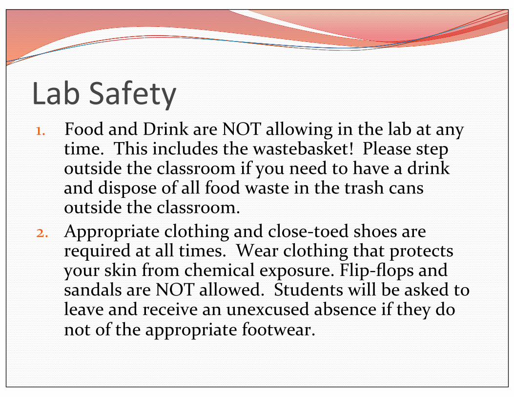
Lab Safety 1. Food and Drink are NOT allowing in the lab at any
time. This includes the wastebasket! Please step outside the classroom if you need to have a drink and dispose of all food waste in the trash cans outside the classroom.
2. Appropriate clothing and close-‐toed shoes are required at all times. Wear clothing that protects your skin from chemical exposure. Flip-‐flops and sandals are NOT allowed. Students will be asked to leave and receive an unexcused absence if they do not of the appropriate footwear.

Lab Safety 3. Safety glasses and gloves are available and are required when instructed by me. Gloves and goggles are always available for student use if desired.
4. An evacuation plan is posted on the door of each lab room. Familiarize yourself with this and make sure you understand the route to leave the building and meeting place for your class outside of the building (such as a specific tree). In the case of a fire everyone must leave the building, stay together as a class, and return to the classroom only when the Fire Marshall excuses you.

Lab Safety 5. Accident report forms are in each lab. If you have an accident, report it to me and fill out an accident report. Students should go to the student health center with any concerns. There is a first aid kit in each room with basics like bandaids.
6. If there is an emergency call 911 from any cell phone (or land line in prep room). Clearly state the location and type the emergency. When in doubt call 911.

Lab Safety 7. During particular labs there will be specific waste containers for biological waste, dissected materials, glass and other sharps. Please check with you TA to ensure you put materials in the correct waste disposal.
8. There are safety showers and eye-‐wash stations in each lab room. In the case of a chemical exposure to the eyes or body they should be used with the help of your TA.
9. A safety plan is found in each lab room and MSDS sheets are available for all chemicals used in lab.
10. Keep lab clean. A cluttered work area increases the risk of accidents.

Lab Safety � Please describe any health concerns (i.e. diabetes, history of fainting) that I should know about.

Plan for today’s lab: � Homeostasis � Anatomic Orientation and Direction � Planes � Landmarks � Body Cavities � Body Quadrants � Epithelial and Connective Tissues
� Rat Dissection
Chapter 1
Chapter 5

What is Anatomy? � Anatomy
� Study of the structure of the human body (and how it relates to other body parts)
� Gross � (anything you can study without a microscope)
� Microscopic � Cytology � Histology

Homeostasis � Maintenance of a stable internal body environment
� MUST be maintained or organ systems will NOT function properly
� Examples: � Oxygen/Carbon Dioxide � Body Temperature � Water content

Homeostasis • Receptor
• Receives the stimulus
• Control center
• Processes the signal and
sends instructions
• Effector
• Carries out instructions

• The Role of Negative
Feedback
• The response of the
effector negates the
stimulus
• Body is brought
back into
homeostasis
• Normal range is
achieved
Nega>ve and Posi>ve Feedback

Nega>ve and Posi>ve Feedback
• The Role of Positive
Feedback
• The response of the
effector increases
change of the stimulus
• Body is moved away
from homeostasis
• Normal range is
lost
• Used to speed up
processes

Anatomical Terminology • Superficial Anatomy
• Locating structures on or near the
body surface
• Anatomical Landmarks
• Anatomical position: hands at
sides, palms forward
• Supine: lying down, face up
• Prone: lying down, face down

Anatomical Terminology

Anatomical Terminology • Sectional Anatomy
• Planes and sections
• Plane: a three-‐dimensional axis
• Section: a slice parallel to a plane
• Used to visualize internal organization and structure
• Important in radiological techniques • MRI
• PET
• CT

Body Landmarks � Divide into groups by table � Label someone in the group or lab model � Present to the class
� You have 10 minutes!
� Refer to figure 1.7 if you need help!

Body Cavi>es • Essential Functions of Body
Cavities
1. Protect organs from accidental shocks
2. Permit changes in size and shape of internal organs
• Ventral body cavity (coelom)
• Divided by the diaphragm
• Thoracic cavity
• Abdominopelvic
cavity

Body Cavi>es • The Thoracic Cavity
• Right and left pleural cavities
• Contain right and left lungs
• Mediastinum
• Upper portion filled with blood vessels, trachea, esophagus, and thymus
• Lower portion contains pericardial cavity
• The heart is located within the pericardial cavity

Body Cavi>es • The Abdominopelvic
Cavity
• Pelvic cavity — inferior
portion
• Within pelvic bones
• Contains
reproductive organs,
rectum, and bladder

Body Cavi>es • Serous Membranes
• Line body cavities and cover organs
• Consist of parietal layer and visceral layer
• Parietal layer — lines cavity
• Visceral layer — covers organ

Body Quadrants • Superficial Anatomy
• Anatomical Landmarks
• References to palpable structures
• Anatomical Regions
• Body regions • Abdominopelvic
quadrants
• Abdominopelvic regions
• Anatomical Directions
• Reference terms based on subject


Get Some

Posi>on 1) On back 2) Arms and legs extended laterally

Make a Loop

Tie it >ght

Run string around back of tray and >e down other arm
Repeat with legs
These are the cuts that you will be making:

Belly BuLon Incision
Under skin and muscle

En>re length of body

1) Should look like this: 2) When cutting up thorax, you may either go above or through the ribcage


Cut just inferior to Diaphragm


Abdominal Cavity

Stuff to ID 1) Liver 2) Stomach 3) Spleen 4) Pancreas 5) Small intestine 6) Large Intestine 7) Cecum 8) Rectum 9) Kidneys
10) Mesentary 11) Bladder

1
2
3
5
6
7

Pancreas
4

11
8
9
Note: The bladder looks like a little balloon (because it is).
Fun fact: That’s real poop in the rectum!

Male Reproduc>on 1) Testes 2) Epididymis 3) Seminal Vesicle 4) Vas Deferens 5) Prostate 6) Penis

5
1
2
6

3
4 Note: Both the seminal vesicle and the vas deferens are paired structures. There is one of each on the other side. Find both!!

Thorax

Stuff to find 1) Heart 2) Lungs 3) Thymus 4) Trachea 5) Diaphragm

Find the Diaphragm. Insert your scissors through it and cut up the sternum
If you are a ninja, you should be able to separate the diaphragm from the thoracic cage with minimal damage.

Note: you may decide to remove the ribs all together

1
2
3
4
5

Disposal

Epithelial Tissue � Covers all body surfaces
� IN and OUT � Primary tissue found in glands � Functions:
� Protect (Skin) � Secrete (Glands) � Absorb (Intestines) � Sensation (Taste Buds) � Filter (Blood within the kidney)

Epithelial Tissue � Classification
� Number of cell layers � Simple = 1 layer � Stratified = >1 layers
� Cell shape � Squamous (squished) = flattened
� Cuboidal = equal height and width
� Columnar = height > width

Simple Squamous Epithelium � Can be found in:
� Lining arteries and veins (Endothelium)
� Lines body cavities ( Mesothelium) � Line alveoli of lungs

Stra>fied Squamous Epithelium � Basal cells may be more cuboidal-‐ polyhedral
� Can be: � Nonkeratinized
� Alive � Oral cavity, pharynx, esophagus, lining vagina and anus
� Keratinized � Dead � Epidermis of skin
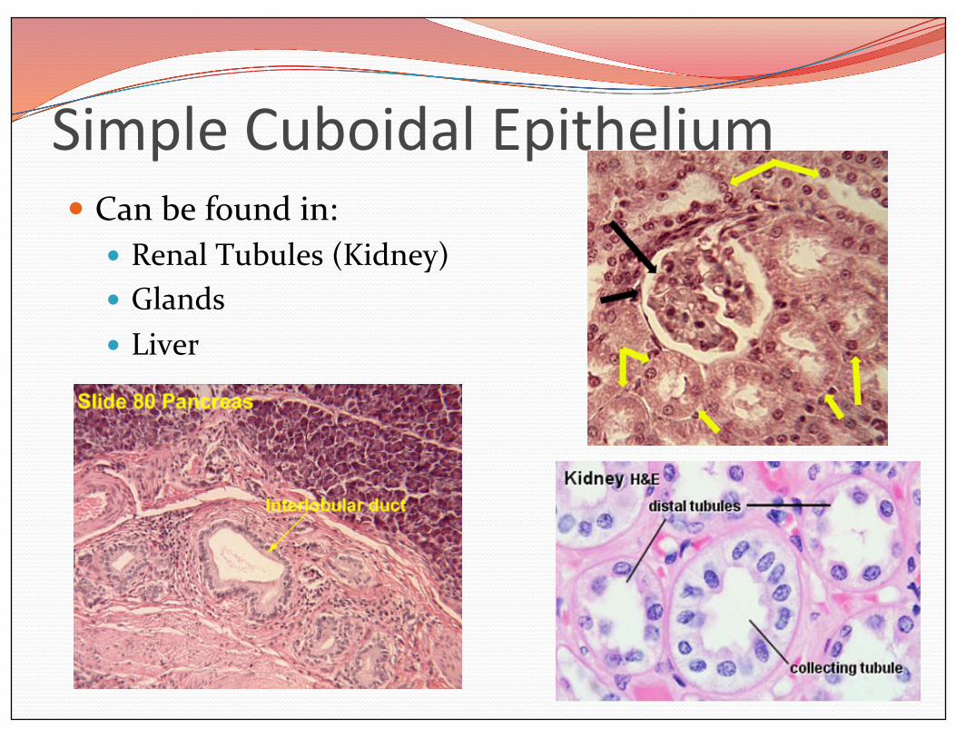
Simple Cuboidal Epithelium � Can be found in:
� Renal Tubules (Kidney) � Glands � Liver

Simple Columnar Epithelium � Can be:
� Ciliated � Respiratory Tract � Uterine Tubes
� Non-‐Ciliated � The inner lining of the digestive tract � Stomach � Small intestines � Large intestines

Pseudostra>fied Columnar Epithelium � Nuclei are located at different levels � Can be found in:
� The inner lining of the trachea and larger airways within the lungs (ciliated) or in the male urethra and epididymus (non-‐ciliated)

Transi>onal Epithelium � Transitional = Changes � Can be found in:
� Inner lining of the urinary tract � Bladder � Ureter � Part of the Urethra

Ques>ons � Which type of epithelium is best for diffusion?
� Which type of epithelium is best for protection?

Connec>ve Tissue � In ALL parts of the body
� Function to: � Connect structures
� Support � Protect � Fill Spaces � Hold Fat � Repair Tissue

Connec>ve Tissue � Cells are loosely associated with each other
� Separated by a matrix � Consists of a ground substance and various protein fibers
� Can be liquid or solid-‐-‐-‐depends on its composition
� Tied together by various adhesion proteins

Connec>ve Tissue � The matrix contains 3 types of fibers
� Collagen � Made of the protein collagen � Thick (dark pink in adjacent image)
� Reticular � Made of the protein collagen � Darkly stain in adjacent image � Branching, often form a network
� Elastic
� Made of the protein elastin � Very fine ( Dark purple in adjacent image)

Connec>ve Tissue � Areolar Connective Tissue
� Loosely arranged connective tissue and cells
� Gel-‐like matrix � Contains many collagen and elastin fibers
� Most nuclei represent fibroblasts
� This type of connective tissue can be found deep to skin but superficial to the muscle

Connec>ve Tissue � Adipose (Fat) Tissue:
� Contains very little matrix � Adipose cells = adipocytes
� Filled with fat � Surrounded by a ring of cytoplasm containing the nucleus

Connec>ve Tissue � Reticular Tissue
� Provide supporting framework for organs in the lymphatic system, liver and kidney
� A fibrous network of reticular fibers

Connec>ve Tissue � Dense Regular Connective Tissue: � Contains densely packed bundles of collagen fibers
� Very strong � Found in:
� Tendons � Ligaments

Suppor>ng Connec>ve Tissue � Cartilage � Bone ( we will discuss later)
� Form strong frameworks in order to protect and support

Connec>ve Tissue � Cartilage
� Contains many clear spaces = lacunae � Each lacuna contains a
cartilage cell à Chondrocyte � Lacunae are separated from
each other by a semisolid matrix
� Types � Hyaline (most common)
� Glossy matrix � Elastic
� Matrix contains more elastic fibers than hyaline cartilage
� Fibrocartilage � Very ordered microscopically � Rows of lacunae separated by alternating roles of collagen
� Shock absorber


Fluid Connec>ve Tissue � Blood (We will discuss later)