DRCR.net Single-Arm Study Assessing the Effects...
40
Gas for Macular Hole Protocol V2.0 7-5-18 DRCR.net Single-Arm Study Assessing the Effects of Pneumatic Vitreolysis on Macular Hole (Protocol AH) IDE Sponsor: Jaeb Center for Health Research (JCHR) Version 2.0 5 July 2018
Transcript of DRCR.net Single-Arm Study Assessing the Effects...
Gas for Macular Hole Protocol V2.0 7-5-18
DRCR.net
Single-Arm Study Assessing the Effects of
Pneumatic Vitreolysis on Macular Hole
(Protocol AH)
IDE Sponsor: Jaeb Center for Health Research (JCHR)
Version 2.0
5 July 2018
Gas for Macular Hole Protocol V2.0 7-5-18
Signature Page
Single-Arm Study Assessing the Effects of Pneumatic
Vitreolysis on Macular Hole
Protocol Identifying Number: DRCR.net AH
IDE Sponsor: Jaeb Center for Health Research
Version Number: 2.0
5 July 2018
JCHR Principal Investigator
Name, degree Adam Glassman, MS
Signature / Date
Jaeb Center for Health Research
Gas for Macular Hole Protocol V2.0 7-5-18
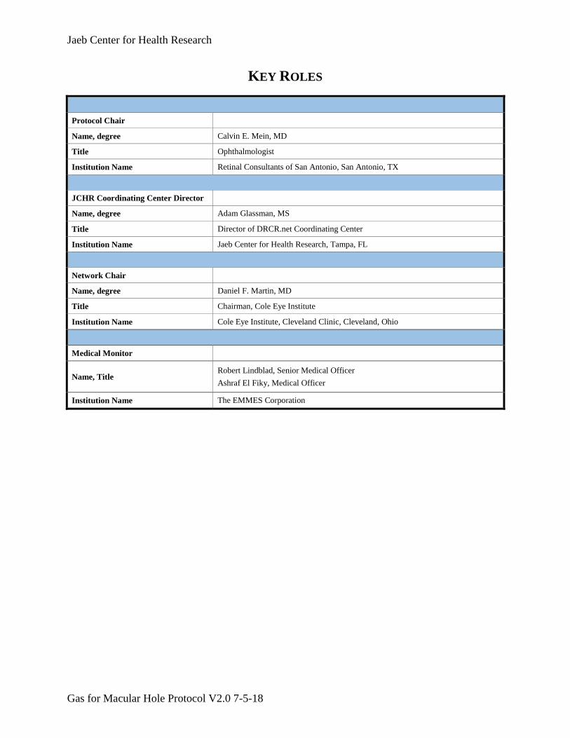
KEY ROLES
Protocol Chair
Name, degree Calvin E. Mein, MD
Title Ophthalmologist
Institution Name Retinal Consultants of San Antonio, San Antonio, TX
JCHR Coordinating Center Director
Name, degree Adam Glassman, MS
Title Director of DRCR.net Coordinating Center
Institution Name Jaeb Center for Health Research, Tampa, FL
Network Chair
Name, degree Daniel F. Martin, MD
Title Chairman, Cole Eye Institute
Institution Name Cole Eye Institute, Cleveland Clinic, Cleveland, Ohio
Medical Monitor
Name, Title Robert Lindblad, Senior Medical Officer
Ashraf El Fiky, Medical Officer
Institution Name The EMMES Corporation
Jaeb Center for Health Research
Gas for Macular Hole Protocol V2.0 7-5-18
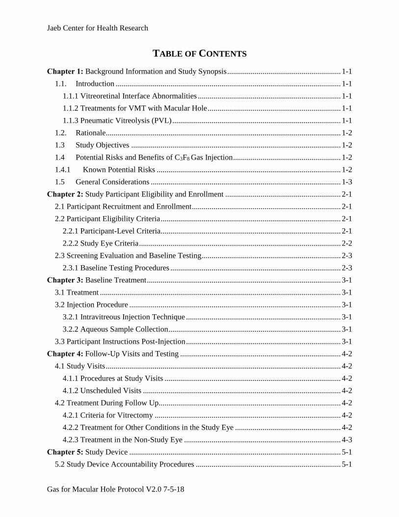
TABLE OF CONTENTS
Chapter 1: Background Information and Study Synopsis .......................................................... 1-1
1.1. Introduction ................................................................................................................... 1-1
1.1.1 Vitreoretinal Interface Abnormalities ......................................................................... 1-1
1.1.2 Treatments for VMT with Macular Hole .................................................................... 1-1
1.1.3 Pneumatic Vitreolysis (PVL) ...................................................................................... 1-1
1.2. Rationale ........................................................................................................................ 1-2
1.3 Study Objectives ........................................................................................................... 1-2
1.4 Potential Risks and Benefits of C3F8 Gas Injection ....................................................... 1-2
1.4.1 Known Potential Risks .............................................................................................. 1-2
1.5 General Considerations ................................................................................................. 1-3
Chapter 2: Study Participant Eligibility and Enrollment ........................................................... 2-1
2.1 Participant Recruitment and Enrollment ............................................................................ 2-1
2.2 Participant Eligibility Criteria ............................................................................................ 2-1
2.2.1 Participant-Level Criteria............................................................................................ 2-1
2.2.2 Study Eye Criteria ....................................................................................................... 2-2
2.3 Screening Evaluation and Baseline Testing ....................................................................... 2-3
2.3.1 Baseline Testing Procedures ....................................................................................... 2-3
Chapter 3: Baseline Treatment ................................................................................................... 3-1
3.1 Treatment ........................................................................................................................... 3-1
3.2 Injection Procedure ............................................................................................................ 3-1
3.2.1 Intravitreous Injection Technique ............................................................................... 3-1
3.2.2 Aqueous Sample Collection ........................................................................................ 3-1
3.3 Participant Instructions Post-Injection ............................................................................... 3-1
Chapter 4: Follow-Up Visits and Testing .................................................................................. 4-2
4.1 Study Visits ........................................................................................................................ 4-2
4.1.1 Procedures at Study Visits .......................................................................................... 4-2
4.1.2 Unscheduled Visits ..................................................................................................... 4-2
4.2 Treatment During Follow Up............................................................................................. 4-2
4.2.1 Criteria for Vitrectomy ............................................................................................... 4-2
4.2.2 Treatment for Other Conditions in the Study Eye ...................................................... 4-2
4.2.3 Treatment in the Non-Study Eye ................................................................................ 4-3
Chapter 5: Study Device ............................................................................................................ 5-1
5.2 Study Device Accountability Procedures .......................................................................... 5-1
Jaeb Center for Health Research
Gas for Macular Hole Protocol V2.0 7-5-18
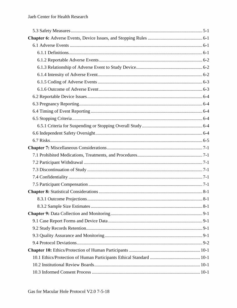
5.3 Safety Measures ................................................................................................................. 5-1
Chapter 6: Adverse Events, Device Issues, and Stopping Rules ............................................... 6-1
6.1 Adverse Events .................................................................................................................. 6-1
6.1.1 Definitions................................................................................................................... 6-1
6.1.2 Reportable Adverse Events ......................................................................................... 6-2
6.1.3 Relationship of Adverse Event to Study Device......................................................... 6-2
6.1.4 Intensity of Adverse Event.......................................................................................... 6-2
6.1.5 Coding of Adverse Events .......................................................................................... 6-3
6.1.6 Outcome of Adverse Event ......................................................................................... 6-3
6.2 Reportable Device Issues ................................................................................................... 6-4
6.3 Pregnancy Reporting .......................................................................................................... 6-4
6.4 Timing of Event Reporting ................................................................................................ 6-4
6.5 Stopping Criteria ................................................................................................................ 6-4
6.5.1 Criteria for Suspending or Stopping Overall Study .................................................... 6-4
6.6 Independent Safety Oversight ............................................................................................ 6-4
6.7 Risks ................................................................................................................................... 6-5
Chapter 7: Miscellaneous Considerations .................................................................................. 7-1
7.1 Prohibited Medications, Treatments, and Procedures ........................................................ 7-1
7.2 Participant Withdrawal ...................................................................................................... 7-1
7.3 Discontinuation of Study ................................................................................................... 7-1
7.4 Confidentiality ................................................................................................................... 7-1
7.5 Participant Compensation .................................................................................................. 7-1
Chapter 8: Statistical Considerations ......................................................................................... 8-1
8.3.1 Outcome Projections ................................................................................................... 8-1
8.3.2 Sample Size Estimates ................................................................................................ 8-1
Chapter 9: Data Collection and Monitoring ............................................................................... 9-1
9.1 Case Report Forms and Device Data ................................................................................. 9-1
9.2 Study Records Retention.................................................................................................... 9-1
9.3 Quality Assurance and Monitoring .................................................................................... 9-1
9.4 Protocol Deviations ............................................................................................................ 9-2
Chapter 10: Ethics/Protection of Human Participants ............................................................. 10-1
10.1 Ethics/Protection of Human Participants Ethical Standard ........................................... 10-1
10.2 Institutional Review Boards ........................................................................................... 10-1
10.3 Informed Consent Process ............................................................................................. 10-1

Jaeb Center for Health Research
Gas for Macular Hole Protocol V2.0 7-5-18
10.3.1 Consent Procedures and Documentation ................................................................ 10-1
10.3.2 Participant and Data Confidentiality ....................................................................... 10-1
10.3.3 Future Use of Stored Specimens ............................................................................. 10-2
Chapter 11: References ............................................................................................................ 11-1

Jaeb Center for Health Research
Gas for Macular Hole Protocol V2.0 7-5-18
LIST OF ABBREVIATIONS
ABBREVIATION DEFINITION
AUC Area Under the Curve
CI Confidence Interval
CRF Case Report Form
DSMC Data and Safety Monitoring Committee
E-ETDRS Electronic-Early Treatment Diabetic Retinopathy Study
ERM Epiretinal Membrane
ETDRS Early Treatment Diabetic Retinopathy Study
FDA Food and Drug Administration
GCP Good Clinical Practice
ICH International Conference on Harmonization
ID Identification
IDE Investigational Device Exemption
IOP Intraocular Pressure
IRB Institutional Review Board
ITT Intention-To-Treat
JCHR Jaeb Center for Health Research
MH Macular Hole
OCT Optical Coherence Tomography
PVD Posterior Vitreous Detachment
PVL Pneumatic Vitreolysis
SADE Serious Adverse Device Event
SAE Serious Adverse Event
SD Standard Deviation
UADE Unanticipated Adverse Device Effect
VMT Vitreomacular Traction
VMA Vitreomacular Adhesion
Jaeb Center for Health Research
Gas for Macular Hole Protocol V2.0 7-5-18
PROTOCOL SUMMARY
PARTICIPANT AREA DESCRIPTION
Title Single-Arm Study Assessing the Effects of Pneumatic Vitreolysis on
Macular Hole
Précis Eyes with vitreomacular traction (VMT) and full-thickness macular
holes(MH) will be enrolled into a non-randomized cohort treated
with PVL to determine the proportion with VMT release and MH
closure and to assess factors associated with success.
Investigational Device Intraocular gas (C3F8) injection
Objectives To obtain estimates of the proportion of eyes with MH closure of the
inner retinal layers for eyes with VMT and full-thickness MHs
treated with PVL.
Rationale Understanding the rates of VMT release and MH closures in eyes
with full-thickness MH treated with PVL is of interest. Surgery
would result in nearly 100% hole closure and VMT release, making
vitrectomy a poor control group choice. Spontaneous resolution of
MH is highly unlikely, making an observation arm unnecessary.
Therefore, these eyes will be enrolled into a non-randomized cohort
treated with PVL to assess the outcomes of treatment.
Study Design Single-arm study
Number of Sites Approximately 50 sites
Endpoint Primary Outcome: Proportion of eyes with MH closure of the inner
retinal layers without rescue treatment
Key Secondary Outcomes: proportion of eyes with foveal VMT
release, proportion of eyes with foveal VMT and vitreopapillary
traction release, proportion of eyes requiring vitrectomy, proportion
of eyes with foveal lucency, mean change in visual acuity, and
ellipsoid zone changes.
Key Safety Outcomes: retinal tear, retinal detachment, traumatic
cataract, cataract extraction, vitreous hemorrhage, intraocular
pressure (IOP) increase, and endophthalmitis.
Population Key Inclusion Criteria
Age ≥ 18 years.
Able and willing to avoid high altitude until gas resolution
(approximately 6 to 8 weeks)
For phakic patients, able and willing to avoid supine
positioning until gas resolution (approximately 6 to 8 weeks)
Jaeb Center for Health Research
Gas for Macular Hole Protocol V2.0 7-5-18
PARTICIPANT AREA DESCRIPTION
Able and willing to position face down for at least 50% of the
time for at least 4 days post-injection
At least 1 eye with:
o Vitreomacular adhesion on OCT that is no larger than
3000 microns, confirmed by a central reading center
o Full-thickness MH that is ≤ 250 microns at the narrowest
point as measured on OCT, confirmed by a central
Reading Center
o Best corrected E-ETDRS visual acuity equivalent of
20/25 to 20/400
Key Exclusion Criteria
Other condition that might affect visual acuity during the
course of the study (e.g., retinal vein occlusion, advanced
age-related macular degeneration, or macular edema induced
by a condition other than VMT)
High level myopia (-8.00 diopters or more myopic if phakic
or retinal abnormalities consistent with pathologic myopia if
phakic or pseudophakic)
Prior gas injection, ocriplasmin injection, or intraocular
injection for any reason
Prior vitrectomy
History of advanced glaucoma that contraindicates intraocular
gas injection
Sample Size 50 eyes
Treatment Groups C3F8 injection only
Participant Duration 24 weeks
Protocol
Overview/Synopsis
1. Informed consent will be obtained.
2. Eligibility will be assessed, including reading center
confirmation of VMT and MH on OCT.
3. Eligible eyes with VMT and MH will be treated with C3F8
injection.
4. Follow-up visits will occur at 1, 4, 8, and 24 weeks and
consist of visual acuity testing, ocular exam, and OCT.
5. The primary outcome assessment will be the proportion of
eyes at 8 weeks with full-thickness MH closure of inner
retinal layers without rescue treatment.
Jaeb Center for Health Research
Gas for Macular Hole Protocol V2.0 7-5-18
SCHEMATIC OF STUDY DESIGN
Dropped IneligibleInformed Consent Obtained
Baseline Testing
Reading Center Review
Injection of C3F8
Gas (0.3 mL)
1 Week
4 Weeks
8 Weeks
(Primary Outcome)
24 Weeks
Dropped
Ineligible
Jaeb Center for Health Research
Gas for Macular Hole Protocol V2.0 7-5-18
SCHEDULE OF STUDY VISITS AND PROCEDURES
Enrollment
Visit*
1, 4, 8, and 24
weeks
E-ETDRS best corrected visual acuity a X X
OCT b X X
Eye exam c X X
Reading center eligibility confirmation d X
Gas injection X
* All baseline testing must occur within 8 days prior to enrollment.
a, Both eyes at all visits; includes protocol refraction in study eye only at each visit and in both eyes at
enrollment and 8 weeks. Electronic ETDRS (E-ETDRS) testing using the Electronic Visual Acuity Tester
that has been validated against 4-meter chart ETDRS testing.
b, Both eyes at baseline; study eye only at follow up visits.
c, Both eyes at baseline; study eye only at each follow-up visit. Includes slit lamp exam (including
assessment of lens), measurement of intraocular pressure, and dilated ophthalmoscopy. Scleral depression
is required at baseline to confirm eligibility. During follow up, the eye exam should be extensive enough
to identify adverse events of interest.
d, Reading center review of the OCT for eligibility must occur prior to enrollment.
Jaeb Center for Health Research
Gas for Macular Hole Protocol V2.0 7-5-18 1-1
CHAPTER 1: BACKGROUND INFORMATION AND STUDY SYNOPSIS 1
1.1. Introduction 2
1.1.1 Vitreoretinal Interface Abnormalities 3
Disorders of the vitreoretinal interface represent a spectrum of abnormalities that develop as the 4
posterior hyaloid separates from the internal limiting membrane. Vitreomacular adhesion (VMA) 5
occurs when the posterior hyaloid remains attached to the internal limiting membrane centrally. 6
Overall, about 1.5% of the population is estimated to have eye diseases caused by or associated 7
with VMA.1 The incidence of VMA diagnoses is expected to increase with widespread use of 8
spectral-domain optical coherence tomography (SD-OCT). Vitreomacular traction (VMT) is 9
diagnosed when VMA results in traction, distortion of retinal architecture, and patient 10
symptomatology.2 11
Advanced VMT can lead to macular holes (MH), in which tractional forces create small full-12
thickness defects on the posterior fundus, often requiring surgical intervention.2 Regarding the 13
incidence of idiopathic full-thickness macular holes, a population-based study showed idiopathic 14
macular holes occur at an age and sex-adjusted incidence in 8.69 eyes per 100,000 population 15
per year.3 The female to male ratio was determined to be 3.3 to 1, and bilateral idiopathic MHs 16
occurred in 11.7% of patients and accounted for 20.9% of the affected eyes.3 In another study of 17
a large population of patients with age-related macular degeneration (15,196 with non 18
neovascular age-related macular degeneration (AMD) and 12,716 with neovascular AMD), 0.7% 19
were found to have MHs (1.1% with non-neovascular AMD and 0.3% with neovascular AMD).4 20
Regarding MH prevalence globally, the Baltimore Eye Study reported a prevalence of 3.3 per 21
1,000 persons in Maryland,5 the Beaver Dam Eye Study showed a prevalence of 2.9 per 1,000 22
persons in Wisconsin, the Blue Mountains Eye Study showed a prevalence of 0.2 per 1,000 23
persons in Australia,6 the Beijing Eye Study showed a prevalence of 0.9 per 1,000 persons in 24
China,7 and Sen et al. reported a prevalence of 1.7 per 1,000 persons in Southern India.8 25
1.1.2 Treatments for VMT with Macular Hole 26
For eyes with VMT with MH, prompt treatment is indicated to restore central vision and prevent 27
retinal detachment. In the MIVI-TRUST trial, ocriplasmin was successful in 60% in eyes with an 28
MH of < 250 microns.9 However, there have been multiple anecdotal reports of substantial 29
ocular complications associated with intraocular administration of ocriplasmin,10-14 including 30
transient vision loss, persistent dyschromatopsia, electroretinographic abnormalities, subluxation 31
of the crystalline lens likely related to zonulolysis, and disturbance or dehiscence of the ellipsoid 32
layer documented by OCT. These adverse events have created major concerns among many 33
retinal surgeons in the clinical use of this drug.10-14 Therefore, vitrectomy is currently the first 34
line therapy for VMT with MH. Although MH closure is usually successful after vitrectomy 35
(approaching 100% success rate in several series), there are associated downsides including cost, 36
patient discomfort, length of time a large bubble resides in the eye, and possible adverse events 37
such as endopthalmitis, retinal detachment, and cataract progression.15 38
1.1.3 Pneumatic Vitreolysis (PVL) 39
Pneumatic vitreolysis (intraocular injection of expansile gas to induce a posterior vitreous 40
detachment [PVD]) has been suggested as a potential treatment alternative to vitrectomy or 41
ocriplasmin for VMT with MH. In 1995, Chan et al. first demonstrated and reported the utility 42
of intraocular gas (C3F8) injection where 13 of 17 (76%) stage 1 or stage 2 macular holes closed 43
after injection.16 Subsequently, Jorge et al. showed success in the induction of PVD (6 of 6 eyes) 44
Jaeb Center for Health Research
Gas for Macular Hole Protocol V2.0 7-5-18 1-2
and macular hole closure (5 of 6 eyes) with C3F8 in small case series.17, 18 Mori et al.19 reported 45
5 of 10 eyes had hole closure after gas.19 Steinle et al. reported a success rate of 83% (25 of 30 46
eyes) with C3F8 gas in a retrospective case series for treatment of VMT syndrome.20 In a 2016 47
retrospective review of 15 consecutive eyes receiving C3F8 gas for pneumatic vitreolysis 48
performed in 2 centers, Chan and Mein reported a success rate of 100% for VMT release and 49
67% for hole closure in eyes with small stage 2 MH ≤ 250 microns.21 50
1.2. Rationale 51
Understanding the rates of MH closure and VMT release in eyes with full-thickness macular 52
holes treated with PVL is of interest given the low cost and convenience of gas injection in the 53
office setting as well as a low rate of adverse events reported in prior retrospective studies. 54
Although spontaneous macular hole closure is possible, this occurs infrequently.22 It is likely that 55
eyes with these characteristics would need prompt treatment, making randomization to a sham 56
arm inappropriate. Prior studies have established the benefit of vitrectomy for treatment of 57
macular holes, reporting 80 to 90% success rates in MH closure with associated visual benefit, 58
making vitrectomy an unnecessary (and expensive) control group choice.22-26 Therefore, these 59
eyes will be enrolled into a non-randomized cohort treated with PVL to assess the outcomes of 60
treatment. 61
62
If a large percentage of eyes can achieve MH closure with a simple in-office, low-cost 63
procedure, while averting the invasiveness and expense of a vitrectomy for this condition, this 64
would provide a viable first-line treatment option. Even if this proposed study finds that PVL is 65
only moderately successful, physicians and patients may decide to attempt PVL in the office 66
first, before proceeding with more costly, invasive surgery. Thus, even without a control group, 67
the results from this study will provide data of value for physicians and patients to make 68
informed decisions about treatment course. 69
1.3 Study Objectives 70
Primary 71
1. To obtain estimates for the proportion of eyes with MH closure of the inner retinal layers 72
for eyes with VMT and full-thickness macular holes treated with PVL 73
1.4 Potential Risks and Benefits of C3F8 Gas Injection 74
1.4.1 Known Potential Risks 75
Potential risks of C3F8 gas injection include the following: 76
Pain, discomfort, redness, or itching lasting for a few days is likely. 77
Subconjunctival hemorrhage or floaters will commonly occur as a result of the injection. The 78
floaters are typically reduced after 6 to 8 weeks, but some floaters may persist. 79
Immediately following the injection, there may be elevation of IOP. Pressure usually returns 80
to normal spontaneously, but may need to be treated with topical drugs or a paracentesis to 81
lower the pressure. The likelihood of permanent loss of vision from elevated IOP is less than 82
1%. 83
Although it has not been reported in prior case series, endophthalmitis could theoretically 84
develop. The risk of endophthalmitis from other intraocular injections is less than 1%. 85
Jaeb Center for Health Research
Gas for Macular Hole Protocol V2.0 7-5-18 1-3
A retinal tear or detachment could occur. The risk of retinal breaks or detachment after gas 86
injection is approximately 1%. 87
There is a possibility of traumatic cataract from the injection. The risk of developing a 88
cataract from the injection is much less than 1 in 1000. 89
If paracentesis is performed, there is a similar risk of traumatic cataract from paracentesis. 90
If vitrectomy is required while gas is in the eye, there is high likelihood of cataract formation 91
during surgery that may require cataract removal at the time of vitrectomy. 92
Limited and transient uveitis may develop after gas injection. Persistent uveitis is uncommon. 93
Limited transient conjunctival or episcleral hemorrhage is common shortly after gas 94
injection. It is usually inconsequential and clears spontaneously from a few days to a week or 95
two. 96
Limited vitreous hemorrhage or opacities after gas injection is uncommon but may occur 97
occasionally after gas injection, particularly given a history of active anticoagulation therapy 98
or predisposing risk for hemorrhage. If present, it usually resolves from a few days to a few 99
months. Marked intraocular hemorrhage requiring a surgical intervention after gas injection 100
is exceedingly rare (< 1%). 101
Pre-existing epiretinal fibrosis may sometimes progress or new epiretinal fibrosis may 102
develop after gas injection. 103
The development of excessive scarring on top of or under the retina after gas injection is 104
exceedingly rare. When this occurs, it is usually associated with advanced retinal 105
detachment, which is also uncommon after gas injection. 106
Additional risks if the participant does not follow post-injection instructions: 107
Intraocular pressure may increase if the patient experiences changes in elevation (i.e. travel 108
by air or over mountain ranges) while the gas bubble is still present in the eye. 109
Loss of vision or blindness is possible if nitrous oxide anesthesia is administered with the gas 110
bubble still present in the eye. 111
Incorrect head positioning following the gas injection may lead to glaucoma or cataracts. 112
113
1.4.2 Known Potential Benefits 114
Potential benefits from participation in the study for eyes treated with PVL include, improved 115
visual acuity, improved quality of vision, closure of MH, and avoidance of more invasive and 116
expensive procedures, i.e., vitrectomy, ocriplasmin. 117
1.4.3 Risk Assessment 118
The risk level is considered to be research involving greater than minimal risk. 119
1.5 General Considerations 120
The study is being conducted in compliance with the policies described in the study policies 121
document, with the ethical principles that have their origin in the Declaration of Helsinki, with 122
the protocol described herein, and with the standards of Good Clinical Practice (GCP). 123
Jaeb Center for Health Research
Gas for Macular Hole Protocol V2.0 7-5-18 1-4
The DRCR.net procedures manuals provide details of the procedures. 124
Data will be directly collected in electronic case report forms, which will be considered the 125
source data. 126
There is no restriction on the number of subjects to be enrolled by each site towards the overall 127
recruitment goal. However, recruitment will be monitored on an ongoing basis and the sponsor 128
can decide to place recruitment at a particular site on hold as needed. 129
All consented participants will be logged. The protocol is considered a significant risk device 130
study because intraocular injection of C3F8 is experimental for this indication. Therefore, an 131
investigational device exemption (IDE) from the U.S. Food and Drug Administration (FDA) is 132
required to conduct the study. 133
Jaeb Center for Health Research
Gas for Macular Hole Protocol V2.0 7-5-18 2-1
CHAPTER 2: STUDY PARTICIPANT ELIGIBILITY AND ENROLLMENT 134
2.1 Participant Recruitment and Enrollment 135
Enrollment will proceed with the goal of at least 50 participants deemed eligible by Reading 136
Center and treated with C3F8. Participants who have signed consent may be permitted to continue 137
into the study, if eligible, even if the enrollment goal has been reached. 138
Study participants will be recruited from approximately 50 clinical centers in the United States. 139
Approximately 5 participants are expected to be enrolled each month, resulting in 10 months of 140
recruitment, for a total study duration of 16 months. 141
All eligible participants will be included without regard to gender, race, or ethnicity. There is no 142
restriction on the number of participants to be enrolled by each site toward the overall 143
recruitment goal. 144
Potential eligibility may be assessed as part of a routine-care examination. Before completing 145
any procedures or collecting any data that are not part of usual care, written informed consent 146
will be obtained. 147
The study protocol will be discussed with the potential study participant by study staff. The 148
potential study participant will be given the Informed Consent Form to read. Potential study 149
participants will be encouraged to discuss the study with family members and their personal 150
physicians(s) before deciding whether to participate in the study. 151
As part of the informed consent process, each participant will be asked to sign an authorization 152
for release of personal information. The investigator, or his or her designee, will review the 153
study-specific information that will be collected and to whom that information will be disclosed. 154
After speaking with the participant, questions will be answered about the details regarding 155
authorization. 156
2.2 Participant Eligibility Criteria 157
2.2.1 Participant-Level Criteria 158
Inclusion 159
To be eligible, the following inclusion criteria must be met: 160
1. Age ≥ 18 years 161
Participants < 18 years old are not being included because the condition is so rare in 162
this age group that the diagnosis may be questionable. 163
2. At least one eye meets the study eye criteria listed in section 2.2.2 164
3. Able and willing to provide informed consent 165
4. Able and willing to avoid high altitude travel, including airline travel, until gas resolution 166
(approximately 6 to 8 weeks) 167
5. For phakic patients, able and willing to avoid supine position until gas resolution 168
(approximately 6 to 8 weeks) 169
6. Able and willing to position face down for at least 50% of the time for at least 4 days post-170
injection (to facilitate macular hole closure) 171
7. Able and willing to wear wristband that informs any medical personnel that the patient has a 172
gas bubble in the eye 173
Jaeb Center for Health Research
Gas for Macular Hole Protocol V2.0 7-5-18 2-2
Exclusion 174
A potential participant is not eligible if any of the following exclusion criteria are present: 175
8. A condition that, in the opinion of the investigator, would preclude participation in the study 176
(e.g., unstable medical status that might preclude completion of follow up) 177
9. Participation in an investigational trial within 30 days of enrollment that involves treatment 178
with any drug or device that has not received regulatory approval for the indication being 179
studied at the time of study entry 180
Note: study participants should not receive another investigational drug or device while 181
participating in the study 182
10. Known contraindication to any component of the treatment 183
11. Known allergy to any drug used in the procedure prep (including povidone iodine) 184
12. Potential participant is expecting to move out of the area of the clinical center to an area not 185
covered by another clinical center during the 6 months following enrollment 186
13. Anticipated surgery requiring anesthesia within the 6 months following enrollment 187
Participants cannot receive nitrous oxide until gas resolution 188
13. For women of child-bearing potential: pregnant at the time of enrollment 189
Women who are potential study participants should be questioned about the potential for 190
pregnancy. Investigator judgement may be used to determine when a pregnancy test is 191
needed 192
2.2.2 Study Eye Criteria 193
The participant must have at least one eye meeting all of the inclusion criteria and none of the 194
exclusion criteria listed below. 195
A participant can have only one study eye. If both eyes are eligible at the time of enrollment, the 196
study eye will be selected by the investigator and participant before injection. 197
The eligibility criteria for a study eye are as follows: 198
Inclusion 199
a. Full-thickness macular hole that is ≤ 250 microns at the narrowest point, confirmed by 200
central reading center 201
b. Vitreomacular adhesion on OCT that is no larger than 3000 microns with visible separation 202
of the vitreous on either side as seen on horizontal and vertical scans , confirmed by central 203
reading center 204
Presence of epiretinal membrane is neither a requirement nor exclusion 205
c. Visual acuity letter score of at least 19 (approximate Snellen equivalent 20/400 or better) 206
and at most 83 (20/25 or worse) 207
Exclusion 208
d. Other ocular condition that might affect visual acuity during the course of the study or 209
require intraocular treatment (e.g., retinal vein occlusion, substantial age-related macular 210
degeneration, or macular edema induced by a condition other than VMT) 211
Jaeb Center for Health Research
Gas for Macular Hole Protocol V2.0 7-5-18 2-3
If diabetic retinopathy is present, severity level must be microaneurysms only or better (≤ 212
diabetic retinopathy severity level 20). 213
Presence of drusen is acceptable; however, eyes with geographic atrophy or neovascular 214
age-related macular degeneration involving the macula are excluded. 215
e. High level of myopia (spherical equivalent of -8.00 diopters or more myopic if phakic, or 216
retinal abnormalities consistent with pathologic myopia if phakic or pseudophakic) 217
f. History of prior gas injection, ocriplasmin injection, or intraocular injection for any reason 218
g. History of prior vitrectomy 219
h. History of uncontrolled glaucoma 220
Intraocular pressure must be < 30 mmHg, with no more than one topical glaucoma 221
medication, and no documented glaucomatous field loss for the eye to be eligible 222
i. History of major ocular surgery (including cataract extraction, scleral buckle, any intraocular 223
surgery, etc.) within prior 4 months or major ocular surgery anticipated within the next 6 224
months following enrollment 225
j. History of YAG capsulotomy performed within 4 months prior to enrollment 226
k. Aphakia or anterior chamber intraocular lens 227
l. Exam evidence of severe external ocular infection, including conjunctivitis, chalazion, or 228
substantial blepharitis 229
m. Uveitis 230
n. Retinal history or pathology that might predispose an eye to an increased risk of retinal 231
detachment from the procedure 232
Untreated retinal tears, not retinal holes, are an exclusion. It is up to the investigator to 233
determine whether extent of lattice degeneration or other pathology might increase the 234
risk of retinal detachment 235
o. Any contraindication to paracentesis (e.g., history of narrow angle glaucoma) 236
p. Lenticular or zonular instability 237
2.3 Screening Evaluation and Baseline Testing 238
After informed consent has been signed, the potential participant will be evaluated for study 239
eligibility through the elicitation of a medical history and performance of an ocular examination 240
by study personnel to screen for exclusionary conditions. 241
Individuals who do not initially meet study eligibility requirements may be rescreened at a later 242
date per investigator discretion. 243
All testing does not need to be completed on the same day provided it is within the windows 244
specified below. Reading Center confirmation of VMT with full-thickness macular hole on OCT 245
must be completed prior to enrollment. 246
2.3.1 Baseline Testing Procedures 247
The following procedures are needed to confirm eligibility and/or to serve as baseline measures 248
for the study: 249
Jaeb Center for Health Research
Gas for Macular Hole Protocol V2.0 7-5-18 2-4
If a procedure has been performed using the study technique and by study certified 250
personnel as part of usual care, then it does not need to be repeated specifically for the 251
study if it was performed within the defined time windows specified below. 252
The testing procedures are detailed in the DRCR.net Procedures Manuals. Visual acuity 253
testing, ocular exam, and OCT will be performed by DRCR.net certified personnel. 254
1. Self-reported demographics (date of birth, sex, race, and ethnicity) 255
2. Medical history (pre-existing medical conditions, concomitant medications, as well as ocular 256
diseases, surgeries, and treatments) 257
Medical history will be obtained by medical charts if available at the enrolling site; 258
otherwise, it will be self-reported 259
3. Electronic-ETDRS visual acuity testing at 3 meters using the Electronic Visual Acuity Tester 260
(including protocol refraction) in each eye (within prior 8 days) 261
4. Spectral-Domain OCT using Zeiss Cirrus or Heidelberg Spectralis on each eye (within prior 262
8 days) 263
OCT scans of the study eye will be promptly sent to the central reading center for 264
grading and a participant cannot be enrolled until reading center confirmation of 265
eligibility has been received. 266
5. Ocular examination on each eye including slit lamp, measurement of IOP, lens assessment, 267
and dilated ophthalmoscopy (within prior 8 days) 268
Scleral depression to rule out any retinal tears pre-operatively will be required for the 269
baseline eye exam to confirm eligibility. 270
Jaeb Center for Health Research
Gas for Macular Hole Protocol V2.0 7-5-18 3-1
CHAPTER 3: BASELINE TREATMENT 271
3.1 Treatment 272
The C3F8 gas injection must be given on the day of enrollment. 273
3.2 Injection Procedure 274
3.2.1 Intravitreous Injection Technique 275
The injection is preceded by a povidone iodine prep of the conjunctiva. A subconjunctival 276
injection of lidocaine may be administered at the discretion of the investigator. The injection will 277
be performed using sterile technique. Pre-injection paracentesis should be considered due to the 278
4x expansion of C3F8 gas and the associated risk of shallowing of the anterior chamber. 279
However, the choice of when or whether or not to do a paracentesis is ultimately at the 280
investigator’s discretion. The full injection procedure is described in the protocol-specific study 281
procedures manual. Topical antibiotics in the pre-, peri-, or post-injection period should not be 282
used without prior approval from the Protocol Chair or Coordinating Center designee. 283
3.2.2 Aqueous Sample Collection 284
Participation in the ancillary sample collection component is not a requirement for participation 285
in this study. It is expected that sites with the capability to ship intraocular fluids will participate. 286
At the time of consent into the main study, participants will have the option of signing the 287
ancillary sample collection portion of the informed consent form to indicate their willingness to 288
provide the sample for future use. If paracentesis is performed and participant consent is 289
obtained, aqueous fluid already being drawn as part of paracentesis may be collected and shipped 290
on dry ice to a central laboratory for storage for future analyses. Sites will be encouraged to 291
collect samples when performing paracentesis, though sample collection will not be required. 292
Details regarding collection, sample labeling, storage, and shipment can be found in the 293
procedures manual. 294
3.3 Participant Instructions Post-Injection 295
Participants will be given a post-injection instruction sheet informing them of all post-injection 296
requirements and risks if they do not follow these requirements. Participants will be instructed to 297
position face down for at least 50% of the time for at least 4 days post-injection. Participants will 298
be instructed to avoid high altitude travel until the surgeon confirms the gas bubble has cleared. 299
Phakic participants will be asked to avoid the supine position and lie on one side or the stomach 300
during sleeping hours until the surgeon confirms that the gas bubble has cleared. All participants 301
will be instructed to wear a wristband to notify healthcare providers that they should not receive 302
nitrous oxide anesthesia until the gas bubble has cleared. 303
Jaeb Center for Health Research
Gas for Macular Hole Protocol V2.0 7-5-18 4-2
CHAPTER 4: FOLLOW-UP VISITS AND TESTING 304
4.1 Study Visits 305
The schedule of protocol-specified follow-up visits is as follows: 306
1 week (±3 days) 307
4 (±1) weeks 308
8 (±2) weeks 309
24 (±4) weeks 310
4.1.1 Procedures at Study Visits 311
The following procedures will be performed at each visit, unless otherwise specified: 312
1. E-ETDRS visual acuity testing (best corrected) in each eye 313
A protocol refraction in the study eye is required at each visit. When a refraction is not 314
performed, the most recently performed refraction is used for the testing. 315
2. Spectral-Domain OCT using Zeiss Cirrus or Heidelberg Spectralis on the study eye 316
The same machine type (Cirrus or Spectralis) used at baseline must be used during follow 317
up. 318
3. Ocular exam in the study eye only, including slit lamp examination with lens assessment, 319
measurement of IOP, and dilated ophthalmoscopy 320
The eye exam should be extensive enough to identify adverse events of interest 321
All of the testing procedures do not need to be performed on the same day, provided that they are 322
completed within the time window of a visit. If data from a testing procedure is unusable (e.g., if 323
OCT is ungradable), the participant may be asked to repeat the procedure during an additional 324
visit, whether part of usual care or solely to repeat the procedure. 325
4.1.2 Unscheduled Visits 326
Additional visits may occur as required for usual care of the study participant. 327
Testing procedures at unscheduled visits are at investigator discretion. However, it is 328
recommended that procedures performed should follow the standard DRCR.net protocol. 329
4.2 Treatment During Follow Up 330
4.2.1 Criteria for Vitrectomy 331
Vitrectomy should not be performed due to failure of macular hole closure prior to 4 weeks 332
without chair approval. Between 4 and 8 weeks, vitrectomy may only be performed (but is not 333
required) if the macular hole size is not improving from baseline. After 8 weeks, vitrectomy can 334
be performed at investigator discretion. 335
4.2.2 Treatment for Other Conditions in the Study Eye 336
An eye should not be enrolled that is anticipated to require intraocular treatment for another 337
condition during the study. If a condition requiring prompt treatment develops during follow up, 338
the treatment is at investigator discretion. 339
Jaeb Center for Health Research
Gas for Macular Hole Protocol V2.0 7-5-18 4-3
4.2.3 Treatment in the Non-Study Eye 340
Treatment in the non-study eye is at investigator discretion, except that gas injection for VMT is 341
not permitted in the non-study eye during the study. 342
Jaeb Center for Health Research
Gas for Macular Hole Protocol V2.0 7-5-18 5-1
CHAPTER 5: STUDY DEVICE 343
5.1 Description of the Investigational Device 344
Perfluoropropane C3F8 is an inert gas under pressure and is administered by injection into the 345
vitreous cavity. It was approved by the FDA in February 1993 (P900066) for the use of placing 346
pressure on detached retina. 347
348
5.2 Study Device Accountability Procedures 349
Each participating site will use their own commercially available perfluoropropane C3F8. It must 350
be stored at room temperature. Prior to each injection, the investigator must confirm that the 351
cylinder pressure is at least 50 psi and that the cylinder is not expired. 352
5.3 Safety Measures 353
Preparation of the perfluoropropane C3F8 injection will be performed in accordance with 354
manufacturer labelling. The full injection procedure is described in the protocol-specific study 355
procedures manual. 356
Jaeb Center for Health Research
Gas for Macular Hole Protocol V2.0 7-5-18 6-1
CHAPTER 6: ADVERSE EVENTS, DEVICE ISSUES, AND STOPPING RULES 357
6.1 Adverse Events 358
6.1.1 Definitions 359
Adverse Event (AE): Any untoward medical occurrence in a study participant, irrespective of the 360
relationship between the adverse event and the device(s) under investigation (see section 6. 2 for 361
reportable adverse events for this protocol). 362
Serious Adverse Event (SAE): Any untoward medical occurrence that: 363
Results in death. 364
Is life-threatening (a non-life-threatening event which, had it been more severe, might 365
have become life-threatening, is not necessarily considered a serious adverse event). 366
Requires inpatient hospitalization or prolongation of existing hospitalization. 367
Results in persistent or significant disability, incapacity, or substantial disruption of the 368
ability to conduct normal life functions (e.g, sight threatening). 369
Is a congenital anomaly or birth defect. 370
Is considered a significant medical event by the investigator based on medical judgment 371
(e.g., may jeopardize the participant or may require medical or surgical intervention to 372
prevent one of the outcomes listed above). 373
In general, an ocular adverse event should be reported as serious (considered sight threatening) if 374
it meets one of the following criteria: 375
1. It causes a decrease of ≥ 30 letters in visual acuity compared with the last visual 376
acuity measurement prior to onset (e.g. central retinal artery occlusion). 377
2. In the opinion of the investigator, it requires prompt surgical intervention (e.g. 378
vitrectomy, vitreous tap, intravitreous antibiotics) to prevent permanent loss of sight. 379
Examples include endophthalmitis or rhegmatogenous retinal detachment. 380
Ocular adverse events that require eventual surgical intervention would not be considered 381
serious. Ocular adverse events that do not have the potential to result in permanent loss of sight 382
also would not be considered serious. 383
Unanticipated Adverse Device Effect (UADE): Any serious adverse effect on health or safety or 384
any life-threatening problem or death caused by, or associated with, a device, if that effect, 385
problem, or death was not previously identified in nature, severity, or degree of incidence in the 386
investigational plan or application (including a supplementary plan or application), or any other 387
unanticipated serious problem associated with a device that relates to the rights, safety, or 388
welfare of participants (21 CFR 812.3(s)). 389
Adverse Device Effect (ADE): Any untoward medical occurrence in a study participant which 390
the device may have caused or to which the device may have contributed (Note that an Adverse 391
Event Form is to be completed in addition to being reported on a Gas Injection Form). 392
Device Complaints: A device complication or complaint is something that happens to a device or 393
related to device performance, whereas an adverse event happens to a participant. A device 394
complaint may occur independently from an AE, or along with an AE. An AE may occur without 395
a device complaint or there may be an AE related to a device complaint. 396
Jaeb Center for Health Research
Gas for Macular Hole Protocol V2.0 7-5-18 6-2
Device Malfunction: Any failure of a device to meet its performance specifications or otherwise 397
perform as intended. Performance specifications include all claims made in the labeling for the 398
device. The intended performance of a device refers to the intended use for which the device is 399
labeled or marketed. (21 CFR 803.3) 400
6.1.2 Reportable Adverse Events 401
For this protocol, a reportable adverse event includes any untoward medical occurrence that 402
meets one of the following criteria: 403
1) an ocular AE (study eye), 404
2) a serious AE, 405
3) an Adverse Device Effect (ADE) as defined in section 6.1.1, or 406
4) An AE occurring in association with a study procedure. 407
All reportable Adverse Events whether they are volunteered by the participant, discovered by 408
study personnel during questioning, or detected through physical examination, testing procedure, 409
or other means, will be reported on an Adverse Event Form online. Each Adverse Event Form is 410
reviewed by the Medical Monitor to verify the coding and the reporting that is required. 411
6.1.3 Relationship of Adverse Event to Study Device 412
The study investigator will assess the relationship of any adverse event to be related or unrelated 413
by determining if there is a reasonable possibility that the adverse event may have been caused 414
by the study device. 415
To ensure consistency of adverse event causality assessments, investigators should apply the 416
following general guideline when determining whether an adverse event is related: 417
Yes 418
There is a plausible temporal relationship between the onset of the adverse event and the study 419
intervention, and the adverse event cannot be readily explained by the participant’s clinical state, 420
intercurrent illness, or concomitant therapies; or the adverse event follows a known pattern of 421
response to the study intervention. 422
No 423
Evidence exists that the adverse event has an etiology other than the study intervention (e.g., 424
preexisting medical condition, underlying disease, intercurrent illness, or concomitant 425
medication); or the adverse event has no plausible temporal relationship to study intervention. 426
6.1.4 Intensity of Adverse Event 427
The intensity of an adverse event will be rated on a 3 point scale: (1) mild, (2) moderate, or (3) 428
severe. It is emphasized that the term severe is a measure of intensity: thus a severe adverse 429
event is not necessarily serious. For example, itching for several days may be rated as severe, but 430
may not be clinically serious. 431
MILD: Usually transient, requires no special treatment, and does not interfere with the 432
participant’s daily activities. 433
MODERATE: Usually causes a low level of inconvenience or concern to the participant and may 434
interfere with daily activities, but is usually ameliorated by simple therapeutic measures. 435
Jaeb Center for Health Research
Gas for Macular Hole Protocol V2.0 7-5-18 6-3
SEVERE: Interrupts a participant’s usual daily activities and generally requires systemic drug 436
therapy or other treatment. 437
6.1.5 Coding of Adverse Events 438
Adverse events will be coded using the MedDRA dictionary. The Medical Monitor will review 439
the investigator’s assessment of causality and may agree or disagree. Both the investigator’s and 440
Medical Monitor’s assessments will be recorded. The Medical Monitor will have the final say in 441
determining the causality. 442
Adverse events that continue after the participant’s discontinuation or completion of the study 443
will be followed until their medical outcome is determined or until no further change in the 444
condition is expected. 445
6.1.6 Outcome of Adverse Event 446
The outcome of each reportable adverse event will be classified by the investigator as follows: 447
COMPLETE RECOVERY/RESOLVED – The participant recovered from the AE/SAE without 448
sequelae. Record the AE/SAE stop date. 449
RECOVERED/RESOLVED WITH SEQUELAE – The event persisted and had stabilized 450
without change in the event anticipated. Record the AE/SAE stop date. 451
FATAL – A fatal outcome is defined as the SAE that resulted in death. Only the event that was 452
the cause of death should be reported as fatal. Adverse events and serious adverse events that 453
were ongoing at the time of death; however, were not the cause of death, will be recorded as 454
resolved at the time of death. 455
ONGOING – An ongoing AE/SAE is defined as the event was ongoing with an undetermined 456
outcome. 457
An ongoing outcome for which further improvement or worsening is possible will 458
require follow up by the site in order to determine the final outcome of the AE/SAE. 459
An ongoing outcome that is medically stable (further change not expected) will be 460
documented as such and will not require additional follow up. 461
The outcome of an ongoing event at the time of death that was not the cause of death, 462
will be updated and recorded as resolved with the date of death recorded as the stop 463
date. 464
All adverse events occurring during the study and continuing at study termination should be 465
followed by the participant’s physician and evaluated with additional tests (if necessary) until 466
diagnosis of the underlying cause, or resolution. Follow-up information should be recorded on 467
source documents. 468
If any reported serious, related, or unexpected adverse events or UADEs are present when a 469
participant completes the study, or if a participant is withdrawn from the study due to a serious, 470
related, or unexpected adverse event of UADE, the participant will be contacted for re-evaluation 471
within 2 weeks. If the adverse event has not resolved, additional follow up will be performed as 472
appropriate. Every effort should be made by the Investigator or delegate to contact the 473
participant until the adverse event has resolved or stabilized. 474

Jaeb Center for Health Research
Gas for Macular Hole Protocol V2.0 7-5-18 6-4
6.2 Reportable Device Issues 475
All UADEs, ADEs, device complaints, and device malfunctions will be reported the Gas 476
Injection Form irrespective of whether an adverse event occurred. 477
6.3 Pregnancy Reporting 478
If pregnancy occurs, the participant will remain in follow up for the duration of the study. The 479
occurrence of pregnancy will be reported on an AE Form. 480
6.4 Timing of Event Reporting 481
Serious adverse events and unexpected device-related adverse events must be reported to the 482
Coordinating Center within 24 hours via completion of the online case report form. 483
The Coordinating Center will notify all participating investigators of any adverse event that is 484
serious, related, and unexpected. Notification will be made within 10 days after the Coordinating 485
Center becomes aware of the event. 486
Each principal investigator is responsible for reporting serious study-related adverse events and 487
abiding by any other reporting requirements specific to his or her Institutional Review Board or 488
Ethics Committee. 489
Upon receipt of a UADE report, the JCHR will investigate the UADE and if indicated, report the 490
results of the investigation to the sites’ IRBs, and the FDA within 10 working days of the JCHR 491
becoming aware of the UADE per 21CFR 812.46(b) (2). The Medical Monitor must determine if 492
the UADE presents an unreasonable risk to participants. If so, the Medical Monitor must ensure 493
that all investigations, or parts of investigations presenting that risk, are terminated as soon as 494
possible but no later than 5 working days after the Medical Monitor makes this determination 495
and no later than 15 working days after first receipt notice of the UADE. 496
Device malfunctions will be handled by the site and manufacturer. 497
6.5 Stopping Criteria 498
6.5.1 Criteria for Suspending or Stopping Overall Study 499
The Data and Safety Monitoring Committee (DSMC) will be informed of all unanticipated 500
adverse device events that occur during the study and will review compiled safety data at 501
periodic intervals. The DSMC may request suspension of study activities or stoppage of the 502
study if deemed necessary based on the totality of safety data available. 503
The study may be discontinued by the Executive Committee (with approval of the DSMC) prior 504
to the preplanned completion of follow up for all study participants. 505
6.6 Independent Safety Oversight 506
A Data and Safety Monitoring Committee will approve the protocol, template Informed Consent 507
Form, and substantive amendments and provide independent monitoring of adverse events. 508
Cumulative adverse event data are tabulated semi-annually for review by the DSMC. Following 509
each DSMC data review, a summary will be provided to IRBs. A list of specific adverse events 510
to be reported expeditiously to the DSMC will be compiled and included as part of the DSMC 511
Standard Operating Procedures document. 512
Jaeb Center for Health Research
Gas for Macular Hole Protocol V2.0 7-5-18 6-5
6.7 Risks 513
The potential risks associated with use of the study device are described in section 1.4.1. 514
Additional risks are minor or infrequent and include the following: 515
Risks Related to Testing Procedures 516
Many of the testing procedures in this study are part of daily ophthalmologic practice in the 517
United States and pose few if any known risks. 518
Dilating eye drops will be used as part of the exam. There is a small risk of inducing a 519
narrow-angle glaucoma attack from the pupil dilation. However, all participants will have 520
had prior pupil dilation usually on multiple occasions and therefore the risk is extremely 521
small. 522
Risks Related Specifically to the Pre-Injection Preparation 523
There are potential side effects to subconjunctival anesthetic, which are rare. They include, 524
but are not limited to, the following: damage to the eyeball by the needle, damage to the optic 525
nerve, double vision lasting 24 hours or more. 526
Complications associated with paracentesis are uncommon, but may include uveitis, flat 527
anterior chamber, corneal wound leak, hyphema, anterior vitreous prolapse, or pupillary 528
block glaucoma and cataract. Under certain circumstances, such complications may lead to 529
vision loss. 530
Risks if Pregnant 531
According to the C3F8 package insert, there are no known teratogenic effects when injected into 532
the eye; however, caution should be used in pregnant women. Therefore, patients will not be 533
allowed to participate in this study if pregnant. Women who become pregnant during the study 534
will be asked to stay in the study since there is no follow-up treatment with the investigational 535
product. 536
Jaeb Center for Health Research
Gas for Macular Hole Protocol V2.0 7-5-18 7-1
CHAPTER 7: MISCELLANEOUS CONSIDERATIONS 537
7.1 Prohibited Medications, Treatments, and Procedures 538
The participant will be instructed that nitrous oxide anesthesia must not be administered unless 539
the investigator has confirmed that the gas bubble is no longer present. Wristbands notifying 540
healthcare providers of this will be given to participants following the intravitreous injection, and 541
must be worn until the investigator confirms that the gas bubble has cleared. 542
7.2 Participant Withdrawal 543
Participation in the study is voluntary and a participant may withdraw at any time. If a study 544
participant is considering withdrawal from the study, the principal investigator should personally 545
speak to the individual about the reasons and every effort should be made to accommodate him 546
or her. 547
The goal for the study is to have as few losses to follow up as possible. The Coordinating Center 548
will assist in the tracking of study participants who cannot be contacted by the site. The 549
Coordinating Center will be responsible for classifying a study participant as lost to follow up. 550
For participants who withdraw, their data will be used up until the time of withdrawal. 551
7.3 Discontinuation of Study 552
The study may be discontinued by the Executive Committee (with approval of the DSMC) prior 553
to the preplanned completion of follow up for all study participants. 554
7.4 Confidentiality 555
For security and confidentiality purposes, participants will be assigned an identifier that will be 556
used instead of their name. Protected health information gathered for this study will be shared 557
with the coordinating center, the Jaeb Center for Health Research in Tampa, FL. The 558
Coordinating Center will be provided with contact information for each study participant. 559
Permission to obtain such information will be included in the Informed Consent Form. The 560
contact information may be maintained in a secure database and will be maintained separately 561
from the study data. Phone contact from the Coordinating Center will be made with each study 562
participant in the first month after enrollment. Additional phone contacts from the Coordinating 563
Center will be made if necessary to facilitate the scheduling of the study participant for follow-564
up visits. A participant-oriented newsletter and a study logo item may be sent once. Study 565
participants will be provided with a summary of the study results in a newsletter format after 566
completion of the study by all participants. 567
7.5 Participant Compensation 568
Participant compensation will be specified in the informed consent form.569
Jaeb Center for Health Research
Gas for Macular Hole Protocol V2.0 7-5-18 8-1
CHAPTER 8: STATISTICAL CONSIDERATIONS 570
8.1 Statistical and Analytical Plans 571
The approach to sample size and statistical analyses are summarized below. A detailed statistical 572
analysis plan will be written and finalized prior to first review of data. The analysis plan synopsis 573
in this chapter contains the framework of the anticipated final analysis plan. 574
8.2 Statistical Hypotheses 575
As this is a single-arm study, there are no formal statistical hypotheses that will be evaluated. 576
8.3 Sample Size 577
8.3.1 Outcome Projections 578
Several case series provide estimates of stage 2 MH closure with PVL using C3F8 gas. Chan et 579
al. (1995) reported MH closure in 3 of 6 eyes (50%) within 9 weeks of injection.16 Jorge et al. 580
(2006) reported MH closure in 5 of 6 eyes (83%) one month after injection.17, 18 Finally, Chan et 581
al. (2017) reported MH closure in 8 of 15 eyes (53%) within 9 weeks of injection.21 582
8.3.2 Sample Size Estimates 583
Table 1 shows anticipated confidence interval half widths for various sample sizes and true 584
proportions of MH closure. These calculations assume a Type I error rate of 5%. 585
Table 1: Anticipated Confidence Interval Half Widths 586
MH Closure Sample Size
25 50 75
85% 13.8% 9.9% 8.1%
70% 16.9% 12.3% 10.2%
50% 18.2% 13.4% 11.0%
The sample size for this study was chosen for convenience and set at 50 eyes. 587
8.4 Outcome Measures 588
Primary Outcome: Proportion of eyes with MH closure of the inner retinal layers* at 8 weeks 589
without rescue treatment (e.g., vitrectomy or ocriplasmin) 590
Secondary Outcomes: 591
MH closure of the inner retinal layers* on OCT through 24 weeks without rescue 592
treatment (time-to-event analysis) 593
Foveal VMT release* on OCT through 24 weeks without rescue treatment (time-to-event 594
analysis) 595
Foveal VMT release and vitreopapillary traction release* on OCT through 24 weeks 596
without rescue treatment (time-to-event analysis) 597
Mean change in E-ETDRS visual acuity letter score from baseline at 8 and 24 weeks 598
Proportion of eyes with at least 10-letter gain (increase) or loss (decrease) in visual acuity 599
from baseline at 8 and 24 weeks 600

Jaeb Center for Health Research
Gas for Macular Hole Protocol V2.0 7-5-18 8-2
Proportion of eyes receiving vitrectomy before the 8-week visit 601
Proportion of eyes receiving vitrectomy before the 24-week visit or for which vitrectomy 602
is planned at the 24-week visit and medical records confirm vitrectomy occurred within 603
subsequent 12 weeks 604
Exploratory Outcomes: 605
Proportion of eyes with MH closure that have foveal lucency* at 8 and 24 weeks.27 606
Ellipsoid zone changes* from baseline at 8 and 24 weeks 607
*Determined by masked grader at the central reading center. 608
To ensure that statistical outliers do not have undue impact on analyses of continuous outcomes, 609
change in continuous outcomes from baseline will be truncated to ± 3 standard deviations based 610
on the overall mean and standard deviation at 8 weeks. 611
8.5 Analysis Cohorts 612
Intention-To-Treat (ITT) Analysis Cohort: all enrolled participants irrespective of 613
treatment received 614
Safety Analysis Cohort: all enrolled participants irrespective of treatment received 615
Per Protocol Analysis Cohort: only participants that complete the initial treatment (PVL) 616
and do not receive any non-protocol treatments during follow up. 617
The primary analysis will follow the ITT principle and include all enrolled participants 618
A per-protocol analysis will be performed to provide additional information regarding the 619
magnitude of the treatment effect. The per-protocol analysis will only be performed if at least 620
10% of enrolled participants would be excluded by these criteria. 621
The ITT analysis is considered the primary analysis. If the results of the per-protocol and ITT 622
analyses give inconsistent results, the per-protocol analysis will be interpreted with caution. In 623
this scenario, exploratory analyses will be performed to evaluate possible factors contributing to 624
the difference. 625
8.6 Analysis of the Primary Outcome 626
The primary outcome of MH closure of the inner retinal layers without rescue treatment through 627
8 weeks is a binary variable that is graded by the central reading center. The proportion of eyes 628
meeting the primary outcome will be determined and the 95% Wilson (Score) confidence 629
interval will be calculated. 630
If an eye has MH closure of inner retinal layers prior to 8 weeks, missing data from any 631
subsequent visit will be imputed using last observation carried forward (LOCF) because re-632
opening prior to 8 weeks is considered highly unlikely. These LOCF-imputed data will be 633
considered as observed. Multiple imputation will then be used to impute missing data for eyes 634
that did not have MH closure prior to 8 weeks. The imputation model will include presence of 635
ERM with 1 mm of the center of the macula at baseline, as previous reports have indicated the 636
rate of VMT release differs by presence of ERM, and MH status at 1, 4 8, and 24 weeks. 637
A sensitivity analysis will be conducted using the same approach as above but without imputing 638
missing data (i.e., complete-case analysis). 639
Jaeb Center for Health Research
Gas for Macular Hole Protocol V2.0 7-5-18 8-3
8.7 Analysis of the Secondary and Exploratory Outcomes 640
The ITT analysis cohort will be used for all secondary and exploratory outcomes. 641
8.7.1 Secondary Efficacy Outcomes 642
Macular hole closure of the inner retinal layers through 24 weeks is a time-to-event outcome. A 643
Kaplan-Meier curve will be constructed and the cumulative probability with 95% confidence 644
interval will be estimated for the final time point. 645
Foveal VMT release and foveal VMT release with vitreopapillary traction release through 24 646
weeks are time-to-event outcomes that will be analyzed as above. 647
Mean change in visual acuity letter score from baseline is a continuous outcome. The mean and 648
95% confidence interval will be calculated for each time point. Missing data will be imputed 649
with multiple imputation. The imputation model will include presence of ERM within 1 mm of 650
the center of the macula at baseline, baseline visual acuity, change in visual acuity from baseline 651
at 1, 4, 8 and 24 weeks, and MH status at 1, 4, 8, and 24 weeks. 652
The proportion of eyes with at least 10-letter gain (increase) or loss (decrease) in visual acuity 653
from baseline are binary variables. Wilson (Score) 95% confidence intervals will be calculated 654
for each time point. The imputed data sets described above for the mean change in visual acuity 655
from baseline will be utilized. 656
The proportion of eyes requiring vitrectomy or with planned vitrectomy is a binary variable. 657
Wilson (Score) 95% confidence intervals will be calculated for each time point. Complete-case 658
analysis (no imputation of missing data) will be used for this outcome. 659
8.7.2 Exploratory Efficacy Outcomes 660
The proportion of eyes with foveal lucency is a binary variable. Wilson (Score) 95% confidence 661
intervals will be calculated for each time point. Complete-case analysis (no imputation of 662
missing data) will be used for this outcome. 663
Ellipsoid zone change from baseline is a categorical variable with three levels (no, incomplete, 664
definite disruption). Wilson (Score) 95% confidence intervals will be calculated for the 665
“incomplete” and “definite disruption” levels at each time point. Complete-case analysis (no 666
imputation of missing data) will be used for this outcome. 667
8.8 Safety Analyses 668
All reportable adverse events will be categorized as study eye or systemic. All events will be 669
tabulated in a listing of each reported Medical Dictionary for Regulatory Activities (MedDRA) 670
term and summarized over each MedDRA System Organ Class. All enrolled participants will be 671
included in safety analyses. 672
The frequency of each ocular adverse event occurring at least once per eye will be calculated. 673
The proportion of eyes experiencing each outcome will be calculated along with 95% Wilson 674
(Score) confidence intervals. The following ocular adverse events are of primary interest: 675
o Endophthalmitis 676
o Retinal detachment 677
o Retinal tear 678
o Traumatic cataract 679
o Cataract extraction in eyes phakic at baseline 680
Jaeb Center for Health Research
Gas for Macular Hole Protocol V2.0 7-5-18 8-4
o Vitreous hemorrhage 681
o Adverse IOP events (composite outcome) 682
Increase in IOP ≥ 10 mmHg from baseline (at a follow-up visit) 683
IOP ≥ 30 mmHg (at a follow-up visit) 684
Initiation of medication to lower IOP that was not in use at baseline 685
Glaucoma procedure 686
The frequency of each systemic adverse event occurring at least once per participant will be 687
calculated. The following systemic adverse events are of primary interest: 688
o Death 689
o Serious adverse event (at least one) 690
The following systemic adverse events are of secondary interest: 691
o For each MedDRA System Organ Class, proportion of participants with at least one 692
serious adverse event 693
In addition, the following will be tabulated: 694
o Number of adverse events thought by investigator to be related to treatment 695
8.9 Intervention Adherence 696
Adherence will be defined as completion of PVL. 697
8.10 Protocol Adherence and Retention 698
Protocol deviations and visit completion rates (excluding deaths) will be tabulated. 699
8.11 Baseline Descriptive Statistics 700
Baseline characteristics will be tabulated and summary statistics appropriate to the distribution 701
will be reported. 702
8.12 Planned Interim Analyses 703
There is no formal interim analysis planned for this study. The Data and Safety Monitory 704
Committee (DSMC) will review safety and outcome data approximately every 6 months while 705
the study is ongoing. 706
8.13 Subgroup Analyses 707
Subgroup analyses, i.e., assessments of effect modification, will be conducted to detect factors 708
associated with the primary outcome. These analyses will be considered exploratory. The general 709
approach for the subgroup analyses will be to add a main effect for the subgroup factor into a 710
logistic regression model. The primary outcome will be the dependent variable of the logistic 711
regression model with robust variance estimation. 712
Subgroup analyses will use data from eyes that complete the 8-week visit or have MH closure 713
prior to 8 weeks but miss the 8-week visit (in which case last observation carried forward will be 714
used to impute missing data). The primary subgroup analysis will evaluate the effect of ERM 715
presence within 1 mm of the center of the macula at baseline. Previous reports have suggested 716
rates of VMT release and MH closure differ depending on the presence of ERM. The P value, 717
Jaeb Center for Health Research
Gas for Macular Hole Protocol V2.0 7-5-18 8-5
odds ratio, and 95% confidence interval for the subgroup factor will be presented. To aid in 718
interpretation of the odds ratio, the observed outcome proportion from each subgroup along with 719
a point estimate of the relative risk will be presented. 720
There are no data to suggest that the release proportions will vary by sex or race/ethnicity. 721
However, both of these factors will be evaluated in exploratory subgroup analyses as mandated 722
by NIH guidelines. Subgroup factors will be analyzed as categorical and continuous/ordinal 723
variables where possible. 724
8.14 Multiple Testing 725
Because this is a single-arm study and no treatment group comparisons, there will be no 726
adjustments made for multiple testing. 727
Jaeb Center for Health Research
Gas for Macular Hole Protocol V2.0 7-5-18 9-1
CHAPTER 9: DATA COLLECTION AND MONITORING 728
9.1 Case Report Forms and Device Data 729
The main study data are collected through electronic case report forms (CRFs). These electronic 730
CRFs from the study website are considered the primary source documentation. 731
Each participating site will maintain appropriate medical and research records for this trial in 732
compliance with ICH E6 and regulatory and institutional requirements for the protection of the 733
confidentiality of participants. 734
9.2 Study Records Retention 735
Study documents should be retained for a minimum of 3 years following the NIH grant cycle for 736
which the last visit was completed (expected to be December 31, 2026). These documents should 737
be retained for a longer period, however, if required by local regulations. No records will be 738
destroyed without the written consent of JCHR. 739
9.3 Quality Assurance and Monitoring 740
Designated personnel from the Coordinating Center will be responsible for maintaining quality 741
assurance (QA) and quality control (QC) systems to ensure that the clinical portion of the trial is 742
conducted and data are generated, documented, and reported in compliance with the protocol, 743
Good Clinical Practice (GCP), and the applicable regulatory requirements. Adverse events will 744
be prioritized for monitoring. 745
A risk-based monitoring (RBM) plan will be developed and revised as needed during the course 746
of the study, consistent with the FDA “Guidance for Industry Oversight of Clinical 747
Investigations — A Risk-Based Approach to Monitoring” (August 2013). Study conduct and 748
monitoring will conform with 21 Code of Federal Regulations (CFR) 812. 749
The most important data for monitoring at the site are participant eligibility and adverse events. 750
Therefore, the RBM plan will focus on these areas. As much as possible, remote monitoring will 751
be performed in real time with on-site monitoring performed to evaluate the verity and 752
completeness of the key site data. Elements of the RBM may include the following: 753
Qualification assessment, training, and certification for sites and site personnel 754
Oversight of Institutional Review Board (IRB) coverage and informed consent 755
procedures 756
Central (remote) data monitoring: validation of data entry, data edits, audit trail, protocol 757
review of entered data and edits, statistical monitoring, study closeout 758
On-site monitoring (site visits): source data verification, site visit report 759
Communications with site staff 760
Patient retention and visit completion 761
Quality control reports 762
Management of noncompliance 763
Documenting monitoring activities 764
Jaeb Center for Health Research
Gas for Macular Hole Protocol V2.0 7-5-18 9-2
Adverse event reporting and monitoring 765
Coordinating Center representatives or their designees may visit the study facilities at any time in 766
order to maintain current and personal knowledge of the study through review of the records, 767
comparison with source documents, or observation and discussion of the conduct and progress of 768
the study. 769
9.4 Protocol Deviations 770
A protocol deviation is any noncompliance with the clinical trial protocol, GCP, or procedure 771
requirements. The noncompliance may be either on the part of the participant, the investigator, or 772
the study site staff. As a result of deviations, corrective actions are to be developed by the site 773
and implemented promptly. 774
The site primary investigator and study staff are responsible for knowing and adhering to their 775
IRB requirements. Further details about the handling of protocol deviations will be included in 776
the monitoring plan.777
Jaeb Center for Health Research
Gas for Macular Hole Protocol V2.0 7-5-18 10-1
CHAPTER 10: ETHICS/PROTECTION OF HUMAN PARTICIPANTS 778
10.1 Ethics/Protection of Human Participants Ethical Standard 779
The investigator will ensure that this study is conducted in full conformity with Regulations for 780
the Protection of Human Participants of Research codified in 45 CFR Part 46, 21 CFR Part 50, 781
21 CFR Part 56, and the ICH E6. 782
10.2 Institutional Review Boards 783
The protocol, informed consent form(s), recruitment materials, and all participant materials will 784
be submitted to the IRB for review and approval. Approval of both the protocol and the consent 785
form must be obtained before any participant is enrolled. Any amendment to the protocol will 786
require review and approval by the IRB before the changes are implemented to the study. All 787
changes to the consent form will be IRB approved. In the event of an amendment, a 788
determination will be made regarding whether previously consented participants need to be re-789
consented. 790
10.3 Informed Consent Process 791
10.3.1 Consent Procedures and Documentation 792
Informed consent is a process that is initiated prior to the individual’s agreeing to participate in 793
the study and continues throughout the individual’s study participation. Extensive discussion of 794
risks and possible benefits of participation will be provided to the participants and their families. 795
Consent forms will be IRB-approved and the participant will be asked to read and review the 796
document. The investigator will explain the research study to the participant and answer any 797
questions that may arise. All participants will receive a verbal explanation in terms suited to their 798
comprehension of the purposes, procedures, and potential risks of the study and of their rights as 799
research participants. Participants will have the opportunity to carefully review the written 800
consent form and ask questions prior to signing. 801
The participants should have the opportunity to discuss the study with their surrogates or think 802
about it prior to agreeing to participate. The participant will sign the informed consent document 803
prior to any procedures being done specifically for the study. The participants may withdraw 804
consent at any time throughout the course of the trial. A copy of the informed consent document 805
will be given to the participants for their records. The rights and welfare of the participants will 806
be protected by emphasizing to them that the quality of their medical care will not be adversely 807
affected if they decline to participate in this study. 808
10.3.2 Participant and Data Confidentiality 809
Participant confidentiality is strictly held in trust by the participating investigators, their staff, 810
and the JCHR and their agents. This confidentiality is extended to cover biological samples in 811
addition to the clinical information relating to participants. Therefore, the study protocol, 812
documentation, data, and all other information generated will be held in strict confidence. No 813
information concerning the study or the data will be released to any unauthorized third party 814
without prior written approval of the JCHR. 815
The study monitor, other authorized representatives of the JCHR, or representatives of the IRB 816
may inspect all documents and records required to be maintained by the investigator, including 817
but not limited to medical records (office, clinic, or hospital) and pharmacy records for the 818
participants in this study. The clinical study site will permit access to such records. 819
Jaeb Center for Health Research
Gas for Macular Hole Protocol V2.0 7-5-18 10-2
The study participant’s contact information will be securely stored at each clinical site for 820
internal use during the study. At the end of the study, all records will continue to be kept in a 821
secure location for as long a period as dictated by local IRB and institutional regulations. 822
Study participant research data that is for the purposes of statistical analysis and scientific 823
reporting will be transmitted to and stored at the coordinating center, the Jaeb Center for Health 824
Research (JCHR) in Tampa, FL. This will not include the participant’s contact or identifying 825
information. Rather, individual participants and their research data will be identified by a unique 826
study identification number. The study data entry and study management systems used by 827
clinical sites and by JCHR research staff will be secured and password protected. At the end of 828
the study, all study databases will be de-identified and archived at JCHR. 829
10.3.3 Future Use of Stored Specimens 830
With the participant’s approval, de-identified biological samples will be stored at a central 831
repository for future research into the causes, complications, and treatments of retinal diseases. 832
The repository will also be provided with a code-link that will allow linking the biological 833
specimens with the phenotypic data from each participant, maintaining the masking of the 834
identity of the participant. 835
During the conduct of the study, an individual participant can choose to withdraw consent to 836
have biological specimens stored for future research. However, withdrawal of consent with 837
regard to biosample storage will not be possible after the study is completed. 838
Jaeb Center for Health Research
Gas for Macular Hole Protocol V2.0 7-5-18 11-1
CHAPTER 11: REFERENCES 839
1. Jackson TL, Nicod E, Simpson A, Angelis A, Grimaccia F, Kanavos P. Symptomatic 840
vitreomacular adhesion. Retina. 2013;33(8):1503-11. 841
2. Duker JS, Kaiser PK, Binder S, et al. The International Vitreomacular Traction Study 842
Group classification of vitreomacular adhesion, traction, and macular hole. 843
Ophthalmology. 2013;120(12):2611-9. 844
3. McCannel CA, Ensminger JL, Diehl NN, Hodge DN. Population-based incidence of 845
macular holes. Ophthalmology. 2009;116(7):1366-9. 846
4. Rao P, Yonekawa Y, Abbey A, al. e. Prevalence and surgical outcomes of macular hole 847
in eyes with age-related macular degeneration. Retina 2017;1(2):158-64. 848
5. Rahmani B, Tielsch JM, Katz J, et al. The cause-specific prevalence of visual impairment 849
in an urban population. The Baltimore Eye Survey. Ophthalmology. 1996;103(11):1721-850
6. 851
6. Mitchell P, Smith W, Chey T, Wang JJ, Chang A. Prevalence and associations of 852
epiretinal membranes. The Blue Mountains Eye Study, Australia. Ophthalmology. 853
1997;104(6):1033-40. 854
7. Wang S, Xu L, Jonas JB. Prevalence of full-thickness macular holes in urban and rural 855
adult Chinese: the Beijing Eye Study. Am J Ophthalmol. 2006;141(3):589-91. 856
8. Sen P, Bhargava A, Vijaya L, George R. Prevalence of idiopathic macular hole in adult 857
rural and urban south Indian population. Clin Exp Ophthalmol. 2008;36(3):257-60. 858
9. Stalmans P, Benz MS, Gandorfer A, et al. Enzymatic vitreolysis with ocriplasmin for 859
vitreomacular traction and macular holes. N Engl J Med. 2012;367(7):606-15. 860
10. Fahim AT, Khan NW, Johnson MW. Acute panretinal structural and functional 861
abnormalities after intravitreous ocriplasmin injection. JAMA Ophthalmol. 862
2014;132(4):484-6. 863
11. Freund KB, Shah SA, Shah VP. Correlation of transient vision loss with outer retinal 864
disruption following intravitreal ocriplasmin. Eye (Lond). 2013;27(6):773-4. 865
12. Hager A, Seibel I, Riechardt A, Rehak M, Joussen AM. Does ocriplasmin affect the RPE-866
photoreceptor adhesion in macular holes? Br J Ophthalmol. 2015;99(5):635-8. 867
13. Kim JE. Safety and complications of ocriplasmin: ocriplasmin, ocriplasmin; oh, how safe 868
art thou? JAMA Ophthalmol. 2014;132(4):379-80. 869
14. Tibbetts MD, Reichel E, Witkin AJ. Vision loss after intravitreal ocriplasmin: correlation 870
of spectral-domain optical coherence tomography and electroretinography. JAMA 871
Ophthalmol. 2014;132(4):487-90. 872
15. Steel DH, Lotery AJ. Idiopathic vitreomacular traction and macular hole: a 873
comprehensive review of pathophysiology, diagnosis, and treatment. Eye (Lond). 874
2013;27 Suppl 1:S1-21. 875
16. Chan CK, Wessels IF, Friedrichsen EJ. Treatment of idiopathic macular holes by induced 876
posterior vitreous detachment. Ophthalmology. 1995;102(5):757-67. 877
17. Costa RA, Cardillo JA, Morales PH, Jorge R, Uno F, Farah ME. Optical coherence 878
tomography evaluation of idiopathic macular hole treatment by gas-assisted posterior 879
vitreous detachment. Am J Ophthalmol. 2001;132(2):264-6. 880
18. Jorge R, Costa RA, Cardillo JA, Uno F, Bonomo PP, Farah ME. Optical coherence 881
tomography evaluation of idiopathic macular hole treatment by gas-assisted posterior 882
vitreous detachment. Am J Ophthalmol. 2006;142(5):869-71. 883
Jaeb Center for Health Research
Gas for Macular Hole Protocol V2.0 7-5-18 11-2
19. Mori K, Saito S, Gehlbach PL, Yoneya S. Treatment of stage 2 macular hole by 884
intravitreous injection of expansile gas and induction of posterior vitreous detachment. 885
Ophthalmology. 2007;114(1):127-33. 886
20. Steinle NC, Dhoot DS, Quezada Ruiz C, et al. Treatment of Vitreomacular Traction with 887
Intravitreal Perfluoropropane (C3f8) Injection. Retina. 2017;37(4):643-50. 888
21. Chan CK, Crosson JN, Mein CE, Daher N. Pneumatic Vitreolysis for Relief of 889
Vitreomacular Traction. Retina. 2017. 890
22. Parravano M, Giansanti F, Eandi CM, Yap YC, Rizzo S, Virgili G. Vitrectomy for 891
idiopathic macular hole. Cochrane Database Syst Rev. 2015(5):CD009080. 892
23. Rao P, Yonekawa Y, Abbey A, Shah A, Wolfe J, Faia L. Prevalence and surgical 893
outcomes of macular hole in eyes with age-related macular degeneration. 894
Ophthalmololgy Retina. 2017;1(2):158-64. 895
24. Kumagai K, Furukawa M, Ogino N, Uemura A, Demizu S, Larson E. Vitreous surgery 896
with and without internal limiting membrane peeling for macular hole repair. Retina. 897
2004;24(5):721-7. 898
25. Spiteri Cornish K, Lois N, Scott NW, et al. Vitrectomy with internal limiting membrane 899
peeling versus no peeling for idiopathic full-thickness macular hole. Ophthalmology. 900
2014;121(3):649-55. 901
26. Tadayoni R, Gaudric A, Haouchine B, Massin P. Relationship between macular hole size 902
and the potential benefit of internal limiting membrane peeling. Br J Ophthalmol. 903
2006;90(10):1239-41. 904
27. Grewal DS, Reddy V, Mahmoud TH. Assessment of Foveal Microstructure and Foveal 905
Lucencies Using Optical Coherence Tomography Radial Scans Following Macular Hole 906
Surgery. Am J Ophthalmol. 2015;160(5):990-9 e1. 907
908