Dr. Maha Al-Sedik. Why do we study respiratory emergency? Respiratory Calls are some of the most...
40
Respiratory Emergencies Dr. Maha Al-Sedik
-
Upload
elfrieda-stephanie-burke -
Category
Documents
-
view
218 -
download
1
Transcript of Dr. Maha Al-Sedik. Why do we study respiratory emergency? Respiratory Calls are some of the most...

Respiratory Emergencies
Dr. Maha Al-Sedik

Respiratory pathophysiology

Why do we study respiratory emergency?
Respiratory Calls are some of the most Common
calls you will see.
Respiratory care is as essential as the ABC’s
Mishandling a respiratory call can be fatal.

•Infectious diseases may affect all parts of the airway.
•The problem is some form of obstruction to the air
flow or the exchange of gases.
Upper or Lower Airway Infection

Examples:
Bronchitis
Common cold
Diphtheria
TB
Pneumonia
Epiglottitis
Influenza

Some form of obstruction causes dyspnea:
Obstruction to flow of air in major passages:
Colds, diphtheria, epiglottitis.
Obstruction to exchange of gases:
Pneumonia.


Assessment:
Administer warm, humidified oxygen.
Do not attempt to suction the airway or insert an
oropharyngeal airway in a patient with suspected
epiglottitis because this may cause a spasm and a
complete airway obstruction.
Transport patient in position of comfort.

Acute Pulmonary Edema
• Heart muscle can’t circulate blood properly.
• Fluid builds up between alveoli and capillaries.
•Signs and symptoms
–Dyspnea
–Frothy pink sputum
•History of chronic congestive heart failure

Fluid betweenalveolus andcapillary
CO2
O2
Gas exchange inhibitedby fluid between alveolusand capillary

Pathophysiology:
Acute pulmonary edema occurs when an excessive
amount of fluid collects in the spaces between the
alveoli and the capillaries.
This fluid disturbs normal gas exchange by reducing
the ability of oxygen and carbon dioxide to diffuse
across the alveolar-capillary surface.
This leads to hypoxia.


Assessment:
Place the patient in a position of comfort, usually
sitting up.
Administer high-flow oxygen.
Provide ventilatory support.

Obstructive Pulmonary Disease
A call for a patient complaining of shortness of breath who has an obstructive pulmonary (lung) disease is common in the pre-hospital environment.
An obstructive lung disease
Obstruction of airflow through the respiratory tract
Reduction in gas exchange
hypoxia

Chronic Obstructive Pulmonary Disease (COPD)A call for a patient complaining of shortness of breath
who has an obstructive pulmonary (lung) disease is common in the pre-hospital environment.
An obstructive lung disease
obstruction of airflow through the respiratory
tract
reduction in gas exchange
hypoxia

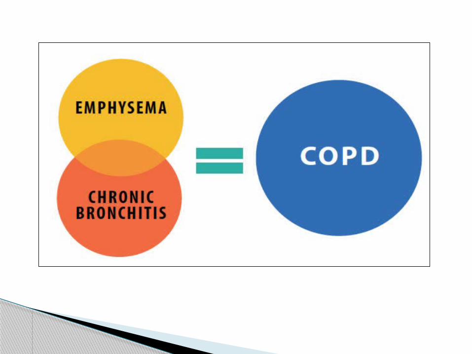
The two most commonly obstructive pulmonary
diseases:
emphysema
chronic bronchitis
Many patients have a combination of chronic bronchitis and emphysema and present with a mixture of signs and symptoms associated with both conditions.

Hypoxic drive: Normally, the amount of carbon dioxide in the blood
is the primary stimulus for breathing.
A secondary stimulus for breathing is hypoxia, a
decrease in oxygen.
COPD patients whose expirations are so inefficient
that their bodies have become accustomed to higher
than normal levels of co2.
So hypoxic drive is a condition in which the body's
stimulus for taking a breath is low oxygen.
Occurs in people with COPD.


Emphysema
More common with smokers.
Emphysema is a permanent disease that is
characterized by destruction of the alveolar walls
and distention of the alveolar sacs.
Severe reduction in the alveolar/capillary area for
gas exchange to occur.


Pathophysiology:
In emphysema, the lung tissue loses its elasticity,
the alveoli become distended with trapped air, and
the walls of the alveoli are destroyed.
Loss of the alveolar wall reduces the surface area in
contact with pulmonary capillaries.
Decrease in gas exchange occurs.
The patient becomes progressively hypoxic and
begins to retain carbon dioxide.


Exhaling becomes an active rather than
a passive process, requiring muscular
contraction; therefore, the patient uses
most of his energy to breathe.

Assessment:
Dyspneic
Thin, barrel-chest appearance from chronic air
trapping in the alveoli causing the anterior–
posterior diameter of the chest to increase

Prolonged exhalation
Pursed-lip breathing (physiologic PEEP)
Diminished breath sounds
Wheezing and rhonchi on auscultation
Extreme difficulty of breathing on minimal exertion
Pink puffer (extra hemoglobin to make up for poor
oxygen pick up).
Tachypnea: breathing rate usually greater than 20 per
minute at rest.
Tachycardia (increased heart rate).

Pursed-lip breathing


Management:
Ensuring an open airway
position of comfort
Administration of supplemental oxygen

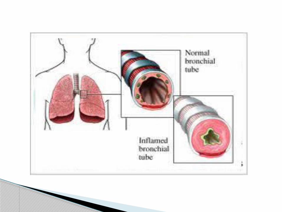
Chronic Bronchitis
• Increased mucus production.
• Decreased alveolar ventilation.
• Underlying problem: Ventilation and inflammation.
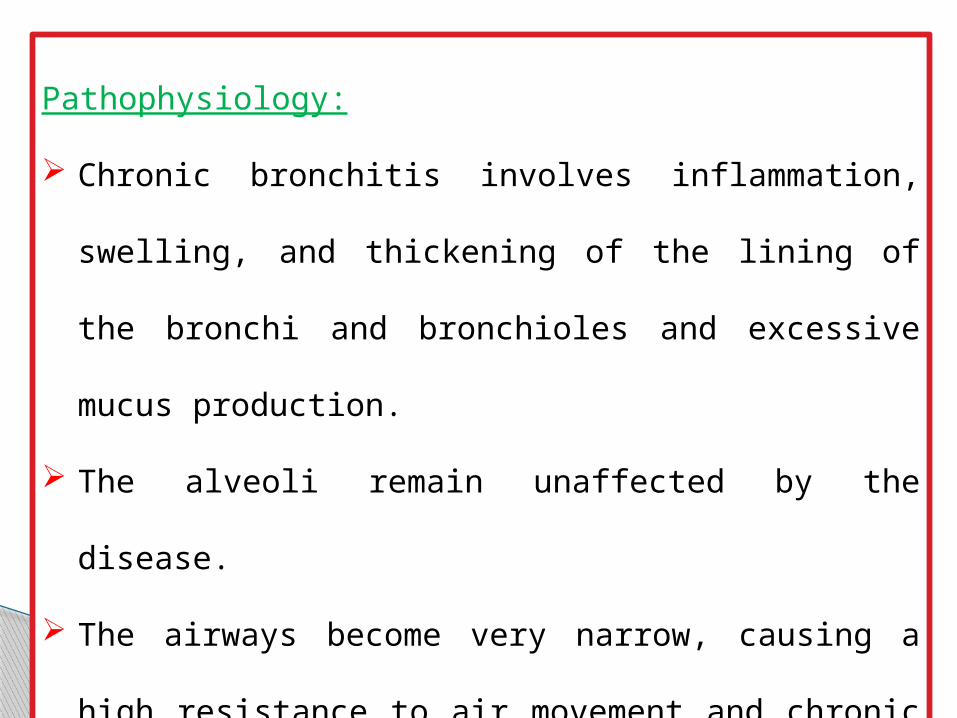
Pathophysiology:
Chronic bronchitis involves inflammation, swelling,
and thickening of the lining of the bronchi and
bronchioles and excessive mucus production.
The alveoli remain unaffected by the disease.
The airways become very narrow, causing a high
resistance to air movement and chronic difficulty in
breathing.

Obstructive Pulmonary Disease
An obstructive lung disease
Obstruction of airflow through the respiratory tract
Reduction in gas exchange
hypoxia


Assessment:
Cough (hallmark sign) is prominent; with sputum
Cyanosis ( so it was called “blue bloaters” )
Minimal difficulty in breathing and anxiety.
Wheezes and, possibly, crackles at the bases of the
lungs

Secure airway
Correct hypoxia
Albuterol bronchodilation if wheezing
Management