Dr copyright Index of Surface Anatomy · Index of Surface Anatomy Abdomen see also Aorta, Trunk...
37
3 The A to Z of Surface Anatomy © A. L. Neill Table of contents Introduction 1 Acknowledgement 1 Dedication 1 How to use this Book 2 Table of Contents 3 Abbreviations 7 Common Terms used in Surface Anatomy 9 Anatomical Planes and Relations 18 Movements – general Upper limb & shoulder 20 Head, Neck & Back 22 Lower limb and Hip 24 Foot and Hand 25 Hand Grips 26 Measurements 28 Proportions and forms of human measurement 30 Human face proportions 32 Human body proportions 34 Vitruvian Man 36 (Symbol of proportionality & derivative of measurements) Index of Surface Anatomy Abdomen see also Aorta, Trunk dermatomes 38 bones + muscles 40 alimentary - GIT 42 non-alimentary - / liver / pancreas / spleen 44 regions 46 scars - from incisions 48 Adam’s apple = Thyroid cartilage see Thyroid Adrenals see Back, Trunk Anatomical snuff box see Thumb Ankle see also Foot 50 Anus see Perineum Aorta 52 Appendix see Abdomen GIT Arm see also Axilla, Forearm, Shoulder 54 Axilla see also Breast bones , muscles boundaries 56 lymph nodes 58 copyright Dr A Neill
Transcript of Dr copyright Index of Surface Anatomy · Index of Surface Anatomy Abdomen see also Aorta, Trunk...
3
The A to Z of Surface Anatomy
© A. L. Neill
Table of contents Introduction 1Acknowledgement 1Dedication 1How to use this Book 2Table of Contents 3Abbreviations 7Common Terms used in Surface Anatomy 9Anatomical Planes and Relations 18Movements – general
Upper limb & shoulder 20Head, Neck & Back 22Lower limb and Hip 24Foot and Hand 25
HandGrips 26Measurements 28
Proportions and forms of human measurement 30Human face proportions 32Human body proportions 34Vitruvian Man 36(Symbol of proportionality & derivative of measurements)
Index of Surface Anatomy Abdomen see also Aorta, Trunk
dermatomes 38 bones + muscles 40alimentary - GIT 42non-alimentary - / liver / pancreas / spleen 44regions 46scars - from incisions 48
Adam’s apple = Thyroid cartilage see Thyroid Adrenals see Back, Trunk Anatomical snuff box see Thumb Ankle see also Foot 50Anus see Perineum Aorta 52Appendix see Abdomen GIT Arm see also Axilla, Forearm, Shoulder 54Axilla see also Breast
bones , muscles boundaries 56lymph nodes 58
copyright
Dr A Neill
The A to Z of Surface Anatomy
© A. L. Neill 4
Back lower 60upper see Chest
Belly see Abdomen Belly button see AbdomenBladder see Kidneys, Pelvis, UterusBreast see also Axilla
arterial 62lymphatic & venous 63
Buttock see Gluteum Caecum see Abdomen GITCarpal tunnel see Hand Chest Wall see also Abdomen, Lungs
great vessels 64heart 65heart valves sounds 66incision or marks = scars 67lungs & pleura 68
Cubital Fossa 72Diaphragm + assoc structures see also Oesophagus 74Duodenum see Abdomen GIT Kidneys Ear 76Elbow see arm, cubital fossa, forearm Eye 78Face
arteries 82bones see also TMJ 88Facial N 90muscles 92veins 94
Femoral trianglecontents & borders 96muscles & bones 98
Finger see Hand Flexor Retinaculum see Hand Foot
dorsumbones 100tendons 102
solefascia / muscle layers 104bones / dermatomes 109
Forearmbones 112muscles 114
copyright
Dr A Neill
5
The A to Z of Surface Anatomy
© A. L. Neill
Gall Bladder see Abdomen GIT, DiaphragmGenitalia –
female 120male see Penis Testis
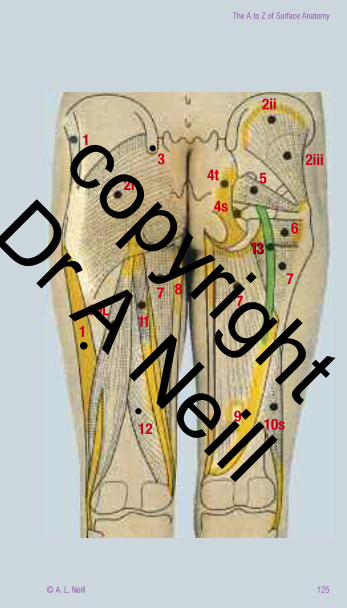
Gluteum / Gluteal regionbones 122muscles / Sciatic N 124
Hand see also gripsbones 126dorsum extensors 128palm
features 130flexors 132flexor retinaculum 134thenar/ hypothenar eminences 136
Head see Face, Neck, Temporomandibular joint Heart see also Chest, Oesophagus 138Hip see also Gluteum 140 Hyoid see Neck, Thyroid Inguinal nodes superficial 142Kidneys see also Back 144Knee see also lower leg, popliteal fossa, Thigh 146Large Intestine see Abdomen GIT 148Leg / Lower leg
muscles and bones 150 Liver see Abdomen non-GIT, Diaphragm Lungs see also Abdomen, Chest 156Lymph nodes see Axilla, Breast, Inguinal, Mouth, NeckMouth
salivary glands 158tonsil & uvula 160
Nail 162Neck
BVs & access points 164LNs and veins 166submandibular regions 168triangles 170
Nipples see Breast Nose 174Oesophagus 184Ovaries see Pelvis, Uterus Pancreas see Abdomen non-GIT, Kidneys Pelvis see Genitalia, Perineum, Uterus also specific organs listed
copyright
Dr A Neill
The A to Z of Surface Anatomy
© A. L. Neill 6
Penis 186Perineum see also Genitalia 188Phrenic N see Oesophagus Pleura see Chest, Lungs Popliteal fossa 192Rectum see Abdomen GIT, Pelvis, Perineum, Sigmoid colonSalivary Glands see Head, Mouth, Neck Sciatic N see also Gluteum 194 Scrotum see Femoral Triangle, Penis, TestisShoulder
bones 196BVs & Ns 198muscles 200
Sigmoid Colon see Abdomen GIT, Large IntestineSinuses 206Small Intestine see Abdomen GIT Spleen see also Abdomen, Back, Diaphragm 208Stomach see also Abdomen 210Teeth see Face, Mouth, TMJ
A to Z of the Head & Neck for complete map Temporomandibular Joint 214Testis/Testes see also Femoral Triangle, Penis, Scrotum Perineum 216Thigh see also Femoral Triangle, Hip & Knee 218
muscle Throat see Mouth Thumb 220Thymus see also Oesophagus 222Thyroid see also Neck 222Tongue see also Mouth 224Tonsil see Mouth, Tongue Trachea see also Neck 228Trunk see Abdomen, Back Umbilicus see Abdomen Ureter see Kidneys Uterus see also Pelvis 230Uvula see Mouth, TongueVagina see Pelvis, Perineum Vagus N see Oesophagus Womb see uterusWrist see also Hand & grips 232
copyright
Dr A Neill
7
The A to Z of Surface Anatomy
© A. L. Neill
A = atrium, (pl atria) / actions / movements of a jointa = arteryabdo = abdomen / abdominal ACF = anterior cranial fossa adj. = adjective AIIS = anterior inferior iliac spine aka = also known as alt. = alternativeAM = arachnoid mater ANS = autonomic nervous system ant = anteriorart. = artery AS = Alternative Spelling, generally referring to the diff. b/n British & American spellingASIS = anterior superior iliac spine assoc.= associated with AV = atrioventricular B = blood BBB = blood brain barrier bc = becauseBF = blood flow BM = basement membrane b/n = between BP = brachial plexus bpm = beats per minute br = branch (of a vessel) BS = blood supply / blood stream BV = blood vessel(s)cap. = capillary c.f. = compared to C = carpalC = cervicalCC = costal borderCC = costal cartilage CH = cerebral hemispheres cm = cell membrane CNS = central nervous system collat. = collateral CP = cervical plexus Cr = cranial CSF = Cerebrospinal fluid CT = connective tissue
CVA = cerebrovascular accident = stroke defn = definitiondiff. = difference(s)dist. = distal DM = dura mater DVT = deep vein thrombosis EAM = external auditory meatus e.g. = example EC = extracellular (outside the cell) ECG = electrocardiogramED = extensor digitorumER = Extensor RetinaculumFDP = Flexor digitorum porofundusFDS = Flexor digitorum superficialisFPB = Flexor pollicus brevisFPL = Flexor pollicus longusFR = Flexor Retinaculum Gk. = Greek H = hormone(s) H = hypochondriumHB = heart beat HF = heart failure HR = heart rate HS = heart sounds IC = intercostalIC = intercarpalICS = intercostal spaceIP = interphalangealIx = investigationIVC = inferior vena cava jt(s) = joints = articulations L = left L = lumbarLA = Left Atrium lat. = lateral LH = left hypochondrium LL = lower limb LIF = left iliac fossa lig = ligament Lt. = Latin m = muscleMC = metacarpal MCF = middle cranial fossa
Abbreviations
copyright
Dr A Neill
The A to Z of Surface Anatomy
© A. L. Neill 8
MCL = mid clavicular lineMCP = metacarpophalangealmed. = medial MI = myocardial infarction MIP = midinguinal point MT = metatarsal N = nerve NAD = normal (size, shape) NAD = no abnormality detected NR = nerve rootNS = nervous system/nerve supply NT = nervous tissuenv = neurovascular bundle P = pressure PAD = peripheral artery disease PaNS = parasympathetic nervous system Ph = phalangesPIIS = posterior inferior iliac spine pl. = pluralPM = pia mater PN = peripheral nerve post. = posteriorproc. = processprox. = proximalPS = pubic symphysis PSIS = posterior superior iliac spine R = right RA = right atriumRH = right hypochondrium RIF = Right Iliac Fossa S = sacral S1 = first heart sound S2 = second heart sound SA = sinoatrial SCM = sternocleidomastoid musclesing. = singular SC = spinal cord SN = spinal nerve SP = spinal process SR = sarcoplasmic reticulum subcut. = subcutaneous supf = superficial
SVC = superior vena cava SyNS = sympathetic nervous system T = thoracic TMJ = temporomandibular jointUL = upper limb, arm V = vertebra V = ventricle VC = vertebral column WM = white matter w/n = within w/o = without wrt = with respect to & = and ∩ = intersection with
copyright
Dr A Neill
The A to Z of Surface Anatomy
© A. L. Neill 42
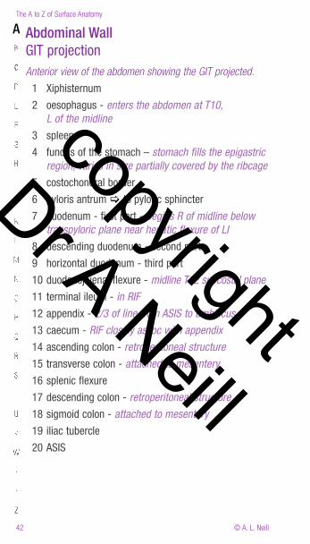
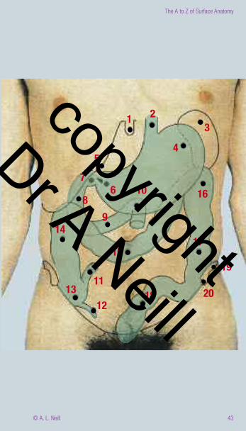
Abdominal Wall GIT projectionAnterior view of the abdomen showing the GIT projected.
1 Xiphisternum
2 oesophagus - enters the abdomen at T10, L of the midline
3 spleen
4 fundus of the stomach – stomach fills the epigastric region, varies in size partially covered by the ribcage
5 costochondral border
6 pyloris antrum [ to pyloric sphincter
7 duodenum - first part - begins R of midline below transpyloric plane near hepatic flexure of LI
8 descending duodenum - second part
9 horizontal duodenum - third part
10 duodenojejenal flexure - midline T12 subcostal plane
11 terminal ileum - in RIF
12 appendix - 2/3 of line from ASIS to umbilicus
13 caecum - RIF closely assoc with appendix
14 ascending colon - retroperitoneal structure
15 transverse colon - attached to mesentery
16 splenic flexure
17 descending colon - retroperitoneal structure
18 sigmoid colon - attached to mesentery
19 iliac tubercle
20 ASIS
A
copyright
Dr A Neill
43
The A to Z of Surface Anatomy
© A. L. Neill
1
10
23
4
16
17
19
2018
15
1113
12
149
86
75
copyright
Dr A Neill
The A to Z of Surface Anatomy
© A. L. Neill 44
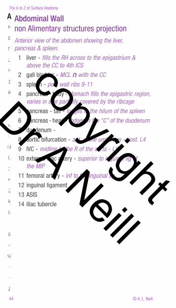
Abdominal Wall non Alimentary structures projectionAnterior view of the abdomen showing the liver, pancreas & spleen.
1 liver - fills the RH across to the epigastrium & above the CC to 4th ICS
2 gall bladder - MCL ∩ with the CC
3 spleen - post wall ribs 9-11
4 pancreas - body - stomach fills the epigastric region, varies in size partially covered by the ribcage
5 pancreas - tail - lodges in the hilum of the spleen
6 pancreas - head - lodges in the “C” of the duodenum
7 duodenum -
8 aortic bifurcation - ant. at the umbilicus - post. L4
9 IVC - midline to the R of the aorta - L5
10 external iliac artery - superior to inguinal lig at the MIP
11 femoral artery - inf to the inguinal lig
12 inguinal ligament
13 ASIS
14 iliac tubercle
A
copyright
Dr A Neill
45
The A to Z of Surface Anatomy
© A. L. Neill
1
35
42
7 6
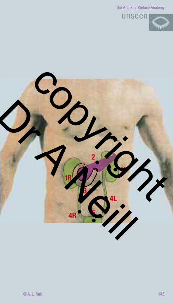
98
1012
11
13
14
copyright
Dr A Neill
The A to Z of Surface Anatomy
© A. L. Neill 46
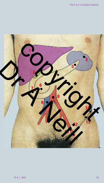
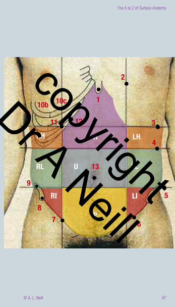
Abdominal Wall RegionsAnterior view of the abdomen showing regions. There are several ways to describe the regions of the Abdomen.
This commonly used schema divides the abdomen into 9 regions,based upon anatomical landmarks - numbered
Regions are coloured areas labelled with letters
1 Xiphisternum 2 midclavicular line (MCL) - vertical line ½ way along the Clavicle 3 transpyloric plane - midway b/n Xiphisternum and umbilicus = L1 passes through pyloric sphincter and 1st part of the duodenum4 subcostal plane - passes through L35 transtubercular plane - passes through L56 Inguinal ligament 7 midinguinal point (MIP) - intersection b/n MCL and inguinal lig 8 anterior superior iliac spine = ASIS 9 iliac tubercle 10 7th rib - b= bone c = cartilage 11 MCL ∩ 9th rib12 CC - lower border of the ribs from the Xiphisternum around13 umbilicus - varies with age & weight approx. T10 dermatome
E = epigastrium - area b/n 3 & 12 H = hypochondrium - area b/n MCL 3 & 4 L = left & R = right I = iliac region - area b/n MCL & 6 L= left & R = rightL = lumbar region - area b/n MCL & 4 & 5 L = left & R = rightP = pelvic area AKA suprapubic region - area below 5 above 6 U = umbilical region - area b/n MCLs 4 & 5
A
copyright
Dr A Neill
47
The A to Z of Surface Anatomy
© A. L. Neill
1
12
10c10b
11RH LHH
4
RL U 13
P
RI
78
9
6
LI
LL
5
3
2copyright
Dr A Neill
51
The A to Z of Surface Anatomy
© A. L. Neill
4b
4L 3 1
78u
8L
12a
2p
9u
9L
2p
6
5
65
2a
2a
78u
8L
2a1
34b4L 4t8
10
copyright
Dr A Neill
The A to Z of Surface Anatomy
© A. L. Neill 52
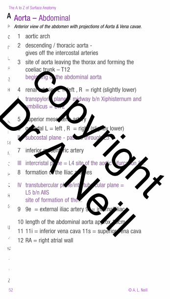
Aorta – Abdominal Anterior view of the abdomen with projections of Aorta & Vena cavae.
1 aortic arch
2 descending / thoracic aorta - gives off the intercostal arteries
3 site of aorta leaving the thorax and forming the coeliac trunk – T12 beginning of the abdominal aorta
4 renal arteries L = left , R = right (slightly lower)
I transpyloric plane – midway b/n Xiphisternum and umbilicus = L1/2
5 superior mesenteric artery
6 gonadal L = left , R = right (slightly lower)
II subcostal plane - passes through L3
7 inferior mesenteric artery
III intercristal plane = L4 site of the aortic bifurcation &...
8 formation of the Iliac arteries
IV transtubercular plane/intertubercular plane = L5 b/n AIIS site of formation of the...
9 9e = external iliac artery 9i = internal iliac
10 length of the abdominal aorta approx 10cm
11 11i = inferior vena cava 11s = superior vena cava
12 RA = right atrial wall
A
copyright
Dr A Neill
53
The A to Z of Surface Anatomy
© A. L. Neill
unseen
111s
12
11i 2
34L
6L78
9i9i 9e
106R
54RL1/2
L3
L4L5
I
II
IIIIV
copyright
Dr A Neill
The A to Z of Surface Anatomy
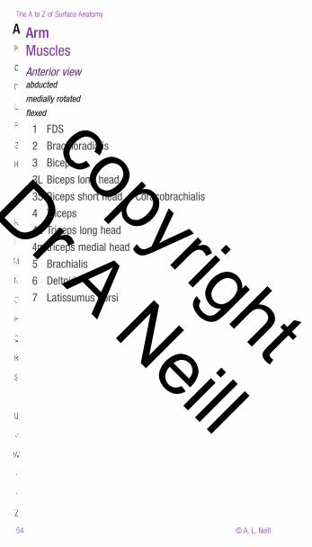
© A. L. Neill 54
Arm Muscles Anterior view abducted
medially rotated
flexed
1 FDS
2 Brachioradialis
3 Biceps
3L Biceps long head
3S Biceps short head + Coracobrachialis
4 Triceps
4L Triceps long head
4m triceps medial head
5 Brachialis
6 Deltoid
7 Latissumus dorsi
A
copyright
Dr A Neill
The A to Z of Surface Anatomy
© A. L. Neill 72
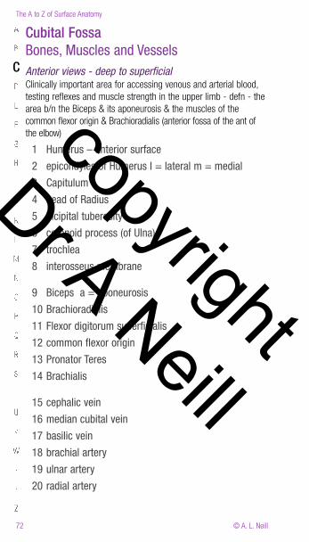
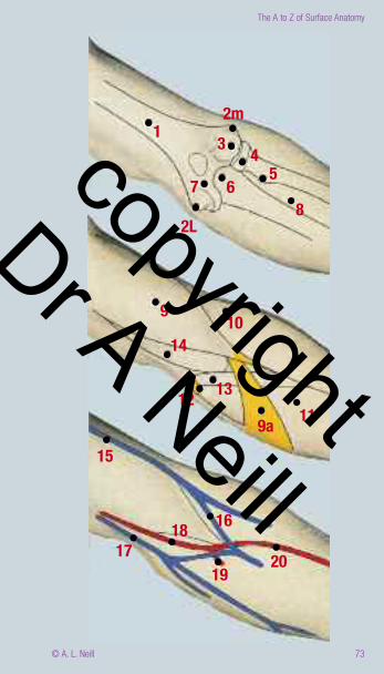
Cubital FossaBones, Muscles and Vessels Anterior views - deep to superficial Clinically important area for accessing venous and arterial blood,testing reflexes and muscle strength in the upper limb - defn - thearea b/n the Biceps & its aponeurosis & the muscles of thecommon flexor origin & Brachioradialis (anterior fossa of the ant ofthe elbow)
1 Humerus – anterior surface
2 epicondyles of Humerus l = lateral m = medial
3 Capitulum
4 head of Radius
5 bicipital tuberosity
6 coronoid process (of Ulna)
7 trochlea
8 interosseus membrane
9 Biceps a = aponeurosis
10 Brachioradialis
11 Flexor digitorum superficialis
12 common flexor origin
13 Pronator Teres
14 Brachialis
15 cephalic vein
16 median cubital vein
17 basilic vein
18 brachial artery
19 ulnar artery
20 radial artery
C
copyright
Dr A Neill
73
The A to Z of Surface Anatomy
© A. L. Neill
12m3
7
2L
68
54
9 10
11
14
12 13
9a15
161817
1920
copyright
Dr A Neill
The A to Z of Surface Anatomy
© A. L. Neill 74
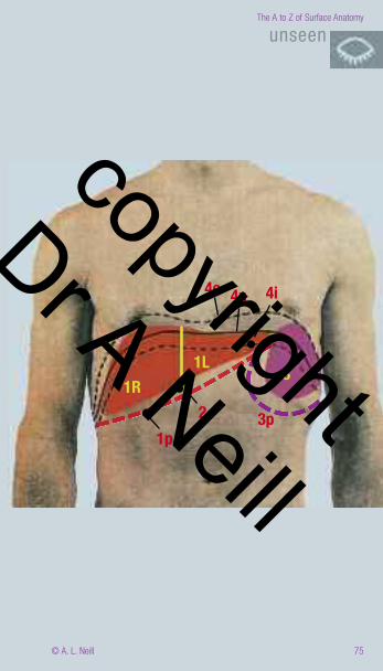
Diaphragm + Gallbladder + Liver + SpleenAnterior view Expiration - Inspiration.
Diaphragm = a musculotendinous dome, dividing the thorax &abdomen, centrally flattened the L(4th ICS T10-11) > R (6th ribT12) – both in the MCL (through the nipple).
Up to 1 rib higher in the supine position and in the broad-chested.
Resting respiration - ± 1cm – but may vary up to 10cm.
Attached - ant. – to the Xiphisternum, costal margin lat. - ribs 6-12post. L1-3
contraction - flattens the dome shape –pulls the IVC open ↑ abdominal P ↑ core strength lowers & moves the Liver to the L lowers & flattens the Spleen to the R
Both the Live r& Spleen are intimately related to the Diaphragmand the IVC and move as described.
1 Liver L = left R = right p = ptosed (ie lowered in inspiration) Liver edge palpable just below the CC on inspiration -
2 Gallbladder (9th CC) – divides the liver into the R & L functional ½
3 Spleen spans ribs 6-9 NAD – but in certain disease states partic parasitic infections - (malaria) may fill down to the LIF p = ptosed (ie lowered in inspiration)
4 Diaphragm e =expiration position i = inspiration position n = neutral position
D
G
L
S
copyright
Dr A Neill
75
The A to Z of Surface Anatomy
© A. L. Neill
unseen
1R
1p2
1L
3p
3
4i4n4e
copyright
Dr A Neill
The A to Z of Surface Anatomy
© A. L. Neill 76
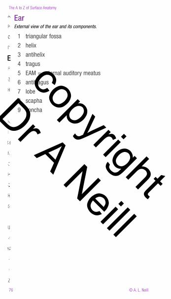
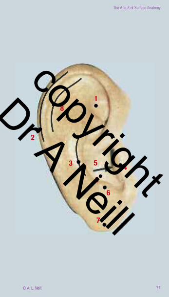
Ear External view of the ear and its components.
1 triangular fossa
2 helix
3 antihelix
4 tragus
5 EAM = external auditory meatus
6 antitragus
7 lobe
8 scapha
9 concha
E copyright
Dr A Neill
77
The A to Z of Surface Anatomy
© A. L. Neill
18
2
3 4
69
7
5
copyright
Dr A Neill
103
The A to Z of Surface Anatomy
© A. L. Neill
2
6
5
2b
4
12L
3
7
copyright
Dr A Neill
The A to Z of Surface Anatomy
© A. L. Neill 104
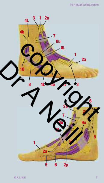
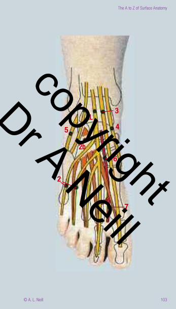
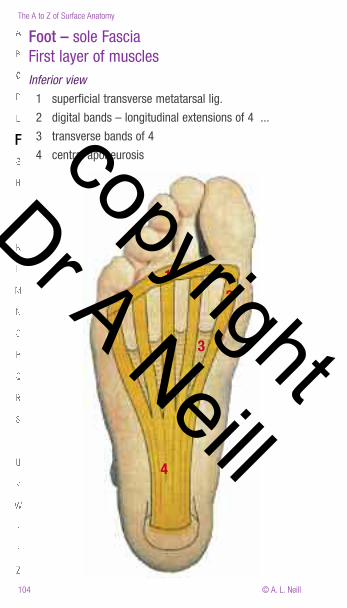
Foot – sole FasciaFirst layer of muscles Inferior view
1 superficial transverse metatarsal lig.
2 digital bands – longitudinal extensions of 4 ...
3 transverse bands of 4
4 central aponeurosisF
12
3
4
copyright
Dr A Neill
105
The A to Z of Surface Anatomy
© A. L. Neill
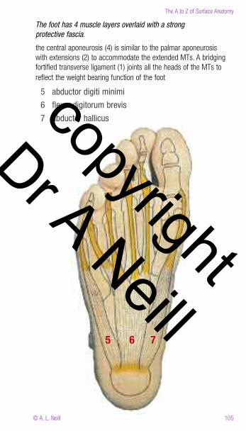
The foot has 4 muscle layers overlaid with a strong protective fascia.
the central aponeurosis (4) is similar to the palmar aponeurosiswith extensions (2) to accommodate the extended MTs. A bridgingfortified transverse ligament (1) joints all the heads of the MTs toreflect the weight bearing function of the foot
5 abductor digiti minimi
6 flexor digitorum brevis
7 abductor hallicus
765
copyright
Dr A Neill
The A to Z of Surface Anatomy
© A. L. Neill 106
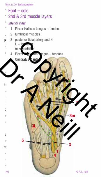
Foot – sole 2nd & 3rd muscle layers Inferior view
1 Flexor Hallicus Longus – tendon
2 lumbrical muscles
3 posterior tibial artery and N L = lateral br m = medial br
4 Flexor Digitorum Longus – tendons
5 Quadratus Plantae
F
2
1
3m
31
4
5
3L
copyright
Dr A Neill
The A to Z of Surface Anatomy
© A. L. Neill 84
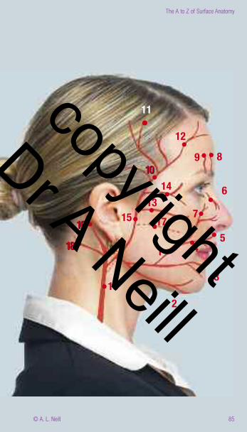
Face – Arteries - major Lateral There are extensive anastomising b/n all the arteries in the face formore detail see the A to Z of the Head and Neck.
1 Facial artery
2 mental br + submental brs
3 inferior labial br
4 superior labial br
5 internal nasal br
6 external nasal br
7 angular br /infraorbital artery
8 Supratrochlear artery
9 Supraorbital artery (may feel pulse 1 in = 2.5 cm from midline)
10 Superficial temporal artery (may feel pulse)
11 parietal br
12 frontal br
13 transverse facial br
14 infraorbital br
15 External carotid artery
16 posterior auricular br
17 deep transverse facial / maxillary
18 occipital
F copyright
Dr A Neill
85
The A to Z of Surface Anatomy
© A. L. Neill
12
9 8
6
7
54
1413171516
18
10
16
18
10
11
1
15 2
3
copyright
Dr A Neill
The A to Z of Surface Anatomy
© A. L. Neill 86
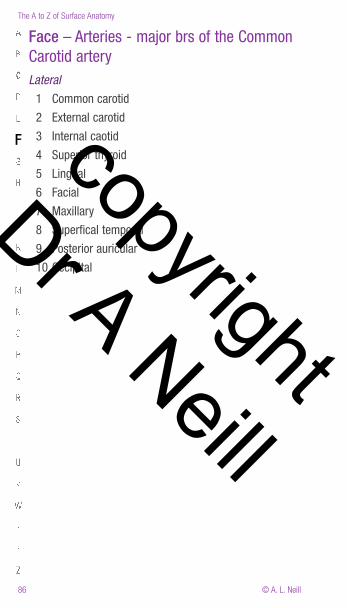
Face – Arteries - major brs of the CommonCarotid arteryLateral
1 Common carotid
2 External carotid
3 Internal caotid
4 Superior thyroid
5 Lingual
6 Facial
7 Maxillary
8 Superfical temporal
9 Posterior auricular
10 Occipital
F copyright
Dr A Neill
125
The A to Z of Surface Anatomy
© A. L. Neill
1
2i3
4t
4s5
2iii
2ii
6
7
1313
7
10s912
10L1 11
7 8
copyright
Dr A Neill
The A to Z of Surface Anatomy
© A. L. Neill 126
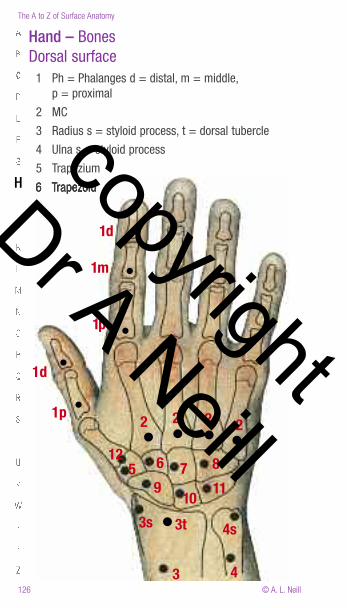
Hand – BonesDorsal surface 1 Ph = Phalanges d = distal, m = middle, p = proximal
2 MC
3 Radius s = styloid process, t = dorsal tubercle
4 Ulna s = styloid process
5 Trapezium
6 Trapezoid H
1d
1m
1p
1d
1p 2 2 2 2
876125
9
3s 3t10 11
4s
43
copyright
Dr A Neill
127
The A to Z of Surface Anatomy
© A. L. Neill
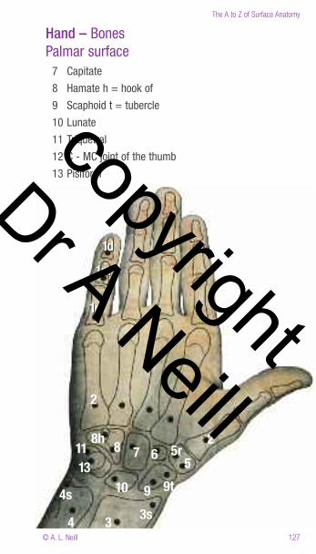
Hand – BonesPalmar surface 7 Capitate
8 Hamate h = hook of
9 Scaphoid t = tubercle
10 Lunate
11 Triquetral
12 C - MC joint of the thumb
13 Pisiform
1d1m
1p
1d1m
1p
2
25
5r6788h1113
10 9 9t
3s34
4s
copyright
Dr A Neill
The A to Z of Surface Anatomy
© A. L. Neill 128
Hand – Dorsum Extensor tendons & synovial sheathsThe hand is highly mobile, hence it must have strong mobile tendinousattachments. To improve mobility these expand to individual synovialsheaths commencing from the base of the fingers.
1 Abductor pollicus longus
2 Extensor pollicus brevis
3 Extensor carpi radialis longus
4 Extensor carpi radialis brevis
5 Extensor pollicus longus
6 Extensor indicis
7 Extensor digitorum (communicans)
8 Extensor digit minimi
9 Extensor carpi ulnaris – in supination / shown more laterally as in pronation
10 Extensor retinaculum
11 shared synovial bursa of extensors
Hcopyright
Dr A Neill
145
The A to Z of Surface Anatomy
© A. L. Neill
unseen
21L
4L
5
3
4R
1R
copyright
Dr A Neill
The A to Z of Surface Anatomy
© A. L. Neill 146
Knee – Flexed Lateral MedialThe knee is the most unstable joint in the body. It relies extensivelyon its ligaments and muscles for structural integrity as the bonysurfaces are not compatible and provide little or no support.
1 Rectus femoris t = tendon
2 Vastus lateralis 2L / V medialis 2m
3 Biceps femoris t = tendon (note bipennate muscle)
4 Iliotibial tract
5 Patella
6 femoral condyle L = lateral m = medial
7 lateral collateral lig
8 meniscus l = lateral, m = medial
9 common tendon insertion = the patella tendon (bursa behind)
10 tibial condyle l = lateral, m = medial
11 common perineal N
12 head of Fibula
13 Semimembranous
14 Sartorius
15 Gracilis
16 Semitendinous
K
copyright
Dr A Neill
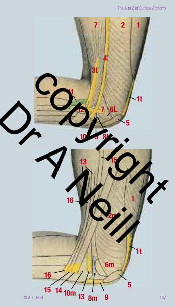
147
The A to Z of Surface Anatomy
© A. L. Neill
1
1513
16
598m1310m1415
166m
2m
1
1t
27
4
1t
56L712
11
10L 9 8L
3tcopyright
Dr A Neill
The A to Z of Surface Anatomy
© A. L. Neill 148
Large Intestine Anterior Surface projection of the LI showing mesenteries and position inAbdomen.
1 ileum
2 caecum 1-2 ilieocaecal junction
3 ascending colon = Right sided colon
4 hepatic flexure
5 Transverse colon (= Horizontal colon) & mesentery
6 splenic flexure
7 descending colon = Left sided colon
8 Sigmoid colon & mesentery
IS - Interspinous plane
MCL - midclavicular line
TP - transpyloric plane
L
copyright
Dr A Neill


![INDEX [sa1c9dbbba90cec02.jimcontent.com]€¦ · index 2. warm up 4. muscles & joints 1. physical fitness 6. judo 7. indoor football 8. artistic gymnastics health anatomy 5. acrosport](https://static.fdocuments.in/doc/165x107/5ec073213824c06650444100/index-index-2-warm-up-4-muscles-joints-1-physical-fitness-6-judo-7.jpg)