Dr Charles De Groot - GP CME 137 deGroot_Charles - What a GP should k… · What a GP should know...
54
Dr Charles De Groot Radiation Oncologist Hamilton
Transcript of Dr Charles De Groot - GP CME 137 deGroot_Charles - What a GP should k… · What a GP should know...

Dr Charles De Groot Radiation Oncologist
Hamilton
No conflict of interest?

What a GP should know about
Radiation Oncology
Charles De Groot
Radiation Oncologist
Waikato Hospital

Overview
• History.
• How does radiation work?
• The role of radiation.
• Different techniques.
• The process of radiation (patient
experience).
• Side effects and how to manage them.
• Future directions.

History of Radiation Delivery
• 1895 Wilhelm
Roentgen discovered
x-rays.
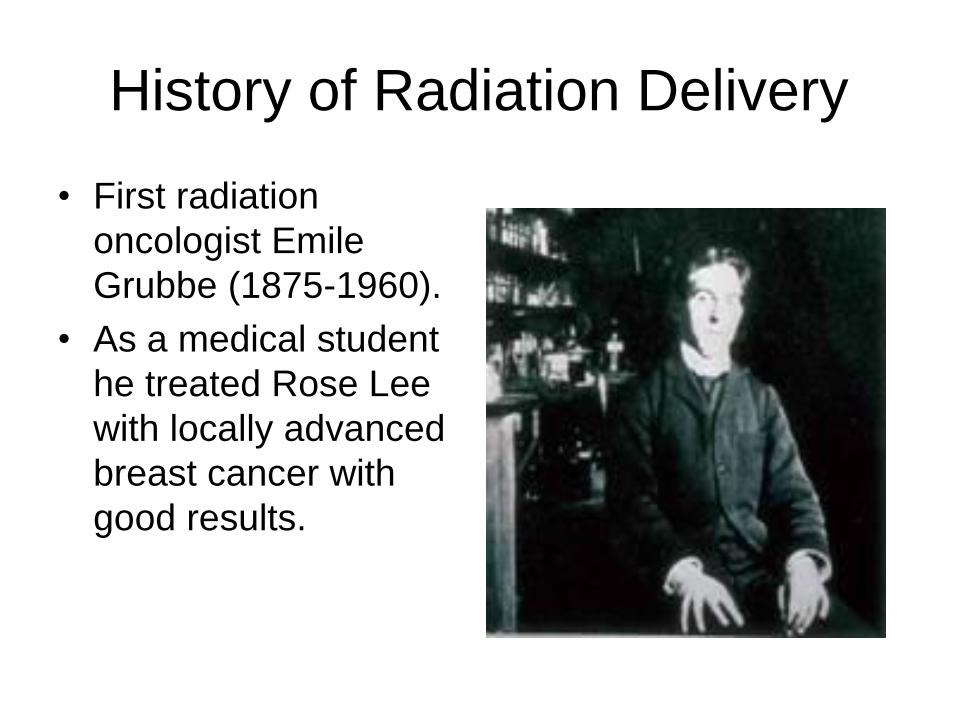
History of Radiation Delivery
• First radiation
oncologist Emile
Grubbe (1875-1960).
• As a medical student
he treated Rose Lee
with locally advanced
breast cancer with
good results.

History of Radiation Delivery
• 1900-1960’s concepts
of fractionation and
field shaping with low
energy RT refined.
• 1960 first linear
accelerator capable of
much higher energies
(more penetrating) of
radiation.

How radiation works

How Radiation Works
• Indirect effects
- Free Radicals
• Direct effects

How radiation works
• The production of free radicals is an
oxygen dependant process so radiation is
less effective in hypoxic tissues.
• Given that anti-oxidants absorb free
radical they may protect both normal
tissues as well as the targeted
malignancy.

How radiation works
• A clinical target is defined, which
encompasses the macroscopic and
expected microscopic extent of the
malignancy.
• The planning team then solves the
geometric and biologic problem of how
best to cover the clinical target with the
appropriate radiation dose – see later..

The role of radiation
• Cancer management
Primary curative
Adjuvant curative
Palliative
Prophylactic
• Benign disease
The role of radiation
• Radiation therapy can cure cancer!


Definitive curative radiation
• Localised head and neck cancer.
• Localised lung cancer.
• Prostate cancer.
• Skin cancer.
• Localised haematological malignancy –
lymphoma, plasmacytoma.
Adjuvant curative radiation
• Head and neck
• Breast cancer
• Rectal cancer
• Sarcoma.
• High grade lymphoma.
• Skin

Curative radiation
• Often but not always high dose.
• Side effects can be significant.

Palliative radiation
• To relieve or prevent localised symptoms
of virtually any malignancy.
• May be life prolonging.
• Can vary from single treatment (e.g.
simple bone metastasis) to multiple
treatments(e.g glioblastoma multiforme).
• Generally aim for low morbidity – lower
doses.

Prophylactic radiation
• Usually preventative radiation to
chemotherapy sanctuary sites – typically
CNS, occasional scrotal.
• Usually relatively low dose.

Radiation for benign disease.
• Benign tumours –meningioma, dermoid.
• Dupuytrens contracture – prevents
progression.
• Thyroid opthalmopathy.
• Heterotopic ossification.
• Keloid scars.
• Arteriovenous malformation.
• Pterygium.

• 5yr progression of 67% observation vs
22% when treated with RT. P=001

Different techniques
• External beam radiation
High energy
Superficial voltage
• Brachytherapy
Interstitial
Mould
Intracavitory
• Isotopes

External beam workhorse
Linear Accelerator
• Complex
• Deeper
dosing

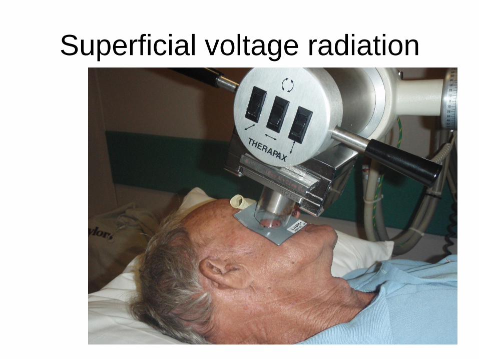
Superficial voltage radiation

Mould brachytherapy
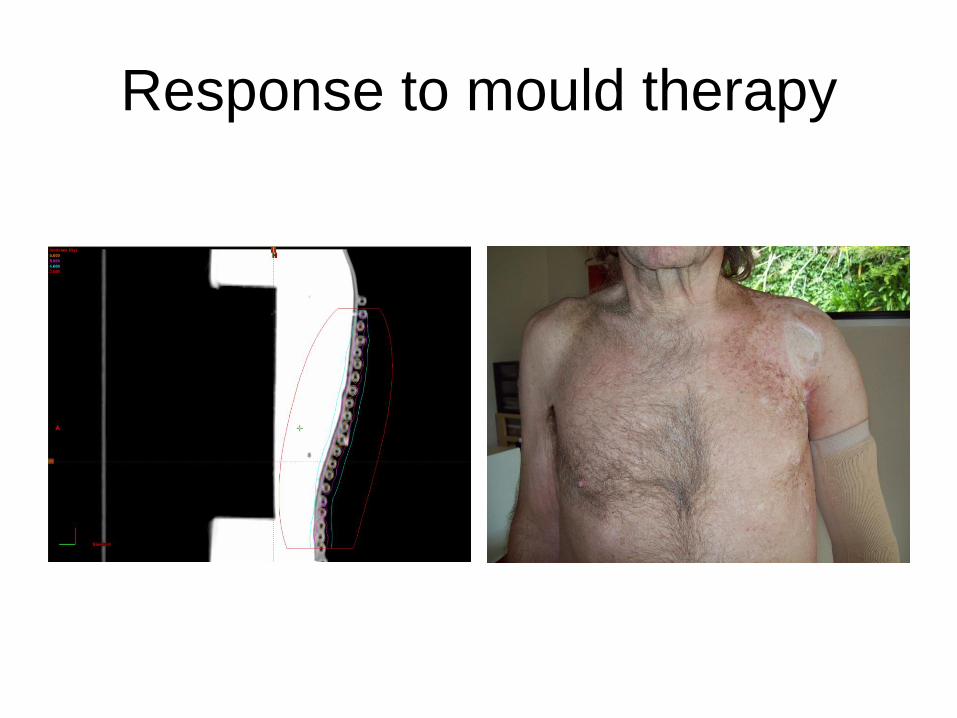
Response to mould therapy

The process of radiation
Patient experience • First specialist assessment
- 1 hour
- explain why needed, or not and side
effects.
- once patient agrees with treatment plan
they will have a RT planning session (may
be same day, usually within 1 week).

Planning
• May be as simple as delineating area to
be treated on skin – sharpie.
• Most common planning requires ct scan of
intended treatment volume, done in
radiotherapy department.
• Patient will be positioned and immobilised
(in comfort) to ensure we can access
target tissue with minimal exposure of
normal tissue.
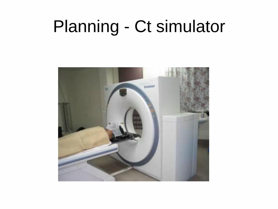
Planning - Ct simulator

Planning - Positioning of breast
cancer treatment

Planning - Shell for head and
neck cancer therapy

Planning
• Once ct completed or clinical target
delineation completed we plan the
radiation.
• This may take from minutes to days,
depending on complexity.

Simple vs Complex plan

Treatment
• From 1-37 treatments, Monday to Friday.
• Treatment session 10-15 minutes, don't
feel anything.
• Accommodation provided.
• Lots of support provided.

Side effects
• With the exception of lethargy, radiation
side effects are confined to the tissues
being irradiated.
• Radiation of the big toe will not cause
heart attacks, syphilis, dementia
etc.etc.etc. It will cause the toenail to fall
out!

Acute Side effects
• Affect rapidly multiplying tissues such as
mucous membranes and skin.
• Stem call depletion leads to loss of
overlying epithelial layers (desquamation,
ulceration).
• Occurs during and shortly after treatment
and usually achieve full recovery.

Chronic Side effects
• Damage of vascular endothelial cells leads
to small vessel ischaemia/hypoxia.
• Chronic hypoxia leads to fibroblast
deposition and clinical fibrosis.
• Fibrosis can lead to stricture, loss elasticity
etc.
• Hypoxia leads to poor healing, poor organ
function.

Chronic side effects
• DNA damage also occurs and leads to a
variable but generally low risk of radiation
induced malignancy.

Chronic Side effects
• Chronic radiation side effects occur > 6
months after therapy and are usually
irreversible.
• The degree of late damage is dependant
on dose given and individuals genetic
ability to repair radiation damage.

Side effects
• Serious chronic effects are rare and
generally would affect < 5% of patients
treated.
Management of common
radiation side effects

Acute radiation dermatitis
• A week 1
• B week 3
• C week 5
• D week 7
End of treatment
• E week 9
• F week 11
• G week 14
• H week 18

Management of radiation
dermatitis • A week 1 aqueous cream
• B week 3
• C week 5 1%hydrocortisone cream
• D week 7 Manuka honey dressings
End of treatment
• E week 9 silver sulphadiazine ointment
• F week 11
• G week 14
• H week 18

Management of radiation
dermatitis Principles:
1. Supportive care, it will run its course.
2. Watch for infection (Bactroban, oral
antibiotics).
3. Non abrasive dressings/ clothing (silk
scarves).
4. Avoid too many dressing changes.

Radiation mucositis

Radiation mucositis
• Very significant issue, especially with
oropharyngeal radiation.
• It will run its course.

Symptoms of RT oral mucositis
• Pain.
• Xerostomia.
• Thick, tacky secretions.
• Loss of taste.
Consequently patients are at high risk of
dehydration and malnourishment.

Management of oral radiation
mucositis
• Analgesia – low index to use opiods, liquid
morphine for topical effect as well as
easier swallowing. Long acting morphine
needs to be used as well.
• Xylocaine gel prn for extreme cases but
watch for aspiration risk.

Management of oral radiation
mucositis
• Mouth care:
Benzydamine (Difflam) is mainstay.
Antifungals – remember lozenges don’t
dissolve in dry mouth. (Mycostatin drops
or fluconazole capsules).
Soda bicarb mouthwash very refreshing,
lifts secretions ( ½ tsp salt, ½ bicarb and ½
glass water).

Management of radiation mucositis
• Make sure they are eating/drinking.
• Liquid food supplements are virtually
always mandatory.
• May require alternate feeding such as NG
tube or gastrostomy.

Mucositis elsewhere
• Symptomatic, so analgesia again.
Xylocaine excellent for anoproctitis as can
steroid suppositories/enemas.
• Loperamide/codiene for loose bowels.
• Sitz bath useful for perineal
dermatitis/mucositis.

Future directions