Afrah Qassim Community Development Worker Inclusion Matters Liverpool & Chair/Cofounder of Savera.

Dr. Afrah Adnan Aldelaimi
B.D.S., C.D.I., M.Sc., Ph.D.
Oral & Maxillofacial Pathologist
University of Anbar - College of Dentistry

is defined as an epithelial-lined pathologic
cavity. In contrast to true cysts,
pseudocysts lack an epithelial lining.
Cysts of the maxilla, mandible, and
perioral regions vary markedly in
histogenesis, incidence, behavior, and
treatment and can be divided into
odontogenic cysts, nonodontogenic cysts,
pseudocysts, and neck cysts.


are the most common
cysts of the jaws. These
inflammatory cysts derive
their epithelial lining from
the proliferation of small
odontogenic epithelial
residues (rests of
Malassez) within the
periodontal ligament.

They develop from a preexisting periapical
granuloma (a focus of chronically inflamed
granulation tissue located at the apex of a nonvital
tooth). Stimulation of resident epithelial rests of
Malassez occurs in response to the products of
inflammation.
Cyst form from epithelial proliferation to separate
the inflammatory stimulus (necrotic pulp) from the
surrounding bone.

Breakdown of cellular debris within the cyst lumen
raises the protein concentration, increasing
osmotic pressure and resulting in fluid transport
across the epithelial lining into the lumen from the
connective tissue side.
With osteoclastic bone resorption, the cyst
expands. Other bone resorption factors, such as
prostaglandins, interleukins, and proteinases, from
inflammatory cells and cells in the peripheral
portion of the lesion causes additional cyst
enlargement.


Radiographically, a periapical cyst cannot be
differentiated from a periapical granuloma.

The periapical cyst is lined
by nonkeratinized stratified
squamous epithelium of
variable thickness with large
numbers of neutrophils
fewer numbers of
lymphocytes involved.
Plasma cell infiltrate and
spherical intracellular
Russell bodies, representing
accumulated gamma
globulin, cholesterol clefts,
and multinucleated giant
cells may be seen.

Antibiotic
Extraction
Root canal filling
Apicoectomy and direct curettage of the lesion.
When the necrotic tooth is extracted but the cyst
lining is incompletely removed, a residual cyst
may develop months to years causing significant
bone resorption and weakening of the mandible or
maxilla.

It is a nonkeratinized
developmental cyst occurring
adjacent or lateral to the root
of a tooth. Its origin from
rests of dental lamina.
Pathogenetically it linked to
the gingival cyst of the adult;
the former is believed to arise
from dental lamina remnants
within bone, and the latter
from dental lamina remnants
in soft tissue between the oral
epithelium and the
periosteum (rests of Serres).

Most lateral periodontal cysts and gingival cysts
of the adult occur in the mandibular premolar
and cuspid regions.
Male predilection has been noted for lateral
periodontal cysts 2:1 distribution while in
gingival cysts show a nearly equal gender
predilection.
The median age for both types of cysts is
between the 5th – 6th decades of life.
Gingival cyst appears
as a small soft tissue
swelling within or
slightly inferior to the
interdental papilla. It
may assume a slightly
bluish discoloration
when it is relatively
large. Most cysts are
less than 1 cm in
diameter.
Radiography reveals
no findings.

Lateral periodontal
cyst presents as an
asymptomatic,well-
delineated, round or
teardrop-shaped
unilocular
radiolucency with an
opaque margin along
the lateral surface of a
vital tooth root. Root
divergence is rarely
seen.

Histopathology Both cysts
are lined by a thin,
nonkeratinized epithelium.
Clusters of glycogen-rich,
clear epithelial cells may be
noted in nodular thickenings
of the cyst lining.
Treatment and Prognosis
Local surgical excision of
both cysts
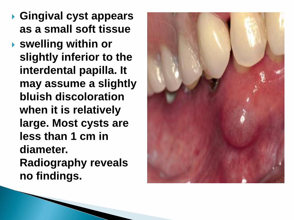
It appear as multiple nodules along the alveolar ridge in neonates as a proliferate of the dental lamina that remain within the alveolar ridge mucosa to form small, keratinized cysts.
These cysts are self-limiting rupture into the oral cavity within a few weeks to a few months.

It is second most common type of odontogenic cyst, and the most common developmental cyst of the jaws.
It develops from proliferation of reduced enamel epithelium which expaned as a result of increase in cyst fluid osmolality and the release of bone resorption factors.

A dentigerous cyst is attached to the tooth
cervix at the cementoenamel junction encloses
the crown of the unerupted tooth. It most
commonly associated with impacted teeth
(third molars and maxillary canines), The
highest incidence of DC occurs during the 2nd
and 3rd decades.
In radiograph, it presents as a well- defined,
unilocular radiolucency with corticated
margins in association with the crown of an
unerupted tooth. Resorption of roots of
adjacent erupted teeth may occasionally be
seen.

The cyst-to-crown relationship variations: Central variety ,Lateral variety, Circumferential variety

Microscopically, the
dentigerous cyst is
formed by a fibrous
connective tissue wall and
is lined by nonkeratinized
stratified squamous
epithelium that arrange in
4-6 cell layers thick.
Mucous cells, ciliated
cells may be found,
although in cases of
secondary inflammation
epithelial hyperplasia may
be noted.
Extraction of the associated tooth with enucleation
Potential complications of untreated dentigerous
cysts include transformation of the epithelial lining
into an ameloblastoma and rarely intraosseous
mucoepidermoid carcinoma.

It results from fluid
accumulation within the
follicular space of an
erupting tooth. The
epithelium lining this
space is simply
reduced enamel
epithelium. With
trauma, blood may
appear within the tissue
space forming an
eruption hematoma.

No treatment is needed
because the tooth erupts
through the lesion.
Subsequent to eruption,
the cyst disappears
spontaneously without
complication.
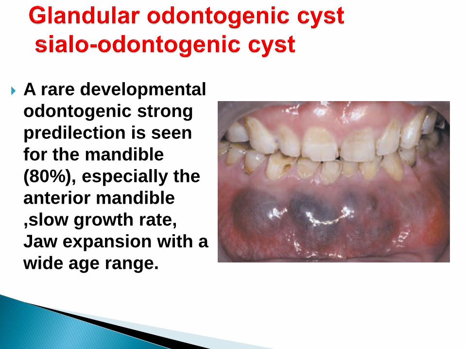
A rare developmental
odontogenic strong
predilection is seen
for the mandible
(80%), especially the
anterior mandible
,slow growth rate,
Jaw expansion with a
wide age range.

It is multilocular lesion
with wide variation in
size, from smaller than
1 cm to involving most
of the mandible
bilaterally with well
defined, sclerotic and
scalloped margin.
Teeth may be
displaced, and root
resorption is noted in
some cases.

It consist of nonkeratinized squamous epithelium
lining with focal thickenings in which the epithelial
cells assume a swirled appearance.
The epithelial lining consists of cuboidal cells,
often with cilia and mucous cells with mucin pools.

This lesion can be considered locally aggressive;
therefore, surgical management with adequate
healthy bone remains beyond the extent of the
cystic lesion. Longterm follow-up is essential given
the local aggressiveness and recurrence rate
(approximately 25%) of this lesion.

It is an unique developmental odontogenic cyst
developed from dental lamina remnants in the
mandible and maxilla and exhibit aggressive clinical
behavior, a relatively high recurrence rate, and an
association with nevoid basal cell carcinoma
syndrome.
Radiographically mimic other types of cysts,
Multilocularity is often present.

It is common jaw cyst
It occurs at any age and have a peak incidence within the 2nd and 3rd decades.
About 5% of patients with OKCs is multiple cysts
Mostly found in the mandible in 2:1 ratio in the ramus and posterior portion. In the maxilla, the third molar area is most commonly affected.

Factors that may contribute to the pathogenesis of
the OKC include a high proliferation rate,
overexpression of the antiapoptotic protein Bcl-2
and several growth factors, and expression of
MMPs 2 and 9.
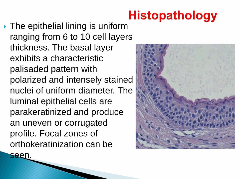
The epithelial lining is uniform
ranging from 6 to 10 cell layers
thickness. The basal layer
exhibits a characteristic
palisaded pattern with
polarized and intensely stained
nuclei of uniform diameter. The
luminal epithelial cells are
parakeratinized and produce
an uneven or corrugated
profile. Focal zones of
orthokeratinization can be
seen.

An orthokeratinized odontogenic cyst has been
described difference from OKC. Histologic
distinction between parakeratinized and
orthokeratinized cysts is made because the latter
type of cyst is less clinically aggressive, has a
lower rate of recurrence, and generally is not
associated with syndrome.

In the orthokeratotic
odontogenic cyst, a
prominent granular
layer is found
immediately below a
flat, noncorrugated
surface. The basal cell
layer is less prominent
and has a more
flattened or squamoid
appearance in
comparison with the
parakeratotic type.

Wide surgical excision with peripheral osseous
curettage is the preferred method of management
because of the aggressive nature the recurrence rate
varies from 10-30% that depend on how the lesion is
managed and is also related to the friable, thin
connective tissue wall of the cyst may lead to
incomplete removal and small daughter satellite cysts
in the bone adjacent to the primary lesion. Also,
cystic proliferation of the overlying oral epithelial
basal cell layer, if not eliminated during cyst removal,
is considered significant by some.
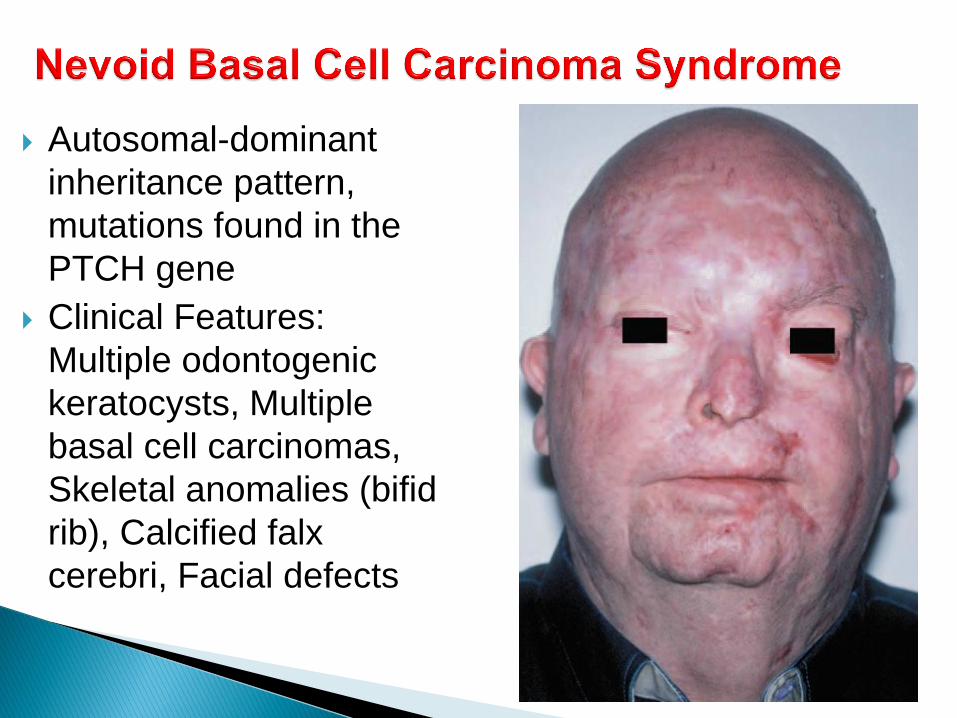
Autosomal-dominant
inheritance pattern,
mutations found in the
PTCH gene
Clinical Features:
Multiple odontogenic
keratocysts, Multiple
basal cell carcinomas,
Skeletal anomalies (bifid
rib), Calcified falx
cerebri, Facial defects


It are developmental odontogenic lesions that
occasionally exhibit recurrence derived from
odontogenic epithelial remnants within the
gingiva or within the mandible or maxilla.
It usually appears in individuals younger than 40
years of age and has a decided predilection for
females. More than 70% of COCs are seen in the
maxilla.

Radiographically,
COCs may present as
unilocular or
multilocular
radiolucencies with
well-demarcated
margins. Within the
radiolucency scattered
irregularly sized
opacities may produce
a salt-and-pepper type
of pattern.
It consists of well-delineated epithelial lining
is of variable thickness with a fibrous
connective tissue wall. Intraluminal epithelial
proliferation obscures the cyst lumen
producing the impression of a solid tumor.
The most prominent and unique microscopic
feature is the presence of Ghost cells which
are anucleate retain the outline of the cell
membrane. These cells undergo dystrophic
mineralization


They are cysts arise from epithelium
entrapped along embryonal lines of fusion.
However, the concept of a fissural origin for
many of these cysts has been questioned in
more recent years. In many instances the
exact pathogenesis of these lesions is still
uncertain. Regardless of their origin, once
cysts develop in the oral and maxillofacial
region, they tend to slowly increase in size,
possibly in response to a slightly elevated
hydrostatic luminal pressure.

Globulomaxillary cysts were
once considered fissural
cysts, located between the
globular and maxillary
processes (between maxillary
lateral incisor and canine).
The theory of origin involved
epithelial entrapment within a
line of embryologic closure
with subsequent cystic
change.

Radiologically, a
globulomaxillary lesion
appears as a well-
defined inverted pear-
shaped radiolucency,
often producing
divergence of the roots
of the maxillary lateral
incisor and canine
teeth.
Radicular cyst and periapical
granuloma can be ruled out with pulp
vitality testing.
Asymptomatic; teeth vital; divergence
of roots
Biopsy necessary to establish
definitive diagnosis

It is a rare soft tissue cyst of
the upper lip. The
pathogenesis of the
nasolabial cyst is unclear the
lesion represents cystic
change in the remnants of
cells that form the
nasolacrimal duct.
a peak incidence noted in the
4th -5th decades. A distinct
female predilection of nearly
4:1 has been noted.
The chief clinical sign is a soft tissue
swelling that may present in the soft tissue
over the canine region or the mucobuccal
fold.
The epithelial lining of this cyst is
characteristically a pseudostratified
columnar type with numerous goblet cells.
Stratified squamous epithelium may be
present in addition to cuboidal epithelium in
some cases. The cyst is treated by curettage
with few recurrences expected.

It is located within the nasopalatine canal
It develops from the proliferation of epithelial
remnants of paired embryonic nasopalatine
ducts within the incisive canal.
Men are affected more often than women, with
differences as 3:1.
Most cases are asymptomatic, with the clinical
sign of swelling usually calling attention to the
lesion.

They are pseudocysts because they appear radiographically as
cyst-like lesions but microscopically exhibit no epithelial lining.
This lesion represents a benign lesion of bone that may arise in
the mandible, the maxilla, or other bones. Within the cranio-
facial complex, The pathogenesis of the aneurysmal bone cyst
is not well understood. Some evidence suggests a reactive
process, and other evidence suggests a tumor. Supporting the
tumor
Histopathology: fibrous connective tissue stroma contains
variable numbers of multinucleated giant cells with blood filled
spaces.
Treatment and Prognosis: A relatively high recurrence rate has
been associated with simple curettage. Excision or curettage
with supplemental cryotherapy is the treatment of choice.

It is an empty intrabony cavity that lacks an epithelial
lining (pseudocyst). It is seen most often in the
mandible.
The pathogenesis is not known, assuming it is a
traumatically-induced hematoma.
The most common site of occurrence is the mandible
and pain is infrequently noted.

Radiographically, a well-defined area of
radiolucency
Microscopic examination identify delicate,
well-vascularized, fibrous connective tissue
without evidence of an epithelial component.
Treatment and Prognosis: Organization of the
bony clot results in complete bony repair
without recurrence.
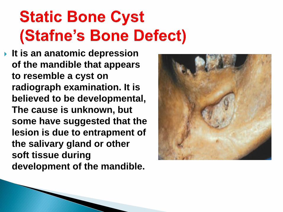
It is an anatomic depression
of the mandible that appears
to resemble a cyst on
radiograph examination. It is
believed to be developmental,
The cause is unknown, but
some have suggested that the
lesion is due to entrapment of
the salivary gland or other
soft tissue during
development of the mandible.

This lesion is entirely
asymptomatic almost all
cases appear in adults,
particularly men.
It appears as a sharply
circumscribed oval
radiolucency beneath the
level of the inferior alveolar
canal
The appearance of a static
bone cyst is usually
pathognomonic, and no
treatment is required.

uncommon lesions, asymptomatic, focal
radiolucencies in areas where
hematopoiesis is normally seen (angle of
the mandible and maxillary tuberosity).
Approximately 70% of these lesions
occur in the posterior mandible; 70%
occur in females.

The pathogenesis of the osteoporotic
marrow defect is unknown, although three
theories have been proposed:
1. abnormal healing following tooth
extraction
2. residual remnants of fetal marrow may
persist into adulthood.
3. a focus of extramedullary hematopoiesis
that becomes hyperplastic in adult life.

A predominance of
hematopoietic cells with
relatively fewer fat cells.
Nonspecific
radiographic findings,
diagnosis by an
incisional biopsy is
generally desirable.

Suggestive Reading
Brad W Neville, Douglas D Damm,
Carl M. Allen, Jerry E Bonguot.
Oral And Maxillofacial Pathology,
4th Edition, Elsevier, 2015