Downregulation of ASPP2 in choriocarcinoma contributes to...
25
Title Downregulation of ASPP2 in choriocarcinoma contributes to increased migratory potential through Src signaling pathway activation Author(s) Mak, CY; Lee, L; Siu, KY; Wong, GW; Lu, X; Ngan, HYS; Wong, ES; Cheung, ANY Citation Carcinogenesis, 2013, v. 34 n. 9, p. 2170-2177 Issued Date 2013 URL http://hdl.handle.net/10722/189574 Rights This is a pre-copy-editing, author-produced PDF of an article accepted for publication in Carcinogenesis following peer review. The definitive publisher-authenticated version Carcinogenesis, 2013, v. 34 n. 9, p. 2170-2177 is available online at: http://carcin.oxfordjournals.org/content/34/9/2170
Transcript of Downregulation of ASPP2 in choriocarcinoma contributes to...
TitleDownregulation of ASPP2 in choriocarcinoma contributes toincreased migratory potential through Src signaling pathwayactivation
Author(s) Mak, CY; Lee, L; Siu, KY; Wong, GW; Lu, X; Ngan, HYS; Wong,ES; Cheung, ANY
Citation Carcinogenesis, 2013, v. 34 n. 9, p. 2170-2177
Issued Date 2013
URL http://hdl.handle.net/10722/189574
Rights
This is a pre-copy-editing, author-produced PDF of an articleaccepted for publication in Carcinogenesis following peerreview. The definitive publisher-authenticated versionCarcinogenesis, 2013, v. 34 n. 9, p. 2170-2177 is available onlineat: http://carcin.oxfordjournals.org/content/34/9/2170
For Peer Review
Downregulation of ASPP2 in choriocarcinoma contributes to
increased migratory potential through Src signaling pathway activation
Journal: Carcinogenesis
Manuscript ID: CARCIN-2012-01361.R1
Manuscript Type: Original Manuscript
Date Submitted by the Author: 29-Apr-2013
Complete List of Authors: Mak, Victor CY; The University of Hong Kong, Pathology
Lee, Lee; The University of Hong Kong, Pathology Siu, Michelle KY; The University of Hong Kong, Obstetrics and Gynaecology Wong, Oscar GW; The University of Hong Kong, Pathology Lu, Xin; University of Oxford, Ludwig Institute for Cancer Research Ltd., Nuffield Department of Clinical Medicine Ngan, Hextan Y.S.; The University of Hong Kong, Obstetrics and Gynaecology Wong, Esther; The University of Hong Kong, Pathology Cheung, Annie; The University of Hong Kong, Pathology
Keywords: Choriocarcinoma, ASPP2, Gestational trophoblastic disease, Src
Carcinogenesis Carcinogenesis Advance Access published May 13, 2013 at U
niversity of Hong K
ong on June 2, 2013http://carcin.oxfordjournals.org/
Dow
nloaded from
For Peer Review
Downregulation of ASPP2 in choriocarcinoma contributes to
increased migratory potential through Src signaling pathway
activation
Victor C. Y. Mak1, Lee Lee
1, Michelle K. Y. Siu
3, Oscar G. W. Wong
1, Xin Lu
2, Hextan Y. S. Ngan
3,
Esther S. Y. Wong1, Annie N. Y. Cheung
1,*
1 Departments of Pathology, The University of Hong Kong, HKSAR, China
2 The Ludwig Institute for Cancer Research Ltd., Nuffield Department of Clinical Medicine, University
of Oxford, UK 3 Departments of Obstetrics and Gynaecology, The University of Hong Kong, HKSAR, China
* To whom correspondence should be addressed: Annie N.Y. Cheung, MD., FRCPath., Department of Pathology,
The University of Hong Kong, Queen Mary Hospital, Pokfulam Road, Hong Kong, China. Tel: +852 22554876;
Fax: +852 28725197; Email: [email protected]
Short title: ASPP2 controls cell migration in choriocarcinoma
Page 1 of 23 Carcinogenesis
123456789101112131415161718192021222324252627282930313233343536373839404142434445464748495051525354555657585960
at University of H
ong Kong on June 2, 2013
http://carcin.oxfordjournals.org/D
ownloaded from
For Peer Review
Abstract
Gestational choriocarcinoma is a malignant tumour derived from placental trophoblast and the most
aggressive member of gestational trophoblastic disease (GTD). Apoptotic stimulating protein of p53-2
(ASPP2) is a member of ASPP family that transactivates p53 and thereby functions as a tumour
suppressor. In this study, the expression profile of ASPP2 in choriocarcinoma was examined in
comparison with normal placentas and hydatidiform moles, the latter being a type of GTD that carries
malignant potential. Downregulation of ASPP2 mRNA and protein was demonstrated in
choriocarcinoma by quantitative PCR and immunohistochemistry. ASPP2-transfected
choriocarcinoma cells (JEG-3 and JAR) showed an increase in apoptosis and a decrease in cell
migration as detected by TUNEL and wound healing assays, respectively, illustrating the complex
action of ASPP2 on cell functions other than programmed cell death. Activated Src is known to be
important in tumour progression. Transfection of ASPP2 but not ASPP1, another tumour suppressive
ASPP, was found to be related to subsequent decreased Src-pY416 phosphorylation, suggesting an
inactivating effect of ASPP2 on Src. Moreover, this ASPP2-mediated inactivation of Src could be
abolished by RNA interference with Csk, a kinase that can inhibit Src activation. Our findings
suggested that the ability of ASPP2 to attenuate Src activation was specific to ASPP2 in a Csk
dependent manner. Taken together, we demonstrated a loss of tumour suppressive ASPP2 in
choriocarcinoma with effects on cell migration as well as apoptosis. We also unveiled a possible
mechanistic link between ASPP2 and Csk/Src signaling pathway, implicating the multiple cellular
functions of ASPP2.
Page 2 of 23Carcinogenesis
123456789101112131415161718192021222324252627282930313233343536373839404142434445464748495051525354555657585960
at University of H
ong Kong on June 2, 2013
http://carcin.oxfordjournals.org/D
ownloaded from
For Peer Review
Introduction
Choriocarcinoma is a prominent member of gestational trophoblastic disease (GTD) which
encompasses a heterogeneous family of allografts arising from placental trophoblasts with varying
potential for local invasion and metastasis [1,2]. GTD can be classified into premalignant hydatidiform
moles and frankly malignant tumours like choriocarcinoma, placental site trophoblastic tumour (PSTT)
and epithelioid trophoblastic tumour (ETT). Hydatidiform moles, including partial and complete moles,
often regress after suction evacuation although around 20% of cases progress to persistent
gestational trophoblastic neoplasia requiring chemotherapy [3]. In contrast, choriocarcinoma, the most
malignant lesion in GTD, is characterized by massive trophoblastic tissue invasion and vascular
permeation leading to haemorrhagic metastasis [4,5].
Unlike other solid tumours, classical tumour suppressor genes p53, RB1 and p21 were found to
be upregulated in choriocarcnoma [6]. The increased expression of these tumour suppressors may
represent inherent but failed mechanisms to antagonize the overgrowth and excessive proliferative
activity in the trophoblast cells. On the other hand, loss or reduced expression of a panel of tumour
suppressors has also been reported. For example, p16, E-cadherin, and TIMP3 were downregulated
in choriocarcinoma through promoter hypermethylation [7,8]. Restoration of certain tumour
suppressor in choriocarcinoma may alter the tumour cell phenotype. For instance, ectopic NECC1
(not expressed in choriocarcinoma clone 1) could suppress the tumorigenicity and induced
differentiation of choriocarcinoma cells [9]. Thus, a unique profile of alternations in tumour suppressor
expression appears to contribute to the malignant phenotype of choriocarcinoma.
Apoptosis stimulating protein of p53 (ASPP) is a family of p53 binding proteins which shares a
common feature containing an Ankyrin repeat domain, a SH3 domain, and a Poly-Proline rich domain
at the C-terminus [10]. To date, three family members, namely ASPP1, ASPP2 and iASPP have been
identified. Our recent report on downregulation of proapoptotic ASPP1 in association with clinical
progression of GTD uncovers the importance of ASPP family in the disease development [11].
Similar to ASPP1, ASPP2 is a proapoptotic regulator that belongs to ASPP family. The
expression of ASPP2 is frequently suppressed in many cancers in relation to enhanced apoptosis
through the binding to p53 for transcriptional transactivation [12-14]. The interaction and regulation of
p53 by the ASPP family members seems to be evolutionarily conserved. Homologs of ASPP family
Page 3 of 23 Carcinogenesis
123456789101112131415161718192021222324252627282930313233343536373839404142434445464748495051525354555657585960
at University of H
ong Kong on June 2, 2013
http://carcin.oxfordjournals.org/D
ownloaded from
For Peer Review
members have been identified in C. elegan (ape-1) and Drosophila (dASPP) [15,16]. Surprisingly, a
number of ASPP2 binding partners that are involved in biological pathways other than apoptosis have
also been identified, suggesting that ASPP2 function is far more complex than simply enhancing p53-
mediated apoptosis [17]. Abnormal activation of Src-family kinases has been implicated in a wide
variety of cancers and is associated with tumour metastasis [18]. A recent study has found that
Drosophila ASPP (dASPP) could maintain epithelial integrity through physical interaction with C-
terminal Src kinase (Csk) to augment the inhibitory phosphorylation of Drosophila Src (dSrc) [16].
However, little is known about these interactions in human and the corresponding biological
significance in cancer cells has not been reported.
In this study, we investigated the expression profile of ASPP2 in GTD, the effects of ASPP2 on
apoptosis, cell migration and the Src signaling pathway.
Page 4 of 23Carcinogenesis
123456789101112131415161718192021222324252627282930313233343536373839404142434445464748495051525354555657585960
at University of H
ong Kong on June 2, 2013
http://carcin.oxfordjournals.org/D
ownloaded from
For Peer Review
Materials and methods
Clinical samples and cell lines
A total 94 trophoblastic tissues, including 20 first trimester placentas, 12 term placentas, 15 partial
moles, 35 complete moles, 12 choriocarcinomas were retrieved from the archives of Department of
Pathology, Queen Mary Hospital, The University of Hong Kong. All tissue sections were histologically
reviewed using generally agreed and accepted diagnostic criteria [1]. First trimester and term placenta
were collected after induced abortion by suction evacuation and normal delivery respectively. The
tissues of hydatidiform moles and choriocarcinomas were obtained from specimens of uterine
evacuate and/or hysterectomy.
Ethics approval for the use of such tissues in this study has been obtained from Institutional
Review Board of the University of Hong Kong/Hospital Authority Hong Kong West Cluster. The
experimental results were delinked from subjects’ personal information and individual’s consent was
considered not necessary. The need for written informed consent from the participants was waived by
the Institutional Review Board.
The clinical diagnosis of persistent gestational trophoblastic neoplasia was made if there was a
plateau in human chorionic gonadotrophin (hCG) level for 4 weeks or a further rise in hCG for three
consecutive weeks after evacuation. In most of the cases, the diagnosis of hydatidiform moles had
been confirmed by fluorescent microsatellite genotyping after microdissection and chromosome in situ
hybridization [19,20]. These trophoblastic tissues have also been assessed earlier by
immunohistochemical studies using M30 Cytodeath (Boehringer Mannheim, Mannheim, German) [21]
and p53 (DO-7, Novocastra Laboratories Ltd., Newcastle, UK) antibodies [22].
For in vitro studies, two choriocarcinoma cell lines (JEG-3 and JAR) (American Type Culture
Collection, Manassas, VA, USA) were cultured in Minimum Essential Eagle’s Medium (Sigma, St
Louis, MO, USA) supplemented with 10% fetal bovine serum (JRH Biosciences, Lenexa, KS, USA),
and 100 U/ml penicillin and streptomycin (Invitrogen, San Diego, CA, USA)[23]. Normal trophoblast
cell lines B6 and PE4 are kind gifts from Professor George S. W. Tsao (the Department of anatomy,
the University of Hong Kong).
Immunohistochemical study
Page 5 of 23 Carcinogenesis
123456789101112131415161718192021222324252627282930313233343536373839404142434445464748495051525354555657585960
at University of H
ong Kong on June 2, 2013
http://carcin.oxfordjournals.org/D
ownloaded from
For Peer Review
Paraffin sections 5 µm thick were cut and deparaffinized. Antigen retrieval was carried out at
95 °C for 10 min in 10 mM sodium citrate buffer at pH 6.0. Immunohistochemistry was performed
using the UltraVision LP Value Detection System Horseradish Peroxidase (HRP) Polymer (LabVision,
Fremont, CA, USA)[24]. A monoclonal mouse anti-human antibody of ASPP2 (Clone DX 54.10)
(Sigma, St.Louis, MO) was applied in 1:1500 dilution and incubated overnight at room temperature.
Freshly-prepared 3.3’-diaminobenzidine tetrahydrochloride (DAB) (Amresco, Solon, Ohio) in PBS with
hydrogen peroxide was applied as chromagen and sections were counter-stained with hematoxylin.
Negative controls were prepared by replacing the primary antibody with PBS. A known positive
control from a normal first trimester placenta was used. The percentage of ASPP2 immunopositive
cells was scored according to the following criteria: 0, negative; 1, 0.1–25.0% of cells immunopositive;
2, 25.1–50.0% immunopositive; 3, 50.1–75.0% immunopositive; 4, 75.1–100% of cells
immunopositive [24,25]. ASPP2 immunoreactivity was further correlated with p53 expression [22].
Quantitative Real-time PCR
Trizol reagent (Invitrogen, Life Technologies Inc, Rockville, MD) was used for total RNA
extraction according to the manufacturer’s instruction. First strand cDNA was synthesized from 2.5 µg
total RNA by SuperScript Reverse Transcriptase system (Invitrogen). Primers used were as followed:
ASPP2 (Forward: 5’-GTG CTG CCT CAT GTA ACA AC-3’; Reverse: 5’-TAT GCC CAT CTT CTC
CTG AAC-3’) and GAPDH (Forward: 5’-TCC ATG ACA ACT TTGGTA TCG CG-3’; Reverse: 5’-ACA
GTC TTC TGG GTG GCA GTG-3’). Quantitative PCR (qPCR) was performed on the ABI PRISM
7900 Sequence Detection System (Applied Biosystems, Foster City, CA). The expression of ASPP2
determined by 2-∆∆CT
method was normalized with respected to that of GAPDH.
Transfection and Western blot analysis
JEG-3 and JAR culture in 6-well plates were transfected with either ASPP2 and ASPP1 construct
(generous gift from Prof. Xin Lu of Ludwig Institute for Cancer Research, UK) using Lipofectamine
2000 (Invitrogen, Carlsbad, CA). The pCDNA 3.1 vector was used as control. Total protein lysate was
extracted with lysis buffer (0.125 M Tris, pH 6.8 at 22ºC containing 1% NP-40 [v/v], 2 mM EDTA, 2
mM phenylmethylsulfonyl fluoride, 1 mM sodium orthovanadate and 0.1 µM sodium okadate). Protein
Page 6 of 23Carcinogenesis
123456789101112131415161718192021222324252627282930313233343536373839404142434445464748495051525354555657585960
at University of H
ong Kong on June 2, 2013
http://carcin.oxfordjournals.org/D
ownloaded from
For Peer Review
concentration was determined by detergent compatible protein assay (Bio-Rad Laboratories, Hercules,
CA). Twenty µg of protein was resolved by SDS-polyacrylamide gel electrophoresis and then
transferred to polyvinylidene difuoride membrane and probed with corresponding antibodies [25].
Primary antibodies used in this study were listed in Table 1.
TdT-Mediated dUTP Nick End Labeling Assay
TdT-mediated dUTP nick end labeling assay (TUNEL) was performed using an In Situ Cell Death
Detection kit (Roche Biochemical, Indianapolis, IN) as previously described [21]. The number of
TUNEL-positive cells in different controls and in JEG-3 and JAR after ASPP2 transient transfection
was counted in three different fields at Χ40 magnification by fluorescence microscopy.
Silencing of Csk by small interfering RNA (siRNA)
To knockdown Csk in choriocarcinoma cell line, Silencer® select Pre-designed siRNA of Csk
(siCsk) (Cat: 4427037; ID:s3614) and non-targeting Negative Control #2 (scramble) siRNA (Cat:
43900846) were used (Ambion, Austin, TX). Co-transfection with 5 nM Csk-specific/non-targeting
siRNA and empty vector pCDNA3.1/ ASPP2 construct in Lipofectamine 2000 (Invitrogen, Carlsbad,
CA) was carried in 6-well culture plate. To assess the knockdown efficiency, Western blotting was
performed using the protein lysate harvested 48 h post-transfection.
Wound healing assay
Migration of the cells was determined by wound healing assay. Cells were first transfected with
specified plasmid constructs or siRNA oligo for one day in twelve-well plates with 90% confluence.
The treated cell monolayer was then scratched with a sterile 200-µl pipette tip. Fresh culturing
medium was added. Photos were retaken at the same position of the wound after 48 hours. Results
expressed in percentage to control were defined as the average percentage change in linear wound
closure in treatment with respect to that in control.
Statistical analysis
Page 7 of 23 Carcinogenesis
123456789101112131415161718192021222324252627282930313233343536373839404142434445464748495051525354555657585960
at University of H
ong Kong on June 2, 2013
http://carcin.oxfordjournals.org/D
ownloaded from

For Peer Review
Statistical analysis was performed using the Statistical Package for Social Science (SPSS) 15.1.
Non-parametric unpaired t-test (Mann-Whitney test) was used for continuous data. Spearman’s rho
test was used for correlation analysis. P values <0.05 were considered as statistically significant.
Page 8 of 23Carcinogenesis
123456789101112131415161718192021222324252627282930313233343536373839404142434445464748495051525354555657585960
at University of H
ong Kong on June 2, 2013
http://carcin.oxfordjournals.org/D
ownloaded from
For Peer Review
Results
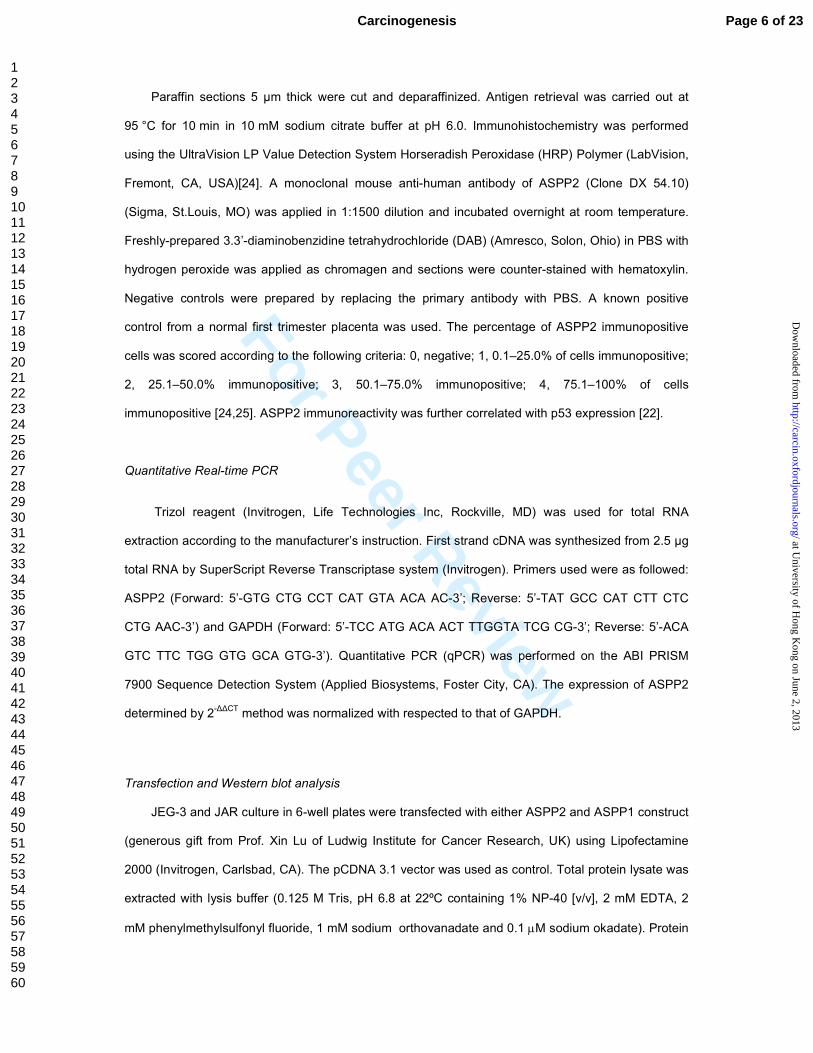
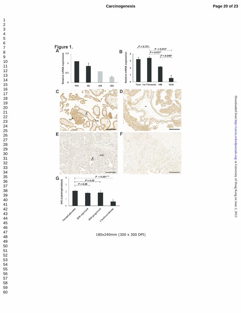
Downregulated expression of ASPP2 mRNA and protein in choriocarcinoma
By qPCR analysis, ASPP2 mRNA was found to be downregulated in choriocarcinoma cell lines,
JEG-3 and JAR, when compared with normal trophoblast cell lines, B6 and PE (Figure. 1A).
Significantly lower ASPP2 mRNA was also detected in choriocarcinoma samples compared with
normal first trimester placenta (P = 0.012). Hydatidiform moles showed an intermediate ASPP2
mRNA expression between normal placentas and choriocarcinomas (Figure 1B).
By immunohistochemistry, both nuclear and cytoplasmic ASPP2 expression was detected in first
trimester samples. ASPP2 was found to be expressed predominantly in the nucleus and moderately
in the cytoplasm of cytotrophoblasts and villous intermediate trophoblasts of normal placenta and
hydatidiform moles in contrast to the absence of expression in choriocarcinoma (Figure 1C - F).
Immunoreactivity to ASPP2 in PSTT was also weak. Moreover, no nuclear immunoreactivity could be
detected in the syncytiotrophoblasts. Concurring with the qPCR findings, significantly lower ASPP2
immunoreactivity was found in the choriocarcinoma compared with normal first trimester samples (P <
0.001, Mann–Whitney test; Figure 1G). There was no significant difference in immunoreactivity
between hydatidiform moles that subsequently developed GTN (progressed mole) and those that
spontaneously regressed (P = 0.786, Mann–Whitney test). Although both regressed and persistent
hydatidiform moles showed a lower nuclear immunoreactivity than normal first trimester placenta, no
statistical significance was reached (P = 0.06 and 0.42 respectively; Figure 1G).
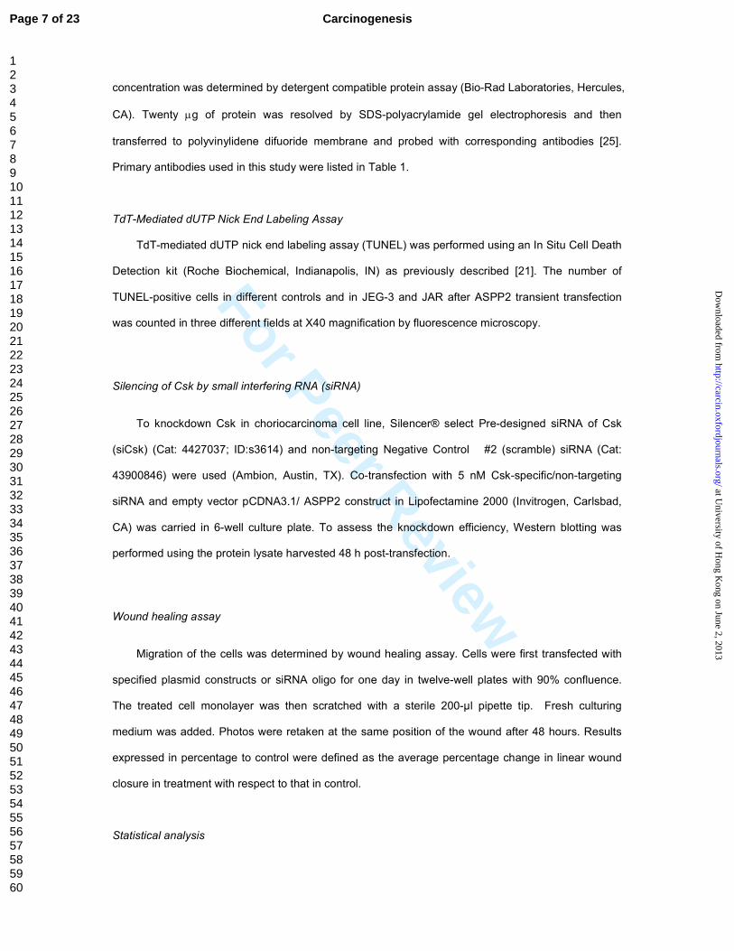
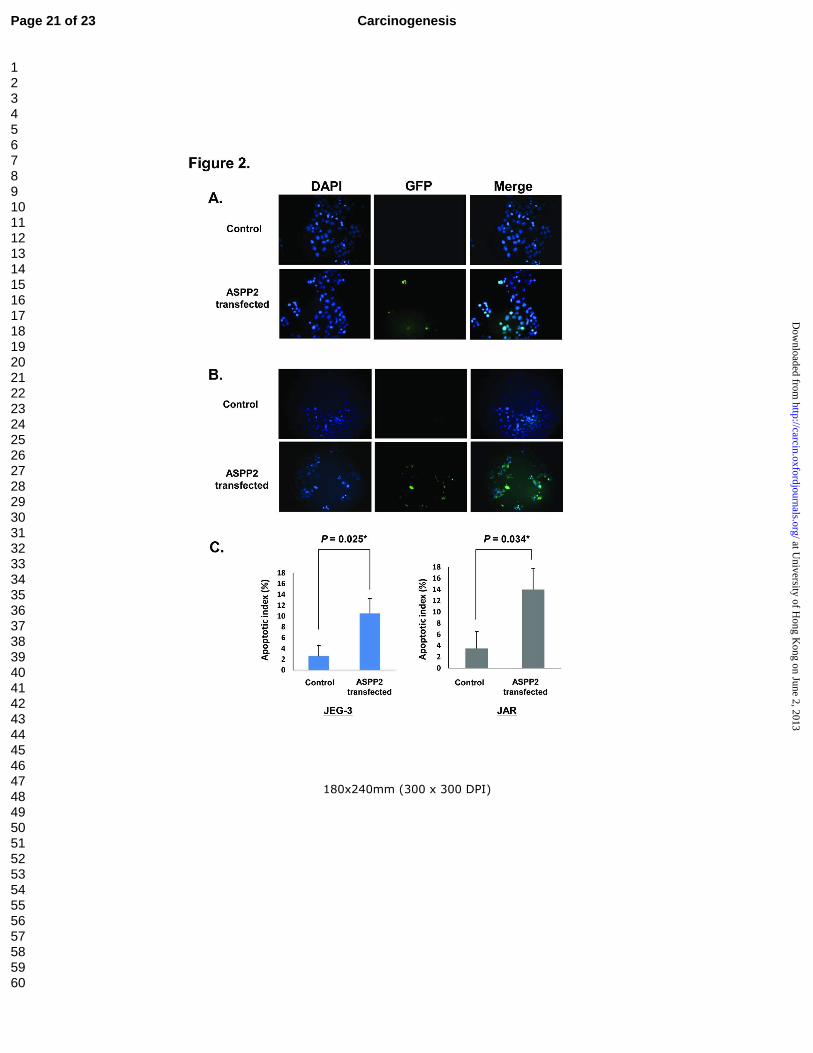
Ectopic ASPP2 increased apoptosis but reduced cell migration in choriocarcinoma cells
Retrieval of data from corresponding cases showed that nuclear immunoreactivity of ASPP2
correlated inversely with p53 (P = 0.032, coefficient= -0.301, Spearman's ρ test) [22]. Such correlation
suggested that loss of ASPP2 in choriocarinoma could modify p53 activity leading to difference in
apoptotic activity among GTD. The effect of ASPP2 on apoptosis in choriocarinoma was assessed by
ASPP2 transfection and TUNEL assay. Our results showed that ectopic ASPP2 expression increased
percentage of apoptotic cells from 2.6+1.9% to 10.5+2.7% in JEG-3 (Figure 2A) and 3.5+2.9 to
Page 9 of 23 Carcinogenesis
123456789101112131415161718192021222324252627282930313233343536373839404142434445464748495051525354555657585960
at University of H
ong Kong on June 2, 2013
http://carcin.oxfordjournals.org/D
ownloaded from
For Peer Review
14.0+3.7% in JAR (Figure 2B) cells in contrast to control respectively (Figure 2), suggesting that loss
of proapoptotic ASPP2 could attenuate apoptosis in choriocarcinoma cells.
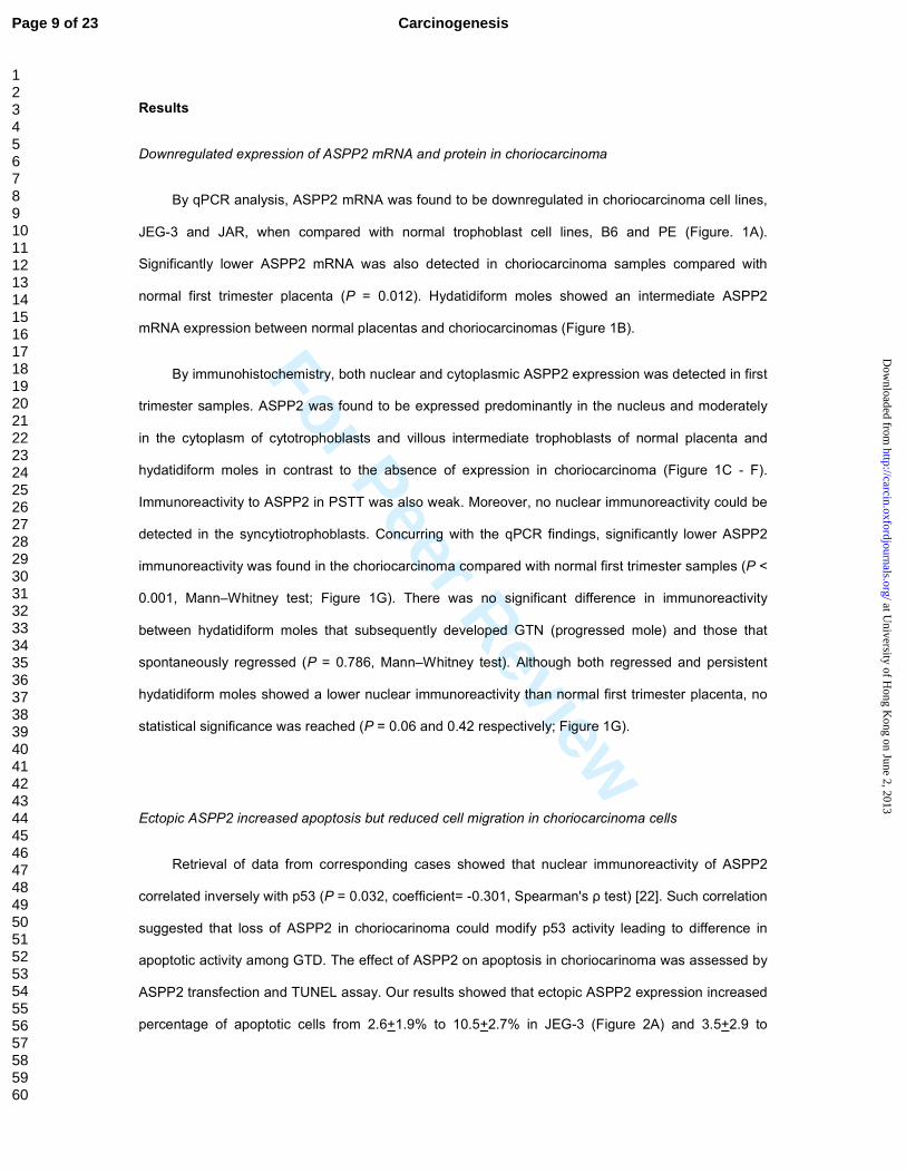
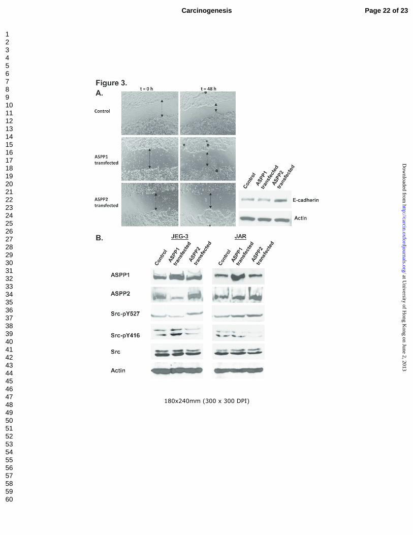
By wound healing assay, slower migration rate was found in JEG-3 transfected with ASPP2 than
those transfected with ASPP1 or the control (Figure 3A, left panel). Our results demonstrated, for the
first time, that ASPP2 negatively regulate cell migration in choriocarcinoma. Furthermore, western blot
analysis also showed an ASPP2-specific induction of E-cadherin expression (Figure 3A, right panel).
ASPP2-specific inactivation of Src
In view of the multi-functional nature of ASPP2 with effect on both apoptosis and cell migration
in choriocarcinoma cells, possible interaction between ASPP2 and Src signaling pathway was
evaluated. Activation and inactivation of Src is known to be tightly regulated by phosphorylation at two
sites with opposite effects. Phosphorylation site at Tyr416 (Y416) in the activation loop of the kinase
domain contributes to augmentation of Src activity. In contrary, Csk-mediated phosphorylation of
Tyr527 (Y527) at C-terminal tail of Src results in a closed structure, through intramolecular interaction
with its SH2 domain, that diminishes the access of substrates to the kinase domain and prevent Y416
autophosphorylation [26]. As shown in Figure 3B, overexpression of ASPP2 in both JEG-3 and JAR
could reduce the expression level of Src-pY416, an activated form of Src. However, such observation
was not found in cells transfected with ASPP1, another member of the ASPP family exerting tumour
suppressive effect. Moreover, complementary increased phosphorylation at inhibitory domain Src-
pY527 accompanied with ectopic ASPP2 expression was also demonstrated. The total form of Src
remained unchanged after ASPP2 or ASPP1 transfection.
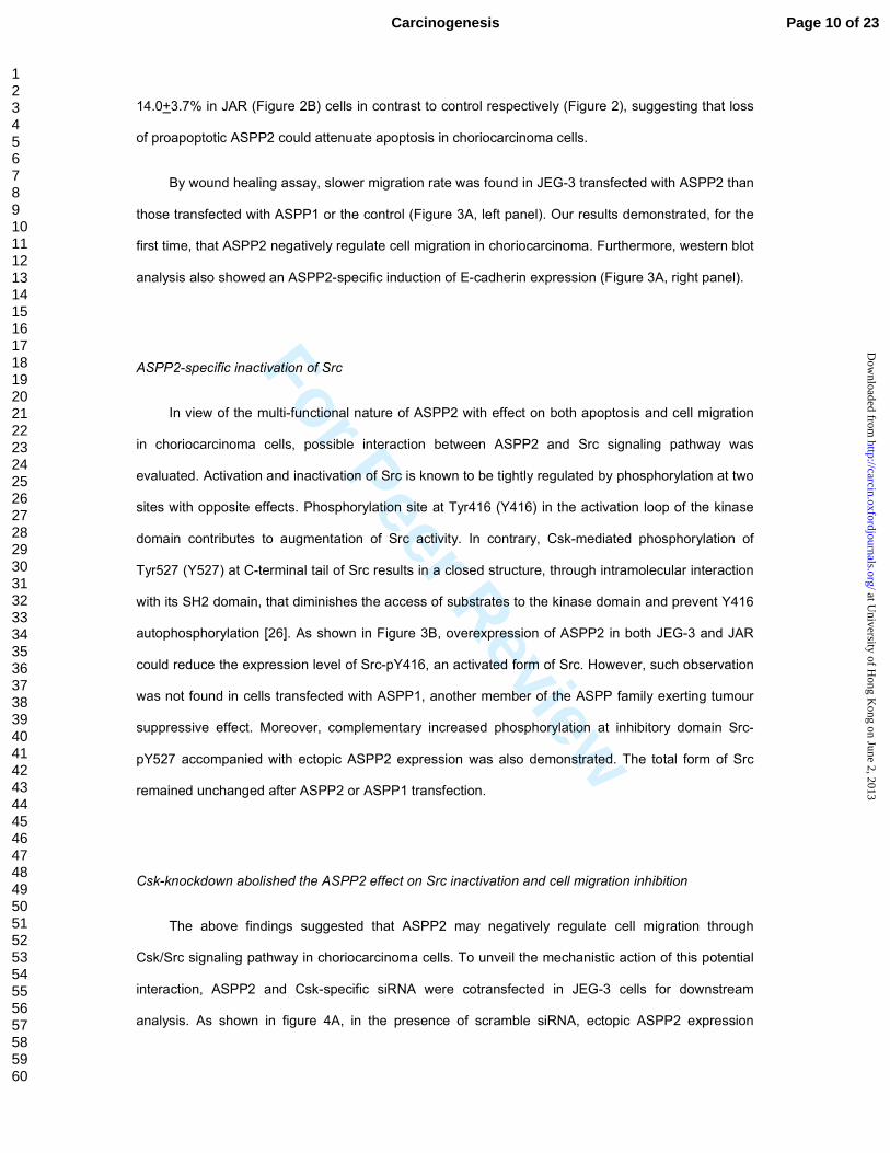
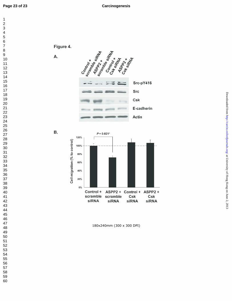
Csk-knockdown abolished the ASPP2 effect on Src inactivation and cell migration inhibition
The above findings suggested that ASPP2 may negatively regulate cell migration through
Csk/Src signaling pathway in choriocarcinoma cells. To unveil the mechanistic action of this potential
interaction, ASPP2 and Csk-specific siRNA were cotransfected in JEG-3 cells for downstream
analysis. As shown in figure 4A, in the presence of scramble siRNA, ectopic ASPP2 expression
Page 10 of 23Carcinogenesis
123456789101112131415161718192021222324252627282930313233343536373839404142434445464748495051525354555657585960
at University of H
ong Kong on June 2, 2013
http://carcin.oxfordjournals.org/D
ownloaded from
For Peer Review
inactivate Src as indicated by suppressing the expression of Src-pY416 and was also able to induce
E-cadherin expression in JEG-3. In contrast, such inactivation and induced E-cadherin expression
were abolished when the JEG-3 cells underwent co-transfection of ASPP2 and Csk siRNA. More
importantly, inhibitory effect on choriocarcinoma cell migration by ectopic ASPP2 was also eliminated
after Csk-specific knockdown as demonstrated in wound healing assay (Figure 4B).
As the Y527 inhibitory phosphorylation site is mediated by Csk, our results demonstrated that
Src could be inactivated specifically by ASPP2 through the action of Csk with effects of cell migration.
Page 11 of 23 Carcinogenesis
123456789101112131415161718192021222324252627282930313233343536373839404142434445464748495051525354555657585960
at University of H
ong Kong on June 2, 2013
http://carcin.oxfordjournals.org/D
ownloaded from
For Peer Review
Discussion
In this study, we have revealed a choriocarcinoma specific loss of ASPP2 compared with normal
placentas and GTD subtypes and a pleiotropic nature of ASPP2 which, in addition to stimulating
apoptosis, negatively regulate cell migration through Src signaling pathway in a Csk dependent
manner. Human trophoblasts exert a crucial role in implantation and placentation of pregnancy and
fetal development. Alterations in molecular mechanism and signal transduction pathways on
trophoblast cell migration and its invasiveness may lead to pathological conditions [27,28]. As
choriocarcinoma is the malignant extreme of the spectrum of GTD, our present data identifies a
critical role of ASPP2 in tumourigenesis of choriocarcinoma with its effects in apoptosis and cell
migration.
Apoptosis is an important process in pathogenesis in GTD [21,24,29,30]. Studies on p53 related
genes and modulation of p53 activity may help understand the development of GTD. ASPP2 is
originally identified as a p53 binding protein. It has been demonstrated that ASPP2 is able to induce
apoptosis through mitochondrial pathway associated with activation of caspase-9 [31]. Our results in
apoptosis in choriocarcinoma cells concur with the central role of ASPP2 in the regulation of
apoptosis as described in these studies.
Real time qPCR showed a downregulation of ASPP2 mRNA level in hydatidiform mole and
choriocarcinoma respectively compared with normal placenta (Figure 1B). Normal placental chorionic
villi are composed of a complex and heterogeneous population of trophoblast cells, including
cytotrophoblast, syncytiotrophoblast and villous intermediate trophoblast. Specific spatial and
subcellular alteration of ASPP2 expression was further evaluated by immunohistochemistry. Since
cytotrophoblast is the progenitor of villous trophoblasts and syncytiotrophoblast [32] and is regarded
as critical for neoplastic transformation in trophoblastic tumours [3], we specifically assessed the
immunoreactivity of nuclear ASPP2 in cytotrophoblast as a function of p53 transactivation (Figure 1G).
Loss of nuclear ASPP2 immunoreactivity particularly in the truly malignant choriocarcinoma concurs
with the fact that choriocarcinoma has a lower apoptotic activity than premalignant hydatidiform moles
as we reported previously [21,29]. Besides the expression in nuclei, cytoplasmic immunoreactivity of
ASPP2 was also detected in both first trimester placenta and hydatidiform mole samples. In contrast,
in choriocarcinoma and PSTT, the malignant forms of GTD, total loss or very weak ASPP2 expression
Page 12 of 23Carcinogenesis
123456789101112131415161718192021222324252627282930313233343536373839404142434445464748495051525354555657585960
at University of H
ong Kong on June 2, 2013
http://carcin.oxfordjournals.org/D
ownloaded from
For Peer Review
in both the nucleus and cytoplasm was observed, respectively. Such findings suggested a possible
interaction between cytoplasmic ASPP2 protein and gene products that are responsible for cellular
functions other than apoptosis, and may therefore account for the aggressiveness of choriocarcinoma
or PSTT.
Although Drosophila homolog of ASPP (dASPP) has been reported to interact with dCsk to
regulate dSrc kinase to maintain epithelial integrity [16], biological significance regarding interaction
between Src signaling pathway and ASPP family members are not available in humans. Our data on
RNA interference and Western blot analysis clearly showed that ectopic ASPP2 could inactivate Src
signaling in a Csk dependent manner in choriocarcinoma cells and reported for the first time the ability
of ASPP2 to negatively regulate cell migration through the Csk/Src axis. Indeed, a wide body of
evidences has shown that activated Src kinase endows migratory phenotype through interaction with
a number of downstream signaling pathways including FAK, paxillin, ERK as well as internalization of
E-cadherin [18,33,34]. As a result, ASPP2 mediated cell migration through Csk/Src axis is noteworthy
to be further investigated in cancer biology.
Metastasis is a multi-step process and is usually initiated by detachments of tumour cells from
primary sites [35,36]. E-cadherin is an extensively studied cell adhesion molecule which mediates cell
adhesion in a homotypic manner [36]. Suppression of E-cadherin expression is frequently involved in
human cancers and is considered as one of the early events of tumourigenesis [37]. Our lab has
earlier reported that E-cadherin is down-regulated by hypermethylation in GTD and choriocarcinoma
has the lowest level of expression [38]. Interestingly, increased Src activity is known to reduce cell-cell
adhesion by promoting the internalization and ubiquitin-mediated protein degradation of E-cadherin
[39,40]. Moreover, Src specific inhibitor PP2 enhanced E-cadherin expression at transcript and
protein levels [41]. These actions exemplify the upregulation of E-cadherin through reduced protein
degradation and increased transcript expression upon Src inactivation. Consistent with ASPP2-
specific interaction with Csk/Src axis demonstrated in this study, E-cadherin expression could be
specifically induced by ectopic ASPP2 in JEG-3 cells whilst this induction was abolished under Csk-
knockdown (Figure 4A). Hence, transfected ASPP2 inactivated Src activity, through which E-cadherin
was upregulated and subsequently accumulated in the cells. Taken together, loss of ASPP2 could
work as an additional mechanism to further down-regulate E-cadherin expression, leading to loss of
Page 13 of 23 Carcinogenesis
123456789101112131415161718192021222324252627282930313233343536373839404142434445464748495051525354555657585960
at University of H
ong Kong on June 2, 2013
http://carcin.oxfordjournals.org/D
ownloaded from
For Peer Review
cell-cell adhesions and, at least in part, contributing to the aggressive and malignant phenotype of
choriocarcinoma.
Unlike ASPP2, another ASPP family member ASPP1 failed to inactivate Src as illustrated by the
decreased expression of Src-pY416. This observation was consistently demonstrated in both JEG-3
and JAR cell lines. While it has been proposed that the interaction between ASPP proteins and Src is
commonly evolutionary conserved from Drosophila to humans [16,42], our in vitro study clearly
indicated that ASPP2, but not ASPP1, interact with Csk/Src axis and highlighted an ASPP2-specific
regulation of cell migration through this signal transduction pathway. Indeed, a recent study showed
that knockdown of ASPP2 was more effective in promoting the growth of hepatocellular carcinoma
cells both in soft-agar transformation assay and in nude mice, when compared with that of ASPP1
[14]. Thus, the loss of ASPP2 might exert a more potent effect than ASPP1 in tumour development
with respect to the pleiotropic nature of this tumour suppressor.
In conclusion, we found loss of expression of the multi-functional ASPP2 in choriocarcinoma
but not the premalignant hydatidiform moles, suggesting its crucial role in tumourigenesis through its
effect on inhibiting cell migration and promoting apoptosis. An ASPP2-specific inactivation of the
oncogenic Src in a Csk dependent manner was further demonstrated, which provides a mechanistic
link between loss of ASPP2 and the aggressive phenotype of choriocarcinoma at molecular basis.
Funding
Areas of Excellence Scheme, Research Grants Council of the Hong Kong Special Administrative
Region
Conflict of Interest Statement: None declared.
References
1. Shih, I., et al. (2002) Gestational trophoblastic disease and related lesions. In Kurman, R. (ed.), Blaustein's pathology of the female genital tract. Springer, New York, pp. 1193-1247.
2. Altieri, A., et al. (2003) Epidemiology and aetiology of gestational trophoblastic diseases. Lancet Oncol, 4, 670-8.
3. Shih Ie, M. (2007) Gestational trophoblastic neoplasia--pathogenesis and potential therapeutic targets. Lancet Oncol, 8, 642-50.
Page 14 of 23Carcinogenesis
123456789101112131415161718192021222324252627282930313233343536373839404142434445464748495051525354555657585960
at University of H
ong Kong on June 2, 2013
http://carcin.oxfordjournals.org/D
ownloaded from
For Peer Review
4. Cheung, A.N., et al. (2009) Pathogenesis of choriocarcinoma: clinical, genetic and stem cell perspectives. Future Oncol, 5, 217-31.
5. Xue, W.C., et al. (2003) Minichromosome maintenance protein 7 expression in gestational trophoblastic disease: correlation with Ki67, PCNA and clinicopathological parameters. Histopathology, 43, 485-90.
6. Li, H.W., et al. (2002) Current understandings of the molecular genetics of gestational trophoblastic diseases. Placenta, 23, 20-31.
7. Feng, H., et al. (2004) Down-regulation and promoter methylation of tissue inhibitor of metalloproteinase 3 in choriocarcinoma. Gynecol Oncol, 94, 375-82.
8. Xue, W.C., et al. (2004) Promoter hypermethylation of multiple genes in hydatidiform mole and choriocarcinoma. J Mol Diagn, 6, 326-34.
9. Asanoma, K., et al. (2003) NECC1, a candidate choriocarcinoma suppressor gene that encodes a homeodomain consensus motif. Genomics, 81, 15-25.
10. Samuels-Lev, Y., et al. (2001) ASPP proteins specifically stimulate the apoptotic function of p53. Mol Cell, 8, 781-94.
11. Mak, V.C., et al. (2011) Downregulation of ASPP1 in gestational trophoblastic disease: correlation with hypermethylation, apoptotic activity and clinical outcome. Mod Pathol, 24, 522-32.
12. Bergamaschi, D., et al. (2004) ASPP1 and ASPP2: common activators of p53 family members. Mol Cell Biol, 24, 1341-50.
13. Lossos, I.S., et al. (2002) Apoptosis stimulating protein of p53 (ASPP2) expression differs in diffuse large B-cell and follicular center lymphoma: correlation with clinical outcome. Leuk Lymphoma, 43, 2309-17.
14. Zhao, J., et al. (2010) Epigenetic silence of ankyrin-repeat-containing, SH3-domain-containing, and proline-rich-region- containing protein 1 (ASPP1) and ASPP2 genes promotes tumor growth in hepatitis B virus-positive hepatocellular carcinoma. Hepatology, 51, 142-53.
15. Bergamaschi, D., et al. (2003) iASPP oncoprotein is a key inhibitor of p53 conserved from worm to human. Nat Genet, 33, 162-7.
16. Langton, P.F., et al. (2007) Drosophila ASPP regulates C-terminal Src kinase activity. Dev Cell, 13, 773-82.
17. Kampa, K.M., et al. (2009) New insights into the expanding complexity of the tumor suppressor ASPP2. Cell Cycle, 8, 2871-6.
18. Yeatman, T.J. (2004) A renaissance for SRC. Nat Rev Cancer, 4, 470-80. 19. Cheung, A.N., et al. (2004) Metastatic trophoblastic disease after an initial diagnosis of partial
hydatidiform mole: genotyping and chromosome in situ hybridization analysis. Cancer, 100, 1411-7.
20. Lai, C.Y., et al. (2004) Analysis of gestational trophoblastic disease by genotyping and chromosome in situ hybridization. Mod Pathol, 17, 40-8.
21. Chiu, P.M., et al. (2001) Apoptotic activity in gestational trophoblastic disease correlates with clinical outcome: assessment by the caspase-related M30 CytoDeath antibody. Histopathology, 38, 243-9.
22. Cheung, A.N., et al. (1999) Immunohistochemical and mutational analysis of p53 tumor suppressor gene in gestational trophoblastic disease: correlation with mdm2, proliferation index, and clinicopathologic parameters. Int J Gynecol Cancer, 9, 123-130.
23. Siu, M.K., et al. (2010) p21-Activated kinase-1 promotes aggressive phenotype, cell proliferation, and invasion in gestational trophoblastic disease. Am J Pathol, 176, 3015-22.
24. Chan, H.Y., et al. (2008) Activated Stat3 expression in gestational trophoblastic disease: correlation with clinicopathological parameters and apoptotic indices. Histopathology, 53, 139-46.
25. Siu, M.K., et al. (2008) Overexpression of NANOG in gestational trophoblastic diseases: effect on apoptosis, cell invasion, and clinical outcome. Am J Pathol, 173, 1165-72.
26. Bjorge, J.D., et al. (2000) Selected glimpses into the activation and function of Src kinase. Oncogene, 19, 5620-35.
27. Knofler, M. (2010) Critical growth factors and signalling pathways controlling human trophoblast invasion. Int J Dev Biol, 54, 269-80.
28. Lunghi, L., et al. (2007) Control of human trophoblast function. Reprod Biol Endocrinol, 5, 6. 29. Wong, S.Y., et al. (1999) Apoptosis in gestational trophoblastic disease is correlated with
clinical outcome and Bcl-2 expression but not Bax expression. Mod Pathol, 12, 1025-33. 30. Fong, P.Y., et al. (2005) Mcl-1 expression in gestational trophoblastic disease correlates with
clinical outcome: a differential expression study. Cancer, 103, 268-76.
Page 15 of 23 Carcinogenesis
123456789101112131415161718192021222324252627282930313233343536373839404142434445464748495051525354555657585960
at University of H
ong Kong on June 2, 2013
http://carcin.oxfordjournals.org/D
ownloaded from
For Peer Review
31. Kobayashi, S., et al. (2005) 53BP2 induces apoptosis through the mitochondrial death pathway. Genes Cells, 10, 253-60.
32. James, J.L., et al. (2005) Cytotrophoblast differentiation in the first trimester of pregnancy: evidence for separate progenitors of extravillous trophoblasts and syncytiotrophoblast. Reproduction, 130, 95-103.
33. Guarino, M. (2010) Src signaling in cancer invasion. J Cell Physiol, 223, 14-26. 34. Webb, D.J., et al. (2004) FAK-Src signalling through paxillin, ERK and MLCK regulates
adhesion disassembly. Nat Cell Biol, 6, 154-61. 35. Geiger, T.R., et al. (2009) Metastasis mechanisms. Biochim Biophys Acta, 1796, 293-308. 36. Makrilia, N., et al. (2009) Cell adhesion molecules: role and clinical significance in cancer.
Cancer Invest, 27, 1023-37. 37. Hirohashi, S. (1998) Inactivation of the E-cadherin-mediated cell adhesion system in human
cancers. Am J Pathol, 153, 333-9. 38. Xue, W.C., et al. (2003) Methylation status and expression of E-cadherin and cadherin-11 in
gestational trophoblastic diseases. Int J Gynecol Cancer, 13, 879-88. 39. Fujita, Y., et al. (2002) Hakai, a c-Cbl-like protein, ubiquitinates and induces endocytosis of
the E-cadherin complex. Nat Cell Biol, 4, 222-31. 40. Palacios, F., et al. (2005) Lysosomal targeting of E-cadherin: a unique mechanism for the
down-regulation of cell-cell adhesion during epithelial to mesenchymal transitions. Mol Cell Biol, 25, 389-402.
41. Nam, J.S., et al. (2002) Src family kinase inhibitor PP2 restores the E-cadherin/catenin cell adhesion system in human cancer cells and reduces cancer metastasis. Clin Cancer Res, 8, 2430-6.
42. Langton, P.F., et al. (2009) The dASPP-dRASSF8 complex regulates cell-cell adhesion during Drosophila retinal morphogenesis. Curr Biol, 19, 1969-78.
Page 16 of 23Carcinogenesis
123456789101112131415161718192021222324252627282930313233343536373839404142434445464748495051525354555657585960
at University of H
ong Kong on June 2, 2013
http://carcin.oxfordjournals.org/D
ownloaded from
For Peer Review
TABLE
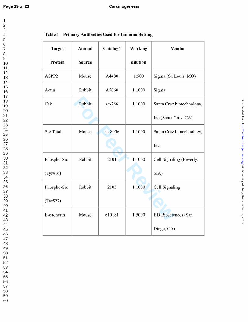
Table 1. Primary Antibodies Used for Immunoblotting
FIGURE LEGENDS
Figure 1 Downregulation of ASPP2 in choriocarcinoma. (A)&(B): Quantitative real-time PCR
analysis on mRNA expression of ASPP2 between (A) normal trophoblast (PE4, B6) and
choriocarcinoma (JAR, JEG) cells as well as (B) in normal placentas (1st trimester and Term),
hydatidiform moles (HM) and choriocarcinoma (CCA) clinical samples. (C)-(G): Immunreactivity of
ASPP2 in trophoblastic tissues. In normal first trimester placenta (C) and hydatidiform mole (D), both
nuclear and cytoplasmic staining could be observed in the cytotrophoblast (CT) and villous
intermediate trophoblast (VIT) whereas predominantly cytoplasmic staining was detected in
syncytiotrophoblast (ST) (indicated by arrows). Central cistern in enlarged villi in hydatidiform mole
was marked by asterisk (*). In choricarcinoma (E), which was composed of both CT and ST, loss of
ASPP2 immunoreactivity was observed. A much weak expression of ASPP2 was also detected in
PSTT, another trophoblastic malignancy (F). Scale bar, 200 µm. (G): Bar chart illustrating the reduced
nuclear immunohistoscore of ASPP2 in cytotrophoblast of choriocarcinoma when compared with
placenta and hydatidiform moles.
Figure 2 ASPP2 induced apoptosis in choriocarcinoma cells. A-B: Photographs of
representative fields of TUNEL assay in choriocarcinoma cell lines JEG-3 (A) and JAR (B) after empty
vector (control) and ASPP2 transfection. (C): Percentage of apoptotic cells (apoptotic cells/total cells
counted) in both cell lines after empty vector (control) and ASPP2 transfection. Approximately 80-100
cells were analyzed per random high power field of view.
Figure 3 Wound healing assay and ASPP2-specific inactivation of Src. (A): Left panel: Effects of
ectopic ASPP1 and ASPP2 expression on cell migration in choriocarcinoma cell JEG-3. Right panel:
Enhanced E-cadherin expression in ASPP1 and ASPP2 transfected JEG-3 cells. (B): Western blot
Page 17 of 23 Carcinogenesis
123456789101112131415161718192021222324252627282930313233343536373839404142434445464748495051525354555657585960
at University of H
ong Kong on June 2, 2013
http://carcin.oxfordjournals.org/D
ownloaded from
For Peer Review
analysis on the effect of ectopic ASPP1 and ASPP2 expression on Src phorspholyation at
autophosphorylation domain Y416 (activated state) and inhibitory domain Y527 (closed structure).
Figure 4 ASPP2 inactivates Src through Csk. (A): Csk-knockdwon abolished the effect of ectopic
ASPP2 on Src inactivation. (B): Bar chart showing effect of Csk-knockdwon on cell migration of
ASPP2 transfected JEG-3 cells as determined by wound healing assay.
Page 18 of 23Carcinogenesis
123456789101112131415161718192021222324252627282930313233343536373839404142434445464748495051525354555657585960
at University of H
ong Kong on June 2, 2013
http://carcin.oxfordjournals.org/D
ownloaded from
For Peer Review
Table 1 Primary Antibodies Used for Immunoblotting
Target
Protein
Animal
Source
Catalog# Working
dilution
Vendor
ASPP2 Mouse A4480 1:500 Sigma (St. Louis, MO)
Actin Rabbit A5060 1:1000 Sigma
Csk Rabbit sc-286 1:1000 Santa Cruz biotechnology,
Inc (Santa Cruz, CA)
Src Total Mouse sc-8056 1:1000 Santa Cruz biotechnology,
Inc
Phospho-Src
(Tyr416)
Rabbit 2101 1:1000 Cell Signaling (Beverly,
MA)
Phospho-Src
(Tyr527)
Rabbit 2105 1:1000 Cell Signaling
E-cadherin Mouse 610181 1:5000 BD Biosciences (San
Diego, CA)
Page 19 of 23 Carcinogenesis
123456789101112131415161718192021222324252627282930313233343536373839404142434445464748495051525354555657585960
at University of H
ong Kong on June 2, 2013
http://carcin.oxfordjournals.org/D
ownloaded from
For Peer Review
180x240mm (300 x 300 DPI)
Page 20 of 23Carcinogenesis
123456789101112131415161718192021222324252627282930313233343536373839404142434445464748495051525354555657585960
at University of H
ong Kong on June 2, 2013
http://carcin.oxfordjournals.org/D
ownloaded from
For Peer Review
180x240mm (300 x 300 DPI)
Page 21 of 23 Carcinogenesis
123456789101112131415161718192021222324252627282930313233343536373839404142434445464748495051525354555657585960
at University of H
ong Kong on June 2, 2013
http://carcin.oxfordjournals.org/D
ownloaded from
For Peer Review
180x240mm (300 x 300 DPI)
Page 22 of 23Carcinogenesis
123456789101112131415161718192021222324252627282930313233343536373839404142434445464748495051525354555657585960
at University of H
ong Kong on June 2, 2013
http://carcin.oxfordjournals.org/D
ownloaded from
For Peer Review
180x240mm (300 x 300 DPI)
Page 23 of 23 Carcinogenesis
123456789101112131415161718192021222324252627282930313233343536373839404142434445464748495051525354555657585960
at University of H
ong Kong on June 2, 2013
http://carcin.oxfordjournals.org/D
ownloaded from