


Downloaded from //iai.asm.org/content/iai/early/2010/04/12/IAI.00173-10.full.pdf · S. Orkiszewski...
38
1 Characterization and Serologic Analysis of the Treponema pallidum Proteome Melanie A. McGill 1† , Diane G. Edmondson 1 , James A. Carroll 2§ , Richard G. Cook 3,4 , Ralph S. Orkiszewski 4 and Steven J. Norris 1* 1 Department of Pathology and Laboratory Medicine, University of Texas-Houston Medical School, 6431 Fannin Street, Houston, TX 77030, USA 2 University of Pittsburgh School of Medicine, Pittsburgh, Pennsylvania, USA 3 Department of Immunology and 4 BCM Protein Chemistry Core Lab, Baylor College of Medicine, One Baylor Plaza, Houston, TX 77030, USA † Current address: Lee Biosolutions, Inc., St. Louis. MO, USA § Current address: Rocky Mountain Laboratories, National Institute of Allergy and Infectious Diseases, National Institutes of Health, Hamilton, MT, USA Running Title: Characterization of the Treponema pallidum proteome * Email: [email protected] Copyright © 2010, American Society for Microbiology and/or the Listed Authors/Institutions. All Rights Reserved. Infect. Immun. doi:10.1128/IAI.00173-10 IAI Accepts, published online ahead of print on 12 April 2010 on February 28, 2019 by guest http://iai.asm.org/ Downloaded from
Transcript of Downloaded from //iai.asm.org/content/iai/early/2010/04/12/IAI.00173-10.full.pdf · S. Orkiszewski...

1
Characterization and Serologic Analysis of the Treponema pallidum Proteome
Melanie A. McGill1†, Diane G. Edmondson1, James A. Carroll2§, Richard G. Cook3,4, Ralph
S. Orkiszewski4 and Steven J. Norris1*
1Department of Pathology and Laboratory Medicine, University of Texas-Houston Medical School,
6431 Fannin Street, Houston, TX 77030, USA
2 University of Pittsburgh School of Medicine, Pittsburgh, Pennsylvania, USA
3 Department of Immunology and 4BCM Protein Chemistry Core Lab, Baylor College of Medicine,
One Baylor Plaza, Houston, TX 77030, USA
† Current address: Lee Biosolutions, Inc., St. Louis. MO, USA
§ Current address: Rocky Mountain Laboratories, National Institute of Allergy and Infectious
Diseases, National Institutes of Health, Hamilton, MT, USA
Running Title: Characterization of the Treponema pallidum proteome
* Email: [email protected]
Copyright © 2010, American Society for Microbiology and/or the Listed Authors/Institutions. All Rights Reserved.Infect. Immun. doi:10.1128/IAI.00173-10 IAI Accepts, published online ahead of print on 12 April 2010
on February 28, 2019 by guest
http://iai.asm.org/
Dow
nloaded from

2
ABSTRACT
Treponema pallidum subsp. pallidum is the causative agent of syphilis, a sexually transmitted
disease characterized by widespread tissue dissemination and chronic infection. In this study,
we analyzed the proteome of T. pallidum by isoelectric focusing (IEF) and non-equilibrating pH
gel electrophoresis (NEPHGE) two-dimensional gel electrophoresis (2DGE) coupled with
matrix-assisted laser desorption ionization time-of-flight (MALDI-TOF) analysis. We determined
the identity of 148 T. pallidum protein spots representing 88 T. pallidum polypeptides; 63 of
these had not been identified previously at the protein level. To examine which of these proteins
are important in the antibody response to syphilis, we performed immunoblot analysis using
infected rabbit serum or human serum from patients at different stages of syphilis infection.
Twenty-nine previously described antigens (predominantly lipoproteins) were detected, as were
a number of previously unidentified antigens. The reactivity patterns obtained with sera from
infected rabbits and humans were similar; these included a subset of antigens reactive with all
serum samples tested, including CfpA, MglB-2, TmpA, TmpB, flagellins, and the 47 kDa, 17
kDa, and 15 kDa lipoproteins. A unique group of antigens specifically reactive with infected
human serum was also identified, and included the previously described antigen TpF1 and the
hypothetical proteins TP0584, TP0608, and TP0965. This combined proteomic and serologic
analysis further delineates the antigens potentially useful as vaccine candidates or diagnostic
markers, and may provide insight into host-pathogen interactions during T. pallidum infection.
on February 28, 2019 by guest
http://iai.asm.org/
Dow
nloaded from

3
INTRODUCTION
Syphilis is a multistage progressive disease caused by the spirochete Treponema
pallidum subsp. pallidum (hereafter called T. pallidum), and is characterized by localized,
disseminated and chronic stages. Manifestations include the development of a localized lesion
called a chancre during the primary stage, disseminated skin lesions and meningovascular
syphilis during the secondary stage followed by a period of latency lasting from months to
decades. Chronic, debilitating symptoms develop during the tertiary stage, including
granuloma-like lesions called gummas, neurosyphilis, and cardiovascular syphilis (38).
Although syphilis can be successfully treated by antibiotics, it remains a significant public health
problem, with an estimated 12 million new cases per year worldwide (41).
Continued improvement of diagnostic tests (particularly point of care tests) as well as the
development of an effective vaccine for syphilis would aid greatly in the control of syphilis (4, 6).
T. pallidum research, including the identification of antigens, has been hindered by the inability
to culture the bacterium continuously in vitro, necessitating the propagation of organisms by
experimental rabbit infection (28). In addition, the fragility and low protein content of the T.
pallidum outer membrane has complicated the identification of surface proteins potentially
useful in vaccines (5, 28).
The T. pallidum genome sequence (15) provides an additional tool for the analysis of
potential antigens. The 1.14 Mb T. pallidum chromosome contains 1,039 open reading frames
(ORFs) encoding predicted protein products, a smaller number than any other spirochete
genome sequenced to date (15). The average size of predicted proteins is 37,771 Da, ranging
from 3,235 to 172,869 Da. Analysis of the translated genome of T. pallidum predicts an
unusually basic proteome with a mean pI of 8.1 and median pI of 8.5, with 66% of proteins
having a pI >7.0 (23). Small genome size and a predominance of basic proteins are more
common in parasitic microorganisms, and the latter is thought to facilitate interaction of the
on February 28, 2019 by guest
http://iai.asm.org/
Dow
nloaded from
4
organism with its host (20). Other pathogenic spirochetes also tend to have basic proteins; for
example, the proteome of Borrelia burgdorferi has a mean pI of 8.36 and median pI of 9.03 (14),
and 69% of Leptospira interrogans serovar Lai str. 56601 proteins have a pI greater than 7.0
(24, 33). A recent analysis of the T. pallidum genome indicates the presence of 46 putative
lipoproteins, many fewer than the 127 predicted for B. burgdorferi (34).
The availability of the genome sequence made it possible to examine predicted T.
pallidum ORFs for potential suitability as diagnostic or immunization tools. McKevitt et al. (22)
and Brinkman et al. (3) created a protein expression library of 900 of the 1039 T. pallidum
proteins predicted from the genome sequence and examined the serologic reactivity of these
proteins by ELISA. They identified 106 antigens reactive with rabbit serum, and 34 antigens
reactive with serum from syphilis patients. This set of antigens was termed the T. pallidum
immunoproteome. This approach permits identification of low abundance T. pallidum antigens,
since they may be expressed as recombinant proteins in much larger quantities. Conversely,
proteins that are poorly expressed in E. coli or do not fold correctly may not be detected, leading
to false negative results.
To provide a complementary set of data regarding the T. pallidum immunoproteome, we
have performed proteomic analysis of T. pallidum proteins expressed during experimental rabbit
infection. We used isoelectric focusing (IEF) and non-equilibrating pH gel electrophoresis
(NEPHGE) forms of 2DGE coupled with matrix-assisted laser desorption ionization-time of flight
(MALDI-TOF) analysis to identify T. pallidum polypeptides. Immunoblotting was subsequently
used to identify antigens reactive with infected rabbit serum and with human sera at different
stages of syphilis. This approach may permit identification of antigens that are not expressed
well in E. coli, and provides a more accurate picture of the level of protein expression in the
intact organism. We have thereby characterized most of the major T. pallidum proteins
expressed in infected tissue, and identified a set of antigens reactive at all stages of infection
on February 28, 2019 by guest
http://iai.asm.org/
Dow
nloaded from
5
which could potentially be useful for the development of improved immunodiagnostic tests or for
vaccines.
MATERIALS AND METHODS
Two dimensional gel electrophoresis and immunoblotting. T. pallidum subsp.
pallidum (Nichols) was extracted from testicular tissue of infected rabbits and purified by
Percoll® density gradient centrifugation as described previously (17, 29). Organisms were
resolved in the first dimension by either isoelectric focusing (IEF, pH 5-7) or non-equilibrium pH
gel electrophoresis (NEPHGE, pH 3.5-10) as described by O’Farrell et al. (30, 31) . For
silverstained gels and subsequent MALDI-TOF, 8x10⁸ organisms were loaded per tube gel; for
immunoblotting, 6x10⁸ organisms were loaded per tube gel. Equilibrated tube gels were
resolved by SDS-PAGE using 8-20% gradient gels in the second dimension, and the gels were
stained using the Silver SNAP kit for Mass Spectrometry (Pierce) or transferred to PVDF
membrane (Millipore, (37)) at 150 V for 1.5 hours at 4°C using a Trans-Blot cell (BioRad). For
immunoblot analysis, membranes were blocked overnight at 4°C in Tris-buffered saline solution
containing 0.05% Tween-20 (TBST) and 1% BSA (blocking solution, Promega). Following
incubation with primary antibody diluted in blocking solution (1:1000 for rabbit serum, 1:500 for
human serum) for one hour at room temperature, the membranes were washed three times for
10 minutes each in TBST and then incubated with secondary antibody (goat anti-rabbit or goat
anti-human IgG, AP conjugate, Promega) at a concentration of 1:5000 diluted in TBST for 30
minutes at room temperature. Membranes were washed three times with TBST, followed by two
washes with TBS to remove inhibitory Tween-20. Membranes were developed for three minutes
with Western Blue Stabilized Substrate for Alkaline Phosphatase (Promega), and development
was stopped by washing with distilled water.
on February 28, 2019 by guest
http://iai.asm.org/
Dow
nloaded from
6
Sera used for immunoblots. Rabbit serum was collected from three individual animals
before infection (prebleed, NRS) or 84 days after intratesticular inoculation with T. pallidum
(infected rabbit serum, IRS). Sera were pooled from the three animals and used for
immunoblotting as described above. Human serum samples were previously collected in Texas
from normal human subjects and from patients diagnosed with primary, secondary, early latent
or late latent syphilis and are summarized in Table S1. Sera were pooled prior to immunoblotting
experiments as normal human (10 sera), primary (3 sera), secondary (3 sera), early latent (8
sera) and late latent (13 sera) pools, and used at a dilution of 1:500. Human syphilitic serum
samples had RPR titers ranging from 1:2 to 1:512. All human sera were collected under
established guidelines with prior approval by the Committee for the Protection of Human
Subjects, University of Texas Health Science Center at Houston.
2D spot preparation and MALDI-TOF MS analysis. Protein spots of interest were
excised manually (1.0 to 3.0 mm in diameter) from a set of four silver stained gels with a
OneTouch 2D gel spotpicker (The Gel Company, San Francisco) and destained using the Silver
SNAP kit for Mass Spectrometry according to the manufacturer’s instructions. Excised spots
were stored in wash buffer (25 mM ammonium bicarbonate, 50% acetonitrile, Sigma) at -20°C
until in-gel trypsin digestion was performed. Destained 2D gel spots were treated with 0.2 M
ammonium bicarbonate /50% acetonitrile for 15 minutes and dried completely in a CentriVap
speed vacuum. Gel pieces were then rehydrated in 50 µl 50mM ammonium bicarbonate
containing 0.2 -0.5 µg modified trypsin (Promega or Sigma) and digested for 20 hours at 37°C.
The supernatant was transferred to a clean microfuge tube, the gel fragments extracted with 50
µl aqueous 50% acetonitrile/2% formic acid for 15 minutes and combined with the initial extract.
The combined supernates were evaporated to 30 µl, acidified with trifluoroacetic acid to a pH of
3, and desalted using a C18 ZipTip (Millipore) as recommended by the manufacturer. Peptides
were eluted from the ZipTip with 5 µl of an aqueous solution of 50% acetonitrile and 2% formic
on February 28, 2019 by guest
http://iai.asm.org/
Dow
nloaded from
7
acid. Two microliters of the sample were spotted onto a 100 well stainless steel MALDI target
plate and allowed to dry partially prior to the addition of 1 µl of a 1 mg/ml matrix solution (alpha-
cyano-4-hydroxycinnamic acid) followed by complete drying. MALDI-TOF analyses were
performed in reflector mode on an ABI/SCIEX 4700 Proteomics Analyzer TOF/TOF mass
spectrometer with the laser intensity adjusted manually to yield the best spectrum for each
sample. The resulting spectra were calibrated manually utilizing the autodigestion products of
trypsin as internal reference peaks with Data Explorer software available from ABI. Proteins
were identified using Protein Prospector (University of California, San Francisco;
http://prospector.ucsf.edu/) set to a mass accuracy of ±20 ppm and a missed cleavage
allowance of 1. Mass fingerprints were compared to the predicted proteins in the NCBI database
using a species-specific filter for T. pallidum, or without the use of a species filter to identify non-
treponemal contaminants.
RESULTS & DISCUSSION
MALDI-TOF MS identification of T. pallidum proteins. Rabbits have been used for
many years to propagate T. pallidum in the absence of an effective in vitro culture system, and
T. pallidum maintained in rabbits retains infectivity and virulence in humans (13, 21). We
extracted T. pallidum subsp. pallidum Nichols from the testicular tissue of infected rabbits and
purified the bacteria by Percoll® gradient density centrifugation. Lysates of purified bacteria
were separated by IEF and NEPHGE 2DGE; NEPHGE permits the separation of highly basic
polypeptides (31). Proteins from silver stained gels were analyzed by MALDI-TOF mass
spectrometry. We identified the polypeptides present in 148 protein spots; of these, 144
corresponded to 88 different T. pallidum proteins, and four corresponded to rabbit proteins (Fig.
1, Table 1). The 56 additional spots represented charge variants, minor size variants, or
degradation products of T. pallidum proteins. The detection of only four rabbit products
on February 28, 2019 by guest
http://iai.asm.org/
Dow
nloaded from
8
demonstrates the high degree of purification achievable by Percoll® density gradient
centrifugation (17).
Multiple distinct spots in the same gel were often identified as the same protein, and
apparently represent charge or molecular mass variants; examples of these variants are
indicated by arrows in Figure 1. This phenomenon was observed at higher frequency with IEF
than NEPHGE 2DGE, most likely due to higher resolution and less compression of the gradient
in the IEF gels (for example, compare spots 142 and 143 on IEF and NEPHGE gels in Figure
1). Fifty-one of the spots identified represented apparent mass variants, based on deviation
from the predicted MW. All of these spots corresponded to abundant proteins for which a major
spot of the expected size and pI was identified; these included the cytoplasmic filament protein
CfpA (9 mass variants), the 47 kDa carboxypeptidase (5 mass variants) and the flagellar
proteins FlaA1, FlaB1, FlaB2, and FlaB3 and the FlaA1 paralog FlaA2 (a total of 16 mass
variants).
The observed Mr and predicted MW of intact polypeptides were compared to further verify
the MS identifications (Table 1, Fig. 2). In this analysis, the MW values were adjusted to take
into account either experimentally verified or predicted cleavage of the polypeptides by either
signal peptidase I or signal peptidase II; for the latter, the cleavage points of 46 predicted
lipoproteins as determined by Setubal et al (34) were utilized. No attempt was made to correct
the predicted MWs for effects of lipidation or other potential modifications. Four polypeptides
(the dodecameric form of TpF1, hypothetical protein TP0179, FtsH, and Tp34 [which migrates
as a smear by SDS-PAGE]) were considered outliers with ratios of observed Mr to predicted
MW of 0.68, 1.57, 0.70 and 1.53, respectively. The TpF1 dodecamer likely migrates aberrantly
because of its multimeric conformation. TP0179 was found to have a much greater Mr than its
predicted size (101.9 kDa as compared to 66.5 kDa); the predicted gene may be truncated by a
sequence error, because inclusion of the adjacent gene (TP0178) in the TP0179 reading frame
results in a predicted MW of ~101 kDa. Tp34 migrates as a smear by SDS-PAGE for unknown
on February 28, 2019 by guest
http://iai.asm.org/
Dow
nloaded from
9
reasons. The reason for the discrepancy in the Mr of FtsH is not known; the spot identified may
be a degradation product.
Exclusion of the mass variants and the four outliers from the list of proteins identified
resulted in a high degree of correlation between the observed Mr and predicted MW (Fig. 2); the
mean ratio of observed/predicted = 1.01 + 0.08 [SD], R2 = 0.9849). In contrast, the mass
variants had a poor correlation (ratio = 0.75 + 0.22, R2 = 0.39; data not shown). Therefore the
mass variants appear to represent degradation products. These may be naturally occurring
breakdown products, or may arise during the purification of T. pallidum from rabbit tissue.
Although the majority of spots identified corresponded to a single protein, there were a few
spots where MALDI-TOF data indicated a mixture of two protein species. Examples are spot 53
(TmpA and FliG), spot 62 (AsnA and 30S ribosomal protein S2), spots 73 and 99 (GroEL and 47
kDa carboxypeptidase degradation products), spot 92 (hypothetical protein TP0453 and
elongation factor Ts), spot 97 (hypothetical protein TP0139 and Lipoprotein Tpn32), and spot
100 (Pgm and hypothetical protein TP0290). Thus these spots appear to be composed of
multiple protein species with similar molecular masses and pIs that were not resolved from one
another under the electrophoretic conditions utilized.
A number of the more abundant polypeptides we describe here have been previously
identified by other methods, such as N-terminal sequencing or immunoblotting with monoclonal
antibodies (18); these proteins include CfpA, GroEL, DnaK, the 47 kDa carboxypeptidase,
TmpA, TmpB, and the 17 kDa and 15 kDa lipoproteins, the purine nucleoside receptor
lipoprotein PnrA (TmpC) (8), the lactoferrin-binding periplasmic lipoprotein Tp34 (TpD) (7), and
the flagellar proteins FlaA1, FlaB1, FlaB2, and FlaB3. As in previous studies, we found that the
most abundant proteins observed by silver staining were flagellins, CfpA, chaperonins, and
several lipoproteins including MglB-2, TmpA, TmpC, and the 47 kDa, 17 kDa and 15 kDa
on February 28, 2019 by guest
http://iai.asm.org/
Dow
nloaded from
10
proteins. High expression of lipoprotein genes is typical of Treponema species and other
spirochetes.
In addition to confirming previous protein identities, we identified 63 proteins that had not
been described previously by electrophoresis or immunoblotting (Fig. 1, Table 1). These
proteins can be categorized by their predicted functions: carbohydrate metabolism (13 proteins),
cell division (2 proteins), lipoproteins or structural proteins (10 proteins), flagella associated
proteins (8 proteins), nucleotide metabolism, degradation or salvage (4 proteins), molecular
chaperones (4 proteins), chemotaxis (2 proteins), energy metabolism enzymes (7 proteins),
ABC transporters (7 proteins), proteases (2 proteins), proteins involved in translation (9
proteins), amino acid and cofactor biosynthesis (3 proteins), iron storage (1 protein), cellular
detoxification (1 protein), and hypothetical proteins with unknown function (12 proteins). We also
determined that the hypothetical protein TP0259 is the lipoprotein TpE; this T. pallidum gene
product had been described previously (19), but its sequence was not published.
The protein expression we observed by 2DGE was consistent with T. pallidum mRNA
level data reported previously (35). We identified the proteins corresponding to nearly all of the
highly expressed mRNAs reported by Smajs, et al. (those with a cDNA/DNA signal ratio of 4.0
or higher) by 2DGE and MALDI-TOF MS (Fig. 1, Table 1). The majority of proteins with
corresponding high transcript levels identified in that study that we did not detect were ribosomal
proteins. We identified only four ribosomal proteins, ribosomal proteins S2 (TP0606, spot 62),
L5 (TP0201, spot 128), L10 (TP0239, spot 132), and L9 (TP0060, spot 138) in contrast to the
eleven ribosomal proteins reported to be transcribed at high levels (35).
Serologic reactivity of T. pallidum proteins. The IEF and NEPHGE 2DGE patterns
obtained were highly reproducible, enabling us to reliably correlate seroreactive proteins in
western blots with the corresponding silver stained gels. We first examined the T. pallidum
proteome for serological reactivity by immunoblotting with pooled sera from rabbits infected for
on February 28, 2019 by guest
http://iai.asm.org/
Dow
nloaded from
11
84 days. At this time post infection, rabbits develop ‘chancre immunity’, i.e. resistance to
reinfection from intradermal inoculation. In addition, the seroreactivity of human serum from
patients at different stages of syphilitic infection to the 2DGE-separated proteins was
determined. Our goal was to identify antigens that were consistently reactive at all stages of
infection, as well as those exhibiting differential reactivity at each stage of infection. A summary
of the serologic reactivity against T. pallidum proteins can be found in Table 1. Degradation
products were excluded from this analysis.
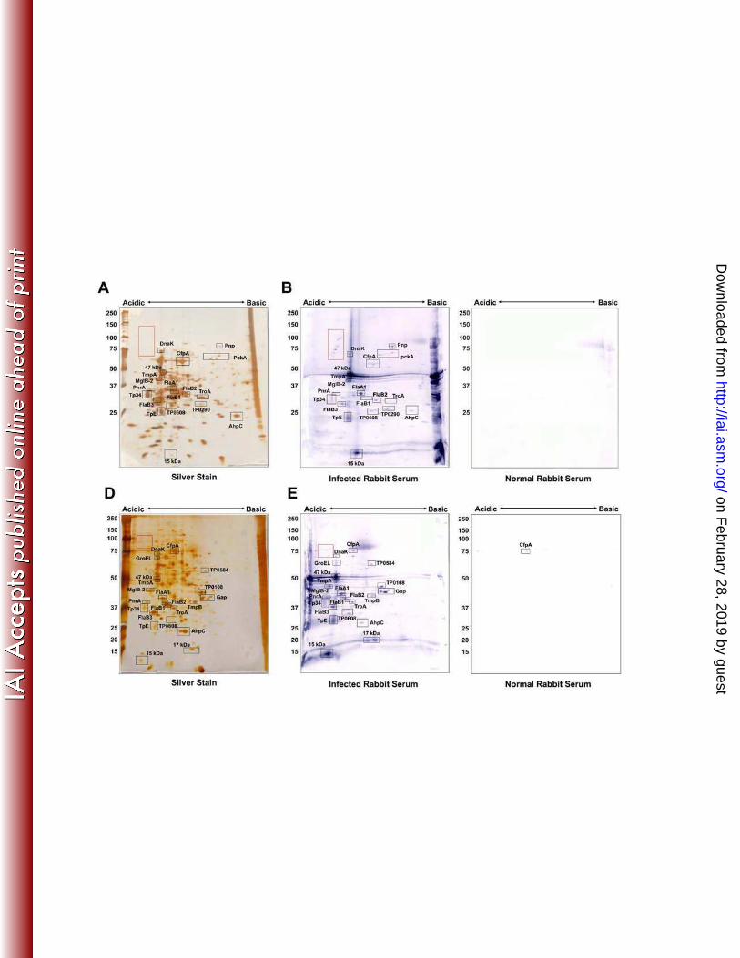
The infected rabbit serum (IRS) pool was reactive with a total of 33 T. pallidum proteins in
the 2DGE immunoblots (Fig. 3B and 3E, Table 1). The majority of proteins reactive in 2DGE
patterns were previously described antigens (26), including flagellar proteins and lipoproteins,
including the ABC transport proteins MglB-2 and TroA (Table 1). This study also confirmed IRS
serologic reactivity against phosphoenolpyruvate carboxykinase (PckA, TP0122, spot 17),
translation elongation factor G (FusA-2, TP0767, spot 6) and chemotaxis protein X (CheX,
TP0365, spot 136), which were identified as antigens by McKevitt et al. (22).
Thirty-two of the 106 T. pallidum proteins found to be reactive with IRS by McKevitt et al.
(22) were identified by MALDI-TOF MS. Surprisingly, only 16 of these 32 antigens were reactive
with rabbit serum in the present study. Although the other 16 antigens reported by McKevitt et
al. were detected by silver staining and MS, they were not reactive with IRS in our study.
However, four of those proteins were reactive with human sera (see below), including
bacterioferritin TpF1 (TP1038, spot 1), oxaloacetate decarboxylase (TP0056, spot 22), the
integral membrane protein (TP0453, spot 92), and peptidyl-prolyl cis-trans isomerase FklB
(TP0862, spot 96), indicating a sufficient amount of these proteins was present for detection of
serological reactivity by immunoblotting. Possible explanations for these results are that
immunoblot reactivity in our studies was relatively less sensitive than the ELISA format utilized
by McKevitt et al., or the human patient sera were more reactive to some antigens than was the
IRS in our analysis. Many of the most reactive antigens identified in the McKevitt study were not
on February 28, 2019 by guest
http://iai.asm.org/
Dow
nloaded from
12
detected by the 2DGE immunoblotting method, including rare lipoprotein A (RlpA, TP0993),
glycerophosphodiester phoshopdiesterase (GlpQ, TP0257), thioredoxin (TP0100), and the
hypothetical proteins TP0957, TP0625, TP0956, TP0463, TP0567, TP0326, and TP0772.
Those proteins were not identified by silver staining and subsequent MALDI-TOF MS, indicating
there may not have been sufficient protein present to detect serological reactivity against those
proteins. Overexpression of those proteins in the McKevitt et al. study may have provided
adequate protein levels for rabbit serological reactivity to be observed (22). Alternately, some of
these proteins may have been among the proteins that were not selected for MALDI-TOF
analysis. For example, several faint spots between 22 and 38 kDa were visible by
immunoblotting, but were of insufficient quantities to be identified by mass spectrometry. All but
two of the antigens not identified by IRS are within that size range.
A number of previously unreported antigens were detected by immunoblotting with IRS in
this study, including diphosphate-fructose-6-phosphate 1-phosphotransferase (TP0108, spot
41), flavodoxin (TP0925, spot 139), the FKBP-type peptidyl-prolyl cis-trans isomerase SlyD
(TP0349, spot 118), polyribonucleotide nucleotidyltransferase (Pnp, TP0886, spot 11),
glyceraldehyde 3-phosphate dehydrogenase (Gap, TP0844, spot 59), and Hypothetical Protein
TP0608 (spots 102 and 105, Fig. 3). We also found one antigen, CfpA, to be weakly reactive
with serum from uninfected animals (Fig. 3C, E).
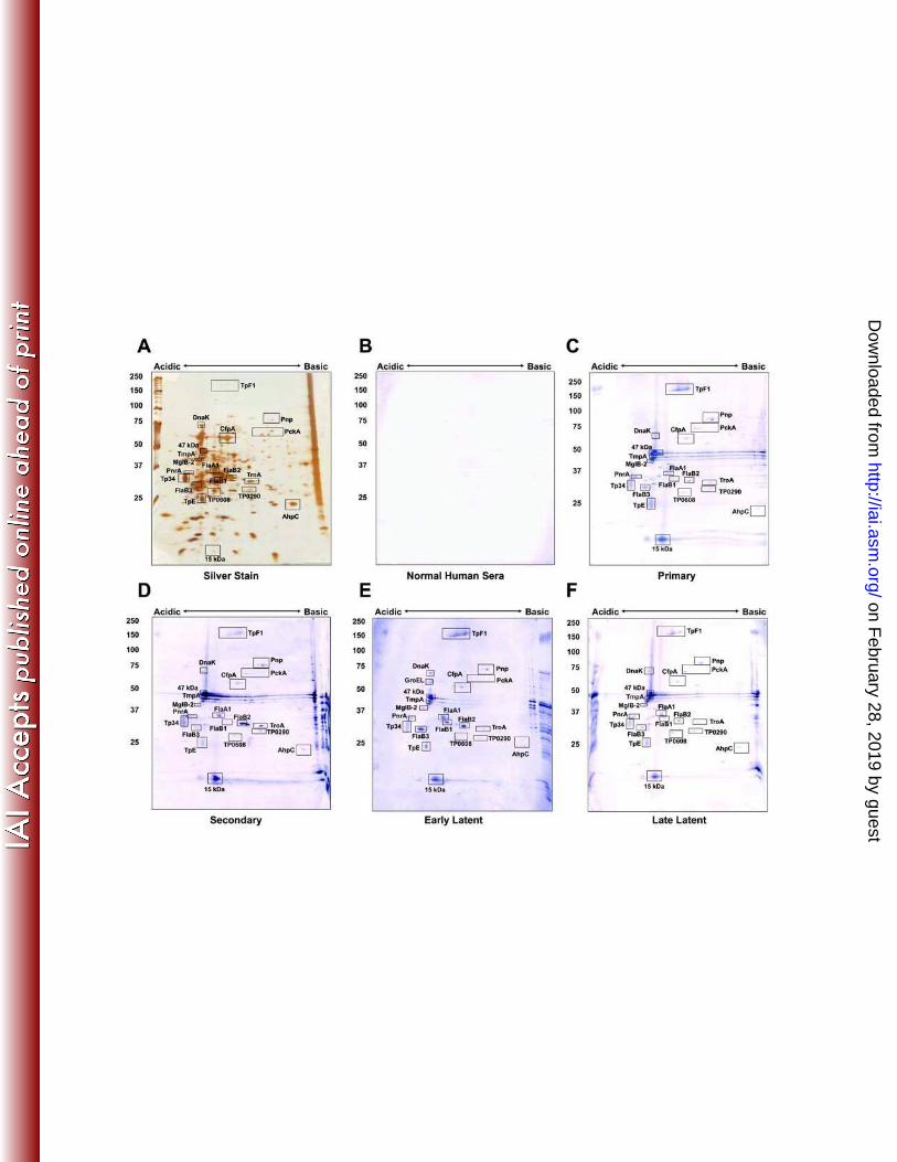
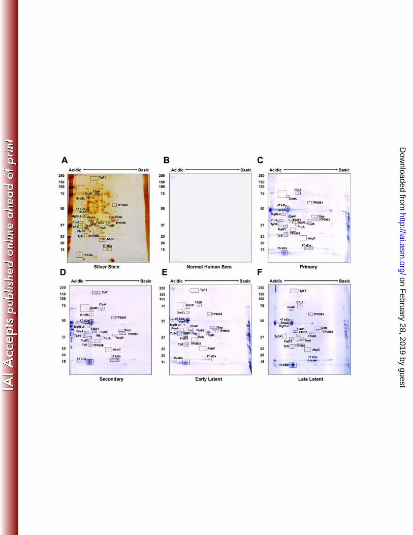
The immunoreactivity of the identified T. pallidum proteins with human sera collected from
patients diagnosed with primary, secondary, early latent or late latent syphilis was also
examined. Sera from each stage were pooled as described in Materials and Methods to provide
an analysis of reactivity during the course of infection. As expected, the highest level of
reactivity occurred with serum from secondary and early latent syphilis patients (Figures 4C and
5C, Table 1). All of the proteins that were strongly reactive with infected rabbit sera were also
reactive with sera from syphilis patients. Many of these are lipoproteins, such as the purine
nucleoside periplasmic binding protein PnrA (TmpC) (8), the lactoferrin-binding periplasmic
on February 28, 2019 by guest
http://iai.asm.org/
Dow
nloaded from
13
lipoprotein Tp34 (TpD) (7), the 47 kDa carboxypeptidase (9), TmpA, TmpB, TpE, and the 17
kDa and 15 kDa lipoproteins (Figures 3 to 5, Table 1) were reactive with sera from patients at all
stages of syphilis. Lipoproteins thus appeared to elicit the strongest antibody response, even if
they are expressed at low levels, as is the case for the 17 kDa lipoprotein (TP0435, spot 141).
This protein, as well as the 47 kDa lipopotein, has been demonstrated previously to be highly
antigenic and is currently used in T. pallidum diagnostic tests (11, 40). The strong
immunogenicity observed with these lipoproteins appears to be dependent on the lipid moiety,
as its removal diminishes the production of inflammatory cytokines and activation of immune
effector cells (1). The induction of antibody responses against the B. burgdorferi lipoprotein
outer surface protein A is highly dependent upon lipidation (10). Therefore, the lipid portion of T.
pallidum lipoproteins is likely acting as an intrinsic adjuvant to stimulate the antibody response
against these proteins.
Tp34 (TpD, Spot 81), also a lipoprotein, was reactive with all the serum pools, although at
lower levels with human late latent serum and infected rabbit serum. Two lipidated periplasmic
ABC transport proteins, MglB-2 and TroA, were also reactive at all stages of infection. In
addition, a number of antigens that have not been previously reported were identified, including
hypothetical protein TP0584 (spot 29), the V-type ATPase AtpA-1 (TP0425, spot 19), and
hypothetical protein TP0608 (spots 102 and 105). These proteins were reactive with all sera
tested (Figs. 3 to 5). Five antigens found to be reactive with infected human serum (but not with
infected rabbit sera) included hexokinase (TP0505, spot 42), hypothetical protein TP0965 (spot
66), phosphate acetyltransferase (Pta, TP0094, spot 85), the integral membrane protein
TP0453 (spot 92) and peptidyl-prolyl cis-trans isomerase, FklB (TP0862, spot 96). These
antigens were not identified as significantly reactive proteins in the Brinkman et al. study of the
reactivity of recombinant T. pallidum proteins with human syphilitic sera (3).
We observed a number of acidic, high molecular weight spots that were strongly reactive
with infected rabbit sera and sera from primary, secondary, and early latent syphilis patients
on February 28, 2019 by guest
http://iai.asm.org/
Dow
nloaded from
14
(red box in figures 3 to 5). These polypeptides ranged in size from approximately 70 kDa to 120
kDa in molecular mass (Figures 3 and 5). Due to their low abundance, we were only able to
detect one of the spots by silver staining/MS (spot 7, CfpA variant). A search of the T. pallidum
genome revealed 13 proteins of appropriate predicted molecular mass with predicted pI < 6.0
that were not identified by silver staining/MS (Table S2); these therefore represent candidate
proteins for this group.
One antigen of interest that appears to be uniquely reactive in human infection is the
oligomeric form of the bacterioferritin protein TpF1 (TP1038, spot 1, Figs. 3 and 4). TpF1
functions as a dodecomer to bind iron (36), but has been observed to migrate at several
molecular weights on SDS-PAGE ranging from160 kDa to >400 kDa in the oligomeric form (12,
25). Multiple identical subunits form a ring structure held together by disulfide bonds, creating a
very stable oligomer. (32). In its unreduced form, the basic TpF1 oligomer typically migrates at
190 kDa, but reduction by mercaptoethanol results in migration of an oligomer at 160 kDa and
dissociated monomers at 19 kDa (32). We observed the 160kDa form by 2DGE and Western
blot. We did not observe serologic reactivity against the monomeric form of TpF1 (spot 134),
which is consistent with previous findings. In prior studies by Borenstein et al. (2), TpF1 was
cloned and expressed as a recombinant protein from E. coli, serologic reactivity from syphilis
patients was observed against a 190 kDa oligomeric form of the protein. However, no reactivity
was observed against the dissociated 19 kDa monomeric form of the expressed protein (2).
Furthermore, immunization of rabbits with recombinant TpF1 provided partial protection against
challenge with viable treponemes (2). It may also be of interest to determine the identity of the
low abundance polypeptides in the ‘red box’ as proteins that might be of diagnostic or
immunogenic value.
We observed stronger reactivity with pooled sera from primary syphilis patients than
expected. The serum pool used in Figures 4C and 5C was comprised of three samples: the two
tested in Figure S1, and an additional serum sample with an RPR titer of 1:64; insufficient
on February 28, 2019 by guest
http://iai.asm.org/
Dow
nloaded from
15
serum from the latter sample was available to perform a separate immunoblot. Therefore, two
of the sera from the primary syphilis patient pool were examined for seroreactivity with NEPHGE
2DGE immunoblots (Fig. S1). Figure S1B shows the reactivity of serum from a primary syphilis
patient with an RPR (Rapid Plasma Reagin test) titer of 1:16, and Figure S1C exhibits the
reactivity of serum from a primary syphilis patient with an RPR titer of 1:64. As expected, the
serum with an RPR titer of 1:16 was reactive to fewer proteins than the serum sample with an
RPR titer of 1:64. For example, reactivity to TpF1, Hypothetical Protein TP0965, and AhpC was
not detectable in the sample with an RPR titer of 1:16 (Fig. S1B). The high RPR titers of two of
these samples correlates with the unexpected strong reactivity we observed with pooled primary
syphilis sera.
The antibody reactivities obtained with human sera and IRS in the 2DGE
immunoproteome analysis correlated well in general, with some differences (Table 1). Of the 87
T. pallidum polypeptides identified by 2DGE/MS, 40 were found to be reactive with sera from
humans at some stage of infection, whereas only 32 were reactive with IRS; 31 of the proteins
were reactive with both human sera and IRS. Nine of the human serum-reactive proteins were
not detectably reactive with IRS, whereas only one of the IRS-reactive proteins was not reactive
with the human sera tested. In most cases, these represented faint reactions indicative of low
antibody titers. However, moderate immunoblot reactivity with some human serum pools was
observed against AtpA-1 (V-type ATPase, subunit a, TP0424), Elongation factor Ts (TP0605),
and FklB (Peptidyl-prolyl cis-trans isomerase, TP0862) (Table 1), whereas a reaction with IRS
was not detected. The IRS used in this study was collected at 84 days post infection and is
considered to be highly reactive. Therefore, the apparent differential reactivity observed for
these three proteins may reflect differences between the antibody responses of humans and
rabbits to certain T. pallidum polypeptides, but additional studies using purified proteins would
be needed to verify this finding. Other possible explanations for differences observed in rabbit
and human immunoreactivity are a) the multiple time points tested from humans compared to
on February 28, 2019 by guest
http://iai.asm.org/
Dow
nloaded from
16
one single time point tested in rabbits b) the use of fewer subjects in the rabbit sera pool as
compared to the human sera pools resulted in a smaller array of immunoreactive proteins, or c)
laboratory rabbits are more inbred than the human population and thus may have reduced
antibody repertoire diversity.
Comparison of the antigenicity results in the present study with prior recombinant protein
immunoproteome analyses (3, 22) indicates that the two approaches provide overlapping but
somewhat disparate results. In the current study, moderate to high reactivity of human serum
pools was observed against CfpA (TP0748), the three flagellar filament core proteins FlaB1,
FlaB2, and FlaB3 (TP0868, TP0792, and TP0870, respectively), flagellar motor protein FliG
(TP0400), the V-type ATPase subunit AtpA-1 (TP0426), hypothetical protein TP0584, and
elongation factor Ts (TP0606) (Figs. 4-5, Fig. S1 and Table 1), whereas these proteins were
nonreactive in both of the prior studies using expression of recombinant proteins in E. coli (3,
22). In addition, Tpp15 (TP0171), GroEL (TP0030), PckA (TP0122), hypothetical protein
TP0453, and membrane fusion protein TP0965 were reactive in the current study and the prior
IRS analysis (22), but not in the prior human serum analysis (3). The lack of reactivity of the
flagellar core proteins and CfpA in the prior immunoproteome studies was particularly
surprising, in that these proteins had been shown previously to be highly immunogenic and to
induce antibody responses during infection (reviewed in 27, 29). The lack of reactivity in the
immunoproteome studies (3, 22) may have been due to poor expression, rapid degradation, or
improper folding with loss of antibody binding activity. For some of the relatively minor spots in
the 2DGE pattern, it is possible that the antigenic reactivity detected in the immunoblots was
due to co-migrating proteins that were not detected in the MS analysis, yielding a ‘false positive’
result. There were also 5 proteins for which clones were not obtained in the previous
recombinant protein studies, but were found to be highly reactive by 2DGE immunoblot
analysis; these were phosphofructokinase (Pfk, TP0108), flagellar sheath protein (FlaA-1,
on February 28, 2019 by guest
http://iai.asm.org/
Dow
nloaded from
17
TP0249), membrane lipoprotein TpE (TP0259), hypothetical protein TP0608, and
polyribonucleotide nucleotidyltransferase (Pnp, TP0886) (Table 1).
Conversely, four proteins identified by 2DGE/MS were not found to be reactive with
human sera by immunoblot analysis in our study, but were reactive in the prior recombinant
protein analyses (3, 22). These included FlaA2/Tromp2 (TP0663), translation elongation factor
G (FusA-2), hypothetical protein TP0789, and lipoprotein Tpn32 (TP0821). All of these are
relatively minor spots in the 2D gels, and may be present in too small of a quantity to yield a
visible antibody reaction under the conditions used in the current study. An even greater
discrepancy was observed with McKevitt et al.’s IRS chemiluminescent EIA recombinant protein
results, in which 17 proteins reactive in this prior study were not detectably reactive by our
2DGE IRS immunoblot analysis. However, 11 and 13 of these proteins were not reactive with
human syphilis sera in the current study (Figs. 4-5 and Fig. S1) or in the Brinkman et al.
analysis (3). Therefore, many of these disparities may have resulted from a low positive value
threshold or procedural differences, resulting in detection of weakly positive or potentially false
positive results.
We assessed whether polypeptides expressed at high levels were more likely to be evoke
a strong antibody response than proteins expressed at low levels. To provide a rough estimate
of expression and relative antigenicity, we compared the apparent amount of protein of 22
polypeptides (Table S3) in the stained gels to the intensity of antibody staining using
ImageQuantTL, version 7.0 (General Electric) software. Immunostaining intensity did not
correlate with silver staining intensity, as exemplified by the values obtained with early latent
human sera and with IRS (Figure S2). Several low abundance proteins exhibited high
immunoreactivity, whereas certain abundant proteins had low immunoreactivity. Of particular
interest was the very strong reactivity observed with for the 15 kDa lipoprotein, TpF1, and the
unidentified polypeptides highlighted in the red box (Figures 3-5 and Figure S1). TpF1 and the
15 kDa lipoprotein account for >25% of the total immunostaining intensity obtained with early
on February 28, 2019 by guest
http://iai.asm.org/
Dow
nloaded from

18
latent human serum. However, when the silver staining intensity of all immunoreactive proteins
was quantitated these proteins represent <0.6% of the total reactive protein. In IRS stained
immunoblots, the proteins in the red box and the 15 kDa lipoprotein account for <1% of the total
reactive protein, while accounting for 20% of the immunoreactivity. An important caveat to note
is that silver staining is not very quantitative, especially for smaller proteins in higher percentage
polyacrylamide (16). However, staining intensity tends to be relatively reduced for highly
expressed proteins rather than low expressed proteins suggesting that our quantitation might
over-represent any correlation between quantity and immunogenicity of a polypeptide. Thus, the
immunogenicity of a polypeptide does not appear to be closely related to its abundance.
In contrast, a relatively good correlation was obtained between the reactivity of individual
proteins with human sera from different stages or with IRS, as exemplified by the early
latent/primary and early latent/IRS comparisons in Fig. S3. This analysis further emphasized
differences in the reactivity of sera from infected humans and rabbits (Fig. S3A). Most notably,
TpF1 (Spot 1) had ~12 fold higher staining intensity with human early latent sera than with IRS,
and the unidentified “red box” antigens were essentially nonreactive with human sera but were
highly reactive with IRS. In addition, FlaB2 (Spot 82) reacted 3.4 times more intensely with
human early latent sera than with IRS, and IRS was ~3-fold more reactive with TpE (Spot 117)
and TmpA (Spot 53) than early latent sera. Removal of these 5 ‘outliers’ from the correlation in
Fig. S3A increased the R2 value from 0.364 to 0.897. These results indicate that the immune
responses to some T. pallidum proteins may differ in humans and experimentally infected
rabbits. Overall, the 22 polypeptides analyzed quantitatively had similar reactivities with human
primary and early latent syphilis serum samples (Fig. S3B).
In reality, it is likely that nearly all bacterial proteins induce an adaptive immune response
during an infection, due to the foreign nature of these proteins and the exquisite sensitivity of the
immune system. The degree of immunogenicity of T. pallidum proteins may therefore represent
a continuum. Only those proteins with the highest responses are potentially useful for
on February 28, 2019 by guest
http://iai.asm.org/
Dow
nloaded from
19
immunodiagnostics, while those that are surface-exposed are most likely to be
immunoprotective. The data presented in this study confirmed the identity of previously
reported antigens. Many new immunoreactive T. pallidum proteins were also revealed by 2DGE
and MS analysis, demonstrating the value of analyzing the immunoproteome by a variety of
methods. These antigens may provide useful future directions for the development of vaccines
and immunodiagnostics. Five antigens of particular interest are the bacterioferritin TpF1, the
integral membrane protein TP0453, TP0965, a putative membrane fusion protein, and the
hypothetical proteins TP0584 and TP0608. All five of these antigens were reactive with serum
from patients with primary syphilis, suggesting they might be useful in early diagnostic studies.
TP0453 has been tested by enzyme immunoassay in the serodiagnosis of syphilis and was
found to be highly reactive with serum from primary syphilis patients, and exhibited 100%
specificity and sensitivity when reacted with serum from syphilis, relapsing fever, Lyme disease
or leptospirosis patients (39). The outer membrane location of TP0453 (18) may also make this
antigen useful for vaccine development. TP0965, TP0584 and TP0608 were reactive with serum
from all syphilis stages, indicating they also may be useful in the serodiagnosis of syphilis. The
cellular location and protective activity of these antigens has yet to be determined. Two
dimensional gel electrophoresis coupled with MALDI-TOF MS and serological analysis is a
valuable tool for the identification of new antigens and virulence factors, and can be applied to a
variety of microbiological systems. These tools are especially useful for organisms like T.
pallidum that cannot be cultured in vitro.
ACKNOWLEDGMENTS
We thank Jerrilyn Howell for her assistance with rabbit infections and immunization.
This work was supported by NIH grants R03 AI69107 and the Greer Professorship in
Biomedical Sciences (S.J.N.).
on February 28, 2019 by guest
http://iai.asm.org/
Dow
nloaded from
21
REFERENCES
1. Akins, D. R., B. K. Purcell, M. M. Mitra, M. V. Norgard, and J. D. Radolf. 1993. Lipid
modification of the 17-kilodalton membrane immunogen of Treponema pallidum
determines macrophage activation as well as amphiphilicity. Infect. Immun. 61:1202-
1210.
2. Borenstein, L. A., J. D. Radolf, T. E. Fehniger, D. R. Blanco, J. N. Miller, and M. A.
Lovett. 1988. Immunization of rabbits with recombinant Treponema pallidum surface
antigen 4D alters the course of experimental syphilis. J. Immunol. 140:2415-2421.
3. Brinkman, M. B., M. McKevitt, M. McLoughlin, C. Perez, J. Howell, G. M. Weinstock,
S. J. Norris, and T. Palzkill. 2006. Reactivity of antibodies from syphilis patients to a
protein array representing the Treponema pallidum proteome. J. Clin. Microbiol. 44:888-
91.
4. Centers for Disease Control and Prevention. 2006. Together we can. The National
Plan to Eliminate Syphilis from the United States. US Department of Health and Human
Services.
5. Cox, D. L., P. Chang, A. W. McDowall, and J. D. Radolf. 1992. The outer membrane,
not a coat of host proteins, limits antigenicity of virulent Treponema pallidum. Infect.
Immun. 60:1076-1083.
6. Cullen, P. A., and C. E. Cameron. 2006. Progress towards an effective syphilis
vaccine: the past, present and future. Expert Rev. Vaccines 5:67-80.
7. Deka, R. K., C. A. Brautigam, F. L. Tomson, S. B. Lumpkins, D. R. Tomchick, M.
Machius, and M. V. Norgard. 2007. Crystal Structure of the Tp34 (TP0971) Lipoprotein
of Treponema pallidum. J. Biol. Chem. 282:5944-5958.
8. Deka, R. K., C. A. Brautigam, X. F. Yang, J. S. Blevins, M. Machius, D. R. Tomchick,
and M. V. Norgard. 2006. The PnrA (Tp0319; TmpC) lipoprotein represents a new
on February 28, 2019 by guest
http://iai.asm.org/
Dow
nloaded from
22
family of bacterial purine nucleoside receptor encoded within an ATP-binding cassette
(ABC)-like operon in Treponema pallidum. J. Biol. Chem. 281:8072-8081.
9. Deka, R. K., M. Machius, M. V. Norgard, and D. R. Tomchick. 2002. Crystal structure
of the 47-kDa lipoprotein of Treponema pallidum reveals a novel penicillin-binding
protein. J. Biol. Chem. 277:41857-64.
10. Erdile, L. F., M. A. Brandt, D. J. Warakomski, G. J. Westrack, A. Sadziene, A. G.
Barbour, and J. P. Mays. 1993. Role of attached lipid in immunogenicity of Borrelia
burgdorferi OspA. Infect. Immun. 61:81-90.
11. Fears, M. B., and V. Pope. 2001. Syphilis Fast latex agglutination test, a rapid
confirmatory test. Clin. Diagn. Lab. Immunol. 8:841-842.
12. Fehniger, T. E., A. M. Walfield, T. M. Cunningham, J. D. Radolf, J. N. Miller, and M.
A. Lovett. 1984. Purification and characterization of a cloned protease-resistant
Treponema pallidum-specific antigen. Infect. Immun. 46:598-607.
13. Fitzgerald, T. J., R. C. Johnson, and M. Smith. 1976. Accidental laboratory infection
with Treponema pallidum. J Am Vener Dis Assoc 3:76-78.
14. Fraser, C. M., S. Casjens, W. M. Huang, G. G. Sutton, R. Clayton, R. Lathigra, O.
White, K. A. Ketchum, R. Dodson, E. K. Hickey, M. Gwinn, B. Dougherty, J. F.
Tomb, R. D. Fleischmann, D. Richardson, J. Peterson, A. R. Kerlavage, J.
Quackenbush, S. Salzberg, M. Hanson, R. van Vugt, N. Palmer, M. D. Adams, J.
Gocayne, J. Weidman, T. Utterback, L. Watthey, L. McDonald, P. Artiach, C.
Bowman, S. Garland, C. Fujii, M. D. Cotton, K. Horst, K. Roberts, B. Hatch, H. O.
Smith, and J. C. Venter. 1997. Genomic sequence of a Lyme disease spirochaete,
Borrelia burgdorferi. Nature 390:580-586.
15. Fraser, C. M., S. J. Norris, G. M. Weinstock, O. White, G. G. Sutton, R. Dodson, M.
Gwinn, E. K. Hickey, R. Clayton, K. A. Ketchum, E. Sodergren, J. M. Hardham, M.
P. McLeod, S. Salzberg, J. Peterson, H. Khalak, D. Richardson, J. K. Howell, M.
on February 28, 2019 by guest
http://iai.asm.org/
Dow
nloaded from
23
Chidambaram, T. Utterback, L. McDonald, P. Artiach, C. Bowman, M. D. Cotton,
and J. C. Venter. 1998. Complete genome sequence of Treponema pallidum, the
syphilis spirochete. Science 281:375-388.
16. Guevara, J. J., D. A. Johnston, L. S. Ramagli, L. S. Martin, B. S. Capetillo, and L. V.
Rodriguez. 1982. Quantitative aspects of silver deposition in proteins resolved in
complex polyacrylamide gels. Electrophoresis 3:197-205.
17. Hanff, P. A., S. J. Norris, M. A. Lovett, and J. N. Miller. 1984. Purification of
Treponema pallidum, Nichols strain, by Percoll density gradient centrifugation. Sex.
Transm. Dis. 11:275-286.
18. Hazlett, K. R., D. L. Cox, M. Decaffmeyer, M. P. Bennett, D. C. Desrosiers, C. J. La
Vake, M. E. La Vake, K. W. Bourell, E. J. Robinson, R. Brasseur, and J. D. Radolf.
2005. TP0453, a concealed outer membrane protein of Treponema pallidum, enhances
membrane permeability. J. Bacteriol. 187:6499-6508.
19. Hindersson, P., D. Thomas, L. Stamm, C. Penn, S. Norris, and L. A. Joens. 1992.
Interaction of spirochetes with the host. Res. Microbiol. 143:629-639.
20. Knight, C. G., R. Kassen, H. Hebestreit, and P. B. Rainey. 2004. Global analysis of
predicted proteomes: functional adaptation of physical properties. Proc. Natl. Acad. Sci.
U. S. A. 101:8390-8395.
21. Magnuson, H. J., E. W. Thomas, S. Olansky, B. I. Kaplan, L. DeMello, and J. C.
Cutler. 1956. Inoculation syphilis in human volunteers. Medicine 35:33-82.
22. McKevitt, M., M. B. Brinkman, M. McLoughlin, C. Perez, J. K. Howell, G. M.
Weinstock, S. J. Norris, and T. Palzkill. 2005. Genome scale identification of
Treponema pallidum antigens. Infect. Immun. 73:4445-4450.
23. Nally, J. E., J. P. Whitelegge, and J. A. Carroll. 2007. Proteomic strategies to
elucidate pathogenic mechanisms of spirochetes. Proteomics Clin. Appl. 1:1185-1197.
on February 28, 2019 by guest
http://iai.asm.org/
Dow
nloaded from

24
24. Nascimento, A. L., A. I. Ko, E. A. Martins, C. B. Monteiro-Vitorello, P. L. Ho, D. A.
Haake, S. Verjovski-Almeida, R. A. Hartskeerl, M. V. Marques, M. C. Oliveira, C. F.
Menck, L. C. Leite, H. Carrer, L. L. Coutinho, W. M. Degrave, O. A. Dellagostin, H.
El-Dorry, E. S. Ferro, M. I. Ferro, L. R. Furlan, M. Gamberini, E. A. Giglioti, A. Goes-
Neto, G. H. Goldman, M. H. Goldman, R. Harakava, S. M. Jeronimo, I. L. Junqueira-
De-Azevedo, E. T. Kimura, E. E. Kuramae, E. G. Lemos, M. V. Lemos, C. L. Marino,
L. R. Nunes, R. C. De Oliveira, G. G. Pereira, M. S. Reis, A. Schriefer, W. J.
Siqueira, P. Sommer, S. M. Tsai, A. J. Simpson, J. A. Ferro, L. E. Camargo, J. P.
Kitajima, J. C. Setubal, and M. A. Van Sluys. 2004. Comparative genomics of two
Leptospira interrogans serovars reveals novel insights into physiology and pathogenesis.
J. Bacteriol. 186:2164-2172.
25. Noordhoek, G. T., A. Cockayne, L. M. Schouls, R. H. Meloen, E. Stolz, and J. D. A.
van Embden. 1990. A new attempt to distinguish serologically the subspecies of
Treponema pallidum causing syphilis and yaws. J. Clin. Microbiol. 28:1600-1607.
26. Norris, S. J., J. F. Alderete, N. H. Axelsen, M. J. Bailey, S. A. Baker-Zander, J. B.
Baseman, P. J. Bassford, R. E. Baughn, A. Cockayne, P. A. Hanff, P. Hindersson,
S. A. Larsen, M. A. Lovett, S. A. Lukehart, J. N. Miller, M. A. Moskiphidis, F. Miller,
M. V. Norgard, C. W. Penn, L. V. Stamm, J. D. van Embden, and K. Wicher. 1987.
Identity of Treponema pallidum susp. pallidum polypeptides: correlation of sodium
dodecyl sulfate-polyacylamide gel electrophoresis results from different laboratories.
Electrophoresis 8:77-92.
27. Norris, S. J., N. W. Charon, R. G. Cook, M. D. Fuentes, and R. J. Limberger. 1988.
Antigenic relatedness and N-terminal sequence homology define two classes of
periplasmic flagellar proteins of Treponema pallidum subsp, pallidum and Treponema
phagedenis. J. Bacteriol. 170:4072-4082.
on February 28, 2019 by guest
http://iai.asm.org/
Dow
nloaded from
25
28. Norris, S. J., D. L. Cox, and G. M. Weinstock. 2001. Biology of Treponema pallidum:
correlation of functional activities with genome sequence data. J. Mol. Microbiol.
Biotechnol. 3:37-62.
29. Norris, S. J., and Treponema pallidum Polypeptide Research Group. 1993.
Polypeptides of Treponema pallidum: progress toward understanding their structural,
functional, and immunologic roles. Microbiol. Rev. 57:750-79.
30. O'Farrel, P. H. 1975. High resolution two-dimensional electrophoresis of proteins. J.
Biol. Chem. 250:4007-4021.
31. O'Farrell, P. Z., H. M. Goodman, and P. H. O'Farrell. 1977. High-resolution two-
dimensional electrophoresis of basic as well as acidic proteins. Cell 12:1133-1142.
32. Radolf, J. D., L. A. Borenstein, J. Y. Kim, T. E. Fehniger, and M. A. Lovett. 1987.
Role of disulfide bonds in the oligomeric structure and protease resistance of
recombinant and native Treponema pallidum surface antigen 4D. J. Bacteriol. 169:1365-
1371.
33. Ren, S. X., G. Fu, X. G. Jiang, R. Zeng, Y. G. Miao, H. Xu, Y. X. Zhang, H. Xiong, G.
Lu, L. F. Lu, H. Q. Jiang, J. Jia, Y. F. Tu, J. X. Jiang, W. Y. Gu, Y. Q. Zhang, Z. Cai,
H. H. Sheng, H. F. Yin, Y. Zhang, G. F. Zhu, M. Wan, H. L. Huang, Z. Qian, S. Y.
Wang, W. Ma, Z. J. Yao, Y. Shen, B. Q. Qiang, Q. C. Xia, X. K. Guo, A. Danchin, I.
Saint Girons, R. L. Somerville, Y. M. Wen, M. H. Shi, Z. Chen, J. G. Xu, and G. P.
Zhao. 2003. Unique physiological and pathogenic features of Leptospira interrogans
revealed by whole-genome sequencing. Nature 422:888-893.
34. Setubal, J. C., M. Reis, J. Matsunaga, and D. A. Haake. 2006. Lipoprotein
computational prediction in spirochaetal genomes. Microbiology 152:113-121.
35. Šmajs, D., M. McKevitt, J. K. Howell, S. J. Norris, W. W. Cai, T. Palzkill, and G. M.
Weinstock. 2005. Transcriptome of Treponema pallidum: gene expression profile during
experimental rabbit infection. J. Bacteriol. 187:1866-1874.
on February 28, 2019 by guest
http://iai.asm.org/
Dow
nloaded from
26
36. Thumiger, A., A. Polenghi, E. Papinutto, R. Battistutta, C. Montecucco, and G.
Zanotti. 2006. Crystal structure of antigen TpF1 from Treponema pallidum. Proteins
62:827-830.
37. Towbin, H., T. Staehelin, and J. Gordon. 1979. Electrophoretic transfer of proteins
from polyacrylamide gels to nitrocellulose sheets: procedure and some applications.
Proc. Natl. Acad. Sci. USA. 76:4350-4354.
38. United States Public Health Service. 1968. Syphilis: a synopsis, vol. U.S. Government
Printing Office, Washington, D.C.
39. Van Voorhis, W. C., L. K. Barrett, S. A. Lukehart, B. Schmidt, M. Schriefer, and C.
E. Cameron. 2003. Serodiagnosis of syphilis: antibodies to recombinant Tp0453, Tp92,
and Gpd proteins are sensitive and specific indicators of infection by Treponema
pallidum. J. Clin. Microbiol. 41:3668-3674.
40. WHO/TDR. 2003. Laboratory-based evaluation of rapid syphilis diagnostics. Geneva.
41. World Health Organization. 2001. Global prevalence and incidence of selected curable
sexually transmitted diseases: Overview and estimates. WHO/HIV_AIDS/2001.02, vol.
World Health Organization, New York.
on February 28, 2019 by guest
http://iai.asm.org/
Dow
nloaded from
27
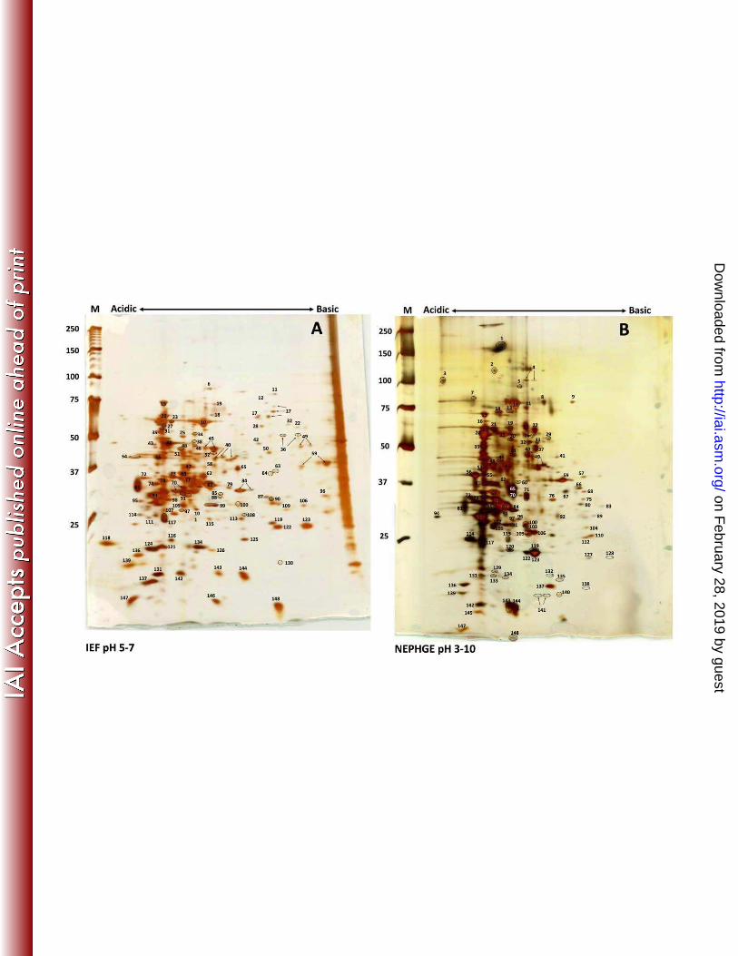
FIGURE LEGENDS Figure 1. Two-dimensional gel electrophoresis of T. pallidum proteins. T. pallidum lysates were
separated by IEF at pH 5-7 (A) or NEPHGE at pH 3.5-10 (B) in the first dimension followed by
8-20% SDS-PAGE in the second dimension. Gels were subsequently silver stained for protein
visualization. Acidic and basic ends are denoted, and relative molecular mass markers are
indicated to the left of each gel. A T. pallidum lysate, resolved in the second dimension only, is
shown at the right side of the IEF pH 5-7 gel (A). Identities of numbered spots are presented in
Table 1. Arrows indicate spots that were submitted separately for MALDI-TOF MS but returned
the same identity. Circles demarcate some closely spaced spots to indicate more clearly which
spots are labeled.
Figure 2. Correlation between predicted molecular weights of T. pallidum proteins and the MR
values obtained in 2DGE patterns in this study. Molecular weights take into account removal of
predicted signal peptides. Apparent degradation products were excluded.
Figure 3. Immunoreactivity of T. pallidum proteins separated by 2DGE with rabbit serum. T.
pallidum lysates were separated by IEF at pH 5-7 (A-C) or NEPHGE at pH 3.5-10 (D-F) in the
first dimension followed by 8-20% SDS-PAGE in the second dimension. Gels were
subsequently silver stained (A, D) or immunoblotted with a 1:1000 dilution of infected (B, E) or
normal rabbit serum (C, F). Black boxed areas indicate major polypeptides that were reactive
with each serum pool. Red boxed areas indicate unidentified acidic proteins. Acidic and basic
ends are denoted, and relative molecular mass markers are indicated to the left of each gel.
Figure 4. Immunoreactivity of T. pallidum proteins separated by IEF pH 5-7 2DGE with human
serum. T. pallidum lysates were separated by IEF at pH 5-7 in the first dimension followed by 8-
on February 28, 2019 by guest
http://iai.asm.org/
Dow
nloaded from
28
20% SDS-PAGE in the second dimension. Gels were subsequently silver stained (A) or
immunoblotted with a 1:500 dilution of pooled human sera from normal blood donors (B),
primary syphilis patients (C), secondary syphilis patients (D), early latent syphilis patients (E) or
late latent syphilis patients (F). Black boxed areas indicate major polypeptides that were
reactive with each serum pool. Acidic and basic ends are denoted, and relative molecular mass
markers are indicated to the left of each gel.
Figure 5. Immunoreactivity of T. pallidum proteins separated by NEPHGE pH 3.5-10 2DGE
with human serum. T. pallidum lysates were separated by NEPHGE at pH 3.5-10 in the first
dimension followed by 8-20% SDS-PAGE in the second dimension. Gels were subsequently
silver stained (A) or immunoblotted with a 1:500 dilution of pooled human sera from normal
blood donors (B), primary syphilis patients (C), secondary syphilis patients (D), early latent
syphilis patients (E) or late latent syphilis patients (F). Black boxed areas indicate major
polypeptides that were reactive with each serum pool. Acidic and basic ends are denoted, and
relative molecular mass markers are indicated to the left of each gel.
on February 28, 2019 by guest
http://iai.asm.org/
Dow
nloaded from
29
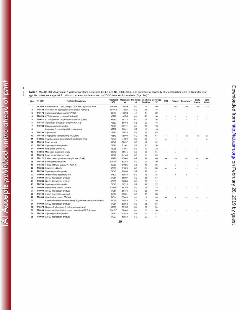
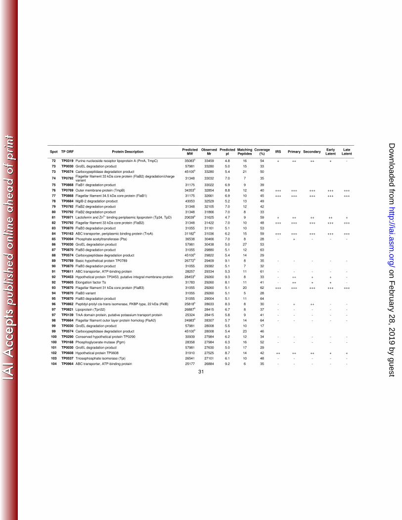
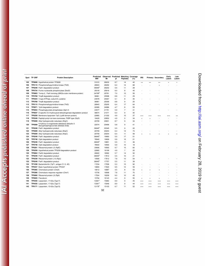
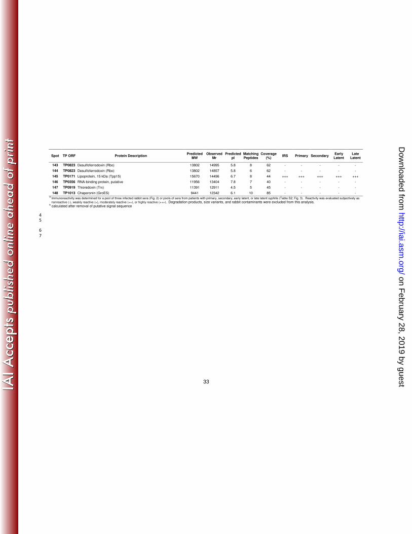
1
Table 1. MALDI-TOF Analysis of T. pallidum proteins separated by IEF and NEPHGE 2DGE and summary of reactivity of infected rabbit sera (IRS) and human 2
syphilis patient sera against T. pallidum proteins, as determined by 2DGE immunoblot analysis (Figs. 2-4) a 3
Spot TP ORF Protein Description Predicted MW
Observed Mr
Predicted pI
Matching Peptides
Coverage (%)
IRS Primary Secondary Early Latent
Late Latent
1 TP1038 Bacterioferritin (TpF1, antigen C1-5, 4D),oligomeric form 206808 166126 5.3 6 50 - +++ +++ +++ +++
2 TP0408 Chromosome segregation SMC protein homolog 124016 118764 5.3 18 18 - - - - -
3 TP0179 Acidic hypothetical protein TP0179 66536 101782 4.3 14 22 - - - - -
4 TP0524 ATP-dependent protease LA (Lon-2) 97729 100742 6.4 23 30 - - - - -
5 TP0071 ATP-dependent Clp protease subunit B (ClpB) 98982 96316 6.0 26 36 - - - - -
6 TP0767 Translation elongation factor G (FusA-2) 76832 86362 5.6 29 46 + - - - -
7 TP0748 CfpA degradation product 78540 83771 5.9 10 18
8 Aconitase 2, probable rabbit contaminant 85762 80021 5.9 10 18
9 TP0748 CfpA variant 78540 79673 5.9 29 45
10 TP0748 Cytoplasmic filament protein A (CfpA) 78540 78809 5.9 46 61 +++ ++ +++ +++ ++
11 TP0886 Polyribonucleotide nucleotidyltransferase (Pnp) 76333 74602 6.2 26 41 ++ ++ ++ ++ ++
12 TP0216 DnaK variant 68042 72297 5.0 17 32
13 TP0748 CfpA degradation product 78540 71561 5.9 23 36
14 TP0984 Heat shock protein 90 72938 71561 5.3 19 32 - - - - -
15 TP0216 Molecular chaperone DnaK 68042 68945 5.0 39 64 +++ + ++ ++ -
16 TP0216 DnaK degradation product 68042 64165 5.0 14 29
17 TP0122 Phosphoenolpyruvate carboxykinase (PckA) 68126 65606 6.2 24 40 ++ ++ ++ ++ ++
18 TP0104 5'-nucleotidase (UshA) 62557b 63286 5.9 24 39 - - - - -
19 TP0426 V-type ATPase, subunit A (AtpA-1) 65020 61025 5.8 19 35 - + ++ + -
20 TP0030 Chaperonin GroEL 57981 61025 5.0 14 30 ++ + ++ ++ -
21 TP0748 CfpA degradation product 78540 60694 5.9 27 42
22 TP0056 Oxaloacetate decarboxylase 64100 59930 6.9 18 35 - + + - -
23 TP0030 GroEL degradation product 57981 58627 5.0 35 57
24 TP0030 GroEL degradation product 57981 57030 5.0 18 41
25 TP0748 CfpA degradation product 78540 56715 5.9 29 37
26 TP0969 Hypothetical protein TP0969 57982b 56354 8.3 18 33 - - - - -
27 TP0030 GroEL degradation product 57981 56136 5.0 36 60
28 TP0426 AtpA-1 degradation product 65020 55661 5.8 15 30
29 TP0584 Hypothetical protein TP0584 53514 55043 9.1 9 20 ++ + ++ ++ +
30 Protein disulfide isomerase family A, probable rabbit contaminant 54096 54449 7.6 8 20
31 TP0030 GroEL degradation product 57981 53852 5.0 35 55
32 TP0478 Glucose-6-phosphate 1-dehydrogenase (Zwf) 58033 53183 6.4 16 34 - - - - -
33 TP0469 Conserved hypothetical protein, containing TPR domains 50471b 52608 6.4 13 30 - - - - -
34 TP0748 CfpA degradation product 78540 51079 5.9 31 41
35 TP0030 GroEL degradation product 57927 50830 5.0 23 41
on February 28, 2019 by guest
http://iai.asm.org/
Dow
nloaded from
30
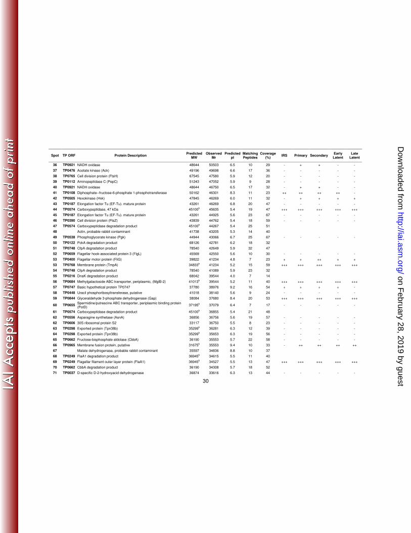
Spot TP ORF Protein Description Predicted MW
Observed Mr
Predicted pI
Matching Peptides
Coverage (%)
IRS Primary Secondary Early Latent
Late Latent
36 TP0921 NADH oxidase 48644 50503 6.5 10 29 - + + - -
37 TP0476 Acetate kinase (Ack) 49196 49698 6.6 17 36 - - - - -
38 TP0765 Cell division protein (FtsH) 67545 47580 5.9 12 20 - - - - -
39 TP0112 Aminopeptidase C (PepC) 51243 47052 5.9 9 28 - - - - -
40 TP0921 NADH oxidase 48644 46750 6.5 17 32 - + + - -
41 TP0108 Diphosphate--fructose-6-phosphate 1-phosphotransferase 50162 46301 8.3 11 23 ++ ++ ++ ++ -
42 TP0505 Hexokinase (Hxk) 47845 46269 6.0 11 32 - + + + +
43 TP0187 Elongation factor Tu (EF-Tu). mature protein 43261 46269 6.8 20 47 - - - - -
44 TP0574 Carboxypeptidase, 47 kDa 45100b 45635 5.4 19 47 +++ +++ +++ +++ +++
45 TP0187 Elongation factor Tu (EF-Tu). mature protein 43261 44925 5.6 23 67 - - - - -
46 TP0390 Cell division protein (FtsZ) 43839 44762 5.4 18 59 - - - - -
47 TP0574 Carboxypeptidase degradation product 45100b 44267 5.4 25 51
48 Actin, probable rabbit contaminant 41738 43205 5.3 14 40
49 TP0538 Phosphoglycerate kinase (Pgk) 44944 43066 6.7 25 67 - - - - -
50 TP0122 PckA degradation product 68126 42781 6.2 18 32
51 TP0748 CfpA degradation product 78540 42649 5.9 32 47
52 TP0659 Flagellar hook-associated protein 3 (FlgL) 45569 42550 5.6 10 30 - - - - -
53 TP0400 Flagellar motor protein (FliG) 39822 41234 4.8 7 23 + + ++ + +
53 TP0768 Membrane protein (TmpA) 34833b 41234 5.2 15 59 +++ +++ +++ +++ +++
54 TP0748 CfpA degradation product 78540 41089 5.9 23 32
55 TP0216 DnaK degradation product 68042 39544 4.0 7 14
56 TP0684 Methylgalactoside ABC transporter, periplasmic, (MglB-2) 41013b 39544 5.2 11 40 +++ +++ +++ +++ +++
57 TP0747 Basic hypothetical protein TP0747 37780 38976 9.2 16 54 + + + + -
58 TP0448 Uracil phosphoribosyltransferase, putative 41018 38140 5.6 9 24 - - - - -
59 TP0844 Glyceraldehyde 3-phosphate dehydrogenase (Gap) 38084 37680 8.4 20 53 +++ +++ +++ +++ +++
60 TP0655 Spermidine/putrescine ABC transporter, periplasmic binding protein (PotD)
37195b 37079 6.4 7 17 - - - - -
61 TP0574 Carboxypeptidase degradation product 45100b 36855 5.4 21 48
62 TP0556 Asparagine synthetase (AsnA) 36856 36756 5.6 19 57 - - - - -
62 TP0606 30S ribosomal protein S2 33117 36750 5.5 8 23 - - - - -
63 TP0298 Exported protein (Tpn38b) 35299b 36281 6.3 12 39 - - - - -
64 TP0298 Exported protein (Tpn38b) 35299b 35653 6.3 19 56 - - - - -
65 TP0662 Fructose-bisphosphate aldolase (CbbA) 36190 35553 5.7 22 58 - - - - -
66 TP0965 Membrane fusion protein, putative 31675b 35553 9.4 10 33 - ++ ++ ++ ++
67 Malate dehydrogenase, probable rabbit contaminant 35597 34836 8.8 10 37
68 TP0249 FlaA1 degradation product 36945b 34615 5.5 11 40
69 TP0249 Flagellar filament outer layer protein (FlaA1) 36945b 34527 5.5 13 47 +++ +++ +++ +++ +++
70 TP0662 CbbA degradation product 36190 34308 5.7 18 52
71 TP0037 D-specific D-2-hydroxyacid dehydrogenase 36874 33616 6.3 13 44 - - - - -
on February 28, 2019 by guest
http://iai.asm.org/
Dow
nloaded from
31
Spot TP ORF Protein Description Predicted MW
Observed Mr
Predicted pI
Matching Peptides
Coverage (%)
IRS Primary Secondary Early Latent
Late Latent
72 TP0319 Purine nucleoside receptor lipoprotein A (PnrA, TmpC) 35083b 33459 4.8 16 54 + ++ ++ + -
73 TP0030 GroEL degradation product 57981 33280 5.0 15 33
73 TP0574 Carboxypeptidase degradation product 45100b 33280 5.4 21 50
74 TP0792 Flagellar filament 33 kDa core protein (FlaB2) degradation/charge variant
31348 33032 7.0 7 35
75 TP0868 FlaB1 degradation product 31175 33022 6.9 9 39
76 TP0769 Outer membrane protein (TmpB) 34353b 32854 8.8 12 40 +++ +++ +++ +++ +++
77 TP0868 Flagellar filament 34.5 kDa core protein (FlaB1) 31175 32661 6.9 10 45 +++ +++ +++ +++ +++
78 TP0684 MglB-2 degradation product 43053 32529 5.2 13 49
79 TP0792 FlaB2 degradation product 31348 32105 7.0 12 42
80 TP0792 FlaB2 degradation product 31348 31866 7.0 8 33
81 TP0971 Lactoferrin and Zn2+
binding periplasmic lipoprotein (Tp34, TpD) 20639b 31625 4.7 9 59 + ++ ++ ++ +
82 TP0792 Flagellar filament 33 kDa core protein (FlaB2) 31348 31422 7.0 10 48 +++ +++ +++ +++ +++
83 TP0870 FlaB3 degradation product 31055 31161 5.1 10 53
84 TP0163 ABC transporter, periplasmic binding protein (TroA) 31182b 31036 6.2 15 59 +++ +++ +++ +++ +++
85 TP0094 Phosphate acetyltransferase (Pta) 36538 30466 7.0 8 28 - + - - -
86 TP0030 GroEL degradation product 57981 30438 5.0 27 53
87 TP0870 FlaB3 degradation product 31055 29880 5.1 12 63
88 TP0574 Carboxypeptidase degradation product 45100b 29822 5.4 14 29
89 TP0789 Basic hypothetical protein TP0789 26772b 29409 9.1 8 35 - - - - -
90 TP0870 FlaB3 degradation product 31055 29382 5.1 7 32
91 TP0611 ABC transporter, ATP-binding protein 28257 29334 5.3 11 61 - - - - -
92 TP0453 Hypothetical protein TP0453, putative integral membrane protein 28453b 29260 9.3 8 33 - ++ + + -
92 TP0605 Elongation factor Ts 31783 29260 8.1 11 41 - ++ + + -
93 TP0870 Flagellar filament 31 kDa core protein (FlaB3) 31055 29260 5.1 20 62 +++ +++ +++ +++ +++
94 TP0870 FlaB3 variant 31055 29260 5.1 5 28
95 TP0870 FlaB3 degradation product 31055 29004 5.1 11 64
96 TP0862 Peptidyl-prolyl cis-trans isomerase, FKBP-type, 22 kDa (FklB) 25818b 28633 8.3 8 30 - + ++ - -
97 TP0821 Lipoprotein (Tpn32) 26887b 28415 6.7 8 37 - - - - -
97 TP0139 TrkA domain protein, putative potassium transport protein 25324 28415 5.8 9 41 - - - - -
98 TP0664 Flagellar filament outer layer protein homolog (FlaA2) 24983b 28307 5.7 14 64 - - - - -
99 TP0030 GroEL degradation product 57981 28008 5.5 10 17
99 TP0574 Carboxypeptidase degradation product 45100b 28008 5.4 23 46
100 TP0290 Conserved hypothetical protein TP0290 30939 27984 6.2 12 34 - - - - -
100 TP0168 Phosphoglycerate mutase (Pgm) 28358 27984 6.3 16 52 - - - - -
101 TP0030 GroEL degradation product 57981 27630 5.0 17 29
102 TP0608 Hypothetical protein TP0608 31910 27525 8.7 14 42 ++ ++ ++ + +
103 TP0537 Triosephosphate isomerase (Tpi) 26541 27101 6.1 10 48 - - - - -
104 TP0964 ABC transporter, ATP-binding protein 25177 26884 9.2 6 35 - - - - -
on February 28, 2019 by guest
http://iai.asm.org/
Dow
nloaded from
32
Spot TP ORF Protein Description Predicted MW
Observed Mr
Predicted pI
Matching Peptides
Coverage (%)
IRS Primary Secondary Early Latent
Late Latent
105 TP0608 Hypothetical protein TP0608 31910 26632 8.7 14 42 ++ ++ ++ + +
106 TP0115 Phosphomethypyrimidine kinase (ThiD) 28943 26292 6.4 14 68 - - + - -
107 TP0249 FlaA1 degradation product 36945b 26202 5.5 11 28
108 TP0734 Purine nucleoside phosphorylase (DeoD) 25318 25916 6.0 8 45 - - - - -
109 TP0663 Tromp-2, FlaA homolog (28KDa outer membrane protein) 24799b 25761 7.0 12 55 - - - - -
110 TP0769 TmpB degradation product 36961 25598 8.8 11 29
111 TP0424 V-type ATPase, subunit E, putative 24978 25467 5.1 8 35 - - - - -
112 TP0769 TmpB degradation product 36961 25306 8.8 9 25
113 TP0115 Phosphomethypyrimidine kinase (ThiD) 28943 25205 6.4 17 64 - - - - -
114 TP0971 Tp34 degradation product 22085 24986 4.7 8 53
115 TP0554 Phosphoglycolate phosphatase (Gph-2) 24577 21751 5.9 7 41 - - - - -
116 TP0037 D-specific D-2-hydroxyacid dehydrogenase degradation product 36874 21371 6.3 9 35
117 TP0259 Membrane lipoprotein TpE (LysM domain protein) 22865 21332 4.9 10 37 +++ +++ +++ +++ ++
118 TP0349 Peptidyl-prolyl cis-trans isomerase, FKBP-type (SlyD) 18429 20852 4.9 5 34 + + - - -
119 TP0509 Alkyl hydroperoxide reductase (AhpC) 20709 20601 8.7 9 34 - - - - -
120 TP0568 4-hydroxy-2-oxoglutarate aldolase/2-dehydro-3-deoxyphosphogluconate aldolase (Eda)
22074 20496 5.8 9 41 - - - - -
121 TP0249 FlaA1 degradation product 36945b 20345 5.5 9 22
122 TP0509 Alkyl hydroperoxide reductase (AhpC) 20709 20204 6.4 14 74 - - - - -
123 TP0509 Alkyl hydroperoxide reductase (AhpC) 20709 20204 6.4 11 59 + + + + +
124 TP0249 FlaA1 degradation product 36945b 19993 5.5 17 51
125 TP0748 CfpA degradation product 78540 19909 5.9 15 21
126 TP0249 FlaA1 degradation product 36945b 19851 5.5 11 36
127 TP0748 CfpA degradation product 78540 19593 5.9 10 18
128 TP0201 Ribosomal protein L5 (RplE) 20806 19593 9.7 15 60 - - - - -
129 TP0259 Hypothetical protein TP0259 degradation product 22865 18199 4.9 7 20
130 TP0664 FlaA2 degradation product 26822 18062 5.7 14 52
131 TP0249 FlaA1 degradation product 36945b 17812 5.5 10 31
132 TP0239 Ribosomal protein L10 (RplJ) 19566 17812 7.8 10 54 - - - - -
133 TP0249 FlaA1 degradation product 36945b 17757 5.5 11 39
134 TP1038 Bacterioferritin (TpF1, antigen C1-5, 4D) 17234 17558 5.3 15 92 - - - - -
135 TP0437 Basic hypothetical protein TP0437 19893 17620 9.5 10 59 - - - - -
136 TP0365 Chemotaxis protein (CheX) 16612 16887 4.5 8 53 + + + - -
137 TP0366 Chemotaxis response regulator (CheY) 15736 16698 7.8 11 70 - - - - -
138 TP0060 Ribosomal protein L9 (RplI) 17504 16209 9.0 10 45 - - - - -
139 TP0925 Flavodoxin 15794 16143 4.4 9 55 + + + - -
140 TP0435 Lipoprotein, 17 kDa (Tpp17) 13361b 15962 8.9 9 45 +++ +++ +++ +++ +++
141 TP0435 Lipoprotein, 17 kDa (Tpp17) 13361b 15946 8.9 9 44 +++ +++ +++ +++ +++
142 TP0171 Lipoprotein, 15 kDa (Tpp15) 13176b 15103 6.7 11 52 +++ +++ +++ +++ +++
on February 28, 2019 by guest
http://iai.asm.org/
Dow
nloaded from
33
Spot TP ORF Protein Description Predicted MW
Observed Mr
Predicted pI
Matching Peptides
Coverage (%)
IRS Primary Secondary Early Latent
Late Latent
143 TP0823 Desulfoferrodoxin (Rbo) 13802 14995 5.8 8 62 - - - - -
144 TP0823 Desulfoferrodoxin (Rbo) 13802 14857 5.8 6 62 - - - - -
145 TP0171 Lipoprotein, 15 kDa (Tpp15) 15670 14496 6.7 8 44 +++ +++ +++ +++ +++
146 TP0356 RNA-binding protein, putative 11956 13404 7.8 7 40 - - - - -
147 TP0919 Thioredoxin (Trx) 11391 12911 4.5 5 45 - - - - -
148 TP1013 Chaperonin (GroES) 9441 12342 6.1 10 85 - - - - - a Immunoreactivity was determined for a pool of three infected rabbit sera (Fig. 2) or pools of sera from patients with primary, secondary, early latent, or late latent syphilis (Table S2; Fig. 3). Reactivity was evaluated subjectively as
nonreactive (-), weakly reactive (+), moderately reactive (++), or highly reactive (+++). Degradation products, size variants, and rabbit contaminants were excluded from this analysis. b calculated after removal of putative signal sequence
4
5
6
7
on February 28, 2019 by guest
http://iai.asm.org/
Dow
nloaded from