
Download (1484Kb) - University of Kent
34
Kent Academic Repository Full text document (pdf) Copyright & reuse Content in the Kent Academic Repository is made available for research purposes. Unless otherwise stated all content is protected by copyright and in the absence of an open licence (eg Creative Commons), permissions for further reuse of content should be sought from the publisher, author or other copyright holder. Versions of research The version in the Kent Academic Repository may differ from the final published version. Users are advised to check http://kar.kent.ac.uk for the status of the paper. Users should always cite the published version of record. Enquiries For any further enquiries regarding the licence status of this document, please contact: [email protected] If you believe this document infringes copyright then please contact the KAR admin team with the take-down information provided at http://kar.kent.ac.uk/contact.html Citation for published version Adamek, Nancy and Lieto-Trivedi, Alena and Geeves, Michael A. and Coluccio, Lynne M. (2010) Modification of Loop 1 Affects the Nucleotide Binding Properties of Myo1c, the Adaptation Motor in the Inner Ear. Biochemistry, 49 (5). pp. 958-971. ISSN 0006-2960. DOI https://doi.org/10.1021/bi901803j Link to record in KAR https://kar.kent.ac.uk/29224/ Document Version UNSPECIFIED
Transcript of Download (1484Kb) - University of Kent
Kent Academic RepositoryFull text document (pdf)
Copyright & reuse
Content in the Kent Academic Repository is made available for research purposes. Unless otherwise stated all
content is protected by copyright and in the absence of an open licence (eg Creative Commons), permissions
for further reuse of content should be sought from the publisher, author or other copyright holder.
Versions of research
The version in the Kent Academic Repository may differ from the final published version.
Users are advised to check http://kar.kent.ac.uk for the status of the paper. Users should always cite the
published version of record.
Enquiries
For any further enquiries regarding the licence status of this document, please contact:
If you believe this document infringes copyright then please contact the KAR admin team with the take-down
information provided at http://kar.kent.ac.uk/contact.html
Citation for published version
Adamek, Nancy and Lieto-Trivedi, Alena and Geeves, Michael A. and Coluccio, Lynne M. (2010)Modification of Loop 1 Affects the Nucleotide Binding Properties of Myo1c, the AdaptationMotor in the Inner Ear. Biochemistry, 49 (5). pp. 958-971. ISSN 0006-2960.
DOI
https://doi.org/10.1021/bi901803j
Link to record in KAR
https://kar.kent.ac.uk/29224/
Document Version
UNSPECIFIED

Modification of Loop 1 Affects the Nucleotide-Binding Properties
of Myo1c, The Adaptation Motor in the Inner Ear †
Nancy Adamek ,University of Kent, Canterbury, Kent, CT2 7NJ, U.K.
Alena Lieto-Trivedi ,Boston Biomedical Research Institute, Watertown, MA 02472, U.S.A.
Michael A. Geeves , andUniversity of Kent, Canterbury, Kent, CT2 7NJ, U.K.
Lynne M. Coluccio *
Boston Biomedical Research Institute, Watertown, MA 02472, U.S.A.
AbstractMyo1c is one of eight members of the mammalian myosin I family of actin-associated molecularmotors. In stereocilia of the hair cells in the inner ear, Myo1c presumably serves as the adaptationmotor, which regulates the opening and closing of transduction channels. Although there isconservation of sequence and structure among all myosins in the N-terminal motor domain, whichcontains the nucleotide- and actin-binding sites, some differences include the length and compositionof surface loops, including loop 1, which lies near the nucleotide-binding domain. To investigate therole of loop 1, we expressed in insect cells mutants of a truncated form of Myo1c, Myo1c1IQ, as wellas chimeras of Myo1c1IQ with the analogous loop from other myosins. We found that replacementof the charged residues in loop 1 with alanines or the whole loop with a series of alanines did notalter the ATPase activity, transient kinetics properties and Ca2+-sensitivity of Myo1c1IQ. Substitutionof loop 1 with that of the corresponding region from tonic smooth muscle myosin II (Myo1c1IQ-tonic) or replacement with a single glycine (Myo1c1IQ-G) accelerated ADP release from A.M 2-3-fold in Ca2+, whereas substitution with loop 1 from phasic muscle myosin II (Myo1c1IQ-phasic)accelerated ADP release 35-fold. Motility assays with chimeras containing a single g-helix, or SAH,domain showed that Myo1cSAH-tonic translocated actin in vitro twice as fast as Myo1cSAH-WT and3-fold faster than Myo1cSAH-G. The studies show that changes induced in Myo1c by modifying loop1 showed no resemblance to the behaviour of the loop donor myosins or to the changes previouslyobserved with similar Myo1b chimeras.
Myosins are a large family of molecular motors that have been subdivided into more than 30subgroups (1,2). Different family members are involved in a wide range of motor activities ineukaryotic cells such as muscle contraction, cell division, pseudopod extension and vesicletransport (3,4). Class I myosins are a diverse group of monomeric myosins implicated in severalactin-mediated processes including organization and maintenance of tension of thecytoskeleton as well as signal transduction (5-7). The mammalian class I myosin, Myo1c,consists of a heavy chain containing an N-terminal motor domain, a neck or lever arm stabilisedby 3 calmodulin molecules and a C-terminal tail region implicated in membrane binding(8-10). Myo1c mediates the cycling of GLUT4 transporters in adipocytes by promoting the
†This work was supported by NIH Grant R01 DC08793 to L. M. C. and Wellcome Trust Grant 070021 to M. A. G.
*To whom correspondence should be addressed: Boston Biomedical Research Institute, 64 Grove Street, Watertown, MA 02472.Telephone: (617) 658-7784. Fax. (617) 972-1761. [email protected].
NIH Public AccessAuthor ManuscriptBiochemistry. Author manuscript; available in PMC 2011 February 9.
Published in final edited form as:Biochemistry. 2010 February 9; 49(5): 958–971. doi:10.1021/bi901803j.
NIH
-PA
Author M
anuscriptN
IH-P
A A
uthor Manuscript
NIH
-PA
Author M
anuscript

fusion of GLUT4-containing vesicles with the cell membrane (11-13). In the specialized haircells of the inner ear, Myo1c is believed to be the adaptation motor, which regulates the tensionon the tip links that connect neighbouring stereocilia thereby controlling the opening andclosing of transduction channels (5).
We recently defined the biochemical kinetics of the ATP-driven interaction of Myo1c withactin and showed that it has an unusual calcium dependence (14). Calcium binding to thecalmodulin closest to the motor domain has little effect on the ATPase or motor activity, butalters specific steps of the ATPase cycle. ATP hydrolysis was inhibited 7-fold by calcium whileADP release from acto-Myo1c was accelerated by 10-fold. These two changes together wouldreduce the lifetime of the actin-attached states and increase the lifetime of the actin-detachedstate without altering the overall cycle time. In combination these properties appear to be idealto modulate the activity of Myo1c in response to a calcium transient of the sort expected tooccur in the inner ear.
The observation that certain events in the ATPase cycle are regulated oppositely in calcium iscurious and in particular the Ca2+-regulation of the ATP hydrolysis step has not been reportedpreviously for any myosin. This raises the question of whether the ATP hydrolysis step iscontrolled by calcium binding to the calmodulin light chain. In fact, the regulation may occurvia the myosin conformational change that precedes the ATP cleavage step and is thought toposition the catalytic residues to allow ATP splitting. This conformational change, known asthe recovery stroke, involves the movement of switch II and the accompanying converter-domain movement to reprime the lever arm/light-chain domain (15,16). Thus, the recoverystroke results in a repositioning of the light chains and therefore could be influenced by acalcium-induced change in the calmodulin/light chain conformation.
Other events regulated by calcium are ATP binding and ADP release. ADP release in smoothand scallop muscle is altered via phosphorylation or calcium binding to light chains (17,18).The exact mechanism by which the conformation of the light chains is communicated to thenucleotide-binding pocket for these dimeric myosins is not defined, but may involve someform of interaction between the two motor domains facilitated by the conformation of the neckdomains (19-21). Although we previously reported a modest regulation of nucleotide releasein calcium for the related myosin I, Myo1b, the behaviour of Myo1c is more dramatic (22).The communication pathway of this novel regulation via the light chains for monomericmyosins is unknown.
The structure of the myosin motor domain is highly conserved across the broad myosin family;however, there are several surface loops that are less well conserved. These loops have beenproposed to tune the activity of myosins to their specific cellular roles (23). One such loop isloop 1 near the entrance to the nucleotide-binding pocket. Loop 1 joins two helices, oneconnected to switch 1 and the other to the P-loop. These two elements form a major part of thebinding site for the gamma phosphate in ATP. Loop 1 is therefore in a position to influenceaccess to the nucleotide pocket and the interaction with the gamma Pi of ATP. Previous studiesof natural variations of loop 1 in vertebrate smooth muscle myosin II (24-26) have shown thatalternately expressed loops can alter the release of ADP from the nucleotide pocket.Furthermore, studies on scallop muscle myosin II (27) have established that alternate splicingof loop 1 alters the affinity of acto-myosin for ADP thereby permitting the cell to producemyosins with differing ATPase and motility properties. However, studies of engineeredvariations in the size and composition of loop 1 in either a smooth muscle myosin II or aDictyostelium myosin II (28) have so far failed to resolve which features of loop 1 areresponsible for this modulation of myosin activity.
Adamek et al. Page 2
Biochemistry. Author manuscript; available in PMC 2011 February 9.
NIH
-PA
Author M
anuscriptN
IH-P
A A
uthor Manuscript
NIH
-PA
Author M
anuscript

Goodson and colleagues showed that the sequences of loop 1 are well conserved when myosinsare grouped according to their kinetic activity and proposed that loop 1 modulates the kineticcharacteristics that distinguish one myosin isoform from another (29). It has also beensuggested that the flexibility of loop 1 or its length determines activity (26) and that longerloops interact with other parts of the myosin molecule including the light chains in someconformations thereby affecting regulation (30).
In a previous study (22), we explored the role of loop 1 on Myo1b by replacing its chargedresidues with alanine, the whole loop with alanine residues or replacing the loop with either asingle glycine, or loop 1 from either phasic or tonic smooth muscle myosin II, a strategy similarto that used by others to explore the effects of loop 1 on smooth muscle myosin II (24,26,27).We found that loop 1 had major effects on the coupling of actin and nucleotide-binding eventsin Myo1b and that it is likely to modulate the load dependence of Myo1b. The role of loop 1can therefore have broad implications for modulating myosin motor activity; however, thesame mutations in different myosin backgrounds can have quite different effects (22,31).
To explore the role of loop 1 in Myo1c we investigated how loop 1 modulates the motor activityof Myo1c, specifically its effects on nucleotide binding and release in the presence and absenceof calcium. We found that all the results obtained with the Myo1c chimeras stand in markedcontrast to those previously obtained with similar constructs using Myo1b.
Experimental Procedures
Preparation of Constructs and Expression in Insect Cells
Using cDNA encoding the entire open reading frame of Myo1c (previously known as myr 2;the kind gift of Dr. Martin Bähler; Institut für Allgemeine Zoologie und Genetik, Westfälische-Wilhelms Universität, Münster, Germany), we prepared a series of constructs in which loop 1of Myo1c1IQ, a truncated form of rat Myo1c representing the motor domains and first IQdomain (amino acids 1-725) (32), was replaced with loops 1 from other myosins; or in whichalanine substitutions were made in the endogenous loop 1. To define the beginning and end ofloop 1 in Myo1c, secondary structure assignments were made by the program DSSP (Definitionof Secondary Structure of Protein; http://swift.cmbi.ru.nl/gv/dssp/descrip.html). All mutantsexcept Myo1c1IQ-phasic were created using polymerase chain reaction (PCR). Overlappingsets of primers having the desired mutations and restriction sites and incorporating a FLAGtag at the C-terminus were used in the first set of PCR reactions. The two resulting fragmentswere then subjected to another round of PCR with the outside primers to generate the in-framefusion proteins. After treatment with the appropriate restriction enzymes, the purified PCRproducts were ligated into the pFastBacDUAL transfer vector (Gibco BRL, Gaithersburg, MD)downstream of the polyhedrin promoter; the p10 promoter cloning site contained the genecoding for calmodulin (32). The plasmids were transformed into DH5g cells and selected byantibiotic resistance. Colonies were grown and the isolated DNA was tested for the presenceof inserts by restriction analysis. Plasmids containing Myo1c inserts were sequenced withinternal and vector-specific oligonucleotides using automated sequencing. Myo1c1IQ-phasicwas made using the Transformer Site-Directed Mutagenesis Kit (Clontech, Mountain View,CA 94043). Briefly, a mutagenic primer and a selection primer were used to generate a mixtureof mutated and unmutated plasmids. The mixture was then subjected to a selective restrictiondigestion, which selectively digested the unmutated plasmid. The mutated plasmid was thentransformed as above into DH5g followed by selection according to antibiotic resistance.Plasmids were isolated from selected colonies and those containing inserts were subjected toautomatic sequencing.
In all cases, the recombinant donor plasmid was transformed into DH10Bac E. coli cells(Invitrogen, Carlsbad, Ca 92008) for transposition into bacmid. Recombinant bacmid DNA
Adamek et al. Page 3
Biochemistry. Author manuscript; available in PMC 2011 February 9.
NIH
-PA
Author M
anuscriptN
IH-P
A A
uthor Manuscript
NIH
-PA
Author M
anuscript

was isolated by potassium acetate precipitation as described by the Bac-to-Bac BaculovirusExpression Systems Instruction Manual supplied by Invitrogen. Virus was produced bytransfecting the recombinant bacmid DNA into Spondoptera frugiperda 9 (Sf9) insect cellswith Cellfectin reagent (Invitrogen) followed by 3 days of growth. Subsequently, amplifiedvirus was used to infect Sf9 cells in suspension. Infection was allowed to proceed for 4 daysafter which time cells were harvested by centrifugation. Cell pellets were either usedimmediately for protein isolation or frozen in liquid N2 and stored at -80°C for future use.
Protein purification
To isolate protein, the insect cell pellets were homogenized in 10 mM Tris, pH 7.5, 0.2 MNaCl, 4 mM MgCl2, and 2 mM ATP in the presence of protease inhibitors and then centrifugedat 183,000 × g for 50 min. The supernatant was applied to an anti-FLAG column, and afterwashing the expressed proteins were eluted with a step gradient of FLAG peptide. Fractionscontaining protein were identified by SDS-polyacrylamide gel electrophoresis, pooled anddialyzed against 10 mM Tris, 50 mM KCL and 1 mM DTT. Proteins were either usedimmediately or stored at -80°C for future use.
Preparation of Constructs with a SAH Domain
Using the clone for rat myosin X (kindly provided by Drs. Erich Boger and Thomas Friedman,NIDCD, NIH) we prepared by PCR a fusion construct (Myo1cSAH-WT) in which the SAHdomain of myosin X (amino acids 805-843) followed by a myc and FLAG tag were added towild-type Myo1c at residue 762 based on sequence analogy between Myo1c and myosin X asdetermined by Clustal X. This results in a LCBD consisting of two complete IQ motifs andmost of the third IQ motif followed by the SAH domain. The SAH constructs express in insectcells at levels equivalent to the 1IQ forms. Similar constructs for the loop 1 mutants, tonic,phasic and G (Myo1cSAH-tonic, Myo1cSAH-phasic and Myo1cSAH-G, respectively) were alsoprepared and expressed in insect cells and purified by affinity purification with anti-FLAG.
ATPase Activity
The steady-state actin-activated Mg2+-ATPase activity of Myo1c and the loop 1 mutants (1IQforms and those incorporating SAH domains) was measured using a colorimetric assaydescribed by Pollard (33) in 10 mM Tris, pH 7.5, 50 mm KCl, 1 mM MgCl2 and CaCl2 andEGTA to effect pCa values ranging from 4 - 8.9.
Motility Assays
The ability of the purified myosins to translocate actin filaments in vitro was determined usinga procedure similar to one previously described (34). Briefly, flow chambers were made byattaching nitrocellulose-coated coverslips face down to a glass microscope slide using double-sided sticky tape. Myosin was prepared by mixing it with 0.05 mg/ml F-actin and 10 mM ATPfor 20 min, then spinning at 245,00 × g and 4°C for 20 min. The flow chambers were coatedwith 痺12 たl 0.05 mg/ml myc antibody (Invitrogen) in PBS for 15 min, then 75 たl 5 mg/mlbovine serum albumin (BSA) in motility buffer (25 mM Imidazole, pH 7.5, 1 mM EGTA, 25mM KCL, 4 mM MgCl2) for 10 min to block non-specific sites. Thirty たl myosin supernatant(at various concentrations) was applied to the flow chamber for 15 min then washed withmotility buffer plus BSA for 10 min. Thirty たl 痺0.14 たM rabbit skeletal muscle F-actin labeledwith rhodamine phalloidin (Molecular Probes, Eugene, OR) was applied to the flow chamberfor 5 min. The chambers were washed with 75 たl motility buffer plus BSA before adding 50たl motility buffer containing 0.5% methylcellulose, 3 mg/ml glucose, 20 mM DTT, 0.1 mg/mlglucose oxidase, 0.05 mg/ml catalase and 2 mM ATP. Slides were examined with afluorescence microscope using a 100× oil immersion lens (Nikon Inc., Melville, NY). Imageswere recorded digitally using a FlashBus MV PCI bus frame grabber (Integral Technologies,
Adamek et al. Page 4
Biochemistry. Author manuscript; available in PMC 2011 February 9.
NIH
-PA
Author M
anuscriptN
IH-P
A A
uthor Manuscript
NIH
-PA
Author M
anuscript
Inc., Indianapolis, IN). The percentage of translocating filaments was determined usingManually Count Objects, and the speed of the filaments was determined using Track Points inMetaMorph bioimaging software (version 6.3r2, Universal Imaging Corp., Downingtown,PA).
Transient enzyme kinetics
Rapid kinetic measurements were carried out using a standard Hi-Tech Scientific SF-61 DX2stopped-flow system fitted with a 75 W Xe/Hg lamp and monochromator for wavelengthselection. Intrinsic tryptophan fluorescence was excited at 295 nm and emission monitoredthrough a WG320 cut-off filter, while pyrene fluorescence was excited at 365 nm and emissionmonitored through a KV 398 nm cut-off filter. The stopped-flow transients were fitted to oneor two exponentials as described previously by non-linear least squares curve fitting using theKinetic Studio software (TgK Scientific). The reactant concentrations stated in the text andfigures are those after 1:1 mixing in the stopped-flow spectrophotometer, unless statedotherwise. In some secondary data plots the plotted concentrations are those prior to mixing,which are then indicated as ‘initial [X]’. All experiments were carried out at 20°C in 20 mMMOPS buffer containing 100 mM KCL, and 5 mM MgCl2. In addition the buffer containedeither 1 mM EGTA for measurements without calcium, or 1.1 mM EGTA and 1.2 mMCaCl2 for measurements carried out in the presence of calcium.
Data Analysis
In the absence of actin the interaction of ATP and ADP with Myo1c was interpreted in termsof the 7-step mechanism for the myosin ATPase cycle (see Scheme 2) in which ATP bindingto myosin occurs in two steps (a binding step and a protein conformational change followedby reversible ATP hydrolysis (35,36)). Pi release and ADP release then occur sequentially,each dissociation event preceded by a protein isomerisation. Our previous work establishedthat ATP binding is accompanied by an increase in tryptophan fluorescence and there is nofluorescence change associated with ADP binding (14). We assume this fluorescence changeis associated with the hydrolysis step because Myo1c has the conserved tryptophan at the endof the relay loop, which is known to signal the protein conformational change associated withswitch II closure and the hydrolysis step in other myosins. The rate constant for the step(k+3+k-3) was inhibited 7-fold by calcium for Myo1c1IQ-WT.
The results of the kinetic interaction of actin-Myo1c1IQ with nucleotide were interpreted interms of the model we described previously for Myo1b (37) and Myo1c (5,14) (Scheme 1).The model assumes that acto-Myo1c exists in two conformations, A.M and A.Mガ, in the absenceof nucleotide and that the interconversion between the two conformations is defined by theequilibrium constant Kg (Kg = k+g / k-g). A.Mガ represents the complex with the nucleotidepocket in the “closed” conformation which must isomerise into the “open-pocket” form, A.M,before nucleotide binding/release can occur. This isomerisation is believed to be coupled to aswing of the converter/IQ regions of the myosin motor domain. The ATP-induced dissociationof actin-myosin is biphasic. The fast phase represents ATP binding to A.M and the observedrate constant is hyperbolically dependent upon ATP concentration with kobs,fast = K1k+2[ATP]/(1+K1[ATP]).
The kobs for the slow phase also shows hyperbolic dependence upon ATP concentration forMyo1c1IQ-WT. This is unusual but reflects the fact that A.Mガ is the predominant species present(Kg <1). An exact solution to the ATP dependence of the slow phase is not possible but threeregions of the ATP dependent behaviour of the slow phase can be described that depend uponthe relative rate constants of ATP binding to A.M and the isomerisation of A.Mガ to A.M. Atvery high [ATP] if k-g 盂 than the rate of ATP binding (K1k+2[ATP]/(1+K1[ATP])) then everyA.M ガ that isomerises to A.M will bind ATP irreversibly and lead to dissociation of the myosin
Adamek et al. Page 5
Biochemistry. Author manuscript; available in PMC 2011 February 9.
NIH
-PA
Author M
anuscriptN
IH-P
A A
uthor Manuscript
NIH
-PA
Author M
anuscript

from the complex. Under these conditions the kobs has it maximal value and kmax,slow = k+g.For WT Myo1c then k+2 is more than ten times the maximum observed value of the slow phaseand therefore the conditions are valid and kmax,slow = k+g.
At very low [ATP], such that K1[ATP]盂1, the kobs of ATP binding to A.M is K1k+2[ATP],and if this is slow compared to k-g + k+g then only a single phase will be observed. The kobsis then defined as K1k+2[ATP]/(1+ Kg), the rate constant for ATP binding to A.M times thefraction of acto-Myo1c in the A.M conformation (= 1/ (1+ Kg)). This condition will always befulfilled if the ATP concentration is low enough. The estimated value of k-g + k+g for the WTis 20-25 s-1 independent of the presence of calcium and therefore this condition will be met at[ATP]盂 1 mM.
If k -g + k+g is of the same order as the fast phase then the observed slow phase will dependupon the relative rate constants of k-g and the rate constant of ATP binding (K1k+2[ATP]/(1+K1[ATP])). The half maximal value of the kobs for the slow phase will be observed at theATP concentration (K0.5) when (K1k+2[ATP]/(1+K1[ATP])) = k-g. Since k-g can be calculatedfrom Kg (obtained from the relative amplitude of the fast and slow phases) and k+g =kmax, slow, then the value of K0.5 can be predicted.
Two similar complexes are also assumed to exist in the presence of ADP, A.M.D and A.Mガ.D,with an open and closed nucleotide pocket, respectively. In the case of Myo1c1IQ-WT thebinding and release of ADP from A.M.D and A.Mガ.D cannot be distinguished because the rateconstants governing ATP binding are slower than ADP release. The apparent ADP affinity canbe estimated by measuring the ADP inhibition of the ATP-induced dissociation of acto-myosin.In the case of Myo1c1IQ-WT, ADP is in rapid equilibrium with A.M and A.Mガ on the timescale of the slow phase of the reaction. Thus, the slow phase, kobs, is reduced with a hyperbolicdependence upon ADP concentration (independent of whether the ADP is premixed with theprotein or the ATP) and K0.5 = (KAD/(1 + 1/Kg), where KAD is the affinity of A.M for ADP.
The observed behaviour of the ATP-induced dissociation reaction and the ADP inhibition ofthe slow phase are similar in the presence or absence of calcium, although the rate andequilibrium constants change. This is not the case for the fast phase of the dissociation reaction,which appears different in the presence and absence of ADP. The fast phase is small anddifficult to characterise in the presence of ADP. When ADP is pre-incubated with the protein,the small amplitude of the fast phase decreases and cannot be measured at ADP concentrationsabove 5 たM. If ADP is pre-incubated with the ATP before mixing with protein then theobserved behaviour is different in the presence vs. absence of calcium. In the absence ofcalcium the system behaves as though the ADP is in rapid equilibrium with A.M and A.Mガ,i.e., ADP binding and release is much faster than the rate constants for ATP binding(K1k+2[ATP]) and ADP slows the kobs with 50% inhibition when [ADP] = KAD. If calcium isnot present then the kobs increases linearly with ADP concentration (38). This is consistentwith the rate constant of ADP and ATP binding being of the same size and once ADP binds itis only released slowly. In this case, kobs = K1k+2[ATP] + k-AD[ADP], and in a plot of kobs vs[ADP] the slope of the linear fit equals the second order binding rate constant k-AD and theintercept equals K1k+2[ATP] (39,40). We therefore see a change in the behaviour frominhibition of ATP-induced dissociation of A.M by ADP in the absence of calcium to a 10-foldacceleration of the rate constant for ADP release (k+AD) in calcium. Thus calcium alters therelative values ofK1k+2[ATP] and k+AD.
Results
There are two clear phases of the changes in pyrene fluorescence when ATP is rapidly mixedwith pyr.actin-Myo1c1IQ; a large slow phase and a small fast phase (50 % and 6 % amplitude,
Adamek et al. Page 6
Biochemistry. Author manuscript; available in PMC 2011 February 9.
NIH
-PA
Author M
anuscriptN
IH-P
A A
uthor Manuscript
NIH
-PA
Author M
anuscript
respectively). The hyperbolic dependence of the fast phase in the absence of calcium wasresolved (Figure 2) in contrast to our earlier work on the wild type, which could only resolvethe data in the presence of calcium (14). The data in the presence of calcium are identical tothis earlier work. The results are summarized in Table 3 and show that the removal of calciumaccelerates the maximum kobs (k+2) from 19.6 s-1 to 41.2 s-1 while the K0.5 (1/K1) valuesdecrease from 1.6 mM to 0.51 mM. Thus, calcium alters both the equilibrium constant for ATPbinding and the isomerisation step, thought to be coupled to switch I movement (41). The stepssensitive to calcium are therefore: the ATP hydrolysis step, which is inhibited 7-fold bycalcium; the ADP release from acto-myosin, which is activated 10-fold by calcium; and theATP-induced dissociation of AM, in which the maximum rate constant is inhibited 2-fold andthe ATP concentration for half maximal inhibition is increased 3-fold by calcium. This differsfrom all the other myosins that show calcium regulation. These have all been reported to haveCa2+-dependence primarily of Pi-release and ADP binding and release events (22,42,43).
ATP-induced dissociation of the acto-Myo1c 1IQ alanine loop 1 chimeras
In the ATP-induced dissociation of acto-Myo1c the reaction remains biphasic for all the alanineloops (the transient for Myo1c1IQ-8A is shown in Figure 3A) with the ratio of amplitudesindependent of ATP concentration and a dominating slow phase. In the absence of calcium thefast phase was 痺10% of the total amplitude for each construct and the addition of calciumreduced the fast phase by a factor of 2 at most, but since fast phase amplitudes are hard todefine with precision the change is not significant. The kmax for the fast phase (k+2) in theabsence of calcium was unaffected by the change in the alanine loops (<20% reduction; Figure3C). In the presence of calcium (data not shown) the 8A construct was unaffected, while theE/A (2-fold) and the R/A (5-fold) did show some acceleration of the reaction compared to WT.The slow phase kmax (k+g) was unaffected in the absence (Figure 3D) or presence of calciumexcept for the R/A construct which showed a 2-fold acceleration in the both the presence andabsence of calcium. The results are summarised in Table 1.
Effect of ADP on the ATP-induced dissociation of acto-Myo1c 1IQ alanine loop 1 chimeras
To obtain an estimate of the affinity of ADP for acto-Myo1c for the loop 1 chimeras, thedissociation by ATP was monitored with increasing concentrations of ADP present (either bypre-incubating ADP with the protein or the ATP). Both experimental approaches producedbiphasic transients for all mutants as observed with Myo1c1IQ-WT (14), in both the presenceand absence of ADP. If ADP binding is in rapid equilibrium with A.M and A.Mガ the twoexperimental approaches will give the same results. This was true on the timescale of the slowphase, but not for the fast phase. The analysis of each phase will therefore be dealt withseparately below. Typical pyrene fluorescence transients for the ATP-induced dissociation ofacto-Myo1c1IQ in the absence and presence of high ADP concentration (preincubated withacto-Myo1c) are shown in Figure 4A for Myo1c1IQ-R/A (-Ca2+) and 4B for Myo1c1IQ-phasic(+Ca2+).
Slow phase
If ADP is in rapid equilibrium with acto-Myo1c1IQ on the time scale of the measurement, thetwo assays will give identical results for the kobs of the slow phase (Figure 4C). As the ADPconcentration was increased, the kobs of the slow phase, which monitors the A.Mガ species, wasslowed for all constructs in a manner similar to that observed for Myo1c1IQ-WT, both in theabsence and presence of calcium. Hyperbolic fits provided the apparent ADP affinities(Kapp) from which the KAD were calculated. The alanine constructs all showed similar Kappand KAD values as Myo1c1IQ-WT, both in the absence (Figure 4C) and presence of calcium(with 10-fold weaker affinities in calcium; Figure 4D). The KAD values are summarised inTable 2.
Adamek et al. Page 7
Biochemistry. Author manuscript; available in PMC 2011 February 9.
NIH
-PA
Author M
anuscriptN
IH-P
A A
uthor Manuscript
NIH
-PA
Author M
anuscript
Fast phase
When ADP was pre-incubated with acto-Myo1c1IQ the already small amplitude of the fastphase disappeared rapidly as ADP was added and no trace of a fast phase was determined atADP > 5 たM, both with and without calcium making the analysis of the fast phase resultsunreliable in this experimental approach (see Figure 4A and 4B). This is consistent with thelow values of KAD calculated above and listed in Table 2.
When ADP was pre-incubated with ATP and then rapidly mixed with acto-Myo1c1IQ thekobs of the fast phase increased linearly with increasing ADP concentration. This diagnosticof a competition between ADP and ATP for the binding to acto-Myo1c1IQ provides the 2nd
order rate constant of ADP binding (slope = k-AD) (data not shown). The R/A and E/Aconstructs had similar k-AD values as WT (痺1 たM-1s-1), while the 8A construct was 2-foldfaster at 2.4 たM-1s-1. The experiment could only be completed reliably in the absence ofcalcium (the fast phase could not be determined reliably in calcium due to the smallamplitudes).
Calculation of the ADP release constants (k+AD) from the KAD and ADP-binding rate constantsin the chimeras in the absence of calcium showed values similar to those found forMyo1c1IQ-WT at 痺2 s-1 for Myo1c1IQ-R/A and Myo1c1IQ-E/A and slightly faster rateconstants at 痺4-5 s-1 for Myo1c1IQ-8A (Table 2). In the presence of calcium, the ADP releaserate constant for Myo1c1IQ-WT was accelerated 痺10-fold to 20 s-1, while the values calculatedfor the alanine chimeras were accelerated between 5-7-fold (Table 2).
ATP-induced dissociation of acto-Myo1c 1IQ-tonic,-Myo1c 1IQ-phasic and -Myo1c 1IQ-G
When the ATP-induced dissociation of acto-Myo1c was performed with Myo1c1IQ-tonic,Myo1c1IQ-phasic and Myo1c1IQ-G in the absence of calcium, the observed amplitudes weresmaller than seen with Myo1c1IQ-WT. The amplitudes in the absence of calcium ofMyo1c1IQ-phasic were as little as 1/5th of those for Myo1c1IQ-WT, with 1.6 and 18 % for fastand slow phases, respectively. The smaller amplitudes make defining the maximal rateconstant, k+2, of the fast phase difficult. Despite the loss in total amplitude, the value for Kgfor Myo1c1IQ-phasic was similar to Myo1c1IQ-WT at 0.09 (Table 1) and this increased to 0.23in the presence of calcium. The amplitudes obtained for Myo1c1IQ-tonic were 痺7 % and 痺32% of total fluorescence, for the fast and slow phases respectively, giving a value of Kg of 0.21in the absence of calcium and this decreased to 0.12 in the presence of calcium. The observedtransients for Myo1c1IQ-G are shown in Figure 3B and in the absence of calcium only a singlephase could be distinguished with an amplitude of 13%. In the presence of calcium the slowphase had an amplitude of 13 %, while the fast-phase amplitude was 33 %, giving Kg a valueof 1.8 in the presence of calcium. Table 1 gives the values of Kg for all the loop 1 chimeras inthe presence and absence of calcium.
The kobs values for the fast and slow phase of the ATP-induced dissociation in the absence ofcalcium for Myo1c1IQ-tonic, Myo1c1IQ-phasic and Myo1c1IQ-G are shown in the secondaryplots in Figures 3E and 3F. The plots for all constructs show hyperbolic dependence of theobserved rate constants of the fast and slow phase on the ATP concentration. Hyperbolic fitsof the data gave values for k+g (slow phase) for Myo1c1IQ-tonic of 15 s-1, 痺8-fold faster thanwith Myo1c1IQ-WT, while k+2 (fast phase) was slightly faster at 51 s-1. In the presence ofcalcium the value for k+g was unchanged for Myo1c1IQ-tonic, while k+2 was 2-fold faster thanwithout calcium. In Myo1c1IQ-phasic, k+g and k+2 were similar to Myo1c1IQ-WT at 痺2 s-1 and痺50 s-1, respectively (Figure 3E). When calcium was present the value of k+g was reduced to1.0 s-1, while k+2 could not be determined reliably due to the very low amplitude. ForMyo1c1IQ-G the single-phase kobs showed hyperbolic dependence on ATP concentration witha kmax of 痺18 s-1 in the absence of calcium. In the presence of calcium the major phase had a
Adamek et al. Page 8
Biochemistry. Author manuscript; available in PMC 2011 February 9.
NIH
-PA
Author M
anuscriptN
IH-P
A A
uthor Manuscript
NIH
-PA
Author M
anuscript

nearly identical kmax at 17.8 s-1 and a kmax at 5.7 s-1 for the slow component (data not shown).Assignments for the two phases for Myo1c1IQ-G construct are not unambiguous since only asingle phase is observed in the absence of calcium. In the presence of calcium two phases areobserved and so the kmax for the fast phase is assigned to k+2 and the kmax of the slow phaseto k+g as previously. The single phase observed in the absence of calcium is problematic. Itcould be that Kg is <0.1 and so no fast phase is observed and thus the kmax value representsk+g = 17 s-1. This is much faster than any other value of k+g observed (<4 s-1), except that ofMyo1c1IQ-tonic (14 s-1). Alternatively k+g and k+2 are of the same order and the two phasescannot be resolved. We assign both values of 17 s-1 with Kg unresolved until we have furtherinformation. The values for k+2 and k+g for all chimeras in the presence and absence of calciumare summarized in Table 1.
Effect of ADP on the ATP-induced dissociation of acto-Myo1c 1IQ-tonic, -Myo1c 1IQ-phasic and-Myo1c 1IQ-G
To obtain an estimate of the affinity of ADP for acto-Myo1c1IQ of the smooth muscle myosinII and G-chimeras, the slow phase kobs of the two experimental approaches (displacement andcompetition) were also plotted together as a function of ADP concentration and fitted tohyperbolae (Figure 4). The values of Kapp in the absence of calcium for Myo1c1IQ-tonic andMyo1c1IQ-phasic were similar to WT at 22 たM (Figure 4E). When calcium was present theapparent affinity was weakened 痺10-fold for Myo1c1IQ-tonic (similar to WT), whileMyo1c1IQ-phasic, had an 痺 2-fold weaker Kapp of 390 たM (Figure 4F). Since the Kg valuesof tonic and phasic were affected differently by the presence of calcium, the KAD were verydifferent as well: Myo1c1IQ-phasic was similar to WT at 1.8 たM (-Ca2+) and was weakened40-fold by calcium, while the KAD of Myo1c1IQ-tonic only weakened 5-fold (from 痺4 たM to痺18 たM).
Unlike Myo1c1IQ-tonic and Myo1c1IQ-phasic, the kobs of Myo1c1IQ-G at 0 たM ADP was 8-fold faster than for Myo1c1IQ-WT and the apparent ADP affinity was at 48 たM, 2-fold weakerthan for Myo1c1IQ-WT (Figure 4E). When calcium was present Myo1c1IQ-G did not produceusable data. Only single exponential transients were obtained, the rates and amplitudes of whichshowed little or no sensitivity to the concentration of ADP present, even at 250 たM, indicatingonly very weak affinity for ADP. The 2nd order ADP-binding rate constants determined fromthe fast phase kobs for Myo1c1IQ-tonic was similar to WT at 1.3 たM-1s-1; however, the ADP-binding rate constant could not be determined for Myo1c1IQ-phasic or Myo1c1IQ-G becausethe amplitudes of the fast phase were too small (for the purposes of the calculation we haveassumed the value of k-AD of these constructs is similar to that of the other constructs at 痺 1.0たM-1s-1).
The ADP release constants (k+AD) calculated from the KAD and ADP-binding rate constantsshowed values in the absence of calcium for Myo1c1IQ-phasic similar to those found forMyo1c1IQ-WT and slightly faster rate constants for Myo1c1IQ-tonic and Myo1c1IQ-G at 痺4-5s-1 (Table 2). In the presence of calcium, the ADP release rate constant for Myo1c1IQ-tonicshowed only a 4-fold acceleration from 5 to 23 s-1 (WT was accelerated 痺 10-fold) andMyo1c1IQ-phasic showed more than a 30-fold accelerated rate constant for ADP release incalcium (due to its much weaker KAD in the presence of calcium). No data could be obtainedfor Myo1c1IQ-G in calcium.
Nucleotide binding and release from the Myo1c 1IQ alanine loop 1 chimeras
The tryptophan fluorescence transients of ATP binding to the Myo1c1IQ alanine loop 1chimeras in the absence of actin were described by a single exponential with similar amplitudesas for WT (5-7 % reduced to 3-5 % in calcium; Figure 5A). The alanine loop constructs showedthe same hyperbolic dependence of the kobs as WT, both in the absence and presence of calcium,
Adamek et al. Page 9
Biochemistry. Author manuscript; available in PMC 2011 February 9.
NIH
-PA
Author M
anuscriptN
IH-P
A A
uthor Manuscript
NIH
-PA
Author M
anuscript
with kmax (=k+3+k-3) values of 痺45 s-1, inhibited 6-8-fold when calcium was present (Table3). In the presence of calcium the values of k+g (痺3 s-1, Table 1) and k+3+k-3 (痺6 s-1) aresimilar, raising the possibility that k+g is limited by a similar process as k+3+k-3. However,only k+3+k-3 is sensitive to calcium suggesting that they are not similar events.
Displacement of ADP from the Myo1c1IQ loop 1 chimeras by ATP was biphasic withamplitudes showing the same hyperbolic dependence on the ADP concentration present asreported previously for WT. The fast phase represents ATP binding to free Myo1c and the slowphase represents Myo1c with a bound ADP that must be released before ATP can bind (asshown in Figures 6A, 6B for Myo1c1IQ-8A). Hyperbolic fits of the amplitudes found similarlytight ADP affinities (5-6 たM) for the alanine constructs as reported for WT (3.3 たM) (14). Thekobs of the slow phase, representing the rate constant of ADP release (k+D), were again similarin the alanine constructs and WT at 痺6 s-1. In the presence of calcium the fast and slow phasecould not be resolved, indicating that calcium slows ATP binding to a similar value as the rateconstant of ADP release, but leaving k+D independent of calcium at 痺4 s-1 (Table 2).
ATP binding to Myo1c 1IQ-tonic, Myo1c 1IQ-phasic and Myo1c 1IQ-G in the absence of actin
ATP binding to Myo1c1IQ-tonic, Myo1c1IQ-phasic and Myo1c1IQ-G differed from the resultsobtained with Myo1c1IQ-WT and the alanine chimeras in so far that the transients of the tonic,phasic and G constructs displayed evidence of a small additional slow phase upon ATP bindingin the absence of calcium, but not in its presence (as observed in Myo1c1IQ-WT). Figure 5Bshows typical transients observed upon binding of 1 mM ATP to Myo1c1IQ-G in the absenceand presence of calcium. The origin of this additional slow phase is unknown and could notbe resolved any further in Myo1c1IQ-tonic and Myo1c1IQ-phasic due to the very smallamplitude (< 0.5 %). The kmax values obtained for the fast phase of these chimeras were 1.5-2-fold faster than found for the wild type, both in the absence and presence of calcium, as shownin the secondary plots of ATP binding in Figures 5C and 5D, respectively (see also Table 3).The 1/K1 values were 75-100 たM for the three chimeras, half the value observed forMyo1c1IQ-WT. Addition of calcium reduced 1/K1 痺3-fold for Myo1c1IQ-tonic (similar in valueto Myo1c1IQ-WT) and much less for Myo1c1IQ-phasic and Myo1c1IQ-G. The presence ofcalcium reduced the kmax 4-5 fold for Myo1c1IQ-tonic and Myo1c1IQ-phasic and 痺6 fold forthe Myo1c1IQ-G, similar to the wild-type results (see Table 3). In the case of Myo1c1IQ-G thesecond phase was reasonably well resolved, both in the absence and presence of calcium, withan amplitude of 痺1.5 % and kmax value of 痺9 s-1 and 痺3 s-1, respectively. It is assumed herethat the fast phase corresponds to the hydrolysis rate constant observed for the other chimeras,since the amplitude is of a similar size (6 %) and behaves in the same manner with K0.5 andK1k+2 values comparable to the tonic and phasic chimeras. However, the binding of ATP tomany myosins occurs in two distinct steps, first the partial closing of switch II, accompaniedby an enhancement in tryptophan fluorescence (reported by W510 in skeletal muscle myosinII and W433 in Myo1c), followed by the hydrolysis step, which fully closes switch II (44, 45).In most myosins this first step cannot be resolved experimentally. The two phases observedhere in Myo1c1IQ-G on ATP binding could therefore represent these two reaction steps: switchII closing at 痺80 s-1 (fast phase) followed by much slower hydrolysis at 痺10 s-1 (slow phase).
ADP displacement from Myo1c 1IQ-tonic, Myo1c 1IQ-phasic and Myo1c 1IQ-G
When ADP was displaced from Myo1c1IQ-tonic, Myo1c1IQ-phasic and Myo1c1IQ-G by a largeexcess of ATP the results differed from those of the WT and the alanine constructs. In theabsence of calcium the tryptophan transients of ADP displacement from these chimeras werealso described by two exponentials (as observed for WT), however the amplitude changed forthese constructs as ADP was increased. For Myo1c1IQ-tonic pre-incubation with increasingADP concentrations in the absence of calcium reduced the total amplitude of the tryptophanfluorescence transient from 5% to 2-3%. The fast amplitude (with kobs = 痺80 s-1) decreased
Adamek et al. Page 10
Biochemistry. Author manuscript; available in PMC 2011 February 9.
NIH
-PA
Author M
anuscriptN
IH-P
A A
uthor Manuscript
NIH
-PA
Author M
anuscript

towards zero and could be described by a hyperbola with a KD of 9.2 たM (Figure 6C). Theamplitude of the slow phase was too small (<2%) to allow reliable analysis of the concentrationdependence, but the kobs (= k+D) was 痺7 s-1, similar to that seen with Myo1c1IQ-WT. The tonicloop therefore weakens the affinity of Myo1c for ADP 3-fold compared to Myo1c1IQ-WT inthe absence of calcium, but has little effect on the rate constant for ADP release (k+D).
For Myo1c1IQ-phasic the total observed amplitude of the reaction was too small (2-3%) toallow the two phases to be distinguished from each other, hence no KD could be determined.However, working at very high ATP concentration (10 mM) in the presence of 50 たM ADP,the ADP release rate constant (k+D) from Myo1c1IQ-phasic could be measured as 37 s-1
(compared to 60 s-1 for k+3+k-3) with an amplitude of 2%. The results are consistent with arelatively weak affinity of ADP for Myo1c1IQ-phasic and a 6-fold faster ADP-dissociationconstant compared to Myo1c1IQ-WT.
The addition of 0.5 mM ATP to Myo1c1IQ-G showed two phases in the tryptophan fluorescencetransient even in the absence of ADP as shown in the secondary plots of Figure 5C. Pre-incubating Myo1c1IQ-G with ADP (2-100 たM) resulted in the kobs of the fast phase slowingfrom 71 s-1 (at zero ADP) to 痺25-30 s-1 (at high ADP, as shown in Figure 6D) and this slowingof the observed rate constant was accompanied by a loss in amplitude of the fast phase from 6to 痺 4 %. The slow-phase kobs and amplitudes were not very well defined (痺2.5 %), but neithershowed any sensitivity to ADP. A hyperbolic fit of the fast-phase kobs values gave a K0.5 of7.9 ± 3.0 たM. This result is indicative of a different type of mechanism at work in Myo1c1IQ-G. The data are compatible with an ADP affinity of 痺8 たM and an ADP-release rate constant(k+D) of 25-30 s-1, indicating that displacement of ADP from myosin is at fast equilibriumcompared to ATP binding in this chimera.
Repeating the ADP displacement measurement in the presence of calcium was limited by thesmall amplitudes of the transients with Myo1c1IQ-phasic and Myo1c1IQ-G and was onlypossible for Myo1c1IQ-tonic (Figure 7). As the ADP concentration was increased from 0 to 50たM the observed fast-phase amplitude of Myo1c1IQ-tonic decreased from 痺5 % to 1 % (Figure7B) and at the same time the kobs slowed from 痺13 to 4 s-1 (Figure 7A). A hyperbolic fit ofthe kobs and amplitude data gave K0.5 values of 痺18 and 11 たM, respectively. Increasing theATP concentration to 10 mM (with ADP at 50 たM) gave an unchanged amplitude and a kobsof 18 s-1 (similar to the kmax for ATP binding) suggesting that the ADP-release rate constant(k+D) is greater than 18 s-1 in the presence of calcium. This suggests that the displacement ofADP from Myo1c1IQ-tonic is at fast equilibrium compared to the maximal rate constant ofATP binding (on the timescale of the measurement) and the ADP affinity of Myo1c1IQ-tonicis weakened up to 2-fold in calcium, resulting in a 2-fold acceleration of the ADP-release rateconstant. This behaviour stands in contrast to results obtained for the alanine chimeras orMyo1c1IQ-WT, where the KD appeared little changed by calcium (Table 2).
Steady-state ATPase activity
As previously observed (7,14,42) the basal ATPase of Myo1c is very low (盂 0.1 s-1) andrelatively insensitive to calcium. The Myo1c1IQ constructs show similar low basal ATPaseexcept for Myo1c1IQ-phasic and Myo1c1IQ-G which had elevated basal ATPase activity athigh, but not low, calcium concentrations with V0 values of 0.23 and 0.48 s-1, respectively.The steady-state actin-activated Mg2+-ATPase activity of Myo1c1IQ-WT is shown in Figure8. The ATPase activity was relatively low with little calcium sensitivity over the range of pCa4.6 - 8.9 (Vmax 2 ± 0.26 s-1 at pCa 4.6; 1.8 ± 0.19 s-1 at pCa 8.9). All of the mutants wereactivated with increasing amounts of actin and their behaviour was not dramatically differentfrom Myo1c1IQ-WT. Exceptions were that the Vmax of Myo1c1IQ-tonic was higher thanMyo1c1IQ-WT at 3.36 ± 0.08 s-1 and the Km of Myo1c1IQ-G at 0.28 ± 0.04 たM at pCa 4.6 was
Adamek et al. Page 11
Biochemistry. Author manuscript; available in PMC 2011 February 9.
NIH
-PA
Author M
anuscriptN
IH-P
A A
uthor Manuscript
NIH
-PA
Author M
anuscript

70-fold lower than Myo1c1IQ-WT (20 ± 7 たM) and Myo1c1IQ-phasic with very low Vmax andKm values.
The same trend was observed for the constructs with 2 IQs and an incorporated SAH domain(data not shown) indicating that modification of the LCBD by incorporation of a single g-helixdid not adversely affect the enzymatic activity of the Myo1c motor domain.
In vitro translocation of actin by Myo1c mutants
The yield of expressed protein from insect cells for the 1-IQ forms of Myo1c exceeds that ofexpressed full-length Myo1c in our hands; however, the light-chain-binding domain (LCBD),which acts as a lever arm and therefore affects the maximum rate of motility (46), is short inthe 1-IQ form. To investigate the ability of the Myo1c loop 1 mutants to translocate actinfilaments in vitro, we prepared fusion proteins of wild-type Myo1c and the loop 1 mutants,phasic, tonic and G, with a recently recognized structural element found in myosin X. MyosinX contains a 36-residue sequence that forms a single g-helix (or SAH) (47). We reasoned thataddition of the SAH domain to the LCBD of Myo1c would act as an extended artificial leverarm much the same way that g-actinin repeats do (47) and make the constructs suitable for usein in vitro motility assays. In 1 mM EGTA Myo1cSAH-WT translocated actin filaments at 12± 4 nm/sec (353 filaments) (Table 4). This is not inconsistent with the previously determinedrate for baculovirus-expressed full-length rat Myo1c of 30 nm/sec (48). The motility ofMyo1cSAH-WT was twice the rate of Myo1cSAH-phasic (5 ± 3 nm/sec, 318 filaments) andabout half that of Myo1cSAH-tonic (22 ± 7 nm/sec, 587 filaments) and Myo1cSAH-G (29 ± 7nm/sec, 455 filaments). The data should be viewed as preliminary because the ideal splicingpoint for fusing the IQ domain with the SAH domain has not yet been explored; however, thevalues for Vmax of the ATPase data for these constructs show the same relationship and wereabout 70% of the values listed in Figure 8 for the 1IQ constructs.
Discussion
In this study we investigated the role of loop 1 in defining the biochemical properties of Myo1c.Loop 1 influences the rate constant of ADP release in a number of different myosins and thenature of the ADP release from acto-myosin is a key element in defining the mechano-chemicalcharacteristics of myosins (23,26,27). For example, ADP release is coupled to a small swingof the motor domain in the same direction as the Pi-coupled power stroke of several myosinsand this is thought to be central to the load sensitivity of the ADP-release step (5,37,38,49).ADP release has also been proposed to limit the velocity of low duty ratio myosins, where itdoes not limit the ATPase cycle (17). In the case of Myo1c we have recently shown that ADPrelease is also calcium sensitive, thereby providing a mechanism in which calcium could alterthe duty ratio of this myosin (14). Thus, understanding how the ADP-release rate is definedfor a given myosin is a core issue in understanding the mechano-chemistry of each myosin. Inaddition, if a simple way to modulate the ADP release by small changes in the sequence ofloop 1 could be defined, these same myosins could be expressed in mice to test how mechano-chemical properties are modulated in vivo. The results presented here show that modifyingloop 1 alters the ADP-release characteristics of Myo1c; however, as for myosin II the resultingproperties cannot be easily predicted from sequence alone.
We show here that the length of loop 1 may be important as suggested previously (26) whereasexchanging residues in loop 1 for alanine residues, individually or as a group of 8, had verylittle effect on any of the parameters measured here including calcium sensitivity. In most casesthe measured parameters were within 10-20% of the wild-type values. The exceptions werethe ATPase Vmax values of Myo1c1IQ-8A, which were 痺40 % higher at both high and lowcalcium and the rate constant of ADP release from A.M.D, k+AD, which was elevated by 痺2fold in the absence of calcium. This increase in k+AD is consistent with ADP release being rate
Adamek et al. Page 12
Biochemistry. Author manuscript; available in PMC 2011 February 9.
NIH
-PA
Author M
anuscriptN
IH-P
A A
uthor Manuscript
NIH
-PA
Author M
anuscript

limiting for the ATPase cycle at low calcium. In contrast, the rate constant for the ATPhydrolysis step of Myo1c1IQ-8A, which is considered to be partially rate limiting for theATPase cycle in the presence of calcium, was altered <10% compared to WT.
Overall, the alanine constructs provided little evidence of a specific role of the amino acid sidechains in the wild-type loop 1, even for the proline side chains, which are expected to providerestricted flexibility in the loop structure. In contrast, shortening the loop to a single glycineor extending it with the longer loops found in smooth muscle myosin II produced significantchanges in the behaviour of the constructs.
The best behaved mutant of the second group of constructs – in that it showed no majoralteration in overall properties - was Myo1c1IQ-tonic. For this construct the Km for actin valueswas relatively unaffected, but Vmax values were elevated by 痺30% at low calcium and almost2-fold at high calcium (Figure 8). A similar increase of 痺2-fold was also observed for thevelocity of the SAH constructs in motility assays (Table 4). If the hydrolysis rate constant iscontributing to limiting the Vmax values at high calcium in Myo1c1IQ-tonic, then an increasein the rate constant of the hydrolysis step (k+3+k-3) is expected and a 2.5-fold increase wasindeed observed. The increase in the rate constant of the hydrolysis step (k+3+k-3) was smallerat low calcium at 痺 60 %. The increase in the rate constant of hydrolysis at high calcium isconsistent with a contribution of the hydrolysis step to the ATPase activity; however, this isunlikely to be direct since the hydrolysis rate constant is 痺 5 times the Vmax value for thisconstruct even at high calcium (3 times for WT).
At low calcium the ADP-release rate constant is thought to limit the ATPase activity. Whereasthe ADP release rate constant (k+AD) and the Vmax were similar for the WT, the ADP releasefor Myo1c1IQ-tonic was up to twice the value of the Vmax (and a little less in the presence ofcalcium). This difference is made clear when the ratio of the k+AD/(k+3+k-3) in the absence ofcalcium for WT and tonic (2.1/44 = 0.048 and 4.9/72.3 = 0.067, respectively) is compared withthe same values in the presence of calcium (20.3/6.3 = 3.2 and 23/15 = 1.53, respectively).
Other differences in the kinetic parameters determined for Myo1c1IQ-tonic were variable.Notable is that the k+g values were 5-fold faster with little impact on Kg and the ADP affinityfor myosin (without actin) was 3-fold weaker with little effect on the rate constant of ADPrelease (k-D). The Myo1c1IQ-phasic construct had a higher basal ATPase, lower Vmax values(30-50% of WT), and the velocities measured in the motility assays were also about 50% ofthe WT (25% of those of the tonic construct) (see Figure 8 and Table 4). It should be notedthat many of the transient kinetic parameters measured here were much harder to evaluate forMyo1c1IQ-phasic since the amplitudes of the fluorescence changes were much smaller in mostcases. Note that in the absence of actin, the affinity of ADP for myosin could not be determined,while the hydrolysis step (-Ca2+) was 1.5- to 2-fold faster, despite a lower Vmax. In the presenceof actin k+g and Kg were not strongly affected by calcium, while the KAD in the presence ofcalcium was weakened 5-fold, but no change was observed in the absence of calcium. This isinterpreted as a large (4-5-fold) increase in k+AD, which only occurs in the presence of calcium.
For the Myo1c1IQ-G construct the basal ATPase was also higher, although the Vmax valueswere similar to WT with a much lower apparent affinity for actin. The unchanged Vmax valueswere not reflected in the motility data, which showed 2.5-fold faster velocities than WT (seeFigure 8 and Table 4). Other major changes for Myo1c1IQ-G were a biphasic fluorescencechange in the absence of actin, while single-phase fluorescence transients were observed in thepresence of actin (-Ca2+).
In summary, each of these three constructs showed a distinct set of properties and no generalpattern was observed. All of the properties assayed here are therefore potentially modulated,
Adamek et al. Page 13
Biochemistry. Author manuscript; available in PMC 2011 February 9.
NIH
-PA
Author M
anuscriptN
IH-P
A A
uthor Manuscript
NIH
-PA
Author M
anuscript

either directly or indirectly, by the structure of the loop 1. A similar set of constructs in theMyo1b backbone also resulted in a wide range of distinct responses, albeit differing to the onesobserved here with Myo1c1IQ (22). The native loops of Myo1b and Myo1c are of similar sizebut different sequence (Figure 1A). In the Myo1b backbone the loss of either charged residueor the change to a series of alanine residues gave distinct properties to the myosin, includingchanges to Kg, KAD and the calcium sensitivity of the two parameters. That the effects of amutation in one myosin do not necessarily correlate with the same mutation in differentbackgrounds has been seen with other myosins, notably in a recent study of the R403Q mutationin myosin II, which is associated with familial cardiomyopathy (31). This is also true of loop1 in smooth muscle myosin II where the longer phasic loop 1 is associated with a weaker ADPaffinity in the presence of actin (KAD) and a faster velocity of muscle shortening (26). Herewe observe little difference in KAD in the absence of calcium (2-fold tighter for phasic thantonic), but 5-fold weaker for phasic in the presence of calcium. In Myo1b the phasic loop gavea 2-fold tighter KAD (2 たM) than for tonic (5.8 たM) in the presence of calcium and the differencedisappeared in the absence of calcium, where for both tonic and phasic loops the KAD affinitywas very tight at 0.7 たM.
Thus we conclude that for the surface loops explored to date, the parent myosin dominates thebehaviour and loop 1 modulates the properties of the parent myosin. The loops themselves donot confer predictable properties on the parent myosin and their roles can only be defined inthe context of the myosin. This is consistent with the loop altering the connecting structuralelements (the helices connecting to the P-loop and switch I) and thereby modulating the myosinbehaviour, rather than the loop having specific interactions with other parts of the myosinmolecule.
AcknowledgmentsWe thank Ms. Sheffali Dash for technical assistance.
References1. Foth BJ, Goedecke MC, Soldati D. New insights into myosin evolution and classification. Proc Natl
Acad Sci U S A 2006;103:3681–3686. [PubMed: 16505385]
2. Odronitz F, Kollmar M. Drawing the tree of eukaryotic life based on the analysis of 2,269 manuallyannotated myosins from 328 species. Genome Biol 2007;8:R196. [PubMed: 17877792]
3. Coluccio, LM. Myosins, A Superfamily of Molecular Motors. Springer; Dordrecht, The Netherlands:2008a.
4. Mermall V, Post PL, Mooseker MS. Unconventional myosins in cell movement, membrane traffic,and signal transduction. Science 1998;279:527–533. [PubMed: 9438839]
5. Batters C, Arthur CP, Lin A, Porter J, Geeves MA, Milligan RA, Molloy JE, Coluccio LM. Myo1c isdesigned for the adaptation response in the inner ear. Embo J 2004a;23:1433–1440. [PubMed:15014434]
6. Sokac AM, Schietroma C, Gundersen CB, Bement WM. Myosin-1c couples assembling actin tomembranes to drive compensatory endocytosis. Dev Cell 2006;11:629–640. [PubMed: 17084356]
7. Stauffer EA, Scarborough JD, Hirono M, Miller ED, Shah K, Mercer JA, Holt JR, Gillespie PG. Fastadaptation in vestibular hair cells requires myosin-1c activity. Neuron 2005;47:541–553. [PubMed:16102537]
8. Barylko B, Jung G, Albanesi JP. Structure, function, and regulation of myosin 1C. Acta Biochim Pol2005;52:373–380. [PubMed: 15933767]
9. Gillespie PG, Cyr JL. Calmodulin binding to recombinant myosin-1c and myosin-1c IQ peptides. BMCBiochem 2002;3:31. [PubMed: 12453307]
10. Reizes O, Barylko B, Li C, Sudhof TC, Albanesi JP. Domain structure of a mammalian myosin I beta.Proc Natl Acad Sci U S A 1994;91:6349–6353. [PubMed: 8022785]
Adamek et al. Page 14
Biochemistry. Author manuscript; available in PMC 2011 February 9.
NIH
-PA
Author M
anuscriptN
IH-P
A A
uthor Manuscript
NIH
-PA
Author M
anuscript

11. Bose A, Guilherme A, Robida SI, Nicoloro SM, Zhou QL, Jiang ZY, Pomerleau DP, Czech MP.Glucose transporter recycling in response to insulin is facilitated by myosin Myo1c. Nature2002;420:821–824. [PubMed: 12490950]
12. Bose A, Robida S, Furcinitti PS, Chawla A, Fogarty K, Corvera S, Czech MP. Unconventional myosinMyo1c promotes membrane fusion in a regulated exocytic pathway. Mol Cell Biol 2004;24:5447–5458. [PubMed: 15169906]
13. Yip MF, Ramm G, Larance M, Hoehn KL, Wagner MC, Guilhaus M, James DE. CaMKII-MediatedPhosphorylation of the Myosin Motor Myo1c Is Required for Insulin-Stimulated GLUT4Translocation in Adipocytes. Cell Metab 2008;8:384–398. [PubMed: 19046570]
14. Adamek N, Coluccio LM, Geeves MA. Calcium sensitivity of the cross-bridge cycle of Myo1c, theadaptation motor in the inner ear. Proc Natl Acad Sci U S A 2008;105:5710–5715. [PubMed:18391215]
15. Fisher AJ, Smith CA, Thoden J, Smith R, Sutoh K, Holden HM, Rayment I. Structural studies ofmyosin:nucleotide complexes: a revised model for the molecular basis of muscle contraction.Biophys 1995;J 68:19S–26S. discussion 27S-28S.
16. Geeves MA, Holmes KC. Structural mechanism of muscle contraction. Annu Rev Biochem1999;68:687–728. [PubMed: 10872464]
17. Nyitrai M, Rossi R, Adamek N, Pellegrino MA, Bottinelli R, Geeves MA. What limits the velocityof fast-skeletal muscle contraction in mammals? J Mol Biol 2006;355:432–442. [PubMed:16325202]
18. Somlyo AV, Khromov AS, Webb MR, Ferenczi MA, Trentham DR, He ZH, Sheng S, Shao Z, SomlyoAP. Smooth muscle myosin: regulation and properties. Philos Trans R Soc Lond B Biol Sci2004;359:1921–1930. [PubMed: 15647168]
19. Nyitrai M, Stafford WF, Szent-Gyorgyi AG, Geeves MA. Ionic interactions play a role in theregulatory mechanism of scallop heavy meromyosin. Biophys J 2003;85:1053–1062. [PubMed:12885652]
20. Rovner AS, Fagnant PM, Trybus KM. Phosphorylation of a single head of smooth muscle myosinactivates the whole molecule. Biochemistry 2006;45:5280–5289. [PubMed: 16618116]
21. Wendt T, Taylor D, Trybus KM, Taylor K. Three-dimensional image reconstruction ofdephosphorylated smooth muscle heavy meromyosin reveals asymmetry in the interaction betweenmyosin heads and placement of subfragment 2. Proc Natl Acad Sci U S A 2001;98:4361–4366.[PubMed: 11287639]
22. Clark R, Ansari MA, Dash S, Geeves MA, Coluccio LM. Loop 1 of transducer region in mammalianclass I myosin, Myo1b, modulates actin affinity, ATPase activity, and nucleotide access. J Biol Chem2005;280:30935–30942. [PubMed: 15980431]
23. Spudich JA. How molecular motors work. Nature 1994;372:515–518. [PubMed: 7990922]
24. Rovner AS, Freyzon Y, Trybus KM. An insert in the motor domain determines the functionalproperties of expressed smooth muscle myosin isoforms. J Muscle Res Cell Motil 1997;18:103–110.[PubMed: 9147986]
25. Somlyo AP. Myosin isoforms in smooth muscle: how may they affect function and structure? J MuscleRes Cell Motil 1993;14:557–563. [PubMed: 8126215]
26. Sweeney HL, Rosenfeld SS, Brown F, Faust L, Smith J, Xing J, Stein LA, Sellers JR. Kinetic tuningof myosin via a flexible loop adjacent to the nucleotide binding pocket. J Biol Chem 1998;273:6262–6270. [PubMed: 9497352]
27. Kurzawa-Goertz SE, Perreault-Micale CL, Trybus KM, Szent-Gyorgyi AG, Geeves MA. Loop I canmodulate ADP affinity, ATPase activity, and motility of different scallop myosins. Transient kineticanalysis of S1 isoforms. Biochemistry 1998;37:7517–7525. [PubMed: 9585566]
28. Murphy CT, Spudich JA. Dictyostelium myosin 25-50K loop substitutions specifically affect ADPrelease rates. Biochemistry 1998;37:6738–6744. [PubMed: 9578557]
29. Goodson HV, Warrick HM, Spudich JA. Specialized conservation of surface loops of myosin:evidence that loops are involved in determining functional characteristics. J Mol Biol 1999;287:173–185. [PubMed: 10074415]
Adamek et al. Page 15
Biochemistry. Author manuscript; available in PMC 2011 February 9.
NIH
-PA
Author M
anuscriptN
IH-P
A A
uthor Manuscript
NIH
-PA
Author M
anuscript

30. Dominguez R, Freyzon Y, Trybus KM, Cohen C. Crystal structure of a vertebrate smooth musclemyosin motor domain and its complex with the essential light chain: visualization of the pre- powerstroke state. Cell 1998;94:559–571. [PubMed: 9741621]
31. Lowey S, Lesko LM, Rovner AS, Hodges AR, White SL, Low RB, Rincon M, Gulick J, Robbins J.Functional effects of the hypertrophic cardiomyopathy R403Q mutation are different in an alpha- orbeta-myosin heavy chain backbone. J Biol Chem 2008;283:20579–20589. [PubMed: 18480046]
32. Perreault-Micale C, Shushan AD, Coluccio LM. Truncation of a mammalian myosin I results in lossof Ca2+-sensitive motility. J Biol Chem 2000;275:21618–21623. [PubMed: 10777479]
33. Pollard TD. Myosin purification and characterisation. Methods Cell Biol 1982;24:333–371. [PubMed:6212751]
34. Sellers JR, Cuda G, Wang F, Homsher E. Myosin-specific adaptations of the motility assay. MethodsCell Biol 1993;39:23–49. [PubMed: 8246800]
35. Bagshaw CR, Trentham DR. The characterization of myosin-product complexes and of product-release steps during the magnesium ion-dependent adenosine triphosphatase reaction. Biochem J1974;141:331–349. [PubMed: 4281653]
36. Trentham DR, Eccleston JF, Bagshaw CR. Kinetic analysis of ATPase mehanisms. Quarterly RevBiophys 1976;9:217–281.
37. Geeves MA, Perreault-Micale C, Coluccio LM. Kinetic analyses of a truncated mammalian myosinI suggest a novel isomerization event preceding nucleotide binding. J Biol Chem 2000;275:21624–21630. [PubMed: 10781577]
38. Siemankowski RF, Wiseman MO, White HD. ADP dissociation from actomyosin subfragment 1 issufficiently slow to limit the unloaded shortening velocity in vertebrate muscle. Proc Natl Acad SciU S A 1985;82:658–662. [PubMed: 3871943]
39. Robblee JP, Cao W, Henn A, Hannemann DE, De La Cruz EM. Thermodynamics of nucleotidebinding to actomyosin V and VI: a positive heat capacity change accompanies strong ADP binding.Biochemistry 2005;44:10238–10249. [PubMed: 16042401]
40. Robblee JP, Olivares AO, de la Cruz EM. Mechanism of nucleotide binding to actomyosin VI:evidence for allosteric head-head communication. J Biological Chemistry 2004;279:38608–38617.
41. Kovacs M, Malnasi-Csizmadia A, Woolley RJ, Bagshaw CR. Analysis of nucleotide binding toDictyostelium myosin II motor domains containing a single tryptophan near the active site. JBiological Chemistry 2002;277:28459–28467.
42. Nyitrai M, Szent-Gyorgyi AG, Geeves MA. A kinetic model of the cooperative binding of calciumand ADP to scallop (Argopecten irradians) heavy meromyosin. Biochem J 2002;365:19–30.[PubMed: 12071838]
43. Olivares AO, Chang W, Mooseker MS, Hackney DD, De La Cruz EM. The tail domain of myosinVa modulates actin binding to one head. J Biological Chemistry 2006;281:31326–31336.
44. Malnasi-Csizmadia A, Pearson DS, Kovacs M, Woolley RJ, Geeves MA, Bagshaw CR. Kineticresolution of a conformational transition and the ATP hydrolysis step using relaxation methods witha Dictyostelium myosin II mutant containing a single tryptophan residue. Biochemistry2001;40:12727–12737. [PubMed: 11601998]
45. Malnasi-Csizmadia A, Woolley RJ, Bagshaw CR. Resolution of conformational states ofDictyostelium myosin II motor domain using tryptophan (W501) mutants: implications for the open-closed transition identified by crystallography. Biochemistry 2000;39:16135–16146. [PubMed:11123942]
46. Uyeda TQ, Abramson PD, Spudich JA. The neck region of the myosin motor domain acts as a leverarm to generate movement. Proc Natl Acad Sci U S A 1996;93:4459–4464. [PubMed: 8633089]
47. Knight PJ, Thirumurugan K, Xu Y, Kalverda AP, Stafford WFI, Sellers JR, Peckham M. The predictedcoiled-coil domain of myosin 10 forms a novel elongated domain that lengthens the head. J BiolChem 2005;280:34702–34708. [PubMed: 16030012]
48. Zhu T, Sata M, Ikebe M. Functional expression of mammalian myosin I beta: analysis of its motoractivity. Biochemistry 1996;35:513–522. [PubMed: 8555222]
49. Coluccio LM, Geeves MA. Transient kinetic analysis of the 130-kDa myosin I (MYR-1 gene product)from rat liver. A myosin I designed for maintenance of tension? J Biol Chem 1999;274:21575–21580.[PubMed: 10419463]
Adamek et al. Page 16
Biochemistry. Author manuscript; available in PMC 2011 February 9.
NIH
-PA
Author M
anuscriptN
IH-P
A A
uthor Manuscript
NIH
-PA
Author M
anuscript

Abbreviations
A actin
BSA bovine serum albumin
D ADP
F-actin filamentous actin
M myosin
Myo1b myosin 1b
Myo1c myosin 1c
PCR polymerase chain reaction
Pyrene pyr, N-(l-pyrenyl) iodoacetamide
SAH single alpha helix
T ATP
WT wild type
Adamek et al. Page 17
Biochemistry. Author manuscript; available in PMC 2011 February 9.
NIH
-PA
Author M
anuscriptN
IH-P
A A
uthor Manuscript
NIH
-PA
Author M
anuscript

Figure 1.A, sequence alignment of the loop 1 region (underlined) in the myosin head starting with theP-loop (bold) and ending after switch-1 (bold). Shown are the sequences of rat Myo1c,Dictyostelium MyoE, rat Myo1b, chicken phasic smooth muscle myosin II, rabbit tonic smoothmuscle myosin II and Dictyostelium myosin II. Secondary structure assignments were madeusing the structure of the motor domain of Dictyostelium MyoE (1LKX) as a guide. Symbols:*, residues identical in all sequences; :, conserved substitutions; ., semi-conservedsubstitutions; H, helical region. B, list of loop 1 constructs used in study.
Adamek et al. Page 18
Biochemistry. Author manuscript; available in PMC 2011 February 9.
NIH
-PA
Author M
anuscriptN
IH-P
A A
uthor Manuscript
NIH
-PA
Author M
anuscript
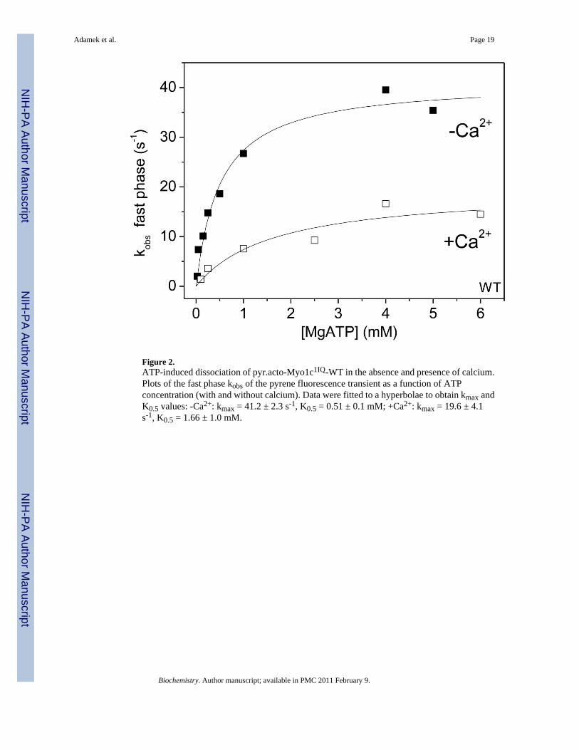
Figure 2.ATP-induced dissociation of pyr.acto-Myo1c1IQ-WT in the absence and presence of calcium.Plots of the fast phase kobs of the pyrene fluorescence transient as a function of ATPconcentration (with and without calcium). Data were fitted to a hyperbolae to obtain kmax andK0.5 values: -Ca2+: kmax = 41.2 ± 2.3 s-1, K0.5 = 0.51 ± 0.1 mM; +Ca2+: kmax = 19.6 ± 4.1s-1, K0.5 = 1.66 ± 1.0 mM.
Adamek et al. Page 19
Biochemistry. Author manuscript; available in PMC 2011 February 9.
NIH
-PA
Author M
anuscriptN
IH-P
A A
uthor Manuscript
NIH
-PA
Author M
anuscript

Figure 3.ATP-induced dissociation of pyr.acto-Myo1c1IQ. Pyrene fluorescence transients observedupon mixing 25 nM pyr.actin-Myo1c1IQ chimeras with 1 mM ATP in the absence and presenceof calcium. A, transients for Myo1c1IQ-8A fitted to two exponentials (best fits superimposed):-Ca2+: kobs1 = 20.8 s-1, A1 = 4.6 %, kobs2 = 1.4 s-1, A2 = 44.2 %; +Ca2+: kobs1 = 11.8 s-1, A1= 2.6 %, kobs2 = 1.3 s-1, A2 = 36.8 %. B, transients for pyr.actin-Myo1c1IQ-G. The −Ca2+
transient was fitted to one exponential, the +Ca2+ transient to two exponentials (best fitssuperimposed): -Ca2+: kobs = 7.9 s-1, A = 15.0 %; +Ca2+: kobs1 = 6.1 s-1, A1 = 31.1 %, kobs2= 1.6 s-1, A2 = 14.8 %. C – F, plots of the fast phase (C & E) and slow phase (D & F) kobs forthe chimeras as a function of [ATP] in the absence of calcium. Data were fitted to hyperbolaeto obtain kmax and K0.5 values: C & D, WT and alanine chimeras: C, fast phase kmax = 41±2s-1 (WT ミ), 41 ± 3 s-1 (R/A ヨ), 35 ± 1 s-1 (E/A メ) and 34 ± 1 s-1 (8A 甌); K0.5 = 507 ± 97たM (WT), 622 ± 182 たM (R/A), 854 ± 75 たM (E/A) and 644 ± 57 たM (8A). D, slow phase:kmax = 2.0 s-1 (WT ミ), 4.3 s-1 (R/A ヨ), 2.8 s-1 (E/A メ) and 2.0 s-1 (8A 甌); K0.5 = 370 ± 33たM (WT), 623 ± 47 たM (R/A), 626 ± 11 たM (E/A) and 506 ± 18 たM (8A). E & F, smoothmuscle loop and G chimeras: E, fast phase : kmax = 41 ± 2 s-1 (WT ミ), 51 ± 2 s-1 (tonic 衲),
Adamek et al. Page 20
Biochemistry. Author manuscript; available in PMC 2011 February 9.
NIH
-PA
Author M
anuscriptN
IH-P
A A
uthor Manuscript
NIH
-PA
Author M
anuscript
痺49 ± 7 s-1 (phasic 衾); K0.5 = 507 ± 97 たM (WT), 445 ± 49 たM (tonic), 1342 ± 479 たM(phasic). F, slow phase: kmax = 2.0 s-1 (WT ミ), 14.6 s-1 (tonic 衲), 1.5 s-1 (phasic 衾) and 17.8s-1 (G 蠹); K0.5 = 370 ± 33 たM (WT), 447 ± 13 たM (tonic), 468 ± 34 たM (phasic) and 888 ±230 たM (G).
Adamek et al. Page 21
Biochemistry. Author manuscript; available in PMC 2011 February 9.
NIH
-PA
Author M
anuscriptN
IH-P
A A
uthor Manuscript
NIH
-PA
Author M
anuscript

Figure 4.Effect of ADP on ATP-induced dissociation of pyr.acto-Myo1c1IQ. (A & B), pyrenefluorescence transients of 25 nM pyr-acto-Myo1c1IQ mixed with 250 たM ATP in the stopped-flow apparatus in the absence or presence of high ADP for Myo1c1IQ-R/A (-Ca2+) andMyo1c1IQ-phasic (+Ca2+). Transients in the absence of ADP were fitted to 2 exponentials,transients in the presence of ADP were fitted to a single exponential: A, R/A: -ADP: kobs1 =11.0 s-1, A1 = 3.0 %, kobs2 = 1.3 s-1 A2 = 51.3 %; +ADP: kobs = 0.3 s-1, A = 52.8 %. B, phasic:-ADP: kobs1 = 16.6 s-1, A1 = 0.8 %, kobs2 = 0.3 s-1, A2 = 6.0 %; +ADP: kobs = 0.9 s-1, A = 6.4%. C to F, Increasing concentrations of ADP were either incubated with pyr.acto-Myo1c1IQ
or ATP and the transients were fitted to two exponentials. The kobs of the slow phases of both
Adamek et al. Page 22
Biochemistry. Author manuscript; available in PMC 2011 February 9.
NIH
-PA
Author M
anuscriptN
IH-P
A A
uthor Manuscript
NIH
-PA
Author M
anuscript

experimental approaches was plotted together and fitted to a hyperbola to determine Kapp andk0 (in the absence (C & E) and presence of calcium (D &F). C & D, alanine chimeras: -Ca2+: Kapp = 16.7 ± 1.9 たM (WT ミ), 22.5 ± 2.9 たM (R/A ヨ), 17.0 ± 1.7 たM (E/A メ), 20.3 ±1.9 たM (8A 甌); k0 = 0.82 s-1 (WT), 1.16 s-1 (R/A), 0.76 s-1 (E/A), 0.62 s-1 (8A); +Ca2+:Kapp = 173.6 ± 17.0 たM (WT), 166.1 ± 12.6 たM (R/A), 172.6 ± 30.3 たM (E/A), 180.0 ± 17.4たM (8A); k0 = 0.51 s-1 (WT), 0.91 s-1 (R/A), 0.59 s-1 (E/A), 0.55 s-1 (8A). (E & F), smoothmuscle myosin II loop 1 and G chimeras: -Ca2+: Kapp = 16.7 ± 1.9 たM (WT ミ), 21.9 ± 4.2たM (tonic 衲), 22.0 ± 3.5 たM (phasic 衾), 47.6 ± 6.3 たM (G蠹); k0 = 0.82 s-1 (WT), 5.5 s-1
(tonic), 0.87 s-1 (phasic), 6.2 s-1 (G); +Ca2+: Kapp = 173.6 ± 17.0 たM (WT), 165.2 ± 24.3 たM(tonic), 390.6 ± 46.1 たM (phasic); k0 = 0.51 s-1 (WT), 4.92 s-1 (tonic), 0.27 s-1 (phasic), (nodata for G).
Adamek et al. Page 23
Biochemistry. Author manuscript; available in PMC 2011 February 9.
NIH
-PA
Author M
anuscriptN
IH-P
A A
uthor Manuscript
NIH
-PA
Author M
anuscript

Figure 5.ATP binding to Myo1c1IQ loop 1 chimera. A & B, Protein fluorescence transients observedupon mixing 50 nM Myo1c1IQwith 1 mM ATP in the absence and presence of calcium(concentrations after mixing). Data were fitted to a single exponential (best fit superimposed):A, Myo1c1IQ-R/A -Ca2+: kobs = 41.6 s-1, A = 6.3 %; +Ca2+: kobs = 5.7 s-1, A = 5.8 %. B,Myo1c1IQ –tonic: -Ca2+: kobs = 66.3 s-1, A = 4.5 %; +Ca2+: kobs = 13.5 s-1, A = 3.5 %. C &D, kobs of each chimera plotted as a function of ATP concentration. Data were fitted tohyperbolae (best fits superimposed): C, fitted values in the absence of calcium kmax = 44.3 ±1.9 s-1 (WT ミ), 72.8 ± 1.1 s-1 (tonic 衲), 61.1 ± 10.3 s-1 (phasic 衾), 9.1 ± 0.2 s-1 (G slow 蠹),82.5 ± 1.9 s-1 (G fast 蠧); K0.5 = 184 ± 22 たM (WT), 94.5 ± 4.8 たM (tonic), 75 ± 30 たM (phasic),100.0 ± 7.7 たM (G fast), 381 ± 102 たM (G slow). D, Fitted values in presence of calcium:kmax = 6.3 ± 0.1 s-1 (WT), 15.0 ± 0.9 s-1 (tonic), 13.0 ± 0.7 s-1 (phasic), 痺3 s-1 (G slow), 13.1± 1.4 s-1 (G fast); and K0.5 = 49.5 ± 3.4 たM (WT), 34.4 ± 9.9 たM (tonic), 52.0 ± 11.9 たM(phasic), 100.3 ± 38.3 たM (G fast), 670 ± 295 たM (G slow).
Adamek et al. Page 24
Biochemistry. Author manuscript; available in PMC 2011 February 9.
NIH
-PA
Author M
anuscriptN
IH-P
A A
uthor Manuscript
NIH
-PA
Author M
anuscript

Figure 6.ADP displacement from Myo1c1IQ loop 1 chimeras in the absence of calcium. A, Proteinfluorescence transient observed upon displacing 10 たM ADP from 50 nM Myo1c1IQ-8A byaddition of 0.5 mM ATP. The change in fluorescence was fitted to two exponentials: kobs1 =8.1 s-1, kobs2 = 2.3 s-1, A1 = 1.7 %, A2 = 5.1 %. B, Plot of the fast and slow amplitudes of theADP displacement experiment for Myo1c1IQ-8A as a function of [ADP]. The data were fittedto a hyperbola to obtain the KD. KD, fast = 5.2 ± 0.8 たM, KD, slow = 5.7 ± 1.1 たM. C, plot ofthe fast phase amplitude of the ADP-displacement experiment with Myo1c1IQ-tonic as afunction of [ADP]. A fit to a hyperbola defined KD = 9.2 ± 1.6 たM. D, plot of the fast phasekobs of the ADP displacement experiment with Myo1c1IQ-G as a function of [ADP]. A fit to ahyperbola defined KD = 7.9 ± 3.0 たM with k0 = 71 s-1.
Adamek et al. Page 25
Biochemistry. Author manuscript; available in PMC 2011 February 9.
NIH
-PA
Author M
anuscriptN
IH-P
A A
uthor Manuscript
NIH
-PA
Author M
anuscript
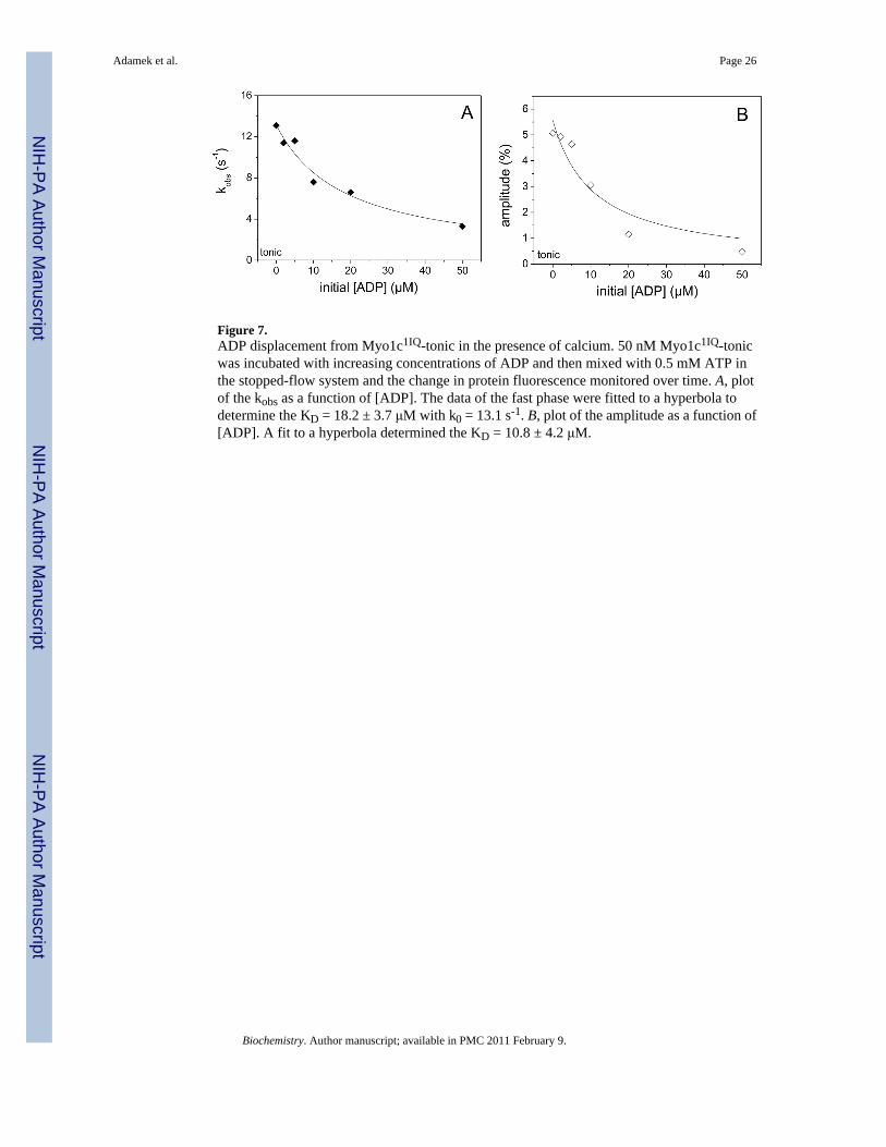
Figure 7.ADP displacement from Myo1c1IQ-tonic in the presence of calcium. 50 nM Myo1c1IQ-tonicwas incubated with increasing concentrations of ADP and then mixed with 0.5 mM ATP inthe stopped-flow system and the change in protein fluorescence monitored over time. A, plotof the kobs as a function of [ADP]. The data of the fast phase were fitted to a hyperbola todetermine the KD = 18.2 ± 3.7 たM with k0 = 13.1 s-1. B, plot of the amplitude as a function of[ADP]. A fit to a hyperbola determined the KD = 10.8 ± 4.2 たM.
Adamek et al. Page 26
Biochemistry. Author manuscript; available in PMC 2011 February 9.
NIH
-PA
Author M
anuscriptN
IH-P
A A
uthor Manuscript
NIH
-PA
Author M
anuscript
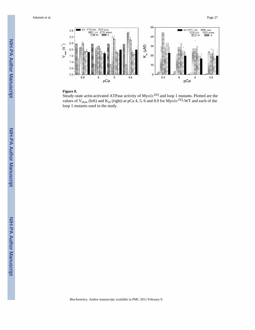
Figure 8.Steady-state actin-activated ATPase activity of Myo1c1IQ and loop 1 mutants. Plotted are thevalues of Vmax (left) and Km (right) at pCa 4, 5, 6 and 8.9 for Myo1c1IQ-WT and each of theloop 1 mutants used in the study.
Adamek et al. Page 27
Biochemistry. Author manuscript; available in PMC 2011 February 9.
NIH
-PA
Author M
anuscriptN
IH-P
A A
uthor Manuscript
NIH
-PA
Author M
anuscript

Scheme 1.
Adamek et al. Page 28
Biochemistry. Author manuscript; available in PMC 2011 February 9.
NIH
-PA
Author M
anuscriptN
IH-P
A A
uthor Manuscript
NIH
-PA
Author M
anuscript

Scheme 2.
Adamek et al. Page 29
Biochemistry. Author manuscript; available in PMC 2011 February 9.
NIH
-PA
Author M
anuscriptN
IH-P
A A
uthor Manuscript
NIH
-PA
Author M
anuscript

NIH-PA Author ManuscriptNIH-PA Author ManuscriptNIH-PA Author Manuscript
Adam
ek et al.P
age 30
Table 1Values for k+2, k+Į and KĮ for the acto-Myo1c1IQ loop 1 chimeras in the absence and presence of calcium
k+Į(s-1) k+2 (s-1) KĮ
-Ca2+ +Ca2+ -Ca2+ +Ca2+ -Ca2+ +Ca2+
WT 2.0 2.7 41 ± 2 20 ± 4 0.12 0.11
R/A 4.3 4.6 41 ± 3 110 ± 34 0.11 0.08
E/A 2.8 3.1 35 ± 1 55 ± 18 0.12 0.05
8A 2.0 2.8 34 ± 1 22 ± 5 0.10 0.08
tonic 14.6 14.5 51 ± 2 95 ± 8 0.21 0.12
phasic 1.5 1.0 49 ± 7 - 0.09 0.23
G 17.8 5.7 17.8 18.4 # nd 1.8
#not defined
Biochem
istry. Author m
anuscript; available in PMC
2011 February 9.

NIH-PA Author ManuscriptNIH-PA Author ManuscriptNIH-PA Author Manuscript
Adam
ek et al.P
age 31
Table 2ADP affinities (KD) and ADP release rate constants (k-D) measured for the loop 1 chimeras in the presence and absence of actin and calcium
KD (ȝM) k+D (s-1) KAD (ȝM) k+AD (s-1) (calculated)
-Ca2+ +Ca2+ -Ca2+ +Ca2+ -Ca2+ +Ca2+ -Ca2+ +Ca2+
WT 3.3 ± 1.5 Nd# 6 4 1.74 16.9 2.1 20.3
R/A 6.4 ± 1.7 Nd# 7 4* 2.22 12.6 2.4 13.9
E/A 4.4 ± 1.0 Nd# 5 4* 1.82 8.6 2.7 12.9
8A 5.5 ± 1.0 Nd# 5 4* 1.85 13.2 4.4 31.7
tonic 9.2 ± 1.6 痺11 7 >15 3.80 17.7 4.9 23.0
phasic Nd Nd 37 Nd 1.83 73.1 痺2 痺73
G 15.1 ± 8.5 - 25-30 Nd 4.32 - 痺4 -
*assumed to be similar to WT with no change in calcium
#assumed to be similar to −Ca2+ as for WT
Nd, not determined
Biochem
istry. Author m
anuscript; available in PMC
2011 February 9.

NIH-PA Author ManuscriptNIH-PA Author ManuscriptNIH-PA Author Manuscript
Adam
ek et al.P
age 32
Table 3ATP binding to Myo1c1IQ loop 1 chimeras in the absence and presence of calcium
kmax (s-1) = k+3+k-3 K0.5 (ȝM) (=1/K1)
-Ca2+ +Ca2+ Ratio -/+Ca2+ -Ca2+ +Ca2+ Ratio -/+Ca2
WT 44.3 ± 1.9 6.3 ± 0.1 7.0 184 ± 22 49.5 ± 3.4 3.7
R/A 45.5 ± 1.9 5.9 ± 0.3 7.7 153 ± 24 54.0 ± 12.2 2.8
E/A 49.6 ± 1.1 7.9 ± 0.2 6.3 255 ± 14 65.6 ± 7.5 3.9
8A 37.2 ± 0.5 5.9 ± 0.3 6.3 179 ± 8 62.2 ± 12.0 2.9
tonic 72.8 ± 1.1 15.0 ± 0.9 4.9 95 ± 5 34.4 ± 9.9 2.8
phasic 61.1 ± 10.3* 13.0 ± 0.7 4.7 75 ± 30 52.0 ± 11.9 1.4
G 82.5 ± 1.9 13.1 ± 1.4 6.3 100 ± 8 100.3 ± 38.3 1
Gslow 9.1 痺3 3 381 ± 102 670 ± 295 0.5
*larger error due to smaller amplitude
Biochem
istry. Author m
anuscript; available in PMC
2011 February 9.
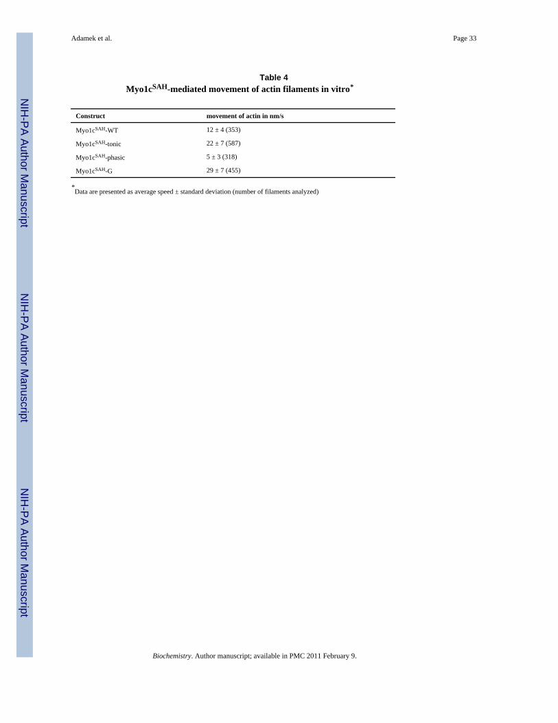
NIH
-PA
Author M
anuscriptN
IH-P
A A
uthor Manuscript
NIH
-PA
Author M
anuscript
Adamek et al. Page 33
Table 4Myo1cSAH-mediated movement of actin filaments in vitro*
Construct movement of actin in nm/s
Myo1cSAH-WT 12 ± 4 (353)
Myo1cSAH-tonic 22 ± 7 (587)
Myo1cSAH-phasic 5 ± 3 (318)
Myo1cSAH-G 29 ± 7 (455)
*Data are presented as average speed ± standard deviation (number of filaments analyzed)
Biochemistry. Author manuscript; available in PMC 2011 February 9.