Double femoral osteotomy fixed with a Puddu plate and … · SOCIEDADE BRASILEIRA DE ORTOPEDIA E...
6
r e v b r a s o r t o p . 2 0 1 8; 5 3(3) :378–383 SOCIEDADE BRASILEIRA DE ORTOPEDIA E TRAUMATOLOGIA www.rbo.org.br Case report Double femoral osteotomy fixed with a Puddu plate and a retrograde intramedullary nail to treat biapical deformity of the femur Patrícia Moreno Grangeiro a , Márcia Uchoa de Rezende a,∗ , Camilo Partezani Helito a , Alessandro Monterroso Felix a , Guilherme Pereira Ocampos a , Roberto Guarniero b a Instituto de Ortopedia e Traumatologia, Hospital das Clínicas, Faculdade de Medicina, Universidade de São Paulo (HC-FM-USP), São Paulo, SP, Brazil b Departamento de Ortopedia e Traumatologia, Faculdade de Medicina, Universidade de São Paulo (FM-USP), São Paulo, SP, Brazil a r t i c l e i n f o Article history: Received 8 February 2017 Accepted 2 May 2017 Available online 5 April 2018 Keywords: Osteotomy Acquired joint deformities Intramedullary nails External fixators Intramedullary fractures fixation a b s t r a c t Biapical femoral deformities are challenging to treat. In order to correct concomitant meta- physeal and diaphyseal deformities of the femur, the authors propose a double femoral controlled osteotomy with combined internal fixation, consisting of a Puddu plate and an intramedullary nail. The method was described in two patients. Results were analyzed using a visual analog scale (VAS), the Lysholm score, and SF-36. No complications were found. Complete consolidation of the osteotomies and radiographic alignment correction were achieved. Results were obtained with a minimum follow-up of 66 months. Both patients had improved for pain (VAS from 60 to 40 and from 50 to 20 at reassessment), function (Lysholm score from 78 to 93 and from 55 to 73) and quality of life (SF-36, both mental – from 40.7 to 57.1 in case one and from 24.7 to 59.7 in case two – and physical – from 27.7 to 45.6 and from 28.2 to 46.8). The authors have found that this technique is a reliable, accurate, and reproducible solution for biapical deformities of the femur. © 2018 Published by Elsevier Editora Ltda. on behalf of Sociedade Brasileira de Ortopedia e Traumatologia. This is an open access article under the CC BY-NC-ND license (http:// creativecommons.org/licenses/by-nc-nd/4.0/). Osteotomia femoral dupla fixada com placa Puddu e haste intramedular retrógrada para tratamento de deformidade femoral biapical Palavras-chave: Osteotomia Deformidades articulares adquiridas r e s u m o O tratamento das deformidades femorais biapicais é desafiador. Para a correc ¸ão das deformidades metafisárias e diafisárias concomitantes do fêmur, os autores propõem uma osteotomia dupla femoral controlada com uma fixac ¸ão interna combinada com uma placa de Puddu e uma haste intramedular. O método foi demonstrado em dois pacientes. Os Study conducted at Instituto de Ortopedia e Traumatologia, Hospital das Clinicas, Faculdade de Medicina, Universidade de São Paulo (HC-FM-USP), São Paulo, SP, Brazil. ∗ Corresponding author. E-mail: [email protected] (M.U. Rezende). https://doi.org/10.1016/j.rboe.2018.03.016 2255-4971/© 2018 Published by Elsevier Editora Ltda. on behalf of Sociedade Brasileira de Ortopedia e Traumatologia. This is an open access article under the CC BY-NC-ND license (http://creativecommons.org/licenses/by-nc-nd/4.0/).
Transcript of Double femoral osteotomy fixed with a Puddu plate and … · SOCIEDADE BRASILEIRA DE ORTOPEDIA E...

r e v b r a s o r t o p . 2 0 1 8;5 3(3):378–383
SOCIEDADE BRASILEIRA DEORTOPEDIA E TRAUMATOLOGIA
www.rbo.org .br
Case report
Double femoral osteotomy fixed with a Puddu plateand a retrograde intramedullary nail to treatbiapical deformity of the femur�
Patrícia Moreno Grangeiroa, Márcia Uchoa de Rezendea,∗, Camilo Partezani Helitoa,Alessandro Monterroso Felixa, Guilherme Pereira Ocamposa, Roberto Guarnierob
a Instituto de Ortopedia e Traumatologia, Hospital das Clínicas, Faculdade de Medicina, Universidade de São Paulo (HC-FM-USP), SãoPaulo, SP, Brazilb Departamento de Ortopedia e Traumatologia, Faculdade de Medicina, Universidade de São Paulo (FM-USP), São Paulo, SP, Brazil
a r t i c l e i n f o
Article history:
Received 8 February 2017
Accepted 2 May 2017
Available online 5 April 2018
Keywords:
Osteotomy
Acquired joint deformities
Intramedullary nails
External fixators
Intramedullary fractures fixation
a b s t r a c t
Biapical femoral deformities are challenging to treat. In order to correct concomitant meta-
physeal and diaphyseal deformities of the femur, the authors propose a double femoral
controlled osteotomy with combined internal fixation, consisting of a Puddu plate and an
intramedullary nail. The method was described in two patients. Results were analyzed using
a visual analog scale (VAS), the Lysholm score, and SF-36. No complications were found.
Complete consolidation of the osteotomies and radiographic alignment correction were
achieved. Results were obtained with a minimum follow-up of 66 months. Both patients
had improved for pain (VAS from 60 to 40 and from 50 to 20 at reassessment), function
(Lysholm score from 78 to 93 and from 55 to 73) and quality of life (SF-36, both mental – from
40.7 to 57.1 in case one and from 24.7 to 59.7 in case two – and physical – from 27.7 to 45.6
and from 28.2 to 46.8). The authors have found that this technique is a reliable, accurate,
and reproducible solution for biapical deformities of the femur.
© 2018 Published by Elsevier Editora Ltda. on behalf of Sociedade Brasileira de Ortopedia
e Traumatologia. This is an open access article under the CC BY-NC-ND license (http://
creativecommons.org/licenses/by-nc-nd/4.0/).
Osteotomia femoral dupla fixada com placa Puddu e haste intramedularretrógrada para tratamento de deformidade femoral biapical
r e s u m o
Palavras-chave:
Osteotomia
Deformidades articulares
adquiridas
O tratamento das deformidades femorais biapicais é desafiador. Para a correcão das
deformidades metafisárias e diafisárias concomitantes do fêmur, os autores propõem uma
osteotomia dupla femoral controlada com uma fixacão interna combinada com uma placa
de Puddu e uma haste intramedular. O método foi demonstrado em dois pacientes. Os
� Study conducted at Instituto de Ortopedia e Traumatologia, Hospital das Clinicas, Faculdade de Medicina, Universidade de São Paulo(HC-FM-USP), São Paulo, SP, Brazil.
∗ Corresponding author.E-mail: [email protected] (M.U. Rezende).
https://doi.org/10.1016/j.rboe.2018.03.0162255-4971/© 2018 Published by Elsevier Editora Ltda. on behalf of Sociedade Brasileira de Ortopedia e Traumatologia. This is an openaccess article under the CC BY-NC-ND license (http://creativecommons.org/licenses/by-nc-nd/4.0/).
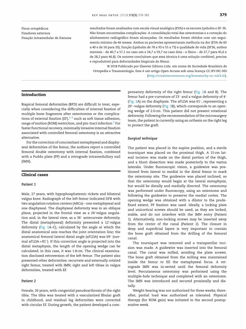
r e v b r a s o r t o p . 2 0 1 8;5 3(3):378–383 379
Pinos ortopédicos
Fixadores externos
Fixacão intramedular de fraturas
resultados foram analisados com escala visual analógica (EVA) e os escores Lysholm e SF-36.
Não foram encontradas complicacões. A consolidacão total das osteotomias e a correcão do
alinhamento radiográfico foram alcancadas. Os resultados foram obtidos com um segui-
mento mínimo de 66 meses. Ambos os pacientes apresentaram melhoria na dor (EVA de 60
a 40 e de 50 para 20), funcão (Lysholm de 78 a 93 e 55 a 73) e qualidade de vida (SF36, ambos
mentais – de 40,7 a 57,1 no caso um e 24,7 a 59,7 no caso dois – e físico – de 27,7 para 45,6 e
de 28,2 para 46,8). Os autores concluíram que essa técnica é uma solucão confiável, precisa
e reprodutível para deformidades biapicais do fêmur.
© 2018 Publicado por Elsevier Editora Ltda. em nome de Sociedade Brasileira de
Ortopedia e Traumatologia. Este e um artigo Open Access sob uma licenca CC BY-NC-ND
I
Bcmtrfaa
sfw(
C
P
MvtoptTddamdctprd
P
Ftiw
ntroduction
iapical femoral deformities (BFD) are difficult to treat, espe-ially when considering the difficulties of internal fixation ofultiple bone fragments after osteotomies or the complica-
ions of external fixation (EF),1–3 such as soft tissue adhesion,ange of motion (ROM) restriction, and pin-tract infection.3 Foraster functional recovery, minimally invasive internal fixationssociated with controlled femoral osteotomy is an attractivelternative.
For the correction of concomitant metaphyseal and diaphy-eal deformities of the femur, the authors report a controlledemoral double osteotomy with internal fixation, combinedith a Puddu plate (PP) and a retrograde intramedullary nail
IMN).
linical cases
atient 1
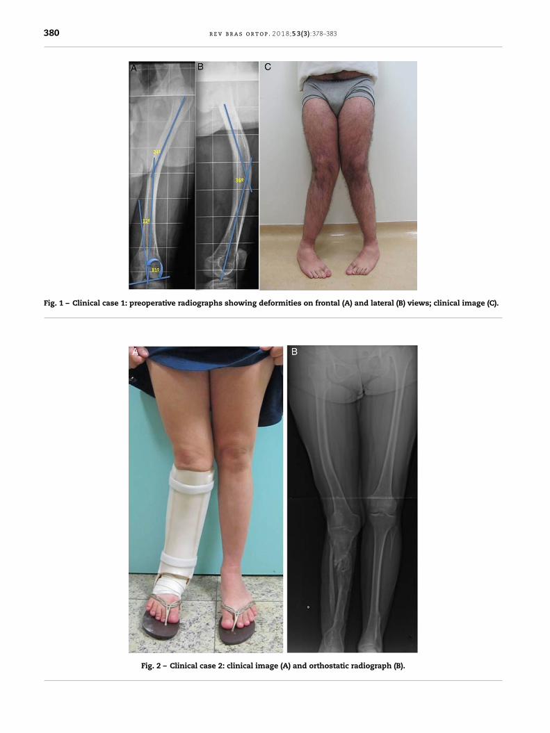
ale, 27 years, with hypophosphatemic rickets and bilateralalgus knee. Radiograph of the left femur indicated DFB withwo angulation rotation centers (ARCs) – one metaphyseal andne diaphyseal. The diaphyseal deformity was in an obliquelane, projected in the frontal view as a 24◦valgus angula-ion and, in the lateral view, as a 36◦ antecurvate deformity.he distal (metaphyseal) deformity presented a 12◦valguseformity (Fig. 1A–C), calculated by the angle at which theistal anatomical axis reaches the joint orientation line; thenatomical femoral lateral distal angle (aFLDA) was 69◦ (nor-al aFLDA = 81◦). If this correction angle is projected into the
istal metaphysis, the length of the opening wedge can bealculated; in this case, it was 1.8 cm. The clinical examina-ion disclosed retroversion of the left femur. The patient alsoresented other deformities: recurvate and externally rotatedight femur, treated with IMN; right and left tibias in valguseformities, treated with EF.
atient 2
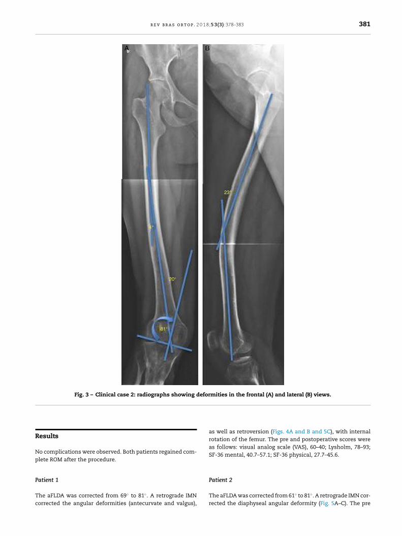
emale, 26 years, with congenital pseudoarthrosis of the rightibia. The tibia was treated with a vascularized fibular graftn childhood, and residual leg deformities were corrected
ith circular EF. During growth, the patient developed a com-
(http://creativecommons.org/licenses/by-nc-nd/4.0/).
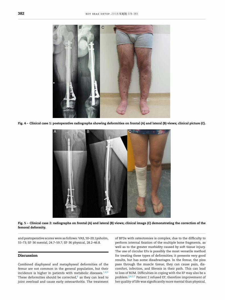
pensatory deformity of the right femur (Fig. 2A and B). Thefemur had a pre-curvature of 23◦ and a valgus deformity of 4◦
(Fig. 3A) on the diaphysis. The aFLDA was 61◦, representing a20◦-valgus deformity (Fig. 3B), which corresponds to an open-ing wedge of 2.0 cm. This patient did not present rotationaldeformity. Following the recommendation of the microsurgeryteam, the patient is currently using an orthesis on the right legto protect the graft.
Surgical technique
The patient was placed in the supine position, and a steriletourniquet was placed on the proximal thigh. A 15 cm lat-eral incision was made on the distal portion of the thigh,and a blunt dissection was made posteriorly to the vastuslateralis. Under fluoroscopic vision, a guidewire was pos-itioned from lateral to medial in the distal femur to markthe osteotomy site. The guidewire was placed inclined, sothat the osteotomy would begin at the lateral metaphysis,but would be distally and medially directed. The osteotomywas performed under fluoroscopy, using an osteotome andfollowing the guidewire to preserve the medial cortex. Theopening wedge was obtained with a dilator to the prede-fined extent; PP fixation was used. Ideally, a locking plateand unicortical screws should be used, as they are locked,stable, and do not interfere with the IMN entry (Patient1). Alternatively, non-locking screws may be inserted awayfrom the center of the canal (Patient 2). The closure indeep and superficial layers is very important to containthe bone graft obtained from the drilling of the femoralcanal.
The tourniquet was removed and a transpatellar inci-sion was made. A guidewire was inserted into the femoralcanal. The canal was milled, avoiding the plate screws.The bone graft obtained from the milling was maintainedinside the femur to fill the metaphyseal focus. A ret-rograde IMN was in-serted until the femoral deformitylevel. Percutaneous osteotomy was performed using themultiple-hole technique and completed with an osteotome.The IMN was introduced and secured proximally and dis-tally.
Weight-bearing was not authorized for three weeks; there-after, partial load was authorized as tolerated. Physicaltherapy (for ROM gain) was initiated in the second postop-erative week.
380 r e v b r a s o r t o p . 2 0 1 8;5 3(3):378–383
Fig. 1 – Clinical case 1: preoperative radiographs showing deformities on frontal (A) and lateral (B) views; clinical image (C).
Fig. 2 – Clinical case 2: clinical image (A) and orthostatic radiograph (B).
r e v b r a s o r t o p . 2 0 1 8;5 3(3):378–383 381
Fig. 3 – Clinical case 2: radiographs showing deformities in the frontal (A) and lateral (B) views.
R
Np
P
Tc
esults
o complications were observed. Both patients regained com-lete ROM after the procedure.
atient 1
he aFLDA was corrected from 69◦ to 81◦. A retrograde IMNorrected the angular deformities (antecurvate and valgus),
as well as retroversion (Figs. 4A and B and 5C), with internalrotation of the femur. The pre and postoperative scores wereas follows: visual analog scale (VAS), 60–40; Lysholm, 78–93;SF-36 mental, 40.7–57.1; SF-36 physical, 27.7–45.6.
Patient 2
The aFLDA was corrected from 61◦ to 81◦. A retrograde IMN cor-rected the diaphyseal angular deformity (Fig. 5A–C). The pre
382 r e v b r a s o r t o p . 2 0 1 8;5 3(3):378–383
Fig. 4 – Clinical case 1: postoperative radiographs showing deformities on frontal (A) and lateral (B) views; clinical picture (C).
(B) v
Fig. 5 – Clinical case 2: radiographs on frontal (A) and lateralfemoral deformity.and postoperative scores were as follows: VAS, 50–20; Lysholm,55–73; SF-36 mental, 24.7–59.7; SF-36 physical, 28.2–46.8.
Discussion
Combined diaphyseal and metaphyseal deformities of the
femur are not common in the general population, but theirincidence is higher in patients with metabolic diseases.1,4,5These deformities should be corrected,6 as they can lead tojoint overload and cause early osteoarthritis. The treatment
iews; clinical image (C) demonstrating the correction of the
of BFDs with osteotomies is complex, due to the difficulty toperform internal fixation of the multiple bone fragments, aswell as to the greater morbidity caused by soft tissue injury.The use of circular EFs is possibly the most versatile methodfor treating these types of deformities; it presents very goodresults, but has some disadvantages. In the femur, the pinspass through the muscle tissue; they can cause pain, dis-
comfort, infection, and fibrosis in their path. This can leadto loss of ROM. Difficulties in coping with the EF may also be aproblem.2,4,5,7 Patient 2 refused EF; therefore improvement ofher quality of life was significantly more mental than physical.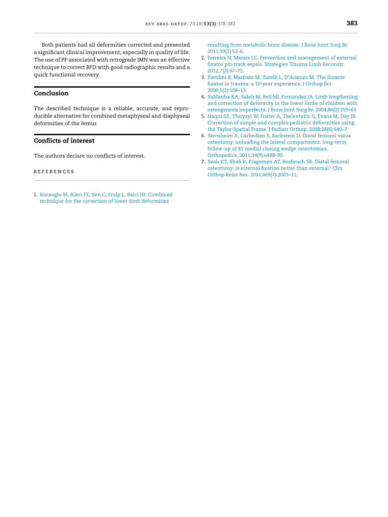
0 1 8
aTtq
C
Tdd
C
T
r
1
2
3
4
5
6
Orthopedics. 2011;34(9):e488–90.7. Seah KT, Shafi R, Fragomen AT, Rozbruch SR. Distal femoral
osteotomy: is internal fixation better than external? ClinOrthop Relat Res. 2011;469(7):2003–11.
r e v b r a s o r t o p . 2
Both patients had all deformities corrected and presented significant clinical improvement, especially in quality of life.he use of PP associated with retrograde IMN was an effective
echnique to correct BFD with good radiographic results and auick functional recovery.
onclusion
he described technique is a reliable, accurate, and repro-ucible alternative for combined metaphyseal and diaphysealeformities of the femur.
onflicts of interest
he authors declare no conflicts of interest.
e f e r e n c e s
. Kocaoglu M, Bilen FE, Sen C, Eralp L, Balci HI. Combinedtechnique for the correction of lower-limb deformities
;5 3(3):378–383 383
resulting from metabolic bone disease. J Bone Joint Surg Br.2011;93(1):52–6.
. Ferreira N, Marais LC. Prevention and management of externalfixator pin track sepsis. Strategies Trauma Limb Reconstr.2012;7(2):67–72.
. Pavolini B, Maritato M, Turelli L, D’Arienzo M. The Ilizarovfixator in trauma: a 10-year experience. J Orthop Sci.2000;5(2):108–13.
. Saldanha KA, Saleh M, Bell MJ, Fernandes JA. Limb lengtheningand correction of deformity in the lower limbs of children withosteogenesis imperfecta. J Bone Joint Surg Br. 2004;86(2):259–65.
. Naqui SZ, Thiryayi W, Foster A, Tselentakis G, Evans M, Day JB.Correction of simple and complex pediatric deformities usingthe Taylor-Spatial Frame. J Pediatr Orthop. 2008;28(6):640–7.
. Sternheim A, Garbedian S, Backstein D. Distal femoral varusosteotomy: unloading the lateral compartment: long-termfollow-up of 45 medial closing wedge osteotomies.