DOTTORATO DI RICERCA IN BIOTECNOLOGIE,...
108
Alma Mater Studiorum – Università di Bologna DOTTORATO DI RICERCA IN BIOTECNOLOGIE, FARMACOLOGIA E TOSSICOLOGIA Progetto Formativo 2: “FARMACOLOGIA E TOSSICOLOGIA” Ciclo XXV Settore Concorsuale di afferenza: 05/G1 Settore Scientifico disciplinare: BIO/14 BIOMOLECULAR STUDIES IN ALZHEIMER’S DISEASE MODELS: INVESTIGATIONS IN VITRO AND IN VIVO Presentata da: FRANCESCA LATTANZIO Coordinatore Dottorato Relatore Chiar.mo Prof. Giorgio Cantelli-Forti Prof.ssa Ester Speroni Correlatori Prof.ssa Patrizia Romualdi Dott.ssa Donatella Carretta Esame finale anno 2013
Transcript of DOTTORATO DI RICERCA IN BIOTECNOLOGIE,...

Alma Mater Studiorum – Università di Bologna
DOTTORATO DI RICERCA IN
BIOTECNOLOGIE, FARMACOLOGIA E TOSSICOLOGIA
Progetto Formativo 2: “FARMACOLOGIA E TOSSICOLOGIA”
Ciclo XXV
Settore Concorsuale di afferenza: 05/G1
Settore Scientifico disciplinare: BIO/14
BIOMOLECULAR STUDIES IN ALZHEIMER’S DISEASE
MODELS: INVESTIGATIONS IN VITRO AND IN VIVO
Presentata da: FRANCESCA LATTANZIO
Coordinatore Dottorato Relatore
Chiar.mo Prof. Giorgio Cantelli-Forti Prof.ssa Ester Speroni
Correlatori
Prof.ssa Patrizia Romualdi
Dott.ssa Donatella Carretta
Esame finale anno 2013
TABLE OF CONTENTS
ABSTRACT 1
1. INTRODUCTION 2
1.1 ALZHEIMER’S DISEASE 2
1.2 ALZHEIMER’S DISEASE NEUROPATHOLOGY 6
1.2.1 ALZHEIMER’S DISEASE PATHOGENESIS: AMYLOID VERSUS TAU
HYPOTHESIS 7
1.3 GENETIC AND OTHER RISK FACTORS 13
1.3.1 GENETIC RISK 13
1.3.2 APOLIPOPROTEIN E: BIOLOGICAL AND PATHOLOGICAL
ROLE 17
1.3.3 OTHER RISK FACTORS IN AD 22
1.3.4 DIET, INSULIN AND ALZHEIMER’S DISEASE 28
1.4 PEPTIDYL-PROLYL CIS/TRANS ISOMERASE 29
1.5 SIRTUIN 1 31
1.6 PRESENILIN 1 33

1.7 BRAIN-DERIVED NEUROTROPHIC FACTOR 34
1.8 OXIDATIVE STRESS AND ALZHEIMER’S DISEASE 36
1.9 ENDOGENOUS ANTIOXIDANT SYSTEMS 38
1.9.1 SUPEROXIDE DISMUTASE, GLUTATIONE PEROXIDASE, CATALASE
AND GLUTATIONE 38
1.9.2 THIOREDOXIN AND GLUTAREDOXIN 39
1.9.3 ROLE OF THIOREDOXIN AND GLUTAREDOXIN IN ALZHEIMER’S
DISEASE 41
2. AIM OF THE STUDY 42
2.1 SPECIFIC AIMS 44
3. MATERIALS AND METHODS 46
3.1 CELL CULTURES 46
3.2 CELL TREATMENTS 46
3.3 MTT CELL VIABILITY ASSAY 47
3.4 REVERSE TRANSCRIPTION AND REAL-TIME QUANTITATIVE
REVERSE TRANSCRIPTION-POLYMERASE CHAIN REACTION
(qRT-PCR) 47

3.5 PROTEIN EXTRACTION AND IMMUNOBLOTTING 50
3.6 ANIMALS 51
3.7 TISSUE SAMPLING 52
3.8 IMMUNOBLOTTING 52
3.9 DATA ANALYSIS 53
4. RESULTS 54
4.1 VIABILITY OF SH-SY5Y CELLS EXPOSED TO Aβ 54
4.2 GENE EXPRESSION IN SH-SY5Y EXPOSED TO Aβ 55
4.3 GENE EXPRESSION IN APOE3/E4 TRANSGENIC MICE 61
4.4 PROTEIN LEVELS IN BE(2)-M17 CELLS EXPOSED TO
APOE3 AND APOE4 68
4.5 PROTEIN LEVELS IN THE APOE3/E4 TRANSGENIC MICE 71
4.6 TRX1 GENE EXPRESSION IN THE APOE3/E4 TRANSGENIC MICE 73
5. DISCUSSION 74
5.1 METHODOLOGICAL CONSIDERATIONS 74
5.1.1 Aβ 25-35 PEPTIDES 74

5.1.2 APOE3 AND APOE4 TRANSGENIC MICE 75
5.2 PIN1 GENE EXPRESSION 77
5.3 SIRT1 GENE EXPRESSION 80
5.4 PSEN1 GENE EXPRESSION 81
5.5 BDNF GENE EXPRESSION 82
5.6 TRX1 AND GRX1 PROTEINS MODULATION 85
5.7 CONCLUDING REMARKS 87
6. REFERENCES 88

1
ABSTRACT
The Alzheimer’s disease (AD), the most prevalent form of age-related dementia, is a
multifactorial and heterogeneous neurodegenerative disease. The molecular mechanisms
underlying the pathogenesis of AD are yet largely unknown. However, the etiopathogenesis
of AD likely resides in the interaction between genetic and environmental risk factors.
Among the different factors that contribute to the pathogenesis of AD, amyloid-beta
peptides and the genetic risk factor apoE4 are prominent on the basis of genetic evidence
and experimental data. ApoE4 transgenic mice have deficits in spatial learning and memory
associated with inflammation and brain atrophy. Evidences suggest that apoE4 is
implicated in amyloid-beta accumulation, imbalance of cellular antioxidant system and in
apoptotic phenomena. The mechanisms by which apoE4 interacts with other AD risk
factors leading to an increased susceptibility to the dementia are still unknown. The aim of
this research was to provide new insights into molecular mechanisms of AD
neurodegeneration, investigating the effect of amyloid-beta peptides and apoE4 genotype
on the modulation of genes and proteins differently involved in cellular processes related to
aging and oxidative balance such as PIN1, SIRT1, PSEN1, BDNF, TRX1 and GRX1. In
particular, we used human neuroblastoma cells exposed to amyloid-beta or apoE3 and
apoE4 proteins at different time-points, and selected brain regions of human apoE3 and
apoE4 targeted replacement mice, as in vitro and in vivo models, respectively. All genes
and proteins studied in the present investigation are modulated by amyloid-beta and apoE4
in different ways, suggesting their involvement in the neurodegenerative mechanisms
underlying the AD. Finally, these proteins might represent novel potential diagnostic and
therapeutic targets in AD.
2
1. INTRODUCTION
1.1 ALZHEIMER’S DISEASE
The Alzheimer’s disease (AD) is a neurodegenerative disorder clinically characterized by a
progressive mental decline; it is the most prevalent form of age-related dementia in the
modern society (Duff and Suleman, 2004). Current estimates indicate that there are about
25-30 million people suffering from AD in the World, and the number of cases will double
during the next twenty years (Ferri et al., 2005). Therefore, with increasing life
expectancy, dementia is a growing socio-economic and medical problem, and AD
represents an important clinical challenge in terms of diagnosis and treatment. Up to now,
since first clinical symptoms of AD overlap with other diseases of the central nervous
system (CNS), a definitive diagnosis is uncertain and it can only be done with post-mortem
histopathological examination of the brain. However, a relative clinical diagnosis based on
physical, neurological and psychological evaluations, laboratory tests and neuroimaging,
can be made with a noteworthy accuracy.
A recent systematic review of epidemiological studies estimates that AD is the fifth cause
of death in elderly population, leading to physical disability more than cardiovascular
diseases, stroke and cancer (WHO report, 2003). Between 2000 and 2008, deaths attributed
to AD increased around 66%, and 5% to 15% of all deaths in people aged ≥65 years can be
ascribed to AD (Miniño et al., 2011).
The first clinical phase of the AD is characterized by an impairment of the episodic
memory that compromises the ability to recall past experiences: the patients develop
symptoms such as difficulty to learn new informations or to remember previously learned
ones (Ballard et al., 2011). This occurs since the functional neuronal degeneration usually
begins in brain regions involved in forming new memories, especially hippocampus and
entorhinal cortex, and in areas of cerebral cortex in the frontal lobe implicated in thinking
and planning. Apathy and depression are also often early symptoms of AD. Later

3
symptoms include impaired judgment, disorientation, confusion, behavior changes, and
difficulty speaking, swallowing, and walking. (Figure 1).
Figure 1. Pathological brain modification during stages of AD progression.
The progression of AD is slow, insidious and implacable, leading the patient to a mental
and physical disability. Several studies indicate that people survive an average of 4 to 8
years after a diagnosis of AD, but others can live as long as 20 years with this
neurodegenerative disease. In fact, severe dementia frequently causes complications such
as immobility, swallowing disorders and malnutrition, increasing the risk of developing
pneumonia which is the most common cause of death among people with AD (Brunnstrom
et al., 2009).
Although some palliative treatments alleviating the AD symptoms are available, no
effective strategies currently exist to inhibit the progression of the disease. The standard
medical treatments used in the clinical practice act modulating neurotransmitters such as
acetylcholine or glutamate; they include acetylcholinesterase inhibitors and N-methyl-D-
aspartate (NMDA) antagonists (Winslow et al, 2011). Antipsychotic drugs are used to treat
secondary symptoms of AD, like depression, agitation, irritability and sleep disorders
(Madhusoodanan et al., 2007).
4
Currently, the stages of AD are often described as early, moderate, or severe stage. In
2011, the National Institute on Aging (NIA) and the Alzheimer’s Association
recommended new diagnostic criteria for AD (Jack CR et al., 2011). These new criteria
propose that the disease begins as preclinical AD, before the early stage. In this preclinical
stage, individuals have measurable earliest signs of the disease in brain, cerebrospinal
fluid, and/or blood (biomarkers), but without symptoms such as memory loss. This
preclinical stage suggests that AD begins several years before symptoms develop. The
second stage is mild cognitive impairment (MCI), in which individuals have mild, but
measurable, changes in thinking performances without affect the ability to carry out
everyday activities. The last stage is the dementia due to AD, characterized by memory,
thinking, and behavioral symptoms, encompassing all AD-related processes.
In particular, MCI is defined as a transitional stage between normal aging and dementia.
MCI has a complex etiology and, even if may present several symptoms, clinically reflects
memory and cognitive impairment with preservation of functional abilities and no
evidence of dementia (Morris JC et al., 2001). When memory loss is predominant, MCI is
termed “amnestic MCI” and it is commonly considered as a prodromal state of AD. MCI is
recognized as potential risk factor of AD development, and, since MCI links and overlaps
normal aging with AD, the clinical diagnosis is a challenge. Moreover, in some cases MCI
can be reverted to normal cognition, it can remain stable or can be actually considered as
an early stage of AD or another dementia.
AD is divided into two subtypes based on the age of onset of the disease: early onset AD
(EOAD) and late-onset AD (LOAD). EOAD accounts for about 1-6% of all clinical cases
and ranges approximately with onset from 30-60 or 65 years; but the most common form
of AD is LOAD, with an age of onset over 60 or 65 years. Both types may occur in people
with a positive family history of AD. Approximately 13% of AD cases are an autosomal
dominant heritage with at least three generations affected (Bekris et al, 2010). With the
exception of few familial autosomal dominant forms of AD resulting from a single-gene
disorder, most AD cases belong to a heterogeneous sporadic disease that involves the

5
complex interaction of aging, multiple gene susceptibility and environmental risk factors
(Alonso Vilatela et al., 2012). Age represent the main risk factors for developing AD,
along with the poor education, low mental ability, traumatic brain injury, stroke and
cardiovascular disease risk factors (eg. physical inactivity, high cholesterol, diabetes,
smoking, and obesity); a history of depression may also predispose to AD. However, the
combination of genetic profile with several environmental factors seems to have the major
role in the increased risk of AD onset (Alzheimer’s Association, 2012).
The neuropathogenic process of AD probably starts decades before the clinical onset of the
disease becomes apparent; during this period a gradual but inexorable neuronal loss occurs,
with brain atrophy and synaptic detriment. The principal pathological hallmarks of AD are
abundant extracellular senile plaques of beta-amyloid peptide (SP) in cerebral blood
vessels and brain parenchyma, deriving from the cleavage of amyloid precursor protein
(APP), and intraneuronal neurofibrillary tangles (NFTs), resulting from aggregation of tau
microtubule-associated protein. (Figure 2). Although SP and NFT deposition in the brain
parenchyma is characteristic, the presence of these lesions is not sufficient to support the
diagnosis of AD since these features also occur in brains of cognitively intact people
(Ballard et al., 2011). Moreover, the elucidation of AD pathological mechanism and the
identification of additional specific biomarkers are needed to improve the accuracy for an
early diagnosis, to distinguish AD from MCI and other dementia forms, and to allow the
discovery of new pharmacological targets and effective therapies for this disabling disease.
Figure 2. Neurofibrillary tangles (a) and senile plaques (b) in the AD brain parenchyma.

6
1.2 ALZHEIMER’S DISEASE NEUROPATHOLOGY
Although the etiology of AD is not yet completely known, it is accepted that the disease,
like other chronic diseases, is the result of multiple factors. However, two pathological
hallmarks characterize the earlier stages of the disease, the deposit of the protein beta-
amyloid (Aβ) outside neurons to form the SP, and the abnormal accumulation of the
protein tau inside neurons to constitute the NTFs. The Aβ and NTFs deposition is supposed
to interfere with the neuron-to-neuron communication at synaptic level and to impair the
transport of nutrients and other essential molecules throughout the cells, contributing to
neuronal death. Further neuropathological features include a massive synaptic neuronal
loss, leading to cortical and hippocampal atrophy, the degeneration of cholinergic basal
forebrain neurons and enlarged ventricles. The SP and the NTFs have a different
distribution through the brain; the deposition of NTFs first starts in the medial temporal
lobe at level of hippocampus and entorhinal cortex, which is near the hippocampus and
directly connected to it, then spanning through other brains regions such as limbic areas
and at last to the cortical association areas and the primary cortex (Braak and Braak,
1991).
The develop of SP begins in the orbitofrontal and temporal cortices, and the spread of this
neuronal damage continues to parietal cortex and throughout the neocortex, usually with
the exclusion of the cerebellum. The first clinical symptoms of these lesions are short-term
memory problems, which reflect the early involvement of the hippocampus, the structure
essential to the formation of short-term and long-term memory and involved in processing
of sensory information. The memory deficit later develops into difficulties with executive
functions mainly controlled by the prefrontal cortex, the connected cortical and subcortical
brain structures, which include planning and initiation of actions, as well as emotional
disturbances and apathy. Moreover, during the AD progression, there is a selective loss of
cholinergic neurons with the reduction of acetylcholine levels in brain areas involved in Aβ
deposition, particularly in the cortex and hippocampus, resulting in the impairment of daily
living activities, behaviour and cognition. Although the mechanism underling the
degeneration of these neurons is still unknown, evidences suggest also that the excessive

7
stimulation of glutamate receptors, and in particular the NMDA receptor, associated to a
chronic neuroinflammation, may contribute to massive neuronal death (Wenk, 2003).
1.2.1 ALZHEIMER’S DISEASE PATHOGENESIS: AMYLOID VERSUS TAU
HYPOTHESIS
The presence of extracellular beta-amyloid plaques in the brain is a central event in the
etiology of AD. Beta-amyloid (Aβ) protein is a peptide of 39-43 amino acids able to form
β-sheets structures and fibrillar aggregates. It derives from the sequential proteolytic
cleavage of the large transmembrane polypeptide APP involving multiple enzymes (Perl,
2010). The primary function of APP is unknown, but it is believed to have a role during
neuronal development and trafficking, to be implicated in synaptic formation and repair,
transmembrane signal transduction and cell adhesion (Walsh et al., 2007). The human APP
gene is located on chromosome 21 with three main isoforms generated by alternative
splicing of exons 7, 8 and 15: APP770, APP751, and APP695, reflecting the number of
amino acids encoded and all including the full-length Aβ peptide (Goate et al. 1991).
APP751 and APP770 contain the Kunitz Protease Inhibitor (KPI) domain, a 57 amino acid
insert within their extracellular region and they are expressed in several tissues; APP695,
instead, lacks of the KPI domain and it is predominantly expressed in neurons (Rohan de
Silva et al., 1997). It has been shown that adult rat brains display higher relative amounts
of KPI-encoding APP isoforms than early post-natal rats, suggesting that specific age-
associated regulation pattern of APP gene is implicated in the AD development (Sandbrink
et al, 1994). Moreover, AD brain presents elevated levels of the protein and mRNA of
KPI-containing APP isoforms and, conversely, reduced levels of KPI lacking APP isoform
(Menendez-Gonzalez et al., 2005). A prolonged activation of NMDA receptors in neuronal
cells also seems to increase the expression of KPI containing APP isoforms, suggesting
that alterations in the APP mRNA splicing can be associated with an increased Aβ
deposition and contribute to AD pathogenesis (Bordji et al., 2010). However, the

8
mechanism of the direct association between different splicing forms of APP and AD
pathology is still to be elucidated.
APP is a type 1 integral cell surface membrane protein that resembles a signal transduction
receptor. It is synthesized in the endoplasmic reticulum, modified in the Golgi apparatus,
and transported to the cell surface through the secretory pathway. The APP is also
endocytosed from the cell surface and metabolized in the endosomal/lysosomal pathway.
APP can undergo two distinct cleavage pathways by the enzymes α-secretase and β-
secretase (also called β-site amyloid precursor protein-cleaving enzyme, BACE), both
active in normal cellular metabolism (Selkoe, 2001). The predominant cleavage of APP is
in the N-terminal portion close to the plasma membrane and within the Aβ peptide region,
and is mediated by α-secretase. This proteolytic process is also called non-amyloidogenic
since prevents Aβ formation and produces the neuroprotective soluble APP α fragment
(sAPPα) released in the extracellular space. The sAPPα, in fact, plays a role in adult
neurogenesis, has neurotrophic effects and is involved in early memory processes (Wang et
al., 2004; Bour et al., 2004). The α-Secretase activity is mediated by one or more enzymes
from the family of disintegrin and metalloproteinase domain proteins (ADAM).
The cleavage of APP leading to Aβ generation, also called amyloidogenic pathway, is
sequentially performed by β-secretase and γ-secretase, an enzymatic complex made up of
presenilin 1 (PSEN1), presenilin 2 (PSEN2) and nicastrin. Firstly, β-secretase cleaves APP
extracellularly producing the soluble β APP (sAPPβ) N-terminal fragment, that lacks most
of the neuroprotective effects associated with sAPPα, and a membrane bound C-terminal
fragment termed CTF99. The C-terminal fragment deriving from α-secretase cleavage is
called CTF83. γ-Secretase proteolysis of CTF83 and CTF99 will result in the generation of
p3 and Aβ, respectively, as well as the APP intracellular domain (AICD). The AICD
fragment has been implicated in the modulation of several cellular processes such as
intracellular trafficking, cytoskeletal dynamics, calcium and ATP levels, and also in the
regulation of Aβ levels through the regulation of neprilysin, one of the main Aβ degrading
enzymes (Wang et al., 2010). (Figure 3).

9
Figure 3. Non-amyloidogenic and amyloidogenic APP cleavage.
It is important to notice that β- and γ-secretases are not only implicated in the cleavage of
Aβ peptide but also in the proteolytic processing of a wide range of substrates, involving in
several cellular activities.
The Aβ peptide generated from the amyloidogenic pathway can then aggregate and
determine neurotoxic effects; alternatively, it can be degraded by different enzymes such
as neprilysin, insulin degrading enzyme or endothelin converting enzyme (Turner et al.,
2004). It still unclear why Aβ aggregates, but the sequence, the levels of the protein and
the conditions that can destabilise the Aβ structure are considered important factors in the
amyloid plaques deposition (Ballard et al., 2011).
Although Aβ has a potential role in the pathogenesis of AD, it has been shown that it also
plays a physiologic role in the CNS (Pearson and Peers, 2006). Moreover, low levels of
Aβ peptide have been shown in the cerebrospinal fluid (CSF) of individuals without signs
of dementia (Selkoe and Schenk, 2003). It has been demonstrated that low concentrations
of Aβ enhance synaptic plasticity and memory, with a positive modulatory role on the
neurotransmission (Puzzo et al., 2012). This positive role of Aβ in the normal
physiological function of cells complicates therapeutic strategies direct to reduce Aβ levels
in the AD.

10
The AD brain displays two forms of amyloid plaques: neuritic or senile plaques, and
diffuse plaques. The neuritic plaques are extracellular deposits of fibrillar Aβ containing
activated microglia cells within the central amyloid core and they are surrounded by
reactive astrocytes. The plaques can also be diffused, without a compacted core and
neuritic dystrophy; it has been suggested that these kinds of plaques are immature
precursors of the neuritic plaques. The activation of microglia by fibrillar Aβ is a very
early phenomenon in the AD pathogenesis, whereas the localization of astrocytes at the
neuritic plaques occurs later, when the dementia is already developing. Microglia is
involved in the clearance of Aβ by phagocytosis, but can also generate toxic products, such
as reactive oxygen species (ROS) and pro-inflammatory cytokines, that contribute to
neurodegeneration (Eikelenboom and van Gool, 2004). It is still unknown if amyloid-beta
plaques themselves cause AD or if they are a by-product of the AD processes.
One of the most accepted theories proposed twenty years ago to elucidate the pathogenesis
of AD is the “amyloid hypothesis”, stating that Aβ deposition plays a central role in the
etiology of the disease. According to this theory, the chronic imbalance between
production and clearance of Aβ leads to synaptic dysfunction, tau pathology, glial
activation and eventually neuronal loss in selected brain areas (Hardy and Allsop, 1991). In
fact, several studies demonstrated that the overproduction/aggregation of Aβ in the brain
can be one of primary causes of AD features. Two main Aβ toxic species are produced
from the amyloidogenic proteolysis of APP: Aβ40 and Aβ42, the Aβ42 being less
abundant but more hydrophobic and prone than Aβ40 to the fibrils formation (Näslund et
al., 2000). It has been demonstrated that the familial AD (FAD) - linked mutations of
PSEN1, PSEN2 and APP causes AD by increasing the extracellular concentrations of
Aβ42, thereby promoting its deposition on diffuse plaques in the earliest stage of the
disease (Iwatsubo et al., 1994; Scheuner et al., 1996). The amyloid hypothesis sustains that
missense mutations in APP, PSEN1 or PSEN2 genes lead to an increased Aβ42 production
and aggregation, forming diffuse plaques widespread in the brain parenchyma. Aβ
oligomers induce a toxic effect directly on synapses and participate to the activation of
microglia and astrocytes, increasing the release of inflammatory mediators and leading to a

11
progressive synaptic and neuritic damage, altered neuronal ionic homeostasis and oxidative
injury. These cellular imbalances produce alterations on kinase/phosphatase activities,
determining an abnormal tau phosphorylation and neurofibrillary tangles deposition, a
widespread neuronal/neuritic dysfunction with transmitter deficit and cell death. The
amyloid hypothesis is supported not only by genetic evidence in AD familial cases, but
also by other observations. In fact, it has been shown that mutations in the gene encoding
the tau protein cause frontotemporal dementia with Parkinsonism characterized by severe
tau deposition without amyloid plaques (Spillantini et al., 1998). Thus, NTFs observed in
AD brains are likely deposited after the initial Aβ plaques formation speculating that
altered APP processing and amyloid deposition predate tau changes and neuronal injury.
However, the “amyloid hypothesis” is not uniformly accepted since it is quite simplistic
and is not able to elucidate the whole complex mechanism behind the AD pathogenesis.
The biggest concern is that this theory does not explain the increased Aβ production in
sporadic AD cases, the form of the disease with the highest incidence, where no mutations
in APP or PSEN 1/2 genes are present. Furthermore, on post-mortem analysis, amyloid
plaques may be undetectable in brains of patients whit severe AD and may be present in
brains of elderly patients without signs of dementia (Davinelli et al., 2011). It has been
shown that the pathological progression of AD and the degree of the cognitive impairment
correlate with the number of neurofibrillary tangles much better than the beta-amyloid
plaques deposition. However, it has also been suggested that the amyloid pathology
correlates with AD progression at the earlier stages of the disease and that subsequent
changes in Aβ levels do not affect cognition, especially late in the disease (Teich and
Arancio, 2012). Moreover, the amyloid cascade hypothesis suggests that changes in tau
stability and neurofibrillary tangles formation are induced by toxic concentrations of Aβ;
although several mechanisms have been proposed, the linkage between Aβ and tau
accumulation is not yet well understood. Despite many efforts to elucidate the deficiencies
of the Aβ hypothesis, an alternative theory explaining the cause and the early pathogenesis
of AD has not been proposed. (Figure 4).

12
Figure 4. Pathological effects of Aβ on neurons.
Tau is a soluble protein that normally binds to and stabilizes axonal and dendritic
microtubules, the essential components of the cytoskeleton, conferring dynamism to the
main support structure for transport and neurotransmission. In the CNS, tau protein is
present as six isoforms deriving from a single gene by alternative splicing; all isoforms are
present in an abnormally hyperphosphorylated state in the NFTs (Goedert et al., 1989).
The microtubule-binding domain of tau protein is the main region involved in the tau
aggregation. When the tau structure is altered by modifications such as an abnormal
hyperphosphorylation in the proline-rich region, the protein loses the affinity to bind
microtubules and begins to self-assemble. The deriving oligomers aggregates into tangles
of hyperphosphorylated tau forming paired helical filaments (PHFs) and straight filaments
(Mandelkow et al., 2007). These tau polymers are present in several types of dementia-
related disorders as well as AD, also known as tauopathies. During NTFs formation, the
destabilization of the microtubular system is involved in the structural and functional
damage of neurons, contributing to the synaptic loss and cell death. The cytoskeleton
alteration seems to be also connected with the disruption of the Golgi apparatus, inducing
abnormal protein processing and increased production of Aβ.
Although the mechanism by which the phosphorylation of tau induces its aggregation is
still unclear, the final effect is the reduction of tau affinity to bind microtubules (Meraz-

13
Ríos et al., 2010). Several kinases are involved in tau phosphorylation, such as glycogen
synthase kinase 3β (GSK3β), cyclin-dependent kinase 5 (Cdk5), MT-affinity regulatory
kinase, cAMP-dependent protein kinase, Tau–tubulin kinase 1, protein kinase A (PKA),
calmodulin-dependent protein kinase 2 and extracellular signal-related kinase (ERK) 1/2.
In physiological conditions, the phosphorylation is regulated by phosphatases like protein
phosphatase 2A (PP2-A) and PP-1 (Wang et al. 2007 a). When the balance between
phosphorylation and dephosphorylation fails, hyperphosphorylation process triggers to tau
aggregation. In fact, it has been reported that the activity of these phosphatases is
decreased in AD brains; the inhibition of the abnormal hyperphosphorylation has been
investigated as potential therapeutic approach to the disease (Iqbal and Grundke-Iqbal,
2008).
1.3 GENETIC AND OTHER RISK FACTORS IN AD
1.3.1 GENETIC RISK
AD can be divided in two subgroups depending on the frequency and on the age of the
disease onset. The familial form of AD (FAD) is usually characterized by an early onset in
the midlife (age<65 years) and it is associated with heritable mutations involving the APP,
PSEN1 and/or PSEN2 genes; they represent less than 5% of AD cases. The sporadic form
of AD (SAD) with late onset (age>65 years) and without heritable gene mutations is the
most common type of dementia, responsible for over 95% of all AD cases, and influenced
by complex interactions between genetic and environmental risk factors.
The mutations within APP gene appear to alter the proteolytic processing of the APP and
generate Aβ deposition. Moreover, APP has a gene-dosing effect on Aβ production and
increased levels of APP seem to enhance the severity of AD pathological features. In fact,
Down’s syndrome patients that have three copy of APP gene on the chromosome 21
usually develop AD over the age of 35 (Tyrrell et al., 2001). All the mutations of APP,

14
PSEN1 and PSEN2 involved in the early-onset familial AD lead to a relative excess in the
production of Aβ42 and amyloid plaques deposition, although not all cases of these AD
form present these genetic mutations.
Up to now the main risk factor for sporadic AD is advancing age, but other risk factors and
potential risk genes involved in the pathology of the disease have been identified. The gene
encoding the cholesterol-carrying apolipoprotein E (apoE) on chromosome 19 is the
strongest and most consistently associated risk gene for the sporadic form of AD,
principally the late-onset one, but also some early-onset cases. The gene is inherited as
three common alleles, ε2, ε3 and ε4, originating six different phenotypes. The ε2 allele is
the least prevalent among the population (frequency of 7-8%) and it is associated with the
lowest risk of developing AD. Epidemiological studies demonstrated that apoE2 displays a
protective effect by delaying the onset of the disease; the apoE2 is also associated with a
reduction of hippocampal atrophy, higher Aβ and lower phosphotau levels in the
cerebrospinal fluid (Caselli and Dueck, 2010; Chiang et al., 2010). The ε3 allele is the
most common (frequency in population of 60-70%) and confers intermediate risk of
developing AD, but less than the ε4. The ε4 presents a gene-dosing effect on the disease
pathology, correlated with increased risk and earlier onset (Finch and Morgan, 2007). In
fact, it has been shown that individuals with two copies of ε4 alleles, compared with those
carrying ε3, have a significantly increased risk (more than seven times) of AD, associated
with an enhanced amyloid deposition, decreased Aβ clearance and cholinergic dysfunction
(Corder et al., 1993). ApoE ε4 carriers have enhanced AD symptoms, accelerated age-
dependent cognitive decline and worse memory performances. Moreover, apoE4 genotype
is also associated with several structural and functional brain changes related to AD
pathogenesis before that the clinical features become evident. Genome-wide association
studies confirmed that the ε4 allele of APOE is the strongest genetic risk factor for AD
(Harold et al., 2009). APOE ε4 probably increases the risk of both early-onset and late-
onset AD by modulating and accelerating Aβ deposition in the brain, and by directly
regulating brain lipid metabolism and synaptic functions through APOE receptors.
Although APOE ε2 is associated with a reduced risk of dementia, both the ε2 and ε4 alleles
of APOE increase amyloid burden compared with APOE ε3 in oldest individuals,

15
suggesting that the protective effects of APOE ε2 might not be associated with Aβ
deposition. However, unlike known genetic mutations, inheriting APOE ε4 is not sufficient
to develop the disease and many patients with AD are not carrying this allele. ApoE
accounts for only 10-20% of late-onset AD risk, suggesting that additional genes are
involved in the disease onset.
Several candidate risk genes have been identified, in particular those implicated in the
cholesterol metabolism, synaptic function and immune response, although the impact of
these genes in the late-onset AD remains to be confirmed (Karch et al., 2012). According
to the Alzgene website meta-analysis, excepting for apoE, the majority of identified
candidates genes have a relative risk for AD onset around 1,5%. In addition to apoE, other
genes involved in the transport or in the metabolism of cholesterol have been suggested as
putative risk factors for AD. Polymorphisms in receptors for the uptake of cholesterol,
such as low-density lipoprotein receptor-related protein (LRP) and the very-low-density
lipoprotein (VLDL) receptor, as well as polymorphisms in enzymes that regulate the
cholesterol catabolism, have been associated with an increased risk for AD (Zerbinatti et
al., 2005).
Among genetic causes of late-onset AD, the lipoprotein receptor sortilin-related receptor
(SORL1) gene has also been identified as an important factor involved in the pathogenesis
of the disease. It has been reported that SORL1 interacts with apoE, is a substrate of γ-
secretase enzyme, affects APP trafficking and it seems able to reduce the interaction
between APP and β-secretase resulting in reduced Aβ production. Some studies reported
that AD brains show a down-regulation of SORL1 gene expression and mutations in this
gene are associated with the late-onset AD pathology (Offe et al., 2006; Rogaeva et al.,
2007).
The GSK3β gene encoding for one of the main tau kinase, together with Cdk5, is
considered as a potential risk gene in AD. It has been proposed that the Aβ peptide, APP
cleavage products and PSEN complexes can activate neuronal GSK3β leading to glia

16
activation, tau increased phosphorylation and tangles deposition. GSK3 polymorphism has
been linked to the sporadic form of AD and it has been reported that apoE4 and Aβ have a
higher effect in the activation of GSK3β (Cedazo-Minguez et al., 2003) suggesting that
potential biochemical interactions between APOE and GSK3B are worth further
investigation. Thus, GSK3β deregulation is suggested to be one of the links between
amyloid deposition and tau protein hyperphosphorylation; several GSK3 inhibitors are
under investigation as a treatment strategy for AD. However, it has been found that some
PSEN1 mutations may activate GSK3β and promote tau phosphorylation by an alternative
pathway from Aβ peptide (Baki et al., 2004).
Tau mutations can also affect splicing of tau protein isoforms and microtubule binding
efficacy. The tau polymorphism is associated with AD, although its relevance in the
pathology of the disease is not completely clear. Polymorphisms of other phosphokinases
as well as GSK3β might be associated with an increased risk of AD and have a role in
explaining the link between Aβ and tau pathology. The DYRK1A is a gene located on the
chromosome 21 encoding for a kinase that plays a significant role in the cell proliferation
and neuronal development. DYRK1A is involved in tau and APP phosphorylation, leading
to an increased amyloidogenic processing, and it might considered as a risk gene on AD
onset (Kimura et al., 2007).
TOMM40 is another gene associated with an increased risk of developing late-onset AD; it
is a channel-forming subunit of the translocase of outer mitochondrial membrane (TOM
complex), which forms the protein-conducting channel facilitating the translocation of
unfolded proteins from the cytosol into the mitochondrial intermembrane space. TOMM40
gene is located on the chromosome 19, next to the APOE gene, and polymorphisms on this
gene can affect the onset age of AD (Roses, 2010).
A recent identified risk gene for AD is CLU, encoding for the chaperone clusterin (also
known as ApoJ) thought to bind and remove Aβ from the brain. Another risk gene
functionally related is PICALM. This gene encodes for the phosphatidylinositol-binding

17
clathrin assembly protein, an endosomal protein involved in synaptic neurotransmitter
release that binds Aβ and may promote its clearance. However, these genes have minimal
effects on the development of AD lesions and are not predicting for AD; they could have
an important role in the identification of pathways involved in the disease (Kok et al.,
2011).
1.3.2 APOLIPOPROTEIN E: BIOLOGICAL AND PATHOLOGICAL ROLE
ApoE is a polymorphic 299-amino acids (~34 kDa) protein responsible for the transport of
cholesterol and other lipids. The three corresponding human apoE isoforms differ only in
the amino acids at positions 112 and 158. ApoE3 has cysteine-112 and arginine-158,
whereas apoE4 only has arginine at both sites and apoE2 only has cysteine. This small
amino acid substitution is able to affect the three-dimensional structure and the lipid-
binding property of the protein, conferring a specific isoform-function in several biological
processes. ApoE contains two independently folded structural domains: the N-terminal,
that includes the receptor binding region, and the C-terminal, that contains the major lipid
binding region (Hatters et al., 2006). ApoE is important for cholesterol and triglycerides
metabolism, transport and homeostasis, in an isoform-dependent manner; it has prominent
functions also in the cell signal transduction pathways, including regulation of
neurotransmission and cell death. ApoE is an integral constituent of many lipid transport
lipoproteins complexes, playing a role in assembly, structure and uptake of lipoproteins by
binding to the cell surface LDLRs (low density lipoprotein receptors) family with a
specific isoform-affinity. These receptors are involved in signal transduction pathways,
although their main function is to provide cells with cholesterol and remove lipoproteins
from the blood.
ApoE has a different preference to tie to specific lipoproteins, depending on the isoform. In
fact, apoE4 preferentially binds to VLDL, whereas apoE3 and apoE2 bind preferentially
high density lipoproteins (HDL) (Strittmatter and Bova, 2002). In peripheral tissues, apoE
is mainly produced by the liver and macrophages. In the CNS it is synthesized

18
predominantly by astrocytes and to some extent by microglia, although also neurons are
able to generate it under physiological and pathological conditions. The main function of
apoE in the brain is to transport cholesterol from astrocytes to neurons via LDLR
receptors, playing a critical role in the distribution and homeostasis of lipids among
neuronal cells. Dysfunctions of LDLR as well as apoE4 are associated with
hyperlipidaemia and hypercholesterolaemia, leading to atherosclerosis, coronary heart
disease and stroke.
Several evidences show that apoE is involved in the maintenance of neuronal structure and
activity, repairing injured neurons through the regulation of lipids homeostasis necessary
for the synaptogenesis, cells proliferation and scavenging toxins (Cedazo-Minguez, 2007).
ApoE4 seems less effective and more detrimental than apoE3 and apoE2 in the normal
maintenance and repair of neuronal cells.
ApoE differentially regulates Aβ production, aggregation and clearance in an isoform-
dependent manner. However, apoE4 can contribute to risk of AD pathogenesis and
cognitive decline also by Aβ-independent mechanisms involving synaptic plasticity,
neurovascular functions and neuroinflammation (Liu et al., 2013). (Figure 5).
Figure 5. ApoE and Aβ metabolism in the brain.

19
Independently of Aβ, apoE4 might be less efficient than apoE3 and apoE2 in delivering
cholesterol and essential lipids for maintenance of synaptic integrity and plasticity.
Immunohistological evidences show that apoE co-localize in senile plaques in the AD
brains and Aβ deposition is more abundant in E4 carriers (Namba et al., 1991). ApoE4 and
Aβ aggregates act synergistically in the induction of neurodegeneration in vivo. Although
apoE4 has an active role in Aβ and NTF formation, it is difficult to find an hypothesis to
explain the mechanism by which apoE4 increases the pathological processes involved in
AD. In vitro studies suggest that apoE isoforms may differently influence tau pathology
and NTF deposition. In particular, it has been shown that apoE3, but not apoE4, forms a
stable complex with non-phosphorylated tau. This interaction between apoE3 and tau is
inhibited by the tau-phosphorylation, suggesting that apoE3 might be able to prevent
abnormal tau hyperphosphorylation and destabilization of the neuronal cytoskeleton
(Strittmatter et al., 1994).
In vivo studies in apoE transgenic mice showed an increased phosphorylation of tau in
mice expressing human apoE4 in neurons, but not in mice expressing apoE4 in astrocytes,
indicating that apoE4 induces tau phosphorylation specifically in neurons (Brecht et al,
2004). An alternative mechanism by which apoE isoforms would differentially contribute
to tau hyperphosphorylation is the modulation of tau kinases and phosphatases.
Intraneuronal accumulation of hyperphosphorylated tau has been also found in apoE
knock-out mice fed with a high cholesterol diet, suggesting a synergic interaction between
cholesterol and lack of apoE function (Rahman et al., 2005). Moreover, apoE4 is less
efficient than other isoforms in promoting cholesterol efflux from neurons and astrocytes;
this is probably related to the structural differences between apoE isoforms (Michikawa et
al., 2000). In AD brains a decreased cholesterol level has been described and several
evidences indicate that cholesterol is directly involved in AD pathogenesis (Reid et al,
2007).
Abnormal lipid metabolism is strongly related to the pathogenesis of AD. Clinical and
epidemiological studies showed that patients with elevated plasma cholesterol levels have
increased susceptibility to AD; the use of statins to inhibit the synthesis of cholesterol

20
seems to decrease the frequency and the progression of the disease. However, the
therapeutic effect of statins on AD pathology is influenced by several factors, such as the
efficiency of blood flow to the brain and the presence of concomitant disease conditions,
including hypertension, diabetes and hypercholesterolaemia. Cholesterol is an essential
component of membranes and is crucial for synaptic integrity and neuronal functions
implicated in learning, memory formation and neuronal repair (Mauch et al., 2001).
Cholesterol levels in hippocampal and cortical areas in patients with AD are lower than
healthy brains. It has been demonstrated that apoE4 is less efficient than apoE3 in
transporting brain cholesterol (Svennerholm and Gottfries, 1994). Furthermore, a number
of studies suggest that cholesterol regulates the Aβ production and increases the activity of
β-secretase. Changes in cholesterol levels or distribution within the membrane have been
shown to alter the localization of APP and its availability to be cleaved by the secretases;
however, the effect of cholesterol on the amyloidogenic processing of APP remains
controversial (Abad-Rodriguez et al., 2004). On the other hand, Aβ modulates the synthesis
and the distribution of cholesterol in neurons. Although the effect of cholesterol on the Aβ
production is complex and not completely clarified, the cholesterol/Aβ interactions are
probably modulated by the apoE genotype.
In the nervous system, the apoE-mediated distribution of lipids plays a fundamental role in
processes such as growth, regeneration and synaptic plasticity. ApoE4 is associated with
impaired synaptic plasticity in the hippocampus and age-dependent disruption of synaptic
organization in APOE knockout transgenic mice (Buttini et al., 1999). In AD and healthy
aged controls, APOE ε4 gene dosage inversely correlates with dendritic spine density in the
hippocampus, suggesting that the effect of ε4 genotype on risk of AD might be mediated,
at least in part, by direct effects on synaptic function (Ji, 2003). ApoE colocalizes with
amyloid plaques and microglia, suggesting that apoE has a role in the innate immune
response in AD. In fact, ApoE4 seems to have pro-inflammatory and/or reduced anti-
inflammatory functions, which could exacerbate AD pathology and cause neurovascular
dysfunction.
ApoE isoforms have differential roles in maintaining vascular health, and a recent meta-
analysis showed increased risk of vascular dementia in individuals with APOE ε4

21
compared with APOE ε3 (Yin et al., 2012). The APOE ε4 genotype combines
synergistically with atherosclerosis, peripheral vascular disease, and type 2 diabetes in
contributing to an increased risk of AD.
Cholinergic signal transduction is well known to be impaired in AD. ApoE4 carriers with
AD show greater deficits than non-carriers in cholinergic activity in the hippocampus and
the cortex, as well as a reduction in the number of cholinergic neurons markers, such as
choline acetyltransferase activity and nicotinic receptor binding. A direct negative
influence of apoE4 on cholinergic signaling may reduce the effectiveness of cholinergic
replacement treatments reported for apoE4-AD patients (Soininen et al., 1995).
ApoE receptors mediate cellular signaling by binding to several extracellular and
intracellular ligands, some of which are relevant to AD pathology. Several studies suggest
that apoE4 is associated with the disruption of multiple signal transduction pathways, loss
of cell protection, and alteration of mitochondrial metabolism.
In neurons, apoE isoforms differentially affect the activity of proteins such as the
extracellular-signal-regulated-kinase (ERK) and the c-Jun N-terminal Kinases (JNK),
principle members of the mitogen-activated protein kinase (MAPK) family, involved in the
regulation of processes including cell proliferation, differentiation and survival (Hoe et al.,
2005). ApoE also has specific isoform-related effects on calcium channels. ApoE4, but not
apoE3, significantly increases calcium levels and NMDA stimulation in cultured
hippocampal neurons, leading to neurotoxicity (Qiu et al., 2003).
Gene expression studies in hippocampus of AD patients demonstrated that apoE4 carriers
have higher expression of negative regulators of cell growth that may lead to increased cell
senescence and apoptosis, and in contrast decreased expression of genes associated with
synaptic plasticity and axonal/neuronal outgrowth. ApoE4 is also associated with the
reduction of the neurotransmitter receptors and Ca2+ homeostasis, disruption of multiple
signal transduction pathways, loss of cell protection, and mitochondrial dysfunction (Xu et
al., 2007). However, apoE derived from various cellular sources might exhibit different
physiological and pathological activity. In order to elucidate the role of apoE in

22
neurodegenerative processes is crucial understanding the mechanisms that govern the apoE
toxicity as well as protection on neurons.
1.3.3 OTHER RISK FACTORS IN AD
DEPRESSION
The role of depression in AD is debated; several results from population-based studies
have been inconsistent. Depressive symptoms occur in 40–50% of patients with AD and
depression may be associated with an increased risk for AD and other dementias. A recent
epidemiological study examining the association between depressive symptoms and
incidence of dementia over a 17-years follow-up period showed an increase of AD and
dementia in participants who were depressed at baseline (Saczynski et al., 2010). One
episode of depression conferred an 87–92% increase in dementia risk, while having more
episodes nearly doubled the risk. It has then been suggested that preventing the recurrence
of depression in older adults may prevent or delay the onset of dementia (Dotson et al.,
2010).
TRAUMATIC BRAIN INJURIES
Head injury and moderate to severe head trauma have been associated with an increased
risk of develop AD as well as other forms of dementia later in life. Moderate head injuries
are associated with twice the risk of developing AD and severe head injuries are associated
with 4.5 times the risk (Lye and Shores, 2000). It has been proposed that traumatic brain
injury leads to accumulation of APP with its proteolytic enzymes at sites of axonal injury,
increased Aβ production and deposition into extracellular plaques (Chen et al., 2004).
Some studies also suggest that ApoE4 carriers who experienced moderate or severe head
injury have a higher risk to develop AD (Katzman et al., 1996).

23
EPIGENETIC MODIFICATIONS
Epigenetic modifications, defined as changes in gene expression that do not alter the
nucleotide sequence of DNA, are the results from gene-environment interactions and are
involved in the regulation of chromatin structure (Goldberg et al., 2007). A key feature
that distinguishes epigenetic modifications from genetic changes is their reversible nature.
Epigenetic alterations such as DNA methylation and histone modifications have been
widely implicated in several age-related diseases, especially in cancer progression. Since
the majority of late-onset AD cases is sporadic, occurs in patients without a family history
of the disease and is characterized by differential susceptibility, epigenetic and
environmental factors may play a role in the etiology of the disease. Recently, epigenetic
phenomena have been recognized as a major contributor to the aging phenotype (Fraga et
al., 2005) and epigenetic modifications seem to be involved in the disruption of synaptic
signaling and neuronal survival. The heterogeneity noticed in clinical phenotypes of AD
patients with PSEN1 mutations suggests that other factors, both genetic and epigenetic,
must contribute to disease phenotype (Larner and Doran, 2006). AD brain cells also
present epigenetic changes on gene expression associated with an increases susceptibility
to oxidative stress. Studies on post-mortem human brain and peripheral leukocytes, as well
as transgenic animal models, showed that aging and AD present epigenetic alterations,
including abnormal DNA methylation and histone modifications (Chouliaras et al., 2010;
Arosio et al., 2012). The hypothesis that epigenetic mechanisms can modulate AD risk is
confirmed also by results obtained in twin studies. Interestingly, a recent study showed that
pharmacological inhibition of DNA methylation in the hippocampus impairs memory
consolidation in mice (Day and Sweatt, 2011). Studies reported that the deregulation of
histone acetylation is related to learning and memory impairment in aged mice models,
suggesting that epigenetic regulation is important in both normal aging and
neurodegenerative processes (Fischer et al., 2007; Peleg et al., 2010). The role of
epigenetic in aging process is a promising field of research, and since epigenetic alterations
are more reversible than genetic alterations this area will be critical in future long-term
studies.

24
CARDIOVASCULAR DISEASES
Growing evidences suggest that the health of the brain depends closely from the overall
health of the heart and blood vessels. In fact, the brain has one of the richest networks of
blood vessels and is necessary ensuring the maximum supply of oxygen and nutrient for its
correct functionality. Some data indicate that cardiovascular disease risk factors such as
physical inactivity, high cholesterol levels, diabetes, smoking and obesity, especially if
present in the midlife, are associated with a higher risk to develop AD and other dementias
(Kivipelto et al., 2005). Unlike genetic risk factors, several of these cardiovascular disease
factors are reversible. Cerebrovascular changes such as infarcts, stroke and vasculopathy
increase the risk of dementia (Pendlebury and Rothwell, 2009). Stroke may lead to
cognitive impairment directly damaging brain regions implicated in memory functions and
inducing inflammatory processes. Experimental animal models of cerebral ischemia
demonstrated the presence of APP and tau in the area of ischemic damage and high level of
amyloid results in progressive increases in infarct size, neuroinflammation, and cognitive
deficits (Whitehead et al., 2007). Other studies also indicated that soluble APP and Aβ42
accumulates in patients with multiinfarct dementia (Jendroska et al., 1997). Brain ischemia
and the following oxidative stress induce the expression and activity of both β- and γ-
secretases, promoting production and aggregation of Aβ peptide which is toxic for
ischemic neuronal cells (Pluta et al., 2013).
Hypertension, especially in midlife, may increase the risk of AD and accelerate the
cognitive decline in patients at risk for dementia (Goldstein et al., 2013). The decrease of
the vascular integrity of the blood-brain barrier (BBB) in patients with hypertension,
determine the protein extravasation into brain tissue leading to cell damage, reduction in
synaptic and neuronal functions, apoptosis and an increase of Aβ accumulation.

25
TYPE 2 DIABETES
Observational studies showed that type 2 diabetes nearly double the risk of AD
(Luchsinger et al., 2001). In cases of hyperinsulinemia, insulin can cross the blood brain
barrier and compete with Aβ for the insulin degrading enzyme (IDE), thereby reducing Aβ
clearance from the brain and increasing its deposition (Craft, 2007). Moreover, a study
showed a reduction in IDE gene expression and protein levels in the hippocampus of
apoE4 AD patients, suggesting that IDE plays a critical role in the degradation of Aβ in the
human brain (Cook et al., 2003). Diabetes and impairment of glucose tolerance lead also to
the formation of advanced glycosylation end products (AGEs). The glycosylation of Aβ
enhances its propensity to aggregate leading to amyloid plaques formation and neuronal
damage (Yan et al., 1996). Insulin is also produced in the brain and alternatively may have
a beneficial effect in amyloid clearance. Elevated insulin blood levels may inhibit brain
insulin production, resulting in lower rate of amyloid clearance. Antidiabetic drugs such as
glitazones, which decrease insulin resistance and peripheral insulin levels, may also be
beneficial in AD.
PLASMA LIPID LEVELS
Epidemiologic studies examining the association between cholesterol and AD, such as the
therapeutic effectiveness of statins for AD and mild cognitive impairment, have reported
conflicting results (Shepardson et al., 2011). The disruption of cholesterol homeostasis in
neuronal membranes caused by oligomeric Aβ may induce AD pathological alterations
including enhanced phosphorylation of tau, impairment of synaptogenesis and synaptic
plasticity, and neurodegeneration (Michikawa, 2003). Many experimental studies suggest
that hypercholesterolemia accelerates the production of Aβ by increasing the
amyloidogenic processing of APP by β- and γ-secretases. The mechanism whereby serum
hypercholesterolemia leads to an increased neuronal content of cholesterol is unknown, but
may be mediated by some cholesterol derivatives implied in its excretion pathways, known

26
as oxysterols (Björkem et al., 2006). One of these products is 27-hydroxycholesterol which
is, in contrast to cholesterol, able to cross into the brain and considered be the link between
circulating cholesterol and dementia (Ghribi, 2008). Nevertheless, dyslipidemia increases
the risk of vascular disease, which in turn is associated with increased risk of AD, and in
people suffering of cardiovascular and cerebrovascular disease, statins are the first-line
treatments for reducing cholesterol levels. Statins may also be beneficial in preventing
dementia however there is not clear effect on the treatment or prevention of this disease.
The potential mechanisms, by which statins can act, may be lowering brain cholesterol
levels leading to reduced neurofibrillary tangles and inflammation (Wong et al., 2012).
CIGARETTE SMOKING
The scientific literature has been reported conflicting results regarding the association
between smoking and the increased risk of AD (Cataldo et al., 2010). Smoking is a strong
risk factor for cerebrovascular diseases but the mechanism by which cigarette smoking can
impair cognitive function and predispose to dementia is unknown. However, an in vivo
study showed that smoking induces the oxidative stress, affect synaptic transmission,
impair the stability of the cytoskeleton and increases the amyloidogenic processing of APP
(Ho et al., 2012). All these pathological alterations could induce neurodegeneration and
might predispose the brain to AD and dementia.
PSYCHOLOGICAL STRESS
Evidences suggest that chronic psychological stress can alter brain morphology exerting a
detrimental effect on its functions such as memory, and might increase the risk of AD
(Aleisa et al., 2006). The hippocampal region of the brain is involved in the response to
stress (Sapolsky et al., 2000). The corticosterone hypersecretion caused by stress down-
regulates the corticosteroid receptors in the hippocampus, reduces the feed-back inhibition
27
of the adrenocortical axis that leads to further hormones hypersecretion, finally causing
permanent loss of hippocampal neurons (McEwen, 2002). In addition, associations
between high concentrations of cortisol, impaired cognitive function, and hippocampal
atrophy have been found in several studies of people with dementia, major depression and
post-traumatic stress disorder (Hull, 2002).
PHYSICAL AND INTELLECTUAL ACTIVITIES
Epidemiological and experimental data suggest that physical exercise may promote brain
health. However, conflicting results have emerged and some studies indicated that physical
activity has a beneficial effect while others showed no association between exercise and
healthy brain. Physical activity could increase cerebral blood flow, oxygen levels and
glucose utilization (Fratiglioni et al., 2004). In animal models exercise promotes structural
brain changes, such as an increase in capillary density, increased brain-derived
neurotrophin factor (BDFN) gene expression and new cells formation in the hippocampus.
Despite these results in animals, the fitness interventions in humans have produced less
reliable effects on cognitive performance (Colcombe and Kramer, 2003). Reports indicate
also that elderly people with higher levels of education had a lower incidence of dementia.
Cognitive activity was suggested to decrease the risk of cognitive decline and several
studies found that people engaged in cognitively stimulating activities, active lifestyle and
rich social network were less likely to develop dementia (Acevedo and Loewenstein, 2007).
Mental stimulation seems to selectively increase synaptogenesis, whereas physical exercise
may enhance other components of the brain, such as vasculature. Although an active and
socially integrated lifestyle in late life protects against dementia and AD, further researches
are necessary to better define the mechanisms of these association.

28
1.3.4 DIET, INSULIN AND ALZHEIMER’S DISEASE
A growing body of evidence support that unhealthy diet can be considered a reversible risk
factor for AD, although preclinical and clinical data are divergent. The first epidemiologic
study providing that diet is linked to cognitive impairment and dementia, found a positive
association between total calories and fat intake and the incidence of the disease (Grant,
1997). This correlation was supported by another study, where the intake of cholesterol
and saturated fats in the middle-age population increased the risk of impaired cognitive
functions (Kalmijn et al, 1997). The hypothesized mechanism of excess dietary fat late in
life on neuronal damage was a combination of oxidative stress and inflammation. AD
seems to be linked to excessive dietary intake of refined carbohydrates and high-saturated
fats animal products, and low intake of fruits and vegetables containing fibers, vitamins,
polyphenols and other antioxidant substances. These important studies pointed toward a
strong environmental component to AD and suggested that dietary improvement might
help to prevent the disease. However, follow-up studies have failed to confirm the link
between healthy dietary modification and decreased risk of dementia, and several studies
exploring preventive strategies with specific vitamin supplementation show no appreciable
results.
Fatty acids can be categorized into saturated (SFA) and unsaturated (UFA). Elevated SFA
levels could have negative effects on age-related cognitive decline, and epidemiological
evidences suggest a possible association between monounsaturated (MUFA) and
polyunsaturated fatty acids (PUFA; particularly, n-3 PUFA) consumption and a reduced
risk of cognitive impairment and dementia. In a recent longitudinal prospective study it has
been found that abundant dietary SFA intake later in life impairs cognitive performance
concerning specific learning and memory tasks (Eskelinen et al., 2008). Dietary fat intake
at midlife is associated with the risk of dementia especially among the apoE4 carriers,
which may be more susceptible to environmental factors (Kivipelto et al., 2008).
Evidences suggest that also high-carbohydrate diet (HC) can increase the risk of AD. This
view is supported by the role of insulin/insulin-like-growth factor (IGF) signaling in aging
and the similar aspects characterizing AD and type II diabetes. In general, insulin signaling

29
and metabolism are reduced in the aging of non-pathologic brains. Moreover, the higher
serum glucose levels in normal aging may directly damage hippocampal structures, up-
regulate the tau kinase GSK-3β and reduce levels of insulin-degrading enzyme in AD
brains (Wu et al., 2008). Patients with advanced AD showed high insulin levels and low
rates of glucose disposal. Furthermore, brain levels of insulin receptors, glucose transport
proteins and other insulin pathway components are reduced in some AD cases (Messier
and Teuntenberg, 2005). The consequent resistance to insulin signaling renders neurons
energy deficient and more vulnerable to oxidative insults, impairing synaptic plasticity.
Endogenous insulin signaling is important for maintaining relatively low phosphorylation
levels of Tau and the down-regulation of insulin signaling may be involved in Tau
hyperphosphorylation and aggregation. HC diet leads to increased levels of insulin and
triglyceride rich lipoproteins, probably related with the reduction of lipid metabolism by
lipoprotein lipases (LPL) (Campos et al., 1995). ApoE4 genotype is frequently associated
with increased insulin sensitivity and elevated plasma lipid levels through the inhibition of
LPL activity. HC diet and apoE4 can synergistically alter lipids metabolism and
homeostasis within the CNS, compromising the integrity of cellular membranes and
decreasing the function of membrane proteins such as glucose transporters and APP. The
results of all these processes lead to decreased glucose utilization, altered APP processing,
consequent Aβ deposition and risk to develop cognitive decline.
1.4 PEPTIDYL-PROLYL CIS/TRANS ISOMERASE
Protein phosphorylation is a central mechanism for the regulation of key cellular functions
and its deregulation seems contribute to age-related pathological conditions such as AD.
Recent studies suggest that dysfunction of the ubiquitous protein peptidyl-prolyl cis/trans
isomerase (PIN1) may play a role in this process, supporting a direct involvement of PIN1
in neurodegenerative diseases. In particular, PIN1 interacts with phosphorylated serine or
threonine preceding proline motifs (pSer/Thr-Pro), promoting the cis/trans isomerization
of the peptide bond and increasing the accessibility for the dephosphorylation by

30
phosphatases. This conformational change modulates catalytic activity, phosphorylation
status, stability and localization of several proteins. PIN1 is considered a mitotic regulator
in the signaling of processes including cell cycle, transcription and neuronal survival (Lu et
al. 1999). An interesting feature of degenerative neurons is an increased mitotic
phosphorylation of certain proteins on Ser/Thr-Pro motifs associated with the activation of
kynases such as Cdk5 and GSK3β. PIN1 is also modified by oxidation, which causes its
inactivation in early stages of AD, suggesting that PIN1 has an important role in the
response to oxidative stress (Sultana et al., 2006).
PIN1 accumulates in neurofibrillary tangles in AD brains and the consequent depletion of
soluble PIN1 may contribute to neuronal death. It has been shown that PIN1 specifically
interacts with phosphorylated tau and promotes tau dephosphorylation through its cis/trans
isomerization (Liou et al., 2003). PIN1 is a modulator of tau metabolism and may
contribute to the pathological processes of taupathies, including AD. Recent studies show
that, although PIN1 expression remains constant, there is a deregulated post-translational
modification of this protein in AD brains. These results suggest that PIN1 post-
translational modifications may also represent interesting biomarkers to follow the severity
of AD and tauopathies (Ando et al., 2013). In AD brain, depletion of PIN1 or its oxidative
modification and inactivation may lead to NFTs formation and Αβ deposition. Knockout
PIN1 mice have age-dependent neurodegeneration with increased levels of phosphorylated
tau (p-tau) and insoluble Aβ, suggesting that PIN1 has a protective role in
neurodegeneration (Pastorino et al.2006). It could be speculated that PIN1 acts as an early
factor in the development of Aβ pathology, because the absence of the PIN1 gene causes
increased amyloidogenic processing of APP. Another study showed that Aβ insult induces
an up-regulation of PIN1 protein associated to decreased p-tau (Bulbarelli et al. 2009).
Thus, in physiological conditions, PIN1 might protect from Aβ toxicity promoting the trans
conformation of APP and increasing its non-amyloidogenic processing. However, the role
of PIN1 in cellular pathways implicated in the protection or promotion of
neurodegenerative disorders it is not completely elucidated. Studies on post-mortem human
brains showed a down-regulation of PIN1 protein levels in advanced AD, with an inverse
correlation between PIN1 and tau accumulation in the hippocampus. Nevertheless, PIN1

31
function may vary on the course of the disease and it has been reported that frontal cortex
of AD brains has increased PIN1 gene expression associated to high p-tau levels (Wang et
al., 2007 b). A recent study also showed a significant increase of PIN1 gene expression
associated to a decreased epigenetic methylation of PIN1 gene promoter in peripheral
blood mononuclear cells (PBMCs) of late onset AD patients, especially in patients carrying
at least one copy of the ε4 allele (Arosio et al., 2012). It is possible suppose that the
decrease of PIN1 may play a role in the initial accumulation of p-tau and Aβ in early stages
of AD pathogenesis, while the increasing p-tau in later stages may induce a protective
compensatory up-regulation of PIN1. Further studies are necessary to elucidate PIN1
functions on the molecular events of AD progression.
1.5 SIRTUIN 1
Sirtuins are a family of highly conserved proteins with deacetylase activity and involved in
mechanisms known to promote healthy ageing and longevity. These enzymes belong to the
class III of NAD-dependent histone deacetylases (HDAC) that remove acetyl groups from
lysines both on histones and nonhistone targets. In humans, sirtuins are present as seven
isoforms (SIRT1-SIRT7) which differ in catalytic activity, cell localization and tissue
expression. The most investigated member of the sirtuins family is SIRT1, which plays a
role in several physiological and pathological conditions. In particular, SIRT1 is involved
in the regulation of numerous neuroprotective functions, including antioxidant and anti-
inflammatory response, anti-apoptotic signaling, regulation of insulin and glucose
homeostasis, gene transcription and mitochondrial metabolism. SIRT1 is a nuclear protein
predominantly expressed in neurons, with high levels in the cortex and hippocampus, and
low levels in white matter (Ramadori et al., 2008).
Increasing evidence suggest that SIRT1 function is necessary for the maintenance of
synaptic plasticity, learning and memory, speculating a role of this protein in AD. Several
studies on in vitro and in vivo models of AD prove a protective role of SIRT1 against
neurodegeneration through the reduction of Aβ accumulation. SIRT1 was initially found to

32
be protective against AD in calorie restriction studies, in which calorie restriction reduced
Aβ generation and senile plaques formation in transgenic AD mice (Patel et al., 2005). The
over-expression of SIRT1 in the hippocampus of AD transgenic mice model protects
against neuronal degeneration and cognitive impairments (Kim et al., 2007). Interestingly,
SIRT1 can attenuate AD onset and Aβ deposition also through the direct activation of the
transcription gene ADAM10 encoding for the α-secretase, enhancing the non-
amyloidogenic processing of APP (Donmez et al., 2010).
SIRT1 is also involved in the reduction of the tau-related AD phenotype and its inhibition
leads to increased levels of phosphorylated tau, suggesting an inverse correlation between
SIRT1 activity and tau accumulation. Furthermore, SIRT1 mRNA and protein level were
found decreased in the parietal cortex of AD patients, supporting that SIRT1 diminution
may be an early event in the disease onset (Julien et al., 2009). SIRT1 displays protective
effects against AD also by inhibiting mitochondrial dysfunction and by preventing
inflammation. In fact, SIRT1 suppress the activity of the nuclear factor kβ in microglia,
reducing neuronal damages resulting from the release of inflammatory cytokines induced
by Aβ peptides exposure (Chen et al., 2005). Although SIRT1 activation exerts a
protective role against neurodegeneration, it has also been show that its inhibition may
have protective effects on neurons (Li et al., 2008).
SIRT1 deacetylase activity makes it a potential target for AD therapy. In fact, increasing
evidences support that the abnormal histone acetylation is involved in the pathology of
AD. HDAC inhibitors have been reported to improve the memory and cognition in mouse
model of AD. The potential mechanism is the inhibition of tau hyperphosphorylation
induced by Aβ deposition, or the regulation of the expression of important genes in the
learning and memory processes (Xu et al., 2011). Because of sirtuins are proteins
regulating several different pathways in the cells, further studies are necessary to elucidate
the specific functions of SIRT1 on molecular events implicated in AD progression.

33
1.6 PRESENILIN 1
Presenilins (PSEN 1 and 2) are integral membrane proteins playing a crucial role in the AD
neurodegenerative process. These enzymes provide the active catalytic components of the
γ-secretase complex, responsible for the APP cleavage into Aβs of different lengths (De
Strooper et al, 1998). PSEN are ubiquitously expressed in neurons and peripheral tissues,
mainly localized in the endoplasmic reticulum and Golgi apparatus. The majority of
mutations associated to familial AD cases have been identified in PSEN1, whereas only a
smaller number of mutations concern PSEN2 and APP genes (Sorbi et al., 2001). The
altered APP cleavage related to PSEN mutations induces a selective enhancement of Aβ42
peptides rate, often together with a descrease of Aβ40 generation, suggesting a toxic gain
of function mechanism in accordance with the amyloid-cascade hypothesis. In addition,
PSEN mutations cause a partial loss of neuronal functions that could contribute to the
neurodegenerative processes. It seems that the increase of Aβ42 and the loss of PSEN
function may independently or in concert contribute to the pathogenesis of the disease.
These observations might explain why patients with PSEN mutations display an earlier
onset and a faster progression of AD than those with APP mutations. However, the
complete loss of PSEN function in the brain of transgenic mice results in a
neurodegeneration without Aβ deposition, leading to the theory that Aβ may be not
sufficient for AD development (Saura et al, 2004).
Besides its involvement in Aβ formation, PSEN regulates the cleavage of other proteins,
modulating different signaling pathways. More importantly, PSEN is implicated in the
maintenance of synaptic functions, in memory formation and synaptic plasticity. The loss
of PSEN activity results in hippocampal-dependent spatial and memory impairments, with
inflammation and progressive neuronal degeneration. It has also been reported that PSEN1
protein levels are reduced in the association neocortex and hippocampus of AD brains
(Davidsson et al., 2001). The functional loss or mutations of PSEN1 are associated to
increased tau phosphorylation, likely through the activation of CDK5 and GSK3β kinases,
with a parallel impairment of axonal transport (Pigino et al., 2003). It is plausible that a

34
large number of distinct PSEN mutations is more consistent with a partial loss of function
than a toxic gain of function pathogenic mechanism of this enzyme.
It has been suggested that a potential therapeutic strategy for the treatment of AD is the
counteraction of the Aβ production by modulating or inhibiting PSEN-mediated γ-
secretase activity (Ballard et al., 2011). However, disruption or loss of γ-secretase function
is also involved in the impairment of many essential physiological processes. Elucidation
of the roles played by presenilins in the brain is necessary for the development of effective
therapeutic strategies in neurodegenerative disorders.
1.7 BRAIN-DERIVED NEUROTROPHIC FACTOR
Neuropathological disorders such as AD are characterized by a decreased neuronal
plasticity defined as “the dynamic capacity of neurons or glial cells to improve or depress
the synaptic efficacy through biochemical or morphological changes”. A family of protein
factors extremely important in the structural and functional plasticity of the brain is
represented by neurotrophins. A current hypothesis sustains that cognitive impairment may
be associated with altered trophic support by neurotrophins to neuronal activity and
survival (Arancio and Chao, 2007). In fact, neurotrophins play an important role for
development, differentiation and protection of neurons against several toxic insults. The
most widely distributed neurotrophin in the CNS is the brain-derived neurotrophic factor
(BDNF), highly expressed in brain areas with high degree of plasticity such as
hippocampus and cortex (Tapia-Arancibia et al. 2008). BDNF regulates synaptic
transmission and neurotransmitters release, but in turn neuronal activity regulates the
expression of BDNF. This neurotrophine is involved in the regulation of axonal and
dendritic growth, having a critical role also in the memory formation and long-term
potentiation. Several studies suggest that BDFN is required for the hippocampus-mediated
learning, probably through the induction of structural changes in the brain. In addition,
BDNF is implicated in the control of gene transcription by the interaction with the p75
receptor, and in the regulation of intracellular signaling by the activation of the TrkB

35
receptor (Reichardt, 2006). The global effects on neuronal differentiation, plasticity and
survival implicate that BDNF may be considered a key molecule in the pathology of
neurodegenerative disorders such as AD.
BDNF is synthesized in the endoplasmic reticulum as a precursor protein proBDNF which
is proteolitically cleaved to generate either the truncated form, or the mature form mBDNF
that is naturally found as a dimer. Recent studies revealed that mBDNF and proBDNF may
elicit opposite biological effects. Specifically, the interaction between mBDNF and TrkB
receptor promotes cell survival, whereas binding of proBDNF to the p75 receptor triggers
apoptotic processes (Greenberg et al., 2009). In addition, mBDNF and proBDNF have
different effects on morphological neuroplasticity. Indeed, in the hippocampus mBDNF
supports dendritic spines formation whereas proBDNF induces spine pruning. With respect
to AD, BDNF has been shown to promote survival and differentiation of basal forebrain
cholinergic neurons stimulating the release of acetylcholine, a neurotransmitter defective in
AD patients. These preclinical observations suggest that deficits of BDNF synthesis might
participate in the impairment of the cellular homeostasis that leads to AD. Sublethal doses
of Aβ42 down-regulate BDNF expression in cultured cortical neurons and impair BDNF
intra-cellular trafficking (Poon et al., 2011). In contrast, BDNF is up-regulated in
astrocytes exposed to Aβ42 suggesting that Aβ has a distinct effect on the expression and
production of BDNF depending on the CNS cellular lineage. The up-regulation of BDNF
mRNA expression and protein is also found in microglia and astrocytes of transgenic mice
model of AD (Burbach et al., 2004). These findings corroborate the hypothesis that an
increase in BDNF levels might be a compensatory mechanism to the amyloid-induced
toxicity in the early stages of AD.
Several clinical evidences have shown that the expression of BDNF and its receptor trkB
are decreased in post-mortem brain regions such as hippocampus, temporal and frontal
cortices of AD patients. Interestingly, neurons containing neurofibrillary tangles do not
show BDNF-immunoreactive material, whereas neurons without tangles display intense
BDNF staining (Murer et al., 1999). The precursor form of BDNF is also decreased in the
hippocampus and parietal cortex even in pre-clinical stages of AD.

36
Despite the controversial findings of genetic studies regarding the association between
BDNF polymorphisms and the risk of AD, large evidence suggests that a reduced
neurotrophic support may play a role in cognitive decline and AD. Decreased serum
concentration of BDNF has been consistently described in AD patients as compared to
healthy controls (Forlenza et al., 2010). In contrast, other studies have reported increased
serum BDNF levels in patients with early AD (Angelucci et al., 2010), suggesting that
there might have a compensatory increase in BDNF levels. Thus, additional studies are
necessary to clarify the dynamics of BDNF changes in early stages of AD.
1.8 OXIDATIVE STRESS AND ALZHEIMER’S DISEASE
In earliest stages of AD pathogenesis a major role is probably played by the interaction of
impaired energy metabolism and oxidative stress. Reactive oxygen species (ROS) are
abundantly produced under physiological conditions by cells, playing helpful roles in
signal transduction or as defense against infectious agents. Although ROS are chemically
unstable and highly reactive, their levels are kept low by efficient antioxidant systems.
Oxidative stress is caused by an imbalance in the pro-oxidant and antioxidant systems,
when ROS are produced in excess or there is a reduction of the endogenous antioxidant
defenses or inability to repair oxidative injuries. ROS damage several biological substrates
such as proteins, DNA, RNA, or polyunsaturated fatty acids of cellular membranes. All of
these markers of oxidative stress have been described in AD brain, suggesting the
contribution of oxidative homeostasis imbalance to neuronal death and dysfunctions
(Nunomura et al., 2006). ROS are also able to induce protein misfolding, Aβ deposition,
tau phosphorylation and impaired mitochondrial functions.
Several studies showed that oxidative metabolic reactions and their by-products can play a
role in the pathogenesis of neurodegenerative diseases. An explanation of the brain
susceptibility to oxidative stress is due to its high oxygen metabolism rate, which generates
higher levels of oxygen-reactive products. In addition, the brain has more limited
antioxidant enzymatic and non-enzymatic systems than other organs, and a large amount of

37
iron, excitatory neurotransmitters and unsaturated lipids, which represent hot spots for
oxidation. Several studies are consistent with the hypothesis that oxidative damage could
be a causative early phenomenon on neurodegenerative pathogenesis and not a secondary
consequence. In fact, in early stages of the disease Aβ peptides induce ROS generation in
the mitochondria, deregulation of calcium homeostasis with energy failure subsequent
oxidative stress prior to the development of plaques (Reddy, 2006). (Figure 6)
Figure 6. Aβ and oxidative stress.
Several studies revealed that polymorphisms in antioxidant genes might be a risk factor for
AD development, and a small variation in antioxidant systems is associated with relevant
modifications of the disease risk (Piacentini et al., 2012). Nevertheless, the relationship
between antioxidant status and the susceptibility to neurodegenerative disorders have been
still poorly investigated. Data concerning the imbalance between oxidative stress markers
and antioxidant scavengers are conflicting. Recent studies showed that low total
antioxidant status is associated with increased risk to develop atrophy in medial temporal
lobe structures, such as hippocampus and entorhinal cortex. The risk of temporal lobe
atrophy is increased by the presence of ε4 allele. In fact, apoE4 genotype plays an
important role in the antioxidant status and its interaction with oxidative stress is related to

38
impaired lipid homeostasis contributing to neurodegeneration (Zito et al., 2013). Numerous
studies demonstrated that AD patients have a global oxidative imbalance status with
increased biological markers of oxidative stress, even if the antioxidant enzymes levels
were unchanged (Gironi et al., 2011). However, the antioxidant capacity is significantly
elevated in AD post-mortem brains and directly related to disease severity. Due to the high
redundancy of the antioxidant system and the dual role of some antioxidant scavengers, the
up-regulation of antioxidant defense might be a counteracting mechanism against the
increased oxidative stress. In fact, in contrast to the general accepted role of the pathologic
hallmarks, also the aggregation of Aβ and tau seems to be a compensatory and protective
response to oxidative insults. Although the exact mechanism is unknown, Aβ plaques and
NTFs oppose to oxidative stress probably chelating redox-active metals such as iron and
copper.
1.9 ENDOGENOUS ANTIOXIDANT SYSTEMS
1.9.1 SUPEROXIDE DISMUTASE, GLUTATIONE PEROXIDASE, CATALASE
AND GLUTATIONE
Cellular ROS are controlled by enzymatic and non-enzymatic antioxidants. As major
antioxidant enzyme, superoxide dismutase (SOD) plays a crucial role in scavenging
superoxide anion radicals primarily produced in the mitochondria as byproducts of oxygen
metabolism and also deriving from extracellular oxidative insults. SOD exists in three
different isoforms in the brain, with similar functions but different protein structures, metal
cofactor requirement and cellular compartmentalization. The copper-zinc superoxide
dismutase (Cu/Zn SOD or SOD1) is localized in the cytosol, lysosomes and mitochondrial
intramembrane space. The manganese superoxide dismutase (Mn SOD or SOD2) is
localized in the mitochondrial matrix, and the SOD3 is localized in the extracellular space.
The function of SOD is to convert superoxide into oxygen and hydrogen peroxide. Other
important antioxidant enzymes are glutathione peroxidase and catalase. Glutathione

39
peroxidase catalyzes the reduction of hydrogen peroxide or organic hydroperoxides to
water or corresponding alcohols using reduced glutathione (GSH). In mammalian tissues,
there are four major selenium-dependent glutathione peroxidases (GPX) and among these
isoforms GPX1 is localized in glial cells, in which its activity is tenfold higher than in
neurons. Catalase is a ferriheme-containing enzyme that converts the hydrogen peroxide
into water. It is localized in peroxisomes and may also be found in cytoplasm and
mitochondria. It has a minor role in the scavenging of hydrogen peroxide at low levels but
becomes more important at higher levels of hydrogen peroxide production (Gandhi and
Abramov, 2012). The main non-enzymatic antioxidant in CNS is glutathione (GSH), a
small tripeptide composed by glutamate, cysteine and glycine characterized by a reactive
thiol group and present in millimolar concentration in the brain. Free glutathione is mainly
present in its reduced form GSH, but under oxidative conditions it can be converted into
the oxidized form GSSG. Reduced GSH can act directly with free radicals, notably
superoxide radicals, hydroxyl radicals, nitric oxide, and carbon radicals for their removal.
GSH peroxidase and GSH reductase can act enzymatically to remove the hydrogen
peroxide and maintain GSH in a reduced state (Dringen and Hirrlinger, 2003). GSH is
essential to preserve the cellular reducing environment to provide the correct activity of
several enzymes. An important non-enzymatic antioxidant molecule in the central nervous
system is the lipid soluble vitamin E. Although its function is not fully understood, it
seems to neutralize the effect of peroxide and prevent lipid peroxidation in membranes.
1.9.2 THIOREDOXIN AND GLUTAREDOXIN
Thioredoxin (TRX) is a multifunctional and ubiquitous small protein (12 kDa) containing
an active thiol (reduced)/disulphide (oxidized) site with oxido-reductase activity. The
major TRX isoforms are cytosolic TRX1 and mitochondrial TRX2. Thioredoxins have a
highly conservative amino acid sequence in the active centre (-Cys-Gly-Pro-Cys-)
containing two cysteine residues that are oxidized into corresponding disulphides. The
disulphides formed in the active centres of TRX1 and TRX2 are reduced by the NADPH-

40
dependent enzyme thioredoxin reductase (TrxR), which is present as two major isoforms
cytosolic (TrxR1) and mitochondrial (TrxR2). The TRX system plays a key role in cell
function by limiting oxidative stress directly via antioxidant effects and indirectly by
protein–protein interactions. Cellular redox regulation of many cellular processes is
provided by the cooperation between the TRX and glutathione system (Cho et al., 2003).
TRX is predominantly a cytosolic protein that can translocate to the nucleus in response to
stress conditions. Into the cells TRX regulates the activity of several redox-sensitive
transcription factors. TRX is able to inhibit apoptosis signaling not only by scavenging
intracellular ROS but also by inhibiting the activity of apoptosis signal-regulating kinase 1
(ASK1) in a redox-sensitive manner. Reduced TRX1 forms a complex with the N-terminal
domain of ASK1, suppressing its activity and stimulating its degradation (Liu and Min,
2002). Studies suggest that TRX1 is involved in the nerve growth factor (NGF) signaling
pathway. NGF is a neurotrophic factor which has profound effects on CNS promoting of
neuronal survival and differentiation. NGF activates TRX1 expression and also induces
nuclear translocation of TRX1. Thus, thioredoxin appears to be a neurotrophic co-factor
that intensifies the effect of NGF on neuronal differentiation and regeneration (Masutani et
al., 2004).
Glutaredoxin (GRX) is a 12 kDa dithiol protein structurally similar to TRX, with a redox-
active site characterized by the amino acid sequence -Cys-Pro-Tyr-Cys-. Mammalian cells
contain three isoforms of GRX: the classical dithiol cytosolic GRX1, mitochondrial and
nuclear GRX2, and a monothiol GRX5 localized in the mitochondria. In presence of an
oxidative environmental, the disulphide site of GRX is reduced by two molecules of
glutathione which in turn is reduced by the NADPH-dependent glutathione reductase
enzyme. GRX possesses the same principal functions of TRX in the maintenance of the
environmental redox homeostasis and participates in cellular processes such as cellular
differentiation, regulation of transcription factors and apoptosis. GRX, such as TRX, acts
as negative regulator of ASK1 by binding the C-terminal domain of the kinase and leading
to an inactive complex (Lillig and Holmgren, 2007).

41
1.9.3 ROLE OF THIOREDOXIN AND GLUTAREDOXIN IN ALZHEIMER’S
DISEASE
The role of TRX1 in AD seems to be important since brain tissues of AD patients show
low TRX1 levels, increased TrxR activity and a marked accumulation of Aβ peptide
(Lovell et al., 2000). These data suggest that loss of TRX1 may contribute to the increased
neuronal oxidative stress and cell death observed in AD. Another study reported increased
GRX1 and decreased TRX1 neuronal levels in the hippocampus and frontal cortex of AD
brains. The loss of TRX1 function was also found in transgenic mice overexpressing Aβ.
Furthermore, in human SH-SY5Y neuroblastoma cells the exposure to Aβ peptides caused
a strong and early oxidation of both intracellular TRX1 and GRX1, whereas
overexpression of these proteins completely protected cells from the Aβ toxicity (Akterin et
al., 2006). Thus, Aβ toxicity might be mediated by oxidation of TRX1 and GRX1 and
subsequent induction of apoptosis process through the activation of ASK1. These data
suggest that the deregulation of TRX1 and GRX1 antioxidant proteins could be an
important event in the pathogenesis of AD. Furthermore, the gradual loss of antioxidant
functions during aging can contribute to oxidative stress and neuronal impairment of
hippocampus and frontal cortex (Venkateshappa et al., 2012). TRX plays also a role in
immune responses acting as chemokine and cytokine, and might be an important link
between oxidative stress and inflammation. All these findings imply that acquired or
genetic dysfunction of TRX and/or GRX could predispose neurons for degeneration and
promote AD pathogenesis.

42
2. AIM OF THE STUDY
AD is a multifactorial disease characterized by the interaction between genetic and
environmental risk factors. Several evidences suggest that lifestyle factors such as dietary
habits may increase cognitive impairment and the risk to develop AD, especially in apoE4
carries (Kivipelto et al., 2008). However, the mechanisms by which apoE4 interacts with
the environmental risk factors leading to an increased susceptibility to the
neurodegeneration are still unknown.
The human apoE3 and apoE4 targeted replacement (h-apoE TR) mice have been used as a
model to study the role of apoE genotype in neurodegenerative processes related to AD.
ApoE4 is associated with age-dependent memory impairments and loss of synaptic
integrity in h-apoE TR mice (Bour et al., 2008; Klein et al., 2010). A recent study
demonstrated that apoE4 mice fed with HCD show cognitive deficits starting at 6 months
of age, suggesting a synergic interaction between HCD and apoE4 in the impairment of
spatial and learning memory. In this animal model, the hippocampus of apoE4 HCD mice
present decreased levels of proteins involved in the regulation of memory processes, such
as Arc, β-catenin and BDNF, and increased levels of the kinase GSK3β although not
associated to changes in tau phosphorylation rate. In contrast, apoE3 mice are resistant to
the deleterious effects of HCD on both behavior and memory-related proteins (Maioli et
al., 2012).
Several studies in asymptomatic elderly individuals demonstrated a significant correlation
between apoE4 genotype, increased Aβ deposition and neurodegeneration. It has been
suggested that the decrease of brain volume in the earliest stages of AD, especially in the
entorhinal cortex, is induced by Aβ deposition only in individuals with phosphorylated tau
(p-tau). A recent study hypothesized that, during preclinical AD, apoE4 genotype
influences Aβ production but not tau phosphorylation and that Aβ-associated
neurodegeneration occurs only in presence of p-tau (Desikan et al., 2012). From this point
of view, apoE4 genotype can be a critical link between Aβ accumulation and tau
phosphorylation in AD brain neurodegeneration.

43
Increasing evidences suggest that the oxidative stress may play a role in the earliest phases
of AD pathogenesis probably affecting the protective functions of cellular antioxidant
systems. The Aβ deposition is implicated in oxidative stress and neuronal death, although
the mechanism underlying its toxicity is not completely understood. The endogenous
antioxidant proteins TRX1 and GRX1 seem to play an important role in the protection of
neurons against the alteration of redox homeostasis and neuronal apoptosis. Although the
role of these proteins in AD is largely unknown, some researches suggest their
involvement in cellular protection against AD-associated oxidative stress. An important
study demonstrated that the neurons of hippocampus and frontal cortex of AD patients
present a significant decrease of TRX1 and increase of GRX1 protein levels (Akterin et al.,
2006). The same study also showed that increasing levels of TRX1 and GRX1 by transient
SH-SY5Y cells transfection protects against Aβ42 toxicity. The Aβ induces an early and
strong oxidation of theses antioxidant proteins and activates apoptosis pathways,
confirming the involvement of these antioxidant proteins in cellular protection. These data
suggest that deregulation of TRX1/GRX1 antioxidant systems could play an important role
in the pathogenesis of AD.
PIN1 plays a central role in the regulation of cellular processes related to aging. In early
stages of AD PIN1 is oxidized, suggesting its function in the response to oxidative stress
(Sultana et al., 2006). PIN1 is also involved in the induction of tau phosphorylation and, in
physiological conditions, might protect against Aβ toxicity promoting the non
amyloidogenic process of APP. Recent studies reported a down-regulation of PIN1 protein
levels in post-mortem AD brains, although PIN1 function may vary on the course of the
disease. In fact, it has been reported that frontal cortex of AD brains has increased PIN1
gene expression associated to high p-tau levels (Wang et al., 2007 b). A recent study also
showed that PBMCs of late onset AD patients display an increased PIN1 gene expression
levels (Arosio et al., 2012).
SIRT1 is a deacetylase enzyme that presents neuroprotective, antioxidant and
transcriptional functions, plays an important role also in the synaptic plasticity, learning
and memory. SIRT1 seems to be involved in the reduction of Aβ deposition and tau
phosphorylation. It has been reported that SIRT1 gene expression is decreased in the

44
parietal cortex of AD patients (Julien et al., 2009). These data support the involvement of
SIRT1 in the normal brain physiology and in the pathogenesis of neurological disorders.
PSEN1 plays a crucial role in the amyloidogenic processing of APP mediated by the
enzyme complex γ-secretase. It is a genetic risk factor strongly associated to familial cases
of AD. PSEN1 also modulates different proteins involved in cellular signaling pathways
and it is implicated in synaptic function and plasticity. It has been demonstrated a reduced
PSEN1 protein level in the hippocampus of AD patients (Davidsson et al., 2001). The
impairment and loss of PSEN1 functions seem also to be involved in tau phosphorylation
(Pigino et al., 2003).
Deficits of BNDF neurotrophin synthesis might participate in the impairment of cellular
survival and differentiation leading to AD pathogenesis. In fact, clinical evidences showed
a decrease of BDNF levels in the hippocampus, and frontal and temporal cortices of AD
patients (Murer et al., 1999). The precursor pro-BDFN is decreased in the hippocampus
and parietal cortex even in pre-clinical AD stages. However, some studies reported an
increased BDFN level in serum of early AD patients and an up-regulation of BDNF gene
expression and protein levels in transgenic mice model of AD (Burbach et al., 2004).
These results suggest that further studies are necessary to clarify the changes of BNDF in
AD process.
2.1 SPECIFIC AIMS
Based on these considerations on the complex mechanisms underlying AD pathogenesis,
the aim of this research project was to evaluate the expression modifications of different
proteins implicated in neurodegenerative processes, using in vitro and in vivo models of
AD.
1) Study of the effects of amyloid beta peptides and apoE genotype on the PIN1,
SIRT1, PSEN1 and BDNF gene expression

45
The present study investigated the modulation of these proteins involved in
neurodegenerative processes evaluating the gene expression levels by Real Time-PCR
assay, using as in vitro model the SH-SY5Y human neuroblastoma cell line exposed to Aβ
peptides for 5, 24 and 48 hours.
It has been investigated the effect of the apoE genotype on the modulation of PIN1, SIRT1,
PSEN1 and pro-BDNF in brain areas involved in AD pathology, using 18 months old
apoE3 and apoE4 transgenic mice model fed with normal diet. The levels of gene
expression were measured in the hippocampus, and in the frontal, entorhinal and parietal
cortices of apoE mice using the apoE3 as control group.
2) Study of the effects of the lipoproteins apoE3 and apoE4 on the antioxidant
proteins TRX1 and GRX1
The first aim of this study was to investigate the effect of human recombinant proteins
apoE3 and apoE4 on the levels of TRX1 and GRX1 in an in vitro model. The protein
variations were assessed by immunoblotting, using BE(2)-M17 human neuroblastoma cells
exposed to the lipoproteins for 5, 24 and 48 hours.
The second aim was to study the interaction of apoE genotype on the regulation of TRX1
and GRX1 antioxidant system. In particular, the interaction of the genotype with different
diets, normal diet (ND) and high carbohydrate diet (HCD), was studied. The protein levels
in the hippocampus of 6 months old apoE3 and apoE4 ND and HCD mice model were
measured, using apoE3 ND as control group.
46
3. MATERIALS AND METHODS
3.1 CELL CULTURES
Human neuroblastoma cell lines SH-SY5Y and BE(2)-M17 were used.
SH-SY5Y cells were cultured in Dulbecco's modified Eagle medium (DMEM) (Lonza,
Italy), supplemented with 10% (v/v) fetal bovine serum (FBS), 100 U/ml penicillin,
100 μg/ml streptomycin, and 2 mM glutamine.
BE(2)-M17 cells were cultured in Opti-MEM medium (Gibco, Sweden) supplemented
with 10% (v/v) of FBS.
Cells were maintained at 37°C in a humidified atmosphere containing 5% CO2. In all
experiments, cells were seeded and allowed to reach 80% confluence before starting the
treatment.
3.2 CELL TREATMENTS
Cells were exposed to different schedule of amyloid beta (25-35) peptides and
apolipoprotein E3 and E4 as follows.
AMYLOID BETA (25-35) PEPTIDE. Amino acid sequence: Gly-Ser-Asn-Lys-Gly-
Ala-Ile-Ile-Gly-Leu-Met. Aβ 25-35 peptides (Sigma-Aldrich, Italy), were dissolved
in sterile distilled water to obtain a stock solution at concentration of 1 mM. The
resulting unaggregated peptides were incubated at 37 °C for 72 h, gently mixing
once in a while to favour the aggregation. The stock solution was stored at -80 °C
until use and diluted into the cell medium supplemented with 2,5% FBS at final
concentration of 25 μM.

47
APOLIPOPROTEIN E3 AND E4: Human recombinant apoE3/4 isoforms
(Reliatech, Sweden) were dissolved in a sterile water solution of sodium phosphate
pH 7.8 or sterile distilled water, respectively, to obtain a stock solution of 10 μM.
The solution was stored to -20°C until use and diluted in serum free Opti-MEM
medium at final concentration of 100 nM.
3.3 MTT CELL VIABILITY ASSAY
Cell viability was measured using a modified MTT (3-(4,5-dimethylthiazol-2-yl)-2,5-
diphenyltetrazolium bromide) assay (Mosmann, 1983). Briefly, cells were plated 24 h
before the treatment at 37°C on 24-well plates at a density of 3 x 10⁴ cells/well and grown
to subconfluence as previously described. After removal of the growth medium, the cells
were rinsed with PBS and treated with 25μM Aβ 25-35 in DMEM containing 2% FBS, for
5, 24 and 48 h. After the required time of exposure, the medium was removed and replaced
with fresh DMEM without phenol red and serum, containing MTT solution (0.5 mg/mL in
PBS). The cells were then incubated in the dark at 37°C and 5% CO2 for 3 h. After removal
of the supernatant, a DMSO–EtOH (4:1) mixture was added to each well for the
solubilization of formazan crystals. The optical densities were then read using a microplate
spectrophotometer (GENIos Tecan) at 590 nm. Data were expressed as a percentage of the
OD value of treated cells compared to untreated ones.
3.4 REVERSE TRANSCRIPTION AND REAL-TIME QUANTITATIVE
REVERSE TRANSCRIPTION-POLYMERASE CHAIN REACTION (qRT-PCR)
Total RNA was extracted according to the method described by Chomczynski and Sacchi
(1987). Briefly, single tissue samples were homogenized in 10-20 volumes of TRI Reagent
solution containing phenol and guanidine thiocyanate (Ambion Inc. Italy). After incubating
for 5 min at room temperature, a volume of 100 μl of chloroform was added to the

48
homogenate, mixed well, incubated at room temperature for 15 min, and then centrifuged
at 12,000 x g for 15 minute at 4°C. The aqueous phase was transferred to a fresh tube and
mixed with 500 μl of isopropanol, incubated for 15 min at room temperature and the RNA
pellet was isolated by centrifugation at 12,000 x g for 8 min at 4°C. The RNA was washed
twice with 75% ethanol, centrifuged at 12,000 x g for 5 min, dried under vacuum and then
dissolved in 25 µl of RNase-free water. Total RNA was digested with DNase RNasefree
enzyme to eliminate genomic DNA content and then quantified by measurement of
absorbance at 260 nm (1 OD/ml = 40 µg RNA/ml). The purity of the total RNA was
provided by a ratio value OD260/OD280 > 2, and its integrity was assessed by
electrophoresis with a 1% agarose gel. The RNA extraction protocol was the same for
treated cells, previous removal of the culture medium and two washes with ice-cold PBS.
A volume of 1 ml of TRI Reagent solution per 5-10 x 106 cells was used.
RNA samples were subjected to DNase treatment and converted to cDNA with the
GeneAmp RNA PCR kit (Applied Biosystems, Foster City, CA, USA) by using random
hexamers (0.45 µg of total RNA in a final reaction volume of 20 µl). The cDNAs were
subsequently diluted x 3 with Nuclease-free water. Relative abundance of each mRNA
species was assessed by real-time RT-PCR employing 2 µl of the diluted cDNA samples in
a final volume of 20 µl, using SYBR Green PCR or TaqMan Gene expression Master Mix
on an StepOne Detection System (Applied Biosystems, Foster City, CA, USA). To provide
precise quantification of initial target in each PCR reaction, the amplification plot was
examined and the point of early logarithmic phase product accumulation was defined by
assigning a fluorescence threshold above background, defined as the threshold cycle (Ct)
number. Differences in threshold cycle number were used to quantify the relative amount
of PCR target contained within each well. Relative expression of different gene transcripts
was calculated by the Delta-Delta Ct (DDCt) method and converted to relative expression
ratio (2-DDCt
) for statistical analysis. All data were normalized to the endogenous reference
gene glyceraldehyde-3-phosphate dehydrogenase (GAPDH) expression. After PCR using
SYBR Green dye, a dissociation curve (melting curve) was constructed in the range of 60
ºC to 95 ºC in order to evaluate the specificity of the amplification products. The primers
used for PCR amplification are listed in the (Table 1 and 2). Each sample was run in

49
triplicate and data were analyzed using StepOne Software v. 2.2 (Applied Biosystems).
Data are expressed as a normalized percentage of control group.
Table 1. Sequence of the designed primers used for PCR amplification of human PIN1, SIRT1,
PSEN1, BDNF and GAPDH genes (Eurofins).
PRIMERS Forward (5’ – 3’) Reverse (3’ – 5’)
PIN1 GACGAGGAGAAGCTGCCGCC CAGGCTCCCCCTGCCCGTTT
SIRT1 GCGATTGGGTACCGAGATAA GTTCGAGGATCTGTGCCAAT
PSEN1 TTGCGGTCCTTAGACAGCTT AGGACAACGGTGCAGGTAAC
BDNF AGAAGGCAGCCCTAGGAAAC GCATCGATGTCGAAAAACCT
GADPH ATTCCACCCATGGCAAATTC TGGGATTTCCATTGATGACAAG
Table 2. TaqMan probes for PCR amplification of mouse PIN1, SIRT1, PSEN1, TRX1 and
GAPDH genes (Applied Biosystems) and sequence of the designed primers used for the
amplification of mouse pro-BDNF and GAPDH genes (Eurofins).
TAQMAN PROBES
PIN1 Mm00777269_mH
SIRT1 Mm00490758_m1
PSEN1 Mm00501184_m1
TRX1 Mm00726847_s1
GADPH Mm99999915_g1
PRIMERS Forward (5’ – 3’) Reverse (3’ – 5’)
pro-BDNF GCGGCAGATAAAAAGACTGC CCTATGAATCGCCAGCCAAT
GADPH AACTTTGGCATTGTGGAAGG ACACATTGGGGGTAGGAACA
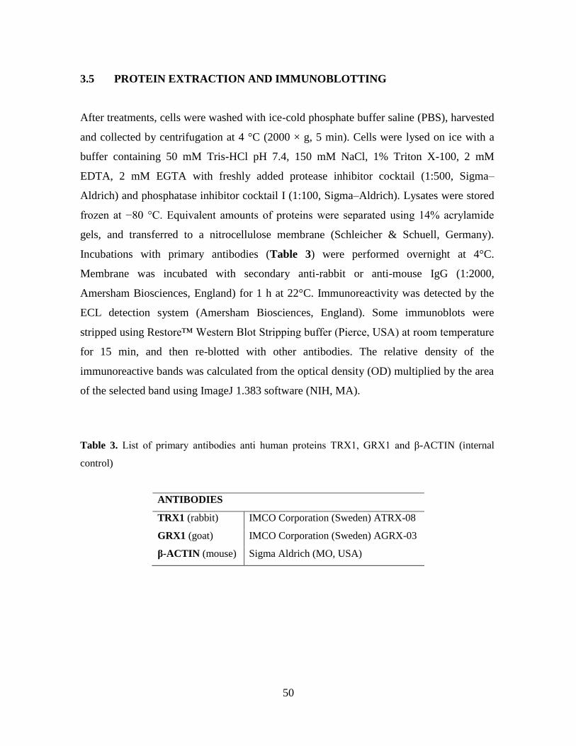
50
3.5 PROTEIN EXTRACTION AND IMMUNOBLOTTING
After treatments, cells were washed with ice-cold phosphate buffer saline (PBS), harvested
and collected by centrifugation at 4 °C (2000 × g, 5 min). Cells were lysed on ice with a
buffer containing 50 mM Tris-HCl pH 7.4, 150 mM NaCl, 1% Triton X-100, 2 mM
EDTA, 2 mM EGTA with freshly added protease inhibitor cocktail (1:500, Sigma–
Aldrich) and phosphatase inhibitor cocktail I (1:100, Sigma–Aldrich). Lysates were stored
frozen at −80 °C. Equivalent amounts of proteins were separated using 14% acrylamide
gels, and transferred to a nitrocellulose membrane (Schleicher & Schuell, Germany).
Incubations with primary antibodies (Table 3) were performed overnight at 4°C.
Membrane was incubated with secondary anti-rabbit or anti-mouse IgG (1:2000,
Amersham Biosciences, England) for 1 h at 22°C. Immunoreactivity was detected by the
ECL detection system (Amersham Biosciences, England). Some immunoblots were
stripped using Restore™ Western Blot Stripping buffer (Pierce, USA) at room temperature
for 15 min, and then re-blotted with other antibodies. The relative density of the
immunoreactive bands was calculated from the optical density (OD) multiplied by the area
of the selected band using ImageJ 1.383 software (NIH, MA).
Table 3. List of primary antibodies anti human proteins TRX1, GRX1 and β-ACTIN (internal
control)
ANTIBODIES
TRX1 (rabbit) IMCO Corporation (Sweden) ATRX-08
GRX1 (goat) IMCO Corporation (Sweden) AGRX-03
β-ACTIN (mouse) Sigma Aldrich (MO, USA)

51
3.6 ANIMALS
In order to investigate the interaction of different genes and proteins involved in
neurodegenerative disorders with the apoE genotype, we used as animal model the human
apoE Targeted Replacement (TR) mice. These transgenic mice express human apoE3 and
apoE4, under the control of the murine apoE regulatory sequences and on the C57BL/6J
background. apoE3 and apoE4 TR mice were purchased by Taconic Farms (USA):
Homozygous B6.129P2-Apoetm3(APOE*4)Mae N8
Homozygous B6.129P2-Apoetm2(APOE*3)Mae N8
The model was created by targeting the murine apoE gene for replacement with the human
apoE4 and apoE3 allele in E14TG2a ES cells and injecting the targeted cells into
blastocysts. Resultant chimeras were backcrossed to C57BL/6 for seven generations (N7).
The mice were backcrossed once more (N8) and embryo transfer derived. The colony was
maintained through mating homozygotes. Research Breeding Agreement was delivered by
Taconic Farms. The pups were suckled by mothers fed with different diets and after
weaning, at 3 weeks of age, they were treated for 6 months with normal diet (ND) or high
carbohydrate diet (HCD) containing 70% of carbohydrates (Mucedola s.r.l., Milano).
The mice were divided in groups of six, housed in individually ventilated cages
(Tecniplast, Italia) with water and food ad libitum and controlled conditions of light (from
7.00 a.m. to 7.00 p.m.), temperature (22±2 °C) and humidity (65%). The experiments were
performed using 6 months and 18 months old mice. The experimental protocol was
approved by a local bioethics committee, while the procedures and animal comfort were
controlled by the University Veterinary Service. All efforts were made to minimize animal
suffering and the number of animals used was kept to a minimum by the experimental
design. All animal experiments were carried out in accordance with the European
Communities Council Directive of 24 November 1986 (86/609/EEC) and National
(Ministry of Health) laws and policies (authorization no. 139/2012-B). Animal experiments
were carried out in the Department of Pharmacology of the University of Bologna with the
approval of the local Ethical committee (Veterinary Service of the University of Bologna).

52
3.7 TISSUES SAMPLING
Mice were killed by cervical dislocation and their brains were quickly removed. The brain
areas (hippocampus, entorhinal, parietal and frontal cortices) were rapidly dissected out,
frozed in dry ice and stored at -80°C until use for RNA and proteins extraction.
3.8 IMMUNOBLOTTING
The dissected brains were homogenized in lysis buffer (50 mM Tris-HCl pH 7.4, 150 mM
NaCl, 1% Triton X-100, 2 mM EDTA, 2 mM EGTA) with protease and phosphatase
inhibitor cocktail (Sigma-Aldrich, MO) at dilution 1:500 added freshly, and incubated 30–
60 minutes on ice before centrifugation (13600 g for 10 min) at 4°C. Protein levels were
quantified using the BCA protein assay kit (Pierce, Rockford, IL, USA). Samples were
mixed with equal volume of tricine gel sample buffer (0.16 M Tris-HCl pH 6.8, 4% SDS,
20% glycerol, 0.01% bromophenol blue, 0.1 M DTT added fresh) and then electrophoresis
was performed. Equal amounts of protein (40 μg) were separated using 12% acrylamide
gel under reducing conditions and then transferred to a nitrocellulose membrane
(Schleicher & Schuell, Germany). After transfer, the membrane was blocked in 5% nonfat
milk buffer in Tris-Buffered saline (TBS) for 1h at room temperature and then incubated
overnight at 4°C with the primary antibody at proper dilutions (Table 3). After being
probed with the primary antibody and washed with TBS-Tween buffer (TBS and 0.01%
Tween 20), membranes were incubated with peroxidase-conjugated anti-mouse, anti-rabbit
or anti-goat immunoglobulin G (IgG) at 1:2000 dilution (Amersham Biosciences, Little
Chalfont, UK) in blocking buffer for 2 hr at 22°C. Immunoreactivity was detected by the
ECL detection system (Amersham Biosciences, Little Chalfont, UK). Some immunoblots
were stripped using RestoreTM Western Blot Stripping buffer (Pierce, Rockford, IL, USA)
at room temperature for 15 minutes, and then re-blotted with other antibodies. The relative
density of the immunoreactive bands was calculated from the optical density (OD)
multiplied by the area of the selected band using ImageJ 1.383 software (NIH, MA). The

53
variations of proteins levels were then evaluated with respect to β-actin as internal control.
The results were expressed as the ratio of protein levels compared to the controls, as 100%.
3.9 DATA ANALYSIS
Results were expressed as mean ± S.E.M (standard error of the mean). Data of
experimental groups were processed by using Student's t-test or by analysis of variance
(one-way and two-way ANOVA) followed by Dunnett post hoc test. Differences were
considered statistically significant at *p< 0.05. Statistical data analyses were performed
using the Program GraphPad version 4.03 (GraphPad Software, San Diego, CA, USA).

54
4. RESULTS
4.1 VIABILITY OF SH-SY5Y CELLS EXPOSED TO Aβ
The exposure of SH-SH5Y neuroblastoma cells to 25 μM Aβ peptides for 5, 24 and 48
hours showed a significant decrease of viability in a time-dependent manner: 88.05 ± 0.99
versus control 100 ± 0.70, p<0.01, at 5 hours; 81.27 ± 1.34 versus control 100 ± 1.06,
p<0.01, at 24 hours; 79.80 ± 0.82 versus control 100 ± 0.83, p<0.01, at 48 hours (Figure 7).
Figure 7. Viability of SH-SY5Y human neuroblastoma cells exposed to 25 μM Aβ (25-35) peptides
for 5, 24 and 48 hours evaluated by the MTT assay. Data are expressed as percentage of absorbance
values of treated cells compared to control untreated cells (100%) and reported as a mean ± S.E.M.
of three independent experiments performed in triplicate. Data were analyzed by one-way Anova,
followed by Dunnett post hoc test (**p < 0.01).

55
4.2 GENE EXPRESSION IN SH-SY5Y EXPOSED TO Aβ
PIN1 gene expression
The level of PIN1 gene expression was significantly decreased in cells exposed to 25 µM
Aβ for 5 hours (0.80 ± 0.04 versus control 1.00 ± 0.02, p<0.001) and for 24 hours (0.86 ±
0.08 versus control 1.00 ± 0.03, p<0.01) compared to unexposed control cells (Figure 8a
and b, respectively). No changes of gene expression were observed after cell exposure to 25
µM Aβ for 48 hours (Figure 8c; Table 4).
a
b

56
Figure 8. PIN1 relative gene expression was measured by real-time PCR in SH-SY5Y
neuroblastoma cells exposed to 25 μM Aβ (25-35) peptides for 5 (a) 24 (b) and 48 (c) hours. Bars
represent 2-DDCt
values calculated by Delta-Delta Ct (DDCt) method. Gene expression was
normalized to glyceraldehyde-3-phosphate dehydrogenase (GAPDH); data are expressed as mean ±
S.E.M percent of control untreated cells for three independent experiments (***p <0.001; **p<0.01
versus controls; Student's t-test)
Table 4
PIN1 gene expression level
Time Control cells Aβ exposed cells
5 hours 1.00 ± 0.02 0.80 ± 0.04***
24 hours 1.00 ± 0.03 0.86 ± 0.08**
48 hours 1.00 ± 0.04 0.96 ± 0.17
**p<0.01; ***p<0.001
SIRT1 gene expression
The level of SIRT1 gene expression was significantly decreased in cells exposed to 25 µM
Aβ for 5 hours (0.78 ± 0.07 versus control 1.00 ± 0.04, p<0.001) and for 24 hours (0.74 ±
0.10 versus control 1.00 ± 0.08, p<0.001) compared to unexposed control cells (Figure 9a
and b, respectively). No changes in the gene expression were observed following cell
exposure to 25 µM Aβ for 48 hours (Figure 9c; Table 5).
c

57
Figure 9. SIRT1 relative gene expression was measured by real-time PCR in SH-SY5Y
neuroblastoma cells exposed to 25 μM Aβ (25-35) peptides for 5 (a) 24 (b) and 48 (c) hours. Bars
represent 2-DDCt
values calculated by Delta-Delta Ct (DDCt) method. Gene expression was
normalized to glyceraldehyde-3-phosphate dehydrogenase (GAPDH); data are expressed as mean ±
S.E.M percent of control untreated cells for three independent experiments (***p <0.001; versus
controls; Student's t-test)
a
b
c
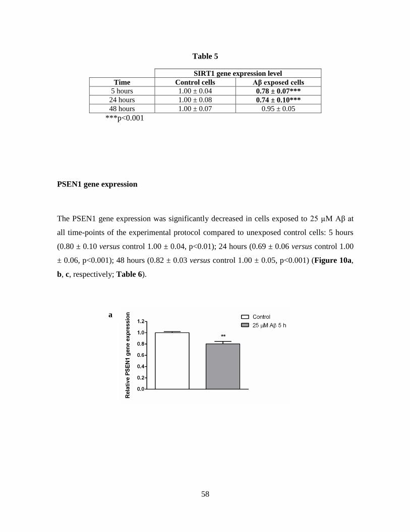
58
Table 5
SIRT1 gene expression level
Time Control cells Aβ exposed cells
5 hours 1.00 ± 0.04 0.78 ± 0.07***
24 hours 1.00 ± 0.08 0.74 ± 0.10***
48 hours 1.00 ± 0.07 0.95 ± 0.05
***p<0.001
PSEN1 gene expression
The PSEN1 gene expression was significantly decreased in cells exposed to 25 µM Aβ at
all time-points of the experimental protocol compared to unexposed control cells: 5 hours
(0.80 ± 0.10 versus control 1.00 ± 0.04, p<0.01); 24 hours (0.69 ± 0.06 versus control 1.00
± 0.06, p<0.001); 48 hours (0.82 ± 0.03 versus control 1.00 ± 0.05, p<0.001) (Figure 10a,
b, c, respectively; Table 6).
a

59
Figure 10. PSEN1 relative gene expression was measured by real-time PCR in SH-SY5Y
neuroblastoma cells exposed to 25 μM Aβ (25-35) peptides for 5 (a) 24 (b) and 48 (c) hours. Bars
represent 2-DDCt
values calculated by Delta-Delta Ct (DDCt) method. Gene expression was
normalized to glyceraldehyde-3-phosphate dehydrogenase (GAPDH); data are expressed as mean ±
S.E.M percent of control untreated cells for three independent experiments (***p <0.001; **p<0.01
versus controls; Student's t-test)
Table 6
PSEN1 gene expression level
Time Control cells Aβ exposed cells
5 hours 1.00 ± 0.04 0.80 ± 0.10**
24 hours 1.00 ± 0.06 0.69 ± 0.06***
48 hours 1.00 ± 0.05 0.82 ± 0.03***
**p<0.01; ***P<0.001
b
c

60
BDNF gene expression
The BDNF gene expression was significantly decreased in cells exposed to 25 µM Aβ for 5
hours compared to unexposed control cells (0.84 ± 0.05 versus control 1.00 ± 0.09, p<0.01)
(Figure 11a). In contrast, the BDFN gene expression was significantly increased in cells
exposed to Aβ for 24 hours (1.76 ± 0.15 versus control 1.00 ± 0.14, p<0.001) and 48 hours
(1.72 ± 0.25 versus control 1.00 ± 0.13, p<0.001) (Figure 11b and c, respectively; Table
7).
a
b

61
Figure 11. BDNF relative gene expression was measured by real-time PCR in SH-SY5Y
neuroblastoma cells exposed to 25 μM Aβ (25-35) peptides for 5 (a) 24 (b) and 48 (c) hours. Bars
represent 2-DDCt
values calculated by Delta-Delta Ct (DDCt) method. Gene expression was
normalized to glyceraldehyde-3-phosphate dehydrogenase (GAPDH); data are expressed as mean ±
S.E.M percent of control untreated cells for three independent experiments (***p <0.001; versus
controls; Student's t-test)
Table 7
BDNF gene expression level
Time Control cells Aβ exposed cells
5 hours 1.00 ± 0.09 0.84 ± 0.05***
24 hours 1.00 ± 0.14 1.76 ± 0.15***
48 hours 1.00 ± 0.13 1.72 ± 0.25***
****p<0.001
4.3 GENE EXPRESSION IN APOE3/E4 TRANSGENIC MICE
PIN1 gene expression
The PIN1 gene expression levels were significantly increased in the hippocampus of 18
months old apoE4 mice (1.23 ± 0.09 versus control apoE3 group = 1.00 ± 0.07, p<0.01)
(Figure 12a). In contrast, apoE4 genotype is associated with a significant reduction of
PIN1 gene expression in the entorhinal cortex (0.75 ± 0.05 versus control apoE3group =
1.00 ± 0.05, p<0.001) and in the parietal cortex (0.68 ± 0.10 versus control group apoE3=
c

62
1.00 ± 0.11, p<0.001) (Figure 12c and d, respectively). No significant changes of PIN1
gene expression were observed in the frontal cortex (Figure 12b; Table 8).
Figure 12. PIN1 gene expression was measured by real-time PCR in hippocampus (a), frontal
cortex (b), entorhinal cortex (c) and parietal cortex (d) of 18 months old apoE 3 and apoE4
transgenic mice fed with normal diet (ND). Bars represent 2-DDCt
value calculated by Delta-Delta Ct
(DDCt) method (n=6). Gene expression was normalized to glyceraldehyde-3-phosphate
dehydrogenase (GAPDH); data are expressed as means ± SEM relative to control apoE3mice (***p
<0.001; **p<0.01. Student's t-test).
a
c
b
d

63
Table 8
PIN1 gene expression level
Brain areas APOE3 mice APOE4 mice
Hippocampus 1.00 ± 0.07 1.23 ± 0.09**
Frontal cortex 1.00 ± 0.08 1.13 ± 0.14
Entorhinal cortex 1.00 ± 0.05 0.75 ± 0.05***
Parietal cortex 1.00 ± 0.11 0.68 ± 0.10***
**p<0.01; ***p<0.001
SIRT1 GENE EXPRESSION
The SIRT1 gene expression was significantly decreased in the frontal cortex of 18 months
old apoE4 mice (0.81 ± 0.04 versus control apoE3 group = 1.00 ± 0.10, p<0.05) (Figure
13b). No changes were observed in the other examined brain areas (Table 9).
a b
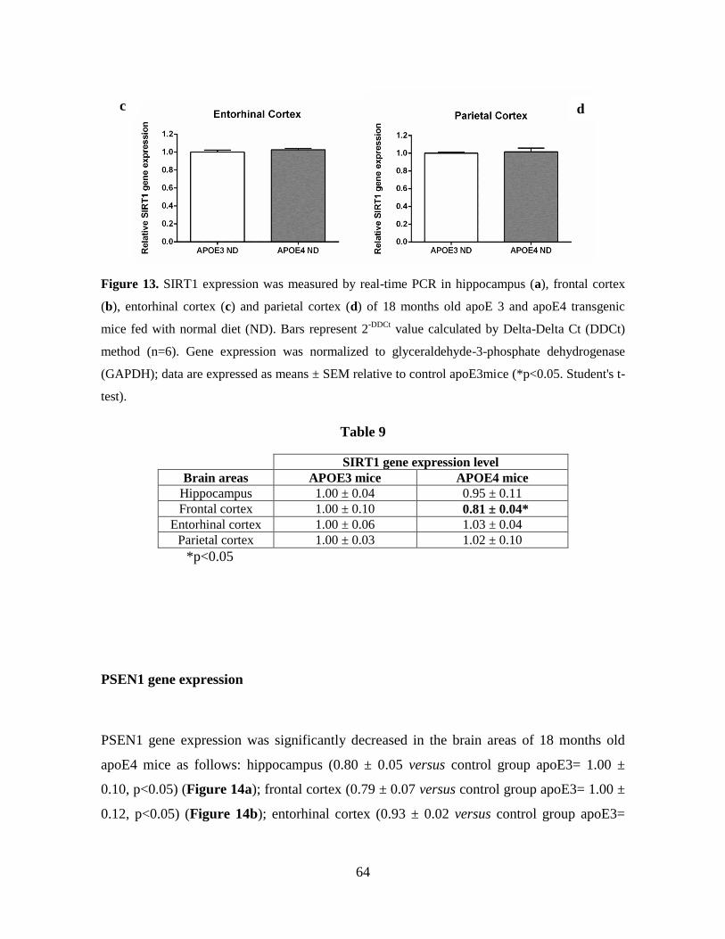
64
Figure 13. SIRT1 expression was measured by real-time PCR in hippocampus (a), frontal cortex
(b), entorhinal cortex (c) and parietal cortex (d) of 18 months old apoE 3 and apoE4 transgenic
mice fed with normal diet (ND). Bars represent 2-DDCt
value calculated by Delta-Delta Ct (DDCt)
method (n=6). Gene expression was normalized to glyceraldehyde-3-phosphate dehydrogenase
(GAPDH); data are expressed as means ± SEM relative to control apoE3mice (*p<0.05. Student's t-
test).
Table 9
SIRT1 gene expression level
Brain areas APOE3 mice APOE4 mice
Hippocampus 1.00 ± 0.04 0.95 ± 0.11
Frontal cortex 1.00 ± 0.10 0.81 ± 0.04*
Entorhinal cortex 1.00 ± 0.06 1.03 ± 0.04
Parietal cortex 1.00 ± 0.03 1.02 ± 0.10
*p<0.05
PSEN1 gene expression
PSEN1 gene expression was significantly decreased in the brain areas of 18 months old
apoE4 mice as follows: hippocampus (0.80 ± 0.05 versus control group apoE3= 1.00 ±
0.10, p<0.05) (Figure 14a); frontal cortex (0.79 ± 0.07 versus control group apoE3= 1.00 ±
0.12, p<0.05) (Figure 14b); entorhinal cortex (0.93 ± 0.02 versus control group apoE3=
c d

65
1.00 ± 0.06, p<0.05) (Figure 14c). No gene expression changes were observed in the
parietal cortex (Figure 14d; Table 10).
Figure 14. PSEN1 expression was measured by real-time PCR in hippocampus (a), frontal cortex
(b), entorhinal cortex (c) and parietal cortex (d) of 18 months old apoE 3 and apoE4 transgenic
mice fed with normal diet (ND). Bars represent 2-DDCt
value calculated by Delta-Delta Ct (DDCt)
method (n=6). Gene expression was normalized to glyceraldehyde-3-phosphate dehydrogenase
(GAPDH); data are expressed as means ± SEM relative to control apoE3mice (**p <0.01; *p<0.05.
Student's t-test).
d c
a b

66
Table 10
PSEN1 gene expression level
Brain areas APOE3 mice APOE4 mice
Hippocampus 1.00 ± 0.10 0.80 ± 0.05*
Frontal cortex 1.00 ± 0.12 0.79 ± 0.07*
Entorhinal cortex 1.00 ± 0.06 0.93 ± 0.02*
Parietal cortex 1.00 ± 0.03 1.02 ± 0.10
*p<0.05
pro-BDNF gene expression
The pro-BDNF gene expression was significantly increased in the hippocampus of 18
months old apoE4 mice (1.30 ± 0.16 versus control apoE3 group = 1.00 ± 0.08, p<0.05)
(Figure 15a). In contrast, apoE4 genotype is associated with a significant reduction of pro-
BDNF gene expression in the entorhinal cortex (0.85 ± 0.10 versus control group apoE3=
1.00 ± 0.04, p<0.01) (Figure 15c). No significant changes of pro-BDNF gene expression
were observed in frontal and parietal cortices (Table 11).
a b

67
Figure 15. pro-BDNF expression was measured by real-time PCR in hippocampus (a), frontal
cortex (b), entorhinal cortex (c) and parietal cortex (d) of 18 months old apoE 3 and apoE4
transgenic mice fed with normal diet (ND). Bars represent 2-DDCt
value calculated by Delta-Delta Ct
(DDCt) method (n=6). Gene expression was normalized to glyceraldehyde-3-phosphate
dehydrogenase (GAPDH); data are expressed as means ± SEM relative to control apoE3mice (**p
<0.01; *p<0.05. Student's t-test). t-test. (n=6)
Table 11
p-BDNF gene expression level
Brain areas APOE3 mice APOE4 mice
Hippocampus 1.00 ± 0.08 1.30 ± 0.16*
Frontal cortex 1.00 ± 0.13 1.16 ± 0.25
Entorhinal cortex 1.00 ± 0.04 0.85 ± 0.10**
Parietal cortex 1.00 ± 0.05 0.98 ± 0.12
*p<0.05; **p<0.01
c d

68
4.4 PROTEIN LEVELS IN BE(2)-M17 CELLS EXPOSED TO APOE3 AND
APOE4
TRX1 levels
The western blot analysis showed a significant decrease of TRX1 protein levels in cells
exposed to 100 nM apoE4 human recombinant protein for 5 hours compared to unexposed
control (64.5 ± 3.19; OD% of control 100 ± 1.95, p<0.01; Figure 16a); cell exposure to
100 nM apoE3 induced no changes of TRX1 protein levels. The levels of the protein were
significantly increased in cells exposed to apoE4 for 48 hours (172.5 ± 10.8; OD% of
control 100 ± 6.79, p<0.01) (Figure 16c). No changes on TRX1 protein levels were
observed in cells exposed to 100 nM apoE4 for 24 hours (Figure 16b; Table 12).
a
b

69
Figure 16. Immunoblotting analysis of TRX1 protein levels (12 kDa) in BE(2)-M17 human
neuroblastoma cells exposed to 100 nM apoE3 and apoE4 for 5 (a), 24 (b) and 48 (c) hours. Bars
represent the mean ± S.E.M of optical density (O.D.) normalized to the internal loading control β-
actin (42 kDa) and expressed as percentage of control untreated cells. **p < 0.01 (one one-way
Anova, followed by Dunnett post hoc test; n=6).
Table 12
TRX1 protein levels
Time Control cells ApoE3 exposed cells ApoE4 exposed cells
5 hours 100 ± 1.95 84.9 ± 6.55 64.5 ± 3.19**
24 hours 100 ± 16.5 87.7 ± 12.2 136.4 ± 10.0
48 hours 100 ± 6.79 102.3 ± 9.86 172.5 ± 10.8**
**p<0.01
GRX1 levels
The western blot analysis showed a significant decrease of GRX1 protein levels in cells
exposed to 100 nM apoE4 human recombinant protein for 5 hours compared to unexposed
control cells (73.3 ± 4.07; OD% of control 100 ± 3.75, p<0.01) (Figure 17a). The protein
levels were significantly increased in cells exposed to apoE4 for 24 hours (128.6 ± 8.05;
OD% of control 100 ± 6.61, p<0.05) and 48 hours (175.9 ± 4.96; OD% of control 100 ±
8.54, p<0.01) (Figure 17b and c, respectively). No changes on GRX1 protein levels were
c

70
observed in cells exposed to 100 nM apoE3 human recombinant protein at 5 and 24 hours.
However, the levels of GRX1 were increased after the apoE3 exposure for 48 hours (149.3
± 18.5; OD% of control 100 ± 8.54, p<0.05) (Figure 17c; Table 13)
a
b
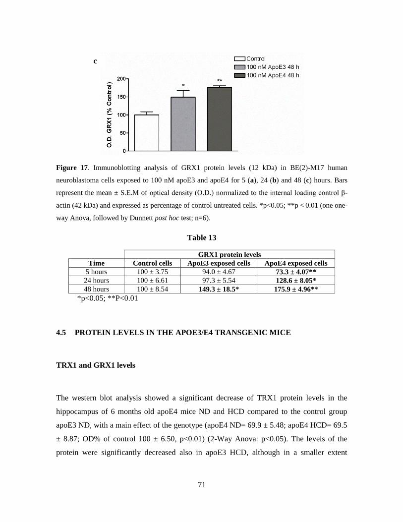
71
Figure 17. Immunoblotting analysis of GRX1 protein levels (12 kDa) in BE(2)-M17 human
neuroblastoma cells exposed to 100 nM apoE3 and apoE4 for 5 (a), 24 (b) and 48 (c) hours. Bars
represent the mean ± S.E.M of optical density (O.D.) normalized to the internal loading control β-
actin (42 kDa) and expressed as percentage of control untreated cells. *p<0.05; **p < 0.01 (one one-
way Anova, followed by Dunnett post hoc test; n=6).
Table 13
GRX1 protein levels
Time Control cells ApoE3 exposed cells ApoE4 exposed cells
5 hours 100 ± 3.75 94.0 ± 4.67 73.3 ± 4.07**
24 hours 100 ± 6.61 97.3 ± 5.54 128.6 ± 8.05*
48 hours 100 ± 8.54 149.3 ± 18.5* 175.9 ± 4.96**
*p<0.05; **P<0.01
4.5 PROTEIN LEVELS IN THE APOE3/E4 TRANSGENIC MICE
TRX1 and GRX1 levels
The western blot analysis showed a significant decrease of TRX1 protein levels in the
hippocampus of 6 months old apoE4 mice ND and HCD compared to the control group
apoE3 ND, with a main effect of the genotype (apoE4 ND= 69.9 ± 5.48; apoE4 HCD= 69.5
± 8.87; OD% of control 100 ± 6.50, p<0.01) (2-Way Anova: p<0.05). The levels of the
protein were significantly decreased also in apoE3 HCD, although in a smaller extent
c

72
compared to apoE4 mice experimental groups (75.9 ± 5.57 OD% of control, p<0.05)
(Figure 18a). The western blot analysis of GRX1 protein levels showed no significantly
differences between experimental groups (Figure 18 b). (Table 14)
Figure 18. Hippocampal samples from 6 months old apoE3 and apoE4 mice fed with normal diet
(ND) or high carbohydrate diet (HCD) were analyzed by Western immunoblotting for TRX1 (a)
and GRX1 (b) protein levels. TRX1 levels (12 kDa) are decreased in apoE4 ND and HCD mice,
and in apoE3 HCD compared to control group apoE3 ND mice (*p<0.05; **p< 0.01, main effect of
the genotype). No significant differences were found for GRX1 protein levels (12 kDa). Results
represent the mean ± S.E.M of optical density (O.D.) normalized to the internal loading control β-
actin (42 kDa) and expressed as percentage of control. 2-way Anova= *p<0.05, main effect of the
genotype; one-way Anova, Dunnett post-test, *p<0.05; ** p<0.01 (n=8)
a
b

73
Table 14
APOE3 ND APOE4 ND APOE3 HCD APOE4 HCD
TRX1 protein levels 100 ± 6.5 69.9 ± 5.48** 75.9 ± 5.57* 69.4 ± 8.87**
GRX1 protein levels 100 ± 5.5 90.9 ± 14.3 114 ± 15.1 125.8 ± 18.1
*p<0.05; **p<0.01
4.6 TRX1 GENE EXPRESSION IN THE APOE3/E4 TRANSGENIC MICE
The TRX1 gene expression was increased in the hippocampus of apoE4 ND and apoE4
HCD mice compared to the control group apoE3 ND (apoE4 ND= 2.10 ± 0.55; apoE4
HCD= 1.97 ± 0.51 versus the control apoE3 group ND= 1.00 ± 0.23, p<0.05), with a main
effect of the genotype (2-Way Anova) (Figure 19). No changes were observed in apoE3
HCD mice (1.46 ± 0.16 versus the control apoE3 group ND).
Figure 19. TRX1 relative gene expression was determined by real-time PCR in
hippocampus of 6 months old apoE 3 and apoE4 transgenic mice fed with normal diet (ND)
or high carbohydrate diet (HCD). Bars represent 2-DDCt value calculated by Delta-Delta
Ct (DDCt) method (n=6). Gene expression was normalized to glyceraldehyde-3-phosphate
dehydrogenase (GAPDH); data are expressed as means ± SEM relative to control
apoE3mice (2-way Anova= #p<0.05, main effect of the genotype)

74
5. DISCUSSION
5.1. METHODOLOGICAL CONSIDERATIONS
5.1.1 Aβ 25-35 PEPTIDES
Several evidences suggest that the excessive production of Aβ peptides and its aggregation
into toxic fibrillar deposits play a central role in the etiology of AD. Potential toxic
mechanisms of Aβ include altered activity of neurotransmitter receptors and signaling
molecules with impairment of axonal transport, disrupted intracellular calcium homeostasis
and mitochondrial functions (Huang and Mucke, 2012). In support of this hypothesis,
several in vitro and in vivo investigations reported neurotoxic effects of Aβ fragments in
neurons of brain regions involved in AD. Nevertheless, the neurodegenerative processes
induced by Aβ in these studies have been observed only in specific experimental conditions
so that the data are somewhat inconsistent.
Human Aβ peptides are present in different assembly states, including monomers,
oligomers, as well as mature fibrils, that may be deposited in brain tissues. Several studies
focusing on functional Aβ effects used synthetic Aβ peptides. However, it is still unknown
the Aβ concentration that should be considered normal or physiological and which
abnormally high or pathological. It has been proposed that the aggregation state also
influences the toxic properties of Aβ, but which form of Aβ is the most toxic and how its
accumulation causes neuronal dysfunction in the brain are unresolved topics (Holscher et
al., 2007). Data obtained in different experimental models suggest that soluble aggregates
of Aβ cause more damages to synaptic and cognitive functions than insoluble fibrils
(Roberson and Mucke, 2006). However, it is impossible to measure Aβ oligomers levels in
the brains of living people, and the kinetic of Aβ peptides assembly in the brain certainly
differs from that observed in vitro. In the earliest stages of amyloid plaques deposition it is
likely that Aβ assembly as small oligomeric species more toxic than large fibrillar products.
It has been suggested that Aβ 25-35 peptide, largely used in the cellular neurotoxicity
studies, represents the biologically active fragment of Aβ. This peptide differs from the
other Aβ peptides used in experimental studies since it displays rapid aggregation
properties forming stable fibrils with large β-structure and it is neurotoxic immediately

75
upon dissolution (Millucci et al., 2009). The Aβ 25-35 is also the shortest peptide that
retains the toxicity of the full-length Aβ 1-42 (D’Ursi et al., 2004). Moreover,
immunohistochemical analyses demonstrated the presence of Aβ 25–35 peptides in the
senile plaques and in the hippocampal neurons of AD brains, but not in age-matched
controls. These results strengthen the hypothesis that soluble Aβ40 is released from plaques
and converted by proteolysis to the toxic Aβ 25–35 (Kubo et al., 2002). On the basis of
these experimental considerations, in the present study we used Aβ 25–35 aggregated
peptides to induce AD-like neurotoxic insults in SH-SY5Y neuroblastoma cells as in vitro
model, and then investigate the modulation of PIN1, SIRT1, PSEN1 and BNDF gene
expression induced by the Aβ exposure. In particular, Aβ 25–35 peptides were “aged” and
aggregated in water through the incubation at 37 °C for 72 h. The consequent aggregation
produces a conformational change of peptides that form β-structures with increased
neurotoxicity as demonstrated by in vitro and in vivo studies (Croce et al., 2011; Maurice
et al., 1996). The in vitro effects of neurotoxic 25 μM Aβ aggregates result in a reduced
time-dependent cell viability, starting to 5 hours after the exposure; the maximum level of
toxicity corresponds to a mortality rate around 20%. This significant but not so elevated
neurotoxicity is useful to investigate the effect of the aggregated Aβ 25-35 peptides on the
early modulation of different genes related to the neurodegenerative processes which can
lead to AD.
5.1.2 APOE3 AND APOE4 TRANSGENIC MICE
Human apoE targeted replacement (h-apoE TR) mice is a validated animal model produced
using a gene targeting strategy which allows the expression of apoE3 or apoE4 mRNA with
a similar levels and tissue distribution (Sullivan et al., 1997). This transgenic line is largely
used to study the role of apoE in neurodegenerative processes related to the onset of AD. In
particular, the spatial memory performances are sensitive to the apoE isoform, with apoE4
mice showing age-dependent memory impairments in spatial and avoidance memory tasks
(Bour et al., 2008). It has been also demonstrated that the synaptic and cholinergic deficits

76
induced by apoE4 preceded the amyloid plaques formation (Buttini et al., 2002). Based on
these data, it has been hypothesized that apoE4 displays neurotoxic properties
independently by Aβ and plaques deposition. Both in vitro and in vivo studies suggest that
several mechanisms apoE4-mediated could contribute to the pathogenesis of AD. These
include the modulation of the Aβ deposition and Aβ clearance, the alteration of the
antioxidant system, the impairment of the neuronal signaling pathways, the disruption of
cytoskeletal structure with increased phosphorylation of tau, and the impairment of glucose
metabolism and mitochondrial function (Huang, 2011). Even if the association of the
apoE4 genotype with increased risk to develop AD has been largely established, the exact
pathogenic mechanism is still poorly understood. In the brain, apoE has been implicated in
lipid metabolism, neuronal repair and remodeling processes. In general, apoE3 is
neurotrophic or neuroprotective, whereas apoE4 and its fragments are toxic. The
neurotoxicity of apoE4 fragments increases the tau phosphorylation, alter the cellular
signaling pathways and/or lipid metabolism leading to impaired neuronal functions
especially in the hippocampus (Li et al., 2009)
The apoE4 genotype is associated to loss of synaptic integrity, spine density and dendritic
arborization in mice, and represents the major genetic risk factor for sporadic AD in
humans. In particular, a recent study showed that apoE4 mice fed with HCD (high
carbohydrate diet) have cognitive deficits starting at 6 months of age, with decreased levels
of proteins that regulate memory processes such as Arc, β-catenin and BDNF, in the
hippocampus, one of the first brain regions that degenerates in earliest stages of AD
pathogenesis (Maioli et al., 2012).
Based on these experimental considerations, in the present study we used h-apoE TR mice
to investigate the effect of the strong genetic AD risk factor apoE4 on the gene expression
of selected proteins involved in the regulation of neurodegenerative processes. In particular,
the interaction between HCD and apoE3 and apoE4 genotype on TRX1/GRX1 antioxidant
protein levels has been investigated in the hippocampus of 6 months old mice. The gene
expression of PIN1, SIRT1, PSEN1 and pro-BDNF was measured in apoE 18 old months
mice fed with normal diet. The modulation of these genes by the apoE4 genotype was
assessed in different brain regions known to be impaired in the first stages of AD, such as
77
hippocampus, and entorhinal, frontal and parietal cortices. Data from the present research
demonstrated selective changes of gene expression in the different examined brain areas.
The apoE TR mice are particularly useful to compare the effects of apoE isoforms on
molecular pathways involved in AD onset before that the pathological brain lesions became
evident. In fact, several in vivo studies suggest that the cognitive impairment can occur in
the absence of Aβ and neurofibrillary tangles deposition. The human apoE TR mice used in
the present study is a validated transgenic model to investigate the pathogenesis of apoE-
related cognitive deficits in the earliest phases of dementia.
5.2 PIN1 GENE EXPRESSION
In vitro studies
The in vitro data show that Aβ 25-35 aggregates exposure determines a significant decrease
of PIN1 gene expression at 5 and 24 hours in SH-SY5Y human neuroblastoma cells,
whereas no changes are observed at 48 hours. At present, no experimental studies
concerning the effects of Aβ on the PIN1 mRNA levels have been performed.
A recent study demonstrated that human hippocampal cultured neurons exposed to Aβ (1–
42) oligomers show a PIN1 protein up-regulation and activation associated with decreased
p-tau, suggesting that Aβ might transiently induce PIN1 expression and prevent tau
hyperphosphorylation (Bulbarelli A et al., 2009). It has been also suggested that, in
physiological conditions, PIN1 may be protective against Aβ toxicity, promoting the trans
conformation of APP and increasing its non-amyloidogenic processing. However, the role
of PIN1 in the protection or promotion of neuronal degeneration it is not completely
elucidated. PIN1 protein was significantly down-regulated and oxidized in AD
hippocampus (Sultana et al., 2006). This oxidation led to the loss of PIN1 isomerase
activity supporting a direct link between oxidative damage to PIN1 and the pathogenesis of
AD. A recent study also showed a compensatory activation/up-regulation of PIN1 protein
in later stages of AD pathogenesis, whereas its reduction seems to play a role in early

78
stages of the disease (Wang et al., 2007 b). These results suggest a pivotal role of PIN1
protein in the protection against p-tau and Aβ deposition during neuronal degenerative
processes.
Therefore, the down-regulation of PIN1 gene expression observed in the present in vitro
study may be considered an early phenomenon induced by the Aβ peptide neuronal
toxicity. On the other hand, PIN1 could be involved in mechanisms of neuronal protection
against Aβ toxicity that probably occur late in the pathogenetic AD-related processes. PIN1
gene expression is in fact down-regulated in a transient manner after the early exposure to
Aβ at 5 and 24 hours, to then revert to a physiological rate following a more prolonged
contact with Aβ peptides (48 hours). We may then hypothesize that this shift of PIN1
mRNA levels might be a compensatory mechanism to counteract the Aβ toxic insults.
In vivo studies
The data obtained from 18 months old apoE4 mice showed an increase of PIN1 gene
expression in the hippocampus and PIN1 down-regulation in the entorhinal and parietal
cortices compared to apoE3 controls. No changes were observed in the frontal cortex.
This is the first study that investigates, in the h-apoE TR mice model, the possible
modulation of PIN1 gene by the apoE4 genotype in brain areas impaired in AD. An
immunohistochemical study in the hippocampus and in the parietal cortex of normal and
AD human brains showed that the regions with lower PIN1 protein levels are the same
prone to the degeneration. These data suggest an inverse correlation between the PIN1
expression and the vulnerability to neuronal cell death (Liou et al., 2003). However, it has
also been reported that hippocampal neurons of AD patients show granular accumulation of
PIN1 without neurofibrillary tangles (Holzer M et al., 2002). These lesions could be
considered alternative pathological alterations, maybe aiming to counteract the neuronal
degeneration. PIN1 is also the first protein whose deletion seems to cause age-dependent
neurodegeneration and tau pathologies. PIN1-knockout mice show hyperphosphorylated
tau, motor-behavioral deficits and neuronal loss, as well as described in AD patients (Liou
et al. 2003). However, in vivo studies on PIN1-knockout mice showed opposite data

79
regarding the effect of PIN1 on Aβ production (Akiyama et al. 2005; Pastorino et al. 2006).
The mechanisms underlying the PIN1 modulation of tau phosphorylation and Aβ
deposition during AD progression are still undefined as well as the role of PIN1 in the
regulation of normal neuronal function. Genetic studies on humans suggest that PIN1
promoter polymorphisms are associated with an increased risk of late-onset AD (Segat et
al. 2006). A recent study also showed an increased PIN1 gene expression in PBMCs of late
onset AD patients, especially if carrying at least one ε4 allele (Arosio et al., 2012).
The hippocampus and entorhinal cortex are brain structures involved in memory formation
and firstly impaired during early neurodegenerative processes of AD (Braak and Braak,
1997). The increase of PIN1 mRNA levels in the hippocampus of apoE4 mice reported in
the present research might be a compensatory/protective mechanism to counteract the
detrimental effects of the apoE4 genotype on neurons. Alternatively, the up-regulation of
PIN1 could represent a reactive biochemical mechanism to counteract the decreased PIN1
protein levels as observed in AD patients (Liou et al., 2003).
The down-regulation of PIN1 in the other brain regions such as entorhinal and parietal
cortices may be considered a consequence of a higher vulnerability of these neurons to the
apoE4 genotype. Otherwise, it is possible that in these regions an increase of PIN1 gene
expression also occurs to counteract the cell damage, but may became evident later on the
apoE4 mice life.
The frontal cortex is a brain region generally impaired during the mild/late stages of AD,
and gives a general description of the full AD pathological features. In the frontal cortex of
apoE4 mice PIN1 gene expression levels are unchanged. It can be hypothesized that PIN1
plays a marginal protective role in this area or that the frontal cortex is overall more able to
counteract the detrimental apoE4-related processes.
It is possible to conclude that, in h-apoE TR mice model used in the present study, PIN1
plays different and specific roles against neurodegenerative processes in the brain regions
involved in AD. Therefore, in AD patient the apoE4 genotype could contribute to the onset
of neurodegeneration also through the modulation of PIN1 gene expression. PIN1 thus
represents a novel potential diagnostic and therapeutic target in AD. However, clinical

80
studies need to confirm and elucidate the role of PIN1 in human neurodegenerative
diseases.
5.3 SIRT1 GENE EXPRESSION
In vitro studies
The in vitro studies show that Aβ 25-35 aggregates exposure induces, in SH-SY5Y human
neuroblastoma cells, a significantly decrease of SIRT1 gene expression at 5 and 24 hours,
whereas no changes are observed at 48 hours. No in vitro studies regarding the modulation
of SIRT1 mRNA levels by Aβ aggregated peptides are currently reported in literature.
SIRT1 is a histone deacetylase (HDAC) protein highly expressed in neurons and involved
in the regulation of several functions, including antioxidant, anti-inflammatory and anti-
apoptotic processes. SIRT1 is implicated in pathways related to synaptic plasticity, learning
and memory. Several studies suggested that SIRT1 protects against AD degenerative
mechanisms, in particular decreasing Aβ accumulation and reducing the tau-related
pathology (Donmez et al., 2010).
The decrease of SIRT1 mRNA levels in the present in vitro study may be the expression of
the Aβ peptide-induced neuronal toxicity. The SIRT1 down-regulation here reported is in
agreement with data coming from humans showing that SIRT1 mRNA and protein levels
are decreased in the parietal cortex of AD patients (Julien et al., 2009). However, no
association between SIRT1 gene polymorphism and AD risk is described.
The present findings showed that SIRT1 gene is down-regulated in a transient manner after
the acute exposure to Aβ at 5 and 24 hours, since it reverts to a physiological level after 48
hours. This time-course modulation of SIRT1 gene expression may be considered a
molecular compensatory mechanism counteracting the Aβ-induced toxicity.
Previous in vitro studies already demonstrated the protective role of SIRT1. SIRT1 has
been shown to protect against microglia-dependent Aβ toxicity through the inhibition of

81
inflammatory signaling (Chen et al., 2005). Moreover, the antioxidant natural compound
resveratrol protects neuronal cells against Aβ also through the activation of SIRT1 protein
expression and activity (Albani et al., 2010).
In vivo studies
The in vivo data from 18 months old apoE4 mice showed a decrease of SIRT1 gene
expression in the frontal cortex compared to apoE3 controls, whereas no changes were
observed in the other brain regions examined. These data could be explained taking into
account the role of SIRT1 in normal and AD brains. SIRT1 plays a crucial role in cognitive
functions and synaptic plasticity in normal mice (Michán et al., 2010). Moreover, a recent
study showed that transgenic mice model lacking of SIRT1 catalytic activity in the brain
show impairment of memory and synaptic plasticity (Gao et al., 2010). On the contrary, the
over-expression of SIRT1 in the hippocampus of AD transgenic mice protects against
neuronal degeneration (Kim et al., 2007). Therefore, the SIRT1 down-regulation we
observed in the frontal cortex of apoE4 mice might be considered as consequence of the
detrimental effects of apoE4 on the SIRT1-related protective functions.
Taking together the in vitro and in vivo results, we can state that Aβ peptides and apoE4
genotype probably contribute to the neuronal impairment also through the reduction of
SIRT1 gene expression.
5.4 PSEN1 GENE EXPRESSION
In vitro and in vivo studies
The in vitro data show that Aβ 25-35 aggregates exposure determines a significantly
decrease of PSEN1 gene expression levels at 5, 24 and 48 hours in SH-SY5Y human
neuroblastoma cells. In particular, the decrease of PSEN1 mRNA levels was more

82
pronounced at the intermediate exposure of 24 hours. Aβ peptides may induce neuronal
toxicity also through the decrease of PSEN1 gene expression in this in vitro model. The in
vivo data in 18 months old apoE4 mice showed a decreased PSEN1 gene expression in the
hippocampus, frontal and entorhinal cortices compared to apoE3 control group. No changes
were observed in the parietal cortex of apoE4 mice.
PSEN1 is a genetic risk factor in the early-onset familial AD. The PSEN1 protein is the
catalytic unit of the enzyme γ-secretase leading to amyloidogenic process of APP.
However, PSEN1 protein is also involved in the regulation of memory formation and
synaptic plasticity. The loss of PSEN1 activity results in hippocampal-dependent spatial
and memory impairments, with inflammation and neuronal degeneration. It has also been
reported that PSEN1 protein levels are reduced in the association neocortex and
hippocampus of AD brains (Davidsson et al., 2001). The functional loss of PSEN1 leads to
increased p-tau levels and impairment of neuronal functions (Pigino et al., 2003). The
decrease of PSEN1 mRNA levels in the brain areas involved in AD degeneration suggests
that the effect of apoE4 also involves PSEN1-related mechanisms.
The in vivo data are consistent with the in vitro PSEN1 gene expression results, suggesting
that the PSEN1 modulation induced by Aβ peptides and apoE4 genotype could in part
contribute to the neuronal loss and impairments in AD processes. However, further
elucidations of the roles played by presenilins in the brain are necessary for the
development of effective therapeutic strategies in neurodegenerative disorders.
5.5 BDNF GENE EXPRESSION
In vitro studies
The in vitro data show that the exposure of SH-SY5Y human neuroblastoma cells to Aβ 25-
35 aggregates induces a significant decrease of BDNF gene expression at 5 hours followed
by an increased mRNA levels at 24 and 48 hours.

83
BDNF is implicated in several brain functions such as synaptic transmission, neuronal
differentiation, protection and survival. It is considered one of the key molecules in the
pathology of neurodegenerative disorders such as AD. In vitro studies recently reported that
long-term Aβ 42 or Aβ 25–35 exposure strongly down-regulates BDNF gene expression in
rat cortical neurons; however, these reduction is preceded by an increase of BDNF mRNA
levels after short-term exposure to amyloid peptides (Aliaga et al., 2010). Oligomeric, but
not fibrillar Aβ 42, significantly decreases BDNF mRNA levels also in differentiated SH-
SY5Y cells (Garzon et al., 2007). However, other studies showed opposite results. BDNF
gene expression is reported up-regulated in astrocytes exposed to Aβ 42 (Kimura et al.,
2006). A recent in vitro study showed increased BDNF mRNA levels in the early stage of
Aβ toxicity in differentiated neuroblastoma cells. In the same study the BDNF exposure
rescues the cells from neuritic degeneration caused by Aβ (Zhang et al., 2012). In SH-
SY5Y cells Aβ also produce an increased release of BDNF protein, maybe reflecting a
compensatory cellular adaptation by enhancing neurotrophic support (Olivieri et al., 2003).
These studies suggest that Aβ triggers distinct effects on the BDNF expression and
production depending on the neuronal cellular lineage and experimental conditions.
The decrease of BDNF mRNA levels at 5 hours may be explained as the result of Aβ-
mediated acute toxicity. The following increased gene expression following a more
prolonged exposure to Aβ (24 and 48 hours) might constitute a neuroprotective BDNF-
mediated mechanism that allows the cells to counteract the Aβ toxic effects.
In vivo studies
The in vivo data from 18 months old apoE4 mice showed an increse of pro-BDNF gene
expression in the hippocampus compared to apoE3 controls, whereas decreased levels in
entorhinal cortex were observed. No changes were observed in the frontal and parietal
cortices.
BDNF is largely expressed in brain areas characterized by a high plasticity potential such as
hippocampus and cortex; several studies suggest that BDNF is required for the
hippocampus-mediated learning (Tapia-Arancibia et al. 2008). In fact, knock-out mice for

84
BDNF or its TrkB receptor show reduced dendritic and axonal arborisation, associated to
learning and memory impairment (Poo, 2001).
BDNF is synthesized as a precursor protein pro-BDNF which is proteolitically cleaved to
generate the mature form BDNF. In h-apoE TR mice model, we measured the levels of pro-
BDNF mRNA since most of the BDNF secreted by the hippocampal neurons is in the
precursor form.
Several clinical evidences showed that the expression of BDNF and its receptor trkB are
decreased in post-mortem brain regions such as hippocampus, and temporal, parietal and
frontal cortices of AD and MCI patients (Connor et al., 1997; Peng et al., 2005). The pro-
BDNF is also decreased in the hippocampus and parietal cortex even in pre-clinical stages
of AD. In our experimental model, the increased pro-BDNF gene expression in the
hippocampus does not match with data reported from human studies; it is possible that, in
h-apoE TR mice, apoE4 induce a first reactive increase of pro-BDNF aiming to react to the
onset of the neurodegenerative process and that, when the disease establishes and
progresses, it possibly decreases as occurs in AD patients. This is even more plausible in
the hippocampus that is one of the first brain area that is involved in the first stages of AD
neurodegenerative process.
In contrast, other studies have reported increased serum BDNF levels in patients with early
AD (Angelucci et al., 2010). In vivo studies regarding mice models of AD also showed an
alteration in BDNF protein and gene expression levels. APP and PS1 double transgenic
mice model of AD have increased hippocampal BDNF protein expression that is consistent
with the pro-BDNF up-regulation we observed in hippocampus (Szapacs et al., 2004). The
up-regulation of BDNF mRNA expression and protein was also found in microglia and
astrocytes of other AD transgenic mice models (Burbach et al., 2004). Some authors
demonstrated increased hippocampal BDNF mRNA expression following intracranial
administration of Aβ peptides (Tang et al., 2000). These findings corroborate the
hypothesis that the increase of BDNF levels might be a compensatory mechanism against
the amyloid toxicity in early stages of AD.

85
In contrast, in the apoE4 mice, pro-BDNF gene expression is decreased; probably, the
entorhinal cortex is not able to activate this neurotrophic support and results more
vulnerable to the neuronal impairment induced by apoE4.
Although the therapy with neurotrophins has been suggested as a treatment for
neurodegenerative disorders, the precise role of BDNF signaling in AD remains to be
clarified.
5.6 TRX1 AND GRX1 PROTEINS MODULATION
In vitro studies
The in vitro data show that the exposure of BE(2)-M17 human neuroblastoma cells to
human recombinant apoE4 induced a significant decrease of the antioxidant protein TRX1
and GRX1 levels following short-term exposure (5 hours). After a more prolonged-term
exposure to apoE4 (24 and 48 hours) both protein levels were increased compared to
untreated control cells. The apoE3 protein induced no changes in TRX1 levels, although it
up-regulates GRX1 at 48 hours. The concentration of apoE3 and apoE4 proteins (100 nM)
used in these experimental procedures is considered physiologically present in CSF of
healthy individuals (Qiu et al., 2003; Riemenschneider, et al., 2002). Based on the evidence
that the acute exposure to apoE4, but not apoE3, reduces TRX1 and GRX1 protein levels, it
is plausible to state that apoE4-related toxicity also impairs these neuronal antioxidant
systems. The neuronal cells then respond to a more prolonged exposure to apoE4 increasing
the expression of TRX1 and GRX1, perhaps as a protective mechanism against the apoE4-
induced damage. Accordingly, an in vitro study showed that, in human SH-SY5Y
neuroblastoma cells, Aβ peptides caused a strong and early oxidation of both TRX1 and
GRX1 proteins, whereas their over-expression completely protected cells from the Aβ
toxicity (Akterin et al., 2006). These evidences indicate that TRX1 and GRX1 antioxidant
proteins are implicated in the protection of neurons against oxidative stress induced by
apoE4 and Aβ.

86
In vivo studies
In this study, TRX1 and GRX1 protein levels have been also measured in the hippocampus
of apoE3 and apoE4 ND and HCD mice. Data showed that TRX1 protein expression was
decreased in apoE4 mice, almost in the same extent independently of the diet, and also in
apoE3 HCD mice compared to control group. In the modulation of TRX1 protein levels in
apoE h-TR mice fed on different diets, the main effect was played by the apoE4 genotype.
GRX1 levels were instead unaffected by both apoE4 genotype and high carbohydrate diet
in this in vivo model. Although the variations in GRX1 expression was not significant, the
immunoblotting showed a trend of protein increasing in apoE HCD mice, suggesting that in
this model the diet plays a major role in the regulation of GRX1.
In this model, it is possible that the apoE4 genotype and HC diet can impair the antioxidant
defense of neuronal cells decreasing TRX1 levels through the indirect activation of
degrading enzymes. The increased gene expression of TRX1 observed in apoE4 mice could
be a compensatory mechanism towards the decrease of the protein levels. ApoE4 and HC
diet affect TRX1/GRX1 cellular antioxidant system, making the hippocampal neurons more
vulnerable to the oxidative stress insults. The hypothesis that the deregulation of these
antioxidant proteins, especially TRX1, could play a role in the early pathogenesis of AD is
supported by previous studies reported in literature. Brain tissues of AD patients showed
decreased TRX1 protein levels, suggesting that the TRX1 alteration can contribute to the
neuronal impairment. Furthermore, TRX1 has a protective effect against the neurotoxicity
of Aβ in hippocampal cell culture (Lovell et al., 2000). The loss of TRX1 activity was also
found in transgenic mice model over-expressing Aβ. A study reported an increased GRX1
and decreased TRX1 neuronal levels in the hippocampus and frontal cortex of AD brains
(Akterin et al., 2006). Furthermore, the gradual loss of antioxidant functions during normal
aging may contribute to the oxidative stress and neuronal impairment of hippocampus and
frontal cortex (Venkateshappa et al., 2012). Several studies in cell lines, in animal models
and humans support the association between apoE4 genotype and the increase of oxidative
stress and inflammation (Jofre-Monseny et al., 2008 a). A recent study showed that the
serum concentration and activities of the enzymes involved in the antioxidant defense are

87
decrease in AD patients carrying apoE4 genotype (Kharrazi et al., 2008). However, in the
brains of healthy apoE3 and apoE4 h-TR mice no significant differences of activity and
levels of antioxidant enzymes were found (Ophir et al., 2005; Jofre-Monseny et al., 2008
b). Further studies are needed to clarify the molecular basis of antioxidant imbalance, and
also to establish the apoE genotype-mediated effects on oxidative status in the pathogenesis
of AD.
TRX1/GRX1 may represent new biomarkers in early neurodegenerative processes;
pharmacological strategies to increase the levels or activity of these antioxidant proteins in
neurons may have future therapeutic applications for AD treatment.
5.7 CONCLUDING REMARKS
The molecular mechanisms underlying the pathogenesis of AD are largely unknown
probably due to the multifactorial etiology of this heterogeneous disease. Among the
different factors that contribute to the pathogenesis of late-onset AD, Aβ peptides and the
genetic risk factor apoE4 are prominent on the basis of genetic evidence and strong
experimental data. Although the roles of apoE4 Aβ-dependent have been widely studied,
apoE4 also has detrimental effects on neurons that are independent of Aβ.
Trying to provide new insights into cellular processes of neurodegeneration, this research
evaluated the role of Aβ peptides and apoE4 genotype on the modulation of genes and
proteins differently involved in aging, using in vitro and in vivo models. In particular, the
effects on the gene and protein expression contributed to clarify selected aspect of the
complex molecular pathways leading to the AD onset. All the genes and proteins studied in
the present investigation are modulated by Aβ and apoE4 in different ways, suggesting
their involvement in the early neurodegenerative mechanisms. Thus, these proteins might
represent novel potential diagnostic and therapeutic targets in AD.

88
6. REFERENCES
Abad-Rodriguez, J. et al. Neuronal membrane cholesterol loss enhances amyloid peptide
generation. J Cell Biol 167, 953-960. 2004
Acevedo A and Loewenstein D A. Nonpharmacological cognitive interventions in aging
and dementia. J Geriatr Psychiatry Neurol 20, 239-249. 2007
Akiyama H, Shin RW, Uchida C, Kitamoto T, Uchida T. Pin1 promotes production of
Alzheimer's amyloid beta from beta-cleaved amyloid precursor protein. Biochem Biophys
Res Commun. Oct 21; 336(2):521-9. 2005
Akterin S, Cowburn RF, Miranda-Vizuete A, Jiménez A, Bogdanovic N, Winblad B,
Cedazo-Minguez A. Involvement of glutaredoxin-1 and thioredoxin-1 in beta-amyloid
toxicity and Alzheimer's disease. Cell Death Differ. Sep; 13(9):1454-65. 2006
Albani D, Polito L, Signorini A, Forloni G. Neuroprotective properties of resveratrol in
different neurodegenerative disorders. Biofactors. Sep-Oct; 36(5):370-6. Review. 2010
Aleisa, A. M., Alzoubi, K. H., Gerges, N. Z. & Alkadhi, K. A. Chronic psychosocial stress-
induced impairment of hippocampal LTP: possible role of BDNF. Neurobiol Dis 22, 453-
462. 2006
Aliaga E, Silhol M, Bonneau N, Maurice T, Arancibia S, Tapia-Arancibia L. Dual response
of BDNF to sublethal concentrations of beta-amyloid peptides in cultured cortical neurons.
Neurobiol Dis. Jan; 37(1):208-17.2010
Alonso Vilatela ME, López López M and Yescas Gómez P. Genetics of Alzheimer’s
disease. Arch Med Res. Nov 7. 2012
Alzheimer’s Association. 2012 Alzheimer's disease facts and figures.8(2):131-68. 2012
Ando K et al. Tau pathology modulates Pin1 post-translational modifications and may be
relevant as biomarker. Neurobiol Aging. Mar;34(3):757-69. 2013
Angelucci F et al. Alzheimer's disease (AD) and Mild Cognitive Impairment (MCI) patients
are characterized by increased BDNF serum levels. Curr Alzheimer Res. Feb;7 (1):15-20.
G. 2010
Arancio O, Chao MV. Neurotrophins, synaptic plasticity and dementia. Curr Opin
Neurobiol. Jun;17(3):325-30. Review. 2007
Arosio B, Bulbarelli A, Bastias Candia S, Lonati E, Mastronardi L, Romualdi P, Candeletti
S, et al. Pin1 contribution to Alzheimer's disease: transcriptional and epigenetic

89
mechanisms in patients with late-onset Alzheimer's disease. Neurodegener Dis.10(1-
4):207-11. 2012
Baki L, Shioi J, Wen P, Shao Z, Schwarzman A, Gama-Sosa M, Neve R, Robakis NK. PS1
activates PI3K thus inhibiting GSK-3 activity and tau overphosphorylation: effects of FAD
mutations. EMBO J. Jul 7; 23(13):2586-96. 2004
Ballard C, Gauthier S, Corbett A, Brayne C, Aarsland D, Jones E. Alzheimer’s disease.
Lancet. Mar 19; 377(9770):1019-31. Epub 2011 Mar 1. Review. 2011
Bekris LM, Yu CE, Bird TD, et al., Genetics of Alzheimer’s disease. J Geriatr Psychiatry
Neurol; 23:213-227, 2010
Björkhem I, Heverin M, Leoni V, Meaney S, Diczfalusy U. Oxysterols and Alzheimer’s
disease. Acta Neurol Scand Suppl.; 185:S43–S49. 2006
Bordji K, Becerril-Ortega J, Nicole O, Buisson A. Activation of extrasynaptic, but not
synaptic, NMDA receptors modifies amyloid precursor protein expression pattern and
increases amyloid-ß production. J Neurosci. Nov 24; 30(47):15927-42. 2010
Bour A, Grootendorst J, et al. Middle-aged human apoE4 targeted-replacement mice show
retentiondeficits on a wide range of spatial memory tasks. Behav BrainRes193,174-182.
2008
Bour A, Little S, Dodart JC, Kelche C, Mathis C.Neurobiol Learn Mem. A secreted form of
the beta-amyloid precursor protein (sAPP695) improves spatial recognition memory in OF1
mice. Jan; 81(1):27-38. 2004
Braak H and Braak E. Diagnostic criteria for neuropathologic assessment of Alzheimer's
disease. Neurobiol Aging. Jul-Aug; 18(4 Suppl):S85-8. 1997
Braak H and Braak E. Neuropathological stageing of Alzheimer-related changes.
ActaNeuropathol 82, 239-259. 1991
Brecht, W. J. et al. Neuron-specific apolipoprotein e4 proteolysis is associated with
increased tau phosphorylation in brains of transgenic mice. J Neurosci 24, 2527-2534. 2004
Brunnstrom HR, Englund EM. Cause of death in patients with dementia disorders. Eur J
Neurol; 16:488–92. 2009
BS McEwen. Sex, stress and the hippocampus: allostasis, allostatic loas and the aging
process. Neurobiol Agin. 23, 921-939. 2002
Bulbarelli, A. et al. Pin1 affects Tau phosphorylation in response to Abeta oligomers.
Molecular and Cellular Neurosciences 42, 75-80. 2009
Burbach GJ, Hellweg R, Haas CA et al. Induction of brain-derived neurotrophic factor in
plaque-associated glial cells of aged APP23 transgenic mice. J Neurosci. Mar 10;
24(10):2421-30. 2004

90
Buttini M, Yu GQ, Shockley K et al. Modulation of Alzheimer-like synaptic and
cholinergic deficits in transgenic mice by human apolipoprotein E depends on isoform,
aging, and overexpression of amyloid beta peptides but not on plaque formation. J
Neurosci. Dec 15; 22(24):10539-48. 2002
Buttini M. et al. Expression of human apolipoprotein E3 or E4 in the brains of Apoe-/-
mice: isoform-specific effects on neurodegeneration. J Neurosci. 19, 4867-4880. 1999
Campos H, Dreon DM, Krauss RM. Associations of hepatic and lipoprotein lipase activities
with changes in dietary composition and low density lipoprotein subclasses. J Lipid Res.
Mar; 36(3):462-72. 1995
Caselli RJ, Dueck AC. APOE varepsilon2 and presymptomatic stage Alzheimer disease:
how much is not enough?. Neurology. Nov 30; 75(22):1952-3. 2010
Cataldo J. K., Prochaska J. J. and Glantz S. A. Cigarette smoking is a risk factor for
Alzheimer's Disease: an analysis controlling for tobacco industry affiliation. J Alzheimers
Dis 19, 465-480. 2010
Cedazo-Mínguez A, Popescu BO, Blanco-Millán JM, Akterin S, Pei JJ, Winblad B,
Cowburn RF. Apolipoprotein E and beta-amyloid (1-42) regulation of glycogen synthase
kinase-3beta. J Neurochem. Dec; 87(5):1152-64. 2003
Cedazo-Mínguez A. Apolipoprotein E and Alzheimer's disease: molecular mechanisms and
therapeutic opportunities. J Cell Mol Med. Nov-Dec; 11(6):1227-38. Review. 2007
Chen J et al. SIRT1 protects against microglia-dependent amyloid-beta toxicity through
inhibiting NF-kappaB signaling.J Biol Chem. Dec 2;280(48):40364-74. 2005
Chen XH, Siman R, Iwata A, Meaney DF, Trojanowski JQ, Smith DH. Long-term
accumulation of amyloid-beta, beta-secretase, presenilin-1, and caspase-3 in damaged
axons following brain trauma. Am J Pathol. Aug;165(2):357-71. 2004
Chiang GC, Insel PS, Tosun D, Schuff N, Truran-Sacrey D, Raptentsetsang ST, Jack CR Jr,
Aisen PS, Petersen RC, Weiner MW. Alzheimer's Disease Neuroimaging Initiative.
Hippocampal atrophy rates and CSF biomarkers in elderly APOE2 normal subjects.
Neurology. Nov 30;75(22):1976-81. 2010
Cho CG, Kim HJ, et al. Modulation of glutathione and thioredoxin systems by calorie
restriction during the aging process. Exp Gerontol. 2003 May;38(5):539-48.
Chomczynski P, Sacchi N. Single-step method of RNA isolation by acid guanidinium
thiocyanate-phenol-chloroform extraction. Anal Biochem. 162:156-159. 1987
Chouliaras L, Rutten BP, Kenis G, Peerbooms O, Visser PJ, Verhey F, van Os J,
Steinbusch HW, van den Hove DL. Epigenetic regulation in the pathophysiology of
Alzheimer's disease. Prog Neurobiol. Apr; 90(4):498-510. 2010

91
Colcombe S. and Kramer, A. F. Fitness effects on the cognitive function of older adults: a
meta-analytic study. Psychol Sci 14, 125-130. 2003
Connor B, Young D, Yan Q, Faull RL, Synek B, Dragunow M. Brain-derived neurotrophic
factor is reduced in Alzheimer's disease. Brain Res Mol Brain Res. Oct 3; 49(1-2):71-81.
1997
Cook, D. G. et al. Reduced hippocampal insulin-degrading enzyme in late-onset
Alzheimer's disease is associated with the apolipoprotein E-epsilon4 allele. Am J Pathol
162, 313-319. 2003
Corder, E. H. et al. Gene dose of apolipoprotein E type 4 allele and the risk of Alzheimer's
disease in late onset families. Science 261, 921-923. 1993
Craft, S. Insulin resistance and Alzheimer's disease pathogenesis: potential mechanisms and
implications for treatment. Curr Alzheimer Res 4, 147-152. 2007
Croce N, Dinallo V, Ricci V et al. Neuroprotective effect of neuropeptide Y against β-
amyloid 25-35 toxicity in SH-SY5Y neuroblastoma cells is associated with increased
neurotrophin production. Neurodegener Dis.; 8(5):300-9. 2011
Davidsson P et al. Reduced expression of amyloid precursor protein, presenilin-1 and rab3a
in cortical brain regions in Alzheimer's disease. Dement Geriatr Cogn Disord. Jul-Aug;
12(4):243-50. 2001
Davinelli S, Intrieri M, Russo C, Di Costanzo A, Zella D, Bosco P, Scapagnini G. The
"Alzheimer's disease signature": potential perspectives for novel biomarkers. Immun
Ageing. Sep 20; 8:7. 2011
Day JJ, Sweatt JD. Epigenetic mechanisms in cognition. Neuron. Jun 9; 70(5):813-29.
Review. 2011
De Strooper B et al. Deficiency of presenilin-1 inhibits the normal cleavage of amyloid
precursor protein. Nature. Jan 22;391(6665):387-90. 1998
Desikan RS, McEvoy LK, et al. Apolipoprotein E ε4 Does Not Modulate Amyloid-β-
Associated Neurodegeneration in Preclinical Alzheimer Disease. for the Alzheimer's
Disease Neuroimaging Initiative. AJNR Am J Neuroradiol. Sep 13. 2012
Donmez G, Wang D, Cohen DE, Guarente L. SIRT1 suppresses beta-amyloid production
by activating the alpha-secretase gene ADAM10. Cell. Jul 23;142(2):320-32. 2010
Dotson VM, Beydoun MA, Zonderman AB. Recurrent depressive symptoms and the
incidence of dementia and mild cognitive impairment. Neurology. Jul 6 2010; 75(1):27-34.
Dringen R, Hirrlinger J. Glutathione pathways in the brain. Biol Chem.; 384(4):505-16.
2003

92
Duff K. and Suleman F., Transgenic mouse models of Alzheimer’s disease: how useful
have they been for therapeutic development?, Briefings in Functional Genomics &
Proteomics, vol. 3, no. 1, pp. 47–59, 2004
D'Ursi AM, Armenante MR, Guerrini R, Salvadori S, Sorrentino G, Picone D. Solution
structure of amyloid beta-peptide (25-35) in different media. J Med Chem. Aug 12;
47(17):4231-8. 2004
Eikelenboom P and van Gool W A. Neuroinflammatory perspectives on the two faces of
Alzheimer's disease. J Neural Transm 2004 Mar; 111(3):281-94. 2004
Eskelinen MH, Ngandu T et al. Fat intake at midlife and cognitive impairment later in life:
a population-based CAIDE study. Int J Geriatr Psychiatry. Jul; 23(7):741-7. 2008
Ferri CP, Prince M, Brayne C, Brodaty H, Fratiglioni L, Ganguli M, Hall K et al. Global
prevalence of dementia: a Delphi consensus study. Alzheimer's Disease International.
Lancet. Dec 17; 366(9503):2112-7, 2005
Finch CE and Morgan TE. Systemic inflammation, infection, ApoE alleles, and Alzheimer
disease: a position paper. Curr Alzheimer Res. Apr;4(2):185-9. Review. 2007
Fischer A, Sananbenesi F, Wang X, Dobbin M, Tsai LH. Recovery of learning and memory
is associated with chromatin remodeling. Nature. May 10; 447(7141):178-82. 2007
Forlenza OV et al. Effect of brain-derived neurotrophic factor Val66Met polymorphism and
serum levels on the progression of mild cognitive impairment.World J Biol Psychiatry.
Sep;11(6):774-80. 2010
Fraga MF, Ballestar E, Paz MF et al. Epigenetic differences arise during the lifetime of
monozygotic twins. Proc Natl Acad Sci U S A 102:10604–10609. 2005.
Fratiglioni, L., Paillard-Borg, S. and Winblad, B. An active and socially integrated lifestyle
in late life might protect against dementia. Lancet Neurol 3, 343-353. 2004
Gandhi S, Abramov AY. Mechanism of oxidative stress in neurodegeneration. Oxid Med
Cell Longev.; 2012:428010. Review. 2012.
Gao J, Wang,YW, Mao WY et al. Anovel pathway regulates memory and plasticity via
SIRT1 and miR-134. Nature, vol. 466, no. 7310, pp. 1105–1109. 2010
Garzon DJ, Fahnestock M. Oligomeric amyloid decreases basal levels of brain-derived
neurotrophic factor (BDNF) mRNA via specific downregulation of BDNF transcripts IV
and V in differentiated human neuroblastoma cells. J Neurosci. Mar 7; 27(10):2628-35.
2007
Ghribi O. Potential mechanisms linking cholesterol to Alzheimer’s disease-like pathology
in rabbit brain, hippocampal organotypic slices, and skeletal muscle. J Alzheimers Dis.;
15(4):673–684. 2008

93
Gironi M, Bianchi A et al. Oxidative imbalance in different neurodegenerative diseases
with memory impairment. Neurodegener Dis; 8(3):129-37. 2011
Goate A, Chartier-Harlin MC, Mullan M, Brown J, Crawford F, Fidani L, Giuffra L,
Haynes A, Irving N, James L, et al. Segregation of a missense mutation in the amyloid
precursor protein gene with familial Alzheimer's disease. Nature. Feb 21;349(6311):704-6.
1991
Goedert M, Spillantini MG, Jakes R, Rutherford D, Crowther RA. Multiple isoforms of
human microtubule-associated protein tau: sequences and localization in neurofibrillary
tangles of Alzheimer's disease. Neuron. Oct;3(4):519-26. 1989
Goldberg AD, Allis CD, and Bernstein E. Epigenetics: a landscape takes shape. Cell 128:
635–638, 2007
Goldstein FC, Levey AI, Steenland NK. High blood pressure and cognitive decline in mild
cognitive impairment. J Am Geriatr Soc. Jan; 61(1):67-73. 2013
Grant WB. Dietary links to Alzheimer’s disease. Alz Dis Rev 2, 42–55. 1997
Greenberg ME, Xu B, Lu B, Hempstead BL. New insights in the biology of BDNF
synthesis and release: implications in CNS function. J Neurosci. Oct 14;29(41):12764-7.
2009. Review
Hardy J and Allsop D. Amyloid deposition as the central event in the aetiology of
Alzheimer's disease. Trends Pharmacol Sci. Oct;12(10):383-8. Review. 1991
Harold, D. et al. Genome-wide association study identifies variants at CLU and PICALM
associated with Alzheimer’s disease. Nat. Genet. 41, 1088–1093. 2009
Hatters DM, Peters-Libeu CA, Weisgraber KH. Apolipoprotein E structure: insights into
function. Trends Biochem Sci. Aug; 31(8):445-54. Review. 2006
Ho YS, Yang X, Yeung SC, Chiu K, Lau CF, Tsang AW, Mak JC, Chang RC. Cigarette
smoking accelerated brain aging and induced pre-Alzheimer-like neuropathology in rats.
PLoS One.; 7(5):e36752. 2012
Hoe HS, Harris DC, Rebeck GW. Multiple pathways of apolipoprotein E signaling in
primary neurons. J Neurochem. Apr; 93(1):145-55. 2005
Holscher C, Gengler S, Gault VA, Harriott P, Mallot HA. Soluble beta-amyloid[25-35]
reversibly impairs hippocampal synaptic plasticity and spatial learning. Eur J Pharmacol.
Apr 30; 561(1-3):85-90. 2007
Holzer M, Gärtner U et al. Inverse association of Pin1 and tau accumulation in Alzheimer's
disease hippocampus. Acta Neuropathol. Nov; 104(5):471-81. Epub 2002 Jul 3. 2002

94
Huang Y, Mucke L. Alzheimer mechanisms and therapeutic strategies. Cell. Mar 16;
148(6):1204-22. Review. 2012
Huang Y. Roles of apolipoprotein E4 (ApoE4) in the pathogenesis of Alzheimer's disease:
lessons from ApoE mouse models. Biochem Soc Trans. Aug; 39(4):924-32. 2011
Hull. AM. Neuroimaging findings in post-traumatic stress disorder. Systematic review. Br J
Psychiatry. 181, 102-110. 2002
Iqbal K and Grundke-Iqbal I. Alzheimer neurofibrillary degeneration: significance,
etiopathogenesis, therapeutics and prevention. J Cell Mol Med. Jan-Feb; 12(1):38-55.
Review. 2008
Iwatsubo T, Odaka A, Suzuki N, Mizusawa H, Nukina N, Ihara Y. Visualization of A beta
42(43) and A beta 40 in senile plaques with end-specific A beta monoclonals: evidence that
an initially deposited species is A beta 42(43). Neuron. Jul;13(1):45-53. 1994
Jack CR, Albert MS, et al. Introduction to the recommendations from the National Institute
on Aging–Alzheimer’s Association workgroups on diagnostic guidelines for Alzheimer’s
disease. Alzheimers Dement. 7:257–62. 2011
Jendroska K, Hoffmann OM, Patt S. Amyloid beta peptide and precursor protein (APP) in
mild and severe brain ischemia. Ann N Y Acad Sci.; 826:401–405. 1997
Ji, Y. et al. Apolipoprotein E isoform-specific regulation of dendritic spine morphology in
apolipoprotein E transgenic mice and Alzheimer’s disease patients. Neuroscience 122, 305–
315. 2003
Jofre-Monseny L, Huebbe P et al. Influence of apolipoprotein E genotype and dietary
alpha-tocopherol on redox status and C-reactive protein levels in apolipoprotein E3 and E4
targeted replacement mice. Br J Nutr. Jul; 100(1):44-53. 2008 (b)
Jofre-Monseny L, Minihane AM, Rimbach G. Impact of apoE genotype on oxidative stress,
inflammation and disease risk. Mol Nutr Food Res. Jan; 52(1):131-45. Review. 2008 (a)
Jorm, A. F. History of depression as a risk factor for dementia: an updated review. Aust N
Z J Psychiatry 35, 776-781. 2001
Julien C, Tremblay C et al. Sirtuin 1 reduction parallels the accumulation of tau in
Alzheimer disease. J Neuropathol Exp Neurol. Jan; 68(1):48-58. 2009
Kalmijn S, Lauher LJ, Ott A, Witteman JCM, Hofman A, Breteler MMB, Dietary fat intake
and the risk of incident dementia in the Rotterdam study, Ann Neurol 42, 776–782. 1997
Karch CM, Jeng AT, Nowotny P, Cady J, Cruchaga C, Goate AM. Expression of novel
Alzheimer's disease risk genes in control and Alzheimer's disease brains. PLoS
One.7(11):e50976. 2012
95
Katzman R, Galasko DR, Saitoh T, Chen X, Pay MM, Booth A, et al. Apolipoprotein-
epsilon 4 and head trauma: synergistic or additive risks? Neurology. 46:889–91. 1996
Kharrazi H, Vaisi-Raygani A et al. Association between enzymatic and non-enzymatic
antioxidant defense mechanism with apolipoprotein E genotypes in Alzheimer disease. Clin
Biochem. Aug; 41(12):932-6. 2008
Kim D, Nguyen MD, Dobbin MM et al. SIRT1 deacetylase protects against
neurodegeneration in models for Alzheimer's disease and amyotrophic lateral sclerosis.
EMBO J. Jul 11; 26(13):3169-79. 2007
Kimura N, Takahashi M, Tashiro T, Terao K. Amyloid beta up-regulates brain-derived
neurotrophic factor production from astrocytes: rescue from amyloid beta-related neuritic
degeneration. J Neurosci Res. Sep; 84(4):782-9. 2006
Kimura R, Kamino K, Yamamoto M, Nuripa A et al. Hum Mol Genet. The DYRK1A gene,
encoded in chromosome 21 Down syndrome critical region, bridges between beta-amyloid
production and tau phosphorylation in Alzheimer disease. Jan 1; 16(1):15-23. 2007
Kivipelto M, Ngandu T, Fratiglioni L, Viitanen M, Kåreholt I, Winblad B, et al. Obesity
and vascular risk factors at midlife and the risk of dementia and Alzheimer disease. Arch
Neurol; 62:1556–60. 2005
Kivipelto M, Rovio S, et al. Apolipoprotein E epsilon4 magnifies lifestyle risks for
dementia: A population-basedstudy. J Cell Mol Med.12, 2762-2771. 2008
Klein RC, Mace BE, Moore SD, Sullivan PM. Progressive loss of synaptic integrity in
human apolipoprotein E4targeted replacement mice and attenuation by apolipoproteinE2.
Neuroscience171,1265-1272. 2010
Kok EH, Luoto T, Haikonen S, Goebeler S, Haapasalo H, Karhunen PJ. CLU, CR1 and
PICALM genes associate with Alzheimer's-related senile plaques. Alzheimers Res Ther.
2011 Apr 5; 3(2):12
Kubo T, Nishimura S, Kumagae Y, Kaneko I. In vivo conversion of racemized beta-
amyloid ([D-Ser 26]A beta 1-40) to truncated and toxic fragments ([D-Ser 26]A beta 25-
35/40) and fragment presence in the brains of Alzheimer's patients. J Neurosci Res. Nov 1;
70(3):474-83. 2002
Larner AJ and Doran M. Clinical phenotypic heterogeneity of Alzheimer's disease
associated with mutations of the presenilin-1 gene. J Neurol. Feb; 253(2):139-58. Review.
2006
Li G, Bien-Ly N et al. GABAergic interneuron dysfunction impairs hippocampal
neurogenesis in adult apolipoprotein E4 knockin mice. Cell Stem Cell. Dec 4;5(6):634-45.
2009

96
Li Y, Xu W, McBurney MW, Longo VD. SirT1 inhibition reduces IGF-I/IRS-
2/Ras/ERK1/2 signaling and protects neurons. Cell Metab. Jul; 8(1):38-48. 2008
Lillig CH, Holmgren A. Thioredoxin and related molecules--from biology to health and
disease. Antioxid Redox Signal. Jan; 9(1):25-47. Review. 2007
Liou Y.C. et al. Role of the prolyl isomerase Pin1 in protecting against age-dependent
neurodegeneration. Nature. 424, 556-561. 2003
Liu CC, Kanekiyo T, Xu H, Bu G. Apolipoprotein E and Alzheimer disease: risk,
mechanisms and therapy. Nat Rev Neurol. Jan 8. 2013
Liu Y, Min W. Thioredoxin promotes ASK1 ubiquitination and degradation to inhibit
ASK1-mediated apoptosis in a redox activity-independent manner. Circ Res. Jun 28;
90(12):1259-66. 2002
Lovell MA, Xie C, Gabbita SP, Markesbery WR. Decreased thioredoxin and increased
thioredoxin reductase levels in Alzheimer's disease brain. Free Radic Biol Med. Feb 1;
28(3):418-27. 2000
Lu P.J. et al. The prolyl isomerase Pin1 restores the function of Alzheimer-associated
phosphorylated tau protein. Nature. 399, 784-788. 1999
Luchsinger, J. A., Tang, M. X., Stern, Y., Shea, S. & Mayeux, R. Diabetes mellitus and risk
of Alzheimer's disease and dementia with stroke in a multiethnic cohort. Am J Epidemiol
154, 635-641. 2001
Lye TC, Shores EA. Traumatic brain injury as a risk factor for Alzheimer’s disease: a
review. Neuropsychol Rev; 10:115–29. 2000
Madhusoodanan S, Shah P, Brenner R, Gupta S. Pharmacological treatment of the
psychosis of Alzheimer's disease: what is the best approach?.CNS Drugs ; 21(2):101-15.
2007
Maioli S, Puerta E, Merino-Serrais P, Fusari L, Gil-Bea F, Rimondini R, Cedazo-Minguez
A. Combination of apolipoprotein E4 and high carbohydrate diet reduces hippocampal
BDNF and arc levels and impairs memory in young mice. J Alzheimers Dis. 32(2):341-55.
2012
Mandelkow E, von Bergen M, Biernat J, Mandelkow EM. Structural principles of tau and
the paired helical filaments of Alzheimer's disease. Brain Pathol. Jan;17(1):83-90. Review.
2007
Masutani H, Bai J, Kim YC, Yodoi J. Thioredoxin as a neurotrophic cofactor and an
important regulator of neuroprotection. Mol Neurobiol. Jun; 29(3):229-42. Review. 2004
97
Mauch DH et al. CNS synaptogenesis promoted by glia-derived cholesterol. Science 294,
1354–1357. 2001
Maurice T, Lockhart BP, Privat A. Amnesia induced in mice by centrally administered
beta-amyloid peptides involves cholinergic dysfunction. Brain Res. Jan 15; 706(2):181-93.
1996
Menéndez-González M, Pérez-Pinera P, Martínez-Rivera M, Calatayud MT, Blázquez
Menes B. APP processing and the APP-KPI domain involvement in the amyloid cascade.
Neurodegener Dis. 2(6):277-83. Review. 2005
Meraz-Ríos MA, Lira-De León KI, Campos-Peña V, De Anda-Hernández MA, Mena-
López R. Tau oligomers and aggregation in Alzheimer's disease. J Neurochem. Mar;
112(6):1353-67. Epub 2009 Nov 27. Review. 2010
Messier C and Teutenberg K. The role of insulin, insulin growth factor, and insulin-
degrading enzyme in brain aging and Alzheimer's disease. Neural Plast. 2005; 12(4):311-
28. Review. 2005
Michán S, Li Y, Chou MM et al. SIRT1 is essential for normal cognitive function and
synaptic plasticity. J Neurosci. Jul 21; 30(29):9695-707. 2010
Michikawa M, Fan QW, Isobe I and Yanagisawa K. Apolipoprotein E exhibits isoform-
specific promotion of lipid efflux from astrocytes and neurons in culture. J Neurochem 74,
1008-1016. 2000
Michikawa M. Cholesterol paradox: is high total or low HDL cholesterol level a risk for
Alzheimer's disease? J Neurosci Res 72, 141-146. 2003
Millucci L, Raggiaschi R, Franceschini D, Terstappen G, Santucci A. J Biosci. Rapid
aggregation and assembly in aqueous solution of A beta (25-35) peptide. Jun; 34(2):293-
303. 2009
Miniño A, Murphy SL, Xu J, Kochanek K. Deaths: final data for 2008. National vital
statistics reports. Hyattsville, MD: National Center for Health Statistics. 2011
Morris JC, Storandt M, Miller JP, McKeel DW, Price JL, Rubin EH, Berg L. Mild
cognitive impairment represents early-stage Alzheimer disease. Arch Neurol. Mar;
58(3):397-405. 2001
Mosmann T. Rapid colorimetric assay for cellular growth and survival: application to
proliferation and cytotoxicity assays. J. Immunol. Methods 65, 55–63. 1983
Murer MG et al. An immunohistochemical study of the distribution of brain-derived
neurotrophic factor in the adult human brain, with particular reference to Alzheimer's
disease. Neuroscience; 88(4):1015-32.1999

98
Namba Y, Tomonaga M, Kawasaki H, Otomo E. and Ikeda K. Apolipoprotein E
immunoreactivity in cerebral amyloid deposits and neurofibrillary tangles in Alzheimer’s
disease and kuru plaque amyloid in Creutzfeldt–Jakob disease. Brain Res. 541, 163–166.
1991
Näslund J, Haroutunian V, Mohs R, Davis KL, Davies P, Greengard P, Buxbaum JD.
JAMA. Mar 22-29;283(12):1571-7. 2000
Nunomura A, Castellani RJ, Zhu X, Moreira PI, Perry G, Smith MA. Involvement of
oxidative stress in Alzheimer disease. J Neuropathol Exp Neurol. Jul; 65(7):631-41.
Review. 2006
Offe K, Dodson SE, Shoemaker JT, Fritz JJ, Gearing M, Levey AI, Lah JJ. The lipoprotein
receptor LR11 regulates amyloid beta production and amyloid precursor protein traffic in
endosomal compartments. J Neurosci. Feb 1;26(5):1596-603. 2006
Olivieri G, Otten U, Meier F et al. Beta-amyloid modulates tyrosine kinase B receptor
expression in SHSY5Y neuroblastoma cells: influence of the antioxidant melatonin.
Neuroscience. 120(3):659-65. 2003
Ophir G, Amariglio N, Jacob-Hirsch J et al. Apolipoprotein E4 enhances brain
inflammation by modulation of the NF-kappaB signaling cascade. Neurobiol Dis. Dec;
20(3):709-18. 2005
Pastorino, L. et al. The prolyl isomerase Pin1 regulates amyloid precursor protein
processing and amyloid-beta production. Nature 440, 528-534. 2006
Patel et al. Caloric restriction attenuates Abeta-deposition in Alzheimer transgenic models.
Neurobiol. Aging. 26, pp. 995–1000. 2005
Pearson HA and Peers Chris. Physiological roles for amyloid β peptides. J Physiol. Aug 15;
575(Pt 1):5-10. Epub 2006 Jun 29. Review. 2006
Peleg S, Sananbenesi F et al. Altered histone acetylation is associated with age-dependent
memory impairment in mice. Science. May 7; 328(5979):753-6. 2010
Pendlebury ST. and Rothwell PM. Prevalence, incidence, and factors associated with pre-
stroke and post-stroke dementia: a systematic review and meta-analysis. Lancet Neurol. 8,
1006-1018. 2009
Peng S, Wuu J, Mufson EJ, Fahnestock M. Precursor form of brain-derived neurotrophic
factor and mature brain-derived neurotrophic factor are decreased in the pre-clinical stages
of Alzheimer's disease. J Neurochem. Jun; 93(6):1412-21. 2005
Perl, D. P. Neuropathology of Alzheimer's disease. Mt Sinai J Med. Jan-Feb;77(1):32-42.
Review. 2010

99
Piacentini S, Polimanti R et al. GSTO1*E155del polymorphism associated with increased
risk for late-onset Alzheimer's disease: association hypothesis for an uncommon genetic
variant.Neurosci Lett. Jan 11; 506(2):203-7. 2012
Pigino G et al. Alzheimer's presenilin 1 mutations impair kinesin-based axonal transport. J
Neurosci. Jun 1;23(11):4499-508. 2003
Pluta R, Furmaga-Jabłońska W, Maciejewski R, Ułamek-Kozioł M, Jabłoński M. Brain
Ischemia Activates β- and γ-Secretase Cleavage of Amyloid Precursor Protein:
Significance in Sporadic Alzheimer's Disease. Mol Neurobiol. Feb; 47(1):425-34. 2013
Poo MM. Neurotrophins as synaptic modulators. Nat Rev Neurosci. Jan;2(1):24-32.
Review. 2001
Poon WW et al. β-Amyloid impairs axonal BDNF retrograde trafficking. Neurobiol Aging.
May; 32(5):821-33. 2011
Puzzo D, Privitera L, Palmeri A. Hormetic effect of amyloid-beta peptide in synaptic
plasticity and memory. Neurobiol Aging. Jul;33(7):1484.e15-24. 2012
Qiu Z et al. ApoE isoforms affect neuronal N-methyl-D-aspartate calcium responses and
toxicity via receptor-mediated processes. Neuroscience. 122(2):291-303. 2003
Rahman, A. et al. High cholesterol diet induces tau hyperphosphorylation in apolipoprotein
E deficient mice. FEBS Lett 579, 6411-6416. 2005
Ramadori et al. Brain SIRT1: anatomical distribution and regulation by energy
availabilityJ. Neurosci., 28 (2008), pp. 9989–9996
Reddy PH. Mitochondrial oxidative damage in aging and Alzheimer's disease: implications
for mitochondrially targeted antioxidant therapeutics. J Biomed Biotechnol; 2006(3):31372.
2006
Reichardt LF. Neurotrophin-regulated signalling pathways. Philos Trans R Soc Lond B
Biol Sci. Sep 29;361(1473):1545-64. Review. 2006
Reid, P. C., Urano, Y., Kodama, T., and Hamakubo, T. Alzheimer's disease: cholesterol,
membrane rafts, isoprenoids and statins. J Cell Mol Med 11, 383-392. 2007
Riemenschneider M, Schmolke M et al. Association of CSF apolipoprotein E, Abeta42 and
cognition in Alzheimer's disease. Neurobiol Aging. Mar-Apr; 23(2):205-11. 2002
RM Sapolsky, LM Romero, AU Munck. How do glucocorticoids influence stress
responses? Integrating permissive, suppressive, stimulatory, and preparative actions.
Endocr Rev, 21, 55-89. 2000
Roberson ED, Mucke L. 100 years and counting: prospects for defeating Alzheimer's
disease. Science. Nov 3;314(5800):781-4. Review. 2006
100
Rogaeva E, Meng Y, Lee JH, Gu Y, Kawarai T, Zou F, Katayama T, Baldwin CT et al. The
neuronal sortilin-related receptor SORL1 is genetically associated with Alzheimer disease.
Nat Genet. Feb; 39(2):168-77. 2007
Rohan de Silva HA, Jen A, Wickenden C, Jen LS, Wilkinson SL, Patel AJ. Cell-specific
expression of beta-amyloid precursor protein isoform mRNAs and proteins in neurons and
astrocytes. Brain Res Mol Brain Res. Jul; 47(1-2):147-56. 1997
Roses AD. An inherited variable poly-T repeat genotype in TOMM40 in Alzheimer
disease. Arch Neurol. May; 67(5):536-41. Review. 2010
Saczynski JS, Beiser A, Seshadri S, Auerbach S, Wolf PA, Au R. Depressive symptoms
and risk of dementia: the Framingham Heart Study. Neurology. Jul 6; 75(1):35-41. 2010
Sandbrink R, Masters CL, Beyreuther K. APP gene family: unique age-associated changes
in splicing of Alzheimer's betaA4-amyloid protein precursor. Neurobiol Dis. Nov;1(1-
2):13-24. 1994
Saura CA et al. Loss of presenilin function causes impairments of memory and synaptic
plasticity followed by age-dependent neurodegeneration. Neuron. Apr 8;42(1):23-36. 2004
Scheuner D, Eckman C, Jensen M et al. Secreted amyloid beta-protein similar to that in the
senile plaques of Alzheimer's disease is increased in vivo by the presenilin 1 and 2 and APP
mutations linked to familial Alzheimer's disease. Nat Med. Aug; 2(8):864-70. 1996
Segat L, Pontillo A, Annoni G et al. PIN1 promoter polymorphisms are associated with
Alzheimer's disease. Neurobiol Aging. Jan; 28(1):69-74. 2007
Selkoe DJ and Schenk D. Alzheimer's disease: molecular understanding predicts amyloid-
based therapeutics. Annu Rev Pharmacol Toxicol.;43:545-84. Review. 2003
Selkoe DJ. Alzheimer’s disease: Genes, proteins and therapy. Physiol Rev; 81:741-766.
Review. 2001
Shepardson NE, Shankar GM, Selkoe DJ. Cholesterol level and statin use in Alzheimer
disease: I. Review of epidemiological and preclinical studies. Arch Neurol.; 68(10):1239–
1244. 2011
Soininen, H. et al. A severe loss of choline acetyltransferase in the frontal cortex of
Alzheimer patients carrying apolipoprotein epsilon 4 allele. Neurosci Lett 187, 79-82. 1995
Sorbi S, Forleo P, Tedde A, Cellini E, Ciantelli M, Bagnoli S, Nacmias B. Genetic risk
factors in familial Alzheimer's disease. Mech Ageing Dev. Nov;122(16):1951-60. Review.
2001
Spillantini MG, Bird TD, Ghetti B. Frontotemporal dementia and Parkinsonism linked to
chromosome 17: a new group of tauopathies.Brain Pathol. Apr; 8(2):387-402. Review.
1998

101
Strittmatter WJ, Bova Hill C. Molecular biology of apolipoprotein E. Curr Opin Lipidol.
Apr;13(2):119-23. Review. 2002
Strittmatter, W. J. et al. Hypothesis: microtubule instability and paired helical filament
formation in the Alzheimer disease brain are related to apolipoprotein E genotype. Exp
Neurol 125, 163-171; discussion 172-164. 1994
Sullivan PM, Mezdour H et al. Targeted replacement of the mouse apolipoprotein E gene
with the common human APOE3 allele enhances diet-induced hypercholesterolemia and
atherosclerosis. J Biol Chem. Jul 18; 272(29):17972-80. 1997
Sultana R. et al. Oxidative modification and down-regulation of Pin1 in Alzheimer's
disease hippocampus: a redox proteomics analysis. Neurobiology of Aging, 918-925. 2006
Svennerholm, L. and Gottfries, C.G. Membrane lipids, selectively diminished in Alzheimer
brains, suggest synapse loss as a primary event in early-onset form (Type I) and
demyelination in late-onset form (Type II). J. Neurochem. 62, 1039–1047. 1994
Szapacs ME, Numis AL, Andrews AM. Late onset loss of hippocampal 5-HT and NE is
accompanied by increases in BDNF protein expression in mice co-expressing mutant APP
and PS1. Neurobiol Dis. Aug; 16(3):572-80. 2004
Tang Y, Yamada K, Kanou Y et al. Spatiotemporal expression of BDNF in the
hippocampus induced by the continuous intracerebroventricular infusion of beta-amyloid in
rats. Brain Res Mol Brain Res. Sep 15; 80(2):188-97. 2000
Tapia-Arancibia L, Aliaga E, Silhol M, Arancibia S. New insights into brain BDNF
function in normal aging and Alzheimer disease. Brain Res Rev. Nov;59(1):201-20.
Review. 2008
Teich AF and Arancio O. Is the amyloid hypothesis of Alzheimer's disease therapeutically
relevant? Biochem J. Sep 1; 446(2):165-77. Review. 2012
Turner AJ, Fisk L, Nalivaeva NN. Targeting amyloid-degrading enzymes as therapeutic
strategies in neurodegeneration. Ann N Y Acad Sci. Dec;1035:1-20. Review. 2004
Tyrrell J, Cosgrave M, McCarron M, McPherson J, Calvert J et al. Dementia in people with
Down's syndrome. Int J Geriatr Psychiatry. Dec;16(12):1168-74. 2001
Venkateshappa C, Harish G, Mahadevan A, Srinivas Bharath MM, Shankar SK. Elevated
oxidative stress and decreased antioxidant function in the human hippocampus and frontal
cortex with increasing age: implications for neurodegeneration in Alzheimer's disease.
Neurochem Res. Aug; 37(8):1601-14. 2012
Walsh DM, Minogue AM, Sala Frigerio C, et al.: The APP family of proteins: similarities
and differences.Biochem Soc Trans. 35:416-420. 2007

102
Wang R, Zhang JY, Yang F, Ji ZJ, Chakraborty G, Sheng SL. A novel neurotrophic
peptide: APP63-73. Neuroreport. Dec 3; 15(17):2677-80. 2004
Wang S, Wang R, Chen L, Bennett DA, Dickson DW, Wang DS. Expression and
functional profiling of neprilysin, insulin-degrading enzyme, and endothelin-converting
enzyme in prospectively studied elderly and Alzheimer's brain. J Neurochem. Oct;
115(1):47-57. 2010
Wang YP, Biernat J, Pickhardt M, Mandelkow E, Mandelkow EM. Stepwise proteolysis
liberates tau fragments that nucleate the Alzheimer-like aggregation of full-length tau in a
neuronal cell model. Proc Natl Acad Sci U S A. Jun 12; 104(24):10252-7. 2007 a
Wang, S. et al. The significance of Pin1 in the development of Alzheimer's disease. Journal
of Alzheimer's Disease 11, 13-23. 2007 b
Wenk GL. Neuropathologic changes in Alzheimer's disease. J Clin Psychiatry; 64 Suppl
9:7-10. Review. 2003
Whitehead SN, Cheng G, Hachinski VC, Cechetto DF. Progressive increase in infarct size,
neuroinflammation, and cognitive deficits in the presence of high levels of amyloid. Stroke.
Dec; 38(12):3245-50. 2007
WHO. World Health Report 2003-Shaping the future. Geneva: WHO, 2003
Winslow BT, Onysko MK, Stob CM, Hazlewood KA. Treatment of Alzheimer disease. Am
Fam Physician ;83(12):1403-12, 2011
Wong WB, Lin VW, Boudreau D, Devine EB. Statins in the prevention of dementia and
Alzheimer's disease: A meta-analysis of observational studies and an assessment of
confounding. Pharmacoepidemiol Drug Saf. Dec 6. 2012
Wu W, Brickman AM, Luchsinger J et al. The brain in the age of old: the hippocampal
formation is targeted differentially by diseases of late life. Ann Neurol. Dec; 64(6):698-
706. 2008
Xu, P. T. et al. A SAGE study of apolipoprotein E3/3, E3/4 and E4/4 allele-specific gene
expression in hippocampus in Alzheimer disease. Mol Cell Neurosci 36, 313-331. 2007
Yan, S. D. et al. RAGE and amyloid-beta peptide neurotoxicity in Alzheimer's disease.
Nature 382, 685-69. 1996
Yin, Y. W. et al. Association between apolipoprotein E gene polymorphism and the risk of
vascular dementia: a meta-analysis. Neurosci. Lett. 514, 6–11. 2012
Zerbinatti, C. V. and Bu, G. LRP and Alzheimer's disease. Rev Neurosci 16, 123-135. 2005
Zhang R, Wang Z, Howson PA et al. Smilagenin attenuates beta amyloid (25-35)-induced
degeneration of neuronal cells via stimulating the gene expression of brain-derived
neurotrophic factor. Neuroscience. May 17; 210:275-85. 2012

103
Zito G, Polimanti R et al. Antioxidant Status and APOE Genotype As Susceptibility
Factors for Neurodegeneration in Alzheimer's Disease and Vascular Dementia.
Rejuvenation Res. Feb; 16(1):51-6. 2013