Dorothy Kozlowski, Ph.D. · Conducted at DePaul University, Lab of Dorothy Kozlowski Ph.D....
12
AD_________________ (Leave blank) Award Number: W81XWH-08-1-0624 TITLE: Neural Plasticity and Neurorehabilitation Following Traumatic Brain PRINCIPAL INVESTIGATOR: Dorothy Kozlowski, Ph.D. ONTRACTING ORGANIZATION: REPORT DATE: October 2010 TYPE OF REPORT: Annual PREPARED FOR: U.S. Army Medical Research and Materiel Command Fort Detrick, Maryland 21702-5012 DISTRIBUTION STATEMENT: (Check one) X Approved for public release; distribution unlimited Distribution limited to U.S. Government agencies only; report contains proprietary information The views, opinions and/or findings contained in this report are those of the author(s) and should not be construed as an official Department of the Army position, policy or decision unless so designated by other documentation.
Transcript of Dorothy Kozlowski, Ph.D. · Conducted at DePaul University, Lab of Dorothy Kozlowski Ph.D....
AD_________________ (Leave blank)
Award Number: W81XWH-08-1-0624
TITLE:
Neural Plasticity and Neurorehabilitation Following Traumatic Brain Injury
PRINCIPAL INVESTIGATOR:
Dorothy Kozlowski, Ph.D.
CONTRACTING ORGANIZATION: Depaul University Chicago, IL 60604
REPORT DATE:
October 2010
TYPE OF REPORT:
Annual
PREPARED FOR: U.S. Army Medical Research and Materiel Command Fort Detrick, Maryland 21702-5012 DISTRIBUTION STATEMENT: (Check one) X Approved for public release; distribution unlimited � Distribution limited to U.S. Government agencies only; report contains proprietary information The views, opinions and/or findings contained in this report are those of the author(s) and should not be construed as an official Department of the Army position, policy or decision unless so designated by other documentation.

REPORT DOCUMENTATION PAGE Form Approved
OMB No. 0704-0188 Public reporting burden for this collection of information is estimated to average 1 hour per response, including the time for reviewing instructions, searching existing data sources, gathering and maintaining the data needed, and completing and reviewing this collection of information. Send comments regarding this burden estimate or any other aspect of this collection of information, including suggestions for reducing this burden to Department of Defense, Washington Headquarters Services, Directorate for Information Operations and Reports (0704-0188), 1215 Jefferson Davis Highway, Suite 1204, Arlington, VA 22202-4302. Respondents should be aware that notwithstanding any other provision of law, no person shall be subject to any penalty for failing to comply with a collection of information if it does not display a currently valid OMB control number. PLEASE DO NOT RETURN YOUR FORM TO THE ABOVE ADDRESS. 1. REPORT DATE (DD-MM-YYYY) 14 -10- 2010
2. REPORT TYPE Annual
3. DATES COVERED (From - To)
4. TITLE AND SUBTITLE
5a. CONTRACT NUMBER W81XWH-08-1-0624
Neural Plasticity and Neurorehabilitation Following Traumatic Brain Injury
5b. GRANT NUMBER
5c. PROGRAM ELEMENT NUMBER
6. AUTHOR(S)
5d. PROJECT NUMBER
Dorothy Kozlowski Email: [email protected]
5e. TASK NUMBER
5f. WORK UNIT NUMBER 7. PERFORMING ORGANIZATION NAME(S) AND ADDRESS(ES)
AND ADDRESS(ES)
8. PERFORMING ORGANIZATION REPORT NUMBER
DePaul University Department of Biology 1 E. Jackson Blvd Chicago, IL 60604
9. SPONSORING / MONITORING AGENCY NAME(S) AND ADDRESS(ES) 10. SPONSOR/MONITOR’S ACRONYM(S) U.S. Army Medical Research and Materiel Command
Fort Detrick, Maryland 21702-5012 11. SPONSOR/MONITOR’S REPORT NUMBER(S) 12. DISTRIBUTION / AVAILABILITY STATEMENT Approved for public release; distribution unlimited 13. SUPPLEMENTARY NOTES
14. ABSTRACT Rehabilitation following traumatic brain injury is not well understood and has relied primarily on findings from studies conducted in stroke. We have demonstrated that following CCI (a rodent model of contusion TBI), behavioral function was most enhanced by combining 3 types of forelimb rehabilitation: tray reaching, exercise, and forelimb constraint. This enhancement was seen in tests of Forelimb Reaching, Coordination, and Forelimb Use. Combining all three rehabilitation therapies also enhanced performance on rehabilitation tasks. CCI results in a drastic loss of movement representations in the motor cortex, an effect far more severe than expected based on stroke models. Despite this, the motor cortex near the contusion maintains the capacity for motor map plasticity. 15. SUBJECT TERMS Controlled Cortical Impact, Reach Training, Exercise, Constraint Induced Therapy, Motor Mapping, Neuroplasticity 16. SECURITY CLASSIFICATION OF:
17. LIMITATION OF ABSTRACT
18. NUMBER OF PAGES
19a. NAME OF RESPONSIBLE PERSON USAMRMC
a. REPORT U
b. ABSTRACT U
c. THIS PAGE U
UU 12
19b. TELEPHONE NUMBER (include area code) Standard Form 298 (Rev. 8-98)
Prescribed by ANSI Std. Z39.18

Table of Contents
Page
Introduction…………………………………………………………….………..….. 4
Body………………………………………………………………………………….. 4
Key Research Accomplishments………………………………………….…….. 10
Reportable Outcomes……………………………………………………………… 10
Conclusion…………………………………………………………………………… 11
References……………………………………………………………………………. 11
Appendices…………………………………………………………………………… 12
4
Research Technical Report - Neural Plasticity and Neurorehabilitation Following Traumatic Brain Injury Dorothy Kozlowski Ph.D. & Theresa Jones, Ph.D. 9/2008 – 10/2010 INTRODUCTION: Individuals with traumatic brain injury (TBI) rarely have the benefit of pharmacological treatments that provide neuroprotection. Instead, they may receive multiple treatments aimed at managing brain injury followed by rehabilitation. While much TBI research is focused on neuroprotection strategies, there remains many individuals who can be expected to either miss the window of opportunity or to benefit incompletely from early neuroprotective treatment. The focus of this proposal is on treatment strategies for individuals who have passed the acute stage of TBI and rely on rehabilitation. The effects of rehabilitation on the brain have been extensively studied in animal models of stroke and have significantly influenced rehabilitation of stroke patients (for review see (T. A. Jones et al., 2009). These findings and practices are being applied to TBI; however, animal studies examining the effectiveness of rehabilitation and its effect on TBI-induced cellular sequelae and neuroplasticity are lacking. The innovation of the proposed research is that it is the first to examine different types of rehabilitation strategies focused on upper limb function following an animal model of TBI: the controlled cortical impact (CCI). The studies assess the therapeutic utility of these rehabilitative strategies, and examine their impact on the injured brain and on compensatory neuroplasticity. BODY: Experiment 1 – Does motor rehabilitative training improve function and promote restorative neural plasticity in remaining brain tissue? Is it improved by adjunctive behavioral therapies? Conducted at DePaul University, Lab of Dorothy Kozlowski Ph.D. Rationale: Our labs have demonstrated that compensatory neural plasticity may not occur following TBI. Given this, we believe that forelimb rehabilitation will need to be more rigorous following TBI than what has been shown to be effective following stroke. Therefore, we will employ combinations of rehabilitative training and examine their effects on behavioral outcome and neural plasticity. Animals: Adult male Hooded Long Evans rats were placed in the groups listed below. All animals have been run in the study. Injuries and behavioral analysis are complete. Group: n= CCI + reach training 9 CCI + reach training + exercise 13 CCI + reach training + exercise + forelimb constraint 11 CCI + forelimb constraint only 9 CCI + yoked control 11 Sham + reach training 6 Sham + reach training + exercise 7 Sham + reach training + exercise + forelimb constraint 8 Sham + forelimb constraint only 6 Sham + yoked control 6 Surgery: Rats received a unilateral CCI over the forelimb representation in the sensorimotor cortex (FL-SMC or Sham surgery (J. E. Minnich et al., 2010, in press) Rehabilitation regime: Reach training - animals are trained on a forelimb reaching task prior to the CCI to determine a baseline and preferred forelimb. The rehabilitative reach training of the impaired forelimb (on a tray reaching task) starts on day 3 and is administered once a day until the end of the study (J. E. Hsu and

5
T. A. Jones, 2006). Reach training + exercise – reach training occurs as above. In addition, starting at 14 days post-CCI, rats have voluntary access to a running wheel for 6 h/day, 3h each in the light and dark cycle and running distance is measured daily. Reach training + exercise + forelimb constraint therapy– reach training and exercise occurs as above. Forelimb constraint therapy- on post-CCI Day 10 rats are placed into limb restricting vests. They are briefly anesthetized with isoflurane so that vests can be customized. The vests are worn continuously for 10 days. Animals in the “CCI+forelimb constraint only” group wear the vests but do not receive reach training or exercise. As a yoked control, non-rehabilitated animals are placed in the reaching chamber and receive pellets on the floor at the same rate that reaching rat retrieves them in the tray. Non-exercised rats receive access to a locked running wheel during exercise periods. Furthermore, animals not receiving forelimb constraint are anesthetized and fitted with control (2-holed) vests during the forelimb constraint period. All vests will be removed on Day 20 prior to final outcome measures. Behavioral analysis: Forelimb function (of both forelimbs) is analyzed pre-CCI and on days 3, 7, 14, 21, & 28, 35 & 42 post-CCI using the forelimb reaching test (single pellet), footfault, vermicelli and limb use tests (J. E. Hsu and T. A. Jones, 2006; J. E. Minnich et al., 2010) Histology: Animals are sacrificed on day 42 post-CCI using intracardiac perfusion. Brains are harvested and sent to Dr. Theresa Jones. Initially we proposed sacrificing on day 30, however preliminary analysis indicated that a slight behavioral effect was starting to be seen on day 30 and that extending the testing period out to day 42 would be beneficial. This change was submitted to ACURO and approved. Therefore, the amount of time spent working with the animals increased from 30 to 42 days. Preliminary Results: Unlike what is seen following stroke (T. A. Jones et al., 1999; J. E. Hsu and T. A. Jones, 2006; M. A. Maldonado et al., 2008), rehabilitation reach training alone was not sufficient to enhance behavioral function following CCI. In a sensitive motor assessment task, the single pellet test, the group that benefited the most was the one that received all three rehabilitative training paradigms (See Figure 1). In this task, animals that received all 3 rehabilitation tasks not only had a greater success rate at retrieving pellets (almost to pre-injury levels) they also retrieved the pellets using a behavioral strategy that did not include abnormal movements. Slow motion analysis of successful reaches showed that injured animals may successfully retrieve pellets but do so using abnormal behaviors (CCI-Yoked- Figure 1). Injured animals receiving all 3 rehab tasks retrieved the pellets in the same manner (i.e. with few abnormalities) as Sham animals. Interestingly, the other rehabilitation tasks, also diminished abnormal behaviors (although the reaching success was not affected)
Figure 1. – Combining all 3 rehabilitation tasks enhances success rate on the single pellet test (left) and eliminates abnormal reaching behaviors (right)
6
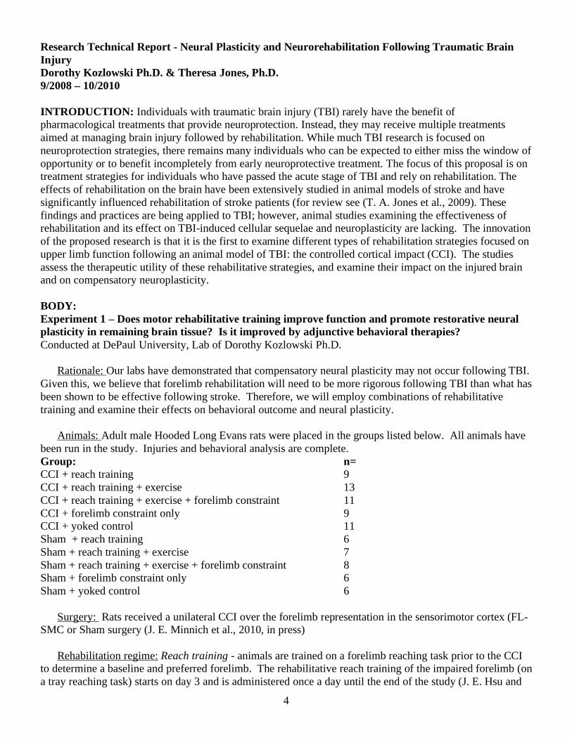
Tests which examine forelimb coordination and asymmetries in forelimb use also demonstrate that combining all 3 rehabilitation tasks (but not each individually) promote enhanced coordination and less symmetrical forelimb use (Figure 2).
Figure 2. Combining all three rehabilitation tasks promotes the recovery of forelimb coordination (left) and the return to more symmetrical limb use (right) following injury. We are still in the process of rating video-tapes of the vermicelli task. The vermicelli tasks is a task of manual dexterity that has shown to be sensitive to deficits following stroke, but has not been used to assess manual dexterity following TBI (CCI). To date we have rated the CCI-Yoked and Sham-Yoked animals. Preliminary data demonstrates that animals that receive CCi definitely show deficits in this task, specifically impairments in adjustments made by the injured paw, increased time to eat the pasta, as well as more abnormal handling behaviors. See Figure 3.
Figure 3. Analysis of Vermicelli Test. Rats that receive a TBI have significant deficits in manual dexterity.

7
Combining all three rehabilitation tasks also seemed to enhance performance on the rehabilitation tasks themselves. Analysis of the time it takes to complete the tray reaching rehabilitation task demonstrated that animals that received all 3 rehab tasks completed the tray reaching faster, and also seemed to run further in the exercise wheel. See Figure 4.
Figure 4. Combining all 3 rehabilitation tasks enhances the performance of the injured animals on the tray reaching tasks (left) and increases the distance they run in the exercise wheel (right). Preliminary Histology Results: The animals listed above have been sacrificed, brains harvested and sent to the lab of Theresa Jones for sectioning and staining. To date, the brains have been sectioned and one set stained for Nissl. Using the Nissl stained sections, Dorothy Kozlowski’s lab has analyzed the size of the contusions. Previous studies have shown that if animals receive too strenuous of a rehabilitation regime too early, it may result in an expansion of the size of the injury. We have demonstrated that there are no significant differences in contusion size at this time. We are still finalizing the analysis. See Figure 5 below.
Figure 5. Volume of remaining cortex is significantly decreased in all CCI groups compared to Sham. However the animals receiving rehabilitation do not have a larger contusion size than CCI+yoked animals. The remaining sets of brain sections are currently being stained with markers of plasticity using immunohistochemistry. We have completed immunohistochemical staining for dendrites (MAP2) and the anatomical quantification of these samples is close to completion. In addition, we are currently processing tissue samples for the other plasticity markers: synaptophysin, spinophilin (pre- and post-synaptic markers,
Distance Run in Exercise Wheel
0
1000
2000
3000
4000
5000
6000
14 15 16 17 18 19 20 21 22 23 24 25 26 27 28 29 30 31 32 33 34 35 36 37 38 39 40 41 42
Day of Study
Dist
ance
Run
(met
ers)
Sham+Reach+Exercise
CCI+Reach+Exercise
CCI+Reach+Exercise+ForelimbConstraint

8
respectively), GAP-43 (axonal growth-related protein), NF200 (axon marker). Quantitative analysis will rely on stereological measures or optical densitometry, as appropriate, given staining patterns. Sample locations will be the remaining sensorimotor cortex around the injury, in the contralateral homotopic cortex and subcortical structures, including the striatum and thalamus (Hsu & Jones, 2006). Experiment 2 – Does motor rehabilitation promote synaptic structural and functional plasticity in remaining motor cortex following TBI? Rationale: After stroke, functional improvements resulting from motor rehabilitation focused on an impaired forelimb has been found to induce functional reorganization and synaptic plasticity in remaining motor cortex. This experiment will determine whether a rehabilitation regime shown to be most effective in improving function in Exp. 1 also results in parallel plasticity of maps and synapses. In addition to validating and extending the results of Exp.1, this study is important for revealing potential mechanisms of functional recovery that might be manipulated or facilitated to further improve function. Animals: Adult male hooded Long Evans rats were placed in the following groups. Power Analysis was conducted as in Exp. 1. An “n” of 13 animals per group was conservatively estimated (the larger sample size that that used in Experiment 2 was needed based on variability in the measure of motor map plasticity evident in preliminary results from a non-DOD funded experiment that were summarized in the last project report.) Group: n= CCI+Reach Training+ Exercise+Constraint) 13 CCI+Control 13 TOTAL =26 Motor Mapping and Electron Microscopy (EM): Synaptic structural and functional plasticity of remaining motor cortex will be assayed using stereological methods for quantitative EM and intracortical microstimulation (ICMS) mapping, respectively, within the same animals. CCI, reach training, exercise and behavioral assays will be as in Experiment 1. Digital videotapes of behavioral measures will be sent to the Kozlowski laboratory for analysis. On day 42-45 post CCI, the caudal and rostral forelimb areas and surrounding regions of both hemispheres will be mapped using standard rodent ICMS techniques under ketamine-xylazine anesthesia, a procedure established in collaboration with Jeffrey Kleim (unpaid consultant). In this method, the organization of motor cortical movement representations are revealed in detail by stimulating primary motor cortex in a systematic (grid like) manner. In this initial experiment, mapping will not be conducted multiple times (because repetitive ICMS may impact the CCI or the EM), but it is expected that this study will set the stage for future within-animal analyses of the time course of map changes. Immediately after mapping, identified cortical forelimb representation regions of either hemisphere will be processed for EM. The density and ultrastructural characteristics of synapses in the motor cortex will be assayed using well-established quantitative approaches. Preliminary Results: Experiment 2 relied on knowing the results of Experiment 1 to reveal the optimal rehabilitation regime, which was found to be Reach Training+ Exercise+ Forelimb Constraint. We have just completed the behavioral training phase of this experiment in which CCI rats received either the optimal regime or served as yoke controls (receiving no reach-training control procedures, access to a locked running wheel and placement in vests permitting use of both forelimbs). The behavioral data are still being analyzed and more behavioral data remain to be collected from video-tapes. However, preliminary analyses have been performed. Analyses of reaching performance indicate, the combined rehabilitation regime resulted in

9
behavioral improvements in reaching performance similar to that found in Experiment 1. Furthermore, it resulted in greater normalization in the way animals moved in the performance of the reaching task, as shown in Figure 6. That is, they were both more successful and had improved movement quality in performance of the task. This provides an important replication of the treatment effects in a different lab, supporting the reliability of the effects on a behavioral level. Microstimulation mapping analyses and post-mortem measures of neural plasticity are currently ongoing, and are in an early phase. Very early motor mapping results point to potential that the behavioral improvements are coupled with motor map plasticity (Figure 7).
Figure 6 - Frame-by-frame movement analysis revealed a reduction in movement abnormalities in performance of the skilled reaching task as a result of rehabilitative training. Larger scores indicated greater abnormality. Movements types are shown left to right in the sequence in which they are performed during reaching: aiming and advancing the limb through the chamber window towards the pellet, then opening the digits, pronating the paw and grasping the pellet, followed by withdrawing the limb back through the window, suppinating at the wrist (which has 2 phases) and releasing the pellet into the mouth).

10
Figure 6 - Example of ICMS derived movement representation maps from an intact motor cortex (top) and following CCI (right). Colors correspond to movements elicited by stimulation. Early results suggest that rehabilitative training with the combination of reach training, constraint therapy and exercise might increase movement representation areas of the forelimb (green and blue in the maps) and mouth (yellow). KEY RESEARCH ACCOMPLISHMENTS:
• Animals in Exp. 1 have been trained, injured, rehabbed and tested in Dr. Kozlowski’s lab • We are completing the final statistical analysis of all behavioral data. • Brains have been harvested and sent to Dr. Jones’ lab • Dr. Jones’ lab has sliced the brains and stained one set with Nissl and is in the midst of completing
immunohistochemistry on the remaining sets. • Dr. Kozlowski’s lab is analyzing contusion size. • Dr. Jones lab has established the CCI model in their lab with the assistance of Dr. Kozlowski. • The Jones’ lab has shown that CCI’s conducted at UT-Austin are comparable to those conducted at
DePaul University • Behavioral results of combined rehabilitative training have been replicated across Kozlowski and
Jones laboratories. • Final motor mapping and neural plasticity measures of the key replication study have been initiated in
the Jones laboratory. REPORTABLE OUTCOMES: Poster/Oral Presentations: K. McDonough, R. De La Torre, L. Ferguson, J. Stamschror, M. Colella, S. Lance, A. Pevstov, E. Ramos, D. Adkins, T. Jones, D. A. Kozlowski. “The vermicelli handling test: A new behavioral test to examine manual dexterity following traumatic brain injury (TBI) in the rat” . Presented at the National Neurotrauma Symposium, 6/10. Received Student Travel Award.
L. Ferguson, J. Stamschror, S. Lance, A. Pevtsov, M. Colella, E. Ramos, K McDonough, R. De La Torre, D. Adkins, T. Jones, D. A. Kozlowski. "Motor rehabilitative training, exercise, and forelimb constraint after a

11
controlled cortical impact (CCI) to the forelimb sensorimotor cortex: Differential effects on skilled motor function and recovery”. Presented at the National Neurotrauma Symposium, 6/10.
E. Ramos, L. Ferguson, J. Stamschror, S. Lance, A. Pevtsov, M. Colella, K. McDonough, R. De La Torre, D. Adkins, T. Jones, D. A. Kozlowski. “Forelimb constraint therapy following a controlled cortical impact (CCI) to the forelimb sensorimotor cortex (FL-SMC) in rats hastens recovery of skilled motor function.” Presented at the National Neurotrauma Symposium, 6/10. – Travel award. Ferguson, L., Adkins, D.L., McDonough, K., Stamschror, J., Colella, M., Jones, T.A., & Kozlowski, D.A. Cortical electrical stimulation of the motor cortex, exercise, and motor rehabilitative training after a controlled cortical impact to the forelimb sensorimotor cortex: Differential effects on skilled motor function. Society for Neuroscience Meeting, 2009. Kozlowski D.A., & Jones, T.A. Neurorehabilitation and Neuroplasticity Following Traumatic Brain Injury. Military Health Research Forum 2009. (poster and oral presentation) Adkins DL, Tennant KA, Donlan N, Jones TJ, Kozlowski D (2009) Cortical electrical stimulation of the motor cortex after a controlled cortical impact to the forelimb sensorimotor cortex improves skilled motor function. National Neurotrauma Society Abstract #226. Peer Reviewed CONCLUSIONS: • Following CCI, behavioral function was most enhanced by combining tray reaching, exercise, and
forelimb constraint on all behaviors measured. • Combining all three rehabilitation therapies enhanced performance on rehabilitation tasks. • Rehabilitation does not increase the size of the contusion. • CCI results in a drastic loss of movement representations in the motor cortex, an effect far more
severe than expected based on stroke models. • Despite this, the motor cortex near the contusion maintains the capacity for motor map plasticity. This study demonstrates that neural plasticity may be limited following TBI and that the rehabilitation protocol required to produce behavioral enhancements is more extensive than that seen in a similarly sized lesion due to stroke. However, it also indicates that the capacity for functionally beneficial reorganization of motor cortex is possible. REFERENCES: Hsu JE, Jones TA (2006) Contralesional neural plasticity and functional changes in the less-affected forelimb
after large and small cortical infarcts in rats. Exp Neurol 201:479-494. Jones TA, Chu CJ, Grande LA, Gregory AD (1999) Motor skills training enhances lesion-induced structural
plasticity in the motor cortex of adult rats. J Neurosci 19:10153-10163. Jones TA, Allred RP, Adkins DL, Hsu JE, O'Bryant A, Maldonado MA (2009) Remodeling the brain with
behavioral experience after stroke. Stroke 40:S136-138.

12
Maldonado MA, Allred RP, Felthauser EL, Jones TA (2008) Motor skill training, but not voluntary exercise, improves skilled reaching after unilateral ischemic lesions of the sensorimotor cortex in rats. Neurorehabil Neural Repair 22:250-261.
Minnich JE, Mann SL, Stock M, Stolzenbach KA, Mortell BM, Soderstrom KE, Bohn MC, Kozlowski DA (2010) Glial cell line-derived neurotrophic factor (GDNF) gene delivery protects cortical neurons from dying following a traumatic brain injury. Restor Neurol Neurosci 28:293-309.
APPENDIX: None.