
Dopamine Levels in the Brain of Rat Models of Human ...
35
University of the Incarnate Word e Athenaeum eses & Dissertations 12-2017 Dopamine Levels in the Brain of Rat Models of Human Rheumatoid Arthritis Amelia Stinson University of the Incarnate Word, [email protected] Follow this and additional works at: hps://athenaeum.uiw.edu/uiw_etds Part of the Animal Sciences Commons , Cell and Developmental Biology Commons , Laboratory and Basic Science Research Commons , Medical Sciences Commons , Medical Specialties Commons , and the Mental and Social Health Commons is esis is brought to you for free and open access by e Athenaeum. It has been accepted for inclusion in eses & Dissertations by an authorized administrator of e Athenaeum. For more information, please contact [email protected]. Recommended Citation Stinson, Amelia, "Dopamine Levels in the Brain of Rat Models of Human Rheumatoid Arthritis" (2017). eses & Dissertations. 335. hps://athenaeum.uiw.edu/uiw_etds/335
Transcript of Dopamine Levels in the Brain of Rat Models of Human ...

University of the Incarnate WordThe Athenaeum
Theses & Dissertations
12-2017
Dopamine Levels in the Brain of Rat Models ofHuman Rheumatoid ArthritisAmelia StinsonUniversity of the Incarnate Word, [email protected]
Follow this and additional works at: https://athenaeum.uiw.edu/uiw_etds
Part of the Animal Sciences Commons, Cell and Developmental Biology Commons, Laboratoryand Basic Science Research Commons, Medical Sciences Commons, Medical Specialties Commons,and the Mental and Social Health Commons
This Thesis is brought to you for free and open access by The Athenaeum. It has been accepted for inclusion in Theses & Dissertations by an authorizedadministrator of The Athenaeum. For more information, please contact [email protected].
Recommended CitationStinson, Amelia, "Dopamine Levels in the Brain of Rat Models of Human Rheumatoid Arthritis" (2017). Theses & Dissertations. 335.https://athenaeum.uiw.edu/uiw_etds/335

DOPAMINE LEVELS IN THE BRAIN OF RAT MODELS OF HUMAN RHEUMATOID ARTHRITIS
by
AMELIA STINSON
A THESIS
Presented to the Faculty of the University of the Incarnate Word in partial fulfillment of the requirements
for the degree of
MASTER OF SCIENCE
UNIVERSITY OF THE INCARNATE WORD
December 2017

ii
© 2017 Amelia Stinson

iii
Acknowledgments
Throughout this academic journey I have experienced a great amount of support,
understanding, and care from my family, friends, coworkers, and colleagues. My mentor
Dr. Carlos Garcia has given me the tools necessary to further advance my knowledge of
academic research. His willingness to work with me through my difficult work schedule
provided me with continuous motivation, and I could not have completed this project
without his extensive guidance and patience.
My supervisor Felix Chavez, and team lead John Garza have both been flexible
and patient with me throughout these two and a half years. I am thankful for their long
discussions regarding proper HPLC procedures and techniques.
My colleagues Han Yang, Jordan Wetz, and Rejeana Stephens provided me with
assistance in Lewis rat care, sacrifice, and experiments. Additionally, I would like to
acknowledge my committee members Christopher Pierce, and David Starkey for teaching
me how to run protein assays and for enhancing my writing. I am thankful for the UIW
Biology and Chemistry Department entirely for providing me with the proper equipment,
solvents, and reagents.

iv
The University of Texas at San Antonio Laboratory Animal Resources Center
(LARC) provided housing and care for the rats. I am thankful for the training
coordinator at LARC Laurie Long, for providing me with a training program to
properly inject and handle rats.
This experience overall has been an intense and difficult process, but I am
grateful for everyone who has pushed me to complete this program.
Amelia Stinson

v
Dedication
This thesis is dedicated to my mother, father, and best friend Marsha Watson. Their love,
support, encouragement, and dedication to see me succeed gave me the strength to become who I
am today.
vi
DOPAMINE LEVELS IN THE BRAIN OF RAT MODELS OF HUMAN RHEUMATOID ARTHRITIS
Amelia Stinson
University of the Incarnate Word, 2017 Research Focus. Rheumatoid arthritis is a chronic, debilitating, autoimmune disease that causes the
destruction of bone tissue and the articular structures of joints. At least 30% of RA patient
populations have cognitive impairment. Acidic dopamine (DA) is the principal
neuroimmunotransmitter that links the central nervous system and peripheral nervous system
together. The aim of the present study was to determine the levels of DA and its two acidic
metabolites: 3,4-dihydroxyphenylacetic acid (DOPAC) and homovanillic acid (HVA) in arthritic
induced rats, and whether their levels vary across four different parts of the brain: amygdala, front
cerebral cortex, hippocampus, and cerebellum. Brain protein was also assessed.
Materials and Methods. 3-month old male Lewis Rats (n = 16) were randomized into either
control (n = 10) or treatment (n = 6) groups. In the treatment group, arthritis was induced in the rats
using Freund’s Adjuvant and all rats were sacrificed on day 28. Dopamine, DOPAC, and HVA
levels were quantified using High Performance Liquid Chromatography technique while proteins
were quantified using Bicinchroninic Acid Protein Assay, in the four brain regions. Two-way
ANOVA test was performed to determine whether brain regions, induce arthritis treatment or their
interactions significantly influenced the levels of the analytes (at p < .05).
Research Results/Findings. Levels of brain protein (C-reactive protein) were elevated in arthritic
rats across all brain regions (p > .05). Dopamine and DOPAC levels were lower in arthritic rats than
vii
controls (p > 0.05). HVA levels were higher in arthritic rats compared to non-arthritic controls.
Conclusions from Research. The present study has demonstrated that C-reactive protein,
dopamine, DOPAC and HVA are involved in the neurophysiology of arthritis. RA patients can
benefit from treatment with dopamine agonists. However, more studies are warranted to determine
the effect of DOPAC and HVA levels in the brain on dopamine utilization in arthritis.
viii
Table of Contents
LIST OF TABLES ............................................................................................................................................ x
LIST OF FIGURES ......................................................................................................................................... xi
CONTEXT OF THIS STUDY ...................................................................................................................... 1
MATERIALS AND METHODS................................................................................................................... 4
Animal Model of Human Rheumatoid Arthritis ...................................................................................... 4
Adjuvant Induced Arthritis .......................................................................................................................... 4
Brain Tissue Preparation .............................................................................................................................. 5
Bicinchroninic Acid (BCA) Protein Assay ................................................................................................ 6
High Performance Liquid Chromatography (HPLC) .............................................................................. 7
Statistical Analyses ........................................................................................................................................ 8
RESULTS ........................................................................................................................................................... 8
Adjuvant-Induced Rheumatoid Arthritis ................................................................................................... 8
Protein levels in four parts of the brain ..................................................................................................... 9
Dopamine levels in four parts of the brain ............................................................................................. 10
DOPAC levels in four parts of the brain ................................................................................................. 11
HVA levels in four parts of the brain ...................................................................................................... 12
DISCUSSION AND CONCLUSION ........................................................................................................ 13
Brain protein ................................................................................................................................................ 13
Dopamine ..................................................................................................................................................... 14
DOPAC and HVA levels ........................................................................................................................... 15 Limitations of the Study and Future Perspective ................................................................................... 16

ix
Conclusion ................................................................................................................................................... 16
REFERENCES ............................................................................................................................................... 17
Appendix A: Two-way ANOVA test of protein levels in four parts of the brain ................................ 20
Appendix B: Two-way ANOVA test of dopamine levels in four parts of the brain ............................ 21
Appendix C: Two-way ANOVA test of DOPAC levels in four parts of the brain .............................. 22
Appendix D: Two-way ANOVA test of HVA levels in four parts of the brain ................................... 23
x
List of Tables Table Page 1: Induced-arthritis Scoring Scale .................................................................................................................... 5 2: Dilution Scheme for Standard Test Tube Procedure ............................................................................... 6 3: Dilution Scheme for Sample Test Tube Procedure .................................................................................. 7

xi
List of Figures
Figure Page 1. Rat model of adjuvant-induced rheumatoid arthritis (RA) ...................................................................... 5 2. Total protein concentration (ug/ml) brains parts of arthritic (treated) versus non-arthritic (control)
Lewis rats ............................................................................................................................................... 9 3. The levels of dopamine in CBL, AMG, HIP and CX. Treated (Arthritic) rats exhibited lower levels of dopamine than control rats, the difference being statistically significant for
CBL (P =0.0170) and CX (0.0095) only ......................................................................................... 10 4. The levels of DOPAC in CBL, AMG, HIP and CX. DOPAC was undetectable in one control (HIP) and one treated brain part (CBL) ............................................................................ 11 5. HVA levels in four parts of the brain ....................................................................................................... 12

1
Context of This Study
Rheumatoid arthritis (RA) is a chronic, debilitating autoimmune disease that causes the
destruction of bone tissue and the articular structures of joints. Thus, RA is characterized by
infiltration of inflammatory immune cells, particularly, T-lymphocytes (T cells), macrophages, and
neutrophils in joints. Activation of microphages immune cells results in the production of
proinflammatory cytokine, particularly interleukin (IL)-1, IL-6, IL-12 and tumor necrosis factor-
alpha (TNF-α), which are involved in the up-regulation of inflammatory reactions (Navegantes et al.
2017). This causes synovial inflammation, eventually leading to joint destruction and disability
(Gorman & Cope, 2008; Tobon et al., 2010). While the etiology of RA is not well understood, it is
thought to be a multifactorial disorder involving a complex interplay between genetic, (Kurkó et al.,
2013; Okada et al., 2013;) and environmental factors, such as tobacco smoking (Tobon et al. 2010),
bodily pain, decreased mobility, fatigue, stiffness, physical disability and the way RA patients have to
cope with these problems during the activities of daily living, significantly affect their health-related
quality of life (HR-QOL) (Matcham et al., 2014; Russell, 2008). There is some evidence that RA also
impacts negatively on mental health (Matcham et al. 2014), indicating that there is a causal
association between RA and psychiatric disorders (Nicassio, 2010). Psychological disorders in RA
include depression, anxiety, emotional problems, and overall cognitive dysfunction a phenomenon
referred to as “brain fog” (Covic et al., 2012; Dickens et al., 2001; Hewlett et al., 2005). Cognitive
impairment in RA patient population is >30% (Joaquim et al., 2004; Shin et al., 2012;), with a
majority exhibiting worse cognitive score in psychometric measures for verbal fluency and memory
(Joaquim et al., 2004). In a longitudinal cohort study, Shin et al. (2012) demonstrated that a
subgroup of RA patients with cognitive impairment exhibited significantly poor fine motor skills
than the rest of RA patients. This implied that there should be a correlation between physical
function and cognitive function in RA. Shin et al. (2013) demonstrated cognitive impairment was

2
significantly associated with greater functional limitations in RA patients, although only depression
and fatigue (due to coping with functional limitations), were significantly associated with perceived
cognitive dysfunction.
Psychological disorders like depression and anxiety in inflammation disorders are associated
with chronic peripheral inflammation mediated by pro-inflammatory cytokines (TNFα, IL-1β, and
IL-6) (Süβ et al., 2015). Initially, cognitive dysfunction in RA patients was thought to be due the
highly elevated systemic markers of inflammation, particularly C-reactive protein, which can
potentially damage the integrity of blood-brain barrier thereby causing neuroinflammation (Eagan et
al., 2012; Elwood et al., 2017). Neuroinflammation was thought to affect hippocampal plasticity,
which triggers depression as repair response (Süβ et al., 2015; Wager-Smith et al. 2010). However, it
has been demonstrated recently that hippocampal immunity, cellular plasticity and function are
maintained despite severe peripheral autoimmune-mediated inflammation (Süβ et al. 2015). By this
account, it can be inferred that there should be neuronal factors underlying psychological disorders
in RA rather than systemic inflammations.
The neural activities in the brain are mediated by a wide repertoire of neurotransmitters in
the brain. However, dopamine, gamma-Aminobutyric acid (GABA), and serotonin are among the
few neurotransmitters frequently studied in relation to mental disorders due to their aggressive
nature from neuropsychology and interdisciplinary perspectives (Narvaes, & Martins de Almeida,
2014). Acidic dopamine (DA) is the principal neurotransmitter involved in both the central nervous
system (CNS) and peripheral nervous system (Basu & Dasgupta, 2000). It is one of the precursors to
norepinephrine and epinephrine allowing it to be involved in the parasympathetic nervous system
(PNS) as well. A decline in DA concentration or inhibition of its synthesis or metabolism rates in
the CNS can result in impairment of critical neurologic functions, such as cognition, behavior and
fine movements.

3
Surprisingly, apart from its principle neurotransmitter function in the CNS, DA also has
some inductive effect on a multitude of T-cells and dendritic cells (DCs), and therefore, regarded as
a neuroimmunotransmitter. The role of DA in the neural-immune communication has been
demonstrated in some in vivo studies reviewed previously by Basu and Dasgupta (2000). The studies
revealed that in vivo injury or stimulation of specific central dopaminergic (DA) system results in the
suppression or enhancement of functional activities of the immune effector cells, especially T-cells
and antigen-presenting DCs. While DCs expresses a complete machinery to synthesize and store
dopamine, regulatory T-cells expresses dopamine receptors (DRs) and, therefore, have the ability to
release, produce, and uptake dopamine (Pacheco et al. 2014). This underpins dopamine-mediated
integration of the immune system to the CNS, where dopaminergic immune regulations could be the
driving force during autoimmunity and associated inflammations (Pacheco et al. 2014). Thus,
malfunctioned DRs on immune cells including T-cells could also be major cause of overall cognitive
impairment. Impairment of the DRs implies a blockade of the interaction between T-cells and
dendritic cells, and therefore, leading to impairment of dopaminergic immune regulations.
Monocyte-derived DCs produce DA and store it in vesicles. The interactions between T-
cells and the DCs affect differentiation, polarization, and the secretion of pro-inflammatory
cytokines (Levite, 2008). It has been shown (Levite, 2008) that high (10-4 - 10-3M) and medium
levels (10-7 - 10-5 M) of DA are toxic to the T-cells and can result in unregulated stimulation.
Through this, it can be inferred that DA affects a multitude of systems. It is necessary to understand
and quantify the amounts present within these systems to link cognitive impairment to the body.
However, the total amount DA in CNS includes its two acidic metabolites: 3,4-
dihydroxyphenylacetic acid (DOPAC) and homovanillic acid (HVA). Thus, to better understand the
amount of DA and its longevity in the brain, it is important to consider its principle acidic
metabolites.
4
The purpose of study was to determine whether there are variations of DA concentration
and its related catecholamines in rat models of RA compared to healthy controls. Different parts of
the brain have different neurologic activities. The following amygdala, hypothalamus, front cerebral
cortex, hippocampus, and cerebellum, which exhibit strong role in memory, balance, emotional
stability, and overall cognition, were evaluated. The limbic system, which consists of the amygdala
and hippocampus, was the main area of interest since this is where spatial reasoning, memories, and
emotional behaviors are affected. The limbic system involves the dopaminergic mesolimbic pathway,
which is made up of movements from the ventral tegmental area to the nucleus accumbens. This
pathway along with the function of the two parts of interest has a role in motivation, and reward. A
difference in catecholamines in any of the affected areas would imply that inflammation, and/or
pain is not the only contributing source of cognitive impairment in patients with RA.
Materials and Methods Animal Model of Human Rheumatoid Arthritis Three-month old Male Lewis Rats (n=16) were purchased from Charles River Laboratories
and maintained at the vivarium in the University of Texas at San Antonio (UTSA). The rats were
maintained on a 12-hour light/12-hour dark cycle in a temperature and humidity controlled
environment.
Adjuvant Induced Arthritis The male rats were randomly assigned to one of two groups: control (non-arthritic), or the
arthritic group. In the arthritic group, arthritis was induced by methods previously described by
Tanimoto et al., (2015). Briefly, an intradermal injection of 0.01 ml of Freund’s incomplete adjuvant
(Sigma Aldrich™) was injected on the base of the tail (1 mg heat killed Mycobacterium tuberculosis
H37Rak, suspended in 1.0 mL of liquid paraffin) at day one and day 7. Arthritis is usually present
within 7 days after injection of the adjuvant. To confirm arthritis, erythema, swelling, and deformity
5
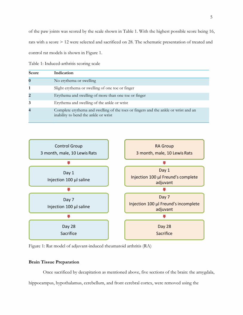
of the paw joints was scored by the scale shown in Table 1. With the highest possible score being 16,
rats with a score > 12 were selected and sacrificed on 28. The schematic presentation of treated and
control rat models is shown in Figure 1.
Table 1: Induced-arthritis scoring scale Score Indication
0 No erythema or swelling
1 Slight erythema or swelling of one toe or finger
2 Erythema and swelling of more than one toe or finger
3 Erythema and swelling of the ankle or wrist
4 Complete erythema and swelling of the toes or fingers and the ankle or wrist and an inability to bend the ankle or wrist
Figure 1: Rat model of adjuvant-induced rheumatoid arthritis (RA) Brain Tissue Preparation Once sacrificed by decapitation as mentioned above, five sections of the brain: the amygdala,
hippocampus, hypothalamus, cerebellum, and front cerebral cortex, were removed using the
RA Group 3 month, male, 10 Lewis Rats
Day 1 Injection 100 µl Freund’s complete
adjuvant
Day 7 Injection 100 µl Freund’s incomplete
adjuvant
Day 28 Sacrifice
Control Group 3 month, male, 10 Lewis Rats
Day 1 Injection 100 µl saline
Day 7 Injection 100 µl saline
Day 28 Sacrifice

6
Stereotaxic Atlas of Paxinos and Watson as a guide. Additional organs of interest (lungs, heart, kidneys,
and eyes) were also collected, removed, and immediately flash frozen in liquid nitrogen for transport
to the University of the Incarnate Word (UIW). The organs were then stored at in the -80ºC UIW
freezer for later analysis.
Bicinchroninic Acid (BCA) Protein Assay Frozen brain tissues were homogenized using a stand homogenizer with 750µL of 0.2N
perchloric acid buffer. The homogenized brain solutions (homogenates) were then centrifuged at
10,000 rpm, 4ºC for 10 minutes. The supernatant was transferred to a different tube and the pellet
discarded. A Microplate BCA Protein Assay Kit, from Thermo Scientific™ was used to calculate the
total amount of protein in a given sample of tissue homogenate. BCA protein assays were used a
standardized curve to determine the amount of protein in each individual supernatant. The scheme
of the standardized curve was as follows:
Table 2: Dilution scheme for standard test-tube procedure
Vial Volume diluent(µL) Volume and source of BSA (µL)
Final BSA concentration (µg/mL)
A 66 200 of stock 1500
B 100 100 of stock 1000
C 100 100 of vial A dilution 750
D 100 100 of vial B dilution 500
E 100 100 of vial D dilution 250
F 100 100 of vial E dilution 125
G 100 100 of vial F dilution 62.5
H 100 0 0=Blank
Samples were then be diluted to achieve an overall R2 value of 0.9 or higher. The scheme of the
sample curve is shown in table 3.

7
Table 3: Dilution scheme for sample test tube procedure
Vial Volume diluent (µL) Volume and source of stock sample supernatant (µL)
Final sample concentration (%)
AS 0 21 100
BS 7 21 75
CS 21 21 50
DS 63 21 25
Once all standard and sample dilution vials were prepared, 9µL of brain samples and standards were
loaded onto a microplate. 4µL of the compatibility reagent solution was then added to each well and
incubated for 15 minutes. Then, 260µL of the working reagent solution prepared earlier (50:1 ratio
of BCA working reagent A to BCA working reagent B provided by Thermo Scientific™) was added
into each well and allowed to incubate for 30 minutes. A protein gradient then begun to form within
the wells. Absorbance was then measured at 562nm using the Infinite 200 PRO Nano Quant Tecan
Microplate Reader. Protein amounts were calculated by plotting the absorbance values read from the
samples vs. the standardized curve, resulting in the protein concentration of each supernatant in
µg/mL.
High Performance Liquid Chromatography (HPLC) High Performance Liquid Chromatography (HPLC) was used for quantification of DA,
HVA, and DOPAC in the brain of both control and arthritic rats. HPLC allows for the distinct
separation of compounds by carrying the injected supernatant through a packed column (stationary
phase) with a solvent (mobile phase). As the sample is passed through the column, it interacts
between the two phases at a different rate. Analytes in the sample that have the least amount of
interaction with the mobile phase, will elute the column sooner, and pass through the detector at
different rates, resulting in varied retention times.
8
A modular design of the Agilent Infinity 1220 HPLC was used since it provides enough
versatility and integration capabilities to overcome analytical challenges. 100µL of brain sample and
standards were prepared and directly injected for analysis. A gradient elution table was used to
ensure clean separation of peaks. Mobile phases consisted of ACN, H2O, and a buffer of 0.1 M
trifluoroacetic acid (TFA). The mobile phase was used to bring the samples through a Zorbax SB-
C18 reversed phased column (Agilent technology). Samples were read at a 250nm absorption
wavelength. Each sample was injected in triplicates at a flow of 0.5µL per minute for ~10 minutes.
The area under each sample’s analyte curve compared to that of the standards curve were
used to determine the concentration of each analyte in ng of specific analyte/ mg of protein. Proper
storage of the column and rinsing before and after each run was carried out using a solution of
50/50 methanol to water, to ensure that no carryover of any analyte peak was in sequential runs.
Statistical Analyses The difference in levels of dopamine, DOPAC, HVA in the five different parts of the brain
of arthritic groups versus non-arthritic controls were compared using t-test and two-way analysis of
variance (two-way ANOVA) test. Differences between groups were considered to be statistically
significant at a P value of <0.05. Statistical analyses were performed using GraphPad Prism version
7.03 for Windows (GraphPad Software, Inc., San Diego, CA, USA, www.graphpad.com).
Results Adjuvant-Induced Rheumatoid Arthritis The levels of dopamine, DOPAC, HVA were measured in the cerebral cortex (CX),
hippocampus (HIP), hypothalamus (HYP), amygdala (AMG), and cerebellum (CBL) regions of the
brains of rats with adjuvant-induced rheumatoid arthritis and non-arthritic controls. However, due
to lack of hypothalamus (HYP) brain tissue samples from the arthritic rats, tests for HYP were
performed for controls only. Therefore, results for HYP were excluded from the analysis.
9
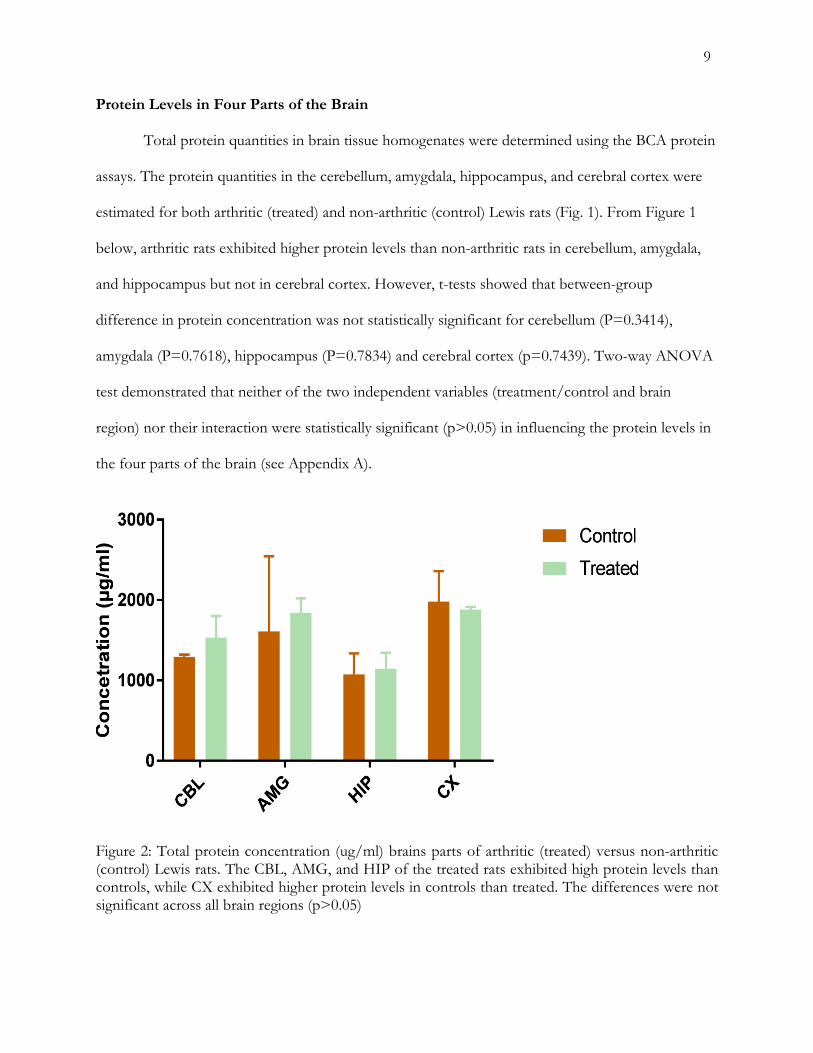
Protein Levels in Four Parts of the Brain
Total protein quantities in brain tissue homogenates were determined using the BCA protein
assays. The protein quantities in the cerebellum, amygdala, hippocampus, and cerebral cortex were
estimated for both arthritic (treated) and non-arthritic (control) Lewis rats (Fig. 1). From Figure 1
below, arthritic rats exhibited higher protein levels than non-arthritic rats in cerebellum, amygdala,
and hippocampus but not in cerebral cortex. However, t-tests showed that between-group
difference in protein concentration was not statistically significant for cerebellum (P=0.3414),
amygdala (P=0.7618), hippocampus (P=0.7834) and cerebral cortex (p=0.7439). Two-way ANOVA
test demonstrated that neither of the two independent variables (treatment/control and brain
region) nor their interaction were statistically significant (p>0.05) in influencing the protein levels in
the four parts of the brain (see Appendix A).
Figure 2: Total protein concentration (ug/ml) brains parts of arthritic (treated) versus non-arthritic (control) Lewis rats. The CBL, AMG, and HIP of the treated rats exhibited high protein levels than controls, while CX exhibited higher protein levels in controls than treated. The differences were not significant across all brain regions (p>0.05)

10
Dopamine Levels in Four Parts of the Brain Dopamine concentrations in the four different parts of the brain from both arthritic
(treated) and non-arthritic (control) Lewis rats were determined using the HPLC (Fig. 3). From
Figure 3 below, arthritic rats exhibited lower dopamine concentration than non-arthritic rats across
all the four parts of the brain. This was statistically significant in the cerebellum (P =0.0170) and
cerebral cortex (0.0095), but not significant in amygdala P=0.2527) and hippocampus (P=0.7779).
Two-way ANOVA test demonstrated that brain regions (cerebellum, amygdala, hippocampus and
cerebral cortex) as an independent variable had statistically significant effect on the dopamine
concentration (P=0.0131), but not its interactions (P=0.4002). On the other hand, treatment and
control or their interactions did not seem to have any statistically significant effect on the
distribution of dopamine across the four brain parts (p>0.05) (see Appendix B).
Figure 3: The levels of dopamine in CBL, AMG, HIP and CX. Treated (Arthritic) rats exhibited lower levels of dopamine than control rats, the difference being statistically significant for CBL (P =0.0170) and CX (0.0095) only.

11
DOPAC Levels in Four Parts of the Brain
Using HPLC, the concentration of DOPAC, a metabolite of dopamine, was determined in
the four different parts of the brain of both arthritic (treated) and non-arthritic (control) Lewis rats
(Fig. 4). From Figure 4 below, arthritic rats exhibited a lower DOPAC levels than non-arthritic rats
in amygdala, though not statistically significant (p=0.4314). Both arthritic and non-arthritic Lewis
rats, exhibited very low concentrations of DOPAC in hippocampus and cerebral cortex, with
undetectable DOPAC observed in cerebellum of arthritic rats and hippocampus of non-arthritic
rats. Two-way ANOVA test demonstrated that neither of the two independent variables
(treatment/control and brain region) or their interaction were statistically significant (p>0.05) (see
Appendix C).
Figure 4: The levels of DOPAC in CBL, AMG, HIP and CX. DOPAC was undetectable in one control (HIP) and one treated brain part (CBL).
12
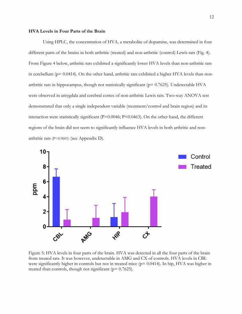
HVA Levels in Four Parts of the Brain Using HPLC, the concentration of HVA, a metabolite of dopamine, was determined in four
different parts of the brains in both arthritic (treated) and non-arthritic (control) Lewis rats (Fig. 4).
From Figure 4 below, arthritic rats exhibited a significantly lower HVA levels than non-arthritic rats
in cerebellum (p= 0.0414). On the other hand, arthritic rats exhibited a higher HVA levels than non-
arthritic rats in hippocampus, though not statistically significant (p= 0.7625). Undetectable HVA
were observed in amygdala and cerebral cortex of non-arthritic Lewis rats. Two-way ANOVA test
demonstrated that only a single independent variable (treatment/control and brain region) and its
interaction were statistically significant (P=0.0046; P=0.0463). On the other hand, the different
regions of the brain did not seem to significantly influence HVA levels in both arthritic and non-
arthritic rats (P=0.9601) (see Appendix D).
Figure 5: HVA levels in four parts of the brain. HVA was detected in all the four parts of the brain from treated rats. It was however, undetectable in AMG and CX of controls. HVA levels in CBL were significantly higher in controls but not in treated mice (p= 0.0414). In hip, HVA was higher in treated than controls, though not significant (p= 0.7625).

13
Discussion And Conclusion More than 30% of populations of population with RA have cognitive impairments (Shin et
al. 2012; Joaquim et al. 2004), with a significant subgroup of this RA patients exhibiting worse
cognitive function in verbal fluency, memory (Joaquim et al. 2004) and poor fine motor skills (Shin
et al. 2012). This study was based on the overarching hypothesis from the literature that acidic
dopamine (DA), which is the principal neuroimmunotransmitter linking the immune system to the
CNS and peripheral nervous system, its changes in the brain produces cognitive impairments in RA.
Furthermore, this study was also guided by the hypothesis, C-reactive protein (CRP), which is a
biomarker of cognitive impairment, should be elevated in the brains of RA patients with cognitive
impairment (Eagan et al. 2012; Elwood et al. 2017). The present study demonstrated variations in
the levels of both brain protein and DA with its two acidic metabolites (DOPAC and HVA) across
four parts of the brain: AMG, CX, HIP, and CBL, which are mainly involved in cognition and
motor function.
Brain Protein
The present study demonstrated elevated levels of total protein CBL, AMG, and HIP of
arthritic than non-arthritic rats, while CX exhibited higher protein levels in non-arthritic than
arthritic rats. Although not significant (p>0.05), the protein elevations in most of the brain regions
of arthritic rats indicated that the proteins could be associated with cognitive impairment in RA.
There several brain proteins involved in cognitive function or decline, including NPTX2—neuronal
pentraxin 2 (Xiao et al. 2017), CRP (Eagan et al. 2012), albumin (Llewellyn et al. 2010). However,
low levels of NPTX2 and albumin have been associated with cognitive impairment and neurological
diseases, such as Alzheimer’s Disease, in the elderly population (Llewellyn et al. 2010; Xiao et al.
2017). Although the present study did not characterize and identify the elevated protein components
in the arthritic rats, CRP, was most likely to be involved (Eagan et al. 2012; Elwood et al. 2017).

14
CRP was elevated in the AMG, which is associated with fear, depression and anxiety disorder
(Forster et al. 2012); CBL, which coordinates movement, motor learning or timing (Mauk et al.
2000); and HIP, which regulates emotions (Binder et al. 2012), but not in CX, which regulated high-
level brain functions, such as memory, cognition, and perception. This demonstrates that CRP was
largely elevated in the limbic system that regulates emotions and therefore, could be largely
responsible for depression in RA. This was supported by findings from previous studies, which
demonstrated that depression scores were slightly, but positively correlated with the CRP levels (r
0.46, P < 0.001) (Kojima et al. 2009), and that CRP was significant predictor of cognitive
impairment in patients with RA (Shin et al. 2012). However, in the present study, CRP was not
significantly elevated across the four brain regions of arthritic rats (p>0.05), indicating that it is not
partly contributes to cognitive impairment. Therefore, there should be neuronal factors that also
contribute to psychological disorders in RA rather in addition to systemic inflammation marker
CRP.
Dopamine
Dopamine, a neurotransmitter in the brain that is important in regulating movement,
emotional responses and sensations of pleasure and pain. It is critically regulating positive emotions
and motivation (Schultz, 2002), and therefore, low levels in the brain could be involved in cognitive
dysfunction in RA. Findings in the present study demonstrated that dopamine levels in the arthritic
rats were generally low across all the four limbic brain regions analyzed, where dopamine levels were
significantly low in CBL (P =0.0170) and CX (0.0095). There is a paucity of published data directly
compares the level of dopamine in arthritic-induced rats or RA patients to healthy non-arthritic
controls. However, low levels and altered dopamine neurotransmission has been observed in
patients with fibromyalgia, a chronic rheumatic condition characterized by generalized pain, fatigue,
and dysregulation of emotion. Pain in both fibromyalgia and RA are all attributed to increased levels
15
of proinflammatory cytokines. According to Miller et al. (2013), the levels and function of
neurotransmitters (including dopamine) are broadly modulated by proinflammatory cytokines, at
multiple levels. This is achieved by alteration of neurotransmitter synthesis, reuptake and release, via
the activation of metabolic and epigenetic processes (Miller et al. 2013). Given that autoimmunity is
coupled to CNS and PNS (Basu & Dasgupta, 2000), and the existence pathways linking systemic
inflammation to dopaminergic reward system (Sturgeon et al. 2016), dopamine could be centrally
involved cognitive impairment in autoimmune inflammatory diseases, including RA. According to
Sturgeon et al. (2016), RA patients are likely to develop alteration in dopaminergic
neurotransmission, and therefore, likely to develop cognitive and behavioral impairments.
This finding suggests that RA patients at increased risk of cognitive impairment can benefit
from treatment with dopamine agonists (Eijsbouts et al. 1999). However, given that autoimmunity in
RA is tightly coupled to CNS (Basu & Dasgupta, 2000), dopamine agonists is likely to suppress
inflammatory response and improve quality of life. A previous case report (Erb et al. 2001),
demonstrated that treating of unremitting RA with prolactin antagonist cabergoline resulted in a
significant remission. D1-like dopamine receptor antagonist have been shown to ameliorated the
severity of collagen-induced arthritis in mice, by promoting anti-inflammatory IFN-γ and inhibiting
pro-inflammatory interleukin (IL)-17 production by T-cells (Nakashioya et al. 2011). Currently, the
potential therapeutic benefits of dopamine-agonists are increasingly being studied for human RA.
DOPAC and HVA Levels
DOPAC and HVA are the acid metabolites of dopamine, and therefore, their levels in the
brain should correlate with the utilization of dopamine in the brain. Findings in the present study,
demonstrated very low levels of DOPAC in arthritic rats, with some brain parts being below
detectable limits. However, levels HVA were higher in arthritic rats compare to non-arthritic
controls. While low DOPAC levels as observed in arthritic rats indicates low utilization of dopamine

16
and low “turnover” of DA in the brain, this does not necessarily imply utilization of dopamine in
the dopaminergic systems (Ebinger et al. 1987). An early in vivo study of dopamine release and
metabolism in rat brain regions, demonstrated DOPAC/HVA ratio in varied between brain regions
in accordance with whole tissue measurements. This is consistent with findings in the present in vivo
study, which demonstrated varied DOPAC and HVA, levels of the difference regions of the arthritic
rats.
Limitations of the Study and Future Perspective The present in vivo study is the first to assess protein levels and utilization of
neurotransmitters in dopaminergic systems in arthritic rats, to assess their roles in cognitive
impairment in RA. Experimental materials were limited (arthritic rat brain samples) and therefore, it
was not possible to evaluate the hypothalamus. Furthermore, the calibration standards for DOPAC
and HVA appeared to be a bit higher, yet DOPAC and HVA are only available in smaller quantities
than the parent neurotransmitter substance (dopamine). This could explain why some parts of the
brain had undetectable levels of DOPAC and HVA. This warrants more in vivo studies to re-evaluate
DOPAC and HVA in arthritic rats using calibration standards of lower concentration. Future,
studies should focus on the potential therapeutic benefits of dopamine-agonists treating human RA
and associated risk of cognitive impairment.
Conclusion
A significant subgroup of RA patients is likely to develop cognitive impairment. The present
study has demonstrated that CRP, dopamine, DOPAC and HVA are involved the neuropathology
and associated cognitive impairments in RA. Therefore, RA patients at increased risk of cognitive
impairment can benefit from treatment with dopamine agonists. However, more studies are
warranted to determine the effect of DOPAC and HVA levels in the brain on dopamine utilization
in arthritic rats.

17
References Basu, S., & Dasgupta, P. S. (2000). Dopamine, a neurotransmitter, influences the immune system. J
Neuroimmunol, 102(2), 113-124. Binder, J., de Quervain, D. J., Friese, M., Luechinger, R., Boesiger, P., & Rasch, B. (2012). Emotion
suppression reduces hippocampal activity during successful memory encoding. Neuroimage, 63(1), 525-532. doi:10.1016/j.neuroimage.2012.07.007
Covic, T., Cumming, S.R., Pallant, J.F., Manolios, N., Emery, P., Conaghan, P.G., et al. (2012.)
Depression and anxiety in patients with rheumatoid arthritis: prevalence rates based on a comparison of the Depression, Anxiety and Stress Scale (DASS) and the hospital, Anxiety and Depression Scale (HADS). BMC Psychiatry 12,6.
Dickens, C., & Creed, F. (2001). The burden of depression in patients with rheumatoid arthritis.
Rheumatology, 40(12), 1327-1330. doi:10.1093/rheumatology/40.12.1327 Eagan, D. E., Gonzales, M. M., Tarumi, T., Tanaka, H., Stautberg, S., & Haley, A. P. (2012).
Elevated Serum C-Reactive Protein Relates to Increased Cerebral Myoinositol Levels in Middle-Aged Adults. Cardiovascular Psychiatry and Neurology, 2012, 9. doi:10.1155/2012/120540
Ebinger, G., Michotte, Y., & Herregodts, P. (1987). The significance of homovanillic acid and 3,4-
dihydroxyphenylacetic acid concentrations in human lumbar cerebrospinal fluid. J Neurochem, 48(6), 1725-1729.
Eijsbouts, A., van den Hoogen, F., Laan, R. F., Hermus, R. M., Sweep, F. C., & van de Putte, L.
(1999). Treatment of rheumatoid arthritis with the dopamine agonist quinagolide. J Rheumatol, 26(10), 2284-2285.
Elwood, E., Lim, Z., Naveed, H., & Galea, I. (2017). The effect of systemic inflammation on human
brain barrier function. Brain, Behavior, and Immunity, 62, 35-40. doi:10.1016/j.bbi.2016.10.020 Erb, N., Pace, A. V., Delamere, J. P., & Kitas, G. D. (2001). Control of unremitting rheumatoid
arthritis by the prolactin antagonist cabergoline. Rheumatology, 40(2), 237-239. doi:10.1093/rheumatology/40.2.237
Forster, G. L., Novick, A. M., Scholl, J. L., & Watt, M. J. (2012). The role of the amygdala in anxiety
disorders. In B. Ferry (Ed.), The amygdala: A discrete multitasking manager (pp. 61-102). Rijeka: InTech.
Gorman, C. L., & Cope, A. P. (2008). Immune-mediated pathways in chronic inflammatory arthritis.
Best Practice & Research Clinical Rheumatology, 22(2), 221-238. doi:10.1016/j.berh.2008.01.003 Hewlett, S., Cockshott, Z., Byron, M., Kitchen, K., Tipler, S., Pope, D., Hehir, M. (2005). Patients’
perceptions of fatigue in rheumatoid arthritis: Overwhelming, uncontrollable, ignored. Arthritis & Rheumatism, 53(5), 697-702. https://www.nature.com/articles/nature12873#supplementary-information
18
Joaquim, A. F., & Appenzeller, S. (2015). Neuropsychiatric manifestations in rheumatoid arthritis.
Autoimmunity Reviews, 14(12), 1116-1122. Kojima, M., Kojima, T., Suzuki, S., Oguchi, T., Oba, M., Tsuchiya, H., . . . Ishiguro, N. (2009). Depression, inflammation, and pain in patients with rheumatoid arthritis. Rheuatoid Arthritis,
61(8), 1018-1024. doi:10.1002/art.24647 Kurkó, J., Besenyei, T., Laki, J., Glant, T. T., Mikecz, K., & Szekanecz, Z. (2013). Genetics of
Rheumatoid Arthritis — A Comprehensive Review. Clinical Reviews in Allergy & Immunology, 45(2), 170-179. doi:10.1007/s12016-012-8346-7
Levite, M., 2008. Neurotransmitters activate T-cells and elicit crucial functions via neurotransmitter
receptors. Current Opinion in Pharmacology, 8(4), 460-471. Llewellyn, D. J., Langa, K. M., Friedland, R. P., & Lang, I. A. (2010). Serum albumin concentration
and cognitive impairment. Current Alzheimer research, 7(1), 91-96. Matcham, F., Scott, I. C., Rayner, L., Hotopf, M., Kingsley, G. H., Norton, S., . . . Steer, S. (2014).
The impact of rheumatoid arthritis on quality-of-life assessed using the SF-36: A systematic review and meta-analysis. Seminars in Arthritis and Rheumatism, 44(2), 123-130. doi:https://doi.org/10.1016/j.semarthrit.2014.05.001
Mauk, M. D., Medina, J. F., Nores, W. L., & Ohyama, T. (2000). Cerebellar function: Coordination,
learning or timing? Current Biology, 10(14), R522-R525. doi: https://doi.org/10.1016/S0960- 9822(00)00584-4
Miller, A. H., Haroon, E., Raison, C. L., & Felger, J. C. (2013). Cytokine targets in the brain: Impact
on neurotransmitters and neurocircuits. Depression and Anxiety, 30(4), 297-306. doi:10.1002/da.22084
Nakashioya, H., Nakano, K., Watanabe, N., Miyasaka, N., Matsushita, S., & Kohsaka, H. (2011). Therapeutic effect of D1-like dopamine receptor antagonist on collagen-induced arthritis of
mice. Modern Rheumatology, 21(3), 260-266. doi:10.3109/s10165-010-0387-2 Narvaes, R., & Martins de Almeida, R. (2014). Aggressive behavior and three neurotransmitters:
Dopamine, GABA, and serotonin—A review of the last 10 years. Psychology & Neuroscience, 7(4). 601-607
Navegantes, K. C., de Souza Gomes, R., Pereira, P. A. T., Czaikoski, P. G., Azevedo, C. H. M., &
Monteiro, M. C. (2017). Immune modulation of some autoimmune diseases: the critical role of macrophages and neutrophils in the innate and adaptive immunity. Journal of Translational Medicine, 15(1), 36. doi:10.1186/s12967-017-1141-8
Nicassio, P. M. (2010). Arthritis and Psychiatric Disorders: Disentangling the Relationship. Journal of
Psychosomatic Research, 68(2), 183-185. doi:10.1016/j.jpsychores.2009.09.008

19
Okada, Y., Wu, D., Trynka, G., Raj, T., Terao, C., Ikari, K., . . . Plenge, R. M. (2013). Genetics of
rheumatoid arthritis contributes to biology and drug discovery. Nature, 506, 376-381. doi:10.1038/nature12873
Pacheco, R., Contreras, F., & Zouali, M. (2014). The dopaminergic system in autoimmune diseases.
Frontiers in Immunology, 5, 117. doi:10.3389/fimmu.2014.00117 Russell, A. S. (2008). Quality-of-life assessment in rheumatoid arthritis. Pharmacoeconomics, 26(10),
831-846. Schultz, W. (2002). Getting formal with dopamine and reward. Neuron, 36(2), 241-263. Shin, S. Y., Katz, P., & Julian, L. (2013). Relationship between perceived cognitive dysfunction and
objective neuropsychological performance in persons with rheumatoid arthritis. Arthritis Care & Reseaerch, 65(3), 481-486. doi:10.1002/acr.21814
Shin, S. Y., Katz, P., Wallhagen, M., & Julian, L. (2012). Cognitive impairment in persons with
rheumatoid arthritis. Arthritis Care & Research, 64(8), 1144-1150. doi:10.1002/acr.21683 Sturgeon, J. A., Finan, P. H., & Zautra, A. J. (2016). Affective disturbance in rheumatoid arthritis:
psychological and disease-related pathways. Nature Reviews. Rheumatology, 12(9), 532-542. doi:10.1038/nrrheum.2016.112
Süβ, P., Kalinichenko L., Baum W., Reichel M., Kornhuber J., Loskarn S., Ettle B., … Schlachetzki,
J. C. (2015). Hippocampal structure and function are maintained despite severe innate peripheral inflammation. Brain, Behavior, & Immunity,
Tanimoto, A., Shinozaki, Y., Nozawa, K., Kimoto, Y., Amano, W., Matsuo, A., Matsushita, M.
(2015). Improvement of spontaneous locomotor activity with JAK inhibition by JTE-052 in rat adjuvant-induced arthritis. BMC Musculoskeletal Disorders, 16, 339.
Tobon, G. J., Youinou, P., & Saraux, A. (2010). The environment, geo-epidemiology, and
autoimmune disease: Rheumatoid arthritis. Journal of Autoimmunity, 35(1), 10-14. doi:10.1016/j.jaut.2009.12.009
Wager-Smith, K., & Markou, A. (2011). Depression: A repair response to stress-induced neuronal
microdamage that can grade into a chronic neuroinflammatory condition? Neuroscience and Biobehavioral Reviews, 35(3), 742-764. doi:10.1016/j.neubiorev.2010.09.010
Xiao, M.-F., Xu, D., Craig, M. T., Pelkey, K. A., Chien, C.-C., Shi, Y., . . . Worley, P. F.
(2017).NPTX2 and cognitive dysfunction in Alzheimer’s Disease. eLife, 6, e23798. doi:10.7554/eLife.23798

20
Appendix A: Two-way ANOVA test of protein levels in four parts of the brain
Table Analyzed Two-way ANOVA, not RM Two-way ANOVA Ordinary Alpha 0.05 Source of Variation % of total variation P value P value
summary Significant?
Interaction 2.655 0.9156 ns No Row Factor 53.28 0.0761 ns No Column Factor 1.694 0.5872 ns No ANOVA table SS DF MS F (DFn, DFd) P value Interaction 77159 3 25720 F (3, 8) =
0.1671 P=0.9156
Row Factor 1548419 3 516140 F (3, 8) = 3.353 P=0.0761 Column Factor 49238 1 49238 F (1, 8) =
0.3198 P=0.5872
Residual 1231624 8 153953 Number of missing values
0

21
Appendix B: Two-way ANOVA test of dopamine levels in four parts of the brain
Table Analyzed Two-way ANOVA, not RM Two-way ANOVA Ordinary Alpha 0.05 Source of Variation % of total variation P
value P value summary
Significant?
Interaction 14.09 0.4002 ns No Row Factor 9.424 0.556 ns No Column Factor 42.65 0.0131 * Yes ANOVA table SS DF MS F (DFn, DFd) P value Interaction 50.11 3 16.7 F (3, 8) = 1.11 P=0.4002 Row Factor 33.53 3 11.18 F (3, 8) =
0.7427 P=0.5560
Column Factor 151.7 1 151.7 F (1, 8) = 10.08
P=0.0131
Residual 120.4 8 15.05 Number of missing values
0

22
Appendix C: Two-way ANOVA test of DOPAC levels in four parts of the brain
Table Analyzed Two-way ANOVA, not RM
Two-way ANOVA Ordinary Alpha 0.05 Source of Variation % of total variation P
value P value summary Significant?
Interaction 19.89 0.4609 ns No Row Factor 16.86 0.525 ns No Column Factor 7.455 0.3314 ns No ANOVA table SS DF MS F (DFn, DFd) P value Interaction 132 3 43.98 F (3, 8) =
0.9507 P=0.4609
Row Factor 111.9 3 37.29 F (3, 8) = 0.8059
P=0.5250
Column Factor 49.46 1 49.46 F (1, 8) = 1.069 P=0.3314 Residual 370.1 8 46.27 Number of missing values
0

23
Appendix D: Two-way ANOVA test of HVA levels in four parts of the brain
Table Analyzed Two-way ANOVA, not RM
Two-way ANOVA Ordinary Alpha 0.05 Source of Variation % of total variation P value P value summary Significant?
Interaction 58.9 0.0046 ** Yes Row Factor 25.15 0.0463 * Yes Column Factor 0.00532 0.9601 ns No ANOVA table SS DF MS F (DFn, DFd) P value Interaction 50.71 3 16.9 F (3, 8) = 9.85 P=0.0046 Row Factor 21.65 3 7.216 F (3, 8) = 4.205 P=0.0463 Column Factor 0.004581 1 0.004581 F (1, 8) =
0.002669 P=0.9601
Residual 13.73 8 1.716 Number of missing values
0


![[3H]Glutamate- binding Sites in Rat Brain](https://static.fdocuments.in/doc/165x107/585ad4351a28ab6e32924c15/3hglutamate-binding-sites-in-rat-brain.jpg)


