Donor Bridge Acceptor
18
This journal is © The Royal Society of Chemistry 2014 Chem. Soc. Rev. Citethis:DOI: 10.1039/c 4cs00221k Photoinduced charge and energy transfer in molecular wires Me ´ lina Gilbert and Bo Albinsson* Expl orin g char ge and ene rgy transport in dono r–b ridg e–acceptor syst ems is an impo rtant rese arch field which is essential for the fundamental knowledge necessary to develop future applications. These studies help creating valuable knowledge to respond to today’s challenges to develop functionalized molecular systems for artificial photosynthesis, photovoltaics or molecular scale electronics. This tutorial revie w focuses on phot o-indu ced charg e/en ergy transfer in covalen tly linked dono r–bri dge–a ccepto r (D–B–A) systems. Of utmost importance in such systems is to understand how to control signal transmis- sion, i.e. how fast electrons or excitation energy could be transferred between the donor and acceptor and the role played by the bridge (the ‘‘molecular wire’’). After a brief description of the electron and energy transfer theory, we aim to give a simple yet accurate picture of the complex role played by the bridge to sustain donor–acceptor electronic communication. Special emphasis is put on understanding brid ge ener geti cs and conf ormation al dyna mics effects on the distance depe ndence of the donor– acceptor electronic coupling and transfer rates. Several examples of donor–bridge–acceptor systems from the literature are described as a support to the discussion. Finally, porphyrin-based molecular wires are introduced, and the relationship between their electronic structure and photophysical properties is outlined. In strongly conjugated porphyrin systems, limitations of the existing electron transfer theory to interpret the distance dependence of the transfer rates are also discussed. Key learning points Photoinduced electron and energy transfer reactions Superexchange theory; coherent tunneling vs. incoherent hopping Molecular bridges (wires) and distance dependence of electron and energy transfer reactions Conformational dependence of electron and energy transfer processes in D–B–A systems Influence of the bridge topology on electron and energy transfer in porphyrin-based molecular wires 1. Introduction Electron (ET) and excitation energy transfer (EET) are key steps of many chemical and physical processes occurring in disciplines ranging from biology, chemistry, and physics to materials science. For instance, both processes constitute the heart of natural photo- synthesis. 1 In any natural photosynt hetic system, light conversi on int o che mica l ene rgy req uir es a ser ies of ste p-wise ele ctro n tran sfe r pr oce sses to cre ate a lo ng li ve d cha rg e se par at ed st at e wi th suffici ent en ergy for w ater o xidat ion and carbon dioxi de reducti on. Over the last two decades, mimicking these elementary steps in designed systems has attracted much interest, motivated by the technological potential of such systems in applications ranging from artificial photosynthesis to molecular electronics . 2 However, going from simple mimics to actual functioning devices is not simple and requires a thorough understanding of all parameters controlling ET and EET processes, especially the kinetics. In this context, considerable theoretical and experimental efforts have been devoted to identifying the factors that govern charge and hole transfer in molecular systems. Valuable knowledge has been acquired by experimentally studying ET and EET processes in donor–bridge–acceptor (D–B–A) mimics of the more complicated natural photosynthetic system. In such D–B–A systems, a donor (D) and an acceptor (A) are covalently linked using a molecular bridge (B). As a bridge component, various structural motifs have been used. In this tutorial review, p-conjugated bridges consisting of covalently connected identical repetitive molecular motifs are considered. A list of such bridge structures, sometimes referred to as molecular wires, reported in the literature are summarized Department of Chemical and Bi ological Engineering/Physical Chemistry, Chalmers University of Technology, 412 96 Go¨teborg, Sweden. E-mail: [email protected] Recei ved 1st July 2014 DOI: 10.1039/c4cs00221k www.rsc.org/csr Chem Soc Rev TUTORIAL REVIEW P u b l i s h e d o n 1 2 S e p t e m b e r 2 0 1 4 . D o w n l o a d e d b y U n i v e r s i t y o f L e e d s o n 0 4 / 0 1 / 2 0 1 5 0 2 : 2 3 : 4 6 . View Article Online View Journal
-
Upload
ana-maria-garrote-canas -
Category
Documents
-
view
240 -
download
0
Transcript of Donor Bridge Acceptor

8/10/2019 Donor Bridge Acceptor
http://slidepdf.com/reader/full/donor-bridge-acceptor 1/18

8/10/2019 Donor Bridge Acceptor
http://slidepdf.com/reader/full/donor-bridge-acceptor 2/18
Chem. Soc. Rev. This journal is © The Royal Society of Chemistry 2014
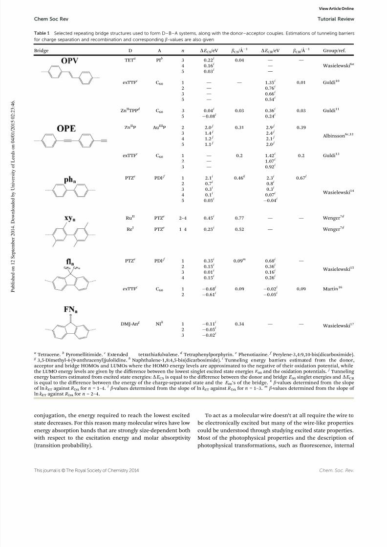
in Table 1 and these examples will form the spine of this discus-sion. The assets of p-conjugated bridges reside in their seeming rigidity, their rod-like structure and more importantly their highdegree of electron delocalization. The latter allows for substantialelectronic communication between the individual bridge unitsand makes them promising candidates for mediating charge/energy over long distances.
In D–B–A systems, ET and EET reactions are traditionally
triggered using light excitation by selectively exciting either thedonor or the acceptor. In the case of ET, these reactions arecalled photo-induced electrontransfer (PET). However there exist other methods to investigate electron transfer in molecularsystems. For instance, electron transfer through molecules canalso be explored by placing single molecules between twometallic electrodes and measuring conductance. Such singlemolecule experiments are becoming increasingly popular as they approach more practical applications by using the molecule asan electrical interconnect between two devices.3
In this tutorial review, focus has been put on photo-inducedelectron and energy transfer in D–B–A systems, more particularly
on the dependence of the transfer rates on distance, energeticsand environment. For long range ET reactions through mole-cular structures governed by quantum mechanical tunneling,the so-called superexchange theory developed by McConnellalready in 1961 is often used.4 The first part of the tutorialprovides the reader with a brief description of the electron andenergy transfer theory, which are intimately related. In the long range electron transfer field, distance dependence studies con-stitute a systematic experiment to probe the ability of a bridge tosustain charge transport. The ability of a bridge structure tomediate electronic coupling for ET have often been summarizedin a single exponential decay constant, namely the attenuationfactor b. However, several studies have shown that the use of theb-value as a simple descriptor of ‘‘conduction’’ is rather limitedas this is not a bridge specific parameter, but instead a donor–bridge–acceptor ensemble parameter.5 In this context, specialemphasis of this tutorial is put on understanding the role of thebridge in charge/energy transfer processes in D–B–A systems.
Several studies in carefully designed D–B–A systems have demon-strated that charge/energy transfer processes (i.e. kinetics andnature of the charge transport) in D–B–A are essentially governedby the bridge energetics6 and conformational dynamics.7 Impli-cations of these studies for obtaining wire-like behavior of themolecular bridge (i.e. low b) are also discussed. To illustrate ourdiscussion, selected examples from Table 1 are described in moredetails. Finally, the last part of the tutorial is dedicated to
porphyrin-based molecular wires as interesting model systemsfor both long-range energy and electron transfer. Once more it is the bridge structure (i.e. the nature of the bridge linker andthe linkage topology) that dictates the excitonic and/or electronicinter-porphyrin coupling in theseconjugated porphyrin oligomers.8
In particular, this was utilized to prepare molecular systems idealfor either energy or electron transfer.9 Limitations of the McConnellmodel to describe electron transfer in strongly conjugatedporphyrin systems are also discussed.
2. Theoretical background
In this section the most essential theoretical framework ispresented. It is by no means complete and the interested readeris referred to any of the excellent text-books on photophysicsand electron transfer theory for an in-depth background.
2.1 Photophysics of molecular wires
This tutorial deals with molecules in electronically excited statesand the fate of excited states is generally described by theso-called photophysical processes. Typically light energies that correspond to visible or UV light are required to excite amolecule to its lowest electronically excited state. A molecular
wire, here defined as a molecule that mediates electronic coupling
between its ends, is often a conjugated organic moleculeconsisting of several subunits linked so that the conjugationextends over a significant part of the molecule. See Table 1 forsome examples of molecular wires discussed in this review.
When the size of the chromophore grows with increased
Melina Gilbert
Melina Gilbert received her Master
of Science in Chemistry from Ecole
Europeenne de Chimie, Polymeres
et Materiaux (ECPM), Strasbourg,
France, in 2010. Currently she isworking towards her PhD in
Physical Chemistry at Chalmers
University of Technology, Sweden.
Her research is focused on under-
standing energy and electron
transfer processes in molecular
wires based on conjugated
porphyrin oligomers. By July
2015, she is expected to defend
her PhD thesis.
Bo Albinsson
Bo Albinsson has been Professor of
Physical Chemistry at Chalmers
University of Technology since
2001. His research interests span
from basic spectroscopy of small molecules to assembly of mole-
cular networks based on DNA.
During most of the time as an
independent researcher he has
been involved in the study of
photoinduced electron and energy
transfer reactions in molecular
donor–bridge–acceptor systems.
Tutorial Review Chem Soc Rev
View Article Online
8/10/2019 Donor Bridge Acceptor
http://slidepdf.com/reader/full/donor-bridge-acceptor 3/18
8/10/2019 Donor Bridge Acceptor
http://slidepdf.com/reader/full/donor-bridge-acceptor 4/18
Chem. Soc. Rev. This journal is © The Royal Society of Chemistry 2014
conversion and intersystem crossing, are similar for molecular wires and small organic molecules. There are however someimportant differences that originate in the degree of delocalizationand the fact that excitation energy (excitons) moves quickly overthese extended molecular structures. When multiple chromo-phores are connected, they interact electronically either only through space in case no conjugated structures connect thechromophores or additionally through the conjugated bonds. In
many cases, a combination of the two interaction mechanismscontributes to the observed effects that range from slow energy migration to ultrafast delocalization and strong alterationsof the electronic spectrum. The through-space interaction,formally between transition dipoles, was quantitatively described by Kasha18 who gave this weak interaction the nameexciton coupling. Even in more strongly interacting molecular
wires and assemblies, where the exciton coupling model isquantitatively inadequate, it may still serve as a qualitativetool to discuss how the arrangement of chromophores (thetopology) contribute to the properties of the assembly.
2.2 Electron and energy transfer rates
Electron and energy transfer are related phenomena that can bedescribed by a common theoretical framework. Provided that the electronic coupling is very small, the Fermi Golden Rule,eqn (1), predicts the rate of transition between two potentialenergy surfaces.
k if = (2p/h)V if 2FCWD (1)
In this so-called diabatic (non-adiabatic) approximation theelectronic coupling, V if , is defined as the effective electronicHamiltonian matrix element that couples the initial (Ci) andfinal (Cf ) states
V if = hCf | H 0|Cii (2) where H 0 is the operator corresponding to the (small) perturba-tion mixing the interacting states. The Franck–Condon
weighted density of states (FCWD) accounts for the conserva-tion of energy and describes the influence from the nuclearmodes of the system. Its specific form has to be adapted to thetransfer reactions studied. For electron transfer, Marcusapproximated the involved potential surfaces by simple para-bolas with equal force constants (curvature) which, whencombined with transition state theory, leads to:19
kET ¼ ffiffiffiffiffiffiffiffiffiffiffiffiffiffiffiffip
h
2
kBT lr V DA
2 exp DG
0 þ l 2
4lkBT ! (3)
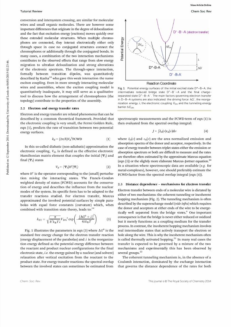
Fig. 1 illustrates the parameters in eqn (3) where DG0 is thestandard free energy change for the electron transfer reaction(energy displacement of the parabolas) and l is the reorganiza-tion energy defined as the potential energy difference betweenthe reactant and product nuclear configurations for the finalelectronic state, i.e. the energy gained by a nuclear (and solvent)relaxation after vertical excitation from the reactant to theproduct state. For energy transfer reactions the spectral overlapbetween the involved states can sometimes be estimated from
spectroscopic measurements and the FCWD-term of eqn (1) isthen evaluated from the spectral overlap integral:
J = Ð
I D(n )e A (n )dn (4)
where I D(n ) and e A (n ) are the area normalized emission andabsorption spectra of the donor and acceptor, respectively. In thecase of energy transfer between triplet states either the emission orabsorption spectrum or both are difficult to measure and the ratesare therefore often estimated by the approximate Marcus equation(eqn (3)) or the slightly more elaborate Marcus–Jortner equation.20
In a situation where spectroscopic data are available (e.g. formetal-complexes), however, one should preferably estimate theFCWD-factor from the spectral overlap integral (eqn (4)).
2.3 Distance dependence – mechanisms for electron transfer
Electron transfer between ends of a molecular wire is dictated by either of two mechanisms: the coherent tunneling or incoherent hopping mechanism (Fig. 2). The tunneling mechanism is oftendescribed by the superexchange model (vide infra) which requiresthe donor and acceptors at either ends of the wire to be energe-tically well separated from the bridge states.4 One important consequence is that the bridge is never either reduced or oxidized
but it merely functions as a coupling medium for the transferprocess. In contrast, the incoherent hopping mechanism involvesreal intermediate states that actively transport the electron orhole along the wire. This is why the incoherent mechanism oftenis called thermally activated hopping.21 In many real cases thetransfer is expected to be governed by a mixture of the twomechanisms and experimentally this has been observed by several groups.22
The coherent tunneling mechanism is, in the absence of aCoulomb interaction, dominated by the exchange interactionthat governs the distance dependence of the rates for both
Fig. 1 Potential energy surfaces of the initial excited state D*–B–A, theintermediate reduced bridge state D+–B–A and the final charge-separated state D+–B–A. The main factors governing electron transferin D–B–A systems are also indicated: the driving force DG1, the reorga-nization energy l, the electronic coupling V DA and the tunneling energybarrier D E DB.
Tutorial Review Chem Soc Rev
View Article Online

8/10/2019 Donor Bridge Acceptor
http://slidepdf.com/reader/full/donor-bridge-acceptor 5/18
This journal is © The Royal Society of Chemistry 2014 Chem. Soc. Rev.
electron and triplet energy transfer (and in some cases also singlet energy transfer). For this mechanism, the distance dependence of the observed electron transfer rates is approximately exponential:
k = k 0 exp(b RDA ) (5)
where RDA is the distance between the donor and acceptor(measured along the wire – not through space), k 0 is thelimiting rate at donor–acceptor contact, and b is the systemspecific attenuation factor. Triplet energy transfer rates alsohave exponential distance dependence but with an attenuationfactor equal to 2b as discussed in Section 4. Numerous experi-mental and computational studies have verified eqn (5) andb-values for a wide range of molecular wires (molecular bridges)have been determined. In general, b-values are larger for systemscomprised of s-bonds than for systems connected by p-conjugated
bridges, which gives more efficient long-range transfer of electronsor excitation energy.
Although b-values only apply for mechanisms that areexpected to decay exponentially (eqn (5)) they have been usedas quality factors also for electron transfer that occur via theincoherent hopping mechanism. As an empirical measure forthe attenuation of the rate vs. distance this is acceptable but since hopping is not expected to decay exponentially it could bequite confusing when comparing b-values with the purposeof trying to understand the mechanistic differences betweenhopping and long range tunneling.
2.4 Superexchange modelFor the coherent tunneling mechanism the influence of theintervening medium has been described by the so-called super-exchange model.4 In this model the magnitude of the electroniccoupling is given by a first-order perturbation theory treatment as:
V DA ¼ V DBV BA
DE DB
V BB
DE DB
n1
(6)
Eqn (6) gives the total electronic coupling (V DA ) in terms of the electronic coupling of the bridge to the donor and acceptor
(V DB and V BA ), the interaction, V BB, between nearest neighborsof a chain composed of n identical units, and the energy gap,D E DB, between the relevant donor and bridge localized states(Fig. 2). If the length of the chain connecting the donor andacceptor is directly proportional to n, i.e. RDA = nR0, where R0 isthe length of one subunit and the electronic coupling betweensubunits is small compared to the energy gap (V BB/D E DB { 1),the distance dependence of the electronic coupling, and there-
fore also the rate, is exponential. Within this approximation theattenuation factor b is given by eqn (7).
b ¼ 2
R0
ln DE DB
V BB
(7)
If the bridge is treated as a single chromophore, i.e. a singlerepeating unit, eqn (6) is simplified to V DA = V DBV BA /D that clearly shows the reciprocal energy gap dependence of the electroniccoupling as will be discussed in the following sections.12a,23
3. Tunneling energy gap effects in
p-conjugated D–B–A systemsThe large interest in p-conjugated bridges arises from their ability to mediate electron transfer over long distances. To evaluate theefficiency of electron transfer over distance, one commonly deter-mines the distance-dependence of the electron–hole transferthrough the bridge in a D–B–A system. In practice, the transferrate constants, k ET, are measured for different donor–acceptordistances RDA achieved by simply varying the number of rigidbridge units separating the donor and acceptor. Results of thisdistance-dependence are then presented in a logarithm plot of k ET
versus RDA . As predicted by the McConnell model (eqn (5)), thetransfer rates k ET are expected to display exponential distance
dependence; in other words, in a semi-logarithmic scale thetransfer rate constants should describe a straight line with a slopecorresponding to the attenuation factor, b. Table 1 provides themeasured b-values of some typical D–B–A systems. These attenua-tion factors could vary quite substantially for different bridges,and ideally a value as close to zero as possible that would allow charge transport over almost ‘‘infinite’’ distances would bepreferable. However, if the electronic coupling gets very largeleading to a very small b-value the distance dependence is nolonger well described by the McConnell model and is no longerexpected to decay exponentially. In Table 1, one can note substan-tial variations of the b-values between the different p-conjugated
bridges, and at first the b-values could seem to be quite randomand difficult to predict. Another important feature to notice isthe different b-values reported for the same bridge when used
with different donor–acceptor couples. Although initially seen as a simple and inactive spectator of
the charge transfer process, it is now evident and quite wellunderstood that the bridge does influence the electron transfer.
According to the McConnell model (eqn (7)), three factors deter-mine the expression of the attenuation factor b: the tunneling energy gap, D E DB, the electronic coupling between individualbridge units, V BB, and the size of the individual bridge units, R0.
Fig. 2 Schematic comparison of the tunneling (left) and hopping (right)mechanisms in a D–B–A system here shown for electron transfer. TheMcConnell superexchange model for electron tunneling through molecularstructures is illustrated in the top drawing. For the tunneling model, thebridge virtual states are represented as dashed lines; while in the hoppingmodel, the local bridge states are drawn as solid lines. DE DB represents thetunneling energy barrier for electron injection onto the bridge.
Chem Soc Rev Tutorial Review
View Article Online

8/10/2019 Donor Bridge Acceptor
http://slidepdf.com/reader/full/donor-bridge-acceptor 6/18
Chem. Soc. Rev. This journal is © The Royal Society of Chemistry 2014
Among these three factors, the most critical parameter is thetunneling energy gap, D E DB. Direct experimental evidence of the tunneling energy gap effect has been lacking for a long timedue to the fact that D E DB is a vertical energy barrier at thetransition state, a quantity which is not easily accessible(Fig. 1). In the last ten years, much effort has been put in thedesign of experiments enabling to test theoretical predictions.Several studies have appeared in the literature providing clear
experimental evidence of the influence of bridge energeticson charge transport in D–B–A systems.6a,d ,12a,24 From thesestudies, the wide range of b-values seen in Table 1 could beascribed in part to substantial differences in the tunneling energy gap between the different D–B–A systems.
The tunneling energy gap effect was first demonstrated by Wasielewski and co-workers. They investigated the distancedependence of electron transfer rates in a series of OPV-bridgedD–B–A systems, with tetracene as a donor and pyromellitimide(PI) as an acceptor (see Table 1).6a,24 For both charge separation(CS) and recombination (CR), they measured the transfer rates(k CS and k CR ) and plotted them as a function of RDA on a semi-
logarithmic scale. While the data points for charge separation inthe shorter members (n = 1, 2) fell into a single line in agreement with a tunneling mechanism, the longer members of the series(n = 3–5) deviated strongly from this line and showed fasterrates and weak distance dependence. This ‘‘irregularity’’ in thedistance dependence was interpreted as the result of a changein the charge transport mechanism. While for the shorterbridges (n = 1, 2), charge transport occurs via superexchangetunneling, bridge-assisted electron hopping predominates inthe longer bridges (n = 3–5). For the charge recombination, asimilar mechanism change was observed for n = 5. As the samedonor–acceptor couple was used throughout the entire series,this crossover from tunneling to hopping could only be related
to properties of the bridge. Indeed for this particular bridge,increasing the bridge length also increases the size of theconjugated system and consequently the energy of the bridgestates, essentially the HOMO–LUMO gap, decreases. This leadsto a decrease of the energy gap between the LUMO of the donorand the LUMO of the bridge, and hence a decrease of the
tunneling energy barrier. In the longer members of this OPV-bridged series,D E DB is small enough that reduction of the bridgebecomes possible. Charge can migrate between the donor andthe acceptor in a multistep fashion, first moving from the donorto the bridge, and then from the bridge to the acceptor. This first example shows that in distance-dependence studies boththe width ( RDA ) and the height (D E DB) of the tunneling energy barrier may vary simultaneously, making the interpretation of
the results more intricate. Thus, when studying charge transport in D–B–A molecules with p-conjugated bridges, it may be desir-able to vary a single parameter at a time, either the width or theheight of the tunneling barrier. With this aim, two approacheshave been proposed to solely focus on a single parameter.
3.1 Using structurally identical bridges
In this first approach, one compares the transfer rates of aseries of D–B–A molecules with constant RDA , that consists of the same donor–acceptor couple connected by structurally similar but electronically different bridge molecules. Albinssonand co-workers have used this approach and designed one
series of D–B–A systems ZnP–RB–AuP
+
where RB denotes anOPE-based bridge (Fig. 3). For the entire series, ZnP acts as anelectron donor while AuP+ is the electron acceptor.6c,12a Theelectronic structure of the OPE bridge (3B) could be modified by replacing the central benzene unit by either naphthalene (NB)or anthracene (AB). This allowed varying the tunneling energy barrier for charge separation D E CS and recombination D E CR whilekeeping a fixed RDA . Fig. 3 provides schematic energy diagrams forthe three D–B–A systems with their respective tunneling energy gaps D E CS andD E CR for charge separation (CS) and recombination(CR), respectively. Note that the tunneling energy gaps are not thetrue energy barriers but are approximations from the donor–acceptor LUMOs. In the present case,photo-excitation of the donor
involves a close to pure HOMO–LUMO transition; hence theLUMOs of both the donor and bridge could be directly relatedto the energy of their first singlet excited states ( E 00). Thus thetunneling energy barriers D E CS and D E CR were calculated as theenergy difference between the donor and bridge E 00 singlet excited states energies, and as the energy difference between
Fig. 3 (top) Molecular structures of the D–B–A systems ZnP–RB–AuP+ with RB = AB (black), NB (red) and 3B (blue). The edge to edge donor–acceptordistance was 19.6 Å in the three D–B–A systems. (bottom) Schematic energy diagrams for charge separation via electron transfer in these systems.Adapted from ref. 6c and 12a.
Tutorial Review Chem Soc Rev
View Article Online

8/10/2019 Donor Bridge Acceptor
http://slidepdf.com/reader/full/donor-bridge-acceptor 7/18
This journal is © The Royal Society of Chemistry 2014 Chem. Soc. Rev.
the charge-separated state and the first singlet excited state of thebridge, respectively. Moving from benzene (3B) to naphthalene(NB) to anthracene (AB), i.e. to lower lying singlet excited stateson the bridging unit, leads to a decreased tunneling energy gap. This decrease in D E CS and D E CR was reflected very well inthe observed electron transfer rates, which increased when going from benzene (3B) to naphthalene (NB) to anthracene (AB).
The solvent dependence of the electron transfer rates for
both charge separation and recombination processes were alsoinvestigated. The transfer rates were measured in six solvents
with different polarity using both steady-state and time-resolvedfluorescence. Along with estimates of the driving forces ( DG1)and reorganization energies (l) accompanying charge separationand recombination, these measurements gave indirect access tothe electronic coupling, V DA . Indeed, in a plot of ln(k ETl
1/2)against (DG1 + l)2/(4lk BT ) the data could be successfully fit to astraight line with a slope of 1 in agreement with the linearizedMarcus equation (eqn (3)). Further, the overall donor–acceptorcoupling for the three different bridge systems could be esti-mated from the intercept of the fitting curves. Finally, the key
finding of this work was the experimental demonstration of the linear correlation between the electronic coupling and theinverse of the tunneling energy gaps, 1/D E CS or 1/D E CR , (Fig. 4)as predicted by the McConnell model.
As an extension, the distance dependence of the electrontransfer rates was studied in the series ZnP–nB–AuP+ where thenumber of OPE units n varied between 2 to 5, spanning edge-to-edge donor–acceptor distances, Ree, between 13 and 33 Å.6c,12b
On a semi-logarithmic plot, the transfer rates ln k against Ree described a straight line, in line with a tunneling chargetransport (Fig. 5). From the slopes of the curves, the attenuationfactor b was estimated to be 0.31 Å 1 for charge separation and0.39 Å 1 for charge recombination. These different b-values reflected
once more tunneling energy gap effects with D E CS o D E CR
(see Fig. 4, as an example for the trimer ZnP–3B–AuP
+
, crudeestimates of the barrier heights give: D E CSE 1.4 eV o D E CR E
2.4 eV). It can be interesting to compare the behavior of OPE-bridged D–B–A molecules with the one of OPV-bridged D–B–A molecules discussed earlier. As for OPV-bridged D–B–A systems,
when the OPE bridge gets longer, the tunneling energy barriersD E CS and D E CR decrease due to increased conjugation. In OPV-bridged D–B–A molecules, we saw that variation of the tunneling energy barrier led to a change in the charge transport mechanism.Here in this particular OPE-bridged series no deviation from theexponential distance-dependence was observed. This indicatesthat the tunneling energy barrier remains large enough forcharge transport to occur via tunneling even for the longer
members of the series. In other words, the distance dependenceof the electron transfer rates in the ZnP–nB–AuP+ series resultsmainly from changes in the barrier width (n or Ree) and to asmaller degree in its height (D E CS and D E CR ).
A more recent study comparing charge transport in structu-rally similar bridges is the work of Wenger et al. on D–B–A molecules linked by phenylene-based bridges.25 They investi-gated hole tunneling in D–B–A systems with a rhenium(I)complex as a hole donor and phenothiazine as a hole acceptor.The donor–acceptor couple was connected either by two phenyls(ph2), two xylene groups (xy 2) or a fluorene group (fl1) (see Table 1).
Again despite the different bridge structure, the donor–acceptor
separation remains identical in the three D–B–A systems. Tran-sient absorption measurements revealed an increase in the holetransfer rates when going from phenylene (k HT = 0.5 108 s1)to xylene (k HT = 2.6 108 s1) and fluorene (k HT = 5.3 108 s1).In parallel, they also estimated the hole tunneling energy gapfrom the redox potentials for the different bridge structures:D E DB = 0.54 eV for ph2, D E DB = 0.25 eV for xy 2 and D E DB =0.16 eV for fl1. The faster transfer rate observed for the fluorenebridge (fl1) reflects nicely the smaller tunneling energy barrier.However the phenylene-bridged D–B–A system, which showsan intermediate transfer rate, has also the highest tunneling
Fig. 4 Electronic coupling, V DA versus the inverse of the energy gap, D E ,for charge separation (filled symbols) and charge recombination (emptysymbols) in the series ZnP–RB–AuP+ with R = AB (black), R = NB (red) andR = 3B (blue). The lines are linear fits obtained when fixing the interceptsV DA(0) = 0. Adapted from ref. 12b.
Fig. 5 Distance dependence of the charge separation (K) and chargerecombination (J) rates in the D–B–A series ZnP–nB–AuP+ with thenumber of OPE units n = 2–5. Adapted from ref. 12b.
Chem Soc Rev Tutorial Review
View Article Online

8/10/2019 Donor Bridge Acceptor
http://slidepdf.com/reader/full/donor-bridge-acceptor 8/18
Chem. Soc. Rev. This journal is © The Royal Society of Chemistry 2014
energy barrier D E DB. The tunneling energy gap argument doesnot solely explain all the kinetic data. When going fromphenylene to xylene to fluorene, the average torsional angle,jph–ph, between the two phenyl planes also varies and decreasesfrom 651 for xy 2 to351 for ph2 to 61 for fl1. This results in increasing the internal bridge electronic coupling, V BB, and thereby increasing the overall electronic coupling, V DA (eqn (6)), when going fromxylene via phenylene to fluorene bridge units. Thus, the faster rate
in the fluorene-bridged D–B–A system presumably results fromthe combined effect of a smaller tunneling energy barrierD E DB and a stronger internal bridge coupling (lower jph–ph).For the phenylene-bridged D–B–A system the intermediateV BB may compensate for the larger tunneling energy barrierD E DB, leading to an intermediate transfer rate.
3.2 Using bridges with length-independent (redox) energies
A second approach to study solely the influence of the width of the tunneling barrier is to use D–B–A systems in which thebridge energetics are independent of the bridge length. Thanksto a combined effort of synthetic and physical chemists, several
examples of p
-conjugated systems with length-independent bridge redox potentials have recently appeared in the literature.Oligo-fluorene (fln), oligo-fluorenone (FNn) and oligo- p-xylenes(xy n) are examples of p-conjugated systems which show nearly length independent redox potentials.15a,17,26 In two separatestudies, Wasielewski and co-workers investigated photoinducedelectron transfer in D–B–A molecules, bridged by either oligo-fluorenes15a or oligo-fluorenone17 (see Table 1). However, bothD–B–A series revealed to be less interesting for investigating electron tunneling as a hopping mechanism dominated thecharge transport (see the next chapter for a more detaileddiscussion). In contrast, charge transport in oligo- p-xylene-bridged D–B–A molecules occurs essentially via electron tunneling.
In a nice series of papers, Wenger and co-workers demonstratethat xylene-bridged D–B–A molecules are interesting modelsystems to isolate the effects of width or height of the tunneling barrier on long-range charge transfer.6d They designed two seriesof xylene-bridged D–B–A molecules and measured the distance-dependent hole transfer rates by varying the number of xylenebridge units (see Table 1). In the first series, the donor–acceptorcouple was phenothiazine (PTZ) and rhenium tricarbonylphenanthroline (Re(I)). In the second series, they employed adifferent donor–acceptor couple with again phenothiazine(PTZ) as a donor but ruthenium(II)tris(2,20-bypridine) (Ru(II))as an acceptor. In both the PTZ–xy n–Re (n = 1–4) and PTZ–xy n–
Ru series (n = 1–4), the absorption spectra remain close-to-identical upon lengthening of the bridge, indicating that thebridge excited states and thereby the tunneling energy gap islength-independent for both series. In accordance with the super-exchange theory, the hole transfer rates decreased exponentially
with the donor–acceptor distance in both series, confirming holetunneling as the dominant charge transport mechanism. Howeverthey determined significantly different attenuation factors withb = 0.52 Å 1 for PTZ–xy n–Re and b = 0.77 Å 1 for PTZ–xy n–Ru.The different b-values could be ascribed to differences in tunneling energy barriers between the two series, that arise from the different
acceptor used: D E BA = 0.25 eV for PTZ–xy n–Re and D E BA = 0.45 eV for PTZ–xy n–Ru. Finally this experimentally demonstrates that the b-value is specific to the donor–bridge–acceptor ensembleand not solely to the bridge unit.
3.3 Crossover from tunneling to hopping
When the tunneling energy gap (D E DB or D E BA ) is small enough,i.e. the bridge and donor (or acceptor) states are nearly resonant,
the bridge reduced (or oxidized) states become thermally acces-sible. Charge transport occurs then predominantly via incoherent hopping. This means that the electron (or hole) resides tempora-rily on the bridge before reaching the electron acceptor (or donor).Thus a clear and simple evidence of incoherent charge hopping is often the detection of the bridge reduced transient stateD+–B–A (or the oxidized transient state D–B+–A ) prior toformation of the fully charge-separated state in transient absorption experiments. As the bridge transient species areoften very short lived (below the time resolution of transient absorption measurements), deviations from the exponentialdistance dependence of the transfer rates have also been used
as an indicator of incoherent charge hopping. Indeed forincoherent hopping, the distance dependence of the transferrates is not anymore exponential or ruled by the superexchange(McConnell) theory, and generally very weak distance depen-dence is observed. Over the last twenty years, several examplesof D–B–A molecules with p-conjugated bridges showing ‘‘irregular’’ distance dependence of their transfer rates attributedto a switch from tunneling to hopping charge transport have beenreported in the literature. The large interest in D–B–A systems
with charge transport occurring in the hopping regime residesin the low distance dependence of charge transport, hence theability of mediating charge over much longer distances than inthe tunneling regime. In the literature, these D–B–A systems
have often been qualified as ‘‘wire-like’’. Crossover fromtunneling to hopping and the appearance of a wire-like beha-
vior has been first observed by Wasielewski and co-workers inOPV-bridged D–B–A molecules with a tetracene donor and apyromellitimide acceptor, as described above.6a In an analogous
work on oligophenylene-bridged D–B–A systems, they observed asimilar switch from tunneling to hopping when increasing thenumber of phenyl bridge units between the donor (PTZ) andacceptor (PDI), see Table 1.14 Once more, this was evidenced by anon-exponential distance dependence of the transfer rates. Likethe OPV-based bridges, oligophenylene bridges possess strongly length-dependent HOMO–LUMO gaps resulting in a tunneling
energy barrier for this D–B–A system, that varies between 2.1 eV (n = 1) and 0.05 eV (n = 5) for charge separation and between2.3 eV and 0.04 eV for charge recombination (Table 1). Thusfor the longer members (n = 4, 5), the tunneling energy barriersare small enough that population of the bridge states might occur and incoherent hopping dominates the charge transport.
Additionally, they measured electron paramagnetic resonance(EPR) spectra and estimated the magnitude of the spin–spinexchange interaction (2J) through the entire series, a quantity
which is proportional to the donor–acceptor coupling V DA . As for the electron transfer rate constants, they observed an
Tutorial Review Chem Soc Rev
View Article Online
8/10/2019 Donor Bridge Acceptor
http://slidepdf.com/reader/full/donor-bridge-acceptor 9/18
This journal is © The Royal Society of Chemistry 2014 Chem. Soc. Rev.
exponential distance dependence of the spin–spin exchangeinteraction 2J only up to n = 4, supporting their conclusion of achange in the charge transport mechanism in the longer system
with n = 5. Using the same donor–acceptor couple (PDI/PTZ), Wasielewski and co-workers have also designed a series of D–B–A molecules, bridged by oligofluorene (fln) with n = 1–4.
As mentioned earlier, fluorene bridges (fln) with n Z 2 possessnearly length-independent redox potentials hence the tunneling
energy barrier remains almost constant for PDI–fln–PTZ withn Z 2. Of much interest in this particular system is the matching of the acceptor and bridge energy levels which is achieved forn = 2–4. This results in incoherent hopping hole transport with a
very small b-value (b = 0.093 Å 1) and gives rise to a ‘‘wire-like’’behavior of the fluorene bridges in the PDI–fln–PTZ systems.
In both examples, we have seen that wire-like chargetransport is achieved by matching the donor (or acceptor)and bridge energy levels. This promotes incoherent hopping charge transport and precludes the strongly-distance depen-dent tunneling charge transport to occur. However in a recent paper, Wasielewski and co-workers reported a high b-value for
hole transfer together with evidence of a hopping mechanismin fluorenone-bridged D–B–A systems (see Table 1).17 In theseparticular D–B–A systems, the selected donor (DMJ-An), bridge(FNn) and acceptor (NI) give rise to a stepwise downhill energy gradient within the D–B–A molecule. Surprisingly, even throughthe downhill energy gradient in DMJ-An–FNn–NI (n = 1–3),they observed an exponential distance dependence of thetransfer rate with a large attenuation factor, b = 0.34 Å 1. Suchlarge attenuation factor would normally be expected forcharge transport in the tunneling regime. However femtosecondtransient absorption measurements confirmed the formation of the intermediate bridge reduced states DMJ+-An–FNn–NI, sup-porting their conclusion of a hopping mechanism. The unusual
distance dependence in the hopping regime was rationalized by invoking the push–pull character of the DMJ-An donor, whichforms upon photo-excitation a primary charge-separated stateDMJ+-An. The electrostatic attraction between the two chargesslows down both the electron injection onto the bridge and thesubsequent electron hopping, resulting in the high attenuationfactor observed. This work demonstrates that a stepwise down-hill energy gradient in a D–B–A molecule does not necessarily imply a ‘‘wire-like’’ behavior of the bridge. It also shows that theattenuation factor b is not a reliable indicator to distinguishbetween tunneling and hopping charge transport.
4. Conformational effects on electrontransfer processes in p-conjugatedsystems
So far we have described a quite simple picture of the factorsinfluencing electronic mediation in D–B–A systems in whichthe tunneling energy barrier and width are the only factorsgoverning the distance dependence and the nature of chargetransport (i.e. coherent tunneling or incoherent hopping). Inreality, conformational dynamics of the bridge, temperature
and solvent may affect the donor–acceptor electronic communica-tion and hence the measured transfer rates in D–B–A systems.This generally complicates the picture, and renders the predic-tion and interpretation of the transfer rates quite difficult. Thefollowing discussion is restricted to the influence of the bridgeconformation on charge transport in D–B–A molecules. Althoughsolvent parameters influence the electron transfer rate throughthe reorganization energy l and driving force DG1 (eqn (3)), in
the interest of clarity we will refrain from discussing thesefactors here.
In the previous discussion, the bridge was considered as arigid structure. However the bridge is a dynamic entity, in whicheach unit may rotate, giving rise to different conformations. Theconformational dynamics of the bridge is both temperature andsolvent dependent. This influences the bridge energetics andthereby the tunneling energy barriers in D–B–A systems. Further-more, variations in the dihedral angles between individual units
within the bridge and between the bridge and the donor oracceptor might strongly modulate the overall donor–acceptorelectronic coupling. Several experimental and theoretical studies
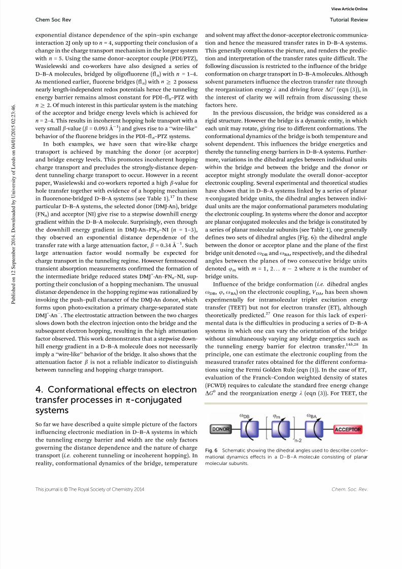
have shown that in D–B–A systems linked by a series of planarp-conjugated bridge units, the dihedral angles between indivi-dual units are the major conformational parameters modulating the electronic coupling. In systems where the donor and acceptorare planar conjugated molecules and the bridge is constituted by a series of planar molecular subunits (see Table 1), one generally defines two sets of dihedral angles (Fig. 6): the dihedral anglebetween the donor or acceptor plane and the plane of the first bridge unit denoted oDB and oBA , respectively, and the dihedralangles between the planes of two consecutive bridge unitsdenoted jm with m = 1, 2. . . n 2 where n is the number of bridge units.
Influence of the bridge conformation (i.e. dihedral anglesoDB, j, oBA ) on the electronic coupling, V DA , has been shownexperimentally for intramolecular triplet excitation energy transfer (TEET) but not for electron transfer (ET), althoughtheoretically predicted.27 One reason for this lack of experi-mental data is the difficulties in producing a series of D–B–A systems in which one can vary the orientation of the bridge
without simultaneously varying any bridge energetics such asthe tunneling energy barrier for electron transfer.14b,28 Inprinciple, one can estimate the electronic coupling from themeasured transfer rates obtained for the different conforma-tions using the Fermi Golden Rule (eqn (1)). In the case of ET,evaluation of the Franck–Condon weighted density of states
(FCWD) requires to calculate the standard free energy changeDG0 and the reorganization energy l (eqn (3)). For TEET, the
Fig. 6 Schematic showing the dihedral angles used to describe confor-mational dynamics effects in a D–B–A molecule consisting of planarmolecular subunits.
Chem Soc Rev Tutorial Review
View Article Online

8/10/2019 Donor Bridge Acceptor
http://slidepdf.com/reader/full/donor-bridge-acceptor 10/18
Chem. Soc. Rev. This journal is © The Royal Society of Chemistry 2014
rate for triplet energy transfer k TEET can also be expressed using the Fermi Golden Rule if the donor–acceptor coupling V DA is
weak. But in contrast to ET, the FWCD is simply given by thespectral overlap of the emission spectra of the donor andacceptor (eqn (4)). The electronic coupling is then the only remaining unknown in eqn (1) and can be obtained from themeasured kinetic data. Another advantage of TEET measure-
ments is the minimal influence of solvent effects due to theabsence of charge movement. Finally, qualitative results fromTEET studies are expected to prevail for ET, as TEET is also anelectron exchange phenomena that can be viewed as twosimultaneous ET reactions (Fig. 7). This qualitative argument
was supported by the work of Closs et al. who experimentally demonstrated the connection between the electronic coupling for triplet energy transfer and electron and hole transfer forsaturated bridges: |V TEET| = const|V ET||V HT|.29
4.1 Geometric control of the electronic coupling
Among the first to show experimentally the influence of the
bridge conformation on the donor–acceptor electronic coupling in D–B–A systems was Harriman and co-workers.30 They probedthe effect of torsional angles within the bridge structure on the
rate of intramolecular triplet excitation energy transfer k TEET
in a series of ruthenium(II)/osmium(II) (tpy)2 donor–acceptorcomplexes linked by ethynylene-substituted biphenyl bridges.They could control and tune the dihedral angle between thephenyl units, jPh–Ph, by attaching a tethering strap between thetwo phenyl units and varying the number of carbon atoms inthe strap (Fig. 8). This gave access to a wide range of dihedralangles while keeping a fixed donor–acceptor distance. To prevent
any undesirable rotation from occurring, triplet energy transferrates were measured in a low temperature organic glass matrix,thus maintaining the bridge frozen into its lowest-energy con-formation. Their results showed a pronounced conformationaldependence of the transfer rates, hence of the electronic coupling
which was largest when the phenyl units were close to coplanar(j = 301). At the largest dihedral angle (j = 901), the electroniccoupling V TEET decreased drastically, resulting in a measuredtransfer rate k TEET that decreased by a factor of 80. Furthermore,they demonstrated experimentally the quadratic dependenceof the electronic coupling with the overlap integral betweentwo p-orbitals: V TEET(j) = V ET(j)V HT(j) = cos2(j).
4.2 Temperature control of the bridge conformational
distribution
Another approach to investigate how bridge conformation affectsthe flow of excitation energy or electrons between the donor andacceptor in D–B–A systems is to use temperature as a way to tunethe Boltzmann distribution of conformations. At first thisapproach seems more accessible, since it is less challenging interms of synthesis. However, interpretation of the temperature-dependent transfer rates is a challenge as several parameters areaffected simultaneously by temperature. Firstly, temperatureimpacts several properties of the solvent, such as the dielectric
constant and the refractive index which influence the driving force DG1 and reorganization energy l for the electron transfer rate(eqn (3)). Thus as stated above, it is easier to study temperature
Fig. 7 Schematic of the triplet excitation energy transfer (TEET) betweena donor and an acceptor, as a simultaneous electron and hole transferreaction. Adapted from ref. 29.
Fig. 8 (top) Schematic of the tethered strap approach used to tune the dihedral angle, j, between the planes of the two consecutive phenylbridge units. Adapted from ref. 7a. (bottom) Molecular structure of one D–B–A system employing the tethered strapped bridge studied by Harrimanand co-workers.30
Tutorial Review Chem Soc Rev
View Article Online

8/10/2019 Donor Bridge Acceptor
http://slidepdf.com/reader/full/donor-bridge-acceptor 11/18
This journal is © The Royal Society of Chemistry 2014 Chem. Soc. Rev.
dependence of TEET so as to minimize the solvent-inducedeffects. Secondly, another temperature dependent property of the solvent is its viscosity that increases as the temperaturedecreases, hence slowing down rotations of the molecularplanes within the bridge structure.
Albinsson and co-workers have investigated the temperaturedependence of TEET, both experimentally and theoretically,in a series of donor–acceptor systems linked by OPE bridges,
ZnP–nB–H2P with n = 2–5.31 The distance-dependent transferrates, k TEET, were measured for temperatures between roomtemperature and 80 K. As for ET, in line with the McConnellmodel, the transfer rates showed exponential distance depen-dence for the entire temperature range, but more interestingly the attenuation factor, b, was found to be temperature depen-dent. In parallel to these experiments, a theoretical model wasderived to calculate the total electronic coupling, V DA , based ona Boltzmann distribution of the bridge conformations.32 Thismodel demonstrates the conformational gating of the electroniccoupling, and hence of the attenuation factor b. The procedureinvolved density functional theory (DFT: B3LYP/6-31G*) calcula-
tions of the potential energy surfaces as a function of thedihedral angles o, Sjm (Fig. 6) followed by calculation of thetotal electronic coupling V DA for each conformational distribu-tion (i.e. each set of o, Sjm values). V DA can be derived fromhalf the triplet excitation energy difference (calculated withtime dependent (TD)-DFT) between the two lowest triplet excited states at the avoided crossing geometry (Fig. 1). In anunsymmetrical system such as here ZnP–nB–H2P, finding thiscrossing point is not straightforward and can be quite complex.Thus, to simplify the procedure, the electronic coupling for thecorresponding symmetrical systems, ZnP–nB–ZnP (n = 2–5),
was calculated. Fig. 9 illustrates the variation of the totalelectronic coupling V DA for ZnP–2B–ZnP as function of the
two dihedral angles o = oDB = oBA and j. Again the cos2j
dependence of the electronic coupling is nicely demonstrated.The electronic coupling V DA reaches a maximum when bothphenyl planes are coplanar (j = 01) and a minimum when bothdihedral angles j and o equal 901 (i.e. all molecular planesin ZnP–2B–ZnP are orthogonal to each other). This showsthe possibility of using conformational changes to tune theelectronic coupling.
The total electronic coupling V DA can be expressed as a product
of one-parameter functions: V DA (o1, j1, j2,. . .,jn
1, o2) =V (o1)V (j1)V (j2). . .V (jn1)V (o2). By introducing a Boltzmann-
weighted contribution for each conformational configuration,the impact of the bridge disorder on the average electroniccoupling hV DA i can be evaluated from (eqn (8)).
V DA o;j1;j2; . . . ;jx1ð Þh ix
¼ V x V ðoÞh i Yx1
m¼1
V jmð Þh i
¼ V x V ðoÞh i V ðjÞh ix1
(8)
where V x is the electronic coupling for a planar bridge and ishere considered to be a bridge-length dependent constant.
hV (a)i is an average given by:
V ðaÞh i ¼
Ð V ðaÞeE rotðaÞ=RT daÐ eE rotðaÞ=RT da
(9)
where a corresponds to any dihedral angle, o or j, and E rot (a) isthe intrinsic energy barrier for rotating coordinate a. In thisexpression, the temperature-dependent conformational distribu-tion is taken into account in the factors hV (o)i and hV (j)i whichalso give the conformational dependence of the total electroniccoupling. From the calculated hV DA i values, the b-values at any temperature can be determined according to eqn (10).
b ¼ 2d ln V DAh i
dRDA
(10)
where lnhV DA i can be re-written as a sum of three terms(eqn (11)).
lnhV DA i = ln V x + lnhV (o)i + (x 1)lnhV (j)i (11)
From eqn (11), one can visualize which terms contributeto the temperature dependence and/or the bridge lengthdependence of the electronic coupling. The first term istemperature-independent (i.e. conformation-independent) but specific to the length of the bridge and the ensemble donor–bridge–acceptor used (i.e. D E DB). The second and third termsdepend on conformation and ‘‘carry’’ the temperature depen-
dence of the electronic coupling. However, the former isindependent of the bridge length, hence will not contributeto b. In contrast the last term varies with the size of thebridge, hence is the only term contributing to the temperaturedependence of b. One can thus express the attenuation factorbtot as the sum of two independent parts, one temperature-independent b0(D E DB) and one temperature-dependent b(T )that reflects the effect of the bridge conformational disorderat temperature T .
btot = b0(D E DB) + b(T ) (12)
Fig. 9 Calculated electronic coupling for TEET, V DA, for the symmetricsystem ZnP–2B–ZnP as function of both porphyrin-bridge (o) andphenyl–phenyl (j) dihedral angles. Reprinted with permission fromref. 32b. Copyright 2006 American Chemical Society.
Chem Soc Rev Tutorial Review
View Article Online

8/10/2019 Donor Bridge Acceptor
http://slidepdf.com/reader/full/donor-bridge-acceptor 12/18
Chem. Soc. Rev. This journal is © The Royal Society of Chemistry 2014
This expression is believed to be quite general and prevailsfor any series of D–B–A systems consisting of several identicalrepeating bridge units. The temperature independent termb0(D E DB) gives the lowest obtainable b-value for a given repeating bridge structure (e.g. for a planar OPE bridge). Fig. 10 comparesthe experimental and calculated b-values for the TEET rates inthe series ZnP–nB–H2P (n = 2–5) as a function of temperature.
At high temperature (i.e. low solvent viscosity), the calculatedb-values agree very well with the experimental values, supporting the validity of the model based on a Boltzmann description of thebridge conformational disorder. However, at low temperature, i.e. at
high viscosity, the experimental b-values decrease much faster thanpredicted. In the high viscosity regime, corrections to the model arerequired in order to take into account the increased solvent viscosity that may hinder rotational motions. In other words, the potentialenergy associated to the conformational configuration E rot (a) ineqn (9) varies with the viscosity of the solvent. To correct for this,
E rot (a) was replaced by a temperature-dependent apparent activationbarrier. This allowed for successful modeling of the experimentaltemperature dependence of the attenuation factor over the entiretemperature range 80–200 K (see solid line in Fig. 10).
More generally it was demonstrated that for D–B–A systemsin which internal rotational dynamics are much faster than the
transfer rates, conformational dependence of the electroniccoupling and the b-value can be well described by considering a pure Boltzmann distribution of conformations. However whenrotational dynamics and transfer rates occur on the same timescale(i.e. at low temperature and/or high viscosity), a pure Boltzmannconformational distribution is not accurate anymore as it tendsto favor the population of conformations with the minimumrotational energy barrier (i.e. intrinsic energy). Finally, it wasestablished that for all bridge structures with potential energy minima associated with a planar conformation (j = 01), themaximum conformational effect on the b-value is simply given
by the number 2 ln 2/ R0 where R0 is the length of a single bridgeunit. This leads to the conclusion that although b0(D E DB) issystem specific, b(T ) is a bridge specific parameter which isindependent of the donor–acceptor couple used and whosemaximum value can be predicted.
5. Porphyrin-based molecular wires
Porphyrins are planar 18p-electron conjugated macrocycles built from four pyrrole units and four methine carbons. One particularity of these structures is the strong sensitivity of their conjugatedelectronic system upon substitution at the peripheric positions,namely a, b and meso (Fig. 11A). In multiporphyrin arrays,perturbations of the conjugated electronic system of individualporphyrins often lead to unusual electronic and optical proper-ties. In the last decade, multiporphyrin arrays, due to theirunusual electronic behavior, have attracted much interest, in
view of potential applications in molecular electronics, photo- voltaics and non-linear optics.9 A wide range of multiporphyrin
arrays have been synthesized with architectures going fromsimple linear structures to cyclic, box, and grid-like structures.To realize such constructs, the porphyrins are connected eitherdirectly to each other or using a linker. Different linkers havebeen used including phenyls,33 alkenes34 or alkynes.34b,c,35 It isnow widely accepted that the type of linker and the porphyrinlinkage topology (a, b or meso) govern the inter-porphyrinelectronic coupling in these arrays, hence their photophysicalproperties. In the following section, we restrict the discussionto only linear multiporphyrin arrays and emphasize a few examplesthat focus on the relationship between the electronic structure andthe photophysical properties. Also synthetic methodologies to obtainthese multiporphyrin arrays fall out of the scope of this tutorial
review. Recent advances in synthesis of multiporphyrin arrays havebeen nicely reviewed by Osuka and co-workers.36
Systems containing one, two or more porphyrin units wereprimarily designed as mimics of the light harvesting antennacomplexes in natural photosynthesis. Donor or acceptors wereattached to the porphyrins to form donor–acceptor systems andthe porphyrins played the role of primary electron donors.In these systems, excitation of the porphyrins lead first to theformation of a charge separated state, thereafter a cascade of electron-transfer events takes place separating the charges bothspatially and temporally.33a,37 In this context, most studies havefocused on producing extremely long-lived charge-separated
states that might allow for subsequent chemical reactions tooccur. The groups of Gust,33a Osuka33c and Fukuzumi37 reportedpromising porphyrin-based systems showing long-lived charge-separated states following photo-excitation. In all these examples,the porphyrins connected using phenyl linkers at their meso
positions are not strongly conjugated (i.e. electronically coupled).Due to steric hindrance with the b-substituents, the phenyllinkers are forced to adopt a nearly orthogonal arrangement
with the porphyrin plane. This orthogonal arrangement breaksthe conjugation and leads to a weak electronic coupling between theindividual porphyrins. This configuration has been used by Lindsey
Fig. 10 Comparison between experimentally determined b-values versustemperature (K) and fits of the theoretical model using a pure Boltzmannaveraging (dashed line) and using viscosity-dependent activation energy (solidline). Reprinted from ‘‘Temperature Dependence of ElectronicCoupling throughOligophenyleneethynylene Bridges’’ Mattias P. Eng, Jerker Mårtensson, BoAlbinsson. Chemistry – A European Journal, 14, 2819. Copyright r 2008WILEY-VCH VerlagGmbH & Co.KGaA, Weinheim. Reproduced with permission.
Tutorial Review Chem Soc Rev
View Article Online

8/10/2019 Donor Bridge Acceptor
http://slidepdf.com/reader/full/donor-bridge-acceptor 13/18
This journal is © The Royal Society of Chemistry 2014 Chem. Soc. Rev.
and co-workers to design a molecular photonic wire for efficient singlet energy transfer.38 They used meso–meso diphenylethyne-linked porphyrin oligomers, in which conjugation (i.e. electroniccoupling) was intentionally minimized to avoid competition betweenenergy transfer and electron transfer quenching. For steric reasons,directly linked meso–meso porphyrins also show weak inter-porphyrin electronic coupling.8c To transport charge over long distances, the porphyrin units need to be connected in a different
way and construct a ‘‘supermolecule’’ with strong electronic com-munication. Several strategies have been reported to construct largenetworks of conjugated porphyrins, that differ essentially by the type
of linker used and the porphyrin linkage topology (b–b, meso–meso,meso–b). The groups of Therien,35 Anderson34b,c and Smith34a haveused unsaturated linkers such as alkynes and alkenes (Fig. 11Band C). Other groups, i.e. Osuka et al.39 and Crossley et al.,40 havesynthesized fused porphyrin arrays, in which the porphyrinmonomers are directly connected using multiple covalent bonds(Fig. 11D) or an aromatic linker.
5.1 Strongly coupled porphyrin oligomers
Bridging porphyrins using alkyne linkers is not a new idea; already in 1978, Arnold and co-workers reported a nickel porphyrin dimer
bridged at its meso position by 1,3-butadyine linker.41 The strong inter-porphyrin electronic coupling in this dimer was evidenced by strong alteration of its ground-state absorption spectrum comparedto the monomer. Since this first report, alkyne-linked porphyrinoligomers have been intensively investigated, in particular withrespect to structure–property relationships. In 1990s, the groups of Therien35 and Anderson34b synthesized zinc porphyrin dimersconnected at their meso position using ethyne and butadiynelinkers, respectively (Fig. 11B and C). In both cases, in contrast tophenyl linkers, alkynes allow a coplanar arrangement of theporphyrin planes resulting in a significant p-overlap between the
porphyrin macrocycles. This ensures a strong inter-porphyrinelectronic coupling in the ground-state. In the excited state, oneobserves an even stronger electronic coupling due, to on average, amore planar conformation of the oligomers than in the ground-state.42 This results from the enhanced charge delocalization in theexcited states.8b Therien and co-workers investigated further theeffect of the porphyrin linkage topology on the electronic coupling by preparing two series of meso–meso, meso–b and b–b linkedporphyrin dimers, related to the ones in Fig. 11B.8a They employedethyne linkers in one series and butadiyne linkers in the otherseries. For both series, the meso–meso connectivity resulted in
Fig. 11 (A) Molecular structure of a free-base porphyrin indicating the different peripheric positions for substitution: a, b and meso. (B–D) Molecularstructures of different strongly conjugated porphyrin oligomers synthesized by the groups of (B) Therien,35 (C) Anderson34b and (D) Osuka.39b
Chem Soc Rev Tutorial Review
View Article Online

8/10/2019 Donor Bridge Acceptor
http://slidepdf.com/reader/full/donor-bridge-acceptor 14/18
Chem. Soc. Rev. This journal is © The Royal Society of Chemistry 2014
maximum inter-porphyrin electronic coupling. The meso–b andb–b connectivity had weaker electronic communication betweenthe porphyrins. For the ethyne-linked dimers, this could beexplained by sterical constraints, forcing the linker to twist out of the porphyrin plane. In the butadiyne-linked series, the reducedconjugation in meso–b and b–b linked dimers was ascribed tosmaller orbital overlap at the b position. In a similar context,
Anderson and co-workers have thoroughly investigated the
effect of enhanced conjugation on the photophysical propertiesof a series of 1,3-butadiyne linked zinc porphyrin oligomersdenoted Pn, spanning from monomer P1 to octamer P8.34c
Fig. 12 shows the absorption spectra of the entire series Pn
(n = 1–4, 6, 8). The ground-state absorption spectra of Pn (n Z 2)are markedly altered by the extensive conjugation and clearly do not resemble the spectrum of the monomer P1. In particularthe lowest absorption band (the Q-band) gradually red-shifts
with the size of the oligomer and gets significantly stronger.The gradual red-shift of the Q band with the length of theoligomer was also nicely reproduced by TD-DFT (B3LYP/6-31G*)calculations of the lowest electronic transitions in the Q band
spectral region (Fig. 12).
43
For simplicity, these calculations were performed assuming coplanar porphyrins for the entireseries Pn (n = 1–4, 6, 8). Also earlier computational calculationsof the electronic spectra of the dimer by Beljonne et al.44 demon-strated the enhanced stabilization of the excited states. Thecalculated and experimental absorption spectra closely agreed only
when considering a reduction of the bondlength alternation in thecentral butadiyne in the excited state geometry of the dimer, henceindicating an increased conjugation in the excited state. Anothermanifestation of the enhanced conjugation is the reduction of theHOMO–LUMO gap which is related to the experimental E 00 energy of the lowest electronic transition. Experimentally E 00 might beevaluated from the wavelength corresponding to the intersection
of the normalized absorption and emission spectra. For theconjugated porphyrin oligomers Pn (n = 1–4, 6, 8), E 00 decreasesgradually from about 1.9 eV for P1 to less than 1.5 eV for P8. Thisreduction of E 00 has been attributed mainly to stabilization of theLUMO orbitals (i.e. the excited states).8b Another advantage that should be highlighted, is the relatively large size of the monomerunit in these conjugated porphyrin oligomers which allows adistance coverage of approximately 12 nm for the longest memberof the series, P8. This, in addition to their attractive electronicproperties, makes them excellent candidates for charge/energy transport over long distances.
Although alkyne linkers seem to be ‘‘ideal’’ linkers to achieve
strong inter-porphyrin electronic coupling, alkyne-linked porphyrinoligomers possess a rod-like structure but are not strictly con-strained planar systems. At room temperature, the alkyne linkersallow an almost barrierless rotation of individual porphyrin units.The individual porphyrin moieties can almost freely move in andout of the plane, reducing the inter-porphyrin electronic coupling.This results in a wide distribution of rotational conformers in theground-state, as evidenced by the spectrally broad Q-band whichfor the longer oligomers increases in width as the number of conformational degrees of freedom increases (Fig. 12). Althoughconformational heterogeneity may seem to constitute a drawback
for application of these conjugated porphyrin oligomers asmolecular wires, inducing conformational changes have beensuccessfully used as a way to regulate the electron flow andcharge separation in D–B–A systems bridged by conjugatedporphyrin oligomers.7b,43,45 To construct strictly coplanarand strongly coupled porphyrin oligomers, several approacheshave been proposed. Fused porphyrin oligomers (also calledporphyrin tapes) are realized by connecting the porphyrins
either indirectly through an aromatic linker or directly by covalent bonds at their meso and b positions. In general, thestrong inter-porphyrin electronic coupling in these porphyrinoligomers results in strongly red-shift absorption bands, that makes them interesting systems for non-linear optical applica-tions. The aromatic-linker approach has been used by Crossley and co-workers who synthesized several b–b fused porphyrinoligomers via aromatic linkers.40 More recently Anderson et al.
also synthesized meso–b fused porphyrin dimers displaying red-shifted absorption bands at 1.15 eV.46 Fused porphyrinoligomers triply linked at their meso, b, b positions by covalent
Fig. 12 (top) Calculated (TDDFT: B3LYP/6-31G*) ground-state stick spec-tra for the planar conformer of P1 (black), P2 (red), P3 (green), P4 (blue), P6(cyan) and P8 (magenta) in the Q-band region. (bottom) Normalizedground-state absorption spectra of P1 (black), P2 (red), P3 (green), P4 (blue),P6 (orange) and P8 (magenta). Note: the electronic absorption spectrum ofa typical zinc porphyrin P1 serves as a reference and consists of a strongtransition band to the second excited state at about 400 nm, generally
called the Soret band, and a weaker transition band to the first excited stateat about 550 nm, called the Q band.
Tutorial Review Chem Soc Rev
View Article Online
8/10/2019 Donor Bridge Acceptor
http://slidepdf.com/reader/full/donor-bridge-acceptor 15/18
This journal is © The Royal Society of Chemistry 2014 Chem. Soc. Rev.
bonds, reported by Osuka and co-workers, exhibit even strongerelectronic coupling with strongly red-shifted Q-bands at only 0.43 eV for a 12-mer fused array (Fig. 11D).39
5.2 Electron transfer in porphyrin-based molecular wires
Owing to their strong inter-porphyrin electronic coupling, alkyneand alkene-linked porphyrin oligomers are expected to sustainefficient charge transport over long distances. This has been
shown experimentally in several studies both at an ensemble leveland single molecule levels. At the ensemble level, spectroscopictechniques such as EPR and pump–probe transient absorptionhave been used. In particular analysis of EPR spectra providesinformation on the number of sites over which a radical cation isdelocalized or hops. For instance, Therien and co-workers haveused EPR spectroscopy to probe the mobility of the radical cationstates in meso–meso ethyne-linked zinc porphyrin oligomersrelated to the ones presented in Fig. 11B.47 They reported a hole-delocalization length of more than 50 Å for the radical cation statein the pentamer over the entire temperature domain 4–298 K. Alsohole motion in this oligomer was found to be temperature-
independent, pointing towards a full delocalization of the holein the ground-state rather than a hole motion by hopping. Later ina similar work, Wasielewski and co-workers demonstrated by EPR spectroscopy efficient charge mediation in singly oxidized meso–meso and meta-phenylene-linked zinc porphyrin dimers, as well asin a dodecameric ring consisting of six meso–meso linked por-phyrin dimers covalently connected by a phenyl linker.48 In thesesystems, charge motion was found to be temperature-dependent,indicating a hopping mechanism. At roomtemperature, rapid holehopping with rates exceeding 107 s1 was observed in the dimerand in the dodecameric ring spanning 8–12 porphyrins. Howeverupon lowering thetemperature to 180 K, hole motion slowed downfor all systems andthe EPR line of all radical cations resembled the
one of the reference monomer, pointing towards trapping of theradical cation on a single porphyrin unit. Nevertheless, thisexample illustrates that despite a weaker inter-porphyrin electroniccoupling (discussed earlier), meso–meso linked porphyrin oligo-mers are capable of mediating charge efficiently. Remarkably, thehole can hop rapidly among 8–12 porphyrins in the cyclic array, adistance that is nearly the same as the one observed among BChlsin the natural light harvesting systems LH1.49
Femtosecond pump–probe transient absorption was used by Winters et al. to investigate electron transfer in butadiyne-linkedporphyrin oligomers.50 Charge recombination was studied in ahomologous series of D–B–A systems Fc+–Pn–C60
(n = 1, 2, 4), in
which butadiyne-linked porphyrin oligomers of different lengths were used to bridge the electron donor (C60) and the electron
acceptor (Fc+). In these D–B–A systems, the fully charge-separated state Fc+–Pn–C60
was formed after photo-excitationof the porphyrin oligomer and a subsequent series of chargetransfer steps. Recombination of this fully charge-separated stateFc+–Pn–C60
was followed by transient absorption. It was shownthat the charges recombine very efficiently, even for the longerbridge P4 corresponding to a donor–acceptor separation of 65 Å.More interestingly, similar recombination rates for n = 2 andn = 4 were observed, giving a very small apparent attenuation
factor, b = 0.003 Å 1. The non-exponential and weak distancedependence of the recombination rates suggested an incoherent hopping process. However no clear evidence of hopping wasobserved and it was concluded that recombination was morelikely to occur via coherent electron tunneling. The fact that thedistance dependence in this case fell out of the McConnellmodel was ascribed to assumptions in the model. As mentionedin the theoretical part, one requirement of the McConnell model
is a weak coupling between the bridge sub-units, V BB, comparedto the tunneling energy barrier D E . In butadiyne-linkedporphyrin oligomers, the inter-porphyrin electronic coupling is quite strong due to the significant p-overlap between theporphyrin macrocyles. The similar recombination rates forFc+–P2–C60
and Fc+–P4–C60 suggest that butadiyne-linked
porphyrin oligomers can be regarded as continuous tunneling barriers. Thus in these D–B–A systems, conditions of theMcConnell model might not be fulfilled. This implies that theMcConnell model is not appropriate to describe the electron-transfer distance dependence in the Fc+–Pn–C60
systems, ormore generally, in any strongly conjugated system.
Recently, although outside the scope of this tutorial review,single molecule experiments that allow probing of the conduc-tivity of porphyrin molecular wires have been reported by thegroups of Anderson and Therien.3a,c This is realized by placing a porphyrin oligomer between two metallic contacts in an STMbreak-junction configuration and measuring its molecularconductance. This molecular conductance can be compared tothe electronic coupling V DA in D–B–A systems obtained throughphotophysical measurements. It generally decreases exponentially upon lengthening of the oligomer with a slope b related to theattenuation factor for electron transfer. Sedghi et al. measured theconductance of a series of butadiyne-linked porphyrin oligomerssimilar to the ones described previously and demonstrated their
wire-like behavior with a low attenuation factor, b = 0.04 Å 1.3a
Their results agreed with the apparent low attenuation factorreported for photo-induced charge transfer in Fc–Pn–C60 systems.50
Nevertheless, one should note that only trends in the attenuationfactor can be compared. The absolute b-values obtained in thesetwo sets of experiments are not comparable due to the different tunneling energy barriers. In an extended study, Sedghi et al. alsocompared the molecular conductance of three types of porphyrinoligomers with a pyridyl anchoring group: meso–meso butadiyne-linked oligomers, twisted meso–meso linked oligomers and meso–meso, b–b, b–b triply linked oligomers.3b As expected, the fusedarrays have the lowest attenuation factor of 0.02 Å 1, followed by
meso–meso butadiyne-linked arrays with b = 0.04 Å 1
and finally twisted meso–meso linked arrays with b = 0.11 Å 1. This can berationalized by the stronger inter-porphyrin electronic coupling that results from the coplanar orientation of the porphyrin macro-cycles in fused porphyrin arrays.
5.3 Excitation energy transfer in porphyrin-based molecular
wires
In the following section, excitonic coupling in multiporphyrinarrays is discussed. In particular we would like to highlight thedifference between exciton coupling and electron delocalization
Chem Soc Rev Tutorial Review
View Article Online

8/10/2019 Donor Bridge Acceptor
http://slidepdf.com/reader/full/donor-bridge-acceptor 16/18
Chem. Soc. Rev. This journal is © The Royal Society of Chemistry 2014
caused by stronger electronic coupling, and emphasize the fact that exciton coupling is a major effect in weakly conjugatedporphyrin systems, while the effects of overlapping p-orbitalsgenerally dominate the spectroscopy of strongly conjugatedporphyrin systems. In a multichromophoric system, excitoncoupling reflects the collective behavior of all individual transi-tion dipole moments. In contrast, stronger electronic coupling can be understood as the extent of electronic delocalization
that results from the p-orbital overlap. Directly meso–mesolinked multiporphyrin arrays which are weakly conjugatedsystems have been thoroughly investigated by Osuka andco-workers.8c They explored the effect of exciton coupling onthe photophysical properties of these systems. A first simplemanifestation of exciton coupling in these systems is thespectral changes in the Soret bands of their absorption spectra.Upon lengthening of the oligomer (for n Z 2), the Soret bandsplits into two components whose relative intensity depends onthe number of porphyrin units. The most red-shifted excitoncoupling band corresponds to a coplanar head-to-tail configu-ration of the individual porphyrin transition moments. As the
size of the oligomer increases, this red-shifted band getsstronger. Both the splitting of the Soret band and relativeintensity of its components can be explained using the point dipole exciton coupling theory developed by Kasha.18 Howeverthe Q-band remains almost unperturbed with the size of theoligomer. This differs from the case of meso–meso butadiyne-linked and fused porphyrin oligomers discussed earlier, wherethe Q-band gradually red-shifts and gets stronger with the sizeof the oligomer. In these systems, strong electronic coupling effects dominate over exciton coupling effects due to the largerp-orbital overlap between the ‘‘close to coplanar’’ porphyrinmacrocycles. In directly meso–meso linked porphyrin oligomers,electron delocalization is hindered by the perpendicular
orientation of the porphyrin moieties, hence the modest spec-tral changes observed in the Q-band with increasing length of the oligomer.
6. Conclusion
Over the last few decades, photoinduced charge and energy transfer in donor–bridge–acceptor (D–B–A) systems has beenthoroughly investigated, as demonstrated by the vast literaturein this field. This tutorial review presents a few representativeexamples to illustrate the role played by the molecular bridge. It
has been shown both theoretically and experimentally that thebridge is not a simple spectator, and that its energetics andconformational dynamics strongly influence the charge/energy transport (kinetics). Numerous studies of the distance depen-dence of transfer rates have shown that the current knowledgeis not sufficient to a priori predict the charge transport mecha-nism or when charge transport will occur over long distances.Interpretation of these studies was often hampered by simulta-neous variation of the width and height of the tunneling energy barrier. However, careful design of the D–B–A systems couldallow access to distance, energy gap (Section 3.1 and 3.2) and
conformational effects (Section 4.1) on the donor–acceptorelectronic coupling, hence the kinetics of the transfer rates.
Achieving a wire-like behavior of the bridge, i.e. small distancedependence of the charge transport, has been demonstrated forfluorene and phenylvinylene-bridged D–B–A systems in whichcharge transport occurs in the hopping regime. However, otherD–B–A system based on fluorenone bridge units, in which chargetransport also occurs via incoherent hopping, showed a stronger
distance dependence of the transfer rate constants. All thesestudies illustrate that there is still a need to establish methodsfor predicting when wire-like properties and weak distancedependence is expected. Other promising systems for chargeand/or energy transport are multiporphyrin-arrays, whose photo-physical properties can be tuned by modifying the type of linker and the porphyrin linkage topology. Finally, the next stepahead is transfer of the knowledge learnt from studies of photoinduced charge/energy transport in D–B–A systems tothe development of molecular-based devices and thereby con-tributing to the solution of huge societal challenges such as thedemand for clean energy.
Acknowledgements
MSc Damir Dzebo is acknowledged for realizing the TOC figure.This work was supported by the Swedish Research Council (VR)and the Nanoscience Area of Advance at Chalmers University of Technology.
References
1 G. D. Scholes, G. R. Fleming, A. Olaya-Castro and R. vanGrondelle, Nat. Chem., 2011, 3, 763.
2 (a) D. Gust, T. A. Moore and A. L. Moore, Faraday Discuss.,2012, 155, 9; (b) M. R. Wasielewski, Acc. Chem. Res., 2009,42, 1910.
3 (a) G. Sedghi, K. Sawada, L. J. Esdaile, M. Hoffmann, H. L. Anderson, D. Bethell, W. Haiss, S. J. Higgins and R. J. Nichols, J. Am. Chem. Soc., 2008, 130, 8582; (b) G. Sedghi, L. J. Esdaile,H. L. Anderson, S. Martin, D. Bethell, S. J. Higgins andR. J. Nichols, Adv. Mater., 2012, 24, 653; (c) Z. Li, T.-H. Park,
J. Rawson, M. J. Therien and E. Borguet, Nano Lett., 2012,12, 2722.
4 H. McConnell, J. Chem. Phys., 1961, 35, 508.5 (a) B. Albinsson and J. Mårtensson, J. Photochem. Photobiol.,
C , 2008, 9, 138; (b) O. S. Wenger, Chem. Soc. Rev., 2011,40, 3538.6 (a) W. B. Davis, W. A. Svec, M. A. Ratner and M. R. Wasielewski,
Nature, 1998, 396, 60; (b) W. B. Davis, M. A. Ratner and M. R. Wasielewski, Chem. Phys., 2002, 281, 333; (c) K. Pettersson, J. Wiberg, T. Ljungdahl, J. Mårtensson and B. Albinsson, J. Phys. Chem. A, 2005, 110, 319; (d ) D. Hanss andO. S. Wenger, Inorg. Chem., 2008, 48, 671.
7 (a) A. C. Benniston and A. Harriman, Chem. Soc. Rev., 2006,35, 169; (b) M. U. Winters, J. Karnbratt, H. E. Blades,C. J. Wilson, M. J. Frampton, H. L. Anderson and
Tutorial Review Chem Soc Rev
View Article Online

8/10/2019 Donor Bridge Acceptor
http://slidepdf.com/reader/full/donor-bridge-acceptor 17/18
This journal is © The Royal Society of Chemistry 2014 Chem. Soc. Rev.
B. Albinsson, Chem. – Eur. J., 2007, 13, 7385; (c) D. Hanss,M. E. Walther and O. S. Wenger, Coord. Chem. Rev., 2010,254, 2584; (d ) C. Schubert, M. Wielopolski, L.-H. Mewes,G. de Miguel Rojas, C. van der Pol, K. C. Moss, M. R. Bryce,
J. E. Moser, T. Clark and D. M. Guldi, Chem. – Eur. J., 2013,19, 7575.
8 (a) V. S. Y. Lin and M. J. Therien, Chem. – Eur. J., 1995,1, 645; (b) H. L. Anderson, Chem. Commun., 1999, 2323;
(c) D. Kim and A. Osuka, Acc. Chem. Res., 2004, 37, 735.9 D. Kim, Multiporphyrin Arrays: Fundamentals and Applications,
Pan Stanford, 2012.10 F. Giacalone, J. L. Segura, N. Martın and D. M. Guldi,
J. Am. Chem. Soc., 2004, 126, 5340.11 G. de la Torre, F. Giacalone, J. L. Segura, N. Martin and
D. M. Guldi, Chem. – Eur. J., 2005, 11, 1267.12 (a) K. Kilså, J. Kajanus, A. N. Macpherson, J. Mårtensson
and B. Albinsson, J. Am. Chem. Soc., 2001, 123, 3069;(b) J. Wiberg, L. Guo, K. Pettersson, D. Nilsson,T. Ljungdahl, J. Mårtensson and B. Albinsson, J. Am. Chem.
Soc., 2006, 129, 155.
13 C. Atienza, N. Martin, M. Wielopolski, N. Haworth, T. Clark and D. M. Guldi, Chem. Commun., 2006, 3202.14 (a) E. A. Weiss, M. J. Ahrens, L. E. Sinks, A. V. Gusev,
M. A. Ratner and M. R. Wasielewski, J. Am. Chem. Soc.,2004, 126, 5577; (b) E. A. Weiss, M. J. Tauber, R. F. Kelley,M. J. Ahrens, M. A. Ratner and M. R. Wasielewski, J. Am.
Chem. Soc., 2005, 127, 11842.15 (a) R. H. Goldsmith, L. E. Sinks, R. F. Kelley, L. J. Betzen,
W. H. Liu, E. A. Weiss, M. A. Ratner and M. R. Wasielewski, Proc. Natl. Acad. Sci. U. S. A., 2005, 102, 3540; (b) T. Miura,R. Carmieli and M. R. Wasielewski, J. Phys. Chem. A, 2010,114, 5769.
16 C. Atienza-Castellanos, M. Wielopolski, D. M. Guldi, C. van
der Pol, M. R. Bryce, S. Filippone and N. Martin, Chem.Commun., 2007, 5164.
17 A. B. Ricks, K. E. Brown, M. Wenninger, S. D. Karlen,Y. A. Berlin, D. T. Co and M. R. Wasielewski, J. Am. Chem.
Soc., 2012, 134, 4581.18 M. Kasha, H. Rawls and M. A. El-Bayoumi, Pure Appl. Chem.,
1965, 11, 371.19 (a) R. A. Marcus, J. Chem. Phys., 1965, 43, 679;
(b) R. A. Marcus, J. Chem. Phys., 1956, 24, 966.20 (a) J. Jortner and M. Bixon, J. Chem. Phys., 1988, 88, 167;
(b) J. Ulstrup and J. Jortner, J. Chem. Phys., 1975,63, 4358.
21 (a) Y. A. Berlin, G. R. Hutchison, P. Rempala, M. A. Ratnerand J. Michl, J. Phys. Chem. A, 2003, 107, 3970;(b) Y. A. Berlin, A. L. Burin and M. A. Ratner, Chem. Phys.,2002, 275, 61.
22 (a) M. U. Winters, K. Pettersson, J. Mårtensson andB. Albinsson, Chem. – Eur. J., 2005, 11, 562; (b) B. P. Paulson,
J. R. Miller, W. X. Gan and G. Closs, J. Am. Chem. Soc., 2005,127, 4860; (c) E. A. Weiss, M. R. Wasielewski andM. A. Ratner, in Molecular Wires: From Design to Properties,ed. L. DeCola, Springer-Verlag Berlin, Berlin, 2005,
vol. 257, p. 103.
23 K. Kilså, J. Kajanus, J. Mårtensson and B. Albinsson, J. Phys.
Chem. B, 1999, 103, 7329.24 W. B. Davis, M. A. Ratner and M. R. Wasielewski, J. Am.
Chem. Soc., 2001, 123, 7877.25 M. E. Walther, J. Grilj, D. Hanss, E. Vauthey and
O. S. Wenger, Eur. J. Inorg. Chem., 2010, 4843.26 D. Hanss and O. S. Wenger, Inorg. Chem., 2008, 47, 9081.27 M. P. Eng and B. Albinsson, Chem. Phys., 2009, 357, 132.
28 D. Hanss and O. S. Wenger, Eur. J. Inorg. Chem., 2009, 3778.29 G. L. Closs, M. D. Johnson, J. R. Miller and P. Piotrowiak,
J. Am. Chem. Soc., 1989, 111, 3751.30 A. C. Benniston, A. Harriman, P. Li, P. V. Patel and
C. A. Sams, Phys. Chem. Chem. Phys., 2005, 7, 3677.31 M. P. Eng, J. Mårtensson and B. Albinsson, Chem. – Eur. J.,
2008, 14, 2819.32 (a) M. P. Eng and B. Albinsson, Angew. Chem., 2006,
118, 5754; (b) M. P. Eng, T. Ljungdahl, J. Mårtensson andB. Albinsson, J. Phys. Chem. B, 2006, 110, 6483.
33 (a) R. E. Palacios, G. Kodis, S. L. Gould, L. de la Garza, A. Brune, D. Gust, T. A. Moore and A. L. Moore, Chem-
PhysChem, 2005, 6, 2359; (b) A. Osuka, S. Nakajima,K. Maruyama, N. Mataga, T. Asahi, I. Yamazaki,Y. Nishimura, T. Ohno and K. Nozaki, J. Am. Chem. Soc.,1993, 115, 4577; (c) A. Osuka, N. Mataga and T. Okada, Pure
Appl. Chem., 1997, 69, 797.34 (a) M. O. Senge, K. R. Gerzevske, M. G. H. Vicente,
T. P. Forsyth and K. M. Smith, Angew. Chem., Int. Ed. Engl.,1993, 32, 750; (b) H. L. Anderson, S. J. Martin andD. D. C. Bradley, Angew. Chem., Int. Ed. Engl., 1994,33, 655; (c) P. N. Taylor, J. Huuskonen, R. T. Aplin,H. L. Anderson, G. Rumbles and E. Williams, Chem. Commun.,1998, 909.
35 V. Lin, S. DiMagno and M. Therien, Science, 1994, 264, 1105.
36 (a) T. Tanaka and A. Osuka, Chem. Soc. Rev., 2014, DOI:10.1039/C3CS60443H; (b) Y. Nakamura, N. Aratani and
A. Osuka, Chem. Soc. Rev., 2007, 36, 831.37 H. Imahori, Y. Sekiguchi, Y. Kashiwagi, T. Sato, Y. Araki,
O. Ito, H. Yamada and S. Fukuzumi, Chem. – Eur. J., 2004,10, 3184.
38 R. W. Wagner and J. S. Lindsey, J. Am. Chem. Soc., 1994,116, 9759.
39 (a) A. Tsuda and A. Osuka, Science, 2001, 293, 79;(b) T. Ikeda, N. Aratani and A. Osuka, Chem. – Asian J.,2009, 4, 1248.
40 T. Khoury and M. J. Crossley, Chem. Commun., 2007, 4851.
41 D. P. Arnold, A. W. Johnson and M. Mahendran, J. Chem.Soc., Perkin Trans. 1, 1978, 366.
42 M. U. Winters, J. Karnbratt, M. Eng, C. J. Wilson,H. L. Anderson and B. Albinsson, J. Phys. Chem. C , 2007,111, 7192.
43 M. Gilbert, L. J. Esdaile, M. Hutin, K. Sawada,H. L. Anderson and B. Albinsson, J. Phys. Chem. C , 2013,117, 26482.
44 D. Beljonne, G. E. O’Keefe, P. J. Hamer, R. H. Friend,H. L. Anderson and J. L. Bredas, J. Chem. Phys., 1997,106, 9439.
Chem Soc Rev Tutorial Review
View Article Online

8/10/2019 Donor Bridge Acceptor
http://slidepdf.com/reader/full/donor-bridge-acceptor 18/18
45 J. Karnbratt, M. Hammarson, S. Li, H. L. Anderson, B. Albinssonand J. Andreasson, Angew. Chem., Int. Ed., 2010, 49, 1854.
46 M. Pawlicki, M. Morisue, N. K. S. Davis, D. G. McLean, J. E. Haley, E. Beuerman, M. Drobizhev, A. Rebane, A. L. Thompson, S. I. Pascu, G. Accorsi, N. Armaroli andH. L. Anderson, Chem. Sci., 2012, 3, 1541.
47 K. Susumu, P. R. Frail, P. J. Angiolillo and M. J. Therien, J. Am. Chem. Soc., 2006, 128, 8380.
48 T. M. Wilson, T. Hori, M.-C. Yoon, N. Aratani, A. Osuka,D. Kim and M. R. Wasielewski, J. Am. Chem. Soc., 2010,132, 1383.
49 D. Kolbasov, N. Srivatsan, N. Ponomarenko, M. Jager and J. R. Norris, J. Phys. Chem. B, 2003, 107, 2386.
50 M. U. Winters, E. Dahlstedt, H. E. Blades, C. J. Wilson,M. J. Frampton, H. L. Anderson and B. Albinsson, J. Am.
Chem. Soc., 2007, 129, 4291.
Tutorial Review Chem Soc Rev
View Article Online