DOI: Morphological and Morphometrical Study of the Human ...
4
Int J Cur Res Rev | Vol 12 • Issue 10 • May 2020 1 Morphological and Morphometrical Study of the Human Acetabulum and its Clinical Implications Ina Bahl 1 , Jyothi K C 2,* , Shailaja Shetty 3 1 Intern, M S Ramaiah Medical College, Bangalore; 2 Assistant Professor, Department of Anatomy, M S Ramaiah Medical College, Bangalore; 3 Professor & HOD, Department of Anatomy, M S Ramaiah Medical College, Bangalore. ABSTRACT Background: The acetabulum is a cup-shaped depression on the hipbone. All three innominate elements, the ilium, ischium and pubis contribute to its formation unequally. The acetabular fossa articulates with the head of femur to form the hip joint. Objectives: To document the relationship between the depth and diameter of the acetabulum and to describe the anterior ac- etabular ridge morphology. Material and Methods: The study was conducted on 73 adult unpaired dry hipbones of unknown age and sex were assessed for two morphometric and one morphological character. The data of the acetabular depth and diameter was determined using Ver- nier calipers. The morphology of anterior acetabular ridge was evaluated and classified as curved, angular, straight and irregular. Results: The mean diameter of acetabular cavity on right side was found to be 48.3 ± 3.4mm and on the left side 48.9 ± 3.5mm. The mean depth on right side was measured to be 27.1 ± 3.4mm and on left side 27 ± 3mm. We observed positive co-relation between the mean and standard deviation of total diameter and depth of acetabular cavity. The Curved shape anterior acetabu- lar ridge was the most predominant type (41.1%) and the least type was straight shaped (5.5%). Conclusion: The morphometric assessment of the acetabulum has a myriad of utilities for Anatomists, anthropologists, experts in forensic medicine and orthopaedic surgeons for better alignment of acetabular cup placement during total hip arthroplasty. Further, the anterior ridge morphology may be vital in diagnosing congenital acetabular dysplasia and during treatment of hip joint fractures. Key Words: Acetabulum, Acetabular ridge, Morphology, Morphometric, Prosthesis, Dysplasia Corresponding Author: Dr. Jyothi K C, Assistant Professor, Department of Anatomy, M S Ramaiah Medical College, MSRIT Post, Bangalore, 560054, India. Phone: 9731346464; E-mail: [email protected] ISSN: 2231-2196 (Print) ISSN: 0975-5241 (Online) Received: 23.03.2020 Revised: 25.04.2020 Accepted: 09.05.2020 Research Article International Journal of Current Research and Review DOI: http://dx.doi.org/10.31782/IJCRR.2020.12101 IJCRR Section: Healthcare Sci. Journal Impact Factor: 6.1 (2018) ICV: 90.90 (2018) INTRODUCTION Age, stature, gender and ethnicity form the corner stone’s of an individual’s biological identity. The distinct morphology of the human hip bone not only helps attribute these parameters to establish a person’s identity but is also fundamental from the orthopedic, anthropologic and forensic point of view 1 . The acetabulum is a deep cup shaped, hemispherical depres- sion on the outer surface of the hipbone. All three innominate elements that is the ilium, ischium and pubis contribute to its formation unequally 2 . The central part of the acetabulum con- sists of the acetabular fossa surrounded by a curved articular lunate surface, which articulates with the head of femur to form the hip joint the major weight bearing joint of the body 3 . The shape of the acetabulum can be altered prenatally due to disruption in its development or during the postnatal period due to damage to the cartilage of lunate articular surface. Acetabular dysplasia is the most common developmental disorder of the hip bone, due to underdeveloped acetabu- lum, wherein acetabular roof remains shallow, superficial and vertically oriented. This results in smaller surface area for weight bearing and hence receives much larger force per unit area while walking and may lead to early degeneration which is the indication for hip arthroplasty 4 . The anthropometric study of the acetabulum aids radiolo- gists in diagnosing congenital hip dysplasia, and orthopae- dicians for planning for an acetabular surgery, during hip arthroplasty and in the treatment of hip fractures.
Transcript of DOI: Morphological and Morphometrical Study of the Human ...

Int J Cur Res Rev | Vol 12 • Issue 10 • May 2020 1
Morphological and Morphometrical Study of the Human Acetabulum and its Clinical Implications
Ina Bahl1, Jyothi K C2,*, Shailaja Shetty3
1Intern, M S Ramaiah Medical College, Bangalore; 2Assistant Professor, Department of Anatomy, M S Ramaiah Medical College, Bangalore; 3Professor & HOD, Department of Anatomy, M S Ramaiah Medical College, Bangalore.
ABSTRACTBackground: The acetabulum is a cup-shaped depression on the hipbone. All three innominate elements, the ilium, ischium and pubis contribute to its formation unequally. The acetabular fossa articulates with the head of femur to form the hip joint. Objectives: To document the relationship between the depth and diameter of the acetabulum and to describe the anterior ac-etabular ridge morphology. Material and Methods: The study was conducted on 73 adult unpaired dry hipbones of unknown age and sex were assessed for two morphometric and one morphological character. The data of the acetabular depth and diameter was determined using Ver-nier calipers. The morphology of anterior acetabular ridge was evaluated and classified as curved, angular, straight and irregular.Results: The mean diameter of acetabular cavity on right side was found to be 48.3 ± 3.4mm and on the left side 48.9 ± 3.5mm. The mean depth on right side was measured to be 27.1 ± 3.4mm and on left side 27 ± 3mm. We observed positive co-relation between the mean and standard deviation of total diameter and depth of acetabular cavity. The Curved shape anterior acetabu-lar ridge was the most predominant type (41.1%) and the least type was straight shaped (5.5%).Conclusion: The morphometric assessment of the acetabulum has a myriad of utilities for Anatomists, anthropologists, experts in forensic medicine and orthopaedic surgeons for better alignment of acetabular cup placement during total hip arthroplasty. Further, the anterior ridge morphology may be vital in diagnosing congenital acetabular dysplasia and during treatment of hip joint fractures.Key Words: Acetabulum, Acetabular ridge, Morphology, Morphometric, Prosthesis, Dysplasia
Corresponding Author:Dr. Jyothi K C, Assistant Professor, Department of Anatomy, M S Ramaiah Medical College, MSRIT Post, Bangalore, 560054, India.Phone: 9731346464; E-mail: [email protected]
ISSN: 2231-2196 (Print) ISSN: 0975-5241 (Online)
Received: 23.03.2020 Revised: 25.04.2020 Accepted: 09.05.2020
Research ArticleInternational Journal of Current Research and ReviewDOI: http://dx.doi.org/10.31782/IJCRR.2020.12101
IJCRRSection: HealthcareSci. Journal Impact Factor: 6.1 (2018)ICV: 90.90 (2018)
INTRODUCTION
Age, stature, gender and ethnicity form the corner stone’s of an individual’s biological identity. The distinct morphology of the human hip bone not only helps attribute these parameters to establish a person’s identity but is also fundamental from the orthopedic, anthropologic and forensic point of view1.
The acetabulum is a deep cup shaped, hemispherical depres-sion on the outer surface of the hipbone. All three innominate elements that is the ilium, ischium and pubis contribute to its formation unequally2. The central part of the acetabulum con-sists of the acetabular fossa surrounded by a curved articular lunate surface, which articulates with the head of femur to form the hip joint the major weight bearing joint of the body3.
The shape of the acetabulum can be altered prenatally due to disruption in its development or during the postnatal period due to damage to the cartilage of lunate articular surface. Acetabular dysplasia is the most common developmental disorder of the hip bone, due to underdeveloped acetabu-lum, wherein acetabular roof remains shallow, superficial and vertically oriented. This results in smaller surface area for weight bearing and hence receives much larger force per unit area while walking and may lead to early degeneration which is the indication for hip arthroplasty4.
The anthropometric study of the acetabulum aids radiolo-gists in diagnosing congenital hip dysplasia, and orthopae-dicians for planning for an acetabular surgery, during hip arthroplasty and in the treatment of hip fractures.
Int J Cur Res Rev | Vol 12 • Issue 10 • May 2020 2
Bahl et al.: Morphological and morphometrical study of the human acetabulum
This study would also be beneficial in understanding the pathophysiology of the hip pathologies such as femoroacetab-ular impingement and preparing prosthesis of desirable sizes5.
The present study becomes all the more vital as acetabular dimensions show regional variations and the study is cru-cial to provide valuable parameters in the Indian population which would exterminate the catastrophic consequences of prosthetic loosening or dislocation6.
The aim and objectives of the present study would be
– To document the acetabular depth and diameter– To demonstrate the relationship between the two pa-
rameters– To describe the morphology of the anterior acetabular
ridge
MATERIAL AND METHODS
The study was conducted on 73 dry adult hip bones of un-known gender and age collected from Department of Anato-my bone bank, Ramaiah Medical College, Bangalore. Bones with gross damage or anomalies were excluded from the study. Vernier calliper was employed for the accurate meas-urements.
Morphometric and Morphological features documented (Figure 1-4).
• The transverse diameter of the acetabulum- The maxi-mum distance between the anterior and posterior ends of the acetabular cavity.
• The vertical diameter of the acetabulum -The greatest dimension between the Upper and Lower Margins of the Acetabular Cavity.
• The total acetabular diameter - the average of the transverse and vertical diameter.
• The depth of the acetabulum - the maximum vertical distance from the deepest point in the acetabular cav-ity to the horizontal plane touching the margins of the acetabular cavity. A plastic ruler was kept across the margins of the acetabular cavity and the depth of the acetabulum was measured on the Vernier calliper from the deepest point in the acetabulum to the ruler.
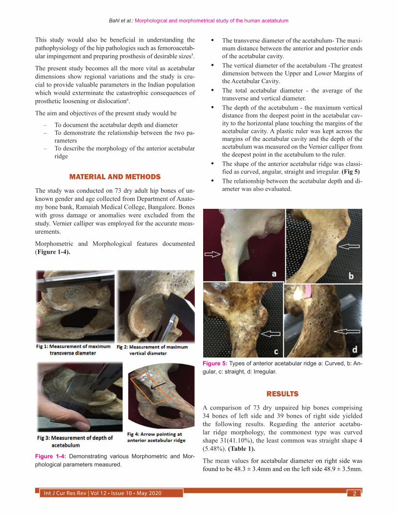
• The shape of the anterior acetabular ridge was classi-fied as curved, angular, straight and irregular. (Fig 5)
• The relationship between the acetabular depth and di-ameter was also evaluated.
Figure 1-4: Demonstrating various Morphometric and Mor-phological parameters measured.
Figure 5: Types of anterior acetabular ridge a: Curved, b: An-gular, c: straight, d: Irregular.
RESULTS
A comparison of 73 dry unpaired hip bones comprising 34 bones of left side and 39 bones of right side yielded the following results. Regarding the anterior acetabu-lar ridge morphology, the commonest type was curved shape 31(41.10%), the least common was straight shape 4 (5.48%). (Table 1).
The mean values for acetabular diameter on right side was found to be 48.3 ± 3.4mm and on the left side 48.9 ± 3.5mm.
Int J Cur Res Rev | Vol 12 • Issue 10 • May 20203
Bahl et al.: Morphological and morphometrical study of the human acetabulum
The mean values for acetabular depth on right side was meas-ured to be 27.1 ± 3.4mm and on left side 27 ± 3mm. (Table 2, 3). The mean and standard deviation of total diameter and
depth of acetabular cavity are shown and a significant posi-tive co-relation was found between them.
DISCUSSION
The acetabular morphology is very important for success-ful hip arthroplasty and for selecting a suitable prosthesis. The curved shaped acetabular ridges were found more fre-quently in our study. The comparison with other authors also showed same result. The percentage of irregular shape bones is significantly higher in the present study as compared to the study by Maruyama et al7.The percentage of bones with straight shape of anterior acetabular ridge is significantly lower as compared to the studies by Vyasa et al8, Prathiba et al9 and AKSU et al 10 (Table 4).
Table 1: Comparison of shape of the anterior acetabular ridge on the right and left side
Shapes Total No Right side Left side
Curved 30 (41.1%) 15 (38.5%) 15 (44.1%)
Angular 19 (26.2%) 11(28.2%) 8 (23.5%)
Straight 4(5.5%) 2 (5.1%) 2(5.9%)
Irregular 20 (27.4%) 11 (28.1%) 9 (26.4%)
Table 2: Mean diameter and depth of the acetabular cavityn=73 Transverse Diameter Vertical Diameter Total Diameter Depth
Mean (mm) 47.2 49.9 48.6 27.1
Standard Deviation 3.3 3.7 3.5 3.2
Maximum 53.1 60.6 55.9 35.4
Minimum 40.3 40.6 40.7 20.1
p=0.0001 ( Positive correlation between Total Diameter and Depth)
Table 3: Comparison of Total diameter and Depth of Acetabular cavity according to sideTotal diameter Depth
Variables RIGHT(mm) LEFT(mm) RIGHT(mm) LEFT(mm)
Mean 48.3 48.9 27.1 27.0
Standard Deviation 3.4 3.5 3.4 3.0
Maximum 54.6 55.9 35.4 32.4
Minimum 40.7 42.2 21.5 20.0
The findings of measurements of Total Acetabular diameter and Depth of acetabular cavity in the present study are con-sistent with other studies (Table 5).
The knowledge of acetabular dimensions will help us in un-derstanding the acetabular pathology and also in identifying disputed person through forensic expertise13.
The differences observed between the values of present study and that of other studies could be attributed to ethnic and racial variations.
Table 4: Comparison of shape of anterior acetabular ridge morphology with other studies
Authors Maruyama et al7
Vyasa et al8 Prathiba Kareddy et al 9
AKSU et al10
Govsa et al11
Present study
Shape
Curved 60.5% 37.5% 38.2% 46.1% 43.3 41.1%
Angular 25.5% 12.5% 11.5% 16.8% 28.3% 26.0%
Irregular 4.5% 18.4% 28% 13.6% 16.3% 27.4%
Straight 4.5% 31.6% 38% 23.3% 11.9% 5.5%

Int J Cur Res Rev | Vol 12 • Issue 10 • May 2020 4
Bahl et al.: Morphological and morphometrical study of the human acetabulum
However, it should be kept in mind, that the present study employed smaller number of hip bones, so it is worthwhile to perform similar studies on more number of hip bones for its theoretical and practical value in the coming years.
CONCLUSION
The findings of our study are vital for the accurate design of side specific prosthetic cups that replicate the curvaceous acetabular profile which would prevent prosthetic overlap, mechanical loosening and reduce the incidence of ilio-psoas impingement. The precise knowledge about the variations in morphology and morphometry of acetabular cavity would help clinicians, orthopedic surgeons, prosthetic surgeons and radiologists for better understanding about the pathologies of hip region which aids in accurate diagnosis and in planning a suitable treatment.
ACKNOWLEDGEMENTS
Authors acknowledge the immense help received from Mrs Radhika our statistician for the statistical analysis. Authors acknowledge the immense help received from the scholars whose articles are cited and included in references of this manuscript. The authors are also grateful to authors/ editors/ publishers of all those articles, journals and books from where the literature for this article has been reviewed and discussed.
Source of Funding: N/A
Conflict of Interest: Nil
Table 5: Comparison of Total acetabular diameter and depth with other studiesAuthors Diameter of acetabulum (cm) Depth of acetabulum ( cm)
Right Left Right Left
Dhindsa et al1 5.1 5.0 2.6 2.6
Vyas et al8 4.8 4.8 2.7 2.6
Prathibha Kareddy et al9 4.8 4.7 3.0 3.0
Chauhan et al12 4.7 4.7 2.7 2.8
Present study 4.9 4.8 2.7 2.7
REFERENCES1. Dhindsa G S, singh P singh Z. Morphometry of the adult human
dry hip bone International Journal of Pharmacy and Pharmaceu-tical Sciences 2013 ;5(2): 505-507.
2. Sinnatamby CS. 2006. Last’s Anatomy, Regional and Applied, 11th Edition, Edinburgh, Churchill Livingstone, 132.
3. Moore KL, Dalley AF, Agur AM. 2010. Pelvis and Perineum: Clinically Oriented Anatomy, 6th Edition, Philadelphia, Lippin-cott Williams and Wilkins, 328.
4. Umer M, Thambyah A, Tan WTJ, das De S. Acetabular morpho-metry for determining hip dysplasia in the Singaporean popula-tion . J Orthop Surg 2006; 14: 21-31.
5. Ukoha U U , Umeasalugo K E, Okafor J I, Ndukwe G U, Nzeakor H C, Ekwunife. Morphology and Morphometry of dry adult acetabula in Nigeria. Rev Arg de Anat Clin 2014 jul, 6 (3): 150-155.
6. Tannast M, Siebenrock KA, Anderson SE. 2007. Femoroacetab-ular Impingement: Radiographic Diagnosis - What the radiolo-gist should know, AJR 188: 1540-52.
7. Maruyama M, Feinberg JR, Capello WN, D’antonio JA.. Mor-phologic features of the acetabulum and femur: anteversion an-gle and implant positioning. Clin Orthop 2001;1: 52-65.
8. Vyas K, Shroff B, Zanzrukiya K. 2013. An Osseous study of morphological aspect of acetabulum of Hip bone. Int J Res Med 2013; 2(1): 78-82.
9. Pratibha K, Hema L, Devishankar. Acetabulum of the hip bone: A morhometric study in south coastal region. International Jour-nal of Recent Trends in Science and Technology 2015; 17(2): 136-139.
10. Aksu FT, Ceri NG, Arman C, Tetik S. Morphology and mor-phometry of the acetabulum. DEÜ Tip Fakültesi Dergisi 2006; 20(3): 143-148.
11. Govsa F, Ozer MA, Ozgur Z. Morphologic features of the ac-etabulum. Arch Orthop Trauma Surg 2005;125:453-461.
12. Chauhan R, Paul S, Dhaon BK.. Anatomical Parameters of North Indian Hip joints – Cadaveric Study. Journal of Anatomi-cal Society of India 2002; 51: 39-42.
13. Parmar G, Rupareliab S, Patel SV, Jethvaa N, “Morphol-ogy and morphometry of Acetabulum.” Int J Biol Med Res 2013;4(1):2924-2926.