DOCTORAL DISSERTATION SUMMARY HEPATO-RENAL DYSFUNCTION … · Hepatorenal syndrome may occur...
43
“GRIGORE T. POPA” UNIVERSITY OF MEDICINE AND PHARMACY OF IASI FACULTY OF MEDICINE DOCTORAL DISSERTATION SUMMARY HEPATO-RENAL DYSFUNCTION IN SEPSIS: ETIOLOGICAL, CLINICAL, PARACLINICAL AND THERAPEUTIC CORRELATIONS BENEFICIAL IN ETIOLOGICAL THERAPY SCIENTIFIC COORDINATOR Professor MD Carmen-Mihaela Dorobat DOCTORAL STUDENT Assistant professor MD Codrina-Veronica Bejan IASI 2014
Transcript of DOCTORAL DISSERTATION SUMMARY HEPATO-RENAL DYSFUNCTION … · Hepatorenal syndrome may occur...

“GRIGORE T. POPA” UNIVERSITY OF MEDICINEAND PHARMACY OF IASIFACULTY OF MEDICINE
DOCTORAL DISSERTATION SUMMARY
HEPATO-RENAL DYSFUNCTION IN SEPSIS:ETIOLOGICAL, CLINICAL, PARACLINICAL ANDTHERAPEUTIC CORRELATIONS BENEFICIAL IN
ETIOLOGICAL THERAPY
SCIENTIFIC COORDINATORProfessor MD Carmen-Mihaela Dorobat
DOCTORAL STUDENTAssistant professor MD Codrina-Veronica Bejan
IASI2014

1
Table of Contents
PART I: LITERATURE OVERVIEWI. INTRODUCTION ………………………………………………….......3II. DEFINITIONS IN SEPSIS ……………………………………………4III. SEPSIS – PATHOGENESIS …………………………………........... 5IV. CLINICAL PRESENTATION IN SEPSIS …………………………..10V. SEVERITY SCORES IN SEPSIS ……………………………………. 11VI. LIVER DYSFUNCTION ………………………………………….. 12VII. RENAL DYSFUNCTION ………………………………………… 14VIII. RESPIRATORY DYSFUNCTION ……………………………….. 15IX. CARDIAC DYSFUNCTION…………………………………………18X. NEUROLOGIC DAMAGE……..……………………………………..19XI. FLUID-COAGULANT EQUILIBRIUM……………………………..20XII. INTESTINAL BARRIER IN SEPSIS ……………………………... 21XIII. HEPATORENAL SYNDROME IN SEPSIS ………………………22XIV. SEPSIS DIAGNOSIS ……………..………………………………..26XV. TERAPEUTIC PRINCIPLES IN HEPATORENAL SYNDROMEIN SEPSIS ……………………………………………………….......….. 29
XVI. ANTIBIOTIC TREATMENT IN SEPSIS ………………..……….. 37XVII. MICORBIAL EPIDEMIOLOGY IN SEPSIS ……………………. 45XVIII. RISK FACTORS ……………………………………………........ 47XIX. SPECIAL POPULATION CATEGORIES ………………………..47PART II: PERSONAL CONTRIBUTIONXX. CURRENT SUBJECT IMPORTANCE. OBJECTIVES ………….. 49XXI. MATERIAL AND GENERAL METHODS ………..…………….. 50XXII. COMPARATIVE STUDY ON CLINICAL, EPIDEMIOLOGICAL,PARACLINICAL DEVELOPMENT AND THERAPEUTIC ASPECTS INPATIENTS WITH SEPSIS AND MULTIPLE ORGANDYSFUNCTIONS…………………………............................................. 531. Introduction ………………………………………………………...... 532. Study objectives …………………………………………………........ 533. Material and methods ……………………………………………........ 534. Results ………………………………………………………………... 56
4.1. Epidemiological data in patients with sepsis hospitalised in theInfectious Diseases Clinical Hospital of Iasi between November 2012 andApril 2014 ………………………………………………………………...56
4.2. Clinical, paraclinical and therapeutic aspects upon admission and duringevolution in patients diagnosed with sepsis hospitalised in the InfectiousDiseases Clinical Hospital of Iasi between November 2012 and April2014………………………………………………………………………. 61

2
5. Discussions …….……………………………………………………… 896. Conclusions …………………………………………………………….93XXIII. COMPARATIVE STUDY ON CLINICAL, EPIDEMIOLOGICAL,PARACLINICAL DEVELOPMENT AND THERAPEUTIC ASPECTS INPATIENTS WITH HEPATORENAL DYSFUNCTION IN SEPSIS ……96
1. Introduction ……………………………………………………..........962. Study objectives ……………………………………………………...963. Material and methods …...................................................................... 964. Results ………………………………………………………………..98
4.1. Epidemiological aspects in patients with liver/ kidney dysfunction insepsis hospitalised in the Infectious Diseases Clinical Hospital of Iasi betweenNovember 2012 and April 2014 …………………………......................... 98
4.2. Clinical, paraclinical and therapeutic aspects and correlations inpatients with hepatorenal dysfunction in sepsis ………………………... 103
5. Discussions …..…………………………………………………........ 1366. Conclusions …………………………………………………………..140
XXIV. RESEARCH ON THE SENSITIVITY/ RESISTANCE PROFILE OFSOME ETIOLOGIC GRAM-POZITIVE AND GRAM-NEGATIVE AGENTSISOLATED FROM PATIENTS WITH SEPSIS hospitalised in the InfectiousDiseases Clinical Hospital of Iasi between November 2012 and April2014……………………………………………………………………… 143
1. Introduction……………………………………………………….......1432. Objectives …………………………………………………………… 1433. Material and methods ………………………..…................................ 1444. Results ………………………………………………………………..146
4.1. Epidemiological data in patients with positive hemocultures ….. 1464.2. Evaluation of sensitivity / resistance profile of some Gram-positive
bacteria ………………………………………............................................1494.3. Evaluation of sensitivity / resistance profile of some Gram-negative
bacteria ……………………………………………..........………..............1535. Discussions …………………………………………………………. 1586. Conclusions …………………………………………………………..163
XXV. GENERAL CONCLUSIONS …………………………………..... 165PERSPECTIVES OPENED BY THE PRESENT DISSERTATION ........ 167BIBLIOGRAPHY ………………………………………………………...168LIST OF ABBREVIATIONS …………………………………………… 182LIST OF SCIENTIFIC PAPERS RELATED TO PERSONALCONTRIBUTIONS …………………………………………………….... 183Appendix A – Scientific papers related to “Personal contributions” publishedin extenso …………………………………………………………………184Appendix B – Informed Consent for Research

3
LITERATURE OVERVIEW
I. IntroductionSepsis is the host's response to a microbial agent and its toxins, while severesepsis and septic shock are most often the causes of anomalies in the infectiousprocess. The concept of sepsis has been the subject of intense debate over time(1, 2), so a huge effort has been made over the past 20 years to find newtherapies to reduce mortality, but unfortunately, leaving aside the progressmade, dissatisfaction is still present (2, 3).
Epidemiological data in sepsisThe major impact of sepsis with multiple organ disorder on the nursing unitswas estimated, in the European Union, at 90.4 cases per 100,000 inhabitants,as compared to 58 per 100,000 in breast cancer (4). It is estimated that thereare 1.8 million cases annually, worldwide, and this in terms of a weakidentification rate or under diagnosis. Recent estimates showed an incidence ofsepsis in intensive care units between 0.25 and 0.38 per 1,000 inhabitants,which suggests two million admissions to intensive care units (5, 6).
III. Sepsis pathogenesisSepsis comprises a great heterogeneity within patients with differentconditions, infection sites, a multitude of microbial agents with high variationof microbial virulence and concentration, with differences in relation to theinflammatory response and immunological context.The response from the host is connected to either the secretion products of thepathogen agent or to one of its fragments. For Gram positive or negativebacteria, teichoic acid and lipopolysaccharides together with pieces of cell wallconstitute components able to induce inflammatory response, sepsis and septicshock. Thus, in the face of this aggression, the hostess triggers, successively, aseries of defensive mechanisms such as: natural barriers (epithelium, mucousmembranes), innate immunity and adaptive immunity. The imbalance of thesedifferent means of defence participates in the evolution toward a misfitresponse, characterized clinically by septic shock (7, 8), Figure 1.

4
Virulence factors
Endotelială PMN macrophages lymphocytescell neutrophils
apoptosis liveracute stageproteines
CNSproinflammatory antiinflammatoryResponse responseTNFα , IL1, IL6 PNS
neuroendocrineinflammation immunodeppression paths
Teichoic acid Lipopolyssacharides
sepsis Severe sepsis Septic shock MSOF
Figure 1. Physiopathogenesis in sepsis
The inflammatory response is often adapted to the etiologic agent or toxicaggression, or, on the contrary, a series of micro-organisms may lead to highlevels of mediators from infected body, having increased expression in patientswith multiple organ dysfunction associated. Currently it is not possible toprovide a quick assessment of organisms’ capacity to have an adequateinflammatory response, and it is considered that the mechanisms related toorgan dysfunction as those which are carried out in the evolution towardsdeath would be similar in the context of different etiologic agents (7, 9).Bacterial toxins such as endotoxins and superantigenes are commonlyinvolved in the pathogenesis of sepsis with Gram-positive and Gram-negative,as they determine signals through cellular mechanisms and induceinflammation through different routes (10, 11).The affected organ plays an important role, being known for example that thedefence mechanisms of the lung and those of the peritoneum are different,leading to different possibilities for evaluation of inflammation (12). Theseantigenic structures and toxins determine the inflammatory process through theconnection with the CD14 receptor of the mononuclear phagocytes incirculation. Tumour necrosis factor (TNF), interleukin 1 (IL-1), IL-6, IL-8 andplatelet activating factor (PAF) are released by monocytes. IL-1 and IL-6activate the T-cells, resulting in the formation of interferon-gamma, IL-2, IL-4,the activation factor of granulocytes monocytes (GM-CSF) (13, 14).

5
XIII. Hepatorenal syndrome (HRS) in sepsisClinical and physiopathogenical aspects in HRS
Hepatorenal syndrome may occur spontaneously, by altering liverfunction or secondarily by precipitation of an event such as infection (15).
Hepatorenal syndrome is caused by intrarenal vasoconstriction thatoccurs in patients with terminal liver disease status and associated circulatorydysfunction.
Circulatory dysfunction is characterized by vasodilatation insplanchnic circulation with relative and insufficient decrease of carbonmonoxide, leading to hypovolemia (16).
HRS: short historyAt the end of the 19th century, assessments made by Frerichs (1861) and Flint(1863) noted an association between advanced liver disease, ascites and renalinsufficiency with oliguria in the absence of renal histological changes (17).Almost 100 years later, in an article signed by Hecker and Sherlock, it wasdescribed the pathogenesis of HRS (18). Detailed studies carried out byEpstein et al. have shown that splanchnic circulation and systemicvasodilatation together with the intense renal vasoconstriction are thephysiopathogenic mechanism of renal HRS (19). However, despite theadvances in knowledge, HRS prognosis remained gloomy at that time, and inthe 1970’s the concept of "functional terminal renal failure" was the equivalentof hepatorenal syndrome (20).In 2006, the definitions for HRS showed that this is a functional reversiblekidney damage that occurs in patients with advanced liver cirrhosis orfulminant liver failure and is characterized by reduction in glomerular andplasma filtration rate in the absence of other causes of renal dysfunction (21).With the rise of knowledge, it has been proved that the original definition wasvery narrow as to a large number of exclusion criteria such as sepsis (22).Until it was reviewed, guidelines proposed in 2007 that bacterial infections areconsidered an exclusion criterion for diagnosis of HRS (23).
Mechanism of the hepatorenal syndrome (HRS)HRS mainly consists of an extreme renal vasoconstriction caused by activationof sodium retention mechanism and vasoconstriction systems, resulting in amajor decline in the glomerular filtration rate (24). The cascade of events isdetermined by vasodilatation in the splanchnic circulation and blood volumedecrease.When data were available regarding HRS type II, a number of studies showedthat terlipressin increases kidney function by more than 65% in patients withHRS type I, with a significant increase in survival (25,26,27). None of these

6
studies included patients with sepsis, which at that time was in agreement withthe original definitions of the HRS (20). More recently, Rodriguez E et al.presented the results of a study that explored the role of terlipressin andalbumin in patients with HRS type I and sepsis, the authors pointing out thatthere are similar results to those in studies where patients with sepsis wereexcluded (20).The condition of hepatorenal syndrome diagnosis is kidneys’ integrity and thishas been repeatedly demonstrated both in terms of morphology and normalfunction of the kidneys. The physiopathologic principle of mechanismsincludes increased renal artery resistance, particularly affecting the renalcortex, which occurs in renal hypoperfusion (28).Renal vasoconstriction per se is not sufficient for the development of HRS.Low blood pressure blood pressure is the key factor even where it does notreach the values in shock, being a simultaneous cause of vasoconstriction andrenal hypoperfusion (29).Thus, the circulating volume, a result of systolic volume is a relevant factor,and its level can be low, normal or increased, but is relatively insufficient toprevent severe decreases in the actual volume of circulating blood caused bysplanchnic arterial vasodilatation in patients with hepatorenal syndrome.
Table VII Hepatorenal syndrome type I and type II
creatinine Evolutionperiod
ascites Liverdamage
norepinephrineand renin
Presenceof
infectiousprocess
prognosis
HRS1
2 X Nor
>2.5mg/dl
2 weeks Possiblyspontaneous
bacterialperitonitis
Rapidprofounddamage
Intense activity possible infaust
HRS2
<2.5mg/dl Slowprogression
Diuretic-resistantascites
Low/minimumdamage
Minimumactivity
mayprecipitateconversion
fromHRS1
To HRS2
>4-6months
HRS type I occurs as a consequence of severe reduction of circulating volumecaused partly by a major splanchnic vasodilatation and, partly by decreasedcardiac output, this type of disorder being characteristic to sepsis. HRS type IIis characterized by a slow progressive loss of renal function, without being,

7
from the clinical point of view, the consequence of an acute deterioration ofrenal function, but the result of refractory ascites, and its impact on prognosisis less negative (30).
HRS diagnosisCurrently, there is no specific diagnostic test for a clear determination of HRS,as it is a diagnosis of exclusion on the basis of the revised criteria of theInternational Ascites Club (15).Primary investigations consist in identifying the glomerular filtration rate, withcreatinine clearance < 40 ml/min or increased serum creatinine > 135micromoles/L in the context of the exclusion of other causes of renaldysfunction. The most relevant indicator of functional character includesnatriuresis < 10 mmol/L, urinary osmolarity > plasma osmolarity, natremia <130 mmol/L and 500 ml diuresis </24 hrs.
The new diagnostic criteria in HRS Cirrhosis with ascites Serum creatinine > 133 micromol/L (1.5 mg/dL) Decreased serum creatinine to 133 micromol/L or less after 2 days
of diuretics and administration of albumin solutions to expandvolemia; the recommended dose of albumin is 1 g/kg bw/day, up toa maximum of 100 g/day
Absence of shock Absence of recent treatment with nephrotoxic substances Absence of parenchymal diseases as an indicator of proteinuria >
500 mg/day, microhematuria (> 50 RBCs/HPF) and/or abnormalrenal ultrasound.
Concerning the diagnostic criteria, it would be useful to distinguish betweenthe concept of hepatorenal syndrome and hepatorenal insufficiency/dysfunction. In both cases, the parameters of renal function clinics are thesame. However, the major difference lies in the circumstances of theoccurrence and prognosis. Hepatorenal syndrome should be considered whenassigning a specific status of renal function deterioration when other causeshave been eliminated and precipitating factors excluded. Thus, hepatorenalsyndrome is a more general term that can be applied to any deterioration ofrenal function in the context of liver disease or sepsis, with the involvement ofportal hypertension (31).

8
XV. Therapeutic principles in HRS therapy in sepsis involving multiple organ dysfunction eliminate use of nephrotoxic substances eliminate gastrointestinal pain and loss compensation therapy eliminate use of nonsteroidal antirheumatics recover lost blood volume through intravascular volume supplement,
preferably by correcting hypoalbuminemia (albumin is the best volumeexpander with the longest impact)
remove diuretics (they increase central hypervolemia, the sympatheticactivity and that of the renin-angiotensin-aldosterone system)
avoid substitution measures to combat hyponatremia because of the riskfor cerebral edema; the restriction on fluids is preferred (31).

9
PERSONAL CONTRIBUTIONS
XX. Subject importanceThe term of hepatorenal syndrome (HRS), after it was described in 1863, wasfirst used in 1932 in an effort to materialize the links between renalinsufficiency which occurs after biliary tract surgery and is later used morebroadly for any association between severe liver damage and kidney damage.The definition of HRS was described as renal insufficiency which arises in thecontext of severe liver disease, acute or chronic, in the absence of pre-existingrenal pathology (32).The first attempt to organize the diagnostic criteria was made by theInternational Ascites Club in 1996, when they were grouped into major andminor criteria (32, 33, 34). The use of these criteria showed, in time, the needfor accuracy and additions due to the ambiguity of HRS definition. In 2006, inSan Francisco, the International Ascites Club redefined HRS in terms of thediagnostic methods and taking into account the possible presence of infection,which had not been a criterion for exclusion (32, 33, 34).The exact incidence of HRS is not known largely due to difficulties indiagnosis. In over 70% of cases of HRS, precipitating factors are identifiedsuch as infection (57%), gastrointestinal hemorrhage (36%), paracentesis (7%)or acute alcoholic hepatitis (35, 36).It is now known that the acceptance of the infection and sepsis in definingHRS determined a decrease in mortality in patients with HRS, from 80% to65% (32).
XXI. Material and General MethodsWe have carried out the three studies in the Infectious Diseases ClinicalHospital of Iasi, here being hospitalised patients with an extremely complexpathology from the whole geographical area of Moldova and beyond, andconsidering the advanced technical possibilities, especially in the IntensiveCare Department, specific to infectious diseases.Both the adult age patients, or persons authorized in circumstances where itwas not possible otherwise, and in the case of children, signed upon admissionan informed consent formulated in accordance with the Helsinki Declarationand approved by the Ethics Committee of the University of Medicine andPharmacy of Iasi, regarding consent to use personal data for the purpose ofresearch.The study included patients with the diagnosis of sepsis according to thedefinition of suspected or proven infection accompanied by the presence ofSIRS (at least 2 of the following criteria: fever/hypothermia, tachycardia,tachypnea, leucocytosis-leukopenia).

10
Data statisticsWe set up a database in EXCEL which has included both the descriptive andqualitative and quantitative parameters, while data processing was performedusing the SPSS version 16.0.
The types of statistical processing included:1. student's t-test2. chi-square test3. Box Plot chart type (with the median to the right of the middle, the
rectangle contains 50% of the data and the extremes marked byvertical lines included data between 10 and 90%)
4. Pearson’s correlation for normally distributed variables5. Spearman correlation - independent of the type of data distribution and
based on the rank order of values6. where data did not have a normal distribution we used the Mann-
Whitney Test for comparing two sets of variables7. Pairwise Comparisons Test for data comparison with small samples8. for comparison of several data sets we used the Kruskal-Wallis Test9. the ROC curve for determining sensitivity/specificity of the diagnostic
tests10. Kaplan Meier survival curve.
XXII. Comparative study regarding the clinical, epidemiological,paraclinical evolution and therapeutic aspects in a group of patients whoassociated organ dysfunction in sepsis2. STUDY OBJECTIVESThrough this study we attempted an evaluation of epidemiological, etiological,paraclinical, laboratory and therapeutic aspects in patients diagnosed withsepsis, for which the clinical evolution context was studied, determiningcorrelations between etiologic agents and severity of infection, establishinginfection site, correlations between paraclinical parameters and APACHE IIand Carmeli prognostic scores, correlations between clinical evolution modeland antibiotherapy, establishing the role of associated comorbidities, andassessing the survival of these patients in the context of the different organdysfunctions associated.
3. MATERIAL AND METHODSThe study included patients diagnosed with sepsis in the Infectious DiseasesClinical Hospital of Iasi, during November 2012 – April 2014, with a total of247 patients grouped as follows:
1. a lot of 100 patients with sepsis associating multiple organdysfunctions (called "MODS");

11
2. a lot of 77 patients with sepsis and hepatorenal dysfunction (alsocalled “HRD”);
3. a lot of 70 patients with sepsis and multiple organ dysfunction, withclinical evolution towards death (called “DECEASED”).
We have also taken into consideration the cases with discharge diagnosis ofsepsis with determined etiology (positive hemocultures) or sepsis withundetermined or clinically suspected etiology (where samples from differentbiological products were positive). These patients had the results of thesamples entered in the registry of the Microbiology Laboratory of theInfectious Diseases Clinical Hospital of Iasi.Patient inclusion criteria were in accordance with the standard definitions ofsepsis.
4. RESULTSWe tried to determine if there is a statistical correlation between inflammatorysyndrome elements throughout the lot of patients with sepsis, using twovariants for the analysis of correlation: Pearson and Spearman.The Pearson correlation is used for normally distributed variables andmeasures a combination of linear type, while extreme values can influencestrongly the value of the coefficient.The Spearman correlation is independent of the type of data distribution and isbased on the rank order of values. Extreme values do not have a majorinfluence on the calculation.This coefficient varies between -1 and 1.We also verify the level of statistical significance (Table XXII. 6., Table XXII.7.).
Table XXII. 6. Establishing statistically significant differences between theelements of the inflammatory syndrome
The table is symmetric, the correlation between white blood cells (WBC) andfibrinogen being equal to the correlation fibrinogen WBC, as the correlationcoefficient is commutative.
Descriptive Statistics
Mean Std. Deviation NWBC upon admission 13307.98 8152.348 247Fibrinogen upon admission 5.8867 1.53384 241ESR upon admission 77.87 37.029 241CRP upon admission 49.10 25.037 226

12
Table XXII. 7. Correlations in inflammatory syndromein all patients with sepsis
WBC uponadmission
Fibrinogenupon
admission
ESR uponadmission
CRP uponadmission
Spearman’srho
WBC uponadmission
CorrelationCoefficient
1.000 .210** .285** .191**
Sig. (2-tailed) . .001 .000 .004N 247 241 241 226
Fibrinogenuponadmission
CorrelationCoefficient
.210** 1.000 .267** .335**
Sig. (2-tailed) .001 . .000 .000N 241 241 238 223
ESR uponadmission
CorrelationCoefficient
.285** .267** 1.000 .418**
Sig. (2-tailed) .000 .000 . .000N 241 238 241 224
CRP uponadmission
CorrelationCoefficient
.191** .335** .418** 1.000
Sig. (2-tailed) .004 .000 .000 .N 226 223 224 226
**. Correlation is significant at the 0.01 level (2-tailed).Therefore, we obtained statistical significance for all combinations, whichmeans that there are statistical correlations between elements of theinflammatory syndrome: an increase in WBC will determine a proportionalincrease in ESR, Fibrinogen, CRP, but reciprocal claims are also true (TableXXII. 8.).
Table XXII. 8. Statistical correlations between the elements of theinflammatory syndrome in patients with sepsis
Coefficient of determination
WBC atadmission
Fibrinogen -admission
ESR-admission
CRP-admission
WBC at admission 1 0.0441 0.081225 0.036481Fibrinogen atadmission 1 0.071289 0.112225
ESR at admission 1 0.174724CRP at admission 1

13
The coefficient of determination multiplied by100 represents, in percentages,the degree to which the variation in a variable is expressed by the othervariable.
Evaluating glycaemia modifications in patients with sepsisOf the metabolic changes that occur during sepsis, hyperglycaemia is the mostimportant. Hyperglycaemia in severe sepsis is not merely a marker of theseverity of the disease but also a predictor of adverse developments and hasmultiple ways of exercising the adverse effects at the level of vital organs. Oneof these side effects is on the innate defence system of the host againstinfection, resulting in a reduction of neutrophils activity such as chemotaxisand bacterial phagocytosis in spite of accelerated diapedesis of leukocytes inperipheral tissues, as well as alteration of cytokines and growth of earlyproinflammatory cytokines, TNF alpha and IL-6, and reducing the formationof endothelial nitric oxide (37).
Table XXII. 9. Statistical correlations between leucocytes and glycaemiaupon admission in patients with sepsis
Spearman’s rhoWBCupon
admission
Fibrinogen upon
admission
ESR uponadmission
CRP uponadmission
Glycaemiaupon
admission
CorrelationCoefficient .170 .079 .057 .058
Sig. (2-tailed) .007 .220 .382 .386
N 247 241 241 226

14
Figure XXII. 12. Statistical correlations between glycaemia uponhospitalization (g/L) and leucocytosis in patients with sepsis (WBC/mm³)
Patients showed a variable number of organ dysfunction, and those whichrequired monitoring in intensive care department had predominantly more thantwo affected organs (Figure XXII. 31.). According to studies, multiple organfailure in sepsis remains the most common cause of death in intensive therapyunits (38).
Figure XXII. 31. Organ dysfunctions in patients who requiredmonitorisation and therapy in the intensive care department
15
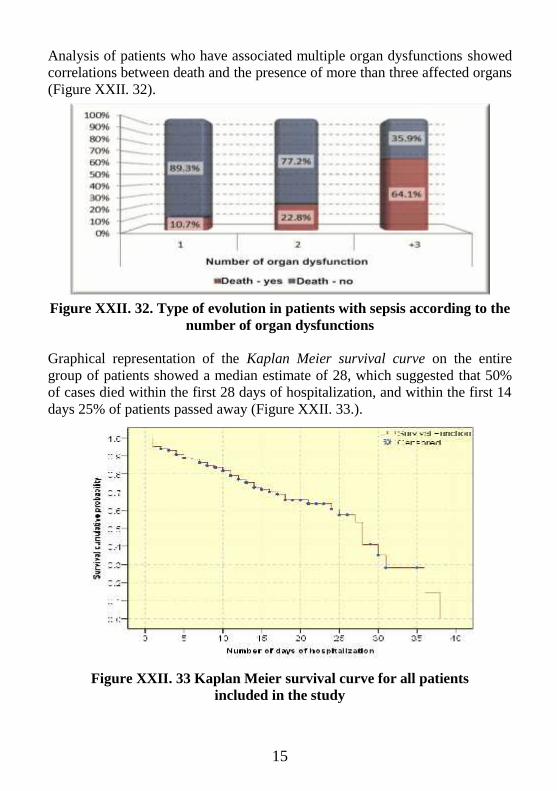
Analysis of patients who have associated multiple organ dysfunctions showedcorrelations between death and the presence of more than three affected organs(Figure XXII. 32).
Figure XXII. 32. Type of evolution in patients with sepsis according to thenumber of organ dysfunctions
Graphical representation of the Kaplan Meier survival curve on the entiregroup of patients showed a median estimate of 28, which suggested that 50%of cases died within the first 28 days of hospitalization, and within the first 14days 25% of patients passed away (Figure XXII. 33.).
Figure XXII. 33 Kaplan Meier survival curve for all patientsincluded in the study

16
Survival analysis in patients hospitalized in intensive care unit showed amedian of 11 days, which means that 50% of patients die within the first 11days, and according to the quartiles of the table, 25% of patients die within thefirst 4 days (Table XXII. 14, Figure XXII. 34.).
Figure XXII. 34. Kaplan Meier survival curve for patients in ICU
5. DISCUSSIONSIn patients with sepsis considered in this study, we tried a comparativeevaluation of parameters related to systemic inflammatory response, so thatstatistical analysis of the variables of the inflammatory syndrome showedcorrelations between ESR, fibrinogen, CRP, leucocytosis and glycaemia,whether patients have associated or not hepatorenal dysfunction indevelopment.We can admit that there were biases concerning data on inflammatorysyndrome, a number of factors being recognised as enhancers in relation toESR values in the absence of inflammation, among them being age, femalegender, obesity, hypercholesterolemia, nephrotic syndrome, renalinsufficiency, cardiac insufficiency.Considering that currently there is no specific diagnostic test for univocaldetermination of HRS, as it is a diagnosis of exclusion on the basis of therevised criteria of the International Ascites Club, we considered defining thesepatients according to the glomerular filtration rate, with a clearance of

17
creatinine < 40 ml/min or increased serum creatinine > 135 micromoles/L inthe context of the exclusion of other causes of renal dysfunction (39).Although an adequate indicator of functional character of hepatorenaldysfunction includes natriuresis < 10 mmol/L, a urinary osmolarity higher thanthe plasma osmolarity, natremia < 130 mmol/L and diuresis < 500 ml/24 hoursin normal practice, it was not possible for a full consideration of these criteriain all cases.According to some authors, the hepatic dysfunction in sepsis represents themoment when total bilirubinemia is higher than 2 mg/dL (>34µmol/L), andalkaline phosphatase or serum aminotransferase values are greater than twicetheir normal value. In a study carried out by Angus and collaborators, from192,980 cases of severe sepsis in the U.S., hepatic dysfunction was present inonly 1.3% (40). However, this low incidence is explained through a highlyrestrictive definition of hepatic dysfunction, with the inclusion only of caseswith acute and sub-acute liver necrosis. In 2001, the International SepsisDefinitions Conference has recommended the use of scoring systems such asSOFA (Sepsis-related Organ Failure Assessment), MODS or LODS (LogisticOrgan Dysfunction System).All of these scores aim to identify the degree of organ disorders during sepsisand use bilirubinemia to define disorders along with prothrombin time forLODS score (41, 42). At this conference, hepatic dysfunction was defined astotal bilirubinemia greater than 4 mg/dL (µmol/L > 70), but interestingly, thiscutoff value has not been proposed by any scoring system known. Anexception is the acute phase of severe sepsis/septic shock where a considerableincrease in the level of aminotransferase (20 times the normal limit) allowsdiagnosis of hypoxic hepatitis, incidentally, total bilirubinemia being used andproposed as a biomarker for the diagnosis of hepatic dysfunction in sepsis.The nature of "systemic" evolution in sepsis, the high number of cell linesinvolved, organs and tissues expanding the area of candidate biomarkers,biochemical modifications determining varied responses over time, making itvery difficult to define a "golden standard" in diagnosing and predicting theevolution of these patients (43).Our analysis showed that each of the parameters studied was an independentvariable in patients with sepsis, this being restrictive in terms of understandingas predicting factors, even when data integration is based on groups ofpatients.

18
XXIII. Comparative study regarding the clinical, epidemiological,paraclinical evolution and therapeutic aspects between a group of patientswho associated hepatorenal dysfunction and another group with variedorgan dysfunction in sepsis
2. STUDY OBJECTIVESThe trend of increase in the number of patients with sepsis hospitalised in theInfectious Diseases Clinic of Iasi in the past years was amplified by a higherdegree of complexity, in many of these patients their development beingaccompanied by hepatorenal syndrome.In the present study, we aimed to address the septic patient with hepatic-renaldysfunction and/or hepatorenal syndrome, from a detailed perspective, incomparative terms with the changes occurred in sepsis development involvingother organ dysfunction.Knowledge of these patients from clinical, etiological, paraclinical, prognosticscore as well as therapeutic perspective represents a challenge inunderstanding the mechanisms underlying the hepatic/renal decompensation insepsis. Since the redefining of the hepatorenal syndrome in 2007 and itsassociation to decompensations in sepsis, studies have tried, in most cases, toidentify similarities between the pathophysiological mechanisms ofhepatorenal decompensation in liver cirrhosis versus sepsis, relatively few databeing available concerning developments in patients with sepsis andhepatorenal syndrome.
3. MATERIAL AND METHODSOur study included patients diagnosed with sepsis in the Infectious DiseasesClinical Hospital of Iasi, during November 2012 - April 2014, with a total of117 patients who associated liver/kidney dysfunction, hepatorenal syndrome,but other organ dysfunction also.Severe sepsis comprised patients with the acute organic dysfunction (cardiac,renal, hepatic, respiratory, circulatory insufficiency, disseminated intravascularcoagulation or shock). Biological products were analysed in the Laboratory ofBacteriology by direct examination, cultures, complement-fixation test, andlatex agglutination.In patients included in the study, we have taken into consideration thefollowing:- Demographic data (age, gender, environment of origin)- Period of hospitalization- Etiology- Paraclinical data at onset and in development

19
- APACHE II and CARMELI prognostic scores- The presence of previous liver/kidney disorders- Liver/kidney disorder in sepsis- The presence of other organ dysfunction- Evolution towards improvement or death-the existence of statistical correlation of evolution parameters
4. RESULTS
Figure XXIII. 5. Patients’ age according to the APACHE II scorecalculated
Patients’ age represented by APACHE II score calculated showed a medianequal to or greater than 55 in patients with hepatorenal syndrome (FigureXXIII. 5.).

20
Table XXIII. 5. Correlations between uremia andcreatininemia upon hospitalization
creatinine i urea i
creatininei
Pearson Correlation 1 .496**
Sig. (2-tailed) .000N 47 47
urea iPearson Correlation .496** 1Sig. (2-tailed) .000N 47 47
**. Correlation is significant at the 0.01 level (2-tailed).
Fig. XXIII. 21. Correlations between uremia (mg/dL) and creatininemia(mg/dL) upon hospitalization in patients with hepatorenal syndrome
in sepsis
Correlations were also found between urea and creatinine values at discharge(Table XXIII. 6., Figure XXIII. 22.).

21
Table XXIII. 6. Correlations between urea and creatinine levelsupon discharge
creatininee
urea e
creatininee
Pearson Correlation 1 .604**
Sig. (2-tailed) .000N 47 46
urea ePearson Correlation .604** 1Sig. (2-tailed) .000N 46 46
**. Correlation is significant at the 0.01 level (2-tailed).
Figure XXIII. 22. Correlations between urea (mg/dL) and creatinine(mg/dL) levels upon discharge, in patients with hepatorenal syndrome
in sepsis
Comparison of the data sets was made using the Kruskal Wallis nonparametrictest for independent samples (ANOVA equivalent), the test yielding a p value

22
of 0.44. By comparing groups of pairs two by two, we found significance onlybetween creatinine upon admission for APACHE I in comparison withAPACHE III. As seen in Figure XXIII. 23., creatininemia is higher in theAPACHE III group.
Figure XXIII. 23. Correlations between creatininemia (mg/dL) andAPACHE II calculated score
For ALT values, we obtained the ROC curve according to the graph in FigureXXIII. 24.
Figure XXIII. 24. ROC curve for TGP values

23
Therefore, the optimal value of diagnostic decision, or the moment when thesensitivity/specificity ratio was at its peak for ALT is 78 IU/l.For AST, we obtained the data from the ROC curve graphical plot in FigureXXIII. 25.
Figure XXIII. 25. ROC curve for AST values
According to the data obtained from the statistical software, the optimal valueof diagnostic decision, or the moment when the sensitivity/specificity ratio wasat its maximum for AST, is 75.5 IU/l.The optimal value of diagnostic decision, or when the sensitivity/specificityratio has been fullest for TB is 25.5 mg ‰ (Figure XXIII. 26.).
Figure XXIII. 26. ROC curve for total bilirubinemia values(TB: mg‰)

24
In those patients with hepatorenal dysfunction in sepsis, we tried to determineif there is a statistical correlation between values of bilirubinemia and alteredneurological status, using the chi-square test (Figure XXIII. 31.).
Figure XXIII. 31. Statistical correlations between bilirubinemia andneurological status alteration
Representation of the correlation between glycaemia levels upon admission inhospital and the number of organ disorders showed higher values of bloodglucose for patients who associated more than three organ dysfunctions(Figure XXIII. 34.).
Figure XXIII. 34. Correlations between glycaemia (g/L) and organdysfunctions in patients with sepsis

25
According to the statistical data presented in Figure XXIII.44, correlationshave been identified between antibiotherapy modification and death in patientswith hepatorenal dysfunction in sepsis, which, on the one hand, could highlightthe increasing risk of inducing resistance to antibiotics by frequently changingthe therapeutic scheme, and on the other hand the difficulty of choosing anappropriate treatment and the complexity of severe cases.
Figure XXIII. 44. Statistical correlations between antibiotherapymodification and death in patients with sepsis
5. DISCUSSIONSAlthough the standard definition of hepatorenal syndrome refers to the precisevalues of biochemical parameters in the development of patients with impairedrenal/liver function as well as consequences related to diselectrolitemia,urinary osmolarity and diuresis, enclosing the critical patient with sepsis in thediagnosis of hepatorenal syndrome remains a challenge for the clinician, as theelements of the clinical development aspect are often not correlatedmathematically with the modified biochemical parameters.In this second study were included by selection patients with sepsis who, inaddition to multiple organ dysfunctions, associated hepatic/renal dysfunction,while in those who developed both types of disorder, we tried to make anevaluation of the hepatorenal syndrome under various aspects: clinical,paraclinical, etiological, as well as from the point of view of patient evolutionand therapy. The absence of shock prior to the diagnosis of hepatorenalsyndrome was considered according to the latest consensus of InternationalAscites Club which allows the association of hepatorenal syndrome in sepsis(44, 45).

26
In our approach, we considered useful to try to determine the significance levelthrough tests of sensitivity/specificity for serum urea, serum creatinine, serumtransaminases, bilirubinemia and even proteinemia, cholesterol in thehepatorenal syndrome in sepsis, the threshold from which these parametersbecome suggestive or not along with elements from the patient's clinical septicevolution.For accuracy in the use of laboratory tests values modified in the hepatorenalsyndrome, we took into considered the data from patients in which thealgorithm for differential diagnosis ruled out other entities that could be causesper se for renal damage, according to currently accepted definitions forhepatorenal syndrome in sepsis (44).At present, there are not any known differences between thepathophysiological mechanisms of HRS type I and type II. However, a numberof precipitating factors type I have been signalled, such as bacterial infection,especially spontaneous bacterial peritonitis, paracentesis with large volumesand without compensating plasma substitute, gastrointestinal bleeding andalcoholic hepatitis, but there are also unknown precipitating factors. Therehave been reported a series of controversies related to dividing HRS type I into2 types according to the existence or not of the precipitating factors in theevolution of the syndrome, and the result was that infection should beconsidered as an element of HRS, such that outside spontaneous bacterialperitonitis, any kind of infection may be considered a precipitating factor ofHRS. Renal dysfunction which occurring after the infection presents thefollowing risk factors: severity of infection, MELD score (Model for end-stageliver of disease) and the lack of response to antibiotherapy (45).

27
XXIV. Research on the sensitivity/resistance profile of the etiologic Gram-positive and Gram-negative agents isolated from patients with sepsishospitalized in the Infectious Diseases Clinical Hospital of Iasi fromNovember 2012 to August 2014
1. INTRODUCTIONWe are currently confronted with the emergence of bacterial resistance tonumerous antibiotics, as the number of systemic infections with multiresistantgerms resister is increasing significantly in Europe and in other parts of theworld (4).In the 60 years since their introduction, millions of tons of antibiotics havebeen produced and used in a huge variety of situations. There is now a "glut"in the use of these "toxic" agents, who contributed significantly to thedevelopment of resistance and the selection pressure, which it is not a naturalprocess, but a process conducted by the man and imposed on nature (4).The successful use of therapies can be compromised by the development oftolerance or resistance, involving a whole range of biochemical andphysiological mechanisms.
2. STUDY OBJECTIVESFor patients with positive hemocultures, hospitalised in the Infectious DiseasesClinic of Iasi from November 2012 to August 2014, we made estimatesconcerning microbiological data, both for Gram-positive and Gram-negativegerms. During this interval, we considered a number of 256 in-patients withpositive hemocultures.
3. MATERIAL AND METHODSWe drew up an evaluation of the microbiological data in terms ofsensitivity/resistance rates for the most common strains identified in thehemocultures of in-patients, admitted from November 2012 until August 2014.Isolation of microorganisms was performed in the Microbiology Laboratory ofthe Infectious Diseases Clinical Hospital of Iasi, by using classical methods.Hemoculture follow-up was carried out on brain-heart infusion broth andtrypticase soy broth, the first 3 days (twice a day), then daily until positiveculture was obtained, extending the examination period to a maximum of 14days. We excluded from the database the stems from the same bacterialspecies isolated during the same infective episode in the same patient.The identification of cultures was based on tracking microorganisms’ culturecharacteristics: morpho-tinctorial, metabolism, and on special tests foridentifying coagulase positive staphylococci (Staphylococcus aureus),

28
coagulase negative (Staphylococcus epidermidis and other species), otherGram-positive bacteria (Enterococcus spp, Streptococcus spp.) and Gram-negative bacteria (Pseudomonas spp., Klebsiella spp., Escherichia coli,Proteus spp., Acinetobacter spp., Enterobacter spp.).
4. RESULTSWe studied if there were statistical correlations between patients who hadreceived antibiotherapy prior to hospitalization in our clinic and the type ofdevelopment classified according to APACHE II score.
Figure XXIV. 4. Correlations between previous antibiotherapy in patientswith positive hemocultures and APACHE II score calculated
Figure XXIV. 4 with chi-square test showed that the proportions of patientswith previous antibiotic differ significantly in the two APACHE groups II andIII.Etiologic agents identified in hemocultures were in close proportions, bothGram positive and Gram negative, and their distribution was represented inFigures XXIV. 5.

29
Figure XXIV. 5. Etiologic agents identified in hemocultures
38% of the Staphylococcus species were Methicillin-resistant strains, whileESBL-producing Enterobacteriaceae were in variable proportions, as follows:Escherichia coli: 27.6% Proteus spp.: 93%Klebsiella spp.: 13.4% Serratia spp.: 32%.
4. 2. Evaluation of sensitivity/resistance profile of isolated Gram-positivebacteria in patients with positive hemocultures during November 2012 -August 2014
Gram-positive bacteriaThe considerably increasing resistance to antibiotics of Gram-positive bacteria,and in particular Staphylococcus aureus in the past 20 years, with thedevelopment of resistance to methicillin is accompanied recently by anemergent resistance to vancomycin.Staphylococci registered a record value with regard to rapidly developedresistance to antibiotics as a consequence of the acquisition and transfer ofantibiotic resistance-carrying plamids, as well as possessing intrinsicmechanisms of resistance (47).In the patients included in our study, we identified 14 strains (36%) ofmethicillin-resistant Staphylococcus aureus, with a resistance rate of 32.3% into oxacillin in the Gram-positive strains isolated.Strains of Staphylococcus coagulase negative were isolated especially fromdiabetic patients and 8 oxacillin-resistant strains were identified.
2%5%
14%
9%
8%
22%
7%
33%
Staphylococcus spp. Klebsiella spp. Escherichia coliEnterococcus spp. Pseudomonas spp. OthersProteus spp. Streptococcus spp.

30
Group B streptococcus was present in 5 patients also in diabetic context, allstrains tested being sensitive to penicillin, ampicillin, oxacillin, 3rd generationcephalosporins (Figure XXIV. 7).
Figure XXIV. 7. The spectrum of sensibility/resistance of Gram-positivegerms (both MRSA and MSSA) to Betalactamines
Figure XXIV. 8 Sensitivity/resistance of Gram-positive bacteria toRifampicin, Trimethoprim-Sulfamethoxazole (STX), Tetracycline

31
The patients under study showed a resistance rate of 15.4% to Gram-positivebacteria with ciprofloxacin and 16% with norfloxacin, while foraminoglycosides there were statistically significant differences in terms ofresistance, sensitivity, and “intermediate” sensitivity between gentamicin andamikacin (Figure XXIV. 9).
Figure XXIV. 9. Sensitivity/resistance of Gram-positive germs toQuinolones/Aminoglycosides
Figure XXIV. 11. Sensitivity/resistance of Gram-positive bacteria toCarbapenems and Oxazolidinones

32
The patients in our study produced a very good sensitivity/resistance ratio ofGram-positive bacteria to carbapenems and oxazolidinones, and so, we can saythat these remain the first choice antibiotics in cases of resistance difficultiesencountered in other classes of antibiotics (Figure XXIV. 11.).
4. 3. Evaluation of sensitivity/resistance profile of Gram-negative bacteria inisolated in hemocultures of patients hospitalized during November 2012-August 2014 in the Infectious Diseases Clinic of IasiGram-negative bacteriaInfections with Gram-negative germs often have severe developments, withhigh mortality, as the difficulty of choosing antibiotherapy is increased by theemergence of numerous resistant strains of Escherichia coli, Klebsiella spp.,Pseudomonas spp., Acinetobacter spp., Proteus spp., and not only.Pseudomonas aeruginosa is an invasive Gram-negative germ responsible for awide variety of systemic infections including pneumonia, urinary tractinfections, and infections of the soft parts (48). This pathogen is inherentlysusceptible to a limited number of antibacterial agents due to low permeabilityof its cell wall (49).Of the total number of patients in our study for which etiology was identified,22% have presented systemic infections with Escherichia coli, 9% withPseudomonas spp., 7% with Klebsiella spp., Proteus spp., 5%, but we alsoidentified in smaller proportions Acinetobacter spp., Morganella spp., Serratiaspp.The profile of susceptibility/resistance to cephalosporins, aminopenicillins(Figure XXIV. 12.) showed a response rate of 62-64% to 3rd generationcephalosporins and a low rate of response to aminopenicillins,aminopenicillins, and inhibitors of beta-lactamases.

33
Figure XXIV. 12. The spectrum of sensitivity/resistance of isolated Gramnegative bacteria (including the beta-lactamase-producing strains) to
Cephalosporins and Aminopenicillins
Our patients have responded with a rate of 64.5% to ciprofloxacin, almost 68%for norfloxacin, and with a response rate of over 60% to aminoglycosides(Figure XXIV. 13).
Carbapenems maintained a high level of efficiency even in the case of ESBL-producing strains, remaining the “rescue” solution in first intention therapy insepsis with Gram-negative germs.In our study, the sensitivity of the Gram-negative bacteria to carbapenems hadvalues above 80% for all three tested antibiotics (ertapenem, imipenem,meropenem), which enabled the appreciation of this therapeutic class as a verygood option of first intention in severe infections with Gram-negative germs.
Figure XXIV. 13. Sensibility/resistance of Gram-negative bacteria toQuinolones and Aminoglycosides

34
.
Figure XXIV. 14. Sensibility/resistance of Gram-negative bacteria toCarbapenems and Piperacillin/Tazobactam
At present, there is limited information regarding resistance mechanisms tocolistin. A number of strains of Pseudomonas aeruginosa showed an increasein sensitivity to other antibiotics such as chloramphenicol or tetracycline (towhich they commonly show resistance) while developing resistance to colistin(50).
Figure XXIV. 15. Sensibility/resistance of Gram-negative bacteria toColistin and Biseptol

35
5. DISCUSSIONSIf classical antibiograms offer information on the sensitivity of germs, theydon't automatically give the solution regarding the optimal association ofantibiotics for targeted treatment of an infection (51). Sepsis with multipleorgan dysfunction often affected patients who were assigned prognostic scoreAPACHE II and III, with values higher than 15-20, requiring associations ofantibiotics from the moment of hospitalization, and these associations do notprevent the emergence of anti-microbial resistance.“Combined” antibiogram could provide important information on the degree
sensitivity of an etiological agent to an antibiotic, in the context of resistanceto another.Laboratory data of the patients studied were integrated into the dynamics,using the results from the study of antibiograms when they were available, andit is difficult to appreciate why a particular choice of antibiotics, although inconformity with microbiological data, did not represent a solution, beingnecessary to reshuffle schemes.Over a third of patients considered in the study of antibiograms of etiologicagents isolated from hemocultures have received antibiotic treatment beforehospitalization and in our clinic. These antibiotics do not induce directlymechanisms of resistance but exert a bacterial selection pressure, and in somecases can facilitate the emergence of some resistant mutants or of pre-existingsubpopulations.While determining the rates of sensitivity/resistance in our patients throughantibiograms, we did not consider separately those who had receivedantibiotics prior to hospitalization in our clinic, so the results include a caseselection bias. Special consideration of patients with previously administeredantibiotics would have led to the formation of a lot too small to obtain relevantmedical and statistical information.

36
CONCLUSSIONS As it is presented in literature, the evolving trend of sepsis, in recent
years, affecting predominantly the advanced ages, according to studiespresented, more than 50% of inpatients in the Infectious DiseasesClinical Hospital of Iasi were over 65 years old, the median age ofpatients falling into the APACHE II score prognostic values of over 30being 68 years.
Research of data sheet concerning inflammatory response showed theexistence of statistical correlations between parameters of theinflammatory syndrome in all patients with sepsis included in thestudy, which reflected an activation of the inflammatory markers onall lines, and also statistical correlations were underlined betweenblood glucose and inflammatory syndrome, and between blood sugarand the number of organ dysfunctions.
Values referring to the inflammatory syndrome and creatinineclearance were statistically correlated with death in patients withsepsis, such data highlighting the role of individual risk factors forrenal insufficiency and inflammatory response in sepsis.
In patients with sepsis studied, hepatorenal syndrome was present in20% of cases which deceased, representing an important percentage ifwe the result is related to the great variety of organ dysfunctions insepsis, however, we consider this value indicative on the reason thatdata were collected only in a single center and targeted a selection ofpatients with liver/kidney dysfunction in sepsis.
The ROC curve (sensitivity/specificity) in hepatorenal dysfunction inthe sepsis revealed a cutoff value of 78 IU/L for TGP: 75.5 IU/L forTGO, 25.5 mg% for total bilirubinemia and 66% for QI, and weconsider these relative values as threshold values for the diagnosis ofhepatorenal syndrome, which is largely a diagnosis of exclusion inaccordance with the definitions.
Total bilirubinemia was statistically correlated with alteration ofneurological status in patients with hepatorenal dysfunction in sepsis,which suggests that the cholestasis syndrome has an influence per seon clinical status changes in these patients.

37
Statistical correlations were found between the antibiotherapymodification and death in patients with hepatorenal dysfunction insepsis, which, on the one hand it could highlight the increasing risk ofresistance to antibiotics by frequent changes in the therapeutic scheme,and on the other hand the difficulty of choosing an appropriatetreatment and the complexity of severe cases.
A very good therapeutic response has been noted in patients whereGram-positive etiologic agents had been identified, for which therewere used glycopeptides and lincosamides, with 86.7% of testedstrains sensitive to vancomycin and 72.5% to clindamycin.
For Gram-positive bacteria, we have obtained a very good report onsensitivity/resistance to carbapenems and oxazolidinones, with rates ofover 90% sensitivity, these antibiotics being used successfully in themost complex clinical situations with Gram-positive germs involved.
Fluoroquinolones were a good therapeutic option, with a sensitivity ofGram-negative germs of up to 68% to norfloxacin, aminoglycosidesshowed a response rate of over 70%, and carbapenemes were alsoeffective, with a sensitivity of up to 87% to imipenem.
Colistin remains the therapeutic option of choice when in sepsis withGram-negative germs resistant to other therapeutic classes, testedsensitivity in our patients being 87%.
The profile study in patients with hepatorenal dysfunction in sepsis isone of the few in the country, and, in the context in which the data inthe literature relating to sepsis bring comparative information in thearea of pathophysiological mechanisms and therapeutic effectivenessin HRS in sepsis vs. HRS in liver cirrhosis, it is a difficult to frame ourresults into data relating to other groups of patients.

38
SELECTIVE BIBLIOGRAPHY1. Eichacker PQ, Natason C, Danner RL. Surviving sepsis: Practiceguidelines, marketing campaigns, and Eli Lilly. N Engl J Med, 2006;355:1640-1642.2. Russel JA. Management of sepsis. N Engl J Med, 2006;355:1699-1713.3. Carlet J. Prescribing indications based on successful clinical trials insepsis: A difficult exercise. Crit Care Med, 2006;34:525-529.4. Davies A, Green C, Hutton J. Severe sepsis: a European estimate ofthe burden of disease in ICU. Int Care Med, 2001;27:S284.5. Blanco J, Muriel-Bombin A, Sagredo V, et al. Incidence, organdysfunction and mortality in severe sepsis: a Spanish multi-centre study. CritCare Med, 2008;12:R158.6. Karlsson S, Varpula M, Ruokonen E. Incidence, treatment andoutcome of severe sepsis in ICU- treated adults in Finland- the FinnsepsisStudy. Int Care Med, 2007;33:435-443.7. Huttunen R, Aittoniemi J. New concepts in the pathogenesis, diagnosisand treatment of bacteriemia and sepsis. J Infect, 2011;63:407-419.8. Roy J, Gatt M. The metabolic response to sepsis: relevance totreatment. Surg, 2012;30:679-686.9. Angus DC, Linde-Zwirble WT, Lidicer J et al. Epidemiology of severesepsis in the United States: Analysis of incidence, outcome and associatedcosts of care. Crit Care Med, 2001;29:1303-1310.10. Russel JA. Management of sepsis. N Engl J Med, 2006;355:1699-1713.11. Hatchkiss RS, Karl IE: The pathophysiology and treatment of sepsis.N Engl J Med, 2003;348:138-150.12. Bagby GJ, Plessala KJ, Wilson LA, et al. Divergent efficacy ofantibody to tumor necrosis factor-alpha in intravascular and peritonitis modelsof sepsis. J Infect Dis, 1991;163:83-88.13. Bone RC. Sepsis, the sepsis syndrome, multiple organ failure: A pleafor comparable definitions. Ann Intern Med, 1991; 114:332-333.14. Cohen J. The immunopathogenesis of sepsis. Nature, 2002;420:885-891.15. Marshall JC, Panacek EA, Teoh L, et al. Modelling organ dysfunctionscore: A reliable descriptor of a complex clinical outcome. Crit Care Med,1995;23:1638-1652.16. Luca V, Dorobat CM, Dorobat G, Intensive Care in Severe InfectiousDiseases. Tehnopress Iasi, 2001:207-212.

39
17. Hecker R, Sherlock S. Electrolyte and circulatory changes in terminalliver failure. Lancet, 1956;271:1121-1125.18. Koppel MH, Coburn JW, Mims MM, Goldstein H, Boyle JD, RubiniME. Transplantation of cadaveric kidneys from patients with hepatorenalsyndrome. Evidence for the functional nature of renal failure in advanced liverdisease. N Engl J Med, 1969;280:1367-1371.19. Epstein M, Berk DP, Hollenberg NK et al. Renal failure in the patientwith cirrhosis. The role of active vasoconstriction. Am J Med, 1970;49:175-185.20. Wadei HM, Mai ML, Ahsan N, Gonwa TA. Hepatorenal Syndrome:Pathophysiology and Management. Clin J Am Soc Nephrol, 2006;1:1066-1079.21. Arroyo V, Gines P, Gerbes AL et al. Definition and diagnostic criteriaof refractory ascites and hepatorenal syndrome in cirrhosis. InternationalAscites Club. Hepatology, 1996;23:164-176.22. Salerno F, Gerbes A, Gines P, Wong F, Arroyo V. Diagnosis,prevention, and treatment of hepatorenal syndrome in cirrhosis. Gut,2007;56:1310-1318.23. Ginès P, Schrier RW. Renal failure in cirrhosis. N Engl J Med, 2009;361:1279-1290.24. Moreau R, Durand F, Poynard T et al. Terlipressin in patients withcirrhosis and type 1 hepatorenal syndrome: a retrospective multicenter study.Gastroenterology, 2002;122:923-930.25. Gluud LL, Christensen K, Christensen E, Krag A. Systematic reviewof randomized trials on vasoconstrictor drugs for hepatorenal syndrome.Hepatology, 2010;51:576-584.26. Martin-Liahi M, Pepin MN, Guevara M et al. Terlipressin and albuminvs. Albumin in patients with cirrhosis and hepatorenal syndrome: arandomized study. Gastroenterology, 2008;134:1352-1359.27. Rodriguez E, Elia C, Sola E et al. Terlipressin and albumin for type 1hepatorenal syndrome associated with sepsis. J Hepatol, 2014; 60: 955-961.28. Sacerdoti D, Bolognesi M, Merkel C, et al. Renal vasoconstriction incirrhosis evaluated by duplex Doppler ultrasonography. Hepatology,1993;17:219-224.29. Moore K. The hepatorenal syndrome. Clin Sci (Lond), 1997;92:433-443.30. American College of Chest Physicians - Society of Critical CareMedicine Consensus Conference. Definitions for sepsis and organ failure andguidelines for the use of innovative therapies in sepsis. Crit Care Med,1992;20:864-874.

40
31. Kopacova M. Hepatorenal syndrome. World J Gastroenterol, 2012;18(36):4978-4984.32. Angeli P, Merkel C. Pathogenesis and management of hepatorenalsyndrome in patients with cirrhosis. Journal of Hepatology, 2008;48:S93-S103.33. Ginès P, Arroyo V. Hepatorenal Syndrome. J Am Soc Nephrol,1999;10:1833-1839.34. Suzuki H, Stanley AJ. Current management and novel therapeuticstrategies for refractory ascites and hepatorenal syndrome. Quarterly Journalof Medicine, 2001;94:293-300.35. Angeli P. Hepatorenal syndrome. In Vincent J-L “2006 Yearbook ofIntensive Care and Emergency Medicine, Ed. Springer-Verlag Berlin,2006;661-670.36. Nietsch HH. Management of portal hypertension. J ClinGastroenterol, 2005;39:232-236.37. Luca V, Dorobat CM, Dorobat G. Intensive Care in Severe InfectiousDiseases. Tehnopress Iasi, 2011;226-232.38. Brun-Buisson C, Doyon F, Carlet J, et al. Incidence, risk factors, andoutcome of severe sepsis and septic shock in adults: a multicenter prospectivestudy in intensive care units. JAMA, 1995;274:968-974.39. Gluud LL, Christensen K, Christensen E, Krag A. Systematic reviewof randomized trials on vasoconstrictor drugs for hepatorenal syndrome.Hepatology, 2010;51:576-584.40. Angus DC, Linde-Zwirble WT, Lidicer J et al. Epidemiology of severesepsis in the United States: Analysis of incidence, outcome and associatedcosts of care. Crit Care Med, 2001;29:1303-1310.41. Gimson AE. Hepatic dysfunction during bacterial sepsis. IntensiveCare Med, 1987;13:162-166.42. Kramer L, Jordan B, Druml W, Bauer P, Metnitz PGH. Incidence andprognosis of early hepatic dysfunction in critically ill patients - a prospectivemulticenter study. Crit Care Med, 2007;35:1099-1104.43. Pierrakos C, Vincent JL. Sepsis biomarkers: a review. Crit Care,2010;14:R15.44. Ginès P, Guevara M, Arroyo V, Rodes J. Hepatorenal syndrome.Lancet, 2003;362:1819-1827.45. International Club of Ascites Guidelines. www.icascites.org46. Salerno F, Gerbes A, Gines P, Wong F, Arroyo V. Diagnosis,prevention, and treatment of hepatorenal syndrome in cirrhosis. Gut,2007;56:1310-1318.

41
47. Kloos WE. Staphylococcus. Topley and Wilson’s Microbiology andMicrobial Infections, London, Edward Arnold 9th Ed, Vol.2, 1998;602-611.48. Talbot GH, Bradley J, Edwards JE, et al. Antimicrobial AvailabilityTask Force of the Infectious Diseases Society of America: Bad bugs needdrugs: an update on the development pipeline from the AntimicrobialAvailability Task Force of the Infectious Diseases Society of America. ClinInfect Dis, 2006;42:657-668.49. Lambert PA. Mechanisms of antibiotic resistance in Pseudomonasaeruginosa. J Roy Soc Med, 2002;95(41):22-26.50. Conard RS, Galanos C. Fatty acid alterations and polymyxin Bbinding by lipopolysaccharides from Pseudomonas aeruginosa adapted topolymyxin B resistance. Antimicrob Agents Chemother, 1989;33:1724-1728.51. Grossi P, Gasperina DD. Antimicrobial treatment of sepsis. Surg Infect(Larchmt), 2006;7:S87-S91.

42
LIST SCIENTIFIC PAPERS REFERING TO PERSONALCONTRIBUTIONS PUBLISHED IN EXTENSO
Codrina Bejan, Isabela Loghin, F. Rosu, G. Dorobat, Carmen-MihaelaDorobat. Clinical features and evolution of organ dysfunctions in sepsis. Rev.Med. Chir. Soc. Med. Nat., Iasi, 2014;118(1):71-74.
Codrina Bejan, Simona Constantinescu, G. Dorobat, Carmen-MihaelaDorobat. Hepatorenal dysfunction in sepsis: epidemiological, clinical andlaboratory aspects. Rev. Med. Chir. Soc. Med. Nat., Iasi, 2014;118(3):759-763.
Codrina Bejan, Gheorghe Dorobat, Erar Kamel, Carmen Dorobat, MadalinaNicoleta Matei. Hepatorenal syndrome in sepsis, microbiological, biochemicaland clinical features. “Dunărea de Jos” Univ. Annals Galati, MedicineFascicule XVII, Nr.1, 2014;107-110.
Isabela Ioana Begezsan (Loghin), Codrina Bejan, Laura Ghibu, Carmen-Mihaela Dorobat, F. Rosu. Infective endocarditis in non-HIVimmunosupressed patients. Rev. Med. Chir. Soc. Med. Nat., Iasi,2012;116(3):687-691.
Carmen-Mihaela Dorobat, G. Dorobat, Codrina Bejan, Laura Ghibu, F. Rosu,Cristina Petrovici, Isabela Loghin, Carmen Manciuc. Antibiotic therapy insevere sepsis in HIV-positive patients. Rev. Med. Chir. Soc. Med. Nat., Iasi,2012;116(3):714-717.
Article cited in Nature Urology 10/2013. Urological aspects of HIV and AIDS.Impact Factor 4.79.
ADMITTED FOR PUBLICATION
Codrina Bejan, Madalina Matei, Carmen Dorobat, Gabriela Juganariu,Gheorghe Dorobat, Simona Constantinescu, Aurel Nechita, Kamel Earar.Biochemical features in hepatorenal dysfunction.Revista de Chimie, ISI-indexed.