Do the cardiovascular effects of ACEI involve ACE...
40
JPET #120873 1 Title page Do the cardiovascular effects of ACEI involve ACE-independent mechanisms? New insights from proline-rich peptides of Bothrops jararaca. Danielle Ianzer, Robson Augusto Souza Santos, Gisele Maia Etelvino, Carlos Henrique Xavier, Jerusa de Almeida Santos, Elizabeth Pereira Mendes, Leonor Tapias Machado, Benedito Carlos Prezoto, Vincent Dive, Antônio Carlos Martins de Camargo Center for Applied Toxinology - CAT-CEPID – Laboratório Especial de Toxinologia Aplicada, Instituto Butantan, Av. Vital Brasil, 1500, Sao Paulo – SP – 05503-900, Brazil (DI, ACMC). Laboratório de Hipertensão, Departamento de Fisiologia e Biofísica, ICB-UFMG, Belo Horizonte, MG – 31270-901, Brazil (RASS, GME,CHX, JAS, EPM, LTM). Laboratório de Farmacologia, Departamento de Farmacologia, Instituto Butantan, Av. Vital Brasil, 1500, Sao Paulo – SP – 05503-900, Brazil (BCP). CEA, iBiTecS, Service d'Ingénierie Moléculaire des Protéines (SIMOPRO), Gif sur Yvette, F-91191, France (VD). JPET Fast Forward. Published on May 2, 2007 as DOI:10.1124/jpet.107.120873 Copyright 2007 by the American Society for Pharmacology and Experimental Therapeutics. This article has not been copyedited and formatted. The final version may differ from this version. JPET Fast Forward. Published on May 2, 2007 as DOI: 10.1124/jpet.107.120873 at ASPET Journals on February 2, 2019 jpet.aspetjournals.org Downloaded from
-
Upload
truongdien -
Category
Documents
-
view
216 -
download
0
Transcript of Do the cardiovascular effects of ACEI involve ACE...
JPET #120873
1
Title page
Do the cardiovascular effects of ACEI involve ACE-independent mechanisms?
New insights from proline-rich peptides of Bothrops jararaca.
Danielle Ianzer, Robson Augusto Souza Santos, Gisele Maia Etelvino, Carlos Henrique
Xavier, Jerusa de Almeida Santos, Elizabeth Pereira Mendes, Leonor Tapias Machado,
Benedito Carlos Prezoto, Vincent Dive, Antônio Carlos Martins de Camargo
Center for Applied Toxinology - CAT-CEPID – Laboratório Especial de Toxinologia
Aplicada, Instituto Butantan, Av. Vital Brasil, 1500, Sao Paulo – SP – 05503-900, Brazil
(DI, ACMC).
Laboratório de Hipertensão, Departamento de Fisiologia e Biofísica, ICB-UFMG, Belo
Horizonte, MG – 31270-901, Brazil (RASS, GME,CHX, JAS, EPM, LTM).
Laboratório de Farmacologia, Departamento de Farmacologia, Instituto Butantan, Av. Vital
Brasil, 1500, Sao Paulo – SP – 05503-900, Brazil (BCP).
CEA, iBiTecS, Service d'Ingénierie Moléculaire des Protéines (SIMOPRO), Gif sur Yvette,
F-91191, France (VD).
JPET Fast Forward. Published on May 2, 2007 as DOI:10.1124/jpet.107.120873
Copyright 2007 by the American Society for Pharmacology and Experimental Therapeutics.
This article has not been copyedited and formatted. The final version may differ from this version.JPET Fast Forward. Published on May 2, 2007 as DOI: 10.1124/jpet.107.120873
at ASPE
T Journals on February 2, 2019
jpet.aspetjournals.orgD
ownloaded from
JPET #120873
2
Running title page
a)
Antihypertensive effects of two BPPs from Bothrops jararaca
b)
Corresponding author:
Dr. Antonio C. M. Camargo
Center for Applied Toxinology - CAT/CEPID
Instituto Butantan
Av. Vital Brasil, 1500
São Paulo, SP, 05530-900
Brazil
Phone/FAX: 11-55-3726 1024
e-mail: [email protected]
c)
number of text pages: 32
number of tables: 2
number of figures: 8
number of references: 40
number of words in the Abstract: 245
number of words in the Introduction: 716
number of words in the Discussion: 1106
This article has not been copyedited and formatted. The final version may differ from this version.JPET Fast Forward. Published on May 2, 2007 as DOI: 10.1124/jpet.107.120873
at ASPE
T Journals on February 2, 2019
jpet.aspetjournals.orgD
ownloaded from
JPET #120873
3
d)
Abbreviations:
ACE, angiotensin-I converting enzyme; ACEI, angiotensin-I converting enzyme inhibitors;
Ang I, angiotensin I; Ang II, angiotensin II; Bj, Bothrops jararaca; BK, bradykinin; BPPs,
bradykinin potentiating peptides; C-, carboxy-; CNP, C-type natriuretic peptide; DpaOH,
N3(2,4-dinitrophenyl)L-2,3-diaminopropionyl; HR, heart rate; I.V., intravenous; MAP,
mean arterial pressure; McaAla, Mca-Ala-Ser-Asp-Lys-DpaOH; McaSer, Mca-Ser-Asp-
Lys-DpaOH; N-, amino-; PAP, pulsatil arterial pressure; sACE, somatic angiotensin
converting enzyme; U, potentiation unit.
e)
Cardiovascular
This article has not been copyedited and formatted. The final version may differ from this version.JPET Fast Forward. Published on May 2, 2007 as DOI: 10.1124/jpet.107.120873
at ASPE
T Journals on February 2, 2019
jpet.aspetjournals.orgD
ownloaded from
JPET #120873
4
Abstract
ACE inhibitors were developed based on proline rich oligopeptides found in the venom of
Bothrops jararaca previously known as bradykinin-potentiating peptides (BPPs). However,
the exact mechanism of action of BPPs remains unclear. The role of the ACE in the
cardiovascular effects of two of naturally proline-rich oligopeptides (Bj-BPP-7a and Bj-
BPP-10c) was evaluated in vitro and in vivo. Bj-BPP-7a does not potentiate the
cardiovascular response to bradykinin and is a weak inhibitor of ACE C- and N-site (Ki =
40,000 and 70,000 nM, respectively), while Bj-BPP-10c is a strong bradykinin potentiator
and inhibitor of ACE C-site (Ki = 0.5 nM vs. 200 nM for N-site). Strikingly, both peptides
in doses ranging from 0.47 to 71 nmol/Kg, produced long-lasting reduction (> 6 hours) in
the mean arterial pressure of conscious SHR (maximal change 45 ± 6 and 53 ± 6 mmHg for
Bj-BPP-7a and Bj-BPP-10c, respectively). The fall in blood pressure was accompanied by
variable degrees of bradycardia. In keeping with the absence of relationship between ACE
inhibitory and antihypertensive activities, no changes in the pressor effect of Ang I or in the
hypotensive effect of bradykinin were observed at the peak of the cardiovascular effects of
both peptides. Our results indicate that the antihypertensive effect of two Bj-BPPs
containing the motif Ile-Pro-Pro, is unrelated to their ability for inhibiting ACE or
potentiating BK indicating as a major component, ACE and BK-independent mechanisms.
These results are in line with previous observations suggesting ACE inhibition-independent
mechanisms for ACEI.
This article has not been copyedited and formatted. The final version may differ from this version.JPET Fast Forward. Published on May 2, 2007 as DOI: 10.1124/jpet.107.120873
at ASPE
T Journals on February 2, 2019
jpet.aspetjournals.orgD
ownloaded from
JPET #120873
5
Introduction
In 1967, John Vane, as consultant to The Squibb Institute for Medical Research, suggested
that the angiotensin-converting enzyme (ACE) should be studied as a possible target for the
treatment of cardiovascular diseases, and its inhibitors found. One should benefit from the
work of his colleague Sergio Ferreira, who had shown that the venom from the Brazilian pit
viper Bothrops jararaca (Bj) contained peptides that greatly enhanced the smooth-muscle
contracting activity of bradykinin (BK) (Ferreira and Rocha e Silva, 1965).
Subsequently, a pentapeptide and a nonapeptide were isolated from the Bj venom and fully
characterized (Ferreira et al., 1970; Ondetti et al., 1971; Stewart et al., 1971). The
corresponding synthetic peptides displayed transient potentiation of BK ex vivo and in vivo.
In addition, one of these peptides, SQ 20.881 (Bj-BPP-9a) produced an antihypertensive
effect, as shown in animal models and human subjects (Bianchi et al., 1973; Gavras et al.,
1975). Vane’s suggestion implied that the antihypertensive activity and the potentiation of
BK by this Bj-BPP were a consequence of sACE (somatic Angiotensin I-converting
Enzyme) inhibition, since the in vivo experiments showed that the Bj-BPPs blocked
bradykinin degradation and inhibited the conversion of Ang I to Ang II (Stewart et al.,
1971). The Bj-venom peptides raised enormous interest and only the lack of oral activity
prevented their general therapeutic use (Ondetti and Cushman, 1981; Hayashi et al., 2003).
Thus, the development of a specific non-peptidic sACE inhibitor, effective by oral route,
became mandatory for pharmaceutical application.
The structure-activity studies by Cushman and Ondetti, from The Squibb Institute,
suggested that the C-terminal proline of the Bj-BPPs specifically interacted with putative
subsites, or pockets, at the active site of the sACE. Extensive studies on the binding to the
Zn2+ of the active site led to the generation of non-peptide compounds that combined
proline with a sulfhydryl group, increasing the inhibitory potency. The best compound was
This article has not been copyedited and formatted. The final version may differ from this version.JPET Fast Forward. Published on May 2, 2007 as DOI: 10.1124/jpet.107.120873
at ASPE
T Journals on February 2, 2019
jpet.aspetjournals.orgD
ownloaded from
JPET #120873
6
named Captopril, a very potent ACE inhibitor, the first truly useful antihypertensive drug
designed to bind to the active site of ACE (Ondetti and Cushman, 1981). Captopril was
shown to reproduce all known pharmacological and enzymatic features of the Bj-BPPs,
consequently strongly reducing the interest in the Bj-peptides themselves (Case et al., 1978;
Antonaccio et al., 1981).
However, recent results revived the interest in these peptides. Three reasons explain the
renewed interest: (i) a number of the Bj-BPPs can distinguish between the N- and the C-
active site of sACE (Cotton et al., 2002). In fact, sACE has two homologous and
functionally distinct active sites (Wei et al., 1991), one of which, the active site at the C-
domain (C- site), is slightly more effective in hydrolysing some vasoactive peptides, like
bradykinin and angiotensin I (Perich et al., 1992; Jaspard et al., 1993). (ii) the isolation and
identification of novel Bj-BPPs in the crude venom (Ianzer et al., 2004). (iii) the presence
of several Bj-BPPs in the CNP-precursor of the snake brain may reveal novel neuropeptides
(Murayama et al., 1997; Hayashi et al., 2003).
We have previously described the isolation of a cDNA coding for a Bj-BPP precursor
protein from the venom gland and found in the neuro-endocryne areas of the Bj-brain,
containing seven tandemly arranged Bj-BPP sequences at the N-terminus, and one C-type
natriuretic peptide (CNP) sequence at the C-terminus (C-site) (Murayama et al., 1997;
Hayashi et al., 2003). Among the endogenous, BPP-9a and BPP-10c, turned out to be the
most efficient inhibitors of the sACE, displaying high selectivity toward the C-active site
(Cotton et al., 2002; Hayashi et al., 2003).
It has been suggested that Bj-BPPs may act by an ACE-independent mechanism (Camargo
and Ferreira, 1971; Green et al., 1972; Mueller et al., 2005; Mueller et al., 2006). A similar
possibility has been raised for conventional ACEI (Marks et al., 1980; Mittra and Singh,
1998; Houben et al., 2000; Ignjatovic et al., 2002). In the present study, we tested whether
This article has not been copyedited and formatted. The final version may differ from this version.JPET Fast Forward. Published on May 2, 2007 as DOI: 10.1124/jpet.107.120873
at ASPE
T Journals on February 2, 2019
jpet.aspetjournals.orgD
ownloaded from
JPET #120873
7
this possibility is true by selecting two peptides, a heptapeptide and a decapeptide, among
19 Bj-BPP sequences according to their totally distinct properties concerning BK
potentiation in isolated guinea pig ileum (Ianzer et al., 2004). We observed a sustained
antihypertensive activity of both Bj-BPPs in SHR. Strikingly, this activity was not related
to the inhibition of sACE in vitro or to the bradykinin potentiation or blockade of the
pressor effect of Angiotensin I in vivo.
This article has not been copyedited and formatted. The final version may differ from this version.JPET Fast Forward. Published on May 2, 2007 as DOI: 10.1124/jpet.107.120873
at ASPE
T Journals on February 2, 2019
jpet.aspetjournals.orgD
ownloaded from

JPET #120873
8
Methods
Drugs
Human wild-type somatic ACE was obtained through stable expression in Chinese hamster
ovary cells transfected with appropriate ACE cDNA. The expression and purification of
ACE were performed as previously described (Wei et al., 1991) and was kindly provided
by Pierre Corvol from the Institut National de la Santé et de la Recherche Médicale
(France). Mca-Ala-Ser-Asp-Lys-DpaOH (Mca-Ala) and Mca-Ser-Asp-Lys-DpaOH (Mca-
Ser) were prepared following the procedure previously described (Dive et al., 1999). BPP-
7a (PyroGlu-Asp-Gly-Pro-Ile-Pro-Pro-OH), BPP-10c (PyroGlu-Asn-Trp-Pro-His-Pro-Gln-
Ile-Pro-Pro-OH), Bradykinin, Captopril, Angiotensin I and Angiotensin II were purchased
from Bachem Chemical Co (USA). The peptides were dissolved in isotonic sterile saline
(0.9% NaCl) (Asterflex, Brazil) just before use in vivo.
In vitro assay: Enzyme assays for selective inhibition of the ACE active sites
The inhibitions of somatic ACE determined for Bj-BPP-7a were performed as previously
described (Cotton et al., 2002). In summary, the synthetic BPP was dissolved in buffer
containing either the Mca-Ala or Mca-Ser substrate. The reactions were initiated by
addition of the recombinant human ACE. The assays were carried out at 25º C in 50 mM
HEPES pH 6.8, 200 mM NaCl and 10 µM ZnCl2. Continuous assays were performed by
recording the fluorescence increase at 390 nm (λex = 340 nm) induced by the cleavage of
Mca-Ala and Mca-Ser substrates by ACE, monitored using a Biolumin 960 photon counter
spectrophotometer (Molecular Dynamics, USA) equipped with a device control and a plate
shaker. The substrate and enzyme concentrations for the experiments were chosen so as to
remain well below 10% of substrate utilization and observe initial rates. Reported inhibition
percentages corresponded to the average of three independent experiments.
This article has not been copyedited and formatted. The final version may differ from this version.JPET Fast Forward. Published on May 2, 2007 as DOI: 10.1124/jpet.107.120873
at ASPE
T Journals on February 2, 2019
jpet.aspetjournals.orgD
ownloaded from
JPET #120873
9
Cell Culture. Commercially available cell lines Chinese hamster ovary (CHO), obtained
from the American Type Culture Collection, Manassas, VA, were used for cell culture
experiments. Cells were cultured 3 days or more as a monolayer (94/16-mm Petri dish) in
culture medium recommended for each cell type. Cells stably transfected with AT1
receptors cDNA driven by a cytomegalovirus promoter and selected by neomycin
(Pesquero et al., 1994), were obtained from Dr. JE Krieger.
Binding Studies in cell cultures. Competition experiments. AT1 receptor-transfected cells
were incubated with 125I-Ang II (0.4 nM) in 24-well plates for 60 min at 4°C in 300 µL of
serum-free medium (DMEM) supplemented with 0.2% BSA, 0.005% bacitracin, 100 µM
phenylmethylsulfonyl fluoride, and 500 µM o-phenanthroline. Competition experiments
were performed by pre-incubating the cells with Ang II (1 µM) (0.01 to 1 µM) or Bj-BPP-
7a and Bj-BPP-10c (1 pM to 10 µM). After two washes with ice-cold serum-free DMEM,
cells were disrupted with 0.1% Triton X-100 in water at 22–24°C. Bound radioactivity in
the cell lysate was measured in a γ-counter (1275 MINIGAMMA LKB Wallac).
Experiments were made in duplicate (n = 3-6 for each peptide). Ang II was labeled with 125I
by the chloramine T method and purified by HPLC, as described (Pinheiro et al., 2004).
Ex vivo assay: BK-potentiation on isolated guinea pig ileum
Animals: Experiments were carried out in 5 female guinea pigs (160–200 g) bred at the
Instituto Butantan (Sao Paulo, Brazil). The animals had free access to food and water and
were submitted to a light-dark cycle (12 hours each) before the preparation for the
experiments. All experimental protocols were performed in accordance to the guidelines for
the human use of laboratory animals of our institute and approved by local authorities.
Experimental protocol: The BK-potentiation assays on isolated guinea pig ileum were
performed as previously described (Ianzer et al., 2006). After a 20 hours fasting period,
approximately 15 cm of the distal ileum of the female guinea pigs were removed
This article has not been copyedited and formatted. The final version may differ from this version.JPET Fast Forward. Published on May 2, 2007 as DOI: 10.1124/jpet.107.120873
at ASPE
T Journals on February 2, 2019
jpet.aspetjournals.orgD
ownloaded from

JPET #120873
10
immediately after death and washed throughly with Tyrode solution (137 mM NaCl; 2.7
mM KCl; 1.36mM CaCl2; 0.49 mM MgCl2; 0.36 mM NaH2PO4; 11.9 mM NaHCO3; 5.04
mM D-glucose). 2.5 cm segments of the ileum were isotonically mounted under a 1 g load
in a 10.5 mL muscle bath containing Tyrode solution at 37 oC and bubbled with air.
Muscular contraction was recorded on a Gould 2600 polygraph (USA). One unit of BK-
potentiation (U) is defined as the amount of potentiator (nanomoles) necessary to transform
the effects of the single dose of BK into that produced by the double dose.
In vivo assays: Blood pressure recording in rats
Animals: Experiments were carried out in 50 male Spontaneously Hypertensive Rats (SHR)
(280 to 350 g) and 33 Wistar rats (250 to 320 g) bred at the animal facility of the Biological
Science Institute (CEBIO, Universidade Federal de Minas Gerais, Minas Gerais, Brazil).
The animals had free access to food and water and were submitted to a light-dark cycle (12
hours each) before the preparation for the experiments. All experimental protocols were
performed in accordance to the guidelines for the human use of laboratory animals of our
institute and approved by local authorities.
Arterial Pressure Measurements: The cardiovascular parameters, pulse arterial pressure
(PAP), mean arterial pressure (MAP) and heart rate (HR) were monitored by a solid-state
strain gauge transducer connected to a computer through a data acquisition system (MP
100; BIOPAC Systems, Inc, USA). The PAP, MAP and HR were monitored jointly during
experiments in different monitor channels and recorded in the computer hard disk for late
analysis.
Experimental Protocols
Protocol 1: Inhibition of the pressor effect of Angiotensin I on blood pressure in
anaesthetized Wistar rats: The surgery was performed under anesthesia with urethane 12%
(1.0 mL/100g of body weight) intraperitoneally. A polyethylene catheter (PE-10 connected
This article has not been copyedited and formatted. The final version may differ from this version.JPET Fast Forward. Published on May 2, 2007 as DOI: 10.1124/jpet.107.120873
at ASPE
T Journals on February 2, 2019
jpet.aspetjournals.orgD
ownloaded from

JPET #120873
11
to PE-50) was introduced into abdominal aorta through the left femoral artery for
measurements of cardiovascular parameters and into right femoral vein for intravenous
injection. During the experimental procedure, the temperature of the animal was maintained
in 36 - 37º C (rectal temperature) through an electrical heating blanket.
Before drug administration, the cardiovascular parameters of the rat were monitored during
10 minutes. After this period, intravenous injections of Ang I (20 and 40 ng) were made.
After administration of Bj-BPP (45nmoles), Ang I (40 ng) was injected in regular intervals
(5, 10, 20, 30, 40, 50, 60, 70 and 90) (n = 4 for each Bj-BPP). ACE inhibition was
estimated by the time (min) needed for 50% recovery of the response to 40 ng of Ang I
before the administration of Bj-BPPs.
Protocol 2: Potentiation of the hypotensive effect of Bradykinin by Bj-BPP in anaesthetized
Wistar rats: This assay was performed according to (Ianzer et al, 2006), as follows: After
surgery (performed according to protocol 1), the cardiovascular parameters were monitored
during 10 minutes. After this period, intravenous injections of BK (0.5 and 1.0 µg) were
made. After administration of Bj-BPP, BK (0.5 µg) was injected in regular intervals (5, 10,
15, 20, 25, 30, 40, 50, 60, 75 and 90 minutes). Different doses of Bj-BPP were injected in
order to achieve the dose corresponding to the potentiating unit (U): dose necessary to
transform the effects of the single dose of BK (0.5 µg) into that produced by the double
dose. Bj-BPP-7a (n = 7) and Bj- BPP-10c (n = 5).
Protocol 3: Effect of Bj-BPP on blood pressure of conscious SHR and Wistar rats: 20 hours
before the experiment, under anesthesia with tribromoetanol 2.5% (1.0 mL/100g of body
weight) intraperitoneally, a polyethylene catheter (PE-10 connected to PE-50) was
introduced into abdominal aorta through a femoral artery for measurements of
cardiovascular parameters and into a femoral vein for intravenous injection. After recovery
This article has not been copyedited and formatted. The final version may differ from this version.JPET Fast Forward. Published on May 2, 2007 as DOI: 10.1124/jpet.107.120873
at ASPE
T Journals on February 2, 2019
jpet.aspetjournals.orgD
ownloaded from

JPET #120873
12
from anesthesia, the rats were kept in individual cages with free access to water and chow
until the end of the experiments.
Before drug administration, the cardiovascular parameters were monitored during 1 hour
(baseline period). After this period, intravenous bolus injection of the Bj-BPPs or vehicle
(NaCl 0.9%) in a total volume of 0.5 mL was made. Bj-BPPs doses of 71; 14.2; 2.37 and
0.47 nmol/Kg and Captopril dose of 71 nmol/Kg were used in SHR rats (n = 5-8 for each
dose); Bj-BPPs doses of 71 and 0.47 nmol/Kg were used in Wistar rats (n = 5-6 for each
dose). The cardiovascular parameters were monitored continuously during 6 hours after
drug administration. MAP and HR values were computed at 2 min interval for the entire
recording period. Each animal received two randomically selected treatments [drugs Bj-
BPPs or Captopril) or vehicle]. In the second experimental day, all animals presented MAP
and HR levels similar to the basal period of the first day.
Protocol 4: Potentiation of bradykinin hypotensive effects and inhibition of angiotensin I
pressor effect during cardiovascular effects of the Bj-BPPs in conscious SHR: The surgery
was performed according to protocol 3. Before drug administration, the cardiovascular
parameters were monitored during 40 minutes (baseline period). After this period,
intravenous bolus injection of BK (0.5 µg and 1.0 µg) and Ang I (20 ng and 40 ng) were
made to obtain the control responses. Bj-BPP-5a and Captopril (71 nmol/Kg), Bj-BPP-10c
(2.37 and 71 nmol/Kg) or saline (0.9%) in a total volume of 0.5 mL were administrated in
SHR (n = 4-5 for each BPP). Injections of BK (0.5 µg) were made at 10 and 210 minutes
after Bj-BPP; injections of Ang I (40 ng) were made at 20 and 220 minutes after Bj-BPP.
MAP and HR values were computed at 2 min interval for the entire recording period.
Statistical analysis
Comparisons were made by Student unpaired t test or one-way ANOVA with Dunnett post-
test when appropriate. GraphPad Prism 4.0, GraphPad Software, Incorporation software
This article has not been copyedited and formatted. The final version may differ from this version.JPET Fast Forward. Published on May 2, 2007 as DOI: 10.1124/jpet.107.120873
at ASPE
T Journals on February 2, 2019
jpet.aspetjournals.orgD
ownloaded from
JPET #120873
13
program (USA) was used in all statistical analysis. The criteria for statistical significance
was set at p < 0.05.
This article has not been copyedited and formatted. The final version may differ from this version.JPET Fast Forward. Published on May 2, 2007 as DOI: 10.1124/jpet.107.120873
at ASPE
T Journals on February 2, 2019
jpet.aspetjournals.orgD
ownloaded from

JPET #120873
14
Results
In vitro and in vivo inhibition of sACE, and ex vivo and in vivo potentiation of BK.
In vitro, Bj-BPP-7a presented a weak inhibition of sACE and showed no clear preference
for either active site of ACE. Its inhibition constants for either site (µM range) was 4 orders
of magnitude higher than that of Bj-BPP-10c (nM range). The potency of sACE inhibition
by the two Bj-BPPs, both, in vitro and in vivo, was proportional to the degree of BK
potentiation in both bioassays. Indeed, in vivo, Bj-BPP-7a neither inhibited ACE nor
potentiated BK. On the other hand, in vivo, the ACE inhibition by Bj-BPP-10c presented a
half-life of about 30 minutes (Table 1).
Effect of Bj-BPPs on rat blood pressure
The cardiovascular effects of intravenous injections of Bj-BPPs were evaluated in
conscious SHR and Wistar rats. Both Bj-BPPs showed potent and long-lasting
antihypertensive activity, which was sustained for more than 6 hours in all doses
administered to SHR. Figure 1, panels A and C, illustrates the effect of the peptides at the
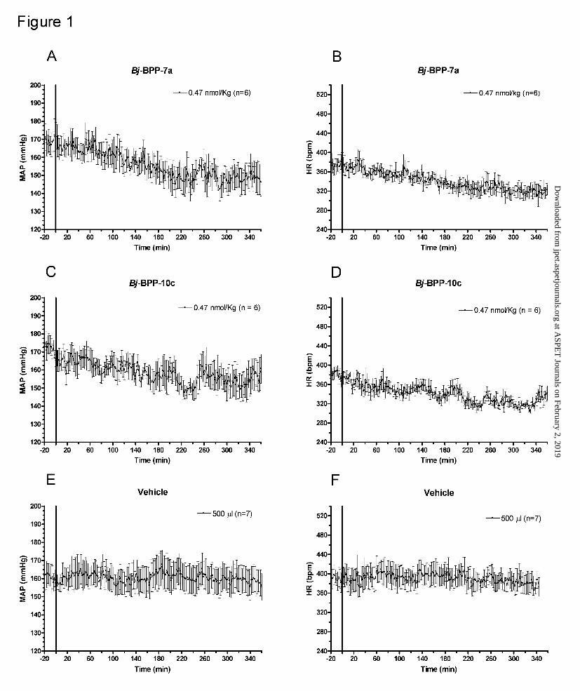
dose of 0.47 nmol/Kg. Interestingly, for the Bj-BPPs in all doses assayed, the fall in blood
pressure was associated with a reduction of the HR (Figure 1, panels B and D).
Figure 2, panels A and C, shows the peak changes in MAP produced by Bj-BPP-7a and Bj-
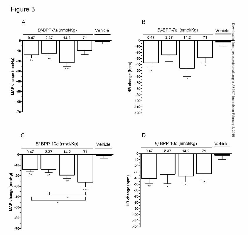
BPP-10c in SHR. Figure 3, panels A and C, shows the mean changes in MAP and HR in
the 6 hours following administration of both peptides. In sharp contrast with its weak
potency as ACE inhibitor or bradykinin potentiator, Bj-BPP-7a produced a marked and
sustained fall in the MAP of SHR even at doses as low as 0.47nmol/Kg (peak change: -33 ±
5 mmHg vs. -11 ± 3 mmHg in vehicle treated SHR, p < 0.05 and mean change over a six
hour period: -14 ± 3 mmHg vs. -1 ± 3 mmHg in vehicle treated SHR, p < 0.01). The
maximum change in MAP was obtained with the dose of 14.2nmol/Kg (peak change: -45 ±
6 mmHg, p < 0.01 and mean change -22 ± 4 mmHg, p < 0.001). However, this effect was
This article has not been copyedited and formatted. The final version may differ from this version.JPET Fast Forward. Published on May 2, 2007 as DOI: 10.1124/jpet.107.120873
at ASPE
T Journals on February 2, 2019
jpet.aspetjournals.orgD
ownloaded from
JPET #120873
15
not statistically different from that obtained with the lowest dose tested (0.47nmol/Kg)
(Figures 2A and 3A). The changes in MAP were associated with significant reductions in
HR (peak change: -78 ± 15 bpm for 0.47 nmol/Kg and -89 ± 18 bpm for 14.2 nmol/Kg, p <
0.05) (Figures 2B and 3B).
For Bj-BPP-10c, a dose-dependent effect was observed in the range of 0.47 nmol/Kg to 71
nmol/Kg (peak change: -36 ± 3 mmHg to -53 ± 6 mmHg and mean change: -14 ± 2 mmHg
to -26 ± 5 mmHg) (Figures 2C and 3C). An inverse relationship between the changes in HR
and the doses used was also observed. The maximum response was observed with the dose
of 0.47 nmol/Kg (peak change: -89 ± 9 bpm, p < 0.05 and mean change: -41 ± 8 bpm, p <
0.01) (Figures 2D and 3D).
The maximum changes in MAP and HR usually occurred after 160 minutes of the Bj-BPP
injection (Table 2). However, no clear relationship could be established between the
changes in MAP and HR.
In Wistar rats, no significant changes in MAP and HR were observed after intravenous
administration of Bj-BPPs with the dose of 0.47 nmol/Kg, considering either the peak
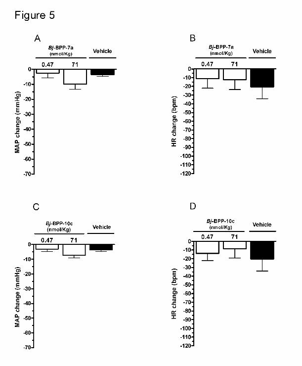
change or mean change (Figures 4 and 5). At the dose of 71 nmol/Kg, both Bj-BPPs caused
a significant decrease in MAP when the peak changes were considered (-28 ± 4 mmHg for
Bj-BPP-7a and -20 ± 1 mmHg for Bj-BPP-10c, p < 0.05 and p < 0.01 respectively; Figure 4
panels A and C). The maximal changes in HR were -50 ± 9 bpm for Bj-BPP-7a (71
nmol/Kg) and -69 ± 8 bpm for Bj-BPP-10c (0.47 nmol/Kg), however these changes were
not statistically significant when compared with vehicle (-40 ± 17 bpm). On the other hand,
considering the average of the changes in the entire period of observation (6 hours) no
noticeable effect on MAP or HR was observed (Figure 5). Similarly to SHR, the maximum
changes in cardiovascular parameters started after 160 minutes of the Bj-BPP injection
(Table 2).
This article has not been copyedited and formatted. The final version may differ from this version.JPET Fast Forward. Published on May 2, 2007 as DOI: 10.1124/jpet.107.120873
at ASPE
T Journals on February 2, 2019
jpet.aspetjournals.orgD
ownloaded from

JPET #120873
16
Effect of BK and Ang I in the arterial blood pressure of conscious SHR before and
after Bj-BPP administration.
The results presented in Figure 6 (panel A) show that the extent of hypotention following
the injection of BK was not affected by the administration of Bj-BPP-7a (71 nmol/Kg),
either after 10 min or 210 min of administration. Accordingly, the extent of the increase in
blood pressure following administration of Ang I, was not affected by the injection of Bj-
BPP-7a (71 nmol/Kg). On the other hand, Bj-BPP-10c at 71 nmol/Kg produced a modest,
but statistically significant, potentiation of BK without interfering with the Ang I pressor
effect, at both time points (Figure 6, panel C). Both Bj-BPPs produced a significant
decrease in MAP (Figure 6, panels B and D). A similar but more intense BK potentiation
was observed with Captopril at the same molar dose (71nmol/Kg or 15.4µg/Kg). As
observed with both Bj-BPPs no significant change in the pressor effect of Ang I were
observed (Figure 6, panel E). In sharp contrast with the Bj-BPPs, despite changing the BK
hypotensive effect, Captopril did not affect blood pressure levels (Figure 6, panel F). With a
lower dose of Bj-BPP-10c (2.37 nmol/Kg) no effect was observed in the BK response at
both time points (10 and 210 minutes after administration) while a significant reduction in
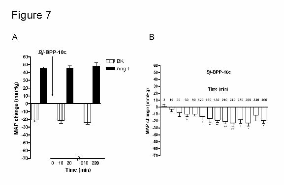
MAP was observed for up to 6 hours (Figure 7, panels A and B).
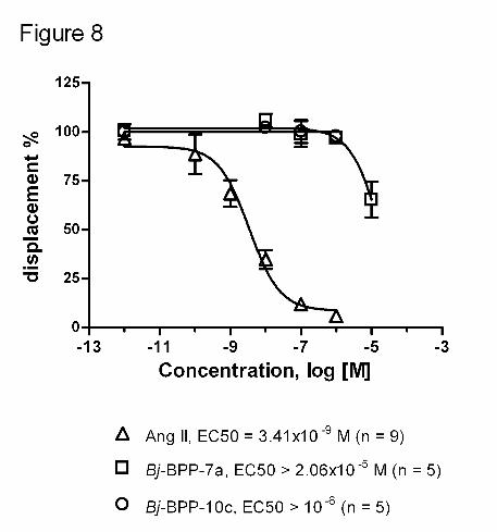
Binding studies
In order to test whether the dissociation of the ACE inhibitory activity of Bj-BPPs from
their antihypertensive effect could be due to blockade of AT1 receptors, we determine the
effect of both peptides on the binding of 125I-Ang II to AT1 receptor-transfected cells. No
significant displacement was observed with both peptides in concentrations ranging from
10-12 to 10-5 M (Figure 8).
This article has not been copyedited and formatted. The final version may differ from this version.JPET Fast Forward. Published on May 2, 2007 as DOI: 10.1124/jpet.107.120873
at ASPE
T Journals on February 2, 2019
jpet.aspetjournals.orgD
ownloaded from

JPET #120873
17
Discussion
The snake venom contains a number of strong inhibitors of the Ang I-converting enzyme
(Ferreira et al., 1970; Chi et al., 1985; Ianzer et al., 2004). They were useful not only to
validate ACE as a target to treat human hypertension, but also as lead molecules for the
development of the active-site directed inhibitors of ACE (Ondetti et al., 1977; Ondetti and
Cushman, 1981). It was demonstrated that once sACE is inhibited, the concentration of
Ang II decreases, while the concentration of BK increases, both reactions concurring to
reduce hypertension (Ondetti and Cushman, 1981). Indeed, a direct relationship between
the duration of the ACE inhibition and the antihypertensive actions of the active-site
directed inhibitors of ACE has been suggested (Sweet et al., 1981). Experimental and
clinical trials have also raised the possible importance of the inhibition of BK degradation,
since it increases the endothelial production of NO and prostacyclin, promoting
vasodilation (Zhang et al., 1999). Accordingly, a similar mechanism has been applied to
explain why Bj-BPPs display antihypertensive activity (Vanhoutte et al., 1995).
However, the mechanisms of the bradykinin potentiation as well as the mechanism of the
antihypertensive effect of ACEI appears to be more complex than previously suspected
(Mittra and Singh, 1998; Marcic et al., 1999; Houben et al., 2000). Bradykinin potentiation
by ACEI might involve beside ACE inhibition, induction of a cross-talk mechanism
between ACE and B2 receptors (Marcic et al., 1999; Mittra and Singh, 1998; Houben et al.,
2000; Mueller et al., 2006). Indeed more than 30 years ago, an ACE-indepentent
mechanism for BK potentiation by ACEI of Bothrops jararaca venom was suggested
(Camargo and Ferreira, 1971; Greene et al., 1972).
Likewise, there are scarce but consistent reports showing that ACEI can produce
vasodilation by mechanism, that cannot be ascribe to ACE inhibition (Marks et al., 1980;
Antonaccio et al., 1981; Mittra and Singh, 1998; Houben et al., 2000; Mueller et al., 2006).
This article has not been copyedited and formatted. The final version may differ from this version.JPET Fast Forward. Published on May 2, 2007 as DOI: 10.1124/jpet.107.120873
at ASPE
T Journals on February 2, 2019
jpet.aspetjournals.orgD
ownloaded from
JPET #120873
18
For example, Houben and co-workers (2000) have found that quinaprilat but not enaprilat
produced significant vasodilation with 15 minutes of administration. At this time both
drugs completely blocked the Ang I pressor effect. Nakamura and co-workers (1992) have
observed that although enalaprilat was without effect on basal forearm blood flow or
vascular resistance, it significantly augmented the increase in blood flow and reduction in
forearm resistance induced by acetylcholine. Captopril lowers blood pressure of animals
with different models hypertension including situations where the renin-angiotensin system
is not responsible for blood pressure maintenance (Marks et al., 1980). In addition,
Captopril has been shown to produce vasodilation in vitro in conditions that lisinopril was
without effect (Mittra and Singh, 1998). Our data are in line with these previous
observations. Taken all together, the current available data show that an ACEI can produce:
1) BK-potentiation without blood pressure lowering effect (captopril in our study); 2)
antihypertensive effect without changing the hypotensive action of BK or the hypertensive
effect of Ang I or Ang II (Bj-BPPs 7a and 10c in our study); 3) blockade of the pressor
effect of Ang I without producing vasodilation (Houben et al., 2000); 4) vasorelaxation
which can be dissociated from ACE inhibition (Captopril) (Mittra and Singh, 1998); 5)
facilitation of NO release induced by acethylcholine without producing vasodilation
directly (Nakamura et al., 1992). Furthermore, the antihypertensive effect of ACE inhibitors
does not correlate with Ang II plasma levels (Marks et al., 1980; Duncan et al., 1999) and a
similar dissociation has been described for bradykinin (Stanziola et al., 1999). In addition, it
has been suggested that ACEI can act as anti-oxidant (de Cavanagh et al., 1997) or can also
act as B1 receptor agonist (Ignjatovic et al., 2002). It should be also pointed out that
Proline-rich peptides which were the basis for the development of ACEI (Ondetti et al.,
1977) having the same C-terminal motif (Ile-Pro-Pro) produced similar and potent
antihypertensive effects, despite the fact that their ACE inhibitory potency differ in about
This article has not been copyedited and formatted. The final version may differ from this version.JPET Fast Forward. Published on May 2, 2007 as DOI: 10.1124/jpet.107.120873
at ASPE
T Journals on February 2, 2019
jpet.aspetjournals.orgD
ownloaded from

JPET #120873
19
80,000-fold (Bj-BPP-7a as compared to Bj-BPP-10c) (Table I, this study). Therefore
although, ACE inhibition is apparently the main mechanism of the antihypertensive effects
of ACEI, other mechanisms unrelated to interference with the hydrolytic activity of ACE
are obviously involved. More important, our study and the available data strongly support
the hypothesis that ACEIs are not a single (uniform) class of antihypertensive drugs.
Actually, they may present completely distinct mechanisms of action. In this regard, the
contribution of other factors such as the increase of the vasodilator Ang-(1-7) should be
considered in future studies (Duncan et al., 1999; Stanziola et al., 1999).
As mentioned above, the present study clearly demonstrates that, at least in SHR, ACE
inhibition cannot be applied to describe the antihypertensive effect of the proline-rich
oligopeptides since the peptide concentrations used were far below those required to inhibit
ACE in vivo. In fact, the hypotensive effect of the Bj-BPPs was also independent of BK
potentiation, whether or not this effect is a consequence of the ACE inhibition. Therefore,
in contrast to what have been suggested for currently used ACEI, our results strongly
suggests that the reduced formation of Ang II and BK potentiation are not essential
mechanisms involved in the antihypertensive action of ACEI. We have also shown that the
antihypertensive effect of Bj-BPPs can not be ascribed to direct effects on AT1 receptors.
Interestingly, Bj-BPPs caused a significant reduction instead of an increase of HR, as would
be expected by the unloading of baroreceptors due to hypotension or if it was mediated by
BK (Bunag et al., 1975). Whether this reflects a consequence of the tendency to a decrease
in the locomotion activity following administration of the two Bj-BPPs (data not shown) or
a direct effect of these peptides on the heart, such as interference in the autonomic activity
modulating the heart pacemaker and/or changes in the baroreflex control of heart rate,
remains to be clarified. It should be pointed out that our current data do not allow to discard
a contribution of heart rate (cardiac output) changes to the blood pressure effects of Bj-
This article has not been copyedited and formatted. The final version may differ from this version.JPET Fast Forward. Published on May 2, 2007 as DOI: 10.1124/jpet.107.120873
at ASPE
T Journals on February 2, 2019
jpet.aspetjournals.orgD
ownloaded from

JPET #120873
20
BPPs, although a clear relationship between changes in BP and HR could not be
established.
We would like to direct the attention to one interesting result presented herein: the
hypotensive effect of Bj-BPPs especially in low doses was observed in SHR but not in
normotensive animals. This observation suggests that the antihypertensive effect produced
in SHR is not due to a nonspecific effect of both peptides on blood vessels or in the heart.
In conclusion, synthetic compounds, displaying properties similar to these Bj-BPPs, which
produced antihypertensive effect without affecting the complex physiological role of ACE,
could represent an attractive alternative for the treatment of human hypertension and other
cardiovascular diseases.
This article has not been copyedited and formatted. The final version may differ from this version.JPET Fast Forward. Published on May 2, 2007 as DOI: 10.1124/jpet.107.120873
at ASPE
T Journals on February 2, 2019
jpet.aspetjournals.orgD
ownloaded from

JPET #120873
21
Acknowledgements
We would like to acknowledge José Roberto Silva for excellent technical assistance; Neusa
Lima for the secretarial assistance. We thank Dr. Jöel Cotton (CEA – Saclay) for technical
assistance in the enzymatic measurements of ACE and Dr. Beatriz L. Fernandes for critical
reading.
This article has not been copyedited and formatted. The final version may differ from this version.JPET Fast Forward. Published on May 2, 2007 as DOI: 10.1124/jpet.107.120873
at ASPE
T Journals on February 2, 2019
jpet.aspetjournals.orgD
ownloaded from

JPET #120873
22
References
Antonaccio MJ, Rubin B, Horovitz ZP, Laffan RJ, Goldberg ME, High JP, Harris DN and
Zaidi I (1979) Effects of chronic treatment with Captopril (SQ 14,225), an orally active
inhibitor of angiotensin I-converting enzyme, in spontaneously hypertensive rats. Jpn J
Pharmacol 29:285-294.
Bianchi A, Evans DB, Cobb M, Peschka MT, Schaeffer TR and Laffan RJ (1973)
Inhibition by SQ 20881 of vasopressor response to angiotensin I in conscious animals.
Eur J Pharmacol 23:90-96.
Bunag RD, Walaszek EJ and Mueting N (1975) Sex differences in reflex tachycardia
induced by hypotensive drugs in unanesthetized rats. Am J Physiol 229:652-656.
Camargo AC and Ferreira SH (1971) Action of bradykinin potentiating factor (BPF) and
dimercaprol (BAL) on the responses to bradykinin of isolated preparations of rat
intestines. Br J Pharmacol 42:305-307.
Case DB, Atlas SA, Laragh JH, Sealey JE, Sullivan PA and McKinstry DN (1978) Clinical
experience with blockade of the renin-angiotensin-aldosterone system by an oral
converting-enzyme inhibitor (SQ 14,225, Captopril) in hypertensive patients. Prog
Cardiovasc Dis 21:195-206.
Chi CW, Wang SZ, Xu LG, Wang MY, Lo SS and Huang WD (1985) Structure-function
studies on the bradykinin potentiating peptide from Chinese snake venom (Agkistrodon
halys Pallas). Peptides 6:339-342.
Cotton J, Hayashi MA, Cuniasse P, Vazeux G, Ianzer D, de Camargo AC and Dive V
(2002) Selective inhibition of the C-domain of angiotensin I converting enzyme by
bradykinin potentiating peptides. Biochemistry 41:6065-6071.
De Cavanagh EM, Fraga CG, Ferder L and Inserra F. (1997) Enalapril and captopril
enhance antioxidant defenses in mouse tissues Am J Physiol.272:R514-R518.
This article has not been copyedited and formatted. The final version may differ from this version.JPET Fast Forward. Published on May 2, 2007 as DOI: 10.1124/jpet.107.120873
at ASPE
T Journals on February 2, 2019
jpet.aspetjournals.orgD
ownloaded from
JPET #120873
23
Dive V, Cotton J, Yiotakis A, Michaud A, Vassiliou S, Jiracek J, Vazeux G, Chauvet MT,
Cuniasse P and Corvol P (1999) RXP 407, a phosphinic peptide, is a potent inhibitor of
angiotensin I converting enzyme able to differentiate between its two active sites. Proc
Natl Acad Sci USA 96:4330-4335.
Duncan AM, James GM, Anastasopoulos F, Kladis A, Briscoe TA and Campbell DJ (1999)
Interaction between neutral endopeptidase and angiotensin converting enzyme inhibition
in rats with myocardial infarction: effects on cardiac hypertrophy and angiotensin and
bradykinin peptide levels. J Pharmacol Exp Ther 289:295-303.
Ferreira SH, Barteld DC and Greene LJ (1970) Isolation of bradykinin potentiating peptides
from Bothrops jararaca venom. Biochemistry 9:2583-2593.
Ferreira SH and Rocha e Silva M (1965) Potentiation of bradykinin and eledoisin by BPF
(bradykinin potentiating factor) from Bothrops jararaca venom. Experientia 21:347-
349.
Gavras H, Brunner HR, Laragh JH, Gavras I and Vukovich RA (1975) The use of
angiotensin-converting enzyme inhibitor in the diagnosis and treatment of hypertension.
Clin Sci Mol Med Suppl 2:57s-60s.
Greene LJ, Camargo ACM, Krieger EM, Stewart JM and Ferreira SH (1972) Inhibition of
the Conversion of Angiotensin I to II and Potentiation of Bradykinin by Small Peptides
Present in Bothrops jararaca Venom. Circ Res 31:62-71.
Hayashi MA, Murbach AF, Ianzer D, Portaro FC, Prezoto BC, Fernandes BL, Silveira PF,
Silva CA, Pires RS, Britto LR, Dive V and Camargo AC (2003) The C-type natriuretic
peptide precursor of snake brain contains highly specific inhibitors of the angiotensin-
converting enzyme. J Neurochem 85:969-977.
This article has not been copyedited and formatted. The final version may differ from this version.JPET Fast Forward. Published on May 2, 2007 as DOI: 10.1124/jpet.107.120873
at ASPE
T Journals on February 2, 2019
jpet.aspetjournals.orgD
ownloaded from

JPET #120873
24
Houben AJ, Kroon AA, de Haan CH, Fuss-Lejeune MJ and de Leeuw PW. (2000)
Quinaprilat-induced vasodilatation in forearm vasculature of patients with essential
hypertension: comparison with enalaprilat. Cardiovasc Drugs Ther.14:657-663.
Ianzer D, Konno K, Marques-Porto R, Vieira Portaro FC, Stöcklin R, Martins De Camargo
AC and Pimenta DC (2004) Identification of five new bradykinin potentiating peptides
(BPPs) from Bothrops jararaca crude venom by using electrospray ionization tandem
mass spectrometry after a two-step liquid chromatography. Peptides 25:1085-1092.
Ianzer D, Konno K, Xavier CH, Stocklin R, Santos RA, de Camargo AC and Pimenta DC.
(2006) Hemorphin and hemorphin-like peptides isolated from dog pancreas and sheep
brain are able to potentiate bradykinin activity in vivo. Peptides 27:2957-2966.
Ignjatovic T, Tan F, Brovkovych V, Skidgel RA and Erdos EG (2002) Novel mode of
action of angiotensin I converting enzyme inhibitors: direct activation of bradykinin B1
receptor. J Biol Chem. 277:16847-16852.
Jaspard E, Wei L and Alhenc-Gelas F (1993) Differences in the properties and enzymatic
specificities of the two active sites of angiotensin I-converting enzyme (kininase II).
Studies with bradykinin and other natural peptides. J Biol Chem 268:9496-9503.
Marcic B, Deddish PA, Jackman HL and Erdos EG. (1999) Enhancement of bradykinin and
resensitization of its B2 receptor. Hypertension 33:835-843.
Marks ES, Bing RF, Thurston H and Swales, JD. (1980) Vasodepressor property of the
converting enzyme inhibitor captopril (SQ 14 225): the role of factors other than renin-
angiotensin blockade in the rat. Clin Sci (Lond) 58:1-6.
Mittra S and Singh M. (1998) Possible mechanism of captopril induced endothelium-
dependent relaxation in isolated rabbit aorta. Mol Cell Biochem.183:63-7
This article has not been copyedited and formatted. The final version may differ from this version.JPET Fast Forward. Published on May 2, 2007 as DOI: 10.1124/jpet.107.120873
at ASPE
T Journals on February 2, 2019
jpet.aspetjournals.orgD
ownloaded from

JPET #120873
25
Mueller S, Gothe R, Siems W-D, Vietinghoff G, Paegelow I and Reissmann S (2005)
Potentiation of bradykinin actions by analogues of the bradykinin potentiating
nonapeptide BPP9α. Peptides 26:1235-1247.
Mueller S, Paegelow I and Reissmann S (2006) Hypothesized and found mechanisms for
potentiation of bradykinin actions. Signal Transduction 6: 5-18.
Murayama N, Hayashi MA, Ohi H, Ferreira LA, Fernandes BL, Yamane T and Camargo
ACM (1997) Cloning and sequence of a Bothrops jararaca cDNA encoding a precursor
of seven bradykinin-potentiating peptides and a C-type netriuretic peptide. Proc Natl
Acad Sci USA 94:1189-1193.
Nakamura M, Funakoshi T, Yoshida H, Arakawa N, Suzuki T and Hiramori K. (1992)
Endothelium-dependent vasodilation is augmented by angiotensin converting enzyme
inhibitors in healthy volunteers. J Cardiovasc Pharmacol.20:949-54
Ondetti MA, Williams NJ, Sabo EF, Pluscec J, Weaver ER and Kocy O (1971)
Angiotensin-converting enzyme inhibitors from the venom of Bothrops jararaca.
Isolation, elucidation of structure, and synthesis. Biochemistry 10:4033-4039.
Ondetti MA and Cushman DW (1981) Inhibitors of Angiotensin-Converting Enzyme. in
Biochemical Regulation of Blood Pressure, R.L. Soffer, Ed. (Wiley, New York) pp. 165-
204.
Ondetti MA, Rubin B and Cushman DW (1977) Design of specific inhibitors of
angiotensin-converting enzyme: new class of orally active antihypertensive agents.
Science 196:441-444.
Perich RB, Jackson B and Johnston CI (1992) Variation in angiotensin-converting enzyme
(ACE) inhibitor affinity at two binding sites on rat pulmonary ACE: influence on
bradykinin hydrolysis. Clin Exp Pharmacol Physiol 19:353-357.
This article has not been copyedited and formatted. The final version may differ from this version.JPET Fast Forward. Published on May 2, 2007 as DOI: 10.1124/jpet.107.120873
at ASPE
T Journals on February 2, 2019
jpet.aspetjournals.orgD
ownloaded from

JPET #120873
26
Pesquero JB, Lindsey CJ, Zeh K, Paiva ACM, Ganten D. and Bader M. (1994) Molecular
structure and expression of rat bradykinin B2 receptor gene. Evidence for alternative
splicing. J. Biol. Chem. 269:26920–26925.
Pinheiro SV, Simoes e Silva AC, Sampaio WO, de Paula RD, Mendes EP, Bontempo ED,
Pesquero JB, Walther T, Alenina N, Bader M, Bleich M and Santos RA. (2004)
Nonpeptide AVE 0991 is an angiotensin-(1-7) receptor Mas agonist in the mouse
kidney. Hypertension 44:490-496.
Stanziola L, Greene LJ and Santos RA (1999) Effect of chronic angiotensin converting
enzyme inhibition on angiotensin I and bradykinin metabolism in rats. Am J Hypertens
12:1021-1029.
Stewart JM, Ferreira SH and Greene LJ (1971) Bradykinin potentiating peptide PCA-Lys-
Trp-Ala-Pro. An inhibitor of the pulmonary inactivation of bradykinin and conversion of
angiotensin I to II. Biochem Pharmacol 20:1557-1567.
Sweet CS, Arbegast PT, Gaul SL, Blaine EH and Gross DM (1981) Relationship between
angiotensin I blockade and antihypertensive properties of single doses of MK-421 and
Captopril in spontaneous and renal hypertensive rats. Eur J Pharmacol 76:167-176.
Vanhoutte PM, Boulanger CM and Mombouli JV (1995) Endothelium-derived relaxing
factors and converting enzyme inhibition. Am J Cardiol 76:3E-12E
Wei L, Alhenc-Gelas F, Corvol P and Clauser E (1991) The two homologous domains of
human angiotensin I-converting enzyme are both catalytically active. J Biol Chem
266:9002-9008.
Zhang X, Recchia FA, Bernstein R, Xu X, Nasjletti A and Hintze TH (1999) Kinin-
mediated coronary nitric oxide production contributes to the therapeutic action of
angiotensin-converting enzyme and neutral endopeptidase inhibitors and amlodipine in
the treatment in heart failure. J Pharmacol Exp Ther 288:742-751.
This article has not been copyedited and formatted. The final version may differ from this version.JPET Fast Forward. Published on May 2, 2007 as DOI: 10.1124/jpet.107.120873
at ASPE
T Journals on February 2, 2019
jpet.aspetjournals.orgD
ownloaded from
JPET #120873
27
Waeber B, Nussberger J, Juillerat L and Brunner HR. (1989) Angiotensin converting
enzyme inhibition: discrepancy between antihypertensive effect and suppression of
enzyme activity. J Cardiovasc Pharmacol.14:S53-S59.
This article has not been copyedited and formatted. The final version may differ from this version.JPET Fast Forward. Published on May 2, 2007 as DOI: 10.1124/jpet.107.120873
at ASPE
T Journals on February 2, 2019
jpet.aspetjournals.orgD
ownloaded from

JPET #120873
28
Footnotes
a)
Financial support
Supported by funds provided by Fundação de Amparo à Pesquisa do Estado de São Paulo
(CAT/CEPID-FAPESP), COINFAR Pesquisa e Desenvolvimento Ltda and Conselho
Nacional de Desenvolvimento Científico e Tecnológico (CNPq).
b)
Corresponding author
Dr. Antonio C. M. Camargo
Center for Applied Toxinology - CAT/CEPID
Instituto Butantan
Av. Vital Brasil, 1500
São Paulo, SP, 05530-900
Brazil
Phone/FAX: 11-55-3726 1024
e-mail: [email protected]
c)
The first 2 authors contributed equally to this work.
This article has not been copyedited and formatted. The final version may differ from this version.JPET Fast Forward. Published on May 2, 2007 as DOI: 10.1124/jpet.107.120873
at ASPE
T Journals on February 2, 2019
jpet.aspetjournals.orgD
ownloaded from

JPET #120873
29
Legends for figures
Figure 1. Time course of the effect of intravenous administration of Bj-BPPs on the
mean arterial pressure (MAP) and heart rate (HR) of spontaneously hypertensive rats
(SHR). The I.V. injections of BPPs (0.47 nmol/Kg) and vehicle were made at the time zero,
indicated by the solid line.
Figure 2. Maximal changes in MAP and HR produced by Bj-BPPs in SHR. Bj-BPPs
were administered in doses ranging from 0.47 to 71 nmol/Kg. The data show the mean ±
SEM of maximal (peak) changes observed in each rat with each dose. * p < 0.05, ** p <
0.01 and *** p < 0.001 compared to the change observed with vehicle.
Figure 3. Average of the changes in MAP over six-hour following administration of
Bj-BPPs in SHR. Bj-BPPs were administered in doses ranging from 0.47 to 71 nmol/Kg.
The data are presented as mean ± SEM of averaged changes of the period of observation (6
hours) * p < 0.05, ** p < 0.01 and *** p < 0.001 compared to the change with vehicle.
Figure 4. Maximal changes in MAP and HR of Bj-BPPs in normotensive Wistar rats.
Bj-BPPs were administered in two different doses: 0.47 and 71 nmol/Kg. The data are
presented as mean ± SEM of maximal (peak) changes observed in each rat with each dose.
* p < 0.05 and ** p < 0.01 compared to the change observed with vehicle.
Figure 5. Average of the changes in MAP over six-hour following administration of
Bj-BPPs in in normotensive Wistar rats. Bj-BPPs were administered in two different
doses: 0.47 and 71 nmol/Kg. The data are presented as mean ± SEM of averaged changes
of period of observation (6 hours).
This article has not been copyedited and formatted. The final version may differ from this version.JPET Fast Forward. Published on May 2, 2007 as DOI: 10.1124/jpet.107.120873
at ASPE
T Journals on February 2, 2019
jpet.aspetjournals.orgD
ownloaded from

JPET #120873
30
Figure 6. Effect of intravenous administration of Bj-BPPs or Captopril (71 nmol/Kg)
on the mean arterial pressure and on the blood pressure change produced by
intravenous injection of BK and Ang I in SHR. Panels A, C, E and G show the influence
of Bj-BPPs, Captopril and vehicle on the depressor effect of BK and the pressor effect of
Ang I. Gray bars: hypotensive effect of bradykinin (0.5 µg); black bars: pressor response of
angiotensin I (40 ng). Panels B, D, F and H show the time course of the effect of Bj-BPPs,
Captopril and vehicle on the MAP of SHR (white bars). The data are presented as mean ±
SEM, (n = 4-6), * p < 0.05; ** p < 0.01 and *** p < 0.001.
Figure 7. Effect of intravenous administration of Bj-BPP-10c (2.37 nmol/Kg) on the
mean arterial pressure and on the blood pressure change produced by intravenous
injection of BK and Ang I in SHR. Panel A shows the influence of Bj-BPP-10c on the
depressor effect of BK and the pressor effect of Ang I. Gray bars: hypotensive effect of
bradykinin (0.5 µg); black bars: pressor response of angiotensin I (40 ng). Panel B shows
the time course of the effect of Bj-BPP-10c on the MAP of SHR (white bars). The data are
presented as mean ± SEM, (n = 5), * p < 0.05; ** p < 0.01 and *** p < 0.001.
Figure 8. Competition for 125I-Ang II binding to AT1-transfected CHO cells by Ang II
and Bj-BPPs. Competition curves were generated by adding increasing concentrations of
Ang II (10-11 to 10-6 M) or Bj-BPP-7a or Bj-BPP-10c (10-12 to 10-5 M) to the incubation
buffer containing 0.4 nmol/L of 125I-Ang II. Data are presented as mean ± SEM of three to
six independent experiments.
This article has not been copyedited and formatted. The final version may differ from this version.JPET Fast Forward. Published on May 2, 2007 as DOI: 10.1124/jpet.107.120873
at ASPE
T Journals on February 2, 2019
jpet.aspetjournals.orgD
ownloaded from

JPET #120873
31
Tables
Table 1 – Inhibition studies with sACE and biological activities of the synthetic BPPs.
ACE inhibition BK Potentiation - U (nmol)
Ki (nM)
N-site C-site
Blood pressure Time for 50% recovery (min)
Isolated ileum Blood pressure
Bj-BPP-7a 70,000 40,000 0 10.77 ± 0.6 > 200
Bj-BPP-10c 200 a 0.5 b 20-30 0.48 ± 0.02 b 5.85
The in vitro inhibition of ACE correspond to the average value of three independent Ki(app)
values determined according to Cotton et al. (2002). The in vivo inhibition of ACE was
estimated by the time required for 50% recovery of the response to Ang I (40 ng) before
administration of 45 nmol of BPPs. The BK potentiation unit (U) is described in methods.
a Cotton et al., 2002
b Hayashi et al., 2003
This article has not been copyedited and formatted. The final version may differ from this version.JPET Fast Forward. Published on May 2, 2007 as DOI: 10.1124/jpet.107.120873
at ASPE
T Journals on February 2, 2019
jpet.aspetjournals.orgD
ownloaded from

JPET #120873
32
Table 2: Time for maximal cardiovascular effect produced by Bj-BPPs at different
doses in SHR and Wistar rats.
Time for maximal change (min) Strain Bj-BPP
0.47 nmol/Kg 2.37 nmol/Kg 14.2 nmol/Kg 71 nmol/Kg
BPP-7a 257 ± 10
(range: 224–290)
308 ± 14
(range:272-348)
248 ± 23
(range:170-342)
323 ± 13
(range:278-354) SHR
BPP-10c 248 ± 28
(range:160–334)
294 ± 25
(range:274-350)
285 ± 29
(range:192-358)
345 ± 7
(range:304-358)
BPP-7a 250 ± 73
(range:168-324)
ND ND 283 ± 13
(range:242-310) Wistar
BPP-10c 230 ± 33
(range:178-356)
ND ND 246 ± 26
(range:222-324)
This article has not been copyedited and formatted. The final version may differ from this version.JPET Fast Forward. Published on May 2, 2007 as DOI: 10.1124/jpet.107.120873
at ASPE
T Journals on February 2, 2019
jpet.aspetjournals.orgD
ownloaded from
This article has not been copyedited and formatted. The final version may differ from this version.JPET Fast Forward. Published on May 2, 2007 as DOI: 10.1124/jpet.107.120873
at ASPE
T Journals on February 2, 2019
jpet.aspetjournals.orgD
ownloaded from

This article has not been copyedited and formatted. The final version may differ from this version.JPET Fast Forward. Published on May 2, 2007 as DOI: 10.1124/jpet.107.120873
at ASPE
T Journals on February 2, 2019
jpet.aspetjournals.orgD
ownloaded from
This article has not been copyedited and formatted. The final version may differ from this version.JPET Fast Forward. Published on May 2, 2007 as DOI: 10.1124/jpet.107.120873
at ASPE
T Journals on February 2, 2019
jpet.aspetjournals.orgD
ownloaded from

This article has not been copyedited and formatted. The final version may differ from this version.JPET Fast Forward. Published on May 2, 2007 as DOI: 10.1124/jpet.107.120873
at ASPE
T Journals on February 2, 2019
jpet.aspetjournals.orgD
ownloaded from
This article has not been copyedited and formatted. The final version may differ from this version.JPET Fast Forward. Published on May 2, 2007 as DOI: 10.1124/jpet.107.120873
at ASPE
T Journals on February 2, 2019
jpet.aspetjournals.orgD
ownloaded from

This article has not been copyedited and formatted. The final version may differ from this version.JPET Fast Forward. Published on May 2, 2007 as DOI: 10.1124/jpet.107.120873
at ASPE
T Journals on February 2, 2019
jpet.aspetjournals.orgD
ownloaded from
This article has not been copyedited and formatted. The final version may differ from this version.JPET Fast Forward. Published on May 2, 2007 as DOI: 10.1124/jpet.107.120873
at ASPE
T Journals on February 2, 2019
jpet.aspetjournals.orgD
ownloaded from
This article has not been copyedited and formatted. The final version may differ from this version.JPET Fast Forward. Published on May 2, 2007 as DOI: 10.1124/jpet.107.120873
at ASPE
T Journals on February 2, 2019
jpet.aspetjournals.orgD
ownloaded from