Do Digital Technologies Enhance Anatomical Education?
26
Practice and Evidence of Scholarship of Teaching and Learning in Higher Education Vol. 13, No. 1, May 2018, pp. 2-27 2 Do Digital Technologies Enhance Anatomical Education? Zoe Pringle, School of Life Sciences, College of Medical, Veterinary and Life Sciences, University of Glasgow [email protected] Paul M. Rea 1* School of Life Sciences, College of Medical, Veterinary and Life Sciences, University of Glasgow [email protected] Abstract Anatomy has been taught by traditional methods for centuries. However, there has been an explosion of a variety of digital training resources for anatomical education. There is also a requirement from regulatory bodies to embrace digital technologies in teaching, yet no formal analysis has been undertaken as to the effectiveness of these products and tools. A comprehensive electronic database search was performed to identify the use, and effectiveness or otherwise, of digital technologies in anatomy, medicine, surgery, dentistry and the allied health professions. The data was pooled, analysed and we identified 164 articles. We identified two groups – those that did, and those that did not, have empirical data for analysis of the effectiveness of digital technologies in anatomical education. We identified three categories within this –pro, neutral and against the use of digital technologies. For the pro category, there were 35 (21.3%) empirically tested articles, and 91 (55.5%) non-empirically tested articles identified. In the neutral category, there were 19 (11.6%) empirically tested articles, and 16 (9.8%) non-empirically tested articles. Only 3 articles were against the use of digital technologies, and were in the empirically tested category. * Corresponding Author ISSN 1750-8428 (online) www.community.dur.ac.uk/pestlhe.org.uk PESTLHE brought to you by CORE View metadata, citation and similar papers at core.ac.uk provided by Enlighten
Transcript of Do Digital Technologies Enhance Anatomical Education?

Practice and Evidence of Scholarship of Teaching and Learning in Higher Education Vol. 13, No. 1, May 2018, pp. 2-27
2
Do Digital Technologies Enhance Anatomical Education?
Zoe Pringle,
School of Life Sciences, College of Medical, Veterinary and Life Sciences,
University of Glasgow [email protected]
Paul M. Rea1*
School of Life Sciences, College of Medical, Veterinary and Life Sciences,
University of Glasgow [email protected]
Abstract
Anatomy has been taught by traditional methods for centuries. However, there has
been an explosion of a variety of digital training resources for anatomical education.
There is also a requirement from regulatory bodies to embrace digital technologies in
teaching, yet no formal analysis has been undertaken as to the effectiveness of these
products and tools. A comprehensive electronic database search was performed to
identify the use, and effectiveness or otherwise, of digital technologies in anatomy,
medicine, surgery, dentistry and the allied health professions. The data was pooled,
analysed and we identified 164 articles. We identified two groups – those that did, and
those that did not, have empirical data for analysis of the effectiveness of digital
technologies in anatomical education. We identified three categories within this –pro,
neutral and against the use of digital technologies. For the pro category, there were 35
(21.3%) empirically tested articles, and 91 (55.5%) non-empirically tested articles
identified. In the neutral category, there were 19 (11.6%) empirically tested articles, and
16 (9.8%) non-empirically tested articles. Only 3 articles were against the use of digital
technologies, and were in the empirically tested category.
* Corresponding Author ISSN 1750-8428 (online) www.community.dur.ac.uk/pestlhe.org.uk
PESTLHE
brought to you by COREView metadata, citation and similar papers at core.ac.uk
provided by Enlighten

Do Digital Technologies Enhance Anatomical Education? May 2018
3
The majority of literature related to digital technologies in anatomical education is
supportive of its use. However, most of the literature is not supported with empirical
data related to the use of digital technologies in anatomy specific education within the
health and related disciplines. Further studies need to be conducted as to the
effectiveness of technology in medical/healthcare related education.
Key words: Anatomy; digital; medical education; surgery; technology
Introduction
Anatomy has been the cornerstone of medical education for thousands of years,
helping to ensure proficient and safe medical practice. Traditionally, the subject has
been taught by didactic lectures and dissection. Dissection has been considered the
‘gold standard’ in education (Balogh et al., 2006) as it offers unique and detailed self-
directed learning opportunities where the student can appreciate the three-dimensional
(3D) human body through self-exploration, developing professionalism and teamwork
(Turney, 2007). Dissection also allows students to develop a patient-centred approach
while developing humanistic values, promoting maturity and empathy (Sugand,
Abrahams & Khurana, 2010).
The teaching of anatomy is relevant across all healthcare specialties (Patel & Moxham,
2008), but the digital technologies that are now available has led to some debate over
the methods of teaching. Turney suggests that anatomy has been slow to adapt to new
developments and has subsequently come under pressure to revolutionise, in order to
keep pace with the modern medical curriculum and to encourage student participation
and interest (Turney, 2007).
Due to various extrinsic pressures, many medical schools have reduced the amount of
time allocated to dissection, or abandoned it altogether, in favour of other teaching
methods (Turney, 2007; Patel & Moxham, 2008; Sugand et al., 2010). The costs of
running and maintaining a dissection laboratory and the holding and dispensing of
cadavers are high, making dissection both costly and time consuming (Turney, 2007;
McMenamin, Quayle, McHenry & Adams, 2014). In addition, reductions in qualified
teaching staff and donors, along with stringent licensing make it more difficult for

Pringle and Rea May 2018
4
institutions to embrace the practice of dissection (Turney, 2007). Dissection can also
require frequent access to laboratories by students, which can be difficult to achieve,
particularly in institutions with restricted resources. Furthermore, in some countries
dissection is prohibited on religious grounds (McMenamin et al., 2014). Such pressures
therefore have invited the introduction of innovative teaching methods.
Since the creation of the Visible Human Project (VHP; Spitzer & Whitlock, 1998) on
cross sectional anatomy, others have followed with similar ones like the Chinese Visible
Human (CVH; Zhang et al., 2004) and the Korean Visible Human (KVH; Park, Chung,
Hwang & Shin, 2006). Since then, there has been an explosion onto the market of a
variety of tools and products to be used as digital training material (Ma, Bale & Rea,
2012; Manson, Poyade & Rea, 2015; Rea, 2016; Raffan, Guevar, Poyade & Rea, 2017;
Visible Body; Primal Pictures; 3D4Medical; Cyber Anatomy HolographicTM; BodyViz). It
has also been stated that technologies could improve both assessment scores and
student satisfaction (Sugand et al., 2010) but that is still controversial (Marsh, Giffin &
Lowrie, 2008; Ruiz, Cook & Levison, 2009). Indeed, there is also a wide range of 3D
virtual reality models (3D VRM; Visible Body; Primal Pictures; 3D4Medical; Cyber
Anatomy HolographicTM; BodyViz; Ruiz et al.; 2009); web based technology (Jastrow &
Hollinderbäumer, 2004; Zhu et al., 2014; Raynor & Iggulden, 2008); tablet and mobile
devices (Wallace, Clark & White., 2012; Lewis, Burnett, Tunstall & Abrahams, 2013;
McCulloch, Hope, Loranger & Rea, 2016); surgical simulation (Madurska et al., 2017)
and computer assisted learning (Ruiz et al., 2009; Varol & Basa, 2009)
However, despite a plethora of digital technologies available in anatomical, medical,
dental and surgical related training, there does not appear to be an analysis as to the
effectiveness of these products. There is no global consensus that has been presented
as to the effectiveness (or otherwise) of the plethora of digital tools that are available for
anatomical education in the healthcare disciplines. Therefore, the purpose of this study
is to examine the current literature in this field to identify what the general consensus is,
related to the use digital tools in anatomical education.

Do Digital Technologies Enhance Anatomical Education? May 2018
5
Method
Study design and data collection
We used the search engines Google Scholar, PubMed and CORE for entering our
keywords. From the analysis of the literature, we selected the common words that
appeared in articles related to the field of anatomy, medicine, dentistry, surgery and
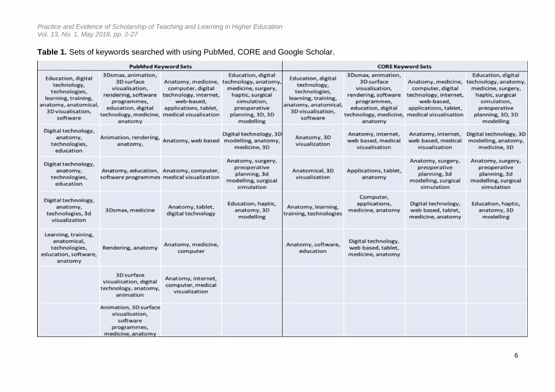
digital technologies. The first search using 10+ key words (row 1, Table 1) was overly
ambitious, and we therefore identified up to 5 key words in each search category and
combination (rows 2-7, Table 1), that generated articles across the three engines. We
selected the first page review, of up to 20 articles in each category on each search
engine.
Practice and Evidence of Scholarship of Teaching and Learning in Higher Education Vol. 13, No. 1, May 2018, pp. 2-27
6
Table 1. Sets of keywords searched with using PubMed, CORE and Google Scholar.

Practice and Evidence of Scholarship of Teaching and Learning in Higher Education Vol. 13, No. 1, May 2018, pp. 2-27
7
Row 1 shows four matching sets of keywords initially searched on in PubMed and
CORE. The four sets of keywords shown in Row 1, Columns 1-4 are the same as
those used in the four searches initiated through Google Scholar. Rows 2-7 show the
series of smaller sets of keywords subsequently searched upon in PubMed and CORE.
In addition, we also applied the following criteria in our search to ensure current
activities were applied to the anatomical and related fields:
1. Only research published within the last 10 years was included due to the ever
changing field of digital technologies, and their applications
2. The article had to include at least one type of digital technology
3. It had to relate to education in the field of anatomy, medicine, dentistry, surgery,
or a related health discipline
4. The article had to be original research
Data analysis
From the articles identified, we then separated them into empirically tested (ET) and
non-empirically tested (NT). Research was considered empirically tested where the
technology was tested on students or experts. Testing was established as either by
formal testing e.g. learner’s knowledge of anatomy and their spatial abilities, or
measured user perception scores based on user satisfaction, ease of use and
encouragement to learn. User perception was deemed a factor that could influence gain
of anatomical knowledge, since being motivated or having access to additional digital
resources to study from could potentially enhance student engagement. Articles were
therefore categorized as pro, neutral or against digital technologies, based on the
criteria established in Table 2. They were also identified as falling into one (or more) of
the educational fields of anatomy, medicine, surgery, dentistry, or other health-related
field. In addition, we identified the type of technology that has been presented by the
article: 3D VRM, 3D printing, m-devices, web-based learning, surgical technologies,
computer aided learning, medical imaging, or other technology, in order to assess the
merits of the different types of digital technology across the various educational fields.

Pringle and Rea May 2018
8
Table 2. Instructions on allocating articles to the categories ‘pro’, ‘neutral’ and ‘against’
for both the empirically tested (ET) and non-tested (NT) groups.
Results
Empirically tested – v – non-empirically tested
A total of 449 articles were identified across the three search engines using the key
words search in Table 1 (rows 2-7). However, only 164 articles met the selection criteria
as 74 were not original work; 192 were not related to the disciplines of anatomy,
medicine, dentistry, surgery, or a related health discipline and 19 were not related to
digital technologies (Figure 1). Of the 164 articles analysed, the majority (107; 65.2%) of
those were non-empirically tested (NT) with only a third having been empirically tested
(ET; 57; 34.8%).

Do Digital Technologies Enhance Anatomical Education? May 2018
9
Figure 1. Workflow chart showing the step-by-step approach to the search. Results on
the number of articles found at each step are shown.
For those articles that were “pro” for the use of digital technologies in discipline specific
specialties, the majority of articles were non-empirically tested (91; 72.2%) compared to
empirically tested (35; 27.8%). Following this, there were approximately equal numbers
of articles that were neutral in their support for digital technologies between non-
empirically tested (16; 45.7%) and empirically tested (19; 54.3%). For those articles
against the use of digital technologies in the related disciplines, only 3 articles were
identified, and all of these were empirically tested. This is summarised in Figure 2.

Pringle and Rea May 2018
10
Figure 2. Graph showing the number of articles for pro, neutral and against categories
for both empirically-tested (ET) and non-empirically tested (NT) groups.
Types of digital technology
The majority of articles were related to 3D VRM (64; 39%) followed by web-based
resources (27; 16.5%) and then computer aided learning activities (19; 11.6%). The
distribution of digital technology types across “pro”, neutral and against in both ET and
NT groups is summarised in Figure 3.

Do Digital Technologies Enhance Anatomical Education? May 2018
11
Figure 3. Graph showing the distribution of digital technologies based on their individual
technology type (3D VRM = 3D virtual reality models, 3D printing, web-based
technologies, M-devices=mobile and tablet devices, surgical technologies, CAL =
computer-assisted learning, medical imaging and other technologies in the pro, neutral
and against categories, in both empirically and non-empirically tested (NT) group.
Non-empirically tested (NT)
Of those articles related to 3D VRM, most were non-empirically tested (44; 68.8%), and
of these, the majority was “pro” use of this digital technology (38; 86.4%), and only 6
articles were neutral and none were against this technology. The proportion of articles
related to 3D printing, web-based technology, surgery, computer aided learning and
medical imaging was low, with those “pro” use representing only 7 – 13 articles, with
those found to be neutral in their opinion from 0 – 2 articles. The number of articles
related to m-devices was also low and this digital technology was the only type in the
NT group to have more articles in the ‘neutral’ category than the ‘pro’ category (four and

Pringle and Rea May 2018
12
two articles respectively).
Empirically tested (ET)
Again, the highest proportion of articles in this category was related to 3D VRM (20;
31.2%). Of these, the majority was “pro” the use of the digital technology (13; 65%), six
neutral (30%) and only one was against 3D VRM technology. For web-based
technology, there were an equal number of articles that were “pro” and neutral for the
specific digital technology, with 8 articles in each category. The third largest category for
empirically tested digital technologies was for computer aided learning packages where
five articles were “pro”, four neutral and two against this technology. Eight articles were
identified which were empirically tested for m-devices; seven of which were “pro” and
one against this type of digital technology. There was only one article each related to
3D printed models and medical imaging, both of which were “pro” use based on
empirically tested data. No articles for surgical technologies were found however in this
category.
Health-related fields
The majority of the articles examined were related to anatomy as a subject, or anatomy
taught for medicine. This represented 66.5% of all 164 articles examined in this study.
Again, the majority of those were “pro” use of the digital technologies (80; 73.4%). This
is summarised in Figures 4a and b.
The results displayed in Figure 4a show that in the non-empirically tested group, a high
number of articles found in the “pro” category for digital technologies were found in the
surgery field (34 articles) and was comparable to anatomy as a specialty for the “pro”
category (32 articles). However, in the empirically tested group, the number of articles
related to surgery in the “pro” category was very low, with only two articles identified.
The numbers of articles related to surgery in the neutral or against categories was very
low in both the NT and ET groups (0 – 2 articles). For dentistry, only 6 articles were
identified in the “pro” (all NT) group, and 4 identified in the neutral (all ET) group for
digital technologies (Figure 4b).

Do Digital Technologies Enhance Anatomical Education? May 2018
13
Figure 4. Graph showing the academic disciplines and the amount of research
completed into the use of digital technology in education for each subject. Results are
split into pro, neutral and against categories as well as the non-tested (NT) and
empirically tested (ET) groups. Figure 4b. Pie chart showing the percentage proportion
of digital technologies relating to academic disciplines.
Discussion
This study has defined some key elements with reference to publishing in the field of
anatomical education using digital technologies. First, the majority of published
literature that has been analysed in this study has been broadly supportive of digital

Pringle and Rea May 2018
14
technologies in anatomy education and training. However, this is not quite as
straightforward as may be first thought.
The majority of the literature analysed here has indeed shown support for digital
technologies in anatomy and education. However, most of the articles related to this are
not actually empirically tested. Of a total 164 articles in this study, we have shown that
107 articles were not empirically tested, and of those 91 articles were “pro” for the
digital technology discussed, representing 85.1%. For those 57 articles which were
empirically tested, 35 were identified as “pro” for the technology, representing a lower
61.4%. It may well be that some researchers and authors are overly confident in their
created products, or digital technologies analysed or used.
However, contrast this with the empirically tested group, which appears more balanced
in its approach to the pros and cons of digital technologies in anatomical education. In
the empirically tested group, 19 articles were identified as neutral, and 35 “pro” the use
of the digital technology discussed. This compares with 16 neutral and 91 “pro”
technology in the non-empirically tested group. This could suggest that the employment
of digital technologies may not be appropriate in all situations, and that educators
should consider carefully when to use them to the best advantage.
Most of the articles we identified were related to the fields of anatomy, and anatomy
within medicine (109; 66.5%), and the majority of those were “pro” use of digital
technologies. A quarter (41) of the articles related to surgical education, and improving
surgical performance, but only two of those were empirically tested. Dentistry on the
other hand was under-represented in the literature, with only 6.1% of articles related to
anatomy within dentistry.
However, some of the articles in the neutral category consisted of research with
conflicting results, such as Vuchkova, Maybury and Farah (2011). They showed
research on 3D VRM showed no significant difference in improvement through
quantitative assessment yet questionnaire evaluations showed a very positive attitude
from students towards the technology as it enhanced their learning and interpretation
skills. This suggests digital technologies have an advantage if only by perceived
effectiveness through encouraging student learning.

Do Digital Technologies Enhance Anatomical Education? May 2018
15
On the other hand, the immediate delivery of information in a new format may require a
different set of interpretation skills and thus students might require more time to
acclimatise to new technologies before testing (Vuchkova et al., 2011).
The small number of articles that were against the use of digital technologies in
education from the empirically tested group (3) argued that there is no correlation
between 3D VRM and enhanced skill acquisition or increased surgical performance
(Roach et al., 2012). Indeed, Khot, Quinlan, Norman and Wainman (2013) had shown
that virtual reality had no advantage over static images, and mentioned “significant
disadvantages” compared to traditional anatomical specimens.
However, with an overwhelming amount of positive evidence for the employment of
digital technologies in education, it is difficult not to be skeptical that these studies are
an exception. Yet, it does indicate that not all digital technologies might be effective in
education, which is important for educators to consider when selecting teaching tools.
3D virtual reality models
3D Virtual Reality Models (3D VRM) had the largest proportion of articles with 64
articles (39.02%) out of a total of 164. Of these, 38 articles were described as ‘pro’ in
the non-empirically tested group, and 13 articles identified as ‘pro’ in the empirically
tested group. This suggests that valuable benefits could be gained from its use as a
learning aid, including the ability to understand human anatomy in 3D.
It has been proposed that 3D VRM facilitates understanding of anatomy, with students
reporting higher levels of confidence when describing anatomy and has been
recommended for use in education by many (Huang, Chen & Chen, 2010; Brown,
Hamilton & Denison, 2012). Most 3D VRM models allow for user interaction allowing the
models to be enlarged, minimised, rotated and even virtually dissected to provide a
deeper understanding of the anatomy than physical models and 2D images which are
limited to specific sizes, viewpoints and cut parts (Falah et al., 2014).
3D VRM offers significant advantages when visualising certain aspects of the body that
may be difficult or impossible to dissect or learn from static images, such as the middle
and inner ear, cerebral ventricular system and the pelvic region. The middle and inner
Pringle and Rea May 2018
16
ear are highly complex and minute in addition to being embedded in bone making
dissection difficult. Nicholson, Chalk, Funnell and Daniel (2006) created a computer 3D
anatomical model to study the ear which significantly enhanced student scores and was
considered a valuable teaching tool. Additionally, the cerebral ventricular system is
difficult to visualise due to its location within the body, making study of its anatomy
difficult. However, the development of 3D virtual models has allowed its full structure to
be visualised in relation to other anatomical parts and enabled spatial orientation to be
grasped. These models have huge potential to aid education and to allow students to
better understand complex anatomy (Adams & Wilson, 2011; Manson et al., 2015).
Web-based technologies
Web-based technology articles represented the second largest category of digital
technologies (Ruiz et al., 2009), showing this to be a popular method of teaching. This
correlates with research that declares web based technologies as favourable to many
other teaching methods (Jastrow & Hollinderbäumer, 2004). This type of digital
technology can offer a great amount of visual material with easy accessibility, enabling
students to engage in self-directed learning at a time convenient to them.
An equal number of articles across “pro” and neutral categories in the empirically tested
group was in contrast to the non-empirically tested group for which ten articles were
“pro” and only one was neutral. This ratio suggests that in practice the employment of
web-based technologies has limitations. It has been suggested that web-based
technologies are not comprehensive enough to teach whole anatomy courses and the
amount of anatomical detail is lacking in some of these online resources (Salajan et al.,
2009). Over-simplification of concepts and topics will not advance knowledge, in
addition reports of navigation and user interactivity problems may discourage students
from learning using such resources (Salajan et al., 2009).
However, there is evidence to suggest web-based technologies assist with anatomical
learning, as Marker, Juluru, Long and Magdid (2012) incorporated web-based
resources into a programme to improve students’ gross anatomy knowledge. The
programme was well organised and corresponded to coursework and learning
objectives, creating a user friendly and valuable resource that increased student

Do Digital Technologies Enhance Anatomical Education? May 2018
17
satisfaction and utilisation (Marker et al., 2012).
This suggests web-based technologies are more useful and favourable by tailoring their
structure to learning outcomes. A great advantage to web-based technologies includes
the substantial accessibility to learning resources they permit, demonstrated by Attardi
and Rogers who suggest these tools also have great pedagogical value enhancing
accessibility, manipulability of models, repeatability and reproducibility (Attardi &
Rogers, 2014).
Additionally, they showed online anatomy courses are equally effective as traditionally
taught courses, which is useful as an alternative method of teaching when access to
institutions and facilities is limited, and aids in the promotion of distance learning.
Computer aided learning
Computer aided learning was ranked third in the overall number of articles identified
related to digital technologies. 19 articles were identified across both the empirically and
non-empirically tested groups, with most in the “pro” category (13), 4 neutral and only
two against their use.
The concept of computer aided learning sounds ideal when there is a declining number
of staff qualified to teach anatomy, coupled with the reduction in hours dedicated to the
teaching of anatomy within curricula (Foreman, Morton, Musolino & Albertine, 2005).
Indeed, computer aided learning is being used with an increasing frequency to
supplement traditional anatomical teaching methods, mainly due to improved
technology and steadily increasing needs for alternative teaching methods (Gould,
Terrell & Fleming, 2008). In addition, student performance has also been identified as
equal to, or better, with the incorporation of this type of teaching methodology
(Shomaker, Ricks & Dale, 2002)
However, there is not always full support of this type of technology, with some preferring
traditional lecture material, textbooks and atlases stated that virtual reality computer
based modules had no benefit over static views, and could actually disadvantage the
user (Khot, Quinlan, Norman & Wainman, 2013; Choi-Lundberg et al., 2016).

Pringle and Rea May 2018
18
Conversely, by identifying the initial problems and limitations of the technologies (e.g.
screen space, annotations obstructing images, and restricted interactivity) could allow
for effective products to be developed (Foreman et al., 2005). The resultant technology,
following user review and validation, could actually be made easier to navigate, and
improve structure identification through enhanced learning and interaction (Foreman et
al., 2005).
Tablets and mobile devices
Mobile, or m-devices, was the only category of digital technologies that had a higher
number of articles in the neutral category (4) than the “pro” category (2), within the non-
empirically tested group. In contrast, there were a higher proportion of articles (7) in the
“pro” category with only one in the neutral category for those technologies that had
been empirically tested.
The use of this type of technology in the literature is questionable to its integrity. No
evidence has been presented within those articles analysed that anatomical accuracy
across all applications exists. Although some offer highly detailed and accurate models,
accuracy in microstructures has been reported to be below satisfactory (Lewis et al.,
2013) Indeed, concerns have also been raised as to whether the ability to access
extensive information on an immediate basis may hinder internalisation of knowledge
creating ‘superficial learners’ (Wallace et al., 2012). Furthermore, there is potential for
m-devices to be highly distracting to students where usage for non-academic purposes
may reduce efficiency of learning.
However, against this, there is considerable evidence which also supports the use of m-
devices. Mayfield et al. (Mayfield, Ohara & O’Sullivan, 2012) and Wilkinson and Barter49
both found significant improvements stemming from the use of iPads in anatomy
classes, showing increased ability to achieve dissection goals and permitting greater
learner independence (Mayfield et al., 2012) in addition to having a positive effect on
attendance, achievement and progression (Wilkinson & Barter, 2016). M-devices have a
clear role in medical and anatomical education and can be used alongside traditional
techniques and methods of training (Lewis et al., 2013). This ensures that the traditional
methods still expose the students to the ‘visual, auditory and tactile pathways’ of
dissection which cannot be replicated with technology (Granger, 2004). In this situation,

Do Digital Technologies Enhance Anatomical Education? May 2018
19
digital technologies could actually be used as a perfect adjunct for differing learner
styles and encourage engagement from a larger audience than just using a single
technique for teaching.
Medical imaging, 3D printing and surgical technologies
The majority of articles relating to medical imaging, 3D printing and surgical
technologies were found in the non-empirically tested category, making it difficult to
objectively evaluate the effectiveness of these digital technologies in education.
However, most were supportive of the technologies, implying there are perceived
benefits for their use in education.
Medical imaging techniques like computerised tomography (CT), magnetic resonance
imaging (MRI) and digital reconstructions from these datasets are ideal for training.
Scans produce high spatial resolution, good contrast between tissues (Rana et al.,
2006) and high level of detail, offering a more effective method than traditional
resources for education52, 53(Anastasi et al., 2007; Macahdo, Barbosa & Ferreira, 2013).
Medical imaging also offers study of living subjects and can show the variation of
different pathologies, enabling students to grasp a strong understanding of how
anatomy and pathologies relate.
A rapidly expanding field is that of 3D printing. This can be employed to create highly
accurate physical 3D replicas of anatomical structures, which have applications in
medical and dental education (McMenamin et al., 2014; Cantin, Munoz & Olate, 2015)
and surgical training and planning (Watson, 2014). These models may be highly
effective in institutions where dissection or prosection is not feasible due to a reduction
in the numbers of donors or funding, or in countries where dissection is banned due to
religious reasons. 3D printed models offer an additional element over virtual 3D images
in that they allow manual manipulation and sense of touch.
Another area showing considerable growth is that of haptic technologies. The
development of haptic technologies in surgical training has delivered a revolutionary
transformation in procedural training, helping to provide safer, repeatable and improved
facilities for surgical practice. A need for innovative tools was identified in order to
develop spatial anatomy and to prepare for the practical demands of surgery (Hochman

Pringle and Rea May 2018
20
et al., 2014) and many studies have shown surgical technologies can enhance surgical
training (Birr et al., 2013; Cohen et al., 2013). However, future studies both in this field,
and for medical imaging and 3D printing, should aim to evaluate their effectiveness as
educational tools, investigating the ratio of employment of digital technologies to
traditional methods in the classroom, since it has been suggested that surgical
technologies act as an aid to anatomical education, rather than as a replacement to
dissection (Hochman et al., 2013).
Considerations
It is important for developers and educators to consider how students learn when
producing or employing digital technologies in education. The cognitive load theory
states working memory only accommodates a limited amount of information and when
exceeded, it creates a ‘cognitive overload’ that impairs future learning (Ruiz et al.,
2009). It has been suggested that some technologies are too detailed and complex to
follow and impede learning due to increasing cognitive load (Ruiz et al., 2009),
therefore, it is important for developers to incorporate only essential information.
Another consideration is that the benefits gained from using digital technologies may
vary according to learner characteristics, such as prior knowledge and spatial ability
(Ruiz et al., 2009) These have been identified to affect learning, however there are valid
arguments for both experienced and novice learners as to who would benefit the most
from their use (Ruiz et al., 2009).
Furthermore, individuals with better spatial ability are argued to gain more from digital
technologies, since those with lower spatial abilities may find it difficult learning from
complex and multi-frame resources (Ruiz et al., 2009). On the other hand, studies have
shown no significant difference between learners with high and low visuospatial ability
(Palomera et al., 2012). Consideration should therefore be given as to what and when
digital technologies should be employed in education in order to ensure that their use is
effective by matching them to individuals’ learning characteristics.
Limitations and future work

Do Digital Technologies Enhance Anatomical Education? May 2018
21
There was a degree of subjectivity when deciding whether articles were ‘pro’, ‘neutral’
or ‘against’, despite having guidelines in place to address this (see Table 2). To reduce
subjectivity, in future a Likert scale could be introduced to rank articles. Additionally, the
allocation of articles to ‘pro’, ‘neutral’ or ‘against’ categories could only be made
according to the level of information given within the article. There were varied
approaches to expressing limitations within articles, implying data may not be equally
robust across all articles; an issue which could apply to any research which examines
published data.
The low number of articles in the ‘against’ category may be due to a lack of publication
rather than lack of research. This could lead to biased opinion if it is assumed that
published research shows digital technologies to be highly effective in education and
might also mean that educators would be unaware which technology to treat with
caution.
In contrast to 3D VRM, there appears to have been much less research conducted on
some of the other digital technologies used for anatomical education in health-related
fields. For example, there is a need for more research to investigate the efficacy of
individual products, such as surgical technologies and medical imaging within the fields
of surgery and dentistry.
Future studies that review the use of digital technologies in anatomical education should
focus on gathering information from those less researched types, and where research
has been empirically tested in order to adequately evaluate the use of them.
Conclusions
The primary objective of this study was to analyse existing research in order to gather
the majority opinion from educators, students and experts as to whether digital
technologies enhance anatomical education in health-related fields. The majority
opinion is that digital technologies do offer considerable advantages in anatomical
education, however there are limitations to these technologies that should be addressed
in future technological developments, and which educators should consider before
deploying in education.

Pringle and Rea May 2018
22
Furthermore, it was found that researchers might be over-confident in use of digital
technologies, highlighting the importance of empirically testing technologies in an
educational setting. It was found that anatomy and medicine benefit greatly from the
use of digital technologies and in surgical education is very important to ensure the
satisfactory development of surgical skills. However, there is a lack of published
research that tests both surgical and dental technologies in an educational setting and
this should be addressed. 3D VRM was found to be the most prevalent and influential
digital technology used, followed by web-based resources and computer aided learning.
The rapid development of digital technologies has resulted in a great impact on
anatomical education offering a unique and alternative learning method, bypassing the
limitations of dissection. To ensure students reach their academic potentials, learning
outcomes should be assessed against the technology in use, in addition to giving an
introduction and allowing time to acclimatise to the new method that has been
employed.
Digital technologies undeniably offer educational enhancement, but traditional teaching
and learning methods remain valuable. It is unlikely that technological products will
completely replace traditional methods in anatomical education due to the unique
knowledge and skills that can be acquired through dissection. However, they do
increase the range of resources suitable for a wide variety of learner styles.
References
3D4Medical. Retrieved March 8, 2017, from http://www.3d4medical.com
Adams CM, Wilson TD. (2011). Virtual cerebral ventricular system: an MR-based three-dimensional
computer model. Anatomical Science Education. 4(6), 340-347.
Anastasi G, Bramanti P, Bella PD, Favaloro A, Trimarchi F, Magaudda L, Gaeta M, Scribano E,
Bruschetta D, Milardi D. (2007). Volume rendering based on magnetic resonance imaging:
advances in understanding the three-dimensional anatomy of the human knee. Journal of
Anatomy. 211(3), 399-406.
Attardi SM, Rogers KA. (2014). Design and implementation of an online systemic human anatomy course
with laboratory. Anatomical Sciences Education. 8(1), 53-62.

Do Digital Technologies Enhance Anatomical Education? May 2018
23
Balogh AA, Preul MC, Laszlo K, Schornak M, Hickman M, Deshmukh P, Spetzler RF. (2006). Multilayer
Image Grid Reconstruction Technology: Four-Dimensional Interactive Image Reconstruction of
Microsurgical Neuroanatomic Dissections. Neurosurgery. 58(1), 157-165.
Birr S, Monch J, Sommerfeld D, Preim U, Preim B. (2013). The Liver Anatomy Explorer: A WebGL-Based
Surgical Teaching Tool. IEEE Computer Graphics and Applications. 33(5), 48-58.
BodyViz. Retrieved March 8, 2017, from: http://www.bodyviz.com
Brown PM, Hamilton NM, Denison AR. (2012). A novel 3D stereoscopic anatomy tutorial. The Clinical
Teacher. 9, 50-53.
Cantin M, Munoz M, Olate S. (2015). Generation of 3D Tooth Models Based on Three dimensional
Scanning to Study the Morphology of Permanent Teeth. International Journal of Morphology.
33(2), 782-787.
Choi-Lundberg DL, Low TF, Patman P, Turner P, Sinha SN. (2016) Medical student preferences for self-
directed study resources in gross anatomy. Anatomical Sciences Education. 9(2), 150-160.
Cohen AR, Lohani S, Manjila S, Natsupakpong S, Brown N, Cavusoglu M. C. (2013). Virtual reality
simulation: basic concepts and use in endoscopic neurosurgery training. Child’s Nervous System.
29(8), 1235-1244.
Cyber Anatomy HolographicTM Retrieved April 8, 2015, from: http://cyber-anatomy.com/Holographic.php
Falah J, Khan S, Alfalah T, Alfalah SFM, Chan W, Harrison DK. (2014). Virtual Reality Medical Training
System for Anatomy Education. Science and Information Conference; [online] Available at:
http://ieeexplore.ieee.org/document/6918271/ [Accessed: March 8th, 2017]
Foreman KB, Morton DA, Musolino GM, Albertine KH. (2005). Design and utility of a web-based
computer-assisted instructional tool for neuroanatomy self-study and review for physical and
occupational therapy graduate students. The Anatomical Record. 285B(1), 26-31.
Granger NA. (2004). Dissection laboratory is vital to medical gross anatomy education. The Anatomical
Record. 281B(1), 6-8.
Gould DJ, Terell MA, Fleming J. (2008). A Usability Study of Users’ Perceptions Toward A Multimedia
Computer-Assisted Learning Tool for Neuroanatomy. Anatomical Sciences Education. 1(4), 175-
183.
Hochman JB, Unger B, Kraut J, Pisa J, Hombach-Klonisch S. (2014). Gesture controlled interactive three
dimensional anatomy: a novel teaching tool in head and neck surgery. Journal of Otolaryngology

Pringle and Rea May 2018
24
– Head & Neck Surgery; [online] 43 Available at:
https://journalotohns.biomedcentral.com/articles/10.1186/s40463-014-0038-2 [Accessed: March
8th, 2017].
Huang H-M, Chen Y-L, Chen K-Y. (2010). Investigation of Three-Dimensional Human Anatomy Applied
in Mobile Learning. Computer Symposium. [online] Available at:
http://ieeexplore.ieee.org/document/5685486/ [Accessed: March 8th, 2017]
Jastrow H, Hollinderbäumer A. (2004). On the use and value of new media and how medical students
assess their effectiveness in learning anatomy. The Anatomical Record. 280B(1), 20-29.
Khot Z, Quinlan K, Norman GR, Wainman B. (2013). The relative effectiveness of computer-based and
traditional resources for education in anatomy. Anatomical Sciences Education. 6(4), 211-215.
Lewis TL, Burnett B, Tunstall RG, Abrahams PH. (2013). Complementing anatomy education using three-
dimensional anatomy mobile software applications on tablet computers. Clinical Anatomy. 27(3),
313-320.
Ma M, Bale K Rea P. Constructionist Learning in Anatomy Education: What Anatomy Students Can Learn
through Serious Games Development. In M. Ma et al. (Eds.): Serious Games Development and
Applications, Lecture Notes in Computer Science, LNCS 2012; 7528, 43-58, Springer-Verlag
Berlin Heidelberg, ISBN 9783642336874
Machado JA, Barbosa JM, Ferreira MA. (2013). Student perspectives of imaging anatomy in
undergraduate medical education. Anatomical Sciences Education. 6(3), 163-169.
Madurska MJ, Poyade M, Eason D, Rea P, Watson AJM. (2017). Development of patient specific 3D
printed liver model for preoperative planning. Surgical Innovation. 2017;
(doi:10.1177/1553350616689414) (PMID:28134003).
Manson A, Poyade M, Rea P. (2015). A recommended workflow methodology in the creation of an
educational and training application incorporating a digital reconstruction of the cerebral
ventricular system and cerebrospinal fluid circulation to aid anatomical understanding. BMC
Medical Imaging. 15:44. doi: 10.1186/s12880-015-0088-6.
Marker RD, Juluru K, Long C, Magid D. (2012). Strategic Improvements for Gross Anatomy Web-based
Teaching. Anatomy Research International. [online] Available at:
https://www.hindawi.com/journals/ari/2012/146262/ [Accessed: March 8th, 2017].
Marsh KR, Giffin BF. Lowrie DJ Jr. (2008). Medical student retention of embryonic development: impact
of the dimensions added by multimedia tutorials. Anatomical Sciences Education. 1(6), 252-257.

Do Digital Technologies Enhance Anatomical Education? May 2018
25
Mayfield CH, Ohara PT, O’Sullivan PS. (2012). Perceptions of a mobile technology on learning strategies
in the anatomy laboratory. Anatomical Sciences Education. 6(2), 81.89.
McCulloch V, Hope S, Loranger B, Rea PM. (2016). Children And Mobile Applications: How to Effectively
Design and Create a Concept Mobile Application to Aid in the Management of Type 1Diabetes In
Adolescents. In: INTED2016: 10th annual International Technology, Education and Development
Conference, Valencia, Spain, 7th-9th March 2016, pp. 6045-6053. ISBN 9788460856177.
McMenamin PG, Quayle MR, McHenry CR, Adams JW. (2014). The Production of Anatomical Teaching
Resources Using Three-Dimensional (3D) Printing Technology. Anatomical Sciences Education.
7:476-486.
Nicholson DT, Chalk C, Funnell WRJ, Daniel SJ. (2006). Can virtual reality improve anatomy education?
A randomised controlled study of a computer-generated three-dimensional anatomical ear model.
Medical Education. 40(11), 1081-1087.
Palomera PR, Babiano PM, Mendez JAJ, Prats-Galino A. (2012). Applied medical informatics for
neuroanatomy training. Computers in Education. [online] Available at:
http://ieeexplore.ieee.org/document/6403169/ [Accessed: March 8th, 2017]
Park JS, Chung MS, Hwang SB, Shin BS. (2006). The visible Korean human: its techniques and
applications. Clinical Anatomy. 19, 216 – 224.
Patel KM, Moxham BJ. (2008). The relationships between learning outcomes and methods of teaching
anatomy as perceived by professional anatomists. Clinical Anatomy. 21, 182-189.
Primal Pictures. Retrieved March 8, 2017, from: https://www.primalpictures.com
Raffan H, Guevar J, Poyade M, Rea PM. (2017). Canine neuroanatomy: Development of a 3D
reconstruction and interactive application for undergraduate veterinary education. PLoS ONE.
2017; 12(2): e0168911. doi:10.1371/journal.pone.0168911.
Rana MA, Setan H, Majid Z, Chong AK. (2006). Soft tissue reconstruction for craniofacial surgery
planning. International Symposium and Exhibition on Geoinformation. p.19-21.
Raynor M, Iggulden H. (2008). Online anatomy and physiology: piloting the use of ananatomy and
physiology e-book–VLE hybrid in pre-registration and post-qualifying nursing programmes at the
University of Salford. Health Information and Libraries Journal. 25(2), 96-108.
Rea P. (2016). Advances in Anatomical and Medical Visualisation. Handbook of Research on Engaging
Digital Natives in Higher Education Settings. Editors M. Pinheiro and D. Pereira. IGI Global. DOI:
10.4018/978-1-5225-0039-1.ch011. Chapter 11, 244-264.

Pringle and Rea May 2018
26
Roach VA, Brandt MG, Moore CC, Wilson TD. (2012). Is three-dimensional videography the cutting edge
of surgical skill acquisition? Anatomical Sciences Education. 5(3),138-145.
Ruiz JG, Cook DA, Levinson AJ. (2009). Computer animations in medical education: a critical literature
review. Medical Education. 43, 838-846.
Salajan FD, Perschbacher S, Cash M, Talwar R, El-Badrawy W, Mount G. J. (2009). Learning with web-
based interactive objects: An investigation into student perceptions of effectiveness. Computers
and Education. 53(3), 632-643.
Shomaker TS, Ricks DJ, Hale DC. (2002). A prospective, randomized controlled study of computer-
assisted learning in parasitology. Academic Medicine. 77(5), 446-450.
Spitzer VM, Whitlock DG. (1998). Atlas of the Visible Human Male: Reverse Engineering of the Human
Body. Sadbury, Jones & Barlett
Sugand K, Abrahams P, Khurana A. (2010). The Anatomy of Anatomy: A Review for its Modernization.
Anatomical Sciences Education. 3:83-93.
Turney BW. (2007). Anatomy in a Modern Medical Curriculum. Annals of The Royal College of Surgeons
of England. 89(2), 104-107.
Varol A, Basa S. (2009). The role of computer-aided 3D surgery and stereolithographic modelling for
vector orientation in premaxillary and trans sinusoidal maxillary distraction osteogenesis. The
International Journal of Medical Robotics and Computer Assisted Surgery. 5(2),198-206.
Vuchkova J, Maybury TS, Farah CS. (2011). Testing the Educational Potential of 3D Visualization
Software in Oral Radiographic Interpretation. Journal of Dental Education. 75(11), 1417-1425.
Visible Body. Retrieved March 8, 2017, from: http://www.visiblebody.com/index.html
Wallace S, Clark M, White J. (2012 ). “It’s on my iPhone”:attitudes to the use of mobile computing devices
in medical education, a mix-methods study. BMJ Open [online]. 2012; 2(4) Available at:
http://bmjopen.bmj.com/content/2/4/e001099.long [Accessed: March 8th, 2017].
Watson RA. (2014). A Low-Cost Surgical Application of Additive Fabrication. Journal of Surgical
Education. 71(1), 14-17.
Wilkinson K., Barter P. (2016 ). ‘Do Mobile Learning Devices Enhance Learning In Higher Education
Anatomy Classrooms?’ Journal of Pedagogic Development. 6(1), 14-23

Do Digital Technologies Enhance Anatomical Education? May 2018
27
Zhang SX, Heng PA, Liu ZJ, Tan LW, Qui MG, Li QY, Liao RX, Li K, Cui GY, Guo YL, Yang XP, Liu GJ,
Shan JL, Liu JJ, Zhang WG, Chen XH, Chen JH, Wang J, Chen W, Lu M, You J, Pang XL, Xiao
H, Xie, YM, Cheng JCY. (2004). The Chinese Visible Human (CVH) datasets incorporate
technical and imaging advances on earlier digital humans. Journal of Anatomy. 204;165 – 173.
Zhu H, Wang W, Sun J, Meng Q, Yu J, Qin J, Heng PA. An Interactive Web-Based Navigation System for
Learning Human Anatomy. Advanced Technologies, Embedded and Multimedia for Human-
centric Computing 260 of the series: Lecture Notes in Electrical Engineering 2014; p.73-81.