DMD #43828 The impact of SNPs on human Aldehyde...
38
DMD #43828 The impact of SNPs on human Aldehyde Oxidase Tobias Hartmann, Mineko Terao, Enrico Garattini, Christian Teutloff, Joshua F. Alfaro, Jeffrey P. Jones, and Silke Leimkühler Department of Molecular Enzymology, Institute of Biochemistry and Biology, University of Potsdam, Karl-Liebknecht-Str. 24-25, 14476 Potsdam, Germany (T.H., S.L.); Laboratory of Molecular Biology, Department of Biochemistry and Molecular Pharmacology, Istituto di Ricerche Farmacologiche Mario Negri, via La Masa 19, 20156 Milano, Italy (M.T., E.G.); Institute for Experimentalphysics, Free University of Berlin, Arnimallee 14, 14195 Berlin, Germany (C.T.); Department of Chemistry, Washington State University, Pullman, WA 99163, USA (J.F.A., J.P.J.). DMD Fast Forward. Published on January 25, 2012 as doi:10.1124/dmd.111.043828 Copyright 2012 by the American Society for Pharmacology and Experimental Therapeutics. This article has not been copyedited and formatted. The final version may differ from this version. DMD Fast Forward. Published on January 25, 2012 as DOI: 10.1124/dmd.111.043828 at ASPET Journals on August 25, 2018 dmd.aspetjournals.org Downloaded from
-
Upload
phungtuyen -
Category
Documents
-
view
215 -
download
0
Transcript of DMD #43828 The impact of SNPs on human Aldehyde...

DMD #43828
1
The impact of SNPs on human Aldehyde Oxidase
Tobias Hartmann, Mineko Terao, Enrico Garattini, Christian Teutloff, Joshua F.
Alfaro, Jeffrey P. Jones, and Silke Leimkühler
Department of Molecular Enzymology, Institute of Biochemistry and Biology, University
of Potsdam, Karl-Liebknecht-Str. 24-25, 14476 Potsdam, Germany (T.H., S.L.);
Laboratory of Molecular Biology, Department of Biochemistry and Molecular
Pharmacology, Istituto di Ricerche Farmacologiche Mario Negri, via La Masa 19, 20156
Milano, Italy (M.T., E.G.);
Institute for Experimentalphysics, Free University of Berlin, Arnimallee 14, 14195 Berlin,
Germany (C.T.);
Department of Chemistry, Washington State University, Pullman, WA 99163, USA
(J.F.A., J.P.J.).
DMD Fast Forward. Published on January 25, 2012 as doi:10.1124/dmd.111.043828
Copyright 2012 by the American Society for Pharmacology and Experimental Therapeutics.
This article has not been copyedited and formatted. The final version may differ from this version.DMD Fast Forward. Published on January 25, 2012 as DOI: 10.1124/dmd.111.043828
at ASPE
T Journals on A
ugust 25, 2018dm
d.aspetjournals.orgD
ownloaded from

DMD #43828
2
Running Title: Characterization of single nucleotide polymorphisms of hAOX1
† Corresponding author:
Silke Leimkühler, Tel: +49-331-977-5603, Fax: +49-331-977-5128,
E-mail: [email protected]
Text Pages (including references): 27
Tables: 3
Figures: 5
References: 43
Abstract : 248
Introduction : 922
Discussion : 1623
Abbreviations: AOX1: aldehyde oxidase 1; AO: aldehyde oxidase; XOR: xanthine
oxidoreductase; XO: xanthine oxidase; XDH: xanthine dehydrogenase; Moco:
molybdopterin cofactor; MCSF: moco sulfurase; DTT: dithiothreitol.
This article has not been copyedited and formatted. The final version may differ from this version.DMD Fast Forward. Published on January 25, 2012 as DOI: 10.1124/dmd.111.043828
at ASPE
T Journals on A
ugust 25, 2018dm
d.aspetjournals.orgD
ownloaded from

DMD #43828
3
Abstract
Aldehyde oxidase (AO) is a complex molybdoflavoprotein that belongs to the xanthine
oxidase family. AO is active as a homodimer and each 150 kDa monomer binds two
distinct [2Fe2S] clusters, FAD and the molybdenum cofactor (Moco). AO has an
important role in the metabolism of drugs based on its broad substrate specificity
oxidizing aromatic aza-heterocycles, e.g. N1-methylnicotinamide and N-
methylphthalazinium, or aldehydes, such as benzaldehyde, retinal and vanillin.
Sequencing of the 35 coding exons of the human AOX1 gene in a sample of 180 Italian
individuals led to the identification of relatively frequent synonymous, missense and
nonsense single nucleotide polymorphisms (SNPs). Human aldehyde oxidase
(hAOX1) was purified after heterologous expression in E. coli. The recombinant protein
was obtained with a purity of 95% and a yield of 50 μg per liter of E. coli culture. Site
directed mutagenesis of the hAOX1 cDNA allowed purification of proteins variants
bearing the amino acid exchanges R802C, R921H, N1135S and H1297R
corresponding to some of the identified SNPs. The hAOX1 variants were purified and
compared to the wild-type protein relative to activity, oligomerization state, and metal
content. Our data show that mutation of each amino acid residue has a variable impact
on hAOX1 ability to metabolize selected substrates. Thus the human population is
characterized by the presence of functionally inactive hAOX1 allelic variants as well as
variants coding for enzymes with different catalytic activity. Our results indicate that the
presence of these allelic variants should be considered for the design of future drugs.
This article has not been copyedited and formatted. The final version may differ from this version.DMD Fast Forward. Published on January 25, 2012 as DOI: 10.1124/dmd.111.043828
at ASPE
T Journals on A
ugust 25, 2018dm
d.aspetjournals.orgD
ownloaded from

DMD #43828
4
Introduction
Aldehyde oxidase (AO, EC1.2.3.1) is a molybdo-flavoenzyme present in the cytosolic
compartment of many tissues in various animal species, including humans (Garattini, et
al., 2008, Garattini, et al., 2009). AO is a member of the xanthine oxidase (XO) family,
which consists of complex metallo-flavoproteins containing two [2Fe2S] clusters, FAD
and the molybdenum cofactor (Moco) as the catalytically active units (Hille, 1996). The
AO holoenzyme is a homodimer and each 150 kDa monomer is characterized by three
separate domains: the 20 kDa N-terminal domain binds the two distinct [2Fe-2S]
clusters, FeSI and FeSII, the 40 kDa central domain binds FAD and the 80 kDa C-
terminal domain binds Moco (Garattini, et al., 2003). Members of the XO family of
molybdoenzymes are characterized by an equatorial sulfur ligand at the Moco,
essential for enzyme activity (Edmondson, et al., 1972, Wahl & Rajagopalan, 1982).
AO is found in most animal species including fish and insects (Garattini, et al.,
2008). In humans, AO is encoded for by a single functional gene, hAOX1. Although
hAOX1 orthologues are found in almost all mammalian organisms, the number of
functional AOX genes varies according to the species considered. Rodents contain the
largest number of AOX functional genes: Aox1, Aox3, Aox4 and Aox3l1 (Garattini, et
al., 2009). These genes arose from a series of gene duplication events from a common
ancestor and are clustered on a short region of mouse chromosome 1 and rat
chromosome 9. All the products of the mammalian AOX genes have high amino acid
sequence similarity and are expressed in a tissue-specific manner in different
organisms (Garattini, et al., 2003, Terao, et al., 2006). It is believed that the various
AOX isoforms recognize distinct substrates and carry out different physiological tasks.
The tissue distribution of mouse mAOX3 is superimposable to that of mAOX1, and the
two enzymes are predominantly synthesized in liver, lung, and testis (Vila, et al., 2004).
The expression of mAOX4 is limited to the harderian gland, oesophagus and skin, while
mAOX3L1 expression is restricted to the nasal mucosa (Terao, et al., 2000). Except for
mAOX4, which metabolizes retinaldehyde into retinoic acid and plays a role in skin
This article has not been copyedited and formatted. The final version may differ from this version.DMD Fast Forward. Published on January 25, 2012 as DOI: 10.1124/dmd.111.043828
at ASPE
T Journals on A
ugust 25, 2018dm
d.aspetjournals.orgD
ownloaded from

DMD #43828
5
homeostasis (Terao, et al., 2009), very little is known about the specific substrates and
the physiological role of mAOX1 or any of the other AOX homologues.
In spite of this lack of information on the physiological significance of the AO
enzymes, hAOX1 has long been recognized as a prominent drug metabolizing enzyme
(Obach, et al., 2004, Pryde, et al., 2010, Garattini & Terao, 2011). AOX1 is
characterized by broad substrate specificity, catalyzing the oxidation of a wide range of
endogenous and exogenous aldehydes as well as N-heterocyclic aromatic compounds
(Kitamura, et al., 2006). In addition, AOX1 catalyzes the reduction of a variety of
functional groups including sulfoxides, N-oxides, azo dyes, and N-hydroxycarbamoyl
substituents in the presence of an appropriate donor. N-heterocyclic drugs such as
methotrexate, 6-mercaptopurine, cinchona alkaloids, and famciclovir are also oxidized
by this enzyme (Obach, et al., 2004, Kitamura, et al., 2006). Finally, AOX1 is involved in
the oxidation of intermediary drug metabolites, such as the conversion of cyclic iminium
ions arising from the cytochrome P450-catalyzed oxidation of pyrrolidines and
piperidines into lactams, or the oxidation of aldehydes deriving from alcohol-containing
drugs (Beedham, 1997).
Marked species differences have been well documented for the aldehyde-catalyzed
metabolism of drugs, including methotrexate and farmciclovir (Rashidi, et al., 1997,
Jordan, et al., 1999, Kitamura, et al., 1999). An inter-individual variability of hAOX1 in
vitro activity has been reported in humans (Kitamura, et al., 1999, Al-Salmy, 2001),
although the underlying determinants of these variations have never been investigated
(Beedham, et al., 2003). Gender may be one such determinant. In fact, male mice
exhibit a 2-4 fold higher AO activity than female mice (Beedham, 1985, Kurosaki, et al.,
1999, Al-Salmy, 2001). It has also been suggested that factors such as age, cigarette
smoking, drug usage, and disease states, such as cancer, may also account for the
inter-individual variability of hAOX1 activity (Pryde, et al., 2010). One final and
prominent source of inter-individual variation is represented by missense single
nucleotide polymorphisms (SNPs) affecting the catalytic activity of the hAOX1 enzyme.
Numerous SNPs of the hAOX1 gene are available in the NCBI dbSNP database,
This article has not been copyedited and formatted. The final version may differ from this version.DMD Fast Forward. Published on January 25, 2012 as DOI: 10.1124/dmd.111.043828
at ASPE
T Journals on A
ugust 25, 2018dm
d.aspetjournals.orgD
ownloaded from

DMD #43828
6
although for the majority of them data on the frequency in the human population are not
available. In addition, no single human SNP has been characterized for its affect on the
catalytic activity of the purified enzyme. The only available functional studies on AOX1
SNPs were reported in Donryu rats (Adachi, et al., 2007, Itoh, et al., 2007, Itoh, et al.,
2007, Itoh, et al., 2007). The functional characterization of the identified SNPs permitted
the classification of these rats into ultrarapid metabolizer, extensive metabolizers and
poor metabolizers according to the AOX1 mutation considered.
Inter-individual differences in hAOX1 activity are of primary importance for the
clinical use of drugs known to be metabolized by the enzyme. They are also very
important factors to be considered in the development of new drugs. For these reasons
we decided to focus on this aspect of hAOX1 biology. Here, we report on the
identification of relatively frequent hAOX1 SNPs and define their frequency in the Italian
population. The most frequent missense SNPs were selected to produce the
corresponding recombinant hAOX1 variant proteins, using an efficient E. coli
expression system. The enzymatic and kinetic characteristics of the canonical hAOX1
and the variant proteins were compared. Our data provide evidence for the existence of
frequent hAOX1 allelic variants defining fast and poor metabolizers in the human
population.
This article has not been copyedited and formatted. The final version may differ from this version.DMD Fast Forward. Published on January 25, 2012 as DOI: 10.1124/dmd.111.043828
at ASPE
T Journals on A
ugust 25, 2018dm
d.aspetjournals.orgD
ownloaded from

DMD #43828
7
Materials and Methods
Identification of SNPs
Blood samples (3 ml) were collected from 180 (67 males and 113 females) volunteers
with following signing of a written consens. Thirty of these individuals were healthy
volunteers, while the remaining 150 were individuals recruited for an unrelated
epidemiological study. All samples were treated anonymously according to the
guidelines of the internal Ethical Committee of the Istituto “Mario Negri”. The genomic
DNA was extracted from blood samples by semi-automated vacuum-based nucleic
acids extractor (AB6100, Applied Biosystems). Oligonucleotides were synthesized by
Invitrogen (San Giuliano Milanese, MI, Italy). The genomic DNA fragments containing
each the exon sequences were amplified using the oligonucleotides listed in
supplementary files (Supplement Table I), using the Taq DNA polymerase kit (Applied
Biosystems, Carlsbad, CA). The amplified PCR products (30-60ng) were sequenced in
96-well plates by Primm srl (Milano, Italy).
Cloning, Expression and Purification of human AOX1 and variants
Cloning of hAOX1 cDNA into pQE-30 Xa vector and PCR mutagenesis of the variants
R921H and N1135S were done after Alfaro et al. (Alfaro, et al., 2009). For site-directed
mutagenesis of hAOX1 R802C and H1297R variants, the expression vector pQE-30 Xa
including hAOX1 cDNA were used as a template, resulting in plasmids pTHAO1 and
pTHAO2. All used constructs express hAOX1 as a N-terminal fusion protein with a His6-
tag.
For expression in E. coli, the constructs were transformed into TP1000 (ΔmobAB) cells
(Palmer, et al., 1996). E. coli was grown at 30°C in LB medium supplemented with 150
µg/ml ampicillin, 1 mM molybdate and 20 µM IPTG. Cells were harvested by
centrifugation after 24 h of growth and resuspended in 50 mM sodium phosphate
buffer, pH 8.0, containing 300 mM NaCl and frozen at -20°C until purification.
Cells were lysed two times in a Cell Disruptor-System at 1.35 kbar (Constant Systems,
Northhampton, UK). Cell fragments were removed by centrifugation and the
This article has not been copyedited and formatted. The final version may differ from this version.DMD Fast Forward. Published on January 25, 2012 as DOI: 10.1124/dmd.111.043828
at ASPE
T Journals on A
ugust 25, 2018dm
d.aspetjournals.orgD
ownloaded from

DMD #43828
8
supernatant was mixed with 4 ml of Ni-nitriloacetic acid (NTA) resin (Qiagen, Hilden,
Germany) per 14 L of cell growth and incubated for 20 min at low stirring speed. The
mixture was transferred to a column and washed with 10 column volumes both of 50
mM sodium phosphate, 300 mM NaCl, pH 8.0 containing 10 mM imidazole and the
same buffer containing 20 mM imidazole. Proteins were eluted from the resin with 50
mM sodium phosphate, 300 mM NaCl, pH 8.0 containing 250 mM imidazole. The buffer
of the eluted proteins was changed to 50 mM Tris, pH 7.5, by using PD10 columns. For
stabilization of the protein, 2.5 mM DTT was added to the buffer. For further purification,
hAOX1 was loaded on a MonoQ 5/50 GL column (GE Healthcare, Munich, Germany)
equilibrated in 50 mM Tris, pH 7.5, containing 1 mM EDTA and eluted with a linear
gradient of the same buffer containing 1 M NaCl. Fractions were analyzed by SDS-
PAGE and the ones containing hAOX1 were combined. hAOX1 was further purified on
a Superdex 200 column (GE Healthcare, Munich, Germany), equilibrated in 50 mM
TrisHCl, pH 7.5, containing 200 mM NaCl and 1 mM EDTA. Only the fractions
containing the dimeric form of hAOX1 were combined and used for the kinetic studies.
SDS-polyacrylamide Gel Electrophoresis (PAGE)
SDS-PAGE was performed using 12% polyacrylamide gels described by Laemmli
(Laemmli, 1970). Staining of the proteins were done by using Coomassie Blue R.
Enzyme assays
Steady state enzyme kinetics were performed with purified hAOX1in 50 mM Tris buffer,
pH 7.5, containing 200 mM NaCl and 1 mM EDTA at 25°C in a final volume of 500 µL.
The substrates benzaldehyde, phthalazine and chloroquinazolinone, were used in a
range of 0.1-100 µM using dichlorphenolindophenol (100 µM) as electron acceptor. For
phenanthridine, molecular oxygen was used as electron acceptor and the product
phenanthridone was detected at 450 nm. Total enzyme concentration varied between
200-600 nM. Reactions monitored over a range of 60 seconds. Activities were
calculated using the extinction coefficient of 16,100 M-1cm-1 at 600 nm for DCPIP and
This article has not been copyedited and formatted. The final version may differ from this version.DMD Fast Forward. Published on January 25, 2012 as DOI: 10.1124/dmd.111.043828
at ASPE
T Journals on A
ugust 25, 2018dm
d.aspetjournals.orgD
ownloaded from

DMD #43828
9
4,775 M-1cm-1 at 450 nm for phenanthridone. Obtained data from three individual
measurements were fitted non-linear using the equation of Michaelis-Menten to obtain
the kinetic constants KM and turnover numbers.
Metal and MPT Analysis
The molybdenum and iron content of purified hAOX1 was determined by induced
coupled plasma optical emission spectroscopy (ICP-OES) on a Perkin-Elmer Optima
2100 DV. 500 µL protein samples with a final concentration of 5 – 10 µM were wet-
ashed in the same volume of 65% nitric acid by incubation at 100°C for 24 hours. The
samples were diluted with 4 ml of water. The multi-element standard solution XVI
(Merck) was used as a reference. The resulting mass concentrations were calculated
as a percent of protein saturation of both molybdenum and two [2Fe2S] in relation to a
theoretical 100% saturation. MPT was converted to its fluorescent derivative Form A by
adjusting the pH of the supernatant to 2.5 with HCl and heating at 95 °C for 30 min in
the presence of iodine (Johnson, et al., 1984). Excess iodine was removed by the
addition of 55 μL of 1% w/v ascorbic acid, and the sample was adjusted with 1 M Tris to
pH 8.3. Form A was obtained from Form A-phospho by the addition of 40 mM MgCl2
and 1 unit of calf intestine alkaline phosphatase. Form A was isolated with 10 mM
acetic acid on a QAE ion exchange column (Sigma), which was equilibrated in H2O.
Form A was identified and quantified by HPLC analysis with a C18 reversed phase
HPLC column (4.6 × 250-mm ODS Hypersil; particle size 5 μm) with 5 mM ammonium
acetate, 15% v/v methanol at an isocratic flow rate of 1 mL/min. In-line fluorescence
was monitored by an Agilent 1100 series detector with an excitation at 383 nm and
emission at 450 nm.
EPR Spectroscopy
Continuous wave electron paramagnetic resonance spectra at 9.4 GHz X-band were
recorded on a home-built spectrometer (microwave bridge ER041MR from Bruker
This article has not been copyedited and formatted. The final version may differ from this version.DMD Fast Forward. Published on January 25, 2012 as DOI: 10.1124/dmd.111.043828
at ASPE
T Journals on A
ugust 25, 2018dm
d.aspetjournals.orgD
ownloaded from

DMD #43828
10
Biospin, Newark, DE, lock-in amplifier SR810 from Stanford Research, Stanford, CA,
microwave counter 53181A from Agilent, Santa Clara, CA) equipped with a Bruker SHQ
resonator. An Oxford ESR 910 helium flow cryostat with an Oxford ITC503 temperature
controller, Oxfordshire, UK, was used for temperature control. The magnetic field was
calibrated using a Li/LiF standard with a known g-value of 2.002293±0.000002. hAOX1
samples with concentrations of 50 µM were reduced with 20-fold excess of dithionite
anaerobically on an argon flow prepared in quartz tubes with 4 mm outer diameter.
Reduction was achieved by adding a small volume of an anaerobic dithionite solution in
order to generate the reduced Fe(II)/Fe(III) in the FeS clusters. Samples tubes were
rapidly frozen in deep-cold ethanol after a color change confirmed successful reduction.
This article has not been copyedited and formatted. The final version may differ from this version.DMD Fast Forward. Published on January 25, 2012 as DOI: 10.1124/dmd.111.043828
at ASPE
T Journals on A
ugust 25, 2018dm
d.aspetjournals.orgD
ownloaded from
DMD #43828
11
Results
Single nucleotide polymorphisms of the human AOX1 gene.
The hAOX1 gene is characterized by a very complex structure consisting of 35 coding
exons (Garattini, et al., 2008). To identify relatively frequent SNPs in the coding region
of the gene, we amplified by PCR and sequenced each hAOX1 exon from genomic
DNA belonging to a cohort of 180 volunteers, representative of the Italian population.
The couple of exon-specific oligonucleotides used for the amplification corresponded to
sequences upstream and downstream of each exon in intronic regions are shown in
Suppl. Table I. The presence of bona-fide SNPs was verified by sequencing in both
directions. In this population, we identified one non-sense mutation, five non-
synonymous and one synonymous SNP (Table I). The non-sense mutation was located
in exon 5 and is predicted to result in a very short and non-functional protein of 126
amino acids. This mutation was relatively frequent and caused haplo-insufficiency in 8
individuals. The most frequent missense mutation was an A-G transition resulting in the
substitution H/R at position 1297. The SNP was localized in exon 34 and it was
observed in 13 individuals, 6 of which were homozygous for the trait. Exon 34 appeared
to be a mutational hotspot, as another relatively frequent SNP (C/T) resulting in the
substitution S/L at position 1271, and a synonymous SNP were located in this exon. All
the individuals carrying the S1271L SNP were heterozygous for the trait. A relatively
frequent missense SNP corresponding to an A-G transition, causing the substitution
N/S at position 1135, was identified in exon 30. Two further and rare non-synonymous
SNPs were identified in exons 22 and 25. They were the result of a C-T (R802C) and
G-A (R921H) transition, respectively.
Expression and purification of hAOX1.
A pre-requisite for the functional characterization of the identified SNPs was
optimization of the system used for the purification of a catalytically active hAOX1
protein (Alfaro, et al., 2009). To this purpose, recombinant hAOX1 was expressed as an
N-terminal His6-tag fusion protein in E. coli TP1000 using the protocol described for
This article has not been copyedited and formatted. The final version may differ from this version.DMD Fast Forward. Published on January 25, 2012 as DOI: 10.1124/dmd.111.043828
at ASPE
T Journals on A
ugust 25, 2018dm
d.aspetjournals.orgD
ownloaded from
DMD #43828
12
mAOX1 (Schumann, et al., 2009) with slight modifications. The protein was purified
using Ni-NTA, anion-exchange and size-exclusion chromatography. We isolated a 95%
pure protein (Figure 1) with a yield of 50 µg per liter of cell growth. After size-exclusion
chromatography two major peaks were detected corresponding to calculated molecular
masses of 300 kDa and 150 kDa, which represented the dimeric and monomeric forms
of hAOX1 (Figure 1). The ratio between dimer and monomer was calculated to be
1.5:1. The formation of protein aggregates was also observed, however, aggregation
was prevented by addition of 2.5 mM dithiothreitol during the purification (data not
shown). Analysis of the purified hAOX1 protein on 12% SDS polyacrylamide gels
revealed a dominant protein band corresponding to the calculated molecular mass of
150 kDa for hAOX1 (Figure 1). Three other less intense bands of lower molecular
weight were visible in all SDS polyacrylamide gels. The three bands were analyzed by
MALDI peptide mapping and identified to be degradation products of hAOX1. Similar
bands were also observed in purified mouse mAOX3 (Mahro, et al., 2011) and AOX1
protein preparations of murine and rat origin (Kundu, et al., 2007, Schumann, et al.,
2009). These products were never detectable upon native PAGE (Kundu, et al., 2007,
Mahro, et al., 2011) and fast protein liquid chromatography (FPLC). All this suggested
that the observed degradation products are generated by the reductive conditions
intrinsic to SDS-PAGE.
The UV-vis spectra of hAOX1 in its oxidized form displayed the typical features of
MFE's (Figure 1). The A280/A450 ratio of 5.0 for the recombinant protein indicated high
purity of the enzyme. The 450/550 absorbance ratio in the UV-vis spectrum was
calculated to be 3.0 for the recombinant enzyme, demonstrating full saturation with
FAD. Additionally, the FAD content of hAOX1 was determined to be 100% by
quantification of the AMP content of the protein (data not shown).
Protein expression over an extended time at low temperature allows E. coli cells
to produce sufficient amounts of Mo-MPT for an almost complete saturation of the
enzyme with this cofactor (Leimkühler, et al., 2004). The main problem of the
recombinant system is represented by poor insertion of the terminal sulfido ligand
This article has not been copyedited and formatted. The final version may differ from this version.DMD Fast Forward. Published on January 25, 2012 as DOI: 10.1124/dmd.111.043828
at ASPE
T Journals on A
ugust 25, 2018dm
d.aspetjournals.orgD
ownloaded from

DMD #43828
13
essential for enzyme activity. We determined a 60% saturation of recombinant hAOX1
with molybdenum and a 70% saturation of the 2x[2Fe2S] clusters with iron (Figure 2).
The amount of active enzyme containing the Mo=S ligand was calculated from the
reduction spectra. To this purpose, we compared the ratio of oxidized over
benzaldehyde-reduced hAOX1 to the amount of enzyme reduced with in relation to the
fully reduced enzyme with dithionite under anaerobic conditions (data not shown). This
method showed a 30% saturation ratio of sulfurated Mo-MPT in hAOX1. Thus, only
50% of the Mo-MPT contained the terminal sulfur ligand. To increase the catalytically
active fraction of hAOX1, we co-expressed the hAOX1 and hMCSF (human Moco-
sulfurase) cDNAs (Ichida, et al., 2001). However, analysis of the purified hAOX1
showed no increase in the levels of sulfurated Mo-MPT (data not shown). A band
corresponding to hMCSF was not detected in cell lysates after separation on SDS-
PAGE, suggesting that the majority of hMCSF was expressed in inclusion bodies in an
inactive form (data not shown). Thus, in all subsequent purification experiments, we
decided to express only the hAOX1 cDNA in TP1000 cells.
EPR spectroscopy of the hAOX1 [2Fe-2S] clusters.
The two [2Fe-2S] clusters were characterized by EPR spectroscopy. Figure 3 shows
the EPR spectra of recombinant and dithionite-reduced wild-type hAOX1 along with the
corresponding simulations. The spectra display signals from several superimposed
paramagnetic species, which are usually observed in enzymes belonging to the XO
family. However, the most prominent signals are the characteristic EPR signals
assigned to the two iron sulfur centers FeSI and FeSII, which are similar for all
members of the XO family that have been described to date (Hille, 1996, Parschat, et
al., 2001). FeSI has EPR properties showing a slightly rhombic g-tensor, similar to
those of many other [2Fe-2S] proteins, being fully developed at relatively high
temperatures (up to T = 80 K), while FeSII has unusual EPR properties for [2Fe-2S]
species with a more rhombic g-tensor, showing lines with varying linewidths that can
only clearly be observed at much lower temperatures (T < 30 K). The g-values and line
This article has not been copyedited and formatted. The final version may differ from this version.DMD Fast Forward. Published on January 25, 2012 as DOI: 10.1124/dmd.111.043828
at ASPE
T Journals on A
ugust 25, 2018dm
d.aspetjournals.orgD
ownloaded from
DMD #43828
14
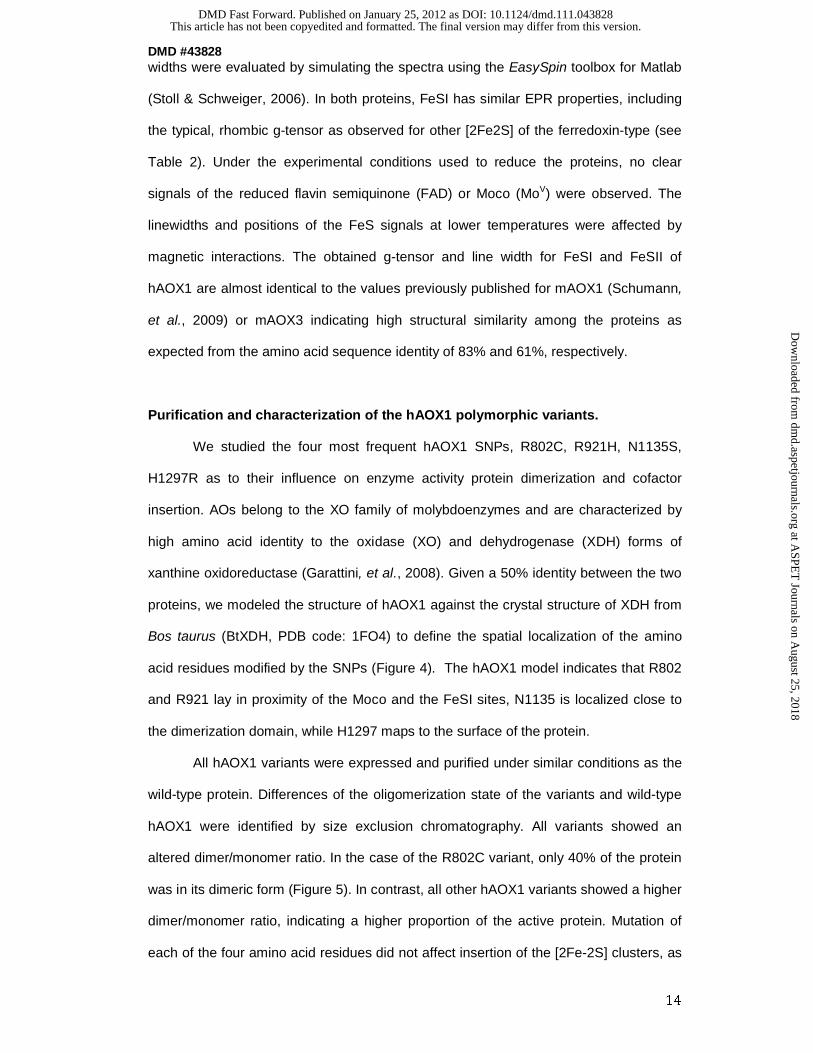
widths were evaluated by simulating the spectra using the EasySpin toolbox for Matlab
(Stoll & Schweiger, 2006). In both proteins, FeSI has similar EPR properties, including
the typical, rhombic g-tensor as observed for other [2Fe2S] of the ferredoxin-type (see
Table 2). Under the experimental conditions used to reduce the proteins, no clear
signals of the reduced flavin semiquinone (FAD) or Moco (MoV) were observed. The
linewidths and positions of the FeS signals at lower temperatures were affected by
magnetic interactions. The obtained g-tensor and line width for FeSI and FeSII of
hAOX1 are almost identical to the values previously published for mAOX1 (Schumann,
et al., 2009) or mAOX3 indicating high structural similarity among the proteins as
expected from the amino acid sequence identity of 83% and 61%, respectively.
Purification and characterization of the hAOX1 polymorphic variants.
We studied the four most frequent hAOX1 SNPs, R802C, R921H, N1135S,
H1297R as to their influence on enzyme activity protein dimerization and cofactor
insertion. AOs belong to the XO family of molybdoenzymes and are characterized by
high amino acid identity to the oxidase (XO) and dehydrogenase (XDH) forms of
xanthine oxidoreductase (Garattini, et al., 2008). Given a 50% identity between the two
proteins, we modeled the structure of hAOX1 against the crystal structure of XDH from
Bos taurus (BtXDH, PDB code: 1FO4) to define the spatial localization of the amino
acid residues modified by the SNPs (Figure 4). The hAOX1 model indicates that R802
and R921 lay in proximity of the Moco and the FeSI sites, N1135 is localized close to
the dimerization domain, while H1297 maps to the surface of the protein.
All hAOX1 variants were expressed and purified under similar conditions as the
wild-type protein. Differences of the oligomerization state of the variants and wild-type
hAOX1 were identified by size exclusion chromatography. All variants showed an
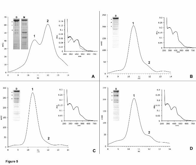
altered dimer/monomer ratio. In the case of the R802C variant, only 40% of the protein
was in its dimeric form (Figure 5). In contrast, all other hAOX1 variants showed a higher
dimer/monomer ratio, indicating a higher proportion of the active protein. Mutation of
each of the four amino acid residues did not affect insertion of the [2Fe-2S] clusters, as
This article has not been copyedited and formatted. The final version may differ from this version.DMD Fast Forward. Published on January 25, 2012 as DOI: 10.1124/dmd.111.043828
at ASPE
T Journals on A
ugust 25, 2018dm
d.aspetjournals.orgD
ownloaded from

DMD #43828
15
the iron content was similar to that of wild-type hAOX1. In fact, similar to wild-type
hAOX1, all variants showed 70% saturation with iron (Figure 2). In R802C molybdenum
saturation was increased to 80%, which is slightly higher than the 70% saturation
observed in the wild-type protein and all the other variants (Figure 2). The variant
hAOX1 proteins were purified and showed a purity comparable to that of the wild-type
counterpart. The UV-vis absorption spectra of the purified variants showed the same
characteristic features of all molybdo-flavoenzymes (Figure 5).
Steady state kinetics of hAOX1 wild-type and variants.
To determine the impact of the SNPs on the enzymatic activity, steady state kinetics
were performed on variants hAOX1-R802C, hAOX1-R921H, hAOX1-N1135S and
hAOX1-H1297R. All generated variants were tested in their ability to catalyze the
conversion of benzaldehyde, phthalazine, phenanthridine and chloroquinazolinone as
substrates (Table 3). The wild-type hAOX1 showed activities with all tested substrates
with a kcat in a range of 5-12 min-1 with KM values between 1 and 7 µM. The best
substrate was phenanthridine with a kcat of 12.2 min-1.
For comparison, the dimeric portion of the hAOX1 variants was also subjected to
steady state kinetic analyzes. The R802C variant showed the most comparable kinetic
data to the wild-type protein. For the hAOX1-N1135S and hAOX1-H1297R variants the
turnover numbers were increased 2.5-fold with phenanthridine as substrate. With other
substrates the kcat and KM values remained in the same range of the wild-type protein
for the two variants. The hAOX1-R921H variant showed a 3.7-1.5-fold decrease in kcat
with most of the substrates in comparison to the wild-type protein, while the KM
remained the comparable. Only with phenanthridine as substrate, the protein was
completely inactive. The results show that the amino acid substitution has different
influences on the kinetic constants depending on the substrate. The inactivity or
reduced activity of the R921H variant might be explained by the fact that the arginine is
highly conserved in all members of the XO family, and with its close proximity to the
pterin molecule of the Mo-MPT cofactor, it might influence the geometry of the bound
This article has not been copyedited and formatted. The final version may differ from this version.DMD Fast Forward. Published on January 25, 2012 as DOI: 10.1124/dmd.111.043828
at ASPE
T Journals on A
ugust 25, 2018dm
d.aspetjournals.orgD
ownloaded from

DMD #43828
16
Mo-MPT molecule, thus affecting catalytic turnover, while not affecting the binding of
the substrate. However, since the different mutations affected the dimer/monomer ratio
of the protein variants, we also considered the total amount of hAOX1 expressed and
calculated the turnover number, taking into account the inactive portion of the
monomeric protein. The normalized values are shown in Table 3. In general, the results
show the same trends as the active portion of the protein.
Discussion
In this report we analyzed the impact of SNPs identified in hAOX1 in an Italian
population. From a cohort of 180 healthy volunteers, representative of the Italian
population, 7 different SNPs were identified which occurred with different frequencies.
In total, 8 individuals were heterozygous for the exchange of Tyr126 to a stop codon.
Another 8 individuals were heterozygous for a silent mutation in Leu1268. The most
frequent mutations resulted in exchanges S1271L, which was identified in 14
individuals heterozygous for this mutation, and amino acid exchange H1297R, for which
7 individuals were heterozygous and 6 individuals were homozygous. Another mutation
resulted in amino acid exchange N1135S, which was not so frequent and was identified
in 1 individual heterozygous for this mutation and 4 individuals homozygous for this
mutation. Two other rather rare mutations resulted in amino acid exchanges R802C
and R921H, for which 2 and 1 individual, respectively, were heterozygous. We decided
to characterize the mutations resulting in AOX1-variants H1297R, N1135S, R802C and
R921H further, since they seemed to be the most interesting SNPs, because either they
were rather frequent and might have an impact on human metabolism, or additionally
homozygous individuals were identified. However, for the characterization of the
hAOX1 variants, first a detailed characterization of the wild-type protein was required. A
heterologous expression system for hAOX1 had been described before (Alfaro, et al.,
2009), however, here the protein was only partially purified and characterized only in
terms of its activity with 6-substituted quinazolinones. In this report, we optimized the
expression and purification of hAOX1 from E. coli. The protein was purified to almost
This article has not been copyedited and formatted. The final version may differ from this version.DMD Fast Forward. Published on January 25, 2012 as DOI: 10.1124/dmd.111.043828
at ASPE
T Journals on A
ugust 25, 2018dm
d.aspetjournals.orgD
ownloaded from

DMD #43828
17
homogeneity, with minor degradation products, which became visible after SDS-PAGE.
Characterization of the active portion of the protein showed that it was purified in a form
with a molybdenum saturation of 70%, of which 50% contained the terminal sulfido
ligand essential for activity. Thus, hAOX1 was purified in a form of which 30% was
active. This is consistent with other reports of the purification of AOX1 and AOX3 from
mouse (Schumann, et al., 2009, Mahro, et al., 2011) or of XDH purified from a
baculovirus insect cell system (Nishino, et al., 2002). Thus, apparently when using
heterologous expression systems, the terminal sulfido ligand seems to be the limiting
step to obtain a fully active mammalian enzyme. The iron saturation was determined to
be 70%. The EPR spectra of hAOX1 were found to be very similar to those from the
mAOX1 protein, showing a slightly rhombic signal for FeSI while FeSII has unusual
EPR properties for [2Fe-2S] species with a pronounced rhombic g tensor, showing
broad lines and being only observed at much lower temperatures (20 K). Overall, the
close similarity of the EPR parameters indicated the presence of the same ligands and
similar geometries of the two redox centers in comparison to mAOX1, which was
expected from the high amino acid sequence identity of the two enzymes of 83%. While
expression of AOs from mouse shows moderate protein yield (Schumann, et al., 2009,
Mahro, et al., 2011), only low yield was achieved for the human AOX1 protein,
suggesting major influences due to the difference in codon usage between H. sapiens
and E. coli. A codon optimized construct for expression in E. coli will be tested in future
studies to increase the protein yield.
Our characterization of the hAOX1 wild-type allowed us to have a good basis for the
characterization of the SNPs. First, modelling of the amino acid exchanges in the
bovine XDH gave us an idea of their locations in the protein. The crystal structure of
eukaryotic AO is not available so far, however, attempts to solve the structure of the
human and mouse proteins are in progress (unpublished data). The amino acid
exchanges resulting from the SNPs were introduced into the protein and the respective
variants were stable and showed an overall molybdenum and iron content similar to the
wild-type protein, with the exception of the hAOX1-R802C variant which showed a
This article has not been copyedited and formatted. The final version may differ from this version.DMD Fast Forward. Published on January 25, 2012 as DOI: 10.1124/dmd.111.043828
at ASPE
T Journals on A
ugust 25, 2018dm
d.aspetjournals.orgD
ownloaded from

DMD #43828
18
slightly higher saturation of Moco. The UV-vis spectra were also comparable to hAOX1
wild-type, suggesting a completely saturation of the FAD cofactor. However, in contrast
to the overall cofactor composition, crucial changes were observed in the protein
quaternary structures. While the wild-type enzyme is stable in its monomeric and
dimeric form in a ratio of 1:1.5, the hAOX1-R921H, hAOX1-N1125S and hAOX1-
H1297R variants were mainly purified as a stable dimer. In contrast, the hAOX1-R802C
variant showed much higher levels of monomer in solution in a ratio of 1.5:1, resulting
in a higher proportion of inactive protein. A change in monomer/dimer ratio has been
reported before for similar variants in AO and XDH enzymes lying in proximity to the
FeS clusters. In SNPs identified in Donryu rat strains the amino acid exchange G101S
in proximity to FeSII also resulted in the production of the monomeric form of AOX1
(Itoh, et al., 2007). In addition, in a human patient suffering from xanthinuria I, a
mutation was identified in the XDH gene resulting in the amino acid exchange R149C
(Sakamoto, et al., 2001). The arginine is located in close proximity to FeSI and when
this mutation was introduced in the Rhodobacter capsulatus xdhB gene and the
corresponding RcXDH-R135C variant was characterized, the purified protein also
existed in two forms, a monomeric inactive form and a dimeric active form (Leimkühler,
et al., 2003). Further analyses of the monomer/dimer behavior of R. capsulatus XDH
resulted in a model in which it was proposed that dimerization of the two requires that
the two FeS clusters are assembled correctly before Moco can be inserted and the
protein can dimerize via the Moco domain (Schumann, et al., 2008). Thus, amino acid
exchanges in proximity to the FeS clusters influence the structure of the protein in a
manner that dimerization is no longer effective.
We studied the activities of hAOX1 wild-type in comparison to the selected variants
based of the SNPs with four selected substrates, benzaldehyde, phthalazine,
phenanthridine and chloroquinazolinone. Our results show that the SNPs can be
classified into three groups in general: fast metabolizers (FM), poor metabolizers (PM)
and no affect on catalytic efficiency. Taking into account only the active portion of the
protein, the hAOX1-R802C variant and even more pronounced the hAOX1-R921H
This article has not been copyedited and formatted. The final version may differ from this version.DMD Fast Forward. Published on January 25, 2012 as DOI: 10.1124/dmd.111.043828
at ASPE
T Journals on A
ugust 25, 2018dm
d.aspetjournals.orgD
ownloaded from

DMD #43828
19
variant are PM, since both variants had a 2.4-1.5-fold reduced activity with most of the
substrates tested. In addition, the R921H variant was identified to be inactive with
phenanthridine, thus, the influence of the amino acid substitution on enzyme activity
varies depending on the substrate. The hAOX1-N1135S and hAOX1-H1297R variants
can be classified into FM, since an increased catalytic efficiency of 2-4-fold was
observed depending on the tested substrate (taking into account only the active portion
of the protein). In general, for the catalytic efficiencies, we calculated the overall activity
of the purified enzyme taking into account the active and the inactive portion of the
protein. In general, the same trends of kinetic values were obtained for the variants in
comparison to the wild-type protein. The hAOX1-R921H variant showed that this
residue is important for maintaining the catalytic activity of hAOX1, in particular with
phenanthridine as substrate. Residue Arg921 lies close to Moco and affected mainly
the monomer/dimer ratio, but also the overall activity of the protein. This might suggest
that not only substrate turnover and binding was impaired, but also the intra-molecular
electron transfer. Thus the positive charge of the arginine might affect substrate binding
and intra-molecular electron transfer, which can only poorly be substituted for by His. In
contrast, in the hAOX1-R802C variant the catalytic efficiency of the protein was not
affected and we obtained similar values in comparison to the hAOX1 wild-type. Both
hAOX1-N1135S and hAOX1-H1297R are considered to be FM variants of hAOX1.
Especially with phthalazine and phenanthridine a 2-4 fold higher catalytic efficiency was
obtained. Both amino acids are located at the surface of the protein and seem to
influence the stability of hAOX1. Thus the more polar and positive charged residues
seem to affect the surface charge of the protein which results in a higher stability of
hAOX1. This also might influence the interaction with other proteins and/or
posttranslational modifications of the protein, as suggested by Itoh et al. (Itoh, et al.,
2007) in a report on the characterization of SNPs in Donryu rats.
Our studies reveal the importance to consider individual differences based on SNPs for
drug design. We analyzed 180 healthy individuals representative of the Italian
population, and identified the total occurrence of 51 SNPs in hAOX1, and a total of 10
This article has not been copyedited and formatted. The final version may differ from this version.DMD Fast Forward. Published on January 25, 2012 as DOI: 10.1124/dmd.111.043828
at ASPE
T Journals on A
ugust 25, 2018dm
d.aspetjournals.orgD
ownloaded from

DMD #43828
20
were homozygous for the SNP. Two of the SNPs resulted in FM and one in PM
individuals. hAOX1 is an important enzyme responsible for the metabolism of a number
of drugs containing aldehydes, and the more prevalent nitrogen heterocycles (Kitamura,
et al., 2006, Torres, et al., 2007). The fraction of drugs metabolized by hAOX1 is likely
to increase over the next decade. Thus, it should be carefully considered which dose of
the drug is administered to an individual, since our results indicate that there might be a
difference in hAOX1 activity in different individuals containing SNPs which additionally
varies depending on the substrate used, thus in FM the drug might be cleared too fast
and have no affect while in PM the drug might reach a toxic dose which would cause
more severe side effects. Additionally, some individuals were identified with SNPs that
resulted in nonsense mutations, which might even affect the overall hAOX1 content and
thus the overall activity more drastically when the active protein amount is expressed
only from one allele. In the future, hAOX1 activities should be measured in individuals
with known SNPs or a pharmacokinetic study with a hAOX1 cleared drug in genotyped
individuals should be conducted to confirm our results on the purified enzyme variants.
This article has not been copyedited and formatted. The final version may differ from this version.DMD Fast Forward. Published on January 25, 2012 as DOI: 10.1124/dmd.111.043828
at ASPE
T Journals on A
ugust 25, 2018dm
d.aspetjournals.orgD
ownloaded from

DMD #43828
21
Acknowledgements:
We thank Manfred Nimtz (Braunschweig) for MALDI peptide mapping. We are grateful
to T. Nishino and T. Matsumura (Nippon Medical School, Tokyo) for providing human
Moco sulfurase cDNA.
This article has not been copyedited and formatted. The final version may differ from this version.DMD Fast Forward. Published on January 25, 2012 as DOI: 10.1124/dmd.111.043828
at ASPE
T Journals on A
ugust 25, 2018dm
d.aspetjournals.orgD
ownloaded from

DMD #43828
22
Authorship Contributions.
Participated in research design: Hartmann, Terao, Garattini, Teutloff, Jones, Leimkühler
Conducted experiments: Hartmann, Terao, Teutloff, Alfaro
Performed data analysis: Hartmann, Terao, Garattini, Teutloff, Leimkühler
Wrote or contributed to the writing of the manuscript: Hartmann, Garattini, Jones,
Leimkühler
This article has not been copyedited and formatted. The final version may differ from this version.DMD Fast Forward. Published on January 25, 2012 as DOI: 10.1124/dmd.111.043828
at ASPE
T Journals on A
ugust 25, 2018dm
d.aspetjournals.orgD
ownloaded from

DMD #43828
23
REFERENCES
Adachi M, Itoh K, Masubuchi A, Watanabe N & Tanaka Y (2007) Construction and
expression of mutant cDNAs responsible for genetic polymorphism in aldehyde
oxidase in Donryu strain rats. Journal of biochemistry and molecular biology 40:
1021-1027.
Al-Salmy HS (2001) Individual variation in hepatic aldehyde oxidase activity. IUBMB life
51: 249-253.
Alfaro JF, Joswig-Jones CA, Ouyang W, Nichols J, Crouch GJ & Jones JP (2009)
Purification and mechanism of human aldehyde oxidase expressed in Escherichia
coli. Drug metabolism and disposition: the biological fate of chemicals 37: 2393-
2398.
Beedham C (1985) Molybdenum hydroxylases as drug-metabolizing enzymes. Drug
Metab. Rev. 16: 119-156.
Beedham C (1997) The role of non-P450 enzymes in drug oxidation. Pharmacy world &
science : PWS 19: 255-263.
Beedham C, Miceli JJ & Obach RS (2003) Ziprasidone metabolism, aldehyde oxidase,
and clinical implications. Journal of clinical psychopharmacology 23: 229-232.
Edmondson D, Massey V, Palmer G, Beacham LM, 3rd & Elion GB (1972) The
resolution of active and inactive xanthine oxidase by affinity chromatography. J Biol
Chem 247: 1597-1604.
Garattini E & Terao M (2011) Increasing recognition of the importance of aldehyde
oxidase in drug development and discovery. Drug metabolism reviews 43: 374-
386.
Garattini E, Fratelli M & Terao M (2008) Mammalian aldehyde oxidases: genetics,
evolution and biochemistry. Cell Mol Life Sci 65: 1019-1048.
Garattini E, Fratelli M & Terao M (2009) The mammalian aldehyde oxidase gene family.
Human genomics 4: 119-130.
This article has not been copyedited and formatted. The final version may differ from this version.DMD Fast Forward. Published on January 25, 2012 as DOI: 10.1124/dmd.111.043828
at ASPE
T Journals on A
ugust 25, 2018dm
d.aspetjournals.orgD
ownloaded from

DMD #43828
24
Garattini E, Mendel R, Romao MJ, Wright R & Terao M (2003) Mammalian molybdo-
flavoenzymes, an expanding family of proteins: structure, genetics, regulation,
function and pathophysiology. Biochem J 372: 15-32.
Hille R (1996) The mononuclear molybdenum enzymes. Chemical Rev 96: 2757-2816.
Ichida K, Matsumura T, Sakuma R, Hosoya T & Nishino T (2001) Mutation of human
molybdenum cofactor sulfurase gene is responsible for classical xanthinuria type II.
Biochem Biophys Res Commun 282: 1194-1200.
Itoh K, Maruyama H, Adachi M, Hoshino K, Watanabe N & Tanaka Y (2007) Lack of
dimer formation ability in rat strains with low aldehyde oxidase activity. Xenobiotica;
the fate of foreign compounds in biological systems 37: 709-716.
Itoh K, Maruyama H, Adachi M, Hoshino K, Watanabe N & Tanaka Y (2007) Lack of
formation of aldehyde oxidase dimer possibly due to 377G>A nucleotide
substitution. Drug metabolism and disposition: the biological fate of chemicals 35:
1860-1864.
Itoh K, Masubuchi A, Sasaki T, et al. (2007) Genetic polymorphism of aldehyde oxidase
in Donryu rats. Drug metabolism and disposition: the biological fate of chemicals
35: 734-739.
Johnson JL, Hainline BE, Rajagopalan KV & Arison BH (1984) The pterin component of
the molybdenum cofactor. Structural characterization of two fluorescent derivatives.
J. Biol. Chem. 259: 5414-5422.
Jordan CG, Rashidi MR, Laljee H, Clarke SE, Brown JE & Beedham C (1999) Aldehyde
oxidase-catalysed oxidation of methotrexate in the liver of guinea-pig, rabbit and
man. The Journal of pharmacy and pharmacology 51: 411-418.
Kitamura S, Sugihara K & Ohta S (2006) Drug-metabolizing ability of molybdenum
hydroxylases. Drug metabolism and pharmacokinetics 21: 83-98.
Kitamura S, Nakatani K, Sugihara K & Ohta S (1999) Strain differences of the ability to
hydroxylate methotrexate in rats. Comparative biochemistry and physiology. Part
C, Pharmacology, toxicology & endocrinology 122: 331-336.
This article has not been copyedited and formatted. The final version may differ from this version.DMD Fast Forward. Published on January 25, 2012 as DOI: 10.1124/dmd.111.043828
at ASPE
T Journals on A
ugust 25, 2018dm
d.aspetjournals.orgD
ownloaded from
DMD #43828
25
Kitamura S, Sugihara K, Nakatani K, et al. (1999) Variation of hepatic methotrexate 7-
hydroxylase activity in animals and humans. IUBMB life 48: 607-611.
Kundu TK, Hille R, Velayutham M & Zweier JL (2007) Characterization of superoxide
production from aldehyde oxidase: an important source of oxidants in biological
tissues. Arch Biochem Biophys 460: 113-121.
Kurosaki M, Demontis S, Barzago MM, Garattini E & Terao M (1999) Molecular cloning
of the cDNA coding for mouse aldehyde oxidase: tissue distribution and regulation
in vivo by testosterone. Vol. 341 ed.^eds.), p.^pp. 71-80.
Laemmli UK (1970) Cleavage of structural proteins during the assembly of the head of
bacteriophage T4. Nature 227: 680-685.
Leimkühler S, Hodson R, George GN & Rajagopalan KV (2003) Recombinant
Rhodobacter capsulatus xanthine dehydrogenase, a useful model system for the
characterization of protein variants leading to xanthinuria I in humans. J Biol Chem
278: 20802-20811.
Leimkühler S, Stockert AL, Igarashi K, Nishino T & Hille R (2004) The role of active site
glutamate residues in catalysis of Rhodobacter capsulatus xanthine
dehydrogenase. J Biol Chem 279: 40437-40444.
Mahro M, Coelho C, Trincao J, et al. (2011) Characterization and Crystallization of
Mouse Aldehyde Oxidase 3: From Mouse Liver to Escherichia coli Heterologous
Protein Expression. Drug metabolism and disposition: the biological fate of
chemicals 39: 1939-1945.
Nishino T, Amaya Y, Kawamoto S, Kashima Y & Okamoto K (2002) Purification and
characterization of multiple forms of rat liver xanthine oxidoreductase expressed in
baculovirus-insect cell system. Journal of biochemistry 132: 597-606.
Obach RS, Huynh P, Allen MC & Beedham C (2004) Human liver aldehyde oxidase:
inhibition by 239 drugs. Journal of clinical pharmacology 44: 7-19.
Palmer T, Santini C-L, Iobbi-Nivol C, Eaves DJ, Boxer DH & Giordano G (1996)
Involvement of the narJ and mob gene products in the biosynthesis of the
molybdoenzyme nitrate reductase in Escherichia coli. Mol Microbiol 20: 875-884.
This article has not been copyedited and formatted. The final version may differ from this version.DMD Fast Forward. Published on January 25, 2012 as DOI: 10.1124/dmd.111.043828
at ASPE
T Journals on A
ugust 25, 2018dm
d.aspetjournals.orgD
ownloaded from

DMD #43828
26
Parschat K, Canne C, Huttermann J, Kappl R & Fetzner S (2001) Xanthine
dehydrogenase from Pseudomonas putida 86: specificity, oxidation-reduction
potentials of its redox-active centers, and first EPR characterization. Biochim
Biophys Acta 1544: 151-165.
Pryde DC, Dalvie D, Hu Q, Jones P, Obach RS & Tran TD (2010) Aldehyde oxidase: an
enzyme of emerging importance in drug discovery. Journal of medicinal chemistry
53: 8441-8460.
Rashidi MR, Smith JA, Clarke SE & Beedham C (1997) In vitro oxidation of famciclovir
and 6-deoxypenciclovir by aldehyde oxidase from human, guinea pig, rabbit, and
rat liver. Drug metabolism and disposition: the biological fate of chemicals 25: 805-
813.
Sakamoto N, Yamamoto T, Moriwaki Y, et al. (2001) Identification of a new point
mutation in the human xanthine dehydrogenase gene responsible for a case of
classical type I xanthinuria. Hum Genet 108: 279-283.
Schumann S, Saggu M, Möller N, Anker SD, Lendzian F, Hildebrandt P & Leimkühler S
(2008) The mechanism of assembly and cofactor insertion into Rhodobacter
capsulatus xanthine dehydrogenase. J Biol Chem 283: 16602-16611.
Schumann S, Terao M, Garattini E, Saggu M, Lendzian F, Hildebrandt P & Leimkuhler
S (2009) Site directed mutagenesis of amino acid residues at the active site of
mouse aldehyde oxidase AOX1. PloS one 4: e5348.
Stoll S & Schweiger A (2006) EasySpin, a comprehensive software package for
spectral simulation and analysis in EPR. . J Magn Reson 178: 42-55.
Terao M, Kurosaki M, Saltini G, Demontis S, Marini M, Salmona M & Garattini E (2000)
Cloning of the cDNAs coding for two novel molybdo-flavoproteins showing high
similarity with aldehyde oxidase and xanthine oxidoreductase. J Biol Chem 275:
30690-30700.
Terao M, Kurosaki M, Barzago MM, et al. (2006) Avian and canine aldehyde oxidases.
Novel insights into the biology and evolution of molybdo-flavoenzymes. The
Journal of biological chemistry 281: 19748-19761.
This article has not been copyedited and formatted. The final version may differ from this version.DMD Fast Forward. Published on January 25, 2012 as DOI: 10.1124/dmd.111.043828
at ASPE
T Journals on A
ugust 25, 2018dm
d.aspetjournals.orgD
ownloaded from

DMD #43828
27
Terao M, Kurosaki M, Barzago MM, et al. (2009) Role of the molybdoflavoenzyme
aldehyde oxidase homolog 2 in the biosynthesis of retinoic acid: generation and
characterization of a knockout mouse. Molecular and cellular biology 29: 357-377.
Torres RA, Korzekwa KR, McMasters DR, Fandozzi CM & Jones JP (2007) Use of
density functional calculations to predict the regioselectivity of drugs and molecules
metabolized by aldehyde oxidase. Journal of medicinal chemistry 50: 4642-4647.
Vila R, Kurosaki M, Barzago MM, et al. (2004) Regulation and biochemistry of mouse
molybdo-flavoenzymes. The DBA/2 mouse is selectively deficient in the expression
of aldehyde oxidase homologues 1 and 2 and represents a unique source for the
purification and characterization of aldehyde oxidase. J Biol Chem 279: 8668-8683.
Wahl RC & Rajagopalan KV (1982) Evidence for the inorganic nature of the
cyanolyzable sulfur of molybdenum hydroxylases. J. Biol. Chem. 257: 1354-1359.
This article has not been copyedited and formatted. The final version may differ from this version.DMD Fast Forward. Published on January 25, 2012 as DOI: 10.1124/dmd.111.043828
at ASPE
T Journals on A
ugust 25, 2018dm
d.aspetjournals.orgD
ownloaded from

DMD #43828
28
Footnotes:
This work was supported by the Cluster of Excellence ‘Unifying Concepts in Catalysis’
(C. Teutloff and S. Leimkühler) coordinated by the Technische Universität Berlin and
funded by the Deutsche Forschungsgemeinschaft and by grants from the Associazione
Italiana per la Ricerca contro il Cancro (AIRC) and the Fondazione Italo Monzino (E.
Garattini).
This article has not been copyedited and formatted. The final version may differ from this version.DMD Fast Forward. Published on January 25, 2012 as DOI: 10.1124/dmd.111.043828
at ASPE
T Journals on A
ugust 25, 2018dm
d.aspetjournals.orgD
ownloaded from

DMD #43828
29
Figure Legends:
Figure 1: Characterization of human aldehyde oxidase
Shown are the size-exclusion chromatogram of Superdex 200 purified hAOX1 and its
UV-vis absorption spectrum. (A) Elution profile of a size-exclusion chromatography
(Superdex 200) shows two peaks of hAOX1 wild-type protein corresponding to the
dimeric (1) and monomeric (2) form in solution. The purity of the protein was
determined by SDS-PAGE (D = dimer, M = monomer). Stars indicate degradation
products of hAOX1 as determined by mass spectroscopy. (B) UV-vis spectrum of air
oxidized hAOX1 in 50 mM Tris, pH 8.0 at 25°C showing characteristic absorptions of
the protein bound FAD at 450 nm and a shoulder for the iron-sulfur clusters at 550 nm.
The spectrum of the protein dimer is shown in black and the spectrum of the
monomeric portion is shown in grey.
Figure 2: Saturation of hAOX1 with molybdenum and iron
Determination of the cofactor saturation of hAOX1 by ICP-OES. The iron content
corresponds to saturation with both, FeSI and FeSII clusters. The wild-type protein and
all variants show a similar saturation of 2x [2Fe2S], but vary in their molybdenum
content between a saturation of 60% and 80% (D = dimer, M = monomer). The %
values are related to theoretical full complement of Moco and the 2x[2Fe2S] clusters.
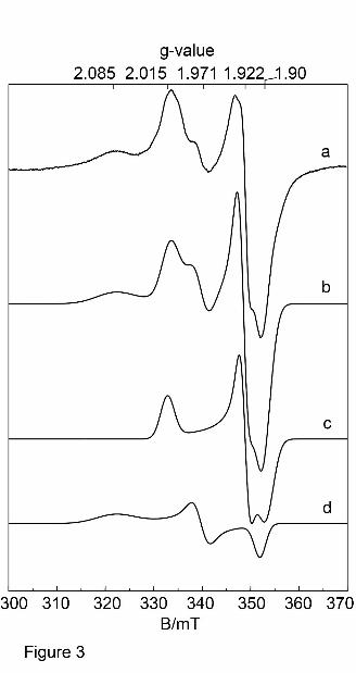
Figure 3: EPR spectra of hAOX1 wild-type.
Experimental cw-EPR spectra of dithionite-reduced mAOX1 wild-type samples at pH
7.0 (trace a) together with the corresponding simulation (trace b). For simulation
parameters see Table 2. The flavin semiquinone and the (MoV) were not detected
under these experimental conditions and therefore neglected in all simulations. (a)
mAOX1 wild-type; (b) simulation of complete spectrum; (c) simulation of FeSI; (d)
simulation of FeSII. Experimental conditions: T = 20 K, 1 mW microwave power,
This article has not been copyedited and formatted. The final version may differ from this version.DMD Fast Forward. Published on January 25, 2012 as DOI: 10.1124/dmd.111.043828
at ASPE
T Journals on A
ugust 25, 2018dm
d.aspetjournals.orgD
ownloaded from

DMD #43828
30
microwave frequency 9.385 GHz, 0.5 mT modulation amplitude, 100 kHz modulation
frequency.
Figure 4: Model of the locations of the amino acid exchanges as a result of SNPs
identified in the hAOX1 gene.
Model with program MacPyMOL using bovine XDH (pdb 1FO4). A = R793 (R802 in
human AOX1), B = R912 (R921 in human AOX1), C = S1126 (N1135 in human AOX1),
D = N1288 (H1297 in human AOX1). The amino acid which is affected by the SNPs is
marked in red. The cofactors (Moco, FeS clusters and FAD) are shown in stick
representations. The overall (α)2 structure is labeled in light grey for one subunit and in
dark grey for the other subunit of the homodimer.
Figure 5: Size-exclusion-chromatography profiles and UV-vis-spectra of hAOX1
variants identified in SNPs.
Elution profiles of hAOX1 variants using size-exclusion chromatography on a Superdex
200 column and UV-vis-spectra of purified proteins are shown (A = R802C, B = R921H,
C = N1135S, D = H1297R). In comparison to the wild-type protein, hAOX1 variants
show different dimer/monomer ratios in solution. R802C is mainly purified in its
monomeric form (A), whereas all other studied variants almost completely exist in their
dimeric forms (B,C,D). For all variants, similar degradation products were visualized by
SDS-PAGE as in the wild-type. (D = Dimer, M = Monomer, stars = degradation
products of hAOX1, 1 = peak corresponding to the AOX1 dimer, 2 = peak
corresponding to the AOX1 monomer)
This article has not been copyedited and formatted. The final version may differ from this version.DMD Fast Forward. Published on January 25, 2012 as DOI: 10.1124/dmd.111.043828
at ASPE
T Journals on A
ugust 25, 2018dm
d.aspetjournals.orgD
ownloaded from
DMD #43828
31
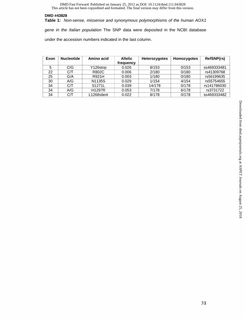
Table 1: Non-sense, missense and synonymous polymorphisms of the human AOX1
gene in the Italian population The SNP data were deposited in the NCBI database
under the accession numbers indicated in the last column.
Exon Nucleotide Amino acid Allelic
frequency Heterozygotes Homozygotes
RefSNP(rs)
5 C/G Y126stop 0.026 8/153 0/153 ss469333481 22 C/T R802C 0.006 2/180 0/180 rs41309768 25 G/A R921H 0.003 1/180 0/180 rs56199635 30 A/G N1135S 0.029 1/154 4/154 rs55754655 34 C/T S1271L 0.039 14/178 0/178 rs141786030 34 A/G H1297R 0.053 7/178 6/178 rs3731722 34 C/T L1268silent 0.022 8/178 0/178 ss469333482
This article has not been copyedited and formatted. The final version may differ from this version.DMD Fast Forward. Published on January 25, 2012 as DOI: 10.1124/dmd.111.043828
at ASPE
T Journals on A
ugust 25, 2018dm
d.aspetjournals.orgD
ownloaded from

DMD #43828
32
Table 2: EPR linewidths and g-values of FeSI and FeSII from hAOX1.
Protein Cluster g-values Linewidth [mT]a
gx gy gz ΔBx ΔBy ΔBz
hAOX1 FeSI FeSII
2.0115 2.085
1.924 1.975
1.900 1.906
4.1 9.2
2.4 3.3
3.7 2.8
mAOX1b FeSI FeSII
2.019 2.085
1.927 1.971
1.912 1.90
2.6 7.4
2.6 4.0
3.2 4.0
a The variable linewidth was included as g-strain in the simulations.
b Values for mAOX1 are taken from ref. (32).
This article has not been copyedited and formatted. The final version may differ from this version.DMD Fast Forward. Published on January 25, 2012 as DOI: 10.1124/dmd.111.043828
at ASPE
T Journals on A
ugust 25, 2018dm
d.aspetjournals.orgD
ownloaded from

DMD #43828
33
Table 3: Steady-state kinetics of hAOX1 and variants corresponding to single
nuclear polymorhpisms (SNPs)
Substrate WT R802C R921H N1135S H1297R
Benzaldehyde
KM [µM] 7.1 ± 0.6 7.6 ± 1.9 6.3 ± 1,2 6.7 ± 2.8 5.2 ± 1.8
active portion kcat [min-1] 6.4 ± 0.1 5.3 ± 0.3 1.7 ± 0,1 6.2 ± 0.3 6.4 ± 0.3
kcat/KM [1/min*µM] 0.91 ± 0.17 0.70 ± 0.16 0.27 ± 0.08 0.92 ± 0.11 1.23 ± 0.17
total protein kcat [min-1] 2.7 ± 0.1 1.8 ± 0.3 1.1 ± 0,1 3.8 ± 0.3 2.6 ± 0.3
kcat/KM [1/min*µM] 0.38 ± 0.17 0.24 ± 0.16 0.17 ± 0.08 0.57 ± 0.11 0.50 ± 0.17
Phthalazine
KM [µM] 1.3 ± 0.3 0.9 ± 0.3 1.6 ± 0.3 1.2 ± 0,1 1.3 ± 0,2
active portion kcat [min-1] 5.6 ± 0.2 5.2 ± 0.3 2.4 ± 0.1 7.2 ± 0,1 5.4 ± 0.1
kcat/KM [1/min*µM] 4.31 ± 0.67 5.78 ± 1.00 1.50 ± 0.33 6.00 ± 1.00 4.15 ± 0.50
total protein kcat [min-1] 2.3 ± 0.2 1.8 ± 0.3 1.5 ± 0.1 4,4 ± 0,1 2.9 ± 0.1
kcat/KM [1/min*µM] 1.77 ± 0.67 2.00 ± 1.00 0.94 ± 0.33 3.67 ± 1.00 2.23 ± 0.50
Phenanthridine
KM [µM] 3.9 ± 0.8 4.4 ± 0.4 n.d. 6.1 ± 1.0 4.1 ± 0.7
active portion kcat [min-1] 12.2 ± 0.5 10.2 ± 0.2 n.d. 32.6 ± 1.1 31.5 ± 1.0
kcat/KM [1/min*µM] 3.13 ± 0.63 2.32 ± 0.50 - 5.34 ± 1.10 7.68 ± 1.43
total protein kcat [min-1] 5.3 ± 0.5 3.4 ± 0.2 n.d. 19.9 ± 1.1 16.9 ± 1.0
kcat/KM [1/min*µM] 1.36 ± 0.63 0.77 ± 0.50 - 3.26 ± 1.10 4.12 ± 1.43
Chloroquinazolinone
KM [µM] 5.2 ± 0.7 4.7 ± 0.7 4.5 ± 0.8 4.1 ± 0.5 5.8 ± 0.5
active portion kcat [min-1] 5.6 ± 0.1 5.4 ± 0.2 3.6 ± 0.1 6.5 ± 0.1 6.7 ± 0.2
kcat/KM [1/min*µM] 1.08 ± 0.14 1.15 ± 0.29 0.80 ± 0.13 1.59 ± 0.20 1.16 ± 0.4
total protein kcat [min-1] 2.3 ± 0.1 1.8 ± 0.2 2.2 ± 0.1 4.0 ± 0.1 3.6 ± 0.2
kcat/KM [1/min*µM] 0.44 ± 0.14 0.38 ± 0.29 0.49 ± 0.13 0.98 ± 0.20 0.62 ± 0.4
Oligomerization in solution
Dimer percentage [%] 58 39 83 90 78
Monomer percentage [%] 38 58 13 7 17
Multimer percentage [%] 4 3 4 3 5
Benzaldehyde, phthalazine, phenanthridine and chloroquinazolinone were used as substrates to cover a range from aldehydes to N-heterocyclic compounds. Assays were performed photometrically using 2,6-dichlorphenol-indophenol as a final electron acceptor. With phenanthridine molecular oxygen was used as electron acceptor. n.d. indicates that no activity is detectable. Data are mean values from three independent measurements (+/- S.D.).
This article has not been copyedited and formatted. The final version may differ from this version.DMD Fast Forward. Published on January 25, 2012 as DOI: 10.1124/dmd.111.043828
at ASPE
T Journals on A
ugust 25, 2018dm
d.aspetjournals.orgD
ownloaded from

This article has not been copyedited and formatted. The final version may differ from this version.DMD Fast Forward. Published on January 25, 2012 as DOI: 10.1124/dmd.111.043828
at ASPE
T Journals on A
ugust 25, 2018dm
d.aspetjournals.orgD
ownloaded from

This article has not been copyedited and formatted. The final version may differ from this version.DMD Fast Forward. Published on January 25, 2012 as DOI: 10.1124/dmd.111.043828
at ASPE
T Journals on A
ugust 25, 2018dm
d.aspetjournals.orgD
ownloaded from
This article has not been copyedited and formatted. The final version may differ from this version.DMD Fast Forward. Published on January 25, 2012 as DOI: 10.1124/dmd.111.043828
at ASPE
T Journals on A
ugust 25, 2018dm
d.aspetjournals.orgD
ownloaded from

This article has not been copyedited and formatted. The final version may differ from this version.DMD Fast Forward. Published on January 25, 2012 as DOI: 10.1124/dmd.111.043828
at ASPE
T Journals on A
ugust 25, 2018dm
d.aspetjournals.orgD
ownloaded from
This article has not been copyedited and formatted. The final version may differ from this version.DMD Fast Forward. Published on January 25, 2012 as DOI: 10.1124/dmd.111.043828
at ASPE
T Journals on A
ugust 25, 2018dm
d.aspetjournals.orgD
ownloaded from