DMD 40:952–962, 2012 Lymphatic Transport and...
11
Lymphatic Transport and Catabolism of Therapeutic Proteins after Subcutaneous Administration to Rats and Dogs Weirong Wang, 1 Nancy Chen, Xiaolan Shen, Paul Cunningham, Scott Fauty, Kimberly Michel, Bo Wang, Xuening Hong, Christine Adreani, Christian N. Nunes, Chris V. Johnson, Kuo-chang Yin, Michelle Groff, Yan Zou, Liming Liu, Lora Hamuro, and Thomayant Prueksaritanont Department of Pharmacokinetics, Pharmacodynamics and Drug Metabolism (W.W., N.C., M.G., Y.Z., L.L., L.H., T.P.), Rahway Laboratory Animal Resources (X.S., P.C., B.W., X.H., C.A., C.N.N., C.V.J.), West Point Toxicological Science (S.F., K.M.), and Vaccine Analytical Development (K.Y.), Merck Sharp and Dohme Corp., West Point, Pennsylvania Received November 7, 2011; accepted February 10, 2012 ABSTRACT: The mechanism underlying subcutaneous absorption of macro- molecules and factors that can influence this process were studied in rats using PEGylated erythropoietins (EPOs) as model com- pounds. Using a thoracic lymph duct cannulation (LDC) model, we showed that PEGylated EPO was absorbed from the subcutaneous injection site mainly via the lymphatic system in rats, which is similar to previous reports in sheep. After subcutaneous adminis- tration, the serum exposure was reduced by 70% in LDC animals compared with that in the control animals, and most of the sys- temically available dose was recovered in the lymph. In both LDC and intact rats, the total radioactivity recoveries in excreta after subcutaneous administration were high (70–80%), indicating that catabolism, not poor absorption, was the main cause for the ob- served low bioavailability (30–40%). Moreover, catabolism of PEGylated EPO was found with both rat subcutaneous tissue ho- mogenate and lymph node cell suspensions, and a significant amount of dose-related breakdown fragments was found in the lymph of LDC rats. In addition, the bioavailability of PEGylated EPOs was shown to be 2- to 4-fold lower in “fat rats,” indicating that physiologic features pertinent to lymphatic transport can have a profound impact on subcutaneous absorption. Limited studies in dogs also suggested similar subcutaneous absorption mecha- nisms. Collectively, our results suggest that the lymphatic absorp- tion mechanism for macromolecules is probably conserved among commonly used preclinical species, e.g., rats and dogs, and that mechanistic understanding of the subcutaneous absorption mech- anism and associated determinants should be helpful in biologic drug discovery and development. Introduction Almost all protein-based biologic drugs are administered parenter- ally. Subcutaneous administration is the preferred route of adminis- tration compared with intravenous administration. There are more than 30 marketed biologic drugs that are administered subcutaneously, including most of the therapeutic proteins/peptides (McDonald et al., 2010). However, despite this wide application, our understanding of the exact mechanism underlying subcutaneous absorption and the factors that can influence this process remain limited (Porter and Charman, 2000; Lin, 2009). Biologic drugs can exhibit a wide range of bioavailability after subcutaneous administration in humans (Tang et al., 2004). Many physiologic factors, e.g., age, body weight, and injection site, have been identified as covariants for the pharmacokinetics (PK) of mac- romolecules administered subcutaneously in humans (Macdougall et al., 1991; Chan et al., 2003; Fishbane et al., 2007; Olsson-Gisleskog et al., 2007; Kakkar et al., 2011). However, the type of factors and extent of their impact are apparently compound-dependent. For ex- ample, when the PK of peginterferon alfa-2a and peginterferon alfa-2b were compared in a randomized 36-patient trial, peginterferon alfa-2a showed much greater variability in patient exposure than peginter- feron alfa-2b (38% versus 20%), and body weight appeared to be a covariant for peginterferon alfa-2a, but not for peginterferon alfa-2b (Silva et al., 2006). At present, there is no established method to predict subcutaneous absorption in humans a priori, i.e., bioavailabil- ity, potential covariants, or the magnitude of PK variability. There- fore, there is a great need for more mechanistic understanding of the subcutaneous absorption process, as well as more preclinical and in vitro tools to help gain insights into its underlying mechanism. Our current understanding of the subcutaneous absorption process mostly came from a sheep lymphatic cannulation model, in which a series of studies demonstrated a molecular weight-dependent lym- phatic contribution to the subcutaneous absorption of macromole- cules; i.e., macromolecules with molecular mass 16 kDa are ab- 1 Current affiliation: Biologics Clinical Pharmacology, Janssen R&D, US, Spring House, Pennsylvania. Article, publication date, and citation information can be found at http://dmd.aspetjournals.org. http://dx.doi.org/10.1124/dmd.111.043604. ABBREVIATIONS: PK, pharmacokinetics; LDC, lymph duct cannulation; EPO, erythropoietin; PEG, polyethylene glycol; CERA, continuous erythropoietin receptor activator; SD, Sprague-Dawley; TCA, trichloroacetic acid; AUC, area under the serum concentration-time curve; hGH, human growth hormone. 1521-009X/12/4005-952–962$25.00 DRUG METABOLISM AND DISPOSITION Vol. 40, No. 5 Copyright © 2012 by The American Society for Pharmacology and Experimental Therapeutics 43604/3764265 DMD 40:952–962, 2012 952 at ASPET Journals on July 5, 2018 dmd.aspetjournals.org Downloaded from
Transcript of DMD 40:952–962, 2012 Lymphatic Transport and...

Lymphatic Transport and Catabolism of Therapeutic Proteins afterSubcutaneous Administration to Rats and Dogs
Weirong Wang,1 Nancy Chen, Xiaolan Shen, Paul Cunningham, Scott Fauty, Kimberly Michel,Bo Wang, Xuening Hong, Christine Adreani, Christian N. Nunes, Chris V. Johnson,
Kuo-chang Yin, Michelle Groff, Yan Zou, Liming Liu, Lora Hamuro,and Thomayant Prueksaritanont
Department of Pharmacokinetics, Pharmacodynamics and Drug Metabolism (W.W., N.C., M.G., Y.Z., L.L., L.H., T.P.), RahwayLaboratory Animal Resources (X.S., P.C., B.W., X.H., C.A., C.N.N., C.V.J.), West Point Toxicological Science (S.F., K.M.),
and Vaccine Analytical Development (K.Y.), Merck Sharp and Dohme Corp., West Point, Pennsylvania
Received November 7, 2011; accepted February 10, 2012
ABSTRACT:
The mechanism underlying subcutaneous absorption of macro-molecules and factors that can influence this process were studiedin rats using PEGylated erythropoietins (EPOs) as model com-pounds. Using a thoracic lymph duct cannulation (LDC) model, weshowed that PEGylated EPO was absorbed from the subcutaneousinjection site mainly via the lymphatic system in rats, which issimilar to previous reports in sheep. After subcutaneous adminis-tration, the serum exposure was reduced by �70% in LDC animalscompared with that in the control animals, and most of the sys-temically available dose was recovered in the lymph. In both LDCand intact rats, the total radioactivity recoveries in excreta aftersubcutaneous administration were high (70–80%), indicating thatcatabolism, not poor absorption, was the main cause for the ob-served low bioavailability (30–40%). Moreover, catabolism of
PEGylated EPO was found with both rat subcutaneous tissue ho-mogenate and lymph node cell suspensions, and a significantamount of dose-related breakdown fragments was found in thelymph of LDC rats. In addition, the bioavailability of PEGylatedEPOs was shown to be 2- to 4-fold lower in “fat rats,” indicatingthat physiologic features pertinent to lymphatic transport can havea profound impact on subcutaneous absorption. Limited studies indogs also suggested similar subcutaneous absorption mecha-nisms. Collectively, our results suggest that the lymphatic absorp-tion mechanism for macromolecules is probably conserved amongcommonly used preclinical species, e.g., rats and dogs, and thatmechanistic understanding of the subcutaneous absorption mech-anism and associated determinants should be helpful in biologicdrug discovery and development.
Introduction
Almost all protein-based biologic drugs are administered parenter-ally. Subcutaneous administration is the preferred route of adminis-tration compared with intravenous administration. There are morethan 30 marketed biologic drugs that are administered subcutaneously,including most of the therapeutic proteins/peptides (McDonald et al.,2010). However, despite this wide application, our understanding ofthe exact mechanism underlying subcutaneous absorption and thefactors that can influence this process remain limited (Porter andCharman, 2000; Lin, 2009).
Biologic drugs can exhibit a wide range of bioavailability aftersubcutaneous administration in humans (Tang et al., 2004). Manyphysiologic factors, e.g., age, body weight, and injection site, havebeen identified as covariants for the pharmacokinetics (PK) of mac-
romolecules administered subcutaneously in humans (Macdougall etal., 1991; Chan et al., 2003; Fishbane et al., 2007; Olsson-Gisleskoget al., 2007; Kakkar et al., 2011). However, the type of factors andextent of their impact are apparently compound-dependent. For ex-ample, when the PK of peginterferon alfa-2a and peginterferon alfa-2bwere compared in a randomized 36-patient trial, peginterferon alfa-2ashowed much greater variability in patient exposure than peginter-feron alfa-2b (38% versus 20%), and body weight appeared to be acovariant for peginterferon alfa-2a, but not for peginterferon alfa-2b(Silva et al., 2006). At present, there is no established method topredict subcutaneous absorption in humans a priori, i.e., bioavailabil-ity, potential covariants, or the magnitude of PK variability. There-fore, there is a great need for more mechanistic understanding of thesubcutaneous absorption process, as well as more preclinical and invitro tools to help gain insights into its underlying mechanism.
Our current understanding of the subcutaneous absorption processmostly came from a sheep lymphatic cannulation model, in which aseries of studies demonstrated a molecular weight-dependent lym-phatic contribution to the subcutaneous absorption of macromole-cules; i.e., macromolecules with molecular mass �16 kDa are ab-
1 Current affiliation: Biologics Clinical Pharmacology, Janssen R&D, US,Spring House, Pennsylvania.
Article, publication date, and citation information can be found athttp://dmd.aspetjournals.org.
http://dx.doi.org/10.1124/dmd.111.043604.
ABBREVIATIONS: PK, pharmacokinetics; LDC, lymph duct cannulation; EPO, erythropoietin; PEG, polyethylene glycol; CERA, continuouserythropoietin receptor activator; SD, Sprague-Dawley; TCA, trichloroacetic acid; AUC, area under the serum concentration-time curve; hGH,human growth hormone.
1521-009X/12/4005-952–962$25.00DRUG METABOLISM AND DISPOSITION Vol. 40, No. 5Copyright © 2012 by The American Society for Pharmacology and Experimental Therapeutics 43604/3764265DMD 40:952–962, 2012
952
at ASPE
T Journals on July 5, 2018
dmd.aspetjournals.org
Dow
nloaded from

sorbed mainly via the lymphatic system (Supersaxo et al., 1990;Porter and Charman, 2000; Porter et al., 2001; McLennan et al.,2006). In addition, the observation of dose-dependent subcutaneousbioavailability for an anti-CD4 monoclonal antibody also providedindirect evidence that monoclonal antibodies are absorbed via thelymphatic pathway in mice (Davis and Bugelski 1998). This theoryaligns well with our knowledge of the anatomic structural differencesbetween blood and lymph capillaries. The lymphatic system is aunidirectional system that transports excess fluid from interstitium tosystemic circulation. Unlike blood capillaries in the subcutaneousspace whose tight endothelial junctions make it difficult for macro-molecules to pass through, lymphatic capillaries have incompletebasal lamina, which enables almost unrestricted drainage of macro-molecules from the interstitial space (Swartz, 2001). Because of thetechnical challenges of cannulating lymph vessels in smaller-sizedlaboratory animals, there have been very limited reports on the con-tribution of the lymphatic system in subcutaneous absorption ofmacromolecules in commonly used preclinical species. Unfortunately,the few available reports in rats and rabbits presented contradictoryevidence (Bocci et al., 1986, 1988; Kojima et al., 1988; Kagan et al.,2007). In the more recent report by Kagan et al. (2007), the contri-bution of the lymphatic system to subcutaneous absorption of mac-romolecules was systematically examined in a rat thoracic lymph ductcannulation (LDC) model, using bovine insulin (5.6 kDa), recombi-nant human erythropoietin (EPO) (30.4 kDa), and bovine albumin (66kDa) as model compounds. The results suggested minimal contribu-tion of the lymphatic system for all three molecules, regardless of theirmolecular masses, i.e., minimal reduction of serum exposure in thethoracic LDC animals, and �3% drug recovery in thoracic duct lymph(Kagan et al., 2007). The only report in which a significant percentage(up to 29%) of subcutaneous dosed radioactivity was recovered in therat thoracic duct lymph was a study using PEGylated polylysinedendrimers, although the amount of dose recovered in the lymph wasstill considerably lower than the systemic bioavailability (94–104%)(Kaminskas et al., 2009). These results raised the question of whetherthe contribution of the lymphatic system in subcutaneous absorptionof macromolecules is conserved among preclinical species and chal-lenged the value, if any, of studying subcutaneous absorption in thesecommonly used preclinical species.
Here, we report our efforts in elucidating the mechanism of sub-cutaneous absorption of macromolecules in thoracic LDC models inrats and dogs. Two PEGylated recombinant human EPOs were se-lected as model compounds because they are likely to be absorbed viathe lymph, and their PK had been well characterized in rats and dogs.Their catabolism after subcutaneous administration and the impact ofphysiologic features pertinent to lymphatic absorption/transport, e.g.,fat content and level of physical activity, on subcutaneous absorptionwere also evaluated in these preclinical models.
Materials and Methods
Materials. Recombinant human EPO is a 30.4-kDa therapeutic protein.PEG30-EPO [continuous erythropoietin receptor activator (CERA)] is a com-mercially available PEGylated recombinant human EPO that contains a �30-kDa methoxy PEG chain linked via amide bonds to the N-terminal aminogroup or the �-amino group of lysines (predominantly lysine-52 or lysine-45)(Macdougall, 2005). CERA (Mircera) was purchased from Myoderm (Norris-town, PA). PEG40-EPO is a PEGylated recombinant human EPO producedin-house, which has an �40-kDa linear PEG conjugated preferably to theamino terminus of the recombinant human EPO (Nett et al., 2012). In additionto the difference in PEG sizes, PEG30-EPO was produced from Chinesehamster ovary cells with enriched tetraantennary sialylated glycoforms at thethree N-glycosylation sites of EPO, whereas PEG40-EPO was produced from
engineered strains of Pichia pastoris with terminally sialylated biantennaryN-glycan structures (Nett et al., 2012).
125I Labeling. 125I labeling of PEG30-EPO and PEG40-EPO was conductedusing Iodobeads according to the manufacturer’s protocol with minor modifi-cation (Thermo Fisher Scientific, Waltham, MA). In brief, 100 �g of proteindialyzed into phosphate-buffered saline was incubated with 1 mCi of 125I(PerkinElmer Life and Analytical Sciences, Waltham, MA) and two Iodobeadsfor 15 min at room temperature. The labeled protein was purified with a Zebacolumn (Thermo Fisher Scientific), and stored at 4°C. The radiochemicalconcentration was determined by gamma counting using a Wallac 1470 auto-mated gamma counter (PerkinElmer Life and Analytical Sciences). The purityof labeled proteins was determined by a size-exclusion high-performanceliquid chromatography system equipped with a gamma detector. The percent-age of free 125I was less than 2% in all preparations. The integrity of labeledproteins was also analyzed with a product-specific immunoassay (QuantikineIVD human erythropoietin kit; R&D Systems, Minneapolis, MN), and alllabeled proteins showed concentration-response curves comparable to those ofthe corresponding unlabeled materials. For in vivo dosing, the dosing solutionwas prepared by mixing unlabeled compound, 125I-labeled compound, andformulation buffer to achieve the desired specific activity and final proteinconcentration.
In Vivo Studies in Sprague-Dawley Rats and Beagle Dogs. All protocolswere approved by the Merck Institutional Animal Care and Use Committee.
Thoracic LDC in rats. Male Sprague-Dawley (SD) rats (350–400 g; Tac-onic Farms, Germantown, NY) were used for all rat lymph cannulation studies.Rats were given 0.5 to 1.0 ml of olive oil by oral gavage 0.5 to 1 h before thesurgery to provide enhanced visualization of lymph vessels. The thoracic ductcannulation technique was based on previous reports (Lee and Hashim, 1966;Ionac, 2003) with modifications for continuous lymph collection.
In brief, rats were anesthetized with isoflurane. The thoracic duct waslocated and separated from the psoas muscle and dorsal aorta. The duct wascannulated with a heparin (500 units/ml) saline-filled catheter. All accessorybranches into the main lymph duct were either bypassed by the cannula tip orligated. Heparin (500 units/ml) was also infused at a constant flow rate of 50�l/h into the catheter that contains collected lymph to prevent the coagulationof lymph fluid ex vivo and therefore maintain the free flow of lymph fluid.Once the lymph flow had been established, the cannula was tied down by silkligatures tunneled subcutaneously and exteriorized at the back between theshoulder blades. A spring tether system was installed and attached to a swivelto allow free movement for the animals. After the surgical procedure, the ratswere monitored until they regained full consciousness. The rats were providedwith ad libitum rodent chow and recovery gel (Diet Gel, Clear H2O). Therewas a 48-h period of recovery and stabilization with continuous lymph fluidcollection before initiation of the study.
For in vivo dosing, 125I-labeled PEG30-EPO (25 �g/kg, 13.5 �Ci/kg) orPEG40-EPO (36 �g/kg, 14 �Ci/kg) was administered subcutaneously to thelateral left lower hind leg (just below the knee) of the thoracic LDC rats as wellas the sham-operated rats. Thoracic duct lymph was continuously collected forall cannulated animals during the entire 7-day study period, and the volumeswere recorded. The lymph flow ranged from 0.5 to 5 ml/h for LDC rats, andthe total volume of lymph collected per animal ranged between 25 and 650 mlduring the study period. Only data from animals with total collected lymphvolume of �70 ml were used. A series of blood samples were collected for allanimals from the carotid artery via implanted cannula at predefined timeintervals and serum was prepared. Lymph and serum samples were stored at�70°C until analysis. For the study with PEG30-EPO, the animals were keptin metabolic cages, and urine and feces samples were also collected.
Thoracic LDC in beagle dogs. Thoracic LDC in beagle dogs was performedsimilarly to that in SD rats. In brief, adult male beagle dogs were surgicallyprepared 16 h before dosing. The lymph duct was cannulated close to thethoracic duct ampulla with a Silastic catheter, which was tunneled under theskin to a small incision over the animal’s shoulder region and exteriorized. Allaccessory branches into the main lymph duct were either bypassed by thecannula tip or ligated. After the surgical procedure, the dogs were monitoreduntil they regained full consciousness. An electrolyte mix was administeredsubcutaneously to the dogs to help compensate for the liquid loss and maintainthe lymph flow. The lymph flow after surgery was monitored; it ranged from1 to 50 ml/h for LDC dogs, and the total volume of lymph collected per animal
953LYMPHATIC TRANSPORT AFTER SUBCUTANEOUS ADMINISTRATION
at ASPE
T Journals on July 5, 2018
dmd.aspetjournals.org
Dow
nloaded from

ranged between 10 and 700 ml during the study period. Only the animals thatmaintained a stable lymph flow at the time of study initiation were used for thefollowing studies.
For in vivo dosing, 125I-labeled PEG40-EPO (10 �g/kg, 7.5 �Ci/kg) wasadministered either intravenously or subcutaneously (at the lateral left lowerhind leg, just below the knee) to thoracic LDC animals. Thoracic duct lymphwas continuously collected, and the volumes were recorded. Only data fromthe two animals with sustained lymph flow for �48 h were used. Bloodsamples were also collected from the jugular vein at predefined time intervals,and serum was prepared. Lymph and serum samples were stored at �70°Cuntil analysis.
PK Study in Fat Rats to Assess Impact of Fat on Subcutaneous Ab-sorption. “Fat rats” were obtained by feeding 2- to 3-month-old regular SDrats (�250 g) with a high-fat diet for �3 months. These fat rats had an averagebody weight of 500 to 600 g, approximately double the body weights of theirnormal counterparts. The back (scruff of the neck) was used as the subcuta-neous injection site in this study, in which the fat rats had significantly moresubcutaneous fat, as evidenced by the skinfold thickness. To determine bio-availability (F), 36 �g/kg either PEG30-EPO or PEG40-EPO was administeredintravenously and subcutaneously in the fat rats (n � 3/intravenous group; n �6/subcutaneous group). As a control, the F of PEG40-EPO was also deter-mined in the regular rats after 36 �g/kg intravenous and subcutaneous admin-istration (n � 6/group). Blood samples were obtained from the jugular vein atspecific time points. Serum samples were prepared and stored at �70°C untilanalysis.
PK Study in Beagle Dogs to Assess Impact of Physical Activity onSubcutaneous Absorption. Beagle dogs with apparent different physicalactivity levels were used in the study. The dog’s physical activity levels weredivided into two categories based on cage side observation: high � excitedbarking, frequent jumping up and down, and vigorous tail wagging; low �quiet, usually laid down with occasional tail wagging.
To evaluate the impact of physical activity on subcutaneous absorption,125I-labeled PEG30-EPO or PEG40-EPO (10 �g/kg, �10 �Ci/kg) was admin-istered subcutaneously to the posterior dorsal region, �10 cm in front of thetail. A modified hand-held sodium iodide detector was used to monitordisappearance of radioactivity from the injection site with minimal interferenceof signals from internal organs such as the thyroid. The sodium iodide detectorwas collimated with lead to limit the detection area to the injection site. Uponadministration, the injection site was monitored at specific time points, andradiation measurements were recorded. Blood samples were obtained from thejugular vein at specific time points. Serum samples were prepared and storedat �70°C until analysis.
Analytical Method and PK Analysis. When 125I-labeled compounds wereused for animal studies, the total radioactivity levels in lymph, serum, urine, orfeces samples were first determined by gamma counting (pre-TCA). Theprotein-associated radioactivity was subsequently quantified by TCA precipi-tation (Bensadoun and Weinstein, 1976). The TCA precipitation was con-ducted as described previously (Vugmeyster et al., 2010). Serum or lymphprotein drug levels were calculated from TCA-precipitable (post-TCA) radio-activity using the specific activity of the dosing solutions with correction for125I decay (nanogram-equivalents per milliliter). The percentage of TCA in asample, calculated as post-TCA/pre-TCA, is an indication of integrity of thelabeled protein, i.e., what percentage of the radioactivity is associated withprotein, instead of being free iodine or very small degraded fragments.
When unlabeled compounds were used for animal studies, the serum druglevels were determined by an immunoassay based on the Quantikine IVDhuman erythropoietin kit and validated for quantifying PEG30-EPO andPEG40-EPO. The assay range was established with spiked quality controlsamples. The accuracy and precision were within 100 � 20%, and the lowerlimit of quantitation was 0.1 ng/ml. The drug levels of selected 125I-labeledPEG30-EPO and PEG40-EPO serum/lymph samples were also determined bythe immunoassay as a comparison to those calculated from post-TCA radio-activity. The results from both assays generally agree with each other, and thedifferences were less than 40%.
The serum drug concentrations were calculated for noncompartmental PKanalysis using WinNonlin (Enterprise Version 5.2.1; Pharsight, MountainView, CA). Both PEG30-EPO and PEG40-EPO exhibited approximately lin-ear PK over the dose ranges (�10 �g/kg) used in our studies, and the systemic
bioavailability (F, percentage) was calculated as follows: AUCsc � dosei.v./AUCi.v. � doses.c..
In Vitro Stability in Rat Subcutaneous Tissue Homogenate and LymphNode Cell Suspensions. The subcutaneous tissue from the back area of naiveSD rats was excised and isolated with a scalpel. After mincing with a scissors,the subcutaneous tissue was mixed with 1.5� volumes of tissue weight ofice-cold homogenization buffer (50 mM Tris-HCl at pH7.4, 150 mM NaCl,0.25 M sucrose, 1% Tween 20, and 1% Triton X-100) and homogenized witha Polytron homogenizer (Kinematica Inc., Newark, NJ) on ice. The lysate wascentrifuged at 14,000g at 4°C for 20 min. The supernatant was removed forfurther analysis.
For lymph node cell suspensions, multiple lymph nodes from naive SD ratswere excised. The cells were released by gently teasing using tweezers andthen collected by straining through 40-�m cell strainers (BD Falcon, FranklinLakes, NJ). The number of viable cells was determined by trypan blue dyeexclusion with a hemacytometer (Gibco Cell Culture; Invitrogen, Carlsbad,CA). The specified numbers of viable cells were incubated with 125I-labeledPEG30-EPO or PEG40-EPO in a six-well tissue culture plate in RPMI 1640medium with 10% fetal calf serum and 5% CO2 at 37°C for 24 h.
For in vitro stability experiments, 125I-labeled compounds were spiked intofreshly prepared subcutaneous tissue homogenate, serum, lymph fluid, orlymph node cell suspensions and incubated at 37°C for 24 h. After incubation,SDS-polyacrylamide gel electrophoresis was performed to evaluate potentialdegradants, and the bands were detected by phosphorimaging (GE Healthcare,Chalfont St. Giles, Buckinghamshire, UK).
Results
Role of Lymphatic System in Subcutaneous Absorption ofPEG30-EPO and PEG40-EPO in Rats. To determine the contribu-tion of the lymphatic system in subcutaneous absorption of macro-molecules in commonly used preclinical species, we first used a ratthoracic LDC model established in-house. This model allows contin-uous thoracic duct lymph fluid collection in conscious and unre-strained SD rats for up to 7 days, considerably longer than what hadbeen reported previously (Kagan et al., 2007; Kaminskas et al., 2009).
Before performing experiments in LDC rats, we examined the PKprofiles of 125I-labeled PEG30-EPO and PEG40-EPO, for which thedrug levels were calculated from post-TCA radioactivity, and foundthem to be similar to the corresponding PK profiles of unlabeledcompounds, for which the drug levels were determined by a product-specific immunoassay: for PEG30-EPO, maximum serum concentra-tion (Cmax) was 114 � 30 versus 73 � 26 ng/ml and AUC0–168 h was7649 � 1566 versus 7059 � 3325 h � ng/ml after 25 �g/kg s.c. dosingin rats; for PEG40-EPO, Cmax was 85 � 42 versus 84 � 26 ng/ml andAUC0–168 h was 5013 � 3635 versus 5193 � 1493 h � ng/ml after 36�g/kg s.c. dosing in rats. These results suggested that 125I-labeling didnot significantly change the PK properties of PEG30-EPO andPEG40-EPO and that the drug levels calculated from post-TCA ra-dioactivity can generally be considered to be representative of thosedetermined by immunoassay. We also determined the systemic bio-availability of PEG30-EPO and PEG40-EPO after subcutaneous ad-ministration using unlabeled compounds in intact control SD rats, andthey were 38 and 30%, respectively (Table 1). In addition, our PKstudies with unlabeled compounds had demonstrated that the clear-ance of PEG40-EPO is linear between 4 and 36 �g/kg in rats (2.0–2.2ml/kg per hour). The clearance of PEG30-EPO (CERA) was alsoreported to be approximately linear between 2.5 and 25 �g/kg in ratsand 3 and 7.5 �g/kg in dogs (http://www.ema.europa.eu/docs/en_GB/document_library/EPAR_-_Scientific_Discussion/human/000739/WC500033669.pdf).
The choice of subcutaneous injection site is very important forstudying lymphatic contribution in rat thoracic LDC models. For allLDC rat studies described, we used the lower hind leg (just below theknee) as the subcutaneous injection site to evaluate the contribution of
954 WANG ET AL.
at ASPE
T Journals on July 5, 2018
dmd.aspetjournals.org
Dow
nloaded from
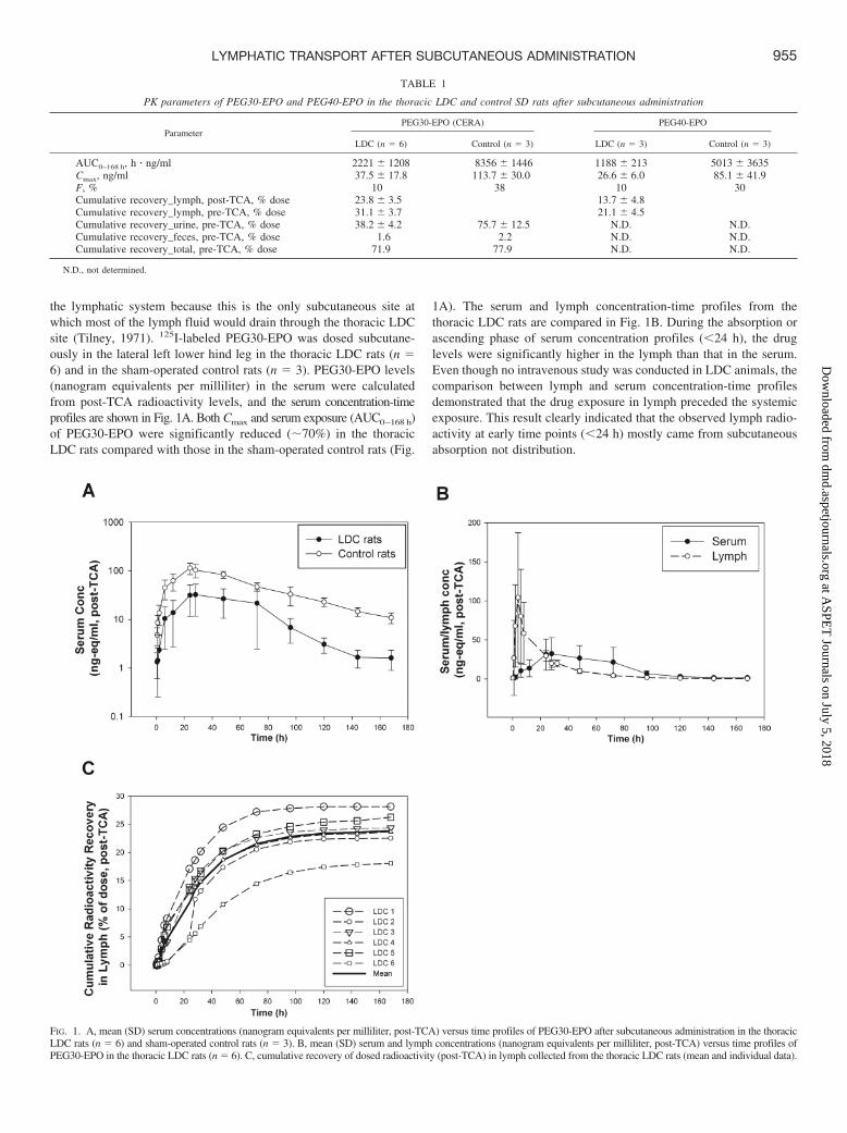
the lymphatic system because this is the only subcutaneous site atwhich most of the lymph fluid would drain through the thoracic LDCsite (Tilney, 1971). 125I-labeled PEG30-EPO was dosed subcutane-ously in the lateral left lower hind leg in the thoracic LDC rats (n �6) and in the sham-operated control rats (n � 3). PEG30-EPO levels(nanogram equivalents per milliliter) in the serum were calculatedfrom post-TCA radioactivity levels, and the serum concentration-timeprofiles are shown in Fig. 1A. Both Cmax and serum exposure (AUC0–168 h)of PEG30-EPO were significantly reduced (�70%) in the thoracicLDC rats compared with those in the sham-operated control rats (Fig.
1A). The serum and lymph concentration-time profiles from thethoracic LDC rats are compared in Fig. 1B. During the absorption orascending phase of serum concentration profiles (�24 h), the druglevels were significantly higher in the lymph than that in the serum.Even though no intravenous study was conducted in LDC animals, thecomparison between lymph and serum concentration-time profilesdemonstrated that the drug exposure in lymph preceded the systemicexposure. This result clearly indicated that the observed lymph radio-activity at early time points (�24 h) mostly came from subcutaneousabsorption not distribution.
FIG. 1. A, mean (SD) serum concentrations (nanogram equivalents per milliliter, post-TCA) versus time profiles of PEG30-EPO after subcutaneous administration in the thoracicLDC rats (n � 6) and sham-operated control rats (n � 3). B, mean (SD) serum and lymph concentrations (nanogram equivalents per milliliter, post-TCA) versus time profiles ofPEG30-EPO in the thoracic LDC rats (n � 6). C, cumulative recovery of dosed radioactivity (post-TCA) in lymph collected from the thoracic LDC rats (mean and individual data).
TABLE 1
PK parameters of PEG30-EPO and PEG40-EPO in the thoracic LDC and control SD rats after subcutaneous administration
ParameterPEG30-EPO (CERA) PEG40-EPO
LDC (n � 6) Control (n � 3) LDC (n � 3) Control (n � 3)
AUC0–168 h, h � ng/ml 2221 � 1208 8356 � 1446 1188 � 213 5013 � 3635Cmax, ng/ml 37.5 � 17.8 113.7 � 30.0 26.6 � 6.0 85.1 � 41.9F, % 10 38 10 30Cumulative recovery_lymph, post-TCA, % dose 23.8 � 3.5 13.7 � 4.8Cumulative recovery_lymph, pre-TCA, % dose 31.1 � 3.7 21.1 � 4.5Cumulative recovery_urine, pre-TCA, % dose 38.2 � 4.2 75.7 � 12.5 N.D. N.D.Cumulative recovery_feces, pre-TCA, % dose 1.6 2.2 N.D. N.D.Cumulative recovery_total, pre-TCA, % dose 71.9 77.9 N.D. N.D.
N.D., not determined.
955LYMPHATIC TRANSPORT AFTER SUBCUTANEOUS ADMINISTRATION
at ASPE
T Journals on July 5, 2018
dmd.aspetjournals.org
Dow
nloaded from

We also collected radioactivity in the lymph over the 7-day studyperiod, and the results are summarized in Table 1. When the totalradioactivity (pre-TCA) was examined, approximately 31% (31.1 �3.7%) of the dose was recovered in the thoracic duct lymph. When thepost-TCA radioactivity (protein-associated) was counted, approximately24% (23.8 � 3.5%) of the dose was recovered. The profiles of post-TCAcumulative lymph recovery in individual animals are also shown in Fig.1C. On the basis of the post-TCA lymphatic recovery, the lymph con-tribution was estimated to be �60 to 70% of what was available system-ically in intact animals (F of �38%). This estimate is consistent with theobserved �70% reduction of serum exposure and consequently thereduced F (to 10%) of PEG30-EPO in the thoracic LDC rats.
As shown in Table 1, nearly 80% of dosed radioactivity was recovered,primarily in urine and minimally in feces, in intact control animals. Thisresult indicates that at least 80% of this protein drug was absorbed fromthe injection site. The total radioactivity recovery (lymph � excreta) inthe LDC animals was comparably high (�72%), with nearly 40% of thedose recovered in urine and �30% of dose in the lymph (Table 1).Considering that the total radioactivity recoveries were significantlyhigher than the observed F of �38% in intact animals, these resultssuggest potential catabolism of PEG30-EPO after subcutaneous admin-istration before reaching the systemic circulation.
125I-labeled PEG40-EPO, a PEGylated EPO produced in-house,was also examined in the rat thoracic LDC model (n � 3). Similar to
the observation with PEG30-EPO, �70% reduction in Cmax andserum exposure (AUC0–168 h) of PEG40-EPO in the thoracic LDCrats was observed (Fig. 2A). Likewise, the post-TCA radioactivitylevels were also significantly higher in the lymph than in the serumduring the absorption phase (�24 h), supporting the fact that theobserved lymph radioactivity at early time points came from subcu-taneous absorption not distribution (Fig. 2B). In addition, a significantamount of dosed radioactivity (approximately 20% as pre-TCA andup to 17% as post-TCA) was recovered in the lymph over the periodstudied, which represents �60% of the F value of �30% (Table 1;Fig. 2C).
Catabolism after Subcutaneous Administration in Rats. Wenext examined potential catabolic activities after subcutaneous admin-istration as possible causes for the significantly lower than expected F,on the basis of the total radioactivity recovered in rats. We firstassessed potential catabolic activity at the subcutaneous injection siteusing freshly prepared rat subcutaneous tissue homogenate from naiveanimals. As shown in Fig. 3A, distinctive degradation products wereobserved for both PEG30-EPO and PEG40-EPO after the 24-h incu-bation, suggesting that there was potential catabolism of these mole-cules at the subcutaneous injection site. The observed catabolic ac-tivity appeared to be specific to the subcutaneous tissue, becausesimilar experiments with freshly prepared rat serum or lymph did notreveal any catabolic activity when either was incubated with PEG30-
FIG. 2. A, mean (SD) serum concentrations (nanogram equivalents per milliliter, post-TCA) versus time profiles of PEG40-EPO after SC administration in the thoracicLDC rats (n � 3) and sham-operated control rats (n � 3). B, mean (SD) serum and lymph concentrations (nanogram equivalents per milliliter, post-TCA) versus timeprofiles of PEG40-EPO in the thoracic LDC rats (n � 3). C, cumulative recovery of dosed radioactivity (post-TCA) in lymph collected from the thoracic LDC rats (meanand individual data).
956 WANG ET AL.
at ASPE
T Journals on July 5, 2018
dmd.aspetjournals.org
Dow
nloaded from

EPO and PEG40-EPO (data not shown). PEG40-EPO appeared tohave slightly more breakdown fragments than PEG30-EPO, but noapparent degradation was seen for the vast majority (90–95%) of bothcompounds over the 24-h incubation period (Fig. 3A). Multiple sub-cutaneous tissue homogenate preparation methods had been evalu-ated, and the one used here exhibited the highest catabolic activity.Despite our efforts to avoid it, potential loss of catabolic activityduring the subcutaneous tissue homogenization and/or incubationprocesses could not be ruled out.
We also investigated potential catabolism during the lymphatictransport as another possible mechanism for the relatively low F in
rats. Charman et al. (2000) had presented evidence of catabolismduring lymphatic transport for hGH, but it has remained the only suchexample. In this study, we first examined the lymph collected from thethoracic LDC rats after subcutaneous administration. Of interest,considerable amounts of non-TCA precipitable, dose-related radioac-tivity were found, especially during early time points (i.e., �8 h) (Fig.3B). The presence of non-TCA-precipitable radioactivity in the lymphsuggested potential catabolism during lymphatic transport, becausethese non-TCA precipitable, small fragments were not expected to beabsorbed via the lymphatic system if they were generated at thesubcutaneous injection site. It was also interesting to note that the
FIG. 3. A, in vitro stability of PEG30-EPO and PEG40-EPO in rat SC tissue homogenates for 24 h. B, percentage of TCA of dose-related radioactivity in lymph collectedfrom the thoracic LDC rats. C, in vitro stability of PEG30-EPO and PEG40-EPO in rat lymph node cell suspensions.
957LYMPHATIC TRANSPORT AFTER SUBCUTANEOUS ADMINISTRATION
at ASPE
T Journals on July 5, 2018
dmd.aspetjournals.org
Dow
nloaded from

percentage of TCA for PEG30-EPO was considerably higher than thatfor PEG40-EPO at early time points (�8 h, p � 0.01) (Fig. 3B),which suggested that the extent of catabolism during lymphatic trans-port could be compound-dependent.
Consistent with the previous report for hGH (Charman et al., 2000),we did not find any catabolic activity when freshly prepared rat lymphwas incubated with PEG30-EPO and PEG40-EPO (data not shown).To investigate the potential source of catabolism during lymphatictransport, we prepared lymph node cell suspensions as describedunder Materials and Methods. Lymph node cell suspensions insteadof lymph node homogenate were used because we anticipated that thecatabolic activity probably came from live phagocytic cells residing inthe lymph nodes. As shown in Fig. 3C, profound catabolic activitywas observed when lymph node cell suspensions were incubated with125I-labeled PEG30-EPO and PEG40-EPO under cell culture condi-tions: the amounts of protein corresponding to the original productwere significantly reduced after the 24-h incubation with 2 � 106 livecells, and there was almost none left after the incubation with 17 �106 live cells. Again, PEG40-EPO appeared to disappear slightlyfaster than PEG30-EPO (Fig. 3C). No distinctive degradant was observedafter the incubation with lymph node cell suspensions, which was differentfrom that for the subcutaneous tissue homogenates.
Subcutaneous Absorption in Fat Rats. We next examined whethersubcutaneous absorption in fat rats was similar to that in regular rats, becauseadipose tissue had been associated with poor lymphatic drainage (Ryan,1995; Swartz, 2001), and body weight is a common negative covariate for Fafter subcutaneous administration in humans (Macdougall et al., 1991; Silvaet al., 2006; Olsson-Gisleskog et al., 2007).
A comparison of the PK profiles of PEG40-EPO in fat rats andregular rats after intravenous and subcutaneous administration isshown in Fig. 4. Because the fat rats had �2-fold higher body weightsthan their normal counterparts, they received approximately two timesmore of the drug when a body weight-normalized dose (36 �g/kg)was given. Consistent with the finding that obese rats have a compa-rable blood volume and lower blood volume-to-body weight ratio thanregular rats (Schreihofer et al., 2005), the serum exposure of PEG40-EPO after intravenous administration was �2-fold higher in fat ratsthan that in regular rats (Fig. 4). However, PEG40-EPO exhibited�2-fold lower exposure in the fat rats after subcutaneous administra-tion, despite the fact that �2-fold more of the drug was administered(Fig. 4). Overall, PEG40-EPO exhibited significantly lower F in fatrats compared with that in regular rats (6 versus 25%).
The F of PEG30-EPO was also determined in fat rats after intra-venous and subcutaneous administration, and the F was found to be�19% (data not shown), which was significantly lower than thereported F of 31 to 45% in regular rats (http://www.ema.europa.eu/docs/en_GB/document_library/EPAR_-_Scientific_Discussion/human/000739/WC500033669.pdf), although the difference betweenfat rats and regular rats appeared to be less for PEG30-EPO (�2-fold)than that for PEG40-EPO (�4-fold).
Subcutaneous Absorption of PEG40-EPO in Dogs. We alsoconducted limited studies in beagle dogs to determine whether thesubcutaneous absorption mechanism is conserved across species.First, 125I-labeled PEG40-EPO was used to study the contribution ofthe lymphatic system in subcutaneous absorption using a thoracicLDC model in beagle dogs. The results obtained in LDC dogs weresimilar to those obtained in LDC rats, despite the limited number ofanimals being used (one after subcutaneous administration and oneafter intravenous administration) (Fig. 5).
After subcutaneous administration, the serum exposure and Cmax ofPEG40-EPO were reduced significantly in the thoracic LDC dogcompared with those in the control dogs (Fig. 5A). In addition, thepost-TCA radioactivity levels in the lymph were significantly higherthan those in serum, especially during the absorption phase (Fig. 5B).Overall, 20% of the dosed radioactivity (post-TCA) was recovered inthe thoracic duct lymph over the 7-day study period after subcutane-ous administration.
In the thoracic LDC dog that received an intravenous dose, theserum PK profile only appeared to deviate significantly from that ofthe control animals after 72 h (Fig. 5C). In contrast to all the subcu-taneous studies in LDC animals, the post-TCA radioactivity levels inlymph were considerably lower than those in serum at all early timepoints (Fig. 5D), consistent with the expectation that the lymphradioactivity came from distribution after intravenous administration.In this LDC dog, much lower radioactivity recovery (�6% afterintravenous versus 20% after subcutaneous) was seen in the lymph,presumably through distribution. Although it was difficult to quantifythe relative difference in the lymph recovery after the intravenousversus subcutaneous doses because we examined only one LDC dogin each arm, the lymph recovery results nevertheless are in line withthe differences observed in the plasma profiles between the twoadministration routes and suggested that the lymphatic system playedan important role in the subcutaneous absorption, not just solely inredistribution, of this macromolecule in dogs.
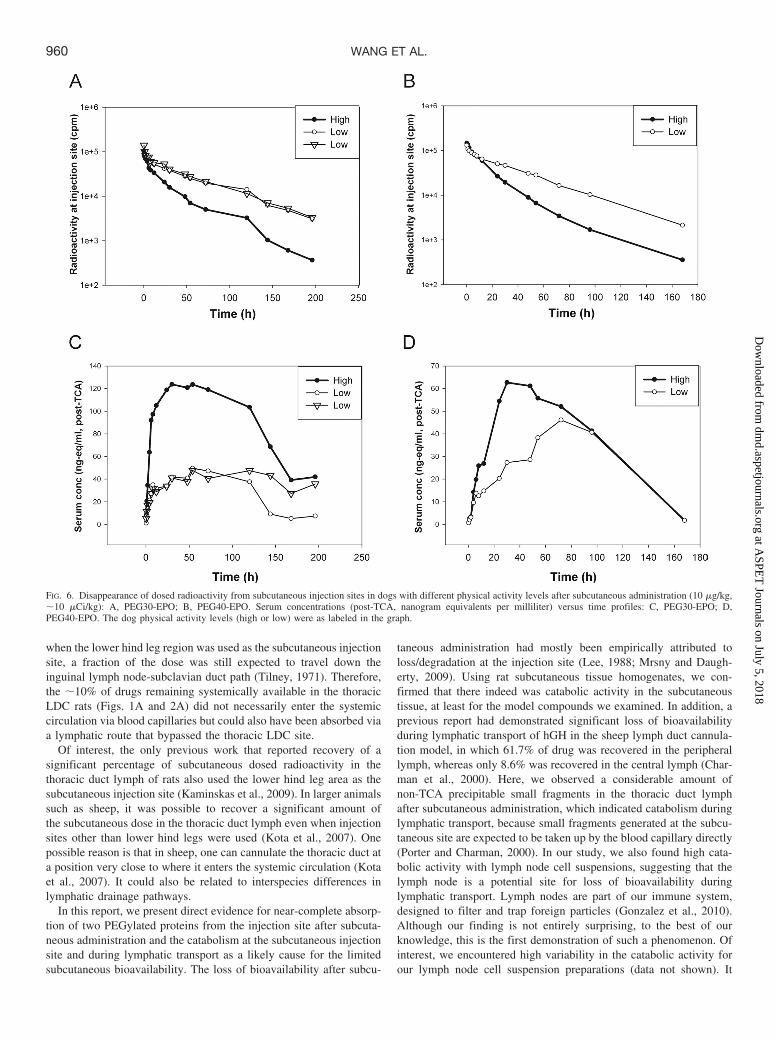
We also examined the impact of physical activity, another physio-logic factor pertinent to lymphatic transport, on subcutaneous absorp-tion in dogs, because physical activity is known to have a directimpact on the lymph flow rate (Swartz, 2001; Downey et al., 2008)and it is possible to identify dogs with apparent differences in physicalactivities (see Materials and Methods). Five dogs with different (highor low) physical activity levels were used in the study. They weredosed subcutaneously with 125I-labeled PEG30-EPO (1� high, 2�low) or PEG40-EPO (1� high, 1� low) �10 cm in front of the tails.This subcutaneous injection site was selected to facilitate monitoringof the disappearance of radioactivity from the injection site withminimal interference of signals from internal organs such as thethyroid. As shown in Fig. 6, A and B, dogs with higher levels ofphysical activity exhibited a faster disappearance of radioactivity fromthe injection site for both compounds and correspondingly signifi-cantly faster appearance of radioactivity in the systemic circulationand higher Cmax and serum exposure than the dogs with lowerphysical activity levels (Fig. 6, C and D). Even though we have yet toestablish an objective method to quantify the physical activity levels,
FIG. 4. Mean (SD) serum concentration (immunoassay, nanograms per milliliter)versus time profiles of PEG40-EPO after intravenous (n � 3) and SC (n � 6)administration in regular and fat rats.
958 WANG ET AL.
at ASPE
T Journals on July 5, 2018
dmd.aspetjournals.org
Dow
nloaded from

these results clearly demonstrated the impact of physical activity onsubcutaneous absorption in this species.
Discussion
Using the thoracic LDC models established in-house and PEGylatedEPOs as model compounds, we showed that these macromoleculeswere mostly absorbed via the lymphatic system after subcutaneousadministration in rats and dogs. The thoracic LDC animals exhibited�70% lower serum exposure than the control animals and up to 70%of the systemically available drugs was recovered in the lymph.Moreover, after subcutaneous administration, lymph exposure wasfound to precede systemic exposure in lymph duct-cannulated ani-mals, indicating that the radioactivity in the lymph during the ascend-ing phase mostly came from absorption not distribution. These resultsagreed well with previous reports in the sheep lymph duct cannulationmodel (Supersaxo et al., 1990; Porter and Charman, 2000; Porter etal., 2001). Taken together, our data and previous reports suggest thatthe role of the lymphatic system in subcutaneous absorption is prob-ably conserved in these preclinical species.
However, our results contradicted a few previous reports, whichsuggested minimal lymphatic contribution to subcutaneous absorptionin rats and rabbits (Bocci et al., 1986, 1988; Kojima et al., 1988;
Kagan et al., 2007). Although factors such as the success of lymphduct cannulation models or the selection of model compounds, e.g.,PEGylated versus non-PEGylated proteins, could have contributed tothe observed differences, one likely explanation for the discrepancy isthe choice of subcutaneous injection sites. In smaller-sized laboratoryanimals, the selection of the subcutaneous injection site is importantfor studying lymphatic contribution. Given the size of a rat, techni-cally it is only feasible to cannulate the thoracic duct at a site justabove the cisterna chyli (Ionac, 2003; Kagan et al., 2007; Kaminskaset al., 2009). The lymphatic drainage routes in adult laboratory ratswere mapped in detail by Tilney (1971). In contrast to the conven-tional wisdom, most of the commonly used subcutaneous injectionsites, including the sites used in those previous reports (thigh), drainedvia the brachial, inguinal, or axillary lymph nodes, which eventuallyenter the systemic circulation through the subclavian duct, bypassingthe thoracic LDC site. The only subcutaneous injection site that woulddrain mostly through the thoracic LDC site is the lower hind legregion (Tilney, 1971).
Using the lower hind leg area as a subcutaneous injection site, wefound that the cumulative recovery of dose in the thoracic duct lymphwas much higher than what had been reported previously, up to 28%of the dose or up to 70% of the systemically available dose. Even
FIG. 5. A, serum concentrations (nanogram equivalents per milliliter, post-TCA) versus time profiles of PEG40-EPO after subcutaneous administration in the thoracic LDC(n � 1) and control (n � 3, mean) dogs. B, serum and lymph concentrations (nanogram equivalents per milliliter, post-TCA) versus time profiles in the thoracic LDC dogafter subcutaneous administration. C, serum concentrations (nanogram equivalents per milliliter, post-TCA) versus time profiles of PEG40-EPO after intravenousadministration in the thoracic LDC (n � 1) and control (n � 3, mean) dogs. D, serum and lymph concentrations (nanogram equivalents per milliliter, post-TCA) versustime profiles in the thoracic LDC dog after intravenous administration.
959LYMPHATIC TRANSPORT AFTER SUBCUTANEOUS ADMINISTRATION
at ASPE
T Journals on July 5, 2018
dmd.aspetjournals.org
Dow
nloaded from
when the lower hind leg region was used as the subcutaneous injectionsite, a fraction of the dose was still expected to travel down theinguinal lymph node-subclavian duct path (Tilney, 1971). Therefore,the �10% of drugs remaining systemically available in the thoracicLDC rats (Figs. 1A and 2A) did not necessarily enter the systemiccirculation via blood capillaries but could also have been absorbed viaa lymphatic route that bypassed the thoracic LDC site.
Of interest, the only previous work that reported recovery of asignificant percentage of subcutaneous dosed radioactivity in thethoracic duct lymph of rats also used the lower hind leg area as thesubcutaneous injection site (Kaminskas et al., 2009). In larger animalssuch as sheep, it was possible to recover a significant amount ofthe subcutaneous dose in the thoracic duct lymph even when injectionsites other than lower hind legs were used (Kota et al., 2007). Onepossible reason is that in sheep, one can cannulate the thoracic duct ata position very close to where it enters the systemic circulation (Kotaet al., 2007). It could also be related to interspecies differences inlymphatic drainage pathways.
In this report, we present direct evidence for near-complete absorp-tion of two PEGylated proteins from the injection site after subcuta-neous administration and the catabolism at the subcutaneous injectionsite and during lymphatic transport as a likely cause for the limitedsubcutaneous bioavailability. The loss of bioavailability after subcu-
taneous administration had mostly been empirically attributed toloss/degradation at the injection site (Lee, 1988; Mrsny and Daugh-erty, 2009). Using rat subcutaneous tissue homogenates, we con-firmed that there indeed was catabolic activity in the subcutaneoustissue, at least for the model compounds we examined. In addition, aprevious report had demonstrated significant loss of bioavailabilityduring lymphatic transport of hGH in the sheep lymph duct cannula-tion model, in which 61.7% of drug was recovered in the peripherallymph, whereas only 8.6% was recovered in the central lymph (Char-man et al., 2000). Here, we observed a considerable amount ofnon-TCA precipitable small fragments in the thoracic duct lymphafter subcutaneous administration, which indicated catabolism duringlymphatic transport, because small fragments generated at the subcu-taneous site are expected to be taken up by the blood capillary directly(Porter and Charman, 2000). In our study, we also found high cata-bolic activity with lymph node cell suspensions, suggesting that thelymph node is a potential site for loss of bioavailability duringlymphatic transport. Lymph nodes are part of our immune system,designed to filter and trap foreign particles (Gonzalez et al., 2010).Although our finding is not entirely surprising, to the best of ourknowledge, this is the first demonstration of such a phenomenon. Ofinterest, we encountered high variability in the catabolic activity forour lymph node cell suspension preparations (data not shown). It
FIG. 6. Disappearance of dosed radioactivity from subcutaneous injection sites in dogs with different physical activity levels after subcutaneous administration (10 �g/kg,�10 �Ci/kg): A, PEG30-EPO; B, PEG40-EPO. Serum concentrations (post-TCA, nanogram equivalents per milliliter) versus time profiles: C, PEG30-EPO; D,PEG40-EPO. The dog physical activity levels (high or low) were as labeled in the graph.
960 WANG ET AL.
at ASPE
T Journals on July 5, 2018
dmd.aspetjournals.org
Dow
nloaded from

remains to be determined whether this was due to the technicalvariability or intrinsic variability of the catabolic activities of lymphnodes.
The differences we observed between PEG30-EPO and PEG40-EPO are interesting, because they suggested that relatively minordifferences in molecular characteristics such as glycosylation or thesize of PEG, may have an impact on subcutaneous absorption/stabil-ity. However, the differences observed here are relatively small andwe have no reason to believe that they would be clinically important.
Many physiologic factors such as age, body weight, and injectionsites have been reported as covariates for subcutaneous bioavailabilityin humans (Macdougall et al., 1991; Chan et al., 2003; Silva et al.,2006; Fishbane et al., 2007; Olsson-Gisleskog et al., 2007; Kakkar etal., 2011). In this investigation, we show that physiologic factors suchas fat content and physical activity can have a profound impact onsubcutaneous absorption of PEGylated proteins in preclinical species.The impacts we observed were consistent with their expected impacton the lymphatic transport; i.e., factors associated with faster lym-phatic transport are associated with faster absorption and higherbioavailability and vice versa. In addition to the results presented here,we also found that factors such as dose, dosing volume/dosing con-centration, and injection site, all could affect subcutaneous absorptionof a macromolecule (data not shown). For all the covariates ofsubcutaneous absorption we evaluated, a common theme is that fasterabsorption usually leads to higher bioavailability. A recent study onsubcutaneous absorption of rituximab in rats reported similar findings(Kagan et al., 2012). Although a slower absorption itself should not bethe reason for lower bioavailability, it is conceivable that the rate oftransport from the injection site and then through the lymphaticsystem determines the extent of catabolism before systemic exposure.For the purpose of comparing bioavailability, these results illustratedthe importance of controlling these potential covariants of subcutane-ous absorption in preclinical and/or clinical studies, because themagnitude of their impact can be significant. Given our observations,we should also actively seek preclinical models more reflective of theintended clinical settings to better assess the impact of a particularphysiologic factor for the compound of interest.
Human PK of biologic drugs after intravenous administration canusually be predicted with reasonable confidence using allometricscaling (Mordenti et al., 1991; Mahmood, 2004; Wang and Prueksari-tanont, 2010), but it is much more challenging to predict their sub-cutaneous absorption process in humans. There are fundamental dif-ferences in subcutaneous tissue structure between preclinical speciesand humans (Magnusson et al., 2001). Therefore, prediction of humanPK after subcutaneous administration probably will rely on mecha-nistic understanding of the subcutaneous absorption process ratherthan on empirical scaling methods. For example, in vitro tools such asthe subcutaneous tissue homogenate and lymph node cell suspensionsshould be very useful in assessing catabolic activity that could play amajor role in determining bioavailability of a given macromoleculecompound. These systems cannot necessarily be used to quantitativelyassess bioavailability but rather can be used for comparison purposesand rank-ordering of compounds. They are also one way to addresspotential interspecies differences in bioavailability associated withinterspecies difference in catabolic stability. Of interest, our resultssuggest that the impact of physiologic factors on subcutaneous ab-sorption can be compound-dependent; i.e., the intrinsic “stability” ofa molecule can affect its sensitivity to such physiologic features. Webelieve that an appropriate use of preclinical models and in vitro toolsshould facilitate mechanistic understanding and aid human PK pre-diction of bioavailability and potential variability. In combinationwith conventional PK studies in preclinical species, the tools pre-
sented here can provide useful information for candidate selection,human PK prediction, clinical study design, and risk mitigation forgiven biologic drug candidates.
Acknowledgments
We thank Lorraine Lipfert for excellent technical assistance in the prepa-ration of 125I-labeled compounds, Carmen Fernandez-Metzler for reviewingthe manuscript, Marissa Vavrek for coordination of in vivo studies, andHuijuan Li for providing PEG40-EPO.
Authorship Contributions
Participated in research design: W. Wang, Chen, Liu, Hamuro, andPrueksaritanont.
Conducted experiments: Chen, Shen, Cunningham, Fauty, Michel, B.Wang, Hong, Adreani, Nunes, Johnson, and Zou.
Contributed new reagents or analytic tools: Chen, Yin, and Groff.Performed data analysis: W. Wang and Chen.Wrote or contributed to the writing of the manuscript: W. Wang and
Prueksaritanont.
References
Bensadoun A and Weinstein D (1976) Assay of proteins in the presence of interfering materials.Anal Biochem 70:241–250.
Bocci V, Muscettola M, Grasso G, Magyar Z, Naldini A, and Szabo G (1986) The lymphaticroute. 1) Albumin and hyaluronidase modify the normal distribution of interferon in lymph andplasma. Experientia 42:432–433.
Bocci V, Pessina GP, Paulesu L, and Nicoletti C (1988) The lymphatic route. VI. Distribution ofrecombinant interferon-�2 in rabbit and pig plasma and lymph. J Biol Response Mod7:390–400.
Chan CC, Ng EH, Chan MM, Tang OS, Lau EY, Yeung WS, and Ho PC (2003) Bioavailabilityof hCG after intramuscular or subcutaneous injection in obese and non-obese women. HumReprod 18:2294–2297.
Charman SA, Segrave AM, Edwards GA, and Porter CJ (2000) Systemic availability andlymphatic transport of human growth hormone administered by subcutaneous injection.J Pharm Sci 89:168–177.
Davis CB and Bugelski PJ (1998) Subcutaneous bioavailability of a PRIMATIZED IgG1anti-human CD4 monoclonal antibody is dose dependent in transgenic mice bearing humanCD4. Drug Deliv 5:95–100.
Downey HF, Durgam P, Williams AG Jr, Rajmane A, King HH, and Stoll ST (2008) Lymph flowin the thoracic duct of conscious dogs during lymphatic pump treatment, exercise, andexpansion of extracellular fluid volume. Lymphat Res Biol 6:3–13.
Fishbane S, Pannier A, Liogier X, Jordan P, Dougherty FC, and Reigner B (2007) Pharmaco-kinetic and pharmacodynamic properties of methoxy polyethylene glycol-epoetin beta areunaffected by the site of subcutaneous administration. J Clin Pharmacol 47:1390–1397.
Gonzalez SF, Kuligowski MP, Pitcher LA, Roozendaal R, and Carroll MC (2010) The role ofinnate immunity in B cell acquisition of antigen within LNs. Adv Immunol 106:1–19.
Ionac M (2003) One technique, two approaches, and results: thoracic duct cannulation in smalllaboratory animals. Microsurgery 23:239–245.
Kagan L, Gershkovich P, Mendelman A, Amsili S, Ezov N, and Hoffman A (2007) The role ofthe lymphatic system in subcutaneous absorption of macromolecules in the rat model. EurJ Pharm Biopharm 67:759–765.
Kagan L, Turner MR, Balu-Iyer SV, and Mager DE (2012) Subcutaneous absorption ofmonoclonal antibodies: role of dose, site of injection, and injection volume on rituximabpharmacokinetics in rats. Pharm Res 29:490–499.
Kakkar T, Sung C, Gibiansky L, Vu T, Narayanan A, Lin SL, Vincent M, Banfield C, ColbertA, Hoofring S, et al. (2011) Population PK and IgE pharmacodynamic analysis of a fullyhuman monoclonal antibody against IL4 receptor. Pharm Res 28:2530–2542.
Kaminskas LM, Kota J, McLeod VM, Kelly BD, Karellas P, and Porter CJ (2009) PEGylationof polylysine dendrimers improves absorption and lymphatic targeting following SC admin-istration in rats. J Control Release 140:108–116.
Kojima K, Takahashi T, and Nakanishi Y (1988) Lymphatic transport of recombinant humantumor necrosis factor in rats. J Pharmacobiodyn 11:700–706.
Kota J, Machavaram KK, McLennan DN, Edwards GA, Porter CJ, and Charman SA (2007)Lymphatic absorption of subcutaneously administered proteins: influence of different injectionsites on the absorption of darbepoetin alfa using a sheep model. Drug Metab Dispos 35:2211–2217.
Lee DS and Hashim SA (1966) A new catheter system for cannulation of thoracic duct of the rat.J Appl Physiol 21:1887–1888.
Lee VH (1988) Enzymatic barriers to peptide and protein absorption. Crit Rev Ther Drug CarrierSyst 5:69–97.
Lin JH (2009) Pharmacokinetics of biotech drugs: peptides, proteins and monoclonal antibodies.Curr Drug Metab 10:661–691.
Mahmood I (2004) Interspecies scaling of protein drugs: prediction of clearance from animals tohumans. J Pharm Sci 93:177–185.
Macdougall IC (2005) CERA (continuous erythropoietin receptor activator): a new erythropoi-esis-stimulating agent for the treatment of anemia. Curr Hematol Rep 4:436–440.
Macdougall IC, Jones JM, Robinson MI, Miles JB, Coles GA, and Williams JD (1991)Subcutaneous erythropoietin therapy: comparison of three different sites of injection. ContribNephrol 88:152–158.
Magnusson BM, Walters KA, and Roberts MS (2001) Veterinary drug delivery: potential for skinpenetration enhancement. Adv Drug Deliv Rev 50:205–227.
961LYMPHATIC TRANSPORT AFTER SUBCUTANEOUS ADMINISTRATION
at ASPE
T Journals on July 5, 2018
dmd.aspetjournals.org
Dow
nloaded from

McDonald TA, Zepeda ML, Tomlinson MJ, Bee WH, and Ivens IA (2010) Subcutaneous administrationof biotherapeutics: current experience in animal models. Curr Opin Mol Ther 12:461–470.
McLennan DN, Porter CJ, Edwards GA, Heatherington AC, Martin SW, and Charman SA (2006)The absorption of darbepoetin alfa occurs predominantly via the lymphatics following sub-cutaneous administration to sheep. Pharm Res 23:2060–2066.
Mordenti J, Chen SA, Moore JA, Ferraiolo BL, and Green JD (1991) Interspecies scaling of clearance andvolume of distribution data for five therapeutic proteins. Pharm Res 8:1351–1359.
Mrsny RJ and Daugherty AL (2009) Proteins and Peptides: Pharmacokinetic, Pharmacody-namic, and Metabolic Outcomes, pp 80–105, Informa Healthcare, New York.
Nett JH, Gomathinayagam S, Hamilton SR, Gong B, Davidson RC, Du M, Hopkins D, MitchellT, Mallem MR, Nylen A, et al. (2012) Optimization of erythropoietin production withcontrolled glycosylation-PEGylated erythropoietin produced in glycoengineered Pichia pas-toris. J Biotechnol 157:198–206.
Olsson-Gisleskog P, Jacqmin P, and Perez-Ruixo JJ (2007) Population pharmacokinetics meta-analysis ofrecombinant human erythropoietin in healthy subjects. Clin Pharmacokinet 46:159–173.
Porter CJ and Charman SA (2000) Lymphatic transport of proteins after subcutaneous admin-istration. J Pharm Sci 89:297–310.
Porter CJ, Edwards GA, and Charman SA (2001) Lymphatic transport of proteins after s.c.injection: implications of animal model selection. Adv Drug Deliv Rev 50:157–171.
Ryan TJ (1995) Lymphatics and adipose tissue. Clin Dermatol 13:493–498.Schreihofer AM, Hair CD, and Stepp DW (2005) Reduced plasma volume and mesenteric vascular
reactivity in obese Zucker rats. Am J Physiol Regul Integr Comp Physiol 288:R253–R261.Silva M, Poo J, Wagner F, Jackson M, Cutler D, Grace M, Bordens R, Cullen C, Harvey J, and
Laughlin M (2006) A randomised trial to compare the pharmacokinetic, pharmacodynamic,and antiviral effects of peginterferon alfa-2b and peginterferon alfa-2a in patients with chronichepatitis C (COMPARE). J Hepatol 45:204–213.
Supersaxo A, Hein WR, and Steffen H (1990) Effect of molecular weight on the lymphaticabsorption of water-soluble compounds following subcutaneous administration. Pharm Res7:167–169.
Swartz MA (2001) The physiology of the lymphatic system. Adv Drug Deliv Rev 50:3–20.Tang L, Persky AM, Hochhaus G, and Meibohm B (2004) Pharmacokinetic aspects of biotech-
nology products. J Pharm Sci 93:2184–2204.Tilney NL (1971) Patterns of lymphatic drainage in the adult laboratory rat. J Anat 109:369–383.Vugmeyster Y, DeFranco D, Szklut P, Wang Q, and Xu X (2010) Biodistribution of [125I]-
labeled therapeutic proteins: application in protein drug development beyond oncology.J Pharm Sci 99:1028–1045.
Wang W and Prueksaritanont T (2010) Prediction of human clearance of therapeutic proteins:simple allometric scaling method revisited. Biopharm Drug Dispos 31:253–263.
Address correspondence to: Dr. Thomayant Prueksaritanont, Departmentof Pharmacokinetics, Pharmacodynamics and Drug Metabolism, WP75A-203,Merck Sharp and Dohme Corp., West Point, PA 19486. E-mail: [email protected]
962 WANG ET AL.
at ASPE
T Journals on July 5, 2018
dmd.aspetjournals.org
Dow
nloaded from