DMD 40:2041–2053, 2012 In Vitro Studies on the...
13
In Vitro Studies on the Oxidative Metabolism of 20(S)-Ginsenoside Rh2 in Human, Monkey, Dog, Rat, and Mouse Liver Microsomes, and Human Liver S9 □ S Liang Li, Xiaoyan Chen, Jialan Zhou, and Dafang Zhong Shanghai Institute of Materia Medica, Chinese Academy of Sciences, Shanghai, China Received May 30, 2012; accepted July 24, 2012 ABSTRACT: 20(S)-Ginsenoside Rh2 (Rh2)-containing products are widely used in Asia, Europe, and North America. However, extremely limited metabolism information greatly impedes the complete under- standing of its clinical safety and effectiveness. The present study aims to systematically investigate the oxidative metabolism of Rh2 using a complementary set of in vitro models. Twenty-five oxida- tive metabolites were found using liquid chromatography-electro- spray ionization ion-trap mass spectrometry. Six metabolites and a metabolic intermediate were synthesized. The metabolites were structurally identified as 26-hydroxy Rh2 (M1-1), (20S,24S)-epoxy- dammarane-12,25-diol-3--D-glucopyranoside (M1-3), (20S,24R)- epoxydammarane-12,25-diol-3--D-glucopyranoside (M1-5), 26,27-dihydroxy Rh2 (M3-6), (20S,24S)-epoxydammarane-12,25,26- triol-3--D-glucopyranoside (M3-10), (20S,24R)-epoxydammarane- 12,25,26-triol-3--D-glucopyranoside (M3-11), and 26-aldehyde Rh2 on the basis of detailed mass spectrometry and nuclear mag- netic resonance data analysis. Double-bond epoxidation followed by rearrangement and vinyl-methyl group hydroxylation represent the initial metabolic pathways generating monooxygenated metab- olites M1-1 to M1-5. Further sequential metabolites (M2–M5) from the dehydrogenation and/or oxygenation of M1 were also de- tected. CYP3A4 was the predominant enzyme involved in the oxi- dative metabolism of Rh2, whereas alcohol dehydrogenase and aldehyde dehydrogenase mainly catalyzed the metabolic conver- sion of alcohol to the corresponding carboxylic acid. No significant differences were observed in the phase I metabolite profiles of Rh2 among the five species tested. Reactive epoxide metabolite for- mation in both humans and animals was evident. However, GSH conjugate M6 was detected only in cynomolgus monkey liver mi- crosomal incubations. In conclusion, Rh2 is a good substrate for CYP3A4 and could undergo extensive oxidative metabolism under the catalysis of CYP3A4. Introduction 20(S)-Ginsenoside Rh2 (Rh2) (Fig. 1), a trace constituent of red ginseng, was first reported by Kitagawa et al. (1983). Structurally, Rh2 is a dammarane-type triterpenoid saponin formed by a glucose sugar moiety glycosidically bonded to the hydroxy group at the C-3 position of 20(S)-protopanaxadiol (PPD). A number of studies have shown that Rh2 exhibits excellent cancer prevention effects, espe- cially the induction of apoptosis (Park et al., 1997; Ham et al., 2003; Cheng et al., 2005). In addition, Rh2 has been reported to exert synergetic effects on some chemotherapeutic agents even at a nonef- fectual dose (Kikuchi et al., 1991; Jia et al., 2004; Wang et al., 2006; Xie et al., 2006). The synergistic mechanisms of Rh2 have yet to be fully characterized. The modulation of efflux transporter activity related to multidrug resistance [e.g., P-glycoprotein (P-gp) and breast cancer resistance protein] has been considered a possible factor (Jin et al., 2006; Zhang et al., 2010). However, metabolism information on Rh2 is scant and mostly limited to its degradation in the gastrointestinal tract to form an aglycone. Various Rh2- containing products are sold over-the-counter in Asian, European, and North American markets. Jinxing capsule, a healthcare product, was approved by the Chinese State Food and Drug Administration for use in neoadjuvant therapy in 2006. A recommended oral dose of 81 mg/day for Rh2 (40.5 mg/time b.i.d.), the main active ingredient, is indicated in the package insert (Asia Pharmaceutical Group, Haikou, China). Jinxing capsule is claimed to improve immune function and increase disease resistance for patients undergoing treatment and rehabilitation for many kinds of cancer. During the preclinical evaluation (D. Zhong, unpublished data), we found that Rh2 could be absorbed moderately with an oral bioavailability of 28 (25 mg/kg) in Wistar rats and 55% (8 mg/kg) in beagles. After an oral administration of 50 mg/kg, the peak Rh2 This work was supported by the National Science and Technology Major Project “Key New Drug Creation and Manufacturing Program,” China [Grant 2009ZX09301-001]. Article, publication date, and citation information can be found at http://dmd.aspetjournals.org. http://dx.doi.org/10.1124/dmd.112.046995. □ S The online version of this article (available at http://dmd.aspetjournals.org) contains supplemental material. ABBREVIATIONS: Rh2, 20(S)-ginsenoside Rh2; PPD, 20(S)-protopanaxadiol; P-gp, P-glycoprotein; P450, cytochrome P450; HLM, human liver microsomes; CyLM, cynomolgus monkey liver microsomes; DLM, dog liver microsomes; RLM, rat liver microsomes; MLM, mouse liver microsomes; GEE, GSH ethyl ester; NAC, N-acetyl-L-cysteine; HPLC, high-performance liquid chromatography; m-CPBA, 3-chloroperoxybenzoic acid; LC, liquid chromatography; MS n , ion-trap mass spectrometry; MSD, mass selective detector; MS, mass spectrometry; NMR, nuclear magnetic resonance; GST, glutathione transferase. 1521-009X/12/4010-2041–2053$25.00 DRUG METABOLISM AND DISPOSITION Vol. 40, No. 10 Copyright © 2012 by The American Society for Pharmacology and Experimental Therapeutics 46995/3797571 DMD 40:2041–2053, 2012 2041 http://dmd.aspetjournals.org/content/suppl/2012/07/24/dmd.112.046995.DC1 Supplemental material to this article can be found at: at ASPET Journals on June 15, 2018 dmd.aspetjournals.org Downloaded from
Transcript of DMD 40:2041–2053, 2012 In Vitro Studies on the...
In Vitro Studies on the Oxidative Metabolism of 20(S)-GinsenosideRh2 in Human, Monkey, Dog, Rat, and Mouse Liver Microsomes,
and Human Liver S9□S
Liang Li, Xiaoyan Chen, Jialan Zhou, and Dafang Zhong
Shanghai Institute of Materia Medica, Chinese Academy of Sciences, Shanghai, China
Received May 30, 2012; accepted July 24, 2012
ABSTRACT:
20(S)-Ginsenoside Rh2 (Rh2)-containing products are widely usedin Asia, Europe, and North America. However, extremely limitedmetabolism information greatly impedes the complete under-standing of its clinical safety and effectiveness. The present studyaims to systematically investigate the oxidative metabolism of Rh2using a complementary set of in vitro models. Twenty-five oxida-tive metabolites were found using liquid chromatography-electro-spray ionization ion-trap mass spectrometry. Six metabolites and ametabolic intermediate were synthesized. The metabolites werestructurally identified as 26-hydroxy Rh2 (M1-1), (20S,24S)-epoxy-dammarane-12,25-diol-3-�-D-glucopyranoside (M1-3), (20S,24R)-epoxydammarane-12,25-diol-3-�-D-glucopyranoside (M1-5),26,27-dihydroxy Rh2 (M3-6), (20S,24S)-epoxydammarane-12,25,26-triol-3-�-D-glucopyranoside (M3-10), (20S,24R)-epoxydammarane-12,25,26-triol-3-�-D-glucopyranoside (M3-11), and 26-aldehydeRh2 on the basis of detailed mass spectrometry and nuclear mag-
netic resonance data analysis. Double-bond epoxidation followedby rearrangement and vinyl-methyl group hydroxylation representthe initial metabolic pathways generating monooxygenated metab-olites M1-1 to M1-5. Further sequential metabolites (M2–M5) fromthe dehydrogenation and/or oxygenation of M1 were also de-tected. CYP3A4 was the predominant enzyme involved in the oxi-dative metabolism of Rh2, whereas alcohol dehydrogenase andaldehyde dehydrogenase mainly catalyzed the metabolic conver-sion of alcohol to the corresponding carboxylic acid. No significantdifferences were observed in the phase I metabolite profiles of Rh2among the five species tested. Reactive epoxide metabolite for-mation in both humans and animals was evident. However, GSHconjugate M6 was detected only in cynomolgus monkey liver mi-crosomal incubations. In conclusion, Rh2 is a good substrate forCYP3A4 and could undergo extensive oxidative metabolism underthe catalysis of CYP3A4.
Introduction
20(S)-Ginsenoside Rh2 (Rh2) (Fig. 1), a trace constituent of redginseng, was first reported by Kitagawa et al. (1983). Structurally,Rh2 is a dammarane-type triterpenoid saponin formed by a glucosesugar moiety glycosidically bonded to the hydroxy group at the C-3position of 20(S)-protopanaxadiol (PPD). A number of studies haveshown that Rh2 exhibits excellent cancer prevention effects, espe-cially the induction of apoptosis (Park et al., 1997; Ham et al., 2003;Cheng et al., 2005). In addition, Rh2 has been reported to exertsynergetic effects on some chemotherapeutic agents even at a nonef-fectual dose (Kikuchi et al., 1991; Jia et al., 2004; Wang et al., 2006;
Xie et al., 2006). The synergistic mechanisms of Rh2 have yet to befully characterized. The modulation of efflux transporter activityrelated to multidrug resistance [e.g., P-glycoprotein (P-gp) andbreast cancer resistance protein] has been considered a possiblefactor (Jin et al., 2006; Zhang et al., 2010). However, metabolisminformation on Rh2 is scant and mostly limited to its degradationin the gastrointestinal tract to form an aglycone. Various Rh2-containing products are sold over-the-counter in Asian, European,and North American markets.
Jinxing capsule, a healthcare product, was approved by the ChineseState Food and Drug Administration for use in neoadjuvant therapy in2006. A recommended oral dose of 81 mg/day for Rh2 (40.5 mg/timeb.i.d.), the main active ingredient, is indicated in the package insert(Asia Pharmaceutical Group, Haikou, China). Jinxing capsule isclaimed to improve immune function and increase disease resistancefor patients undergoing treatment and rehabilitation for many kinds ofcancer. During the preclinical evaluation (D. Zhong, unpublisheddata), we found that Rh2 could be absorbed moderately with an oralbioavailability of 28 (25 mg/kg) in Wistar rats and 55% (8 mg/kg) inbeagles. After an oral administration of 50 mg/kg, the peak Rh2
This work was supported by the National Science and Technology MajorProject “Key New Drug Creation and Manufacturing Program,” China [Grant2009ZX09301-001].
Article, publication date, and citation information can be found athttp://dmd.aspetjournals.org.
http://dx.doi.org/10.1124/dmd.112.046995.□S The online version of this article (available at http://dmd.aspetjournals.org)
contains supplemental material.
ABBREVIATIONS: Rh2, 20(S)-ginsenoside Rh2; PPD, 20(S)-protopanaxadiol; P-gp, P-glycoprotein; P450, cytochrome P450; HLM, human livermicrosomes; CyLM, cynomolgus monkey liver microsomes; DLM, dog liver microsomes; RLM, rat liver microsomes; MLM, mouse livermicrosomes; GEE, GSH ethyl ester; NAC, N-acetyl-L-cysteine; HPLC, high-performance liquid chromatography; m-CPBA, 3-chloroperoxybenzoicacid; LC, liquid chromatography; MSn, ion-trap mass spectrometry; MSD, mass selective detector; MS, mass spectrometry; NMR, nuclearmagnetic resonance; GST, glutathione transferase.
1521-009X/12/4010-2041–2053$25.00DRUG METABOLISM AND DISPOSITION Vol. 40, No. 10Copyright © 2012 by The American Society for Pharmacology and Experimental Therapeutics 46995/3797571DMD 40:2041–2053, 2012
2041
http://dmd.aspetjournals.org/content/suppl/2012/07/24/dmd.112.046995.DC1Supplemental material to this article can be found at:
at ASPE
T Journals on June 15, 2018
dmd.aspetjournals.org
Dow
nloaded from
concentration (60 �M) in rat liver was approximately 20 times higherthan that in plasma. No Rh2-related material was detected in rat urine,and minimal Rh2 (less than 10%) was detected in rat feces and bile,which imply that extensive metabolism followed by secretion into bileis the major route of Rh2 excretion.
Rh2 metabolism in rats after a single oral administration (50 mg/kg)was subsequently studied in our laboratory (Deng et al., 2009). Majormetabolic pathways observed included deglycosylation, oxygenation,and both sulfate and glucuronate conjugation. Small amounts of PPDwere detected in rat plasma, whereas monooxygenated, dioxygenated,and carboxylated derivatives of both Rh2 and PPD represented themajor circulating metabolites. A GSH conjugate derived from mo-nooxygenated Rh2 was determined as one of the major metabolites inrat bile. Cysteine adducts of both Rh2 and PPD were also detected inrat feces. Thus, some chemically reactive intermediates were formedin vivo. To the best of our knowledge, the metabolites of Rh2 inhumans have not been reported to date.
Chemically reactive metabolite formation is an unwanted featureof any drug or drug candidate during discovery and development.Therefore, substantially decreasing or eliminating the metabolicactivation liability of drug candidates has become the ultimate goal
of researchers in the field of drug metabolism and toxicology(Kumar et al., 2008). Detecting and characterizing reactive metab-olites with appropriate trapping agents in both experimental ani-mals and humans are highly important initial steps so that informedevaluation of hidden risks can be made in a timely manner (Evansand Baillie, 2005).
A range of rodent (mice and rats) and nonrodent (dogs and monkeys)animal species are routinely used for preclinical metabolism and toxicol-ogy tests in many pharmaceutical companies to evaluate the absorption,distribution, metabolism, excretion, and toxicology profiles of new can-didate drugs. Nevertheless, given the differences in isoform composition,expression, and catalytic activities of drug-metabolizing enzymes acrossspecies, extensive differences in drug metabolism and toxicity betweenanimals and humans have been found. In this context, choosing the mostrelevant animal species on which to conduct studies and extrapolating theresults to humans are huge challenges for researchers (Martignoni et al.,2006; Baillie and Rettie, 2011).
The objectives of the present study were as follows: 1) to charac-terize the structures of oxidative metabolites, including reactive inter-mediates of Rh2 and 2) to identify the cytochrome P450 (P450) andnon-P450 enzymes responsible for Rh2 biotransformation.
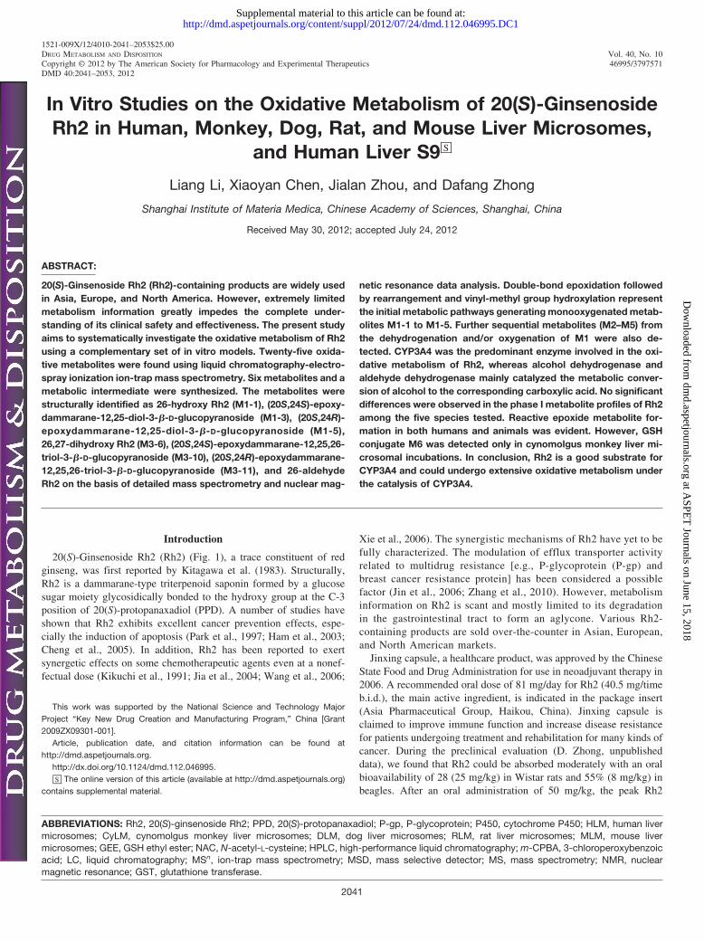
FIG. 1. Chemical structure. Full-scan mass spectrum (A) and MS2
spectrum (B) of Rh2. MS2 data were obtained from the 587.5 m/zion as the precursor for collision-induced dissociation. Intens.,intensity.
2042 LI ET AL.
at ASPE
T Journals on June 15, 2018
dmd.aspetjournals.org
Dow
nloaded from
Materials and Methods
Materials. 20(S)-Ginsenoside Rh2 (purity �98%) was purchased fromShanghai Huayi Bio Technology Co., Ltd. (Shanghai, China). Pooled humanliver microsomes (HLM), S9, and cytosol (mixed-gender pool of 50 donors),male cynomolgus monkey liver microsomes (CyLM), male beagles [dog livermicrosomes (DLM)], male Sprague-Dawley rats [rat liver microsomes(RLM)], and male CD-1 mice [mouse liver microsomes (MLM)], as well asrecombinant P450 enzymes CYP1A2, CYP1B1, CYP2A6, CYP2B6,CYP2C8, CYP2C9, CYP2C19, CYP2D6, CYP2E1, CYP3A4, CYP3A5, andCYP4A11 were purchased from BD Gentest (Woburn, MA). NADPH, NAD�,GSH, GSH ethyl ester (GEE), N-acetyl-L-cysteine (NAC), 4-methylpyrazolehydrochloride, raloxifene, and disulfiram were purchased from Sigma-Aldrich(St. Louis, MO). All solvents used for high-performance liquid chromatogra-phy (HPLC) were of HPLC grade (Merck, Darmstadt, Germany). 3-Chloroper-oxybenzoic acid (m-CPBA) was purchased from Alfa Aesar China (Tianjin)Co., Ltd. (Beijing, China). Analytical-grade dichloromethane and methanol,selenium dioxide (SeO2), tert-butyl hydroperoxide, and salicylic acid werepurchased from Sinopharm Chemical Reagent Co., Ltd. (Shanghai, China).Purified water was generated with a Milli-Q Gradient system (MilliporeCorporation, Molsheim, France). C18 reversed-phase silica gel (150–200 mesh;Merck) was used for column chromatography. Deuterium methanol and pyridinewere obtained from Cambridge Isotope Laboratories, Inc. (Andover, MA).
Microsomal and S9 Fraction Incubations of Rh2 and Isolated Metab-olites. Stock solutions of Rh2 (3, 10, and 50 mM) were prepared in methanol.The final methanol concentration in the incubation was 0.1% (v/v). The livermicrosomes and S9 fraction were carefully thawed on ice before the experi-ment. NADPH (2 mM) was added into open-to-air polyethylene tubes heatedin a water-bath shaker at 37°C containing 10 �M Rh2 solution in 100 mMpotassium phosphate buffer (pH 7.4) with or without GSH, GEE, or NAC at afinal concentration of 2 mM. The total incubation volume was 200 �l. After 3min preincubation at 37°C, the incubation reactions were initiated with theaddition of microsome proteins (1 mg/ml). After undergoing incubation for 60min, the reactions were terminated with an equal volume of ice-cold acetoni-trile. Control samples without NADPH were included. Each incubation wasperformed in duplicate. Individual incubations of Rh2 (3 and 50 �M) and sixisolated metabolites (M1-1, M1-3, M1-5, M3-6, M3-10, and M3-11; 10 �M)in HLM and M1-1 (10 �M) in human liver S9 fraction were performed underthe same incubation conditions described above.
Microsomal Incubations in the Presence of Inhibitors. The specific P450enzymes involved in the oxidative metabolism of Rh2 were determined bytesting six chemical inhibitors as follows (concentration and enzyme in paren-thesis): �-naphthoflavone (0.1 �M CYP1A2), sulfaphenazole (1.0 �MCYP2C9), ticlopidine (0.4 �M CYP2B6 and 6.0 �M CYP2C19), quinidine(2.0 �M CYP2D6), chlormethiazole (60 �M CYP2E1), and ketoconazole (1.0and 10.0 �M CYP3A). The inhibitor concentrations used were referred fromthe U.S. Food and Drug Administration Guidance for Industry on DrugInteraction Studies (http://www.fda.gov/downloads/Drugs/GuidanceCompli-anceRegulatoryInformation/ Guidances/UCM072101.pdf, 2006). Under theexperimental conditions, ketoconazole shows inhibitory activities againstmidazolam (5 �M) 1-hydroxylation and testosterone (50 �M) 6�-hydroxyla-tion with IC50 values of 0.044 and 0.026 �M, respectively. The production ofM1-2, M1-3, and M1-4 � M1-5 was monitored and quantified by liquidchromatography (LC)/ion-trap mass spectrometry (MSn). The incubation mix-tures included 1 mg/ml pooled HLM, 10 �M Rh2, 2 mM NADPH, and theinhibitors at various concentrations in 100 mM potassium phosphate buffer(pH 7.4). The final incubation volume was 200 �l. Incubations were performedat 37°C for 60 min, and reactions were terminated with an equal volume ofice-cold acetonitrile. Controls without chemical inhibitors were included. In aseparate study, ketoconazole (1.0 �M) was incubated with CyLM (1 mg/ml),GSH (2 mM), and Rh2 (10 �M) for 10 min before the addition of NADPH(2 mM). Each incubation was performed in duplicate.
Recombinant Enzyme Incubations. Rh2 (10 �M) was incubated in du-plicate at 37°C for 60 min with a panel of recombinant human P450 enzymes(CYP1A2, CYP1B1, CYP2A6, CYP2B6, CYP2C8, CYP2C9, CYP2C19,CYP2D6, CYP2E1, CYP3A4, CYP3A5, and CYP4A11) at 50 nM. In aseparate study, 10 �M M1-1 was incubated with CYP3A4. The incubation
conditions followed the microsomal incubations described under Microsomaland S9 Fraction Incubations of Rh2 and Isolated Metabolites.
Pooled Human Liver Cytosol Incubations of M1-1 in the Presence ofInhibitors. The involvement of cytosol enzymes in the conversion of alcoholmetabolite M1-1 to the corresponding acid M2-3 was investigated by incubat-ing M1-1 and the aldehyde metabolite with pooled human liver cytosol withone of the following chemical inhibitors (concentration and enzyme in paren-thesis): 4-methylpyrazole (50 �M alcohol dehydrogenase; Deng et al., 2011),raloxifene (100 nM aldehyde oxidase; Obach, 2004), and disulfiram (50 �Maldehyde dehydrogenase; Lam et al., 1997). The inhibitors were dissolved indimethyl sulfoxide, and the final concentration of dimethyl sulfoxide in theincubation was 0.1% (v/v). The incubation mixtures contained 1 mg/ml humanliver cytosol, 2 mM NAD�, and the inhibitors at various concentrations in 100mM potassium phosphate buffer (pH 7.4). The final incubation volume was200 �l. After 15 min of preincubation at 37°C, the reactions were initiated withthe addition of 10 �M M1-1 (or 26-aldehyde-Rh2). The reactions wereterminated with an equal volume of ice-cold acetonitrile after 60 min ofincubation. Controls without chemical inhibitors were included. Each incuba-tion was performed in duplicate.
Sample Preparation for LC/MSn Analysis. All the in vitro incubationsamples were prepared using the same methods. Methanol (200 �l) was addedto a 200-�l aliquot of the in vitro incubations. This sample was vortex-mixedand centrifuged at 14,000g for 5 min. The supernatant was transferred into aglass tube, evaporated to dryness under a stream of nitrogen at 40°C, and thenreconstituted in 100 �l of 0.1% formic acid in methanol with 0.1% formic acidin 5 mM ammonium acetate (70:30, v/v). A 20-�l aliquot of the reconstitutedsolution was injected into the LC/MSn system for analysis.
Chromatography. The Agilent 1200 HPLC system was equipped with areversed-phase column (Eclipse XDB-C18, 4.6 � 150 mm i.d., 5 �m; AgilentTechnologies, Santa Clara, CA) protected by a 4.0 � 3.0 mm i.d. SecurityGuard (5 �m) C18 guard column (Phenomenex, Torrance, CA). The mobilephase was a mixture of 0.1% formic acid in methanol (A) and 0.1% formic acidin 5 mM ammonium acetate (B) with gradients programmed as follows: initial70% A maintained for 3 min, increased to 100% in 17 min, maintained for 5min, and then finally decreased to 70% A in 0.1 min and maintained for 10min. The flow rate was 0.5 ml/min, and the injection volume was 20 �l. Theeffluent from the LC column was diverted to waste for the first 1.5 min afterthe injection to avoid contamination with nonvolatile salts and backgroundinterferences.
Ion-Trap Mass Spectrometry. The LC/MSn experiment was performedon an Agilent 6330 LC/MSD Trap XCT ultra (Agilent Technologies,Waldbronn, Germany). The mass spectrometer (MSD) was equipped withan electrospray ionization source. The ionization mode was positive. Theinterface and MSD parameters were as follows: nebulizer pressure, 50 psi(N2); dry gas, 12 ml/min (N2); dry gas temperature, 350°C; spray capillaryvoltage, 3500 V; scan range, m/z 100 to 1000; spectra average, 3; and dwelltime, 200 ms. For the MSn spectra, the fragmentation amplitude variedbetween 0.7 and 1.0 V. The MSn product ion spectra were produced via thecollision-induced dissociation of the molecular ions [M � H]� of allanalytes at their respective HPLC retention times. Data acquisition wasperformed in full-scan LC/mass spectrometry (MS) and MSn modes. Alldata acquired were processed using ChemStation software (revisionB.01.03; Agilent Technologies).
Synthesis and Isolation of M1-1, M3-6, and 26-Aldehyde-Rh2. Up to 22�l (200 �mol) of tert-butyl hydroperoxide was added into a 100-ml flaskcontaining a magnetically stirred suspension of 1.1 (10 �mol) of SeO2 and 7mg of (50 �mol) salicylic acid in 25 ml of dichloromethane. The resultingsolution was placed in a 30°C water bath, and 40 mg (64 �mol) of Rh2 wasintroduced. After 30 h, the reaction mixture was filtered and concentrated in avacuum to obtain a residue. The residue was subjected to semipreparativeHPLC (equipped with two Shimadzu LC-6AD pumps and a Shimadzu SPD-20A UV detector; Shimadzu, Kyoto, Japan) for further purification. Chromato-graphic separation was performed on a YMC-Pack ODS-A column (10 � 250mm i.d., 5 �m; YMC Company Ltd., Kyoto, Japan) eluted with a methanol/H2O/formic acid solution (90:10:0.1, v/v/v) at a flow rate of 3 ml/min. Thedetection wavelength used was 210 nm. The fractions with retention times at5.4 min (M3-6, 4.7 mg), 7.6 min (M1-1, 15.7 mg), and 8.9 min (26-aldehyde-Rh2, 5.0 mg) were collected.
2043OXIDATIVE METABOLISM OF 20(S)-GINSENOSIDE Rh2 IN VITRO
at ASPE
T Journals on June 15, 2018
dmd.aspetjournals.org
Dow
nloaded from
Synthesis and Isolation of M1-3 and M1-5. m-CPBA (25 mg, 160 �mol)was slowly added to Rh2 solution (20 mg, 32 �mol) in 25 ml of dichloro-methane at room temperature. After 15 h of stirring, the reaction mixture wasadded to sodium carbonate-saturated ice water and extracted three times withethyl acetate. The organic extract was evaporated to dryness in a vacuum toobtain a residue. The residue was subjected to column chromatography on aC18 reversed-phase silica gel eluted with methanol/H2O (87:13) to produceM1-3 (11.2 mg) and M1-5 (5.4 mg).
Synthesis and Isolation of M3-10 and M3-11. m-CPBA (25 mg, 160�mol) was slowly added to a M1-1 solution (10 mg, 16 �mol) in 30 ml ofdichloromethane at room temperature. The resulting solution was refluxed ina 60°C water bath for 24 h. The reaction mixture was added to sodiumcarbonate-saturated ice water and extracted three times with ethyl acetate. Theorganic extract was evaporated to dryness in a vacuum to obtain a residue. Theresidue was subjected to column chromatography on a C18 reversed-phasesilica gel eluted with methanol/H2O (80:20) to produce M3-10 (2.2 mg) andM3-11 (5.5 mg).
Nuclear Magnetic Resonance Instruments. 1H and 13C nuclear magneticresonance (NMR) spectra (distortionless enhancement by polarization transfer)were recorded with a Varian Mercury 400 spectrometer (400 for 1H and 100MHz for 13C; Varian, Inc., Palo Alto, CA), and chemical shifts were recordedin parts per million referenced to the solvent signal, respectively. Deuteriumpyridine (C5D5N) was the solvent used for Rh2, M1-3, M1-5, M3-10, andM3-11, whereas deuterium methanol (CD3OD) was used for M1-1, M3-6, and26-aldehyde-Rh2. Rotating-frame Overhauser effect spectroscopy and nuclearOverhauser enhancement difference spectroscopy were performed with a Var-ian Unity Inova 600 spectrometer (600 MHz; Varian, Inc.).
Isolation and Incubation of M1-4. An up-scaled HLM incubation withRh2 was performed (1 ml) under similar experimental conditions as describedpreviously under Microsomal and S9 Fraction Incubations of Rh2 and Isolated
Metabolites. After 60 min of incubation, 2 ml ice-cold acetonitrile was intro-duced. This sample was vortex-mixed and centrifuged at 14,000g for 5 min.The resulting supernatant was concentrated under a stream of nitrogen andsubjected to HPLC separation under the same chromatography conditions asmentioned previously under Chromatography. The fractions with retentiontimes between 19.8 and 20.5 min (M1-4 and M1-5) were collected andpooled. The pooled fractions containing M1-4 and M1-5 were divided intofour equal parts, evaporated to dryness under a stream of nitrogen at 40°C,and individually incubated with one of the following: 1) CyLM and 2 mMGSH; 2) 2 mM GSH; 3) 0.1 M hydrochloric acid; or 4) 100 mM potassiumphosphate buffer only (pH 7.4).
Results
Mass Spectral Properties of Rh2. A comprehensive understand-ing of the fragmentation behavior of the parent compound to be testedcan be very helpful in metabolite identification using LC/MSn. Ahigher mass spectrometry signal response was gained by setting thesource temperature to 350°C in performing the experiments. Underthe experimental conditions, the protonated Rh2 molecule (m/z 623)was not detected under positive scan mode (Fig. 1A). The LC peak at22.9-min retention time showed typical in-source dissociation frag-ment ions at m/z 605 ([M � H � H2O]�), 587 ([M � H � 2H2O]�),425 ([M � H � glucose � H2O]�), and 407 ([M � H � glucose �2H2O]�), as well as adduct ions at m/z 645 ([M � Na]�). In the MS2
spectrum (Fig. 1B), Rh2 did not display diagnostic fragment ions.Structure Elucidation of Rh2 Metabolites in HLM and Human
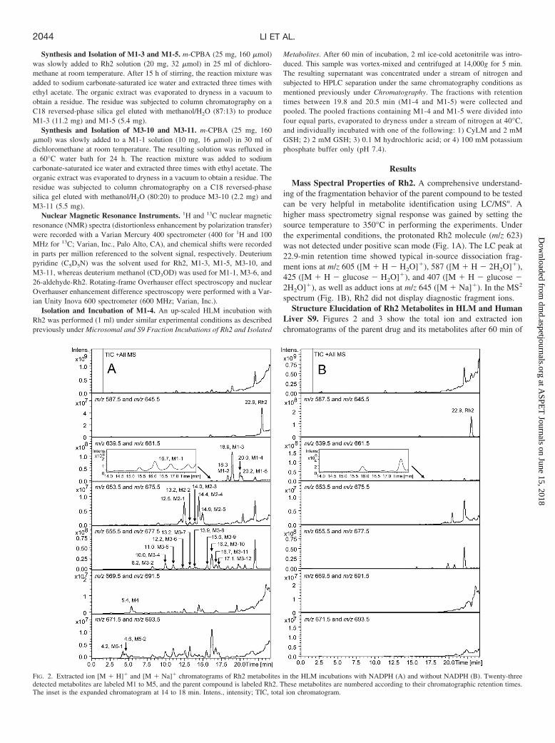
Liver S9. Figures 2 and 3 show the total ion and extracted ionchromatograms of the parent drug and its metabolites after 60 min of
FIG. 2. Extracted ion [M � H]� and [M � Na]� chromatograms of Rh2 metabolites in the HLM incubations with NADPH (A) and without NADPH (B). Twenty-threedetected metabolites are labeled M1 to M5, and the parent compound is labeled Rh2. These metabolites are numbered according to their chromatographic retention times.The inset is the expanded chromatogram at 14 to 18 min. Intens., intensity; TIC, total ion chromatogram.
2044 LI ET AL.
at ASPE
T Journals on June 15, 2018
dmd.aspetjournals.org
Dow
nloaded from
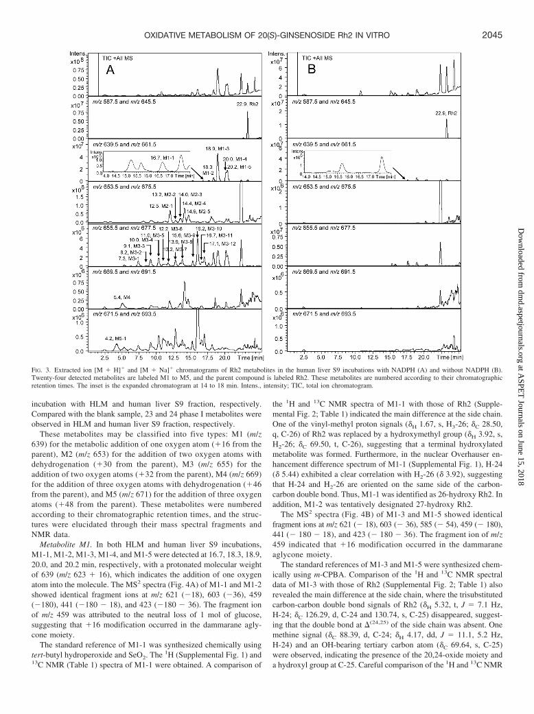
incubation with HLM and human liver S9 fraction, respectively.Compared with the blank sample, 23 and 24 phase I metabolites wereobserved in HLM and human liver S9 fraction, respectively.
These metabolites may be classified into five types: M1 (m/z639) for the metabolic addition of one oxygen atom (�16 from theparent), M2 (m/z 653) for the addition of two oxygen atoms withdehydrogenation (�30 from the parent), M3 (m/z 655) for theaddition of two oxygen atoms (�32 from the parent), M4 (m/z 669)for the addition of three oxygen atoms with dehydrogenation (�46from the parent), and M5 (m/z 671) for the addition of three oxygenatoms (�48 from the parent). These metabolites were numberedaccording to their chromatographic retention times, and the struc-tures were elucidated through their mass spectral fragments andNMR data.
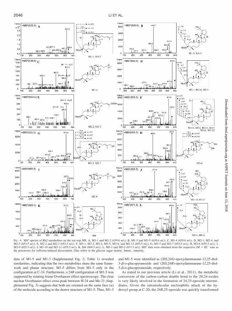
Metabolite M1. In both HLM and human liver S9 incubations,M1-1, M1-2, M1-3, M1-4, and M1-5 were detected at 16.7, 18.3, 18.9,20.0, and 20.2 min, respectively, with a protonated molecular weightof 639 (m/z 623 � 16), which indicates the addition of one oxygenatom into the molecule. The MS2 spectra (Fig. 4A) of M1-1 and M1-2showed identical fragment ions at m/z 621 (�18), 603 (�36), 459(�180), 441 (�180 � 18), and 423 (�180 � 36). The fragment ionof m/z 459 was attributed to the neutral loss of 1 mol of glucose,suggesting that �16 modification occurred in the dammarane agly-cone moiety.
The standard reference of M1-1 was synthesized chemically usingtert-butyl hydroperoxide and SeO2. The 1H (Supplemental Fig. 1) and13C NMR (Table 1) spectra of M1-1 were obtained. A comparison of
the 1H and 13C NMR spectra of M1-1 with those of Rh2 (Supple-mental Fig. 2; Table 1) indicated the main difference at the side chain.One of the vinyl-methyl proton signals (�H 1.67, s, H3-26; �C 28.50,q, C-26) of Rh2 was replaced by a hydroxymethyl group (�H 3.92, s,H2-26; �C 69.50, t, C-26), suggesting that a terminal hydroxylatedmetabolite was formed. Furthermore, in the nuclear Overhauser en-hancement difference spectrum of M1-1 (Supplemental Fig. 1), H-24(� 5.44) exhibited a clear correlation with H2-26 (� 3.92), suggestingthat H-24 and H2-26 are oriented on the same side of the carbon-carbon double bond. Thus, M1-1 was identified as 26-hydroxy Rh2. Inaddition, M1-2 was tentatively designated 27-hydroxy Rh2.
The MS2 spectra (Fig. 4B) of M1-3 and M1-5 showed identicalfragment ions at m/z 621 (� 18), 603 (� 36), 585 (� 54), 459 (� 180),441 (� 180 � 18), and 423 (� 180 � 36). The fragment ion of m/z459 indicated that �16 modification occurred in the dammaraneaglycone moiety.
The standard references of M1-3 and M1-5 were synthesized chem-ically using m-CPBA. Comparison of the 1H and 13C NMR spectraldata of M1-3 with those of Rh2 (Supplemental Fig. 2; Table 1) alsorevealed the main difference at the side chain, where the trisubstitutedcarbon-carbon double bond signals of Rh2 (�H 5.32, t, J � 7.1 Hz,H-24; �C 126.29, d, C-24 and 130.74, s, C-25) disappeared, suggest-ing that the double bond at �(24,25) of the side chain was absent. Onemethine signal (�C 88.39, d, C-24; �H 4.17, dd, J � 11.1, 5.2 Hz,H-24) and an OH-bearing tertiary carbon atom (�C 69.64, s, C-25)were observed, indicating the presence of the 20,24-oxide moiety anda hydroxyl group at C-25. Careful comparison of the 1H and 13C NMR
FIG. 3. Extracted ion [M � H]� and [M � Na]� chromatograms of Rh2 metabolites in the human liver S9 incubations with NADPH (A) and without NADPH (B).Twenty-four detected metabolites are labeled M1 to M5, and the parent compound is labeled Rh2. These metabolites are numbered according to their chromatographicretention times. The inset is the expanded chromatogram at 14 to 18 min. Intens., intensity; TIC, total ion chromatogram.
2045OXIDATIVE METABOLISM OF 20(S)-GINSENOSIDE Rh2 IN VITRO
at ASPE
T Journals on June 15, 2018
dmd.aspetjournals.org
Dow
nloaded from
data of M1-5 and M1-3 (Supplemental Fig. 2; Table 1) revealedsimilarities, indicating that the two metabolites share the same frame-work and planar structure. M1-5 differs from M1-3 only in theconfiguration at C-24. Furthermore, a 24R configuration of M1-5 wassupported by rotating frame Overhauser effect spectroscopy. The clearnuclear Overhauser effect cross-peak between H-24 and Me-21 (Sup-plemental Fig. 3) suggests that both are oriented on the same face (�)of the molecule according to the drawn structure of M1-5. Thus, M1-3
and M1-5 were identified as (20S,24S)-epoxydammarane-12,25-diol-3-�-D-glucopyranoside and (20S,24R)-epoxydammarane-12,25-diol-3-�-D-glucopyranoside, respectively.
As stated in our previous article (Li et al., 2011), the metabolicconversion of the carbon-carbon double bond to the 20,24-oxidesis very likely involved in the formation of 24,25-epoxide interme-diates. Given the intramolecular nucleophilic attack of the hy-droxyl group at C-20, the 24R,25-epoxide was quickly transformed
FIG. 4. MS2 spectra of Rh2 metabolites on the ion trap MS. A, M1-1 and M1-2 (639.6 m/z). B, M1-3 and M1-5 (639.6 m/z). C, M1-4 (639.6 m/z). D, M2-1, M2-4, andM2-5 (653.5 m/z). E, M2-2 and M2-3 (653.5 m/z). F, M3-1, M3-2, M3-4, M3-5, M3-8, and M3-12 (655.5 m/z). G, M3-3 and M3-7 (655.5 m/z). H, M3-6 (655.5 m/z). I,M3-9 (655.5 m/z). J, M3-10 and M3-11 (655.5 m/z). K, M4 (669.5 m/z). L, M5-1 and M5-2 (671.5 m/z). MS2 data were obtained from the respective [M � H]� ions asthe precursors for collision-induced dissociation. Gluc refers to the glucose sugar moiety. Intens., intensity.
2046 LI ET AL.
at ASPE
T Journals on June 15, 2018
dmd.aspetjournals.org
Dow
nloaded from

to 20S,24S-oxide (M1-3). By contrast, considering the steric hin-drance posed by the � hydrogen atom at C-24 and the hydroxylgroup at C-20, the 24S,25-epoxide was more stable against theintramolecular nucleophilic attack reaction (presumably to formM1-5) than the 24R-epimer; thus, it was detected in the incubationmixture. M1-4 was hypothesized to be 24S,25-epoxide Rh2. Weproved our hypothesis by isolating M1-4 (mixed with M1-5) from up-scaled HLM incubations with Rh2, sequentially adjusting the pH to 1,and incubating the mixture at 37°C. After 1 h of incubation, the chro-matographic peak of M1-4 (tR � 20.0 min) disappeared, and the massspectral responses of M1-5 (tR � 20.2 min) increased (Fig. 5) comparedwith the control sample. Thus, M1-4 might rearrange to form M1-5 underacidic conditions. Thus, M1-4 was identified as (24S,25)-epoxydamma-rane-12,20-diol-3-�-D-glucopyranoside.
Metabolite M2. The protonated molecular weight of M2 wasdetected at 653, which was 30 higher than that of the protonatedparent drug, indicating the introduction of two oxygen atoms withdehydrogenation. M2-1, M2-4, and M2-5 were detected at 12.5,14.4, and 14.9 min, respectively, in the HLM and human liver S9incubations. The MS2 spectra (Fig. 4D) showed fragment ions atm/z 635 (� 18), 617 (� 36), 455 (� 180 � 18), and 437 (� 180 �36), suggesting that �30 modification occurred in the dammaraneaglycone moiety.
Metabolites M2-2 and M2-3 were detected in the HLM and humanliver S9 incubations at 13.2 and 14.0 min, respectively. The MS2 spectra(Fig. 4E) showed fragment ions at m/z 635 (� 18) and 609 (� 44). Thefragment ion of m/z 609 was attributed to the neutral loss of one mole ofcarbon dioxide, which suggests that two carboxylic acid derivatives were
TABLE 113C NMR data of Rh2, M1-1, M1-3, M1-5, M3-6, M3-10, M3-11, and 26-aldehyde-Rh2
No. Rh2a M1-1 b M1-3 a M1-5 a M3-6 b M3-10 a M3-11a 26-Aldehyde-Rh2b
1 39.08, t 40.84, t 39.23, t 39.15, t 38.81, t 39.11, t 38.99, t 38.80, t2 26.82, t 27.68, t 28.62, t 28.73, t 25.77, t 28.80, t 28.59, t 25.94, t3 88.73, d 91.10, d 88.72, d 88.64, d 89.20, d 88.57, d 88.47, d 89.20, d4 39.97, s 41.45, s 39.96, s 39.64, s 39.57, s 40.05, s 39.75, s 39.57, s5 56.32, d 58.82, d 56.40, d 56.36, d 56.14, d 56.28, d 56.19, d 56.14, d6 18.41, t 19.73, t 18.45, t 18.39, t 17.84, t 18.46, t 18.21, t 17.84, t7 35.85, t 36.46, t 35.11, t 35.06, t 34.81, t 34.99, t 34.87, t 34.52, t8 36.92, s 38.44, s 39.67, s 39.92, s 36.54, s 39.26, s 39.46, s 36.55, s9 50.34, d 51.83, d 50.51, d 50.67, d 49.96, d 50.44, d 50.50, d 49.95, d
10 39.66, s 40.69, s 36.96, s 36.90, s 38.94, s 36.81, s 36.74, s 38.94, s11 32.05, t 32.53, t 32.63, t 32.34, t 30.66, t 32.76, t 32.56, t 30.69, t12 70.96, d 72.12, d 70.73, d 71.07, d 70.27, d 70.25, d 70.85, d 70.26, d13 48.53, d 49.70, d 49.43, d 49.69, d 48.06, d 48.00, d 48.19, d 47.55, d14 51.68, s 53.08, s 52.22, s 52.12, s 51.18, s 52.21, s 51.93, s 51.19, s15 31.31, t 32.49, t 32.56, t 32.75, t 30.58, t 32.18, t 32.31, t 30.57, t16 26.70, t 26.93, t 25.73, t 25.43, t 25.03, t 25.66, t 25.07, t 25.78, t17 54.79, d 55.63, d 49.45, d 48.31, d 53.76, d 49.50, d 49.47, d 53.81, d18 16.78, q 17.29, q 18.06, q 18.28, q 15.70, q 17.94, q 18.10, q 15.40, q19 16.34, q 16.65, q 16.56, q 16.68, q 15.39, q 16.35, q 16.33, q 15.40, q20 72.90, s 72.60, s 87.02, s 86.65, s 72.92, s 86.57, s 86.10, s 72.76, s21 27.06, q 27.86, q 26.97, q 27.18, q 25.93, q 26.33, q 26.82, q 24.98, q22 35.11, t 36.40, t 32.14, t 31.60, t 34.52, t 31.91, t 31.67, t 33.07, t23 22.97, t 23.38, t 26.71, t 26.66, t 21.24, t 27.42, t 27.27, t 23.22, t24 126.29, d 127.87, d 88.39, d 85.56, d 129.76, d 86.24, d 82.27, d 156.54, d25 130.74, s 136.20, s 69.64, s 70.26, s 137.65, s 72.77, s 73.08, s 138.79, s26 25.80, q 69.50, t 26.55, q 26.90, q 64.16, t 68.24, t 68.48, t 195.86, d27 17.66, q 18.87, q 28.07, q 27.60, q 56.79, t 27.84, q 26.82, q 7.66, q28 28.12, q 28.89, q 28.98, q 28.04, q 27.00, q 29.00, q 27.86, q 27.02, q29 15.79, q 14.17, q 15.57, q 15.46, q 14.77, q 15.30 15.30 14.79, q30 16.98, q 17.60, q 16.72, q 16.49, q 16.96, q 16.81, q 16.49, q 15.75, q1� 106.98, d 107.24, d 106.97, d 106.96, d 105.34, d 106.78, d 106.79, d 105.34, d2� 75.79, d 76.14, d 75.77, d 75.75, d 74.26, d 75.63, d 75.60, d 74.26, d3� 78.75, d 78.75, d 78.74, d 78.72, d 76.87, d 78.54, d 78.56, d 76.87, d4� 71.83, d 72.12, d 71.80, d 71.79, d 70.75, d 71.65, d 71.64, d 70.72, d5� 78.40, d 78.19, d 78.40, d 78.39, d 76.30, d 78.17, d 78.22, d 76.30, d6� 63.04, t 63.27, t 63.01, t 62.98, t 61.41, t 62.77, t 62.83, t 61.41, t
s, singlet; d, doublet; t, triplet; q, quartet.a In C5D5N.b In CD3OD.
FIG. 5. Extracted ion [M � H]� and [M �Na]� chromatograms of the incubationmixture of M1-4 and M1-5 after 1 h ofincubation at 37°C. Top, with 100 mM po-tassium phosphate buffer (pH 7.4). Bottom,with 0.1 M hydrochloric acid. Left column,full chromatograms. Right column, ex-panded chromatogram at 19.5 to 20.7 min.Intens., intensity.
2047OXIDATIVE METABOLISM OF 20(S)-GINSENOSIDE Rh2 IN VITRO
at ASPE
T Journals on June 15, 2018
dmd.aspetjournals.org
Dow
nloaded from
formed. In addition, M2-3 was detected in the HLM and human liver S9incubations with M1-1. This finding implies that M2-3 was derived from26-hydroxy Rh2. Thus, M2-3 was identified to be 26-carboxy Rh2. Thus,M2-2 was tentatively designated as 27-carboxy Rh2.
Metabolite M3. M3 was detected with a protonated molecularweight of 655, which was 32 higher than that of the protonated parentdrug. This finding indicates that there was an introduction of twooxygen atoms. Ten and 12 independent chromatographic peaks withprotonated ions at 655 m/z were detected in the HLM and human liverS9 incubations, respectively.
Metabolites M3-1, M3-2, M3-4, M3-5, M3-8, and M3-12 were de-tected at 7.3, 8.2, 10.0, 11.0, 13.9, and 17.1 min, respectively. The MS2
spectra (Fig. 4F) showed fragment ions at m/z 637 (� 18), 619 (� 36),475 (� 180), 457 (� 180 � 18), 439 (� 180 � 36), 421 (� 180 � 54),and 403 (� 180 � 72). M3-1, M3-2, M3-4, M3-5, M3-8, and M3-12 weretentatively identified as dioxygenation products of Rh2. Moreover, the twooxygen atoms were introduced into the dammarane aglycone moiety.
Metabolites M3-3 and M3-7 were detected at 9.1 and 13.2 min,respectively. The MS2 spectra (Fig. 4G) showed fragment ions at m/z637 (� 18), 619 (� 36), 457 (� 180 � 18), 439 (� 180 � 36), and421 (� 180 � 54). M3-3 and M3-7 were also identified as dioxy-genation products of Rh2. The two oxygen atoms were introduced intothe dammarane aglycone moiety.
Metabolite M3-6 was detected at 12.2 min. The MS2 spectra (Fig.4H) showed major fragment ions at m/z 637 (� 18), 619 (� 36), 457(� 180 � 18), and 439 (� 180 � 36). The standard reference of M3-6was synthesized chemically using tert-butyl hydroperoxide and SeO2.The 1H (Supplemental Fig. 2) and 13C NMR (Table 1) spectra ofM3-6 were obtained. The comparison between the 1H and 13C NMRspectra of M3-6 with those of Rh2 (Supplemental Fig. 2; Table 1)indicated the main difference at the side chain, where two of thevinyl-methyl proton signals (�H 1.67, s, H3-26; �C 28.50, q, C-26 and�H 1.62, s, H3-27; �C 17.66, q, C-27) of Rh2 were replaced by twohydroxymethyl groups (�H 4.17, s and �H 4.09, s, H2-26 and H2-27;�C 64.16, t and �C 56.79, t, C-26 and C-27). This result suggests thata dihydroxylated metabolite was formed. Thus, M3-6 was identifiedas 26,27-dihydroxy Rh2.
Metabolite M3-9 was detected at 15.6 min. The MS2 spectra (Fig.4I) showed major fragment ions at m/z 637 (� 18) and 619 (� 36).M3-9 was detected in the HLM and human liver S9 incubations withM1-1. M1-1 oxidation with m-CPBA can also produce M3-9 (data notshown). Thus, M3-9 was tentatively identified as (24S,25)-epoxydammarane-12,20, 26-triol-3-�-D-glucopyranoside.
Metabolites M3-10 and M3-11 were detected at 16.2 and 16.7 min,respectively. The MS2 spectra (Fig. 4J) showed fragment ions at m/z637 (� 18), 619 (� 36), 457 (� 180 � 18), 439 (� 180 � 36), and421 (� 180 � 54). The standard reference of M3-10 and M3-11 waschemically synthesized using m-CPBA. The 1H (Supplemental Fig. 2)and 13C NMR (Table 1) spectra of M3-10 and M3-11 were obtained.
The comparison between the 1H and 13C NMR data of M3-10 withthose of M1-3 revealed that M3-10 shares the same nucleus as M1-3.M3-10 differs from M1-3 only at the substituted tetrahydrofuran ringwhere one of the methyl groups was hydroxylated. This finding wasin agreement with the mass data. The introduction of the oxygen atomcaused the 1H (from � 1.31–3.74) and 13C NMR resonance (from �26.55–68.24) of C-26 to shift downfield significantly. Thus, M3-10was identified as (20S,24S)-epoxydammarane-12,25,26-triol-3-�-D-glucopyranoside.
M3-11 was identified as (20S,24R)-epoxydammarane-12,25,26-triol-3-�-D-glucopyranoside by comparing the 1H and 13C NMR data(Supplemental Fig. 2; Table 1) with those of M1-5. Likewise, theintroduction of the oxygen atom caused the 1H (from � 1.27–3.96 and
3.91) and 13C NMR resonance (from � 26.90–68.48) of C-26 to shiftdownfield significantly.
Metabolite M4. M4 was detected at 5.4 min with a protonatedmolecular weight of 669. Its weight was 46 higher than the protonatedRh2, which indicates that there was an introduction of three oxygenatoms with dehydrogenation. The MS2 spectra (Fig. 4K) showedfragment ions at m/z 651 (� 18), 637 (� 36), 615 (� 54), and 453 (�180 � 36). M4 was tentatively identified as the trioxygenation anddehydrogenation product of Rh2. All of the modifications occurred atthe dammarane aglycone moiety.
Metabolite M5. Metabolites M5-1 and M5-2 gave a precursor ionat 671 m/z, which was 48 higher than that of Rh2. M5-1 and M5-2were eluted at 4.2 and 4.6 min, respectively. Their MS2 spectra(Fig. 4L) were identical, and the major fragment ions were at m/z653 (� 18), 635 (� 36), 617 (� 54), and 473 (� 180 � 18). M5-1and M5-2 were tentatively identified as trioxygenation products ofRh2. All oxygen atoms were introduced into the dammarane agly-cone moiety.
Incubations of Isolated Metabolites in HLM. Individual incuba-tions in HLM were performed for six isolated metabolites (M1-1,M1-3, M1-5, M3-6, M3-10, and M3-11). For M1-1, M1-3, M1-5,M3-6, M3-10, and M3-11, 8, 8, 5, 0, 1, and 1 metabolites weredetected compared with the control samples, respectively (data notshown). M1-1 underwent extensive oxidative metabolism in HLMincubations and major metabolic pathways, including hydroxylation,to form M3-3, M3-6, and M3-7. In addition, it underwent epoxidationto form M3-9 and its 24-epimer (not detected because of the quickrearrangement) and their rearrangement products M3-10 and M3-11.A small amount of M5-2 generated from the hydroxylation of M3-11and a carboxylic acid derivative M2-3 was also detected. The metab-olites from M1-3 may be classified into the following three types:M2-1 and M2-4 from hydroxylation with dehydrogenation; M3-1,M3-2, M3-4, M3-5, and M3-10 from hydroxylation; and M5-1 fromdihydroxylation. Hydroxylation and dehydrogenation were the majormetabolic pathways observed for M1-5 to generate metabolites M2-5,M3-8, M3-11, M3-12, and M4. No obvious oxidative metaboliteswere detected in the HLM incubation with M3-6. M3-10 and M3-11only underwent hydroxylation to produce M5-1 and M5-2 in the HLMincubations, respectively.
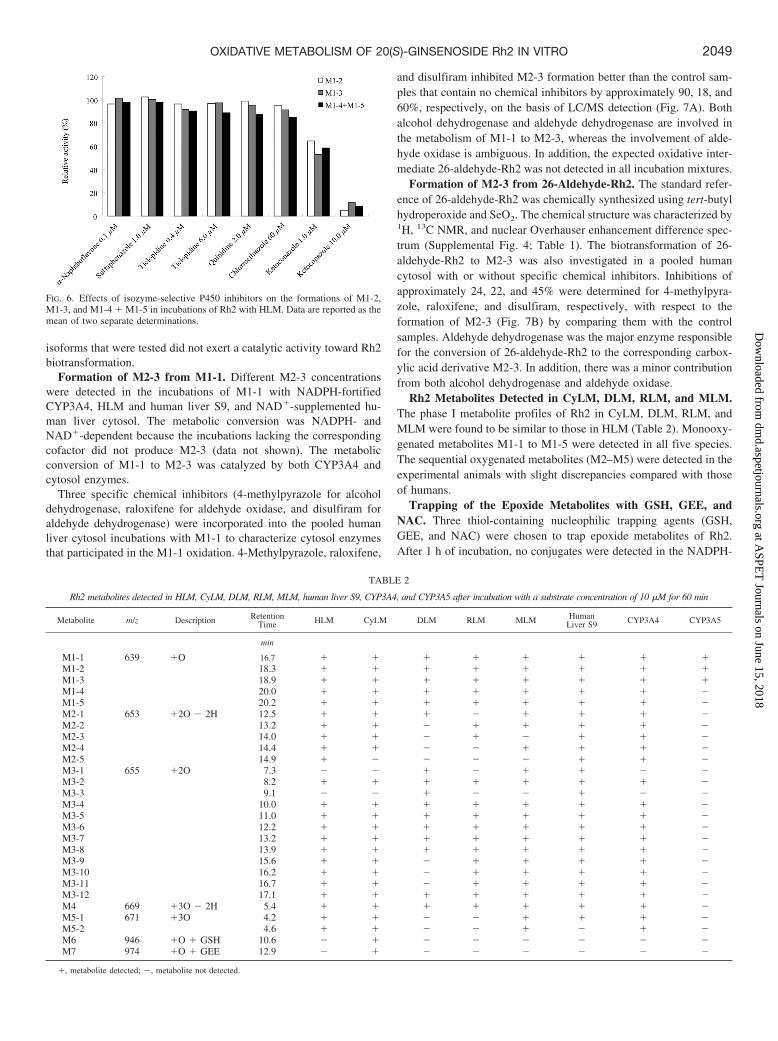
Identification of P450 Enzymes Responsible for Rh2 Metabo-lism. P450 enzymes responsible for the oxidative metabolism of Rh2were probed through coincubation with isoenzyme-selective inhibi-tors, including �-naphthoflavone (CYP1A2), ticlopidine (CYP2B6and CYP2C19), sulfaphenazole (CYP2C9), quinidine (CYP2D6),chlormethiazole (CYP2E1), or ketoconazole (CYP3A), with HLM inthe presence of NADPH. The formations of M1-2, M1-3, and M1-4 �M1-5 were monitored because of their higher MS responses comparedwith the sequential oxidative metabolites. Among the inhibitors used,only ketoconazole showed a significant inhibitory effect on the oxi-dative metabolism of Rh2. No such inhibition was observed for theother inhibitors (Fig. 6). P450 3As were probably the major enzymesresponsible for the oxidative metabolism of Rh2.
Catalytic activities of 12 individual recombinant human P450s,including CYP1A2, CYP1B1, CYP2A6, CYP2B6, CYP2C8,CYP2C9, CYP2C19, CYP2D6, CYP2E1, CYP3A4, CYP3A5, andCYP4A11, in Rh2 biotransformation were evaluated. As expected, theoxidative metabolites of Rh2 were detected in the incubations onlywith CYP3A4 and CYP3A5 to a minor extent. The phase I metaboliteprofile of Rh2 in CYP3A4 was the same as that in HLM, whereasCYP3A5 produced only small amounts (less than 5% that ofCYP3A4, respectively) of M1-1, M1-2, and M1-3 (Table 2). Other
2048 LI ET AL.
at ASPE
T Journals on June 15, 2018
dmd.aspetjournals.org
Dow
nloaded from
isoforms that were tested did not exert a catalytic activity toward Rh2biotransformation.
Formation of M2-3 from M1-1. Different M2-3 concentrationswere detected in the incubations of M1-1 with NADPH-fortifiedCYP3A4, HLM and human liver S9, and NAD�-supplemented hu-man liver cytosol. The metabolic conversion was NADPH- andNAD�-dependent because the incubations lacking the correspondingcofactor did not produce M2-3 (data not shown). The metabolicconversion of M1-1 to M2-3 was catalyzed by both CYP3A4 andcytosol enzymes.
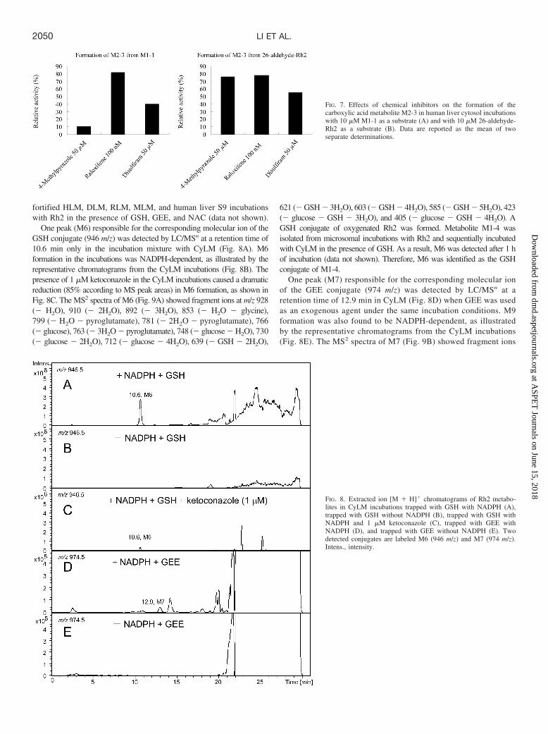
Three specific chemical inhibitors (4-methylpyrazole for alcoholdehydrogenase, raloxifene for aldehyde oxidase, and disulfiram foraldehyde dehydrogenase) were incorporated into the pooled humanliver cytosol incubations with M1-1 to characterize cytosol enzymesthat participated in the M1-1 oxidation. 4-Methylpyrazole, raloxifene,
and disulfiram inhibited M2-3 formation better than the control sam-ples that contain no chemical inhibitors by approximately 90, 18, and60%, respectively, on the basis of LC/MS detection (Fig. 7A). Bothalcohol dehydrogenase and aldehyde dehydrogenase are involved inthe metabolism of M1-1 to M2-3, whereas the involvement of alde-hyde oxidase is ambiguous. In addition, the expected oxidative inter-mediate 26-aldehyde-Rh2 was not detected in all incubation mixtures.
Formation of M2-3 from 26-Aldehyde-Rh2. The standard refer-ence of 26-aldehyde-Rh2 was chemically synthesized using tert-butylhydroperoxide and SeO2. The chemical structure was characterized by1H, 13C NMR, and nuclear Overhauser enhancement difference spec-trum (Supplemental Fig. 4; Table 1). The biotransformation of 26-aldehyde-Rh2 to M2-3 was also investigated in a pooled humancytosol with or without specific chemical inhibitors. Inhibitions ofapproximately 24, 22, and 45% were determined for 4-methylpyra-zole, raloxifene, and disulfiram, respectively, with respect to theformation of M2-3 (Fig. 7B) by comparing them with the controlsamples. Aldehyde dehydrogenase was the major enzyme responsiblefor the conversion of 26-aldehyde-Rh2 to the corresponding carbox-ylic acid derivative M2-3. In addition, there was a minor contributionfrom both alcohol dehydrogenase and aldehyde oxidase.
Rh2 Metabolites Detected in CyLM, DLM, RLM, and MLM.The phase I metabolite profiles of Rh2 in CyLM, DLM, RLM, andMLM were found to be similar to those in HLM (Table 2). Monooxy-genated metabolites M1-1 to M1-5 were detected in all five species.The sequential oxygenated metabolites (M2–M5) were detected in theexperimental animals with slight discrepancies compared with thoseof humans.
Trapping of the Epoxide Metabolites with GSH, GEE, andNAC. Three thiol-containing nucleophilic trapping agents (GSH,GEE, and NAC) were chosen to trap epoxide metabolites of Rh2.After 1 h of incubation, no conjugates were detected in the NADPH-
TABLE 2
Rh2 metabolites detected in HLM, CyLM, DLM, RLM, MLM, human liver S9, CYP3A4, and CYP3A5 after incubation with a substrate concentration of 10 �M for 60 min
Metabolite m/z Description RetentionTime HLM CyLM DLM RLM MLM Human
Liver S9 CYP3A4 CYP3A5
min
M1-1 639 �O 16.7 � � � � � � � �M1-2 18.3 � � � � � � � �M1-3 18.9 � � � � � � � �M1-4 20.0 � � � � � � � �M1-5 20.2 � � � � � � � �M2-1 653 �2O � 2H 12.5 � � � � � � � �M2-2 13.2 � � � � � � � �M2-3 14.0 � � � � � � � �M2-4 14.4 � � � � � � � �M2-5 14.9 � � � � � � � �M3-1 655 �2O 7.3 � � � � � � � �M3-2 8.2 � � � � � � � �M3-3 9.1 � � � � � � � �M3-4 10.0 � � � � � � � �M3-5 11.0 � � � � � � � �M3-6 12.2 � � � � � � � �M3-7 13.2 � � � � � � � �M3-8 13.9 � � � � � � � �M3-9 15.6 � � � � � � � �M3-10 16.2 � � � � � � � �M3-11 16.7 � � � � � � � �M3-12 17.1 � � � � � � � �M4 669 �3O � 2H 5.4 � � � � � � � �M5-1 671 �3O 4.2 � � � � � � � �M5-2 4.6 � � � � � � � �M6 946 �O � GSH 10.6 � � � � � � � �M7 974 �O � GEE 12.9 � � � � � � � �
�, metabolite detected; �, metabolite not detected.
FIG. 6. Effects of isozyme-selective P450 inhibitors on the formations of M1-2,M1-3, and M1-4 � M1-5 in incubations of Rh2 with HLM. Data are reported as themean of two separate determinations.
2049OXIDATIVE METABOLISM OF 20(S)-GINSENOSIDE Rh2 IN VITRO
at ASPE
T Journals on June 15, 2018
dmd.aspetjournals.org
Dow
nloaded from
fortified HLM, DLM, RLM, MLM, and human liver S9 incubationswith Rh2 in the presence of GSH, GEE, and NAC (data not shown).
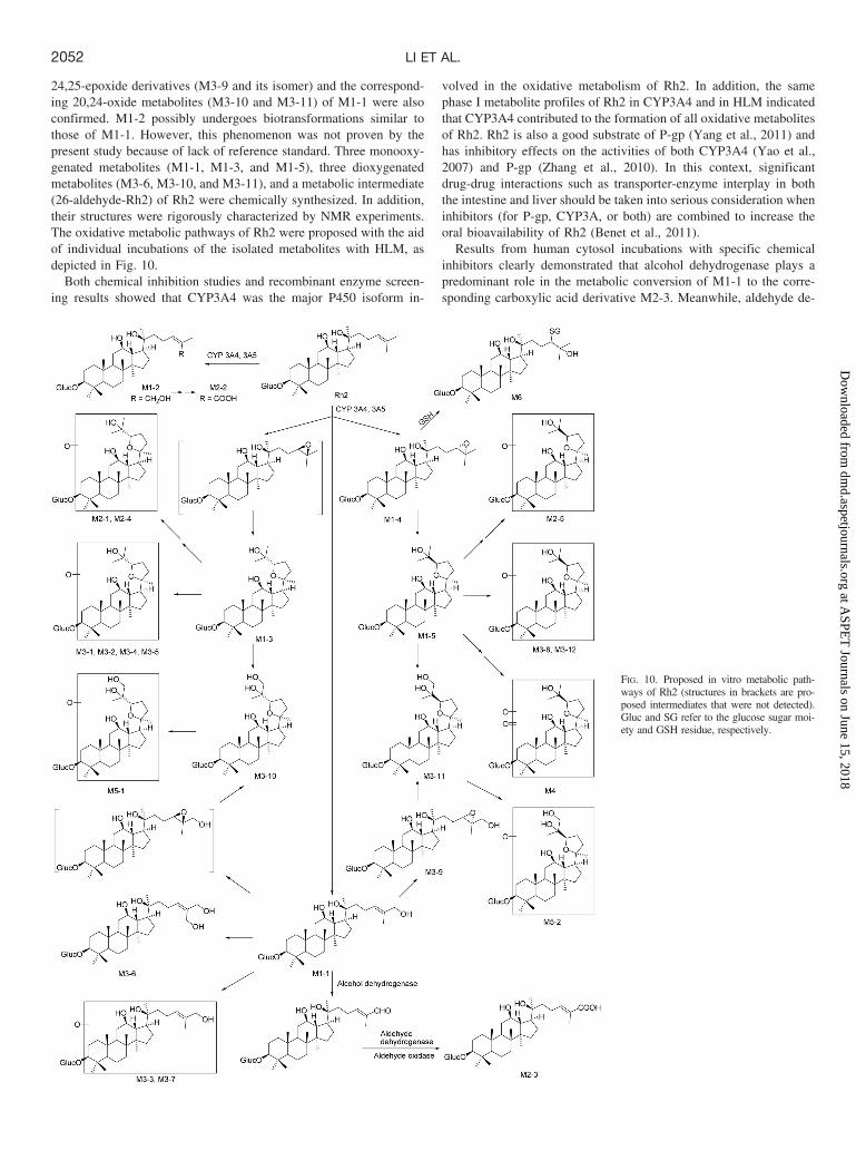
One peak (M6) responsible for the corresponding molecular ion of theGSH conjugate (946 m/z) was detected by LC/MSn at a retention time of10.6 min only in the incubation mixture with CyLM (Fig. 8A). M6formation in the incubations was NADPH-dependent, as illustrated by therepresentative chromatograms from the CyLM incubations (Fig. 8B). Thepresence of 1 �M ketoconazole in the CyLM incubations caused a dramaticreduction (85% according to MS peak areas) in M6 formation, as shown inFig. 8C. The MS2 spectra of M6 (Fig. 9A) showed fragment ions at m/z 928(� H2O), 910 (� 2H2O), 892 (� 3H2O), 853 (� H2O � glycine),799 (� H2O � pyroglutamate), 781 (� 2H2O � pyroglutamate), 766(� glucose), 763 (� 3H2O � pyroglutamate), 748 (� glucose � H2O), 730(� glucose � 2H2O), 712 (� glucose � 4H2O), 639 (� GSH � 2H2O),
621 (� GSH � 3H2O), 603 (� GSH � 4H2O), 585 (� GSH � 5H2O), 423(� glucose � GSH � 3H2O), and 405 (� glucose � GSH � 4H2O). AGSH conjugate of oxygenated Rh2 was formed. Metabolite M1-4 wasisolated from microsomal incubations with Rh2 and sequentially incubatedwith CyLM in the presence of GSH. As a result, M6 was detected after 1 hof incubation (data not shown). Therefore, M6 was identified as the GSHconjugate of M1-4.
One peak (M7) responsible for the corresponding molecular ionof the GEE conjugate (974 m/z) was detected by LC/MSn at aretention time of 12.9 min in CyLM (Fig. 8D) when GEE was usedas an exogenous agent under the same incubation conditions. M9formation was also found to be NADPH-dependent, as illustratedby the representative chromatograms from the CyLM incubations(Fig. 8E). The MS2 spectra of M7 (Fig. 9B) showed fragment ions
FIG. 7. Effects of chemical inhibitors on the formation of thecarboxylic acid metabolite M2-3 in human liver cytosol incubationswith 10 �M M1-1 as a substrate (A) and with 10 �M 26-aldehyde-Rh2 as a substrate (B). Data are reported as the mean of twoseparate determinations.
FIG. 8. Extracted ion [M � H]� chromatograms of Rh2 metabo-lites in CyLM incubations trapped with GSH with NADPH (A),trapped with GSH without NADPH (B), trapped with GSH withNADPH and 1 �M ketoconazole (C), trapped with GEE withNADPH (D), and trapped with GEE without NADPH (E). Twodetected conjugates are labeled M6 (946 m/z) and M7 (974 m/z).Intens., intensity.
2050 LI ET AL.
at ASPE
T Journals on June 15, 2018
dmd.aspetjournals.org
Dow
nloaded from
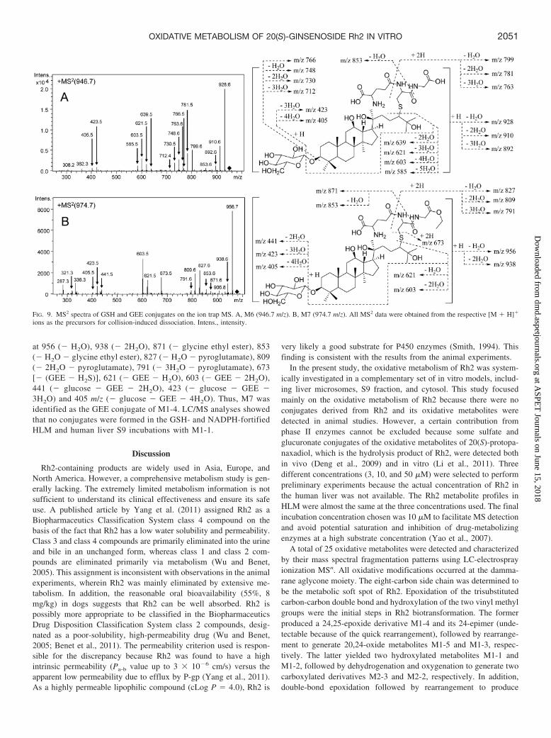
at 956 (� H2O), 938 (� 2H2O), 871 (� glycine ethyl ester), 853(� H2O � glycine ethyl ester), 827 (� H2O � pyroglutamate), 809(� 2H2O � pyroglutamate), 791 (� 3H2O � pyroglutamate), 673[� (GEE � H2S)], 621 (� GEE � H2O), 603 (� GEE � 2H2O),441 (� glucose � GEE � 2H2O), 423 (� glucose � GEE �3H2O) and 405 m/z (� glucose � GEE � 4H2O). Thus, M7 wasidentified as the GEE conjugate of M1-4. LC/MS analyses showedthat no conjugates were formed in the GSH- and NADPH-fortifiedHLM and human liver S9 incubations with M1-1.
Discussion
Rh2-containing products are widely used in Asia, Europe, andNorth America. However, a comprehensive metabolism study is gen-erally lacking. The extremely limited metabolism information is notsufficient to understand its clinical effectiveness and ensure its safeuse. A published article by Yang et al. (2011) assigned Rh2 as aBiopharmaceutics Classification System class 4 compound on thebasis of the fact that Rh2 has a low water solubility and permeability.Class 3 and class 4 compounds are primarily eliminated into the urineand bile in an unchanged form, whereas class 1 and class 2 com-pounds are eliminated primarily via metabolism (Wu and Benet,2005). This assignment is inconsistent with observations in the animalexperiments, wherein Rh2 was mainly eliminated by extensive me-tabolism. In addition, the reasonable oral bioavailability (55%, 8mg/kg) in dogs suggests that Rh2 can be well absorbed. Rh2 ispossibly more appropriate to be classified in the BiopharmaceuticsDrug Disposition Classification System class 2 compounds, desig-nated as a poor-solubility, high-permeability drug (Wu and Benet,2005; Benet et al., 2011). The permeability criterion used is respon-sible for the discrepancy because Rh2 was found to have a highintrinsic permeability (Pa-b value up to 3 � 10�6 cm/s) versus theapparent low permeability due to efflux by P-gp (Yang et al., 2011).As a highly permeable lipophilic compound (cLog P � 4.0), Rh2 is
very likely a good substrate for P450 enzymes (Smith, 1994). Thisfinding is consistent with the results from the animal experiments.
In the present study, the oxidative metabolism of Rh2 was system-ically investigated in a complementary set of in vitro models, includ-ing liver microsomes, S9 fraction, and cytosol. This study focusedmainly on the oxidative metabolism of Rh2 because there were noconjugates derived from Rh2 and its oxidative metabolites weredetected in animal studies. However, a certain contribution fromphase II enzymes cannot be excluded because some sulfate andglucuronate conjugates of the oxidative metabolites of 20(S)-protopa-naxadiol, which is the hydrolysis product of Rh2, were detected bothin vivo (Deng et al., 2009) and in vitro (Li et al., 2011). Threedifferent concentrations (3, 10, and 50 �M) were selected to performpreliminary experiments because the actual concentration of Rh2 inthe human liver was not available. The Rh2 metabolite profiles inHLM were almost the same at the three concentrations used. The finalincubation concentration chosen was 10 �M to facilitate MS detectionand avoid potential saturation and inhibition of drug-metabolizingenzymes at a high substrate concentration (Yao et al., 2007).
A total of 25 oxidative metabolites were detected and characterizedby their mass spectral fragmentation patterns using LC-electrosprayionization MSn. All oxidative modifications occurred at the damma-rane aglycone moiety. The eight-carbon side chain was determined tobe the metabolic soft spot of Rh2. Epoxidation of the trisubstitutedcarbon-carbon double bond and hydroxylation of the two vinyl methylgroups were the initial steps in Rh2 biotransformation. The formerproduced a 24,25-epoxide derivative M1-4 and its 24-epimer (unde-tectable because of the quick rearrangement), followed by rearrange-ment to generate 20,24-oxide metabolites M1-5 and M1-3, respec-tively. The latter yielded two hydroxylated metabolites M1-1 andM1-2, followed by dehydrogenation and oxygenation to generate twocarboxylated derivatives M2-3 and M2-2, respectively. In addition,double-bond epoxidation followed by rearrangement to produce
FIG. 9. MS2 spectra of GSH and GEE conjugates on the ion trap MS. A, M6 (946.7 m/z). B, M7 (974.7 m/z). All MS2 data were obtained from the respective [M � H]�
ions as the precursors for collision-induced dissociation. Intens., intensity.
2051OXIDATIVE METABOLISM OF 20(S)-GINSENOSIDE Rh2 IN VITRO
at ASPE
T Journals on June 15, 2018
dmd.aspetjournals.org
Dow
nloaded from
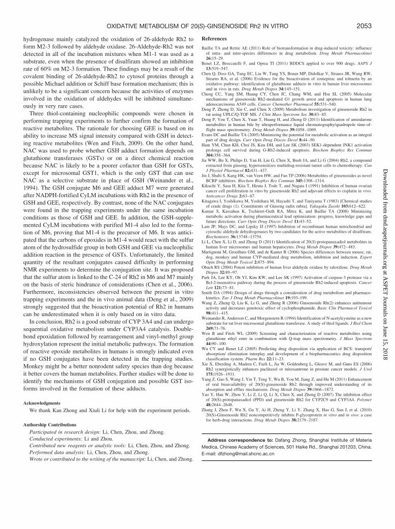
24,25-epoxide derivatives (M3-9 and its isomer) and the correspond-ing 20,24-oxide metabolites (M3-10 and M3-11) of M1-1 were alsoconfirmed. M1-2 possibly undergoes biotransformations similar tothose of M1-1. However, this phenomenon was not proven by thepresent study because of lack of reference standard. Three monooxy-genated metabolites (M1-1, M1-3, and M1-5), three dioxygenatedmetabolites (M3-6, M3-10, and M3-11), and a metabolic intermediate(26-aldehyde-Rh2) of Rh2 were chemically synthesized. In addition,their structures were rigorously characterized by NMR experiments.The oxidative metabolic pathways of Rh2 were proposed with the aidof individual incubations of the isolated metabolites with HLM, asdepicted in Fig. 10.
Both chemical inhibition studies and recombinant enzyme screen-ing results showed that CYP3A4 was the major P450 isoform in-
volved in the oxidative metabolism of Rh2. In addition, the samephase I metabolite profiles of Rh2 in CYP3A4 and in HLM indicatedthat CYP3A4 contributed to the formation of all oxidative metabolitesof Rh2. Rh2 is also a good substrate of P-gp (Yang et al., 2011) andhas inhibitory effects on the activities of both CYP3A4 (Yao et al.,2007) and P-gp (Zhang et al., 2010). In this context, significantdrug-drug interactions such as transporter-enzyme interplay in boththe intestine and liver should be taken into serious consideration wheninhibitors (for P-gp, CYP3A, or both) are combined to increase theoral bioavailability of Rh2 (Benet et al., 2011).
Results from human cytosol incubations with specific chemicalinhibitors clearly demonstrated that alcohol dehydrogenase plays apredominant role in the metabolic conversion of M1-1 to the corre-sponding carboxylic acid derivative M2-3. Meanwhile, aldehyde de-
FIG. 10. Proposed in vitro metabolic path-ways of Rh2 (structures in brackets are pro-posed intermediates that were not detected).Gluc and SG refer to the glucose sugar moi-ety and GSH residue, respectively.
2052 LI ET AL.
at ASPE
T Journals on June 15, 2018
dmd.aspetjournals.org
Dow
nloaded from
hydrogenase mainly catalyzed the oxidation of 26-aldehyde Rh2 toform M2-3 followed by aldehyde oxidase. 26-Aldehyde-Rh2 was notdetected in all of the incubation mixtures when M1-1 was used as asubstrate, even when the presence of disulfiram showed an inhibitionrate of 60% on M2-3 formation. These findings may be a result of thecovalent binding of 26-aldehyde-Rh2 to cytosol proteins through apossible Michael addition or Schiff base formation mechanism; this isunlikely to be a significant concern because the activities of enzymesinvolved in the oxidation of aldehydes will be inhibited simultane-ously in very rare cases.
Three thiol-containing nucleophilic compounds were chosen inperforming trapping experiments to further confirm the formation ofreactive metabolites. The rationale for choosing GEE is based on itsability to increase MS signal intensity compared with GSH in detect-ing reactive metabolites (Wen and Fitch, 2009). On the other hand,NAC was used to probe whether GSH adduct formation depends onglutathione transferases (GSTs) or on a direct chemical reactionbecause NAC is likely to be a poorer cofactor than GSH for GSTs,except for microsomal GST1, which is the only GST that can useNAC as a selective substrate in place of GSH (Weinander et al.,1994). The GSH conjugate M6 and GEE adduct M7 were generatedafter NADPH-fortified CyLM incubations with Rh2 in the presence ofGSH and GEE, respectively. By contrast, none of the NAC conjugateswere found in the trapping experiments under the same incubationconditions as those of GSH and GEE. In addition, the GSH-supple-mented CyLM incubations with purified M1-4 also led to the forma-tion of M6, proving that M1-4 is the precursor of M6. It was antici-pated that the carbons of epoxides in M1-4 would react with the sulfuratom of the hydrosulfide group in both GSH and GEE via nucleophilicaddition reaction in the presence of GSTs. Unfortunately, the limitedquantity of the resultant conjugates caused difficulty in performingNMR experiments to determine the conjugation site. It was proposedthat the sulfur atom is linked to the C-24 of Rh2 in M6 and M7 mainlyon the basis of steric hindrance of considerations (Chen et al., 2006).Furthermore, inconsistencies observed between the present in vitrotrapping experiments and the in vivo animal data (Deng et al., 2009)strongly suggested that the bioactivation potential of Rh2 in humanscan be underestimated when it is only based on in vitro data.
In conclusion, Rh2 is a good substrate of CYP 3A4 and can undergosequential oxidative metabolism under CYP3A4 catalysis. Double-bond epoxidation followed by rearrangement and vinyl-methyl grouphydroxylation represent the initial metabolic pathways. The formationof reactive epoxide metabolites in humans is strongly indicated evenif no GSH conjugates have been detected in the trapping studies.Monkey might be a better nonrodent safety species than dog becauseit better covers the human metabolites. Further studies will be done toidentify the mechanisms of GSH conjugation and possible GST iso-forms involved in the formation of these adducts.
Acknowledgments
We thank Kan Zhong and Xiuli Li for help with the experiment periods.
Authorship Contributions
Participated in research design: Li, Chen, Zhou, and Zhong.Conducted experiments: Li and Zhou.Contributed new reagents or analytic tools: Li, Chen, Zhou, and Zhong.Performed data analysis: Li, Chen, Zhou, and Zhong.Wrote or contributed to the writing of the manuscript: Li, Chen, and Zhong.
References
Baillie TA and Rettie AE (2011) Role of biotransformation in drug-induced toxicity: influenceof intra- and inter-species differences in drug metabolism. Drug Metab Pharmacokinet26:15–29.
Benet LZ, Broccatelli F, and Oprea TI (2011) BDDCS applied to over 900 drugs. AAPS J13:519–547.
Chen Q, Doss GA, Tung EC, Liu W, Tang YS, Braun MP, Didolkar V, Strauss JR, Wang RW,Stearns RA, et al. (2006) Evidence for the bioactivation of zomepirac and tolmetin by anoxidative pathway: identification of glutathione adducts in vitro in human liver microsomesand in vivo in rats. Drug Metab Dispos 34:145–151.
Cheng CC, Yang SM, Huang CY, Chen JC, Chang WM, and Hsu SL (2005) Molecularmechanisms of ginsenoside Rh2-mediated G1 growth arrest and apoptosis in human lungadenocarcinoma A549 cells. Cancer Chemother Pharmacol 55:531–540.
Deng P, Zhong D, Xie C, and Chen X (2009) Metabolism investigation of ginsenoside Rh2 inrat using UPLC/Q-TOF MS. J Chin Mass Spectrom Soc 30:83–85.
Deng P, You T, Chen X, Yuan T, Huang H, and Zhong D (2011) Identification of amiodaronemetabolites in human bile by ultraperformance liquid chromatography/quadrupole time-of-flight mass spectrometry. Drug Metab Dispos 39:1058–1069.
Evans DC and Baillie TA (2005) Minimizing the potential for metabolic activation as an integralpart of drug design. Curr Opin Drug Discov Devel 8:44–50.
Ham YM, Chun KH, Choi JS, Kim DH, and Lee SK (2003) SEK1-dependent JNK1 activationprolongs cell survival during G-Rh2-induced apoptosis. Biochem Biophys Res Commun304:358–364.
Jia WW, Bu X, Philips D, Yan H, Liu G, Chen X, Bush JA, and Li G (2004) Rh2, a compoundextracted from ginseng, hypersensitizes multidrug-resistant tumor cells to chemotherapy. CanJ Physiol Pharmacol 82:431–437.
Jin J, Shahi S, Kang HK, van Veen HW, and Fan TP (2006) Metabolites of ginsenosides as novelBCRP inhibitors. Biochem Biophys Res Commun 345:1308–1314.
Kikuchi Y, Sasa H, Kita T, Hirata J, Tode T, and Nagata I (1991) Inhibition of human ovariancancer cell proliferation in vitro by ginsenoside Rh2 and adjuvant effects to cisplatin in vivo.Anticancer Drugs 2:63–67.
Kitagawa I, Yoshikawa M, Yoshihara M, Hayashi T, and Taniyama T (1983) [Chemical studiesof crude drugs (1). Constituents of Ginseng radix rubra]. Yakugaku Zasshi 103:612–622.
Kumar S, Kassahun K, Tschirret-Guth RA, Mitra K, and Baillie TA (2008) Minimizingmetabolic activation during pharmaceutical lead optimization: progress, knowledge gaps andfuture directions. Curr Opin Drug Discov Devel 11:43–52.
Lam JP, Mays DC, and Lipsky JJ (1997) Inhibition of recombinant human mitochondrial andcytosolic aldehyde dehydrogenases by two candidates for the active metabolites of disulfiram.Biochemistry 36:13748–13754.
Li L, Chen X, Li D, and Zhong D (2011) Identification of 20(S)-protopanaxadiol metabolites inhuman liver microsomes and human hepatocytes. Drug Metab Dispos 39:472–483.
Martignoni M, Groothuis GM, and de Kanter R (2006) Species differences between mouse, rat,dog, monkey and human CYP-mediated drug metabolism, inhibition and induction. ExpertOpin Drug Metab Toxicol 2:875–894.
Obach RS (2004) Potent inhibition of human liver aldehyde oxidase by raloxifene. Drug MetabDispos 32:89–97.
Park JA, Lee KY, Oh YJ, Kim KW, and Lee SK (1997) Activation of caspase-3 protease via aBcl-2-insensitive pathway during the process of ginsenoside Rh2-induced apoptosis. CancerLett 121:73–81.
Smith DA (1994) Design of drugs through a consideration of drug metabolism and pharmaco-kinetics. Eur J Drug Metab Pharmacokinet 19:193–199.
Wang Z, Zheng Q, Liu K, Li G, and Zheng R (2006) Ginsenoside Rh(2) enhances antitumouractivity and decreases genotoxic effect of cyclophosphamide. Basic Clin Pharmacol Toxicol98:411–415.
Weinander R, Anderson C, and Morgenstern R (1994) Identification of N-acetylcysteine as a newsubstrate for rat liver microsomal glutathione transferase. A study of thiol ligands. J Biol Chem269:71–76.
Wen B and Fitch WL (2009) Screening and characterization of reactive metabolites usingglutathione ethyl ester in combination with Q-trap mass spectrometry. J Mass Spectrom44:90–100.
Wu CY and Benet LZ (2005) Predicting drug disposition via application of BCS: transport/absorption/ elimination interplay and development of a biopharmaceutics drug dispositionclassification system. Pharm Res 22:11–23.
Xie X, Eberding A, Madera C, Fazli L, Jia W, Goldenberg L, Gleave M, and Guns ES (2006)Rh2 synergistically enhances paclitaxel or mitoxantrone in prostate cancer models. J Urol175:1926–1931.
Yang Z, Gao S, Wang J, Yin T, Teng Y, Wu B, You M, Jiang Z, and Hu M (2011) Enhancementof oral bioavailability of 20(S)-ginsenoside Rh2 through improved understanding of itsabsorption and efflux mechanisms. Drug Metab Dispos 39:1866–1872.
Yao Y, Han W, Zhou Y, Li Z, Li Q, Li X, Chen X, and Zhong D (2007) The inhibition effectof 20(S)-protopanaxadiol (PPD) and ginsenoside Rh2 for CYP2C9 and CYP3A4. Polymer48:2644–2648.
Zhang J, Zhou F, Wu X, Gu Y, Ai H, Zheng Y, Li Y, Zhang X, Hao G, Sun J, et al. (2010)20(S)-Ginsenoside Rh2 noncompetitively inhibits P-glycoprotein in vitro and in vivo: a casefor herb-drug interactions. Drug Metab Dispos 38:2179–2187.
Address correspondence to: Dafang Zhong, Shanghai Institute of MateriaMedica, Chinese Academy of Sciences, 501 Haike Rd., Shanghai 201203, China.E-mail: [email protected]
2053OXIDATIVE METABOLISM OF 20(S)-GINSENOSIDE Rh2 IN VITRO
at ASPE
T Journals on June 15, 2018
dmd.aspetjournals.org
Dow
nloaded from