Division’s Revised Proposed Package Insert Imagent Kit for ... · Kit for the Preparation of...
19
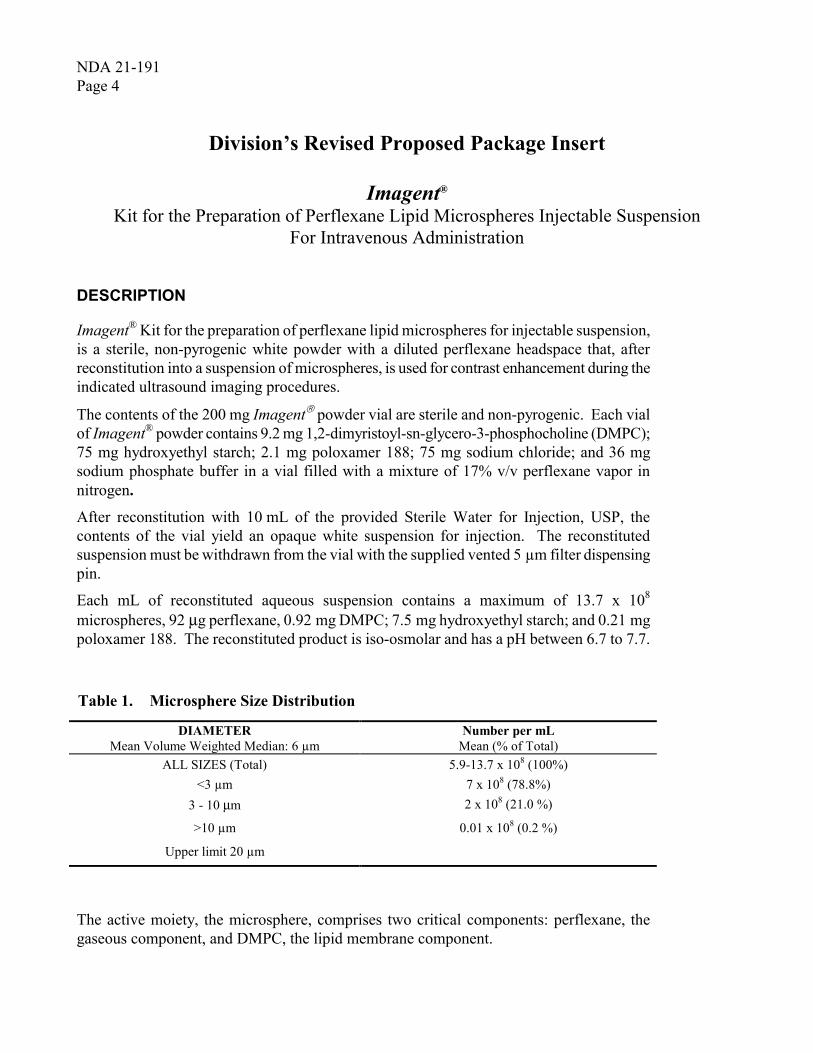
NDA 21-191 Page 4 Division’s Revised Proposed Package Insert Imagent ® Kit for the Preparation of Perflexane Lipid Microspheres Injectable Suspension For Intravenous Administration DESCRIPTION Imagent ® Kit for the preparation of perflexane lipid microspheres for injectable suspension, is a sterile, non-pyrogenic white powder with a diluted perflexane headspace that, after reconstitution into a suspension of microspheres, is used for contrast enhancement during the indicated ultrasound imaging procedures. The contents of the 200 mg Imagent â powder vial are sterile and non-pyrogenic. Each vial of Imagent ® powder contains 9.2 mg 1,2-dimyristoyl-sn-glycero-3-phosphocholine (DMPC); 75 mg hydroxyethyl starch; 2.1 mg poloxamer 188; 75 mg sodium chloride; and 36 mg sodium phosphate buffer in a vial filled with a mixture of 17% v/v perflexane vapor in nitrogen. After reconstitution with 10 mL of the provided Sterile Water for Injection, USP, the contents of the vial yield an opaque white suspension for injection. The reconstituted suspension must be withdrawn from the vial with the supplied vented 5 μm filter dispensing pin. Each mL of reconstituted aqueous suspension contains a maximum of 13.7 x 10 8 microspheres, 92 µg perflexane, 0.92 mg DMPC; 7.5 mg hydroxyethyl starch; and 0.21 mg poloxamer 188. The reconstituted product is iso-osmolar and has a pH between 6.7 to 7.7. Table 1. Microsphere Size Distribution DIAMETER Mean Volume Weighted Median: 6 μm Number per mL Mean (% of Total) ALL SIZES (Total) 5.9-13.7 x 10 8 (100%) <3 μm 7 x 10 8 (78.8%) 3 - 10 µm 2 x 10 8 (21.0 %) >10 μm 0.01 x 10 8 (0.2 %) Upper limit 20 μm The active moiety, the microsphere, comprises two critical components: perflexane, the gaseous component, and DMPC, the lipid membrane component.
-
Upload
truonglien -
Category
Documents
-
view
218 -
download
0
Transcript of Division’s Revised Proposed Package Insert Imagent Kit for ... · Kit for the Preparation of...
NDA 21-191 Page 4
Division’s Revised Proposed Package Insert
Imagent® Kit for the Preparation of Perflexane Lipid Microspheres Injectable Suspension
For Intravenous Administration
DESCRIPTION
Imagent® Kit for the preparation of perflexane lipid microspheres for injectable suspension, is a sterile, non-pyrogenic white powder with a diluted perflexane headspace that, after reconstitution into a suspension of microspheres, is used for contrast enhancement during the indicated ultrasound imaging procedures.
The contents of the 200 mg Imagent� powder vial are sterile and non-pyrogenic. Each vial of Imagent® powder contains 9.2 mg 1,2-dimyristoyl-sn-glycero-3-phosphocholine (DMPC); 75 mg hydroxyethyl starch; 2.1 mg poloxamer 188; 75 mg sodium chloride; and 36 mg sodium phosphate buffer in a vial filled with a mixture of 17% v/v perflexane vapor in nitrogen.
After reconstitution with 10 mL of the provided Sterile Water for Injection, USP, the contents of the vial yield an opaque white suspension for injection. The reconstituted suspension must be withdrawn from the vial with the supplied vented 5 µm filter dispensing pin.
Each mL of reconstituted aqueous suspension contains a maximum of 13.7 x 10⁸
microspheres, 92 µg perflexane, 0.92 mg DMPC; 7.5 mg hydroxyethyl starch; and 0.21 mg poloxamer 188. The reconstituted product is iso-osmolar and has a pH between 6.7 to 7.7.
Table 1. Microsphere Size Distribution
DIAMETER Mean Volume Weighted Median: 6 µm
Number per mL Mean (% of Total)
ALL SIZES (Total) 5.9-13.7 x 10⁸ (100%) <3 µm 7 x 10⁸ (78.8%)
3 - 10 µm 2 x 10⁸ (21.0 %)
>10 µm 0.01 x 10⁸ (0.2 %)
Upper limit 20 µm
The active moiety, the microsphere, comprises two critical components: perflexane, the gaseous component, and DMPC, the lipid membrane component.

NDA 21-191 Page 5
Perflexane is chemically characterized as n-perfluorohexane with a molecular weight of 338 atomic mass units and an empirical formula of C6F14. Perflexane has the following structural formula:
F F F F F
F
F
FFF
FF
F F DMPC is a semi-synthetic (not of animal origin) phospholipid and is chemically characterized as 1, 2,-dimyristoyl-sn-glycero-3-phosphocholine with a molecular weight of 678 atomic mass units and an empirical formula of C36H72NO8P. DMPC has the following structural formula:
CO H
O
O CH2
H2C
O
OP
O O
ON(CH3)3-+
Imagent� Kit for the Preparation of Perflexane-Lipid Microspheres Injectable Suspension is supplied for single-use and each kit contains a 10-mL glass vial containing 200 mg of Imagent� powder, a 20-mL plastic vial of Sterile Water for Injection, a 10-mL disposable plastic sterile syringe, a sterile, vented 5 µm filter dispensing pin, and a package insert.
The powder vial must be reconstituted with 10 mL supplied Sterile Water for Injection and then withdrawn from the vial with the provided vented 5 µm filter dispensing pin as described under DOSAGE AND ADMINISTRATION – Drug Handling and Preparation.
CLINICAL PHARMACOLOGY
Pharmacodynamics
After reconstitution and intravenous injection, Imagent® increases the ultrasound reflectivity of blood in the left ventricle, thereby enhancing the ultrasound signal.
Using continuous echocardiographic imaging, after intravenous injections of 0.125 mg/kg, the onset of the echogenic effect occurs within approximately 40 seconds and the mean duration of useful contrast enhancement is approximately 2.6 minutes.
Imagent® microspheres are destroyed and contrast enhancement decreases as the mechanical index increases. The mechanical index setting when using Imagent® is below 1.0. (See Warnings and Precautions).

NDA 21-191 Page 6
Pharmacokinetics
The single or multiple intravenous dose pharmacokinetics of the intact microsphere and of the other components, including DMPC, has not been studied in humans. However, pharmacokinetic information is available for perflexane gas from 12 healthy volunteers (7 men and 5 women.)
Protein Binding
Results of an in vitro study indicate that the solubility of perflexane, a critical component of Imagent®, is very low in albumin solutions, comparable to its solubility in water, suggesting a low affinity for protein binding. Protein binding studies have not been conducted with the intact microsphere or it’s other critical component DMPC.
Metabolism
Perflexane is a stable compound that is not metabolized.
DMPC is a semi-synthetic phospholipid compound, and is expected to be handled by the normal metabolic routes for phospholipids.
Elimination
The elimination of perflexane was evaluated in healthy volunteers. Following administration of a single dose of Imagent� (4 mg reconstituted powder/kg [mg/kg] or 20 µg perflexane/kg) to 12 healthy volunteers, perflexane concentrations in both blood and expired air were shown to decline in a multi-exponential fashion with a mean terminal elimination half-life (± SD) in blood and expired air of approximately 5.3 hours (± 6.1 hours) and 9.0 hours (± 5.0 hours), respectively.
Total clearance and lung clearance of perflexane were 716 L/hr (± 735 L/hr) and 603 L/hr (± 94 L/hr), respectively. Approximately 75% of the administered dose of perflexane was recovered in expired air within 3 hours, and approximately 87% was recovered in expired air within 24 hours.
Special Populations
The pharmacokinetics of Imagent® have not been studied in subjects with hepatic or renal dysfunction.
Pulmonary
The pharmacokinetics of Imagent® have not been studied in subjects with respiratory dysfunction.
Age, Race
The effects of age and race on the pharmacokinetics of Imagent® have not been studied.

NDA 21-191 Page 7
Gender
Females eliminate perflexane through the expired air more slowly than males (female terminal elimination half-life = 13 + 4 hours, N = 5; male terminal elimination half-life = 6 + 3 hours, N = 7). The clinical relevance of the gender differences observed is not known.
Pediatrics
The pharmacokinetics of Imagent® in pediatric subjects has not been studied (See WARNINGS).
DRUG-DRUG INTERACTIONS
Drug-drug interactions for Imagent® have not been studied.
CLINICAL TRIALS
Reconstituted Imagent® was evaluated in two multicenter randomized-blinded image interpretation clinical studies (Studies A and B) of subjects with suboptimal echocardiograms. In Study A, subjects were randomized in single-blind fashion to Imagent® or to saline.
These two studies evaluated a total of 409 adults (206 in Study A and 203 in Study B). In this group, 267 (65.3%) were male and 142 (34.7%) were female; 340 (83.1%) were White, 54 (13.2%) were Black, and 15 (3.7%) were classified as other racial or ethnic groups. The mean age was 58.9 years (range 22 to 84).
Eligible subjects had a suboptimal echocardiogram, defined as 2 to 9 non-visualized segments out of 12 segments in the apical 4- and 2-chamber views of the baseline continuous fundamental, two-dimensional echocardiogram. All Imagent® treated subjects received a single intravenous bolus injection of 0.125 mg/kg.
Two-dimensional echocardiography was performed before and following the administration of Imagent�. Three independent echocardiographers read the baseline non-contrast and the contrast echocardiograms in a blinded and randomized manner. The endpoints were the ability of Imagent� to improve endocardial border delineation, ejection fraction, and wall motion scores / measurements using continuous fundamental echocardiographic imaging.
Independent blinded readers (3 for each study) scored 16 left ventricular endocardial border segments on a four-point ordinal scale for delineation and on a six-point scale for wall motion, and measured ejection fraction. These 16 segments were in three views: the apical 4-chamber, apical 2-chamber, and apical long-axis views.

NDA 21-191 Page 8
Endocardial Border Delineation
Table 2 presents data comparing a single bolus dose of 0.125 mg/kg reconstituted Imagent� to baseline. The mean change in endocardial border delineation score was statistically significant for all readers for all three views in both studies.
Table 2 MEAN (SD) ENDOCARDIAL BORDER DELINEATION SCORE BY
APICAL 2- AND 4-CHAMBER VIEWS AND LONG AXIS VIEW BY STUDY, INTENT TO TREAT SUBJECTS
Endocardial Border Delineation Scorea – Blinded Read
Mean (SD)
Study/View Reader 1 Reader 2 Reader 3
Study A: (N = 206) Apical 2-chamber Baseline Post-Imagent� Apical 4-chamber Baseline Post-Imagent� Apical Long axis Baseline Post-Imagent�
7.5 (2.8) 11.4 (3.9)*
8.4 (2.7)
12.6 (3.4)*
5.5 (2.0) 7.9 (2.8)*
9.7 (2.6) 11.5 (3.2)*
10.3 (2.4) 12.3 (3.0)*
6.3 (1.9) 7.4 (2.3)*
7.2 (3.3) 10.0 (3.1)*
7.7 (3.6)
11.7 (3.3)*
4.8 (2.5) 7.3 (2.5)*
Study B: (N = 203) Apical 2-chamber Baseline Post-Imagent� Apical 4-chamber Baseline Post-Imagent� Apical Long axis Baseline Post-Imagent�
6.1 (3.6) 9.6 (4.2)*
5.6 (3.7)
10.5 (4.5)*
3.7 (2.4) 6.1 (3.0)*
5.2 (3.3) 8.2 (3.5)*
5.8 (3.4) 9.5 (3.6)*
3.3 (2.4) 5.5 (2.6)*
8.1 (2.0) 11.1 (1.9)*
7.5 (1.8)
11.2 (2.0)*
5.6 (1.6) 7.4 (1.4)*
Reconstituted Imagent� Bolus Dose = 0.125 mg/kg a Total score for the view; maximum total score = 18 for the 2- and 4-chamber views and
12 for the long axis view. Missing values were imputed to assume there was no change between non-contrast and contrast images.
* Significant change from baseline (ANOVA model with effect for site, p<0.05)

NDA 21-191 Page 9
Segmental Wall Motion:
In a retrospectively analyzed, subset of subjects (n=23 to 25, depending on reader) having at least 2 adjacent segments non-evaluable in at least 2 of the 3 views on non-contrast imaging, reconstituted Imagent� converted a baseline non-evaluable image to an evaluable image in 43 to 79% of the subjects, depending on the reader. In the converted images, the ability to interpret wall motion (i.e., normal versus abnormal) improved in 10-46% of the subjects, depending on the reader, however, improvement in the specific diagnostic assessments (e.g., hypokinetic, akinetic etc.) was not established. Also, in 20% of the subjects for one reader, reconstituted Imagent was found to obscure the wall motion rendering the image non-evaluable.
Ejection Fraction: When ejection fractions derived from echocardiography were compared to radionuclide ventriculography, no improvement over non-contrast studies was observed.
Optimal reconstituted Imagent� doses and device settings for harmonic imaging have not been established.
INDICATIONS AND USAGE
Reconstituted Imagent� (Perflexane Lipid Microspheres) Injectable Suspension is indicated for use in subjects with suboptimal echocardiograms to opacify the left ventricular chamber and to improve the delineation of the left ventricular endocardial border.
CONTRAINDICATIONS
None known.
WARNINGS
Cardiac Shunts: The safety of Imagent� in subjects with right to left, bi-directional, or transient right to left cardiac shunts has not been studied. In these subjects, microspheres can bypass filtering by the lung and directly enter the arterial circulation. In a microcirculatory study of the musculature in both normal and hyperlipidemic rats after intra-arterial administration of Imagent� at a dose of 40 mg/kg (52-fold human dose based on body surface area), microsphere trapping was seen in small arterioles and capillaries �7 um. The extent of microsphere trapping tended to be greater in hiperlipdemic rats than in normal rats. Extreme caution should be exercised when considering the administration of Imagent� to subjects that may have cardiac shunts.

NDA 21-191 Page 10
Pulmonary Vascular Compromise: The safety of Imagent� in subjects with compromised pulmonary vascular beds or with small cross-sectional pulmonary vascular surface area has not been studied. The relationship of the size and concentration of microspheres with the possibility of pulmonary microembolism has not been established. In an animal model with artificially induced acute pulmonary hypertension, Imagent� did not alter pulmonary artery pressures; however, this acute model does not test the effects on pulmonary occlusion of a histopathologically compromised vasculature. Therefore, Imagent� should be administered with caution to subjects with severe emphysema, pulmonary vasculitis, or a history of pulmonary embolism.
Mechanical Index: Imagent� microsphere destruction increases (and contrast enhancement diminishes) as mechanical index increases. The mechanical index setting when using Imagent� should be below 1.0. For microsphere products, reports in the medical literature note the development of ventricular arrhythmias and endothelial damage in association with systolic triggering or microsphere destruction at a high mechanical index.
PRECAUTIONS
General
The safety of microspheres in subjects on mechanical ventilation has not been studied.
Diagnostic procedures involving the use of ultrasound microspheres should be conducted under supervision of a physician with the prerequisite training and a thorough knowledge of the procedure to be performed. Appropriate facilities should be immediately available to treat any complication of the procedure, and to treat severe reactions.
Physical stress (such as treadmill test) and pharmacological stress with Imagent� have not been studied in humans.

NDA 21-191 Page 11
Electrocardiographic (ECG) Changes High ultrasound mechanical index values may cause microsphere cavitation or rupture and lead to ventricular arrhythmias. Additionally, end-systolic triggering with high mechanical indices has been reported to cause ventricular arrhythmias with microsphere products. The safety of reconstituted Imagent� at mechanical indices greater than 1.0 has not been established. The safety of reconstituted Imagent� with the use of end-systolic triggering has not been established.
ECG parameters for the 0.125 mg/kg dose were monitored in 445 subjects baseline; 5 minutes, 1 hour, and 24 hours after Imagent� injection. QTc prolongations of >30 msec were noted in 75 (17%) subjects. Malignant cardiac arrhythmias were not reported in these subjects, and the results were similar to those in 81 subjects who received placebo.
Information for subjects
Subjects receiving Imagent� should be instructed to:
1. Inform your physician or health care provider if you may be pregnant, are trying to become pregnant, or are nursing.
2. Inform your physician or health care provider if you have a congenital heart defect (See WARNINGS).
Laboratory Tests
In a clinical study in healthy volunteers (N=64), a temporary increase in the serum complement marker C3a was observed following administration of Imagent� in some subjects. Clinical sequelae were not noted following these increases. Subjects should be observed for the possibility of hypersensitivity-like reactions.
Carcinogenesis, Mutagenesis, and Impairment of Fertility
The carcinogenic potential of Imagent� has not been studied in animals.
Imagent� was negative in the following genetic toxicity studies: 1) an in vitro bacterial reverse mutation assay, 2) an in vitro chromosomal aberration assay with human lymphocytes, 3) an in vitro forward mutation assay with mouse lymphoma cells, and 4) an in vivo micronucleus assay in mice.
Potential impairment of fertility in either males or females for Imagent� has not been studied in humans. After daily intravenous administration of Imagent� for a minimum of 28 successive days at a dose of 200 mg/kg/day (259 times the human dose based on body surface area) the no observable effect level (NOAEL) for male fertility in rats was 100 mg/kg/day (130 times the human dose based on body surface area). There were no effects on fertility or other reproductive performance parameters in female rats.

NDA 21-191 Page 12
Pregnancy Category C
An increase in the incidence of fetal malformations, including fetal external and skeletal anomalies (such as microphthalmia, spina bifida, fused and forked ribs), was seen in rabbits given Imagent� intravenously in single daily doses of 100 and 200 mg/kg (260 and 520 times the human dose based on body surface area) during the period of organogenesis (gestation days 7–20). The NOAEL for the embryofetal toxicity in rabbits was 50 mg/kg/day (130 times the human dose based on body surface.) Similar effects were not seen in a study conducted in rats treated with the same doses of Imagent�.
Imagent� has also been associated with an increase in total postnatal deaths (by 2-fold) and decreases in both neonate live birth (by 7%) and gestation survival index (by 5%) in rats when given intravenously at a daily dose of 200 mg/kg (259 times the human dose based on body surface area) during the prenatal and postnatal development period (from gestation day 6 through lactation day 20). The NOAEL for the neonatal toxicity in rats was 100 mg/kg/day (130 times the human dose based on body surface area) .
Adequate and well-controlled studies have not been conducted in pregnant women. Imagent� should be used during pregnancy only if the potential benefit justifies the potential risk to the fetus.
Nursing mothers
It is not known whether Imagent� is excreted in human milk. Because many drugs are excreted in human milk, caution should be exercised when Imagent� is administered to a nursing mother.
Pediatric Use
The safety and effectiveness of Imagent� in pediatric subjects has not been established.
Geriatric Use
Imagent� was administered to 173 subjects ≥65 years of age in the pivotal clinical studies. Differences in the safety and efficacy with Imagent� in the geriatric population were not found when compared to other adult populations, but greater sensitivity to adverse events for some geriatric subjects cannot be excluded.
ADVERSE REACTIONS
A total of 777 subjects were evaluated in clinical trials of Imagent�. Of these, 676 subjects received Imagent� and 101 subjects received saline. In the Imagent� treated participants, there were 432 men and 244 women with a mean age of 57 years (range 19–87). The racial and ethnic representation was 79% Caucasian, 15% Black, and 6% other races.

NDA 21-191 Page 13
Deaths and serious adverse events: Among the 676 Imagent� subjects, 4 subjects (0.6%) reported a total of 8 serious adverse events. One of these events (myocardial infarct) resulted in death, which occurred 3 days after Imagent� administration. Serious adverse events reported for the 3 other subjects included chest pain, atrial fibrillation, heart failure, dyspnea, hypotension, cardiogenic shock, and heart arrest. All 8 serious adverse events appeared related to the course of the illness (i.e., recent myocardial infarction) and the relationship to Imagent� administration is not clear.
Overall, of the 561 subjects who received Imagent�, 77 (14%) reported at least one adverse event, compared to 11 (11%) of the 101 subjects who received saline.
Table 3 Overall Incidence of Adverse Events Reported in����0.5% of Subjects that
Received Imagent� Body System
Preferred Term Imagent� (n=561)
n (%) Any 77 (13.7%) Body as a Whole 27 (4.8%)
Headache 14 (2.5%) Asthenia 4 (0.7%) Chest pain 3 (0.5%) Abdominal Pain 3 (0.5%)
Cardiovascular System 27 (4.8%) Hypertension 7 (1.2%) Hypotension 5 (0.9%) Atrial Fibrillation 3 (0.5%) Vasodilatation 3 (0.5%)
Digestive 16 (2.9%) Nausea 7 (1.2%) Diarrhea 5 (0.9%)
Metabolic and Nutritional Disorders 8 (1.4%) Creatine Phosphokinase Increased 3 (0.5%)
Nervous 6 (1.1%) Dizziness 3 (0.5%)
Special Senses 5 (0.9%) Taste perversion 4 (0.7%)

NDA 21-191 Page 14
Adverse events following the administration of Imagent� that occurred in <0.5% of subjects include the following:
Body as a Whole Chills, fever, injection site hypersensitivity, injection site reaction, and malaise
Cardiovascular System Angina pectoris, atrial flutter, bradycardia, congestive heart failure, electrocardiogram abnormal, extrasystoles, heart arrest, heart failure, myocardial infarct, palpitation, shock, sinus bradycardia, supraventricular tachycardia, syncope, T inverted, and tachycardia
Digestive System Anorexia, dyspepsia, flatulence, tongue disorder, and vomiting Musculoskeletal System Myalgia Nervous System Confusion, dry mouth, hallucinations, insomnia, paresthesia, and
vasodilatation Respiratory System Asthma, dyspnea, epistaxis, hypoxia, lung edema, pneumonia, and rhinitis Skin and Appendages Sweating Special Senses Conjunctivitis and eye pain
OVERDOSAGE
The clinical consequences of overdosing with Imagent� are not known. Treatment of an overdose should be directed toward the support of all vital functions and prompt institution of symptomatic therapy. (See Warnings and Precautions.)
DOSAGE AND ADMINISTRATION
Imagent� must be reconstituted and withdrawn from the vial via the supplied vented 5 µm filter dispensing pin.
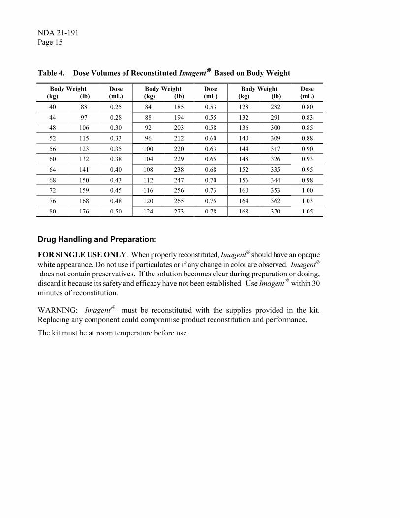
The recommended dose is 0.00625 mL/kg (0.125 mg/kg) administered as a single intravenous bolus over a period of not less than 10 seconds and immediately followed by a saline flush. Imagent� must be used within 30 minutes of reconstitution. Table 4 provides the dose volume (mL) for body kilogram (and pound) weights. Discard any unused portion.
The safety and clinical benefit of repeated doses of Imagent� have not been established.
The mechanical index setting when using Imagent� should be below 1.0. Higher settings will cause microsphere rupture (See Warnings and Precautions).
NDA 21-191 Page 15
Table 4. Dose Volumes of Reconstituted Imagent���� Based on Body Weight
Body Weight (kg) (lb)
Dose (mL)
Body Weight (kg) (lb)
Dose (mL)
Body Weight (kg) (lb)
Dose (mL)
40 88 0.25 84 185 0.53 128 282 0.80 44 97 0.28 88 194 0.55 132 291 0.83 48 106 0.30 92 203 0.58 136 300 0.85 52 115 0.33 96 212 0.60 140 309 0.88 56 123 0.35 100 220 0.63 144 317 0.90 60 132 0.38 104 229 0.65 148 326 0.93 64 141 0.40 108 238 0.68 152 335 0.95 68 150 0.43 112 247 0.70 156 344 0.98 72 159 0.45 116 256 0.73 160 353 1.00 76 168 0.48 120 265 0.75 164 362 1.03 80 176 0.50 124 273 0.78 168 370 1.05
Drug Handling and Preparation:
FOR SINGLE USE ONLY. When properly reconstituted, Imagent� should have an opaque white appearance. Do not use if particulates or if any change in color are observed. Imagent� does not contain preservatives. If the solution becomes clear during preparation or dosing, discard it because its safety and efficacy have not been established Use Imagent� within 30 minutes of reconstitution.
WARNING: Imagent� must be reconstituted with the supplies provided in the kit. Replacing any component could compromise product reconstitution and performance.
The kit must be at room temperature before use.

NDA 21-191 Page 16
Patient Preparation 1. Prepare patient with intravenous catheter of 20 gauge
or larger. 2. Determine the appropriate volume to administer a
dose of 0.125 mg/kg (or 0.00625 mL/kg) based on the weight of the patient (see Table 4, Dosage and Administration section).
Reconstitution Procedure for Imagent���� 1. Connect the 10 mL syringe and dispensing pin into
one assembly. Fully depress syringe plunger to expel air (Fig. 1).
2. Grasp the dispensing pin and push down through the center of the Sterile Water for Injection (SWFI) stopper.
3. Invert the SWFI and dispensing pin assembly and slowly withdraw 10 mL into the syringe (Fig. 2).
4. Remove the dispensing pin assembly from the SWFI. 5. Push and twist the dispensing pin assembly into the
Imagent� vial until fully inserted. DO NOT INJECT AIR INTO THE IMAGENT� VIAL
6. Holding the vial and syringe at a 45-degree angle with the side vent facing up and the syringe facing down, slowly (over 10 seconds) inject 7 mL SWFI. Briefly swirl to wet the powder (Fig. 3).
7. Add the remaining SWFI (about 3 mL) and shake until dissolved and the solution is an opaque white liquid that may have a foamy appearance (Fig. 4).
8. Do not allow reconstituted Imagent���� to sit for longer than 30 minutes before administration. Imagent���� must be discarded after 30 minutes of reconstitution.
Fig. 1
Fig. 2
Fig. 3
Fig. 4

NDA 21-191 Page 17
Administration Procedure for Imagent����
1. Immediately (within one minute) before injection, repeat agitation of vial (Fig. 4) to resuspend its contents, invert and allow foam to rise to the surface of the liquid, avoiding the foam that was allowed to rise.
2. Withdraw 1 mL of reconstituted Imagent� to remove any residual water from the dispensing pin assembly. Detach the 10-mL syringe and contents from the dispensing pin and discard (Fig. 5). The product is now ready for administration.
3. Based on the calculated volume need, connect an appropriately calibrated 1-3 mL syringe (not supplied) to the dispensing pin. Slowly withdraw the dose. (Fig. 6). Do not push/pull the plunger rapidly. If excessive pressure/vacuum is applied and the microspheres are damaged, the product may become clear and should not be used. Immediately after withdrawing the dose, inject it over 30 seconds into the intravenous line.
4. Immediately after injection slowly (over a 30 second period) flush the catheter with 1-2 mL of saline to clear the catheter.
HOW SUPPLIED
Imagent� Kit for the preparation of Perflexane-Lipid Microspheres for Injectable Suspension is supplied for single-use. Each kit contains the following parts:
• One 10-mL glass vial containing 200 mg of Imagent� Powder for Injectable Suspension.
• One 20-mL plastic vial of Sterile Water for Injection, USP.
• One 10-mL disposable plastic sterile syringe.
• One vented 5 µm filter dispensing pin.
• One package insert.
The powder in the vial must be reconstituted with 10 mL supplied SWFI and withdrawn from the vial via the supplied vented 5 µm filter dispensing pin as described under DOSAGE AND ADMINISTRATION – Reconstitution Procedure for Imagent�.
U.S. Patent Nos: (5,605,673); (5,626,833); (5,639,443); (5,695,741); (5,720,938); (5,798,091); (6,258,339); (6,280,704); (6,280,705); and (6,287,539)
Fig. 5
Fig. 6

NDA 21-191 Page 18
NDC xxxxx-xxxx-xx.
STORAGE
Store the kit components and reconstituted Imagent� at 25°C (77°F); excursions permitted to 15 – 30°C (59° – 86°F). [see USP Controlled Room Temperature]
Use reconstituted Imagent� Injectable Suspension within 30 minutes.
Month, 2002
Manufactured by Alliance Pharmaceutical Corp., San Diego, CA U.S.A 92121
For Customer Service call 1-800-IMAGENT (1-800-462-4368)

NDA 21-191 Page 19
LABEL FOR IMMEDIATE CONTAINER , 10ML GLASS VIAL-
(OPTION 2 OF EMAIL DATED 1-MAY-02)
Lot # 000000 Exp: MMMYY ------------------------------------------------------------------------------------------------------------------------------
Imagent
Perflexane Lipid Microspheres Powder For Injectable Suspension
200 mg For intravenous use only
Single–dose vial. Contains no preservatives. Rx Only Use within 30 minutes of reconstitution. Discard unused portion. See package insert for Product Description, Dosage, Reconstitution, and Administration instructions. Do not use if seal is broken. Store at 25 °°°°C (77 °°°°F); excursions permitted to 15-30 °°°°C ( 59-86 °°°°F) ---------------------------------------------------------------------------------------------------------------------------- Reconstitute with 10 mL SWFI prior to use and remove from vial using provided vented 5 µµµµm filter dispensing pin. Upon reconstitution, each mL contains: Maximum of 13.7 x 10⁸ microspheres 92 µµµµg perflexane 0.92 mg DMPC 7.5 mg hydroxyethyl starch 0.21 mg poloxamer 188. Product is iso-osmotic, pH 6.7 to 7.7 Alliance Pharmaceutical Corp. San Diego, CA 92121 U.S.A. NDC # xxxxx-xxxx-xx Artwork Rev. xx/xx

NDA 21-191 Page 20
TOP PANEL - Carton Actual Size: 9 1/8 x 7 3/4
Imagent® Kit for the Preparation of Perflexane Lipid Microspheres Injectable Suspension
Rx Only
For intravenous use only.
Each kit contains one 10-mL glass vial containing 200 mg of Imagent Powder for Injectable Suspension, one 20-mL plastic vial of Sterile Water for Injection, USP, one 10-mL disposable plastic sterile syringe,
one vented Mini-Spike 5 µm filter dispensing pin, one package insert.
Carton contains 4 kits.
Catalog Number XXXXXXX
For Customer Service Call 1 - XXX – XXX- XXXX
Alliance Pharmaceutical Corp.
FRONT SIDE PANEL - Carton Actual Size: 9 1/8 x 5 3/4
Imagent® Kit for the Preparation of Perflexane Lipid Microspheres Injectable Suspension Carton contains 4 kits.
Alliance Pharmaceutical Corp.
See package insert for Product Description, Dosage, Reconstitution, and Administration.
REAR SIDE PANEL - Carton Actual Size: 9 1/8 x 5 ¾
LOT # 000000
EXP. DATE: XX/XXXX

NDA 21-191 Page 21
LEFT SIDE PANEL - Carton Actual Size: 7 3/4 x 5 3/4
Imagent® Kit for the Preparation of
Perflexane Lipid Microspheres Injectable Suspension
Store at 25 °C (77 °F); excursions permitted to 15-30 °C (59-86 °F)
[see USP Controlled Room Temperature]
Patents, see Insert
Alliance Pharmaceutical Corp., San Diego, CA 92121
RIGHT SIDE PANEL - Carton Actual Size: 7 3/4 x 5 3/4
Imagent® Kit for the Preparation of Perflexane Lipid Microspheres
Injectable Suspension Rx Only
For intravenous use only.
Alliance Pharmaceutical Corp., San Diego, CA 92121
Made in USA
NDC #xxxxx-xxxxxx
Artwork revision number will be placed inside box flap

---------------------------------------------------------------------------------------------------------------------This is a representation of an electronic record that was signed electronically andthis page is the manifestation of the electronic signature.--------------------------------------------------------------------------------------------------------------------- /s/---------------------Florence Houn5/31/02 11:22:51 AM