P-I Snake Venom Metalloproteinase Is Able to Activate the ...
Diversity of Micrurus Snake Species Related to TheirVenom Toxic Effects and the Prospective of AntivenomNeutralizationGabriela D. Tanaka1, Maria de Fatima D. Furtado2, Fernanda C. V. Portaro1, Osvaldo Augusto
Sant’Anna1, Denise V. Tambourgi1*
1 Immunochemistry Laboratory, Butantan Institute, Sao Paulo, SP, Brazil, 2 Herpetology Laboratory, Butantan Institute, Sao Paulo, SP, Brazil
Abstract
Background: Micrurus snake bites can cause death by muscle paralysis and respiratory arrest, few hours after envenomation.The specific treatment for coral snake envenomation is the intravenous application of heterologous antivenom and, inBrazil, it is produced by horse immunization with a mixture of M. corallinus and M. frontalis venoms, snakes that inhabit theSouth and Southeastern regions of the country. However, this antivenom might be inefficient, considering the existence ofintra- and inter-specific variations in the composition of the venoms. Therefore, the aim of the present study was toinvestigate the toxic properties of venoms from nine species of Micrurus: eight present in different geographic regions ofBrazil (M. frontalis, M. corallinus, M. hemprichii, M. spixii, M. altirostris, M. surinamensis, M. ibiboboca, M. lemniscatus) and one(M. fulvius) with large distribution in Southeastern United States and Mexico. This study also analyzed the antigenic cross-reactivity and the neutralizing potential of the Brazilian coral snake antivenom against these Micrurus venoms.
Methodology/Principal Findings: Analysis of protein composition and toxicity revealed a large diversity of venoms fromthe nine Micrurus species. ELISA and Western blot assays showed a varied capability of the therapeutic antivenom torecognize the diverse species venom components. In vivo and in vitro neutralization assays indicated that the antivenom isnot able to fully neutralize the toxic activities of all venoms.
Conclusion: These results indicate the existence of a large range of both qualitative and quantitative variations in Micrurusvenoms, probably reflecting the adaptation of the snakes from this genus to vastly dissimilar habitats. The data also showthat the antivenom used for human therapy in Brazil is not fully able to neutralize the main toxic activities present in thevenoms from all Micrurus species occurring in the country. It suggests that modifications in the immunization scheme, withthe inclusion of other venoms in the antigenic mixture, should occur in order to generate effective therapeutic coral snakeantivenom.
Citation: Tanaka GD, Furtado MdFD, Portaro FCV, Sant’Anna OA, Tambourgi DV (2010) Diversity of Micrurus Snake Species Related to Their Venom Toxic Effectsand the Prospective of Antivenom Neutralization. PLoS Negl Trop Dis 4(3): e622. doi:10.1371/journal.pntd.0000622
Editor: H. Janaka de Silva, University of Kelaniya, Ragama, Sri Lanka
Received October 1, 2009; Accepted January 20, 2010; Published March 9, 2010
Copyright: � 2010 Tanaka et al. This is an open-access article distributed under the terms of the Creative Commons Attribution License, which permitsunrestricted use, distribution, and reproduction in any medium, provided the original author and source are credited.
Funding: The study was funded by FAPESP, CNPq, and INCTTox. The funders had no role in study design, data collection and analysis, decision to publish, orpreparation of manuscript.
Competing Interests: The authors have declared that no competing interests exist.
* E-mail: [email protected]
Introduction
The Elapidae family has about 250 species, distributed from the
Southeastern and Southwestern United States, through Mexico,
Central America and South America, and are also found in Asia,
Africa and Australia [1]. In the Americas, there is a group of more
than 120 species and subspecies, divided into three genera:
Micruroides, with one species; Leptomicrurus with three, and Micrurus,
with almost 70 species [1,2,3,4].
Coral snakes have a large geographical distribution in Americas,
inhabiting extremely diverse environments, from lowland rain-
forests and deserts to highland cloudy forests [1,5]. Most of the
snakes of the Micrurus genus has terrestrial to subfossorial habits,
however, some species are semi-aquatic, such as M. surinamensis
and M. lemniscatus [1].
Most coral snakes have a color pattern of some combination of
the red, yellow or white, and black, usually disposed in rings.
They are proterogliphous animals, presenting the fixed small
teeth at the forefront of the mouth. Food is generally composed of
small snakes, but may also include lizards and amphisbaenians.
Certain species have specialized nutritional habits, feeding on
caecilians, swamp eels, and other type of fishes and even
onycophorans and other invertebrates [1]. Snakes such as M.
lemniscatus and M. surinamensis feed on fish and M. hemprichii of
peripatus [6,7,5].
The Micrurus species of public health importance are M. fulvius in the
United States and Mexico, M. alleni, M. diastema and M. nigrocintus in
Central America and M. altirostris, M. corallinus, M. dumerilii, M. frontalis,
M. mipartitus, M. spixii, M. surinamensis and M. isozonus in South America
[8,9,10]. In Brazil, some species are quite common and widespread in
large areas of the territory, such as M. corallinus, M. frontalis, M. ibiboboca,
M. lemniscastus, M. spixii and M. surinamensis.
Human envenomations by coral snakes are relatively rare due to
their subfossorial habits; however, the case fatality, attributable to
www.plosntds.org 1 March 2010 | Volume 4 | Issue 3 | e622
respiratory paralysis, may be high [11]. A variety of local and
systemic manifestations of envenoming has been described in
patients bitten by different species of coral snakes [5,12,11]. The
main feature of the venom action is the neurotoxicity, although,
experimentally, it has been reported that some Micrurus venoms
may induce myotoxicity and local lesions [13,14]. Neurotoxicity
can be produced by a post-synaptic action, through blockage of
the end-plate receptors by alpha neurotoxins, as determined for M.
frontalis venom, or by a pre-synaptic-like activity, which causes
inhibition of acetylcholine release at the motor nerve endings, as
induced by M. corallinus venom. There are also venom toxins, such
as cardiotoxins and myotoxic phospholipases A2 from M.
nigrocinctus and M. fulvius, which block the end-plate receptors
and depolarize the muscle fiber membrane [15].
Experimental studies have shown that Micrurus venoms are
cardiotoxic, myotoxic, hemolytic, hemorrhagic and edemato-
genic [13,14,16,17,18,19,20,21,22,23,24]. Furthermore, many
enzymatic activities have been detected including those derived
from phospholipase A2, hyaluronidase, phosphodiesterase, leu-
cine amino peptidase, L-amino acid dehydrogenase, acetylcho-
linesterase, alkaline phosphomonoesterase and L-amino acid
oxidase actions [25,26]. Anticoagulant action was also identified
in some coral venom species, but none or little proteolytic
activity was detected [26]. Therefore, common characteristics, as
well as variability of some biological properties, have been
demonstrated in comparative studies of Micrurus venoms
[23,25,26,27,28].
The transcriptomic analysis of a Micrurus snake venom gland (M.
corallinus) was recently described [29]. Toxin transcripts represent-
ed 46% of the total ESTs and the main toxin classes were
neurotoxins, i.e, three-finger toxins (3FTx) and phospholipases A2
(PLA2s). It was also showed that the post-synaptic components
(3FTx) were very diverse in terms of sequences, possibly aiming to
achieve different types of receptors, whereas the pre-synaptic
component (PLA2) was more conserved. The high expression of
both types of these neurotoxins is in agreement with the known
presence of pre- and post-synaptic activities in the Micrurus
venoms. However, eight other classes of toxins were found,
including C-type lectins, natriuretic peptide precursors and high-
molecular mass components such as metalloproteases and L-
amino acid oxidases.
The specific treatment for Micrurus envenomation is the
intravenous application of heterologous antivenom. In Brazil, the
coral snake therapeutic antivenom produced by Butantan Institute
is obtained by the immunization of horses with a mixture
containing equivalent amounts of M. corallinus and M. frontalis
venoms [30]. In view of the fact that Micrurus venoms can exhibit a
diversity of composition and toxicity, the therapeutic antivenom
may not be capable to fully recognize all the major components of
the distinct venom species occurring in the country. Therefore, the
aim of this study was to characterize some biological properties of
venoms from nine species of Micrurus, including those used for
serum preparation, i.e., M. frontalis and M. corallinus, evaluate their
antigenic cross-reactivity, using the Brazilian coral snake anti-
venom, as well as to test the ability of this antivenom to neutralize
the main toxic activities of these venoms.
Materials and Methods
Chemicals and reagentsTriton X-100, Tween 20, bovine serum albumin (BSA), L-a-
phosphatidylcholine, ortho-phenylenediamine (OPD), hyaluro-
nic acid and goat anti-horse (GAH) IgG horseradish peroxidase
labeled (IgG-HRPO) were purchased from Sigma (St. Louis,
Missouri, USA). Goat anti-horse (GAH) IgG labeled with
alkaline phosphatase (IgG-AP), 5-bromo-4-chloro-3-indolyl-
phosphate (BCIP) and nitroblue tetrazolium (NBT) were from
Promega Corp. (Madison, Wisconsin, USA). The P2E Fluores-
cent Resonance Energy Transfer (Abz-FEPFRQ-EDnp) sub-
strate was synthesized and purified as described in Hirata et al.
[31].
VenomsVenoms from Micrurus frontalis, M. corallinus, M. ibiboboca, M.
hemprichii, M. spixii, M. fulvius, M. altirostris, M. surinamensis and M.
lemniscatus were supplied by Herpetology Laboratory from
Butantan Institute, SP, Brazil. Stock solutions were prepared in
PBS (10 mM sodium phosphate containing 150 mM NaCl,
pH 7.2) at 1.0 mg/mL.
Coral snake antivenomThe Brazilian therapeutic coral snake antivenom, produced by
hyperimmunization of horses with venoms from M. corallinus (50%)
and M. frontalis (50%), was obtained from Butantan Institute, SP,
Brazil.
Electrophoresis and western blotSamples of 20 mg of Micrurus venoms were solubilised in non-
reducing sample buffer and run on 7.5 to 15% SDS-PAGE
gradient gels [32]. Gels were stained with silver [33] or blotted
onto nitrocellulose [34]. After transfering, the membrane was
blocked with PBS containing 5% BSA and incubated with the
coral snake antivenom (diluted 1:2,000) for 1 h at room
temperature. The membrane was washed 3 times for 10 min with
PBS/0.05% Tween 20, and incubated with GAH/IgG-AP
(1:7,500) in PBS/1% BSA for 1 h at room temperature. After
washing 3 times for 10 min with PBS/0.05% Tween 20, the blot
was developed using NBT/BCIP according to the manufacturer’s
instructions (Promega).
Author Summary
The Elapidae family is represented in America by threegenera of coral snakes: Micruroides, Leptomicrurus andMicrurus, the latter being the most abundant anddiversified group. Micrurus bites can cause death bymuscle paralysis and respiratory arrest few hours afterenvenomation. The specific treatment for Micrurus enven-omation is the application of heterologous antivenom. Theaim of this study was to compare the toxicity of venomsfrom nine species of coral snakes and analyze theneutralization potential of the Brazilian coral snakeantivenom. In vitro assays showed that the majority ofthe Micrurus venoms are endowed with phospholipaseand hyaluronidase and low proteolytic activities. Theseenzymes are not equally neutralized in all venoms by thetherapeutic antivenom. Moreover, in vivo assays showedthat some of the Micrurus venoms are extremely lethal,such as the ones from M. altirostris, M. corallinus, M.frontalis, M. lemniscatus and M. spixii. Neutralization tests,performed in vivo, showed that the therapeutic antivenomwas able to neutralize better the venoms from M. frontalis,M. corallinus, and M. spixii but not from M. altirostris and M.lemniscatus. Taken together, these results suggest thatmodifications in the immunization antigenic mixtureshould occur in order to generate more comprehensivetherapeutic antivenom.
Diversity of Micrurus Snake Venoms
www.plosntds.org 2 March 2010 | Volume 4 | Issue 3 | e622
Determination of LD50
The lethal potential of Micrurus venoms was assessed in Swiss
mice by intraperitoneal injection of different amounts of venoms in
500 mL of PBS. Four animals were used for each venom dose (five
doses). The LD50 was calculated by probit analysis of death
occurring within 48 h of venom injection [35]. All animal
experiments were approved in advance by the Laboratory Animal
Ethics Committee of Butantan Institute.
Phospholipase activityThe phospholipase A2 activity of Micrurus venoms was
determined as described by Price III [36], with some modifica-
tions. Samples of the venoms (4 mg) and PBS were added to a
final volume of 200 mL. Samples of 180 mL of the mixture
containing: 5 mM Triton X-100, 5 mM phosphatidylcholine
(Sigma), 2 mM HEPES, 10 mM calcium chloride and 0,124%
(wt/vol) bromothymol blue dye in water, at pH 7.5 and at 37uC,
were added. After a pre-incubation of 5 min at 37uC, the
absorbance of the samples was determined at l 620 nm in a
Multiskan spectrophotometer EX (Labsystems, Finland). Results
were expressed in nanomoles of acid per minute per mg of venom
(compared on pH changes in standard curves of the reaction
mixture using HCl).
Proteolytic activitySamples of the Micrurus venoms (50 mg) were mixed with
5 mM of the Fluorescent Resonance Energy Transfer (FRET)
substrate, Abz-FEPFRQ-EDnp, and PBS, for a final volume of
100 mL, and the reactions monitored by measuring the
fluorescence (lem 420 nm and lex 320 nm) in a spectrofluorim-
eter (Victor 3TM, Perkin-Elmer, USA) at 37uC, as described by
Araujo et al. [37]. The specific proteolytic activity was expressed
as units of free fluorescence per minute per mg of venom (UF/
min/mg).
Hyaluronidase activityHyaluronidase activity was measured as described [38], with
slight modifications. Samples of Micrurus venoms (30 mg) were
added to 100 mL of the hyaluronic acid substrate (1 mg/mL) and
acetate buffer (pH 6.0) for a final volume of 500 mL. The mixtures
were incubated for 15 min at 37uC. After the incubation, it was
added to the samples 1 mL of cetyltrimethylammonium bromide
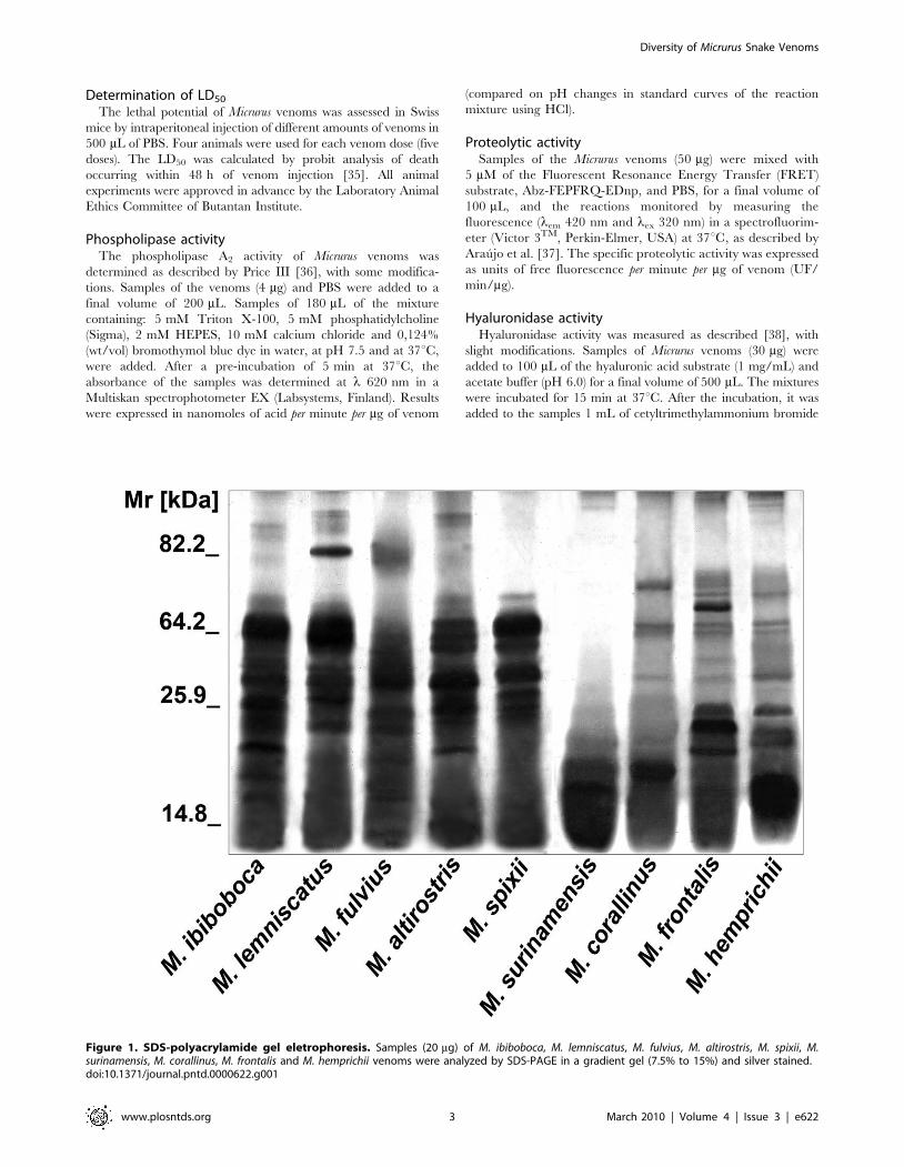
Figure 1. SDS-polyacrylamide gel eletrophoresis. Samples (20 mg) of M. ibiboboca, M. lemniscatus, M. fulvius, M. altirostris, M. spixii, M.surinamensis, M. corallinus, M. frontalis and M. hemprichii venoms were analyzed by SDS-PAGE in a gradient gel (7.5% to 15%) and silver stained.doi:10.1371/journal.pntd.0000622.g001
Diversity of Micrurus Snake Venoms
www.plosntds.org 3 March 2010 | Volume 4 | Issue 3 | e622
2.5% in NaOH 2%, to develop the turbidity in the mixtures, and
the absorbance measured in a spectrophotometer (Multiskan EX)
at lem 405 nm. Results were expressed in units of turbidity
reduction (UTR) per mg of venom.
Enzyme linked immunosorbent assay (ELISA)Microtitre plates were coated with 100 mL of Micrurus venoms
(10 mg/mL; overnight at 4uC). Plates were blocked with 5% BSA
in PBS and increased dilutions of the therapeutic coral snake
antivenom were added. After 1 h of incubation at room
temperature, plates were washed with PBS/0.05% Tween 20
and incubated with GAH-IgG-HRPO diluted 1:3,000, for 1 h at
room temperature. Plates were washed and the reactions
developed with OPD substrate according to the manufacturers
conditions (Sigma). The absorbances were recorded in an ELISA
reader (Multiskan spectrophotometer EX) at l 492 nm. The titer
was established as the highest antivenom dilution, in which an
absorbance five times greater than that determined for the normal
horse serum was measured.
Table 1. Lethal dose 50% (LD50) of Micrurus spp snakevenoms determined in murine model.
Venoms LD50 (mg)
Micrurus ibiboboca 76 (67–89)
Micrurus lemniscatus 13 (7–22)
Micrurus fulvius 64 (52–88)
Micrurus altirostris 9 (7–13)
Micrurus spixii 8 (6–16)
Micrurus surinamensis 58 (43–87)
Micrurus corallinus 7 (5–27)
Micrurus frontalis 22 (4–29)
Micrurus hemprichii 47 (20–88)
Results are expressed in mg venom/mouse (18–22 g), and the 95% confidencelimits are included in parenthesis.doi:10.1371/journal.pntd.0000622.t001
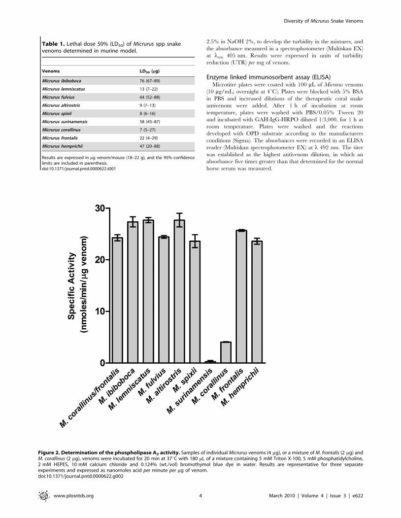
Figure 2. Determination of the phospholipase A2 activity. Samples of individual Micrurus venoms (4 mg), or a mixture of M. frontalis (2 mg) andM. corallinus (2 mg), venoms were incubated for 20 min at 37uC with 180 mL of a mixture containing 5 mM Triton X-100, 5 mM phosphatidylcholine,2 mM HEPES, 10 mM calcium chloride and 0.124% (wt./vol) bromothymol blue dye in water. Results are representative for three separateexperiments and expressed as nanomoles acid per minute per mg of venom.doi:10.1371/journal.pntd.0000622.g002
Diversity of Micrurus Snake Venoms
www.plosntds.org 4 March 2010 | Volume 4 | Issue 3 | e622
Serum neutralization assays performed in vitroThe ability of the therapeutic Brazilian coral snake antivenom
to neutralize the venoms phospholipase, hyaluronidase and
proteolytic activities was estimated by incubating Micrurus venoms
with the antivenom. The antivenom volume, amount of venoms
and the pre-incubation time, for each tested enzymatic activity,
was standardized using the immunization pool, composed by 50%
of M. corallinus and 50% of M. frontalis venoms. For serum
neutralization measurements of the phospholipase activity,
samples of 4 mg of the venoms were incubated with the antivenom,
diluted 1:10; for the hyaluronidase activity, samples of Micrurus
venoms (30 mg) and the antivenom (1:20) were incubated for
20 min at room temperature; for the proteolytic activity, samples
of Micrurus venoms (50 mg) and the antivenom (1:4) were incubated
for 10 min at room temperature. Venoms residual toxic activities
were measured as described above.
Neutralization of the lethal activityThe capacity of the therapeutic coral snake antivenom to
neutralize the lethal activity of Micrurus venoms was determined by
mixing the venoms, corresponding to 2 LD50, with serial dilutions
of the horse antivenom. The mixtures were incubated for 30 min
at 37uC and the animals received 0.5 mL by the intraperitoneal
route. The effective dose (ED50) was calculated from the number
of deaths within 48 h of injection of the venom/antivenom
mixture using probit analysis, as described above. The ED50 was
expressed as mL of antivenom per mg of venom.
Results
Eletrophoretic characterization of Micrurus venomsThe protein profiles of Micrurus venoms were analyzed by SDS-
PAGE followed by silver staining. Figure 1 shows that the
venoms from the nine coral species differ in composition, number
and intensity of bands. The majority of the components of these
venoms present Mr inferior to 64 kDa. Venom from M.
surinamensis differs more from the others, by the presence of a
few number of components with Mr lower than 20 kDa.
Enzymatic activities of the Micrurus venomsIn order to assess whether the venoms of Micrurus displayed the
same biological activities, some functional assays were carried out.
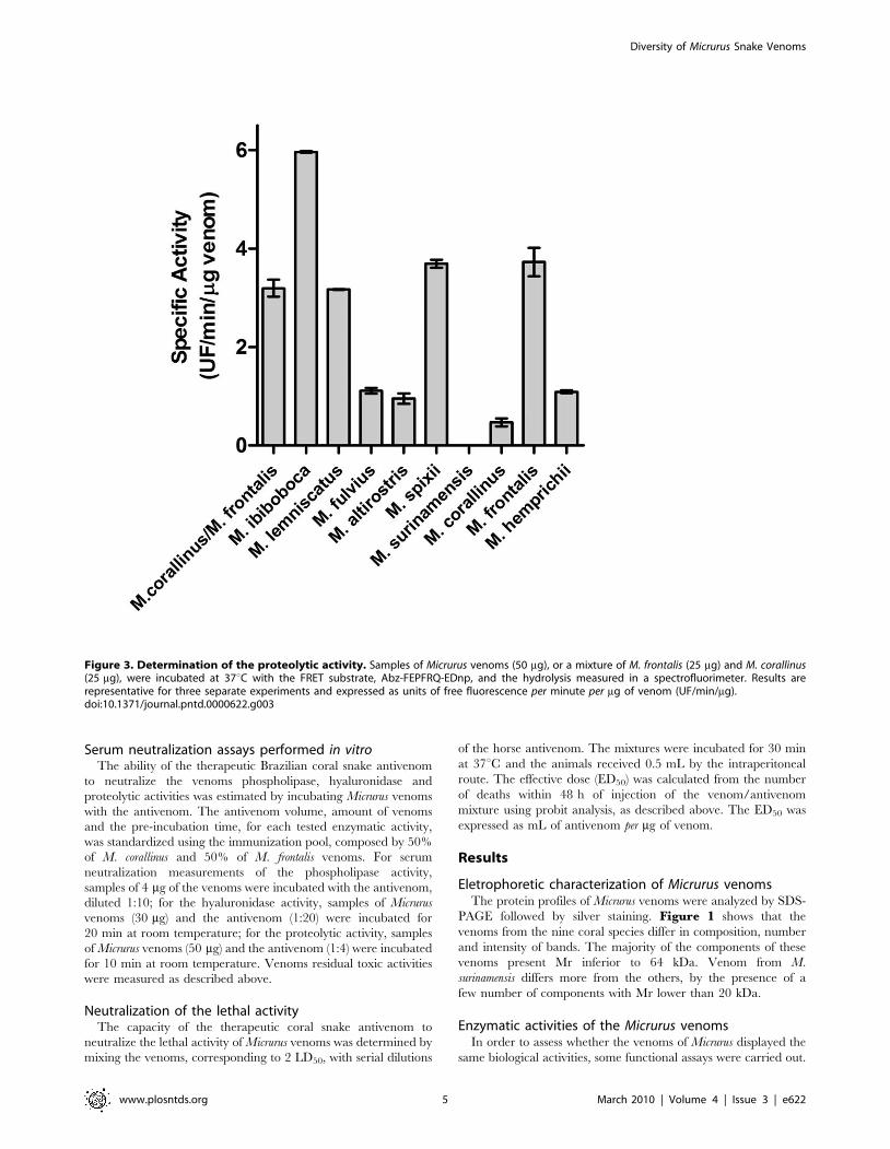
Figure 3. Determination of the proteolytic activity. Samples of Micrurus venoms (50 mg), or a mixture of M. frontalis (25 mg) and M. corallinus(25 mg), were incubated at 37uC with the FRET substrate, Abz-FEPFRQ-EDnp, and the hydrolysis measured in a spectrofluorimeter. Results arerepresentative for three separate experiments and expressed as units of free fluorescence per minute per mg of venom (UF/min/mg).doi:10.1371/journal.pntd.0000622.g003
Diversity of Micrurus Snake Venoms
www.plosntds.org 5 March 2010 | Volume 4 | Issue 3 | e622
The LD50, used as a parameter of the Micrurus venom
neurotoxicity, was tested in groups of mice, after intraperitoneal
injection of different concentrations of the venoms, and the
number of deaths recorded during 48 h. The LD50 values,
calculated by probit analysis at 95% confidence, were variable
among Micrurus venoms, being the most lethal those from M.
lemniscatus, M. altirostris, M. spixii, M. corallinus and M. frontalis
(Table 1).
Figure 2 shows that venoms contain variable levels of PLA2
activity, toxin also associated with Micrurus envenomation
neurotoxicity. M. ibiboboca, M. lemniscatus, M. fulvius, M. altirostris,
M. spixii, M. frontalis, M. hemprichii and the mixture of M. corallinus
and M. frontalis venoms present an intense hydrolytic activity. In
the same experimental conditions, the venoms from M. corallinus
and M. surinamensis showed, respectively, low and none phospho-
lipase activity.
The proteolytic activity of the Micrurus venoms was tested using
a FRET substrate, Abz-FEPFRQ-EDnp. Figure 3 demonstrates
that the venoms from M. ibiboboca, M. lemniscatus, M. fulvius, M.
altirostris, M. spixii, M. corallinus, M. frontalis and M. hemprichii present
hydrolytic activity on this substrate. However, no proteolytic could
be measured in the venom from M. surinamensis.
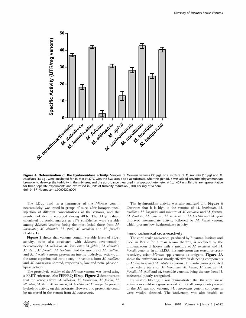
The hyaluronidase activity was also analyzed and Figure 4illustrates that it is high in the venoms of M. lemniscatus, M.
corallinus, M. hemprichii and mixture of M. corallinus and M. frontalis.
M. ibiboboca, M. altirostris, M. surinamensis, M. frontalis and M. spixii
displayed intermediate activity followed by M. fulvius venom,
which presents low hyaluronidase activity.
Immunochemical cross-reactivityThe coral snake antivenom, produced by Butantan Institute and
used in Brazil for human serum therapy, is obtained by the
immunization of horses with a mixture of M. corallinus and M.
frontalis venoms. In an ELISA, this antivenom was tested for cross-
reactivity, using Micrurus spp venoms as antigens. Figure 5Ashows the antivenom was mostly effective in detecting components
of M. corallinus and M. ibiboboca venoms. This antivenom presented
intermediary titers for M. lemniscatus, M. fulvius, M. altirostris, M.
frontalis, M. spixii and M. hemprichii venoms, being the one from M.
surinamensis poorly recognized.
By western blotting, it was demonstrated that the coral snake
antivenom could recognize several but not all components present
in the Micrurus spp venoms. M. surinamensis venom components
were weakly detected. The antivenom was also unable to
Figure 4. Determination of the hyaluronidase activity. Samples of Micrurus venoms (30 mg), or a mixture of M. frontalis (15 mg) and M.corallinus (15 mg), were incubated for 15 min at 37uC with the hyaluronic acid as substrate. After this period, it was added cetyltrimethylammoniumbromide, to develop the turbidity in the mixtures, and the absorbance measured in a spectrophotometer at lem 405 nm. Results are representativefor three separate experiments and expressed in units of turbidity reduction (UTR) per mg of venom.doi:10.1371/journal.pntd.0000622.g004
Diversity of Micrurus Snake Venoms
www.plosntds.org 6 March 2010 | Volume 4 | Issue 3 | e622
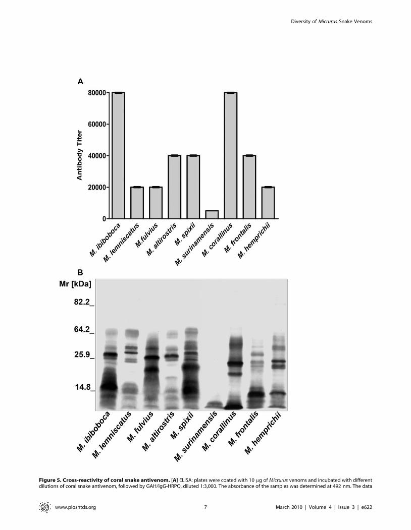
Figure 5. Cross-reactivity of coral snake antivenom. [A] ELISA: plates were coated with 10 mg of Micrurus venoms and incubated with differentdilutions of coral snake antivenom, followed by GAH/IgG-HRPO, diluted 1:3,000. The absorbance of the samples was determined at 492 nm. The data
Diversity of Micrurus Snake Venoms
www.plosntds.org 7 March 2010 | Volume 4 | Issue 3 | e622
recognize molecules with Mr above 64 kDa present in the majority
of the venoms (Fig. 5B).
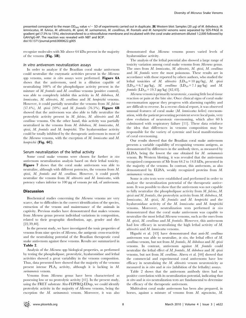
In vitro antivenom neutralization assaysIn order to analyze if the Brazilian coral snake antivenom
could neutralize the enzymatic activities present in the Micrurus
spp venoms, some in vitro assays were performed. Figure 6Ashows that the antivenom, used in a dilution capable of
neutralizing 100% of the phospholipase activity present in the
mixture of M. frontalis and M. corallinus venoms (positive control),
was able to completely inhibit this activity in M. ibiboboca, M.
lemniscatus, M. altirostris, M. corallinus and M. hemprichii venoms.
However, it could partially neutralize the venoms from M. fulvius
(57.4%), M. spixii (59%) and M. frontalis (76.5%). Figure 6Bshowed that the antivenom was also able to fully neutralize the
proteolytic activity present in M. fulvius, M. altirostris and M.
corallinus venoms. On the other hand, this activity was partially
neutralized in the venoms from M. ibiboboca, M. lemniscatus, M.
spixii, M. frontalis and M. hemprichii. The hyaluronidase activity
could be totally inhibited by the therapeutic antivenom in most of
the Micrurus venoms, with the exception of M. lemniscatus and M.
hemprichii (Fig. 6C).
Serum neutralization of the lethal activitySome coral snake venoms were chosen for further in vivo
antivenom neutralization analysis based on their lethal toxicity.
Figure 7 shows that the coral snake antivenom was able to
neutralize, although with different potencies, the venoms from M.
spixii, M. frontalis and M. corallinus. However, it could poorly
neutralize the venoms from M. altirostris and M. lemniscatus, with
potency values inferior to 100 mg of venom per mL of antivenom.
Discussion
Biochemical studies concerning the Micrurus venoms are very
scarce, due to difficulties in the correct identification of the species,
extraction of the venom and maintenance of the animals in
captivity. Previous studies have demonstrated that snakes venoms
from Micrurus genus present individual variations in composition,
related to their geographic distribution, age, gender and diet
[22,39,40].
In the present study, we have investigated the toxic properties of
venoms from nine species of Micrurus, the antigenic cross-reactivity
and the neutralizing potential of the Brazilian therapeutic coral
snake antivenom against these venoms. Results are summarized in
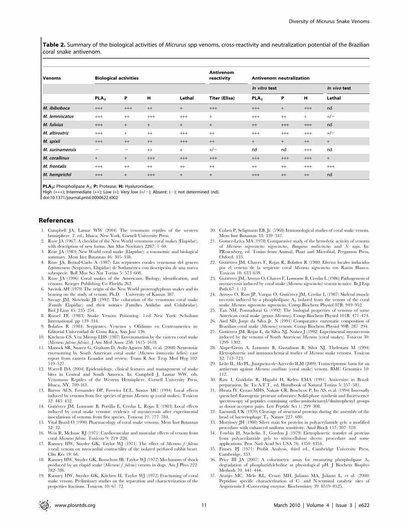
Table 2.
Analysis of the Micrurus spp biological properties, as performed
by testing the phospholipase, proteolytic, hyaluronidase and lethal
activities showed a great variability in the venoms composition.
Thus, data presented here showed that the majority of the venoms
present intense PLA2 activity, although it is lacking in M.
surinamensis venom.
Venoms from Micrurus genus have been characterized as
possessing low or no proteolytic activity [41]. In the present study,
using the FRET substrate Abz-FEPFRQ-EDnp, we could identify
proteolytic activity in the majority of Micrurus venoms, being the
exception the M. surinamensis venom. Moreover, it was also
demonstrated that Micrurus venoms posses varied levels of
hyaluronidase activity.
The analysis of the lethal potential also showed a large range of
toxicity variation among coral snake venoms from Micrurus genus.
The ones from M. lemniscatus, M. altirostris, M. spixii, M. corallinus
and M. frontalis were the most poisonous. These results are in
accordance with those reported by others authors, who studied the
lethal toxicities of M. altirostris (LD50 = 10 mg/kg), M. spixii
(LD50 = 6.7 mg/kg), M. corallinus (LD50 = 7.1 mg/kg) and M.
frontalis (LD50 = 19.3 mg/kg) [42,43].
Micrurus venom is primarily neurotoxic, causing little local tissue
reaction or pain at the bite site. Once clinical signs of coral snake
envenomation appear they progress with alarming rapidity and
are difficult to reverse. In a recent clinical report, it was observed
unusual features of coral snake (M. lemniscatus helleri) envenom-
ation, with the patient presenting persistent severe local pain, very
slow evolution of neurotoxic envenoming, which after 60 h
culminated with respiratory failure [11]. These data reinforce
the idea that differences in venoms composition may be
responsible for the variety of systemic and local manifestations
of coral envenoming.
Our results showed that the Brazilian coral snake antivenom
presents a variable capability of recognizing venoms antigens, as
demonstrated by differences in the antibody titers, as measured by
ELISA, being the lowest the one obtained for M. surinamensis
venom. By Western blotting, it was revealed that the antivenom
recognized components of Mr from 64.2 to 14.8 kDa, presented in
the majority of the venoms. In contrast, this antivenom, as also
demonstrated by ELISA, weakly recognized proteins from M.
surinamensis venom.
Some in vitro tests were established and performed in order to
analyze the neutralization potential of the therapeutic antive-
nom. It was possible to show that the antivenom was not capable
to fully neutralize the phospholipase activity from M. fulvius, M.
spixii and M. frontalis, the proteolytic activity from M. ibiboboca, M.
lemniscatus, M. spixii, M. frontalis and M. hemprichii and the
hyaluronidase activity of the M. lemniscatus and M. hemprichii
venoms. Moreover, neutralization tests performed in vivo
demonstrated that the coral snake antivenom was capable to
neutralize the most lethal Micrurus venoms, such as the ones from
M. spixii, M. corallinus and M. frontalis. However, this antivenom
had low efficacy in neutralizing the high lethal activity of M.
altirostris and M. lemniscatus venoms.
Higashi et al. [43] have demonstrated that anti-M. corallinus
antivenom was able to neutralize, in vivo, the lethal effect of M.
corallinus venom, but not from M. frontalis, M. ibiboboca and M. spixii
venoms. In contrast, antivenom against M. frontalis could
neutralize the lethal effect of M. frontalis, M. ibiboboca and M. spixii
venoms, but not from M. corallinus. Abreu et al. [44] showed that
the commercial and experimental coral antivenoms have low
efficacy in neutralizing the M. altirostris venom neurotoxicity as
measured in in vitro and in vivo (inhibition of the lethality) assays.
Table 2 shows that the antivenom antibody titers had no
positive correlation with its neutralization potential, indicating that
in vitro and in vivo neutralization tests are fundamental to determine
the efficacy of the therapeutic antivenom.
Multivalent coral snake antivenom has been also prepared, in
horses, against a mixture of venoms from M. nigrocinctus, M.
presented correspond to the mean OD492 value +/2 SD of experiments carried out in duplicate. [B] Western blot: Samples (20 mg) of M. ibiboboca, M.lemniscatus, M. fulvius, M. altirostris, M. spixii, M. surinamensis, M. corallinus, M. frontalis and M. hemprichii venoms were separated by SDS-PAGE ingradient gel (7.5% to 15%), electrotransfered to a nitrocellulose membrane and incubated with the coral snake antivenom diluted 1:2,000 followed byGAH/IgG-AP. The reaction was revealed with NBT and BCIP.doi:10.1371/journal.pntd.0000622.g005
Diversity of Micrurus Snake Venoms
www.plosntds.org 8 March 2010 | Volume 4 | Issue 3 | e622
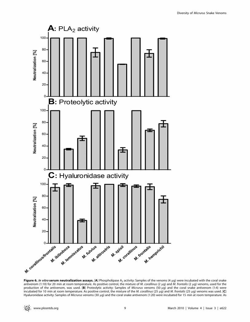
Figure 6. In vitro serum neutralization assays. [A] Phospholipase A2 activity: Samples of the venoms (4 mg) were incubated with the coral snakeantivenom (1:10) for 20 min at room temperature. As positive control, the mixture of M. corallinus (2 mg) and M. frontalis (2 mg) venoms, used for theproduction of the antivenom, was used. [B] Proteolytic activity: Samples of Micrurus venoms (50 mg) and the coral snake antivenom (1:4) wereincubated for 10 min at room temperature. As positive control, the mixture of the M. corallinus (25 mg) and M. frontalis (25 mg) venoms was used. [C]Hyaluronidase activity: Samples of Micrurus venoms (30 mg) and the coral snake antivenom (1:20) were incubated for 15 min at room temperature. As
Diversity of Micrurus Snake Venoms
www.plosntds.org 9 March 2010 | Volume 4 | Issue 3 | e622
mipartitus and M. frontalis species [45]. In this study it was suggested
that it would be useful in treating bites from most of the important
coral snake species in North and South America, such as M. fulvius,
M. alleni, M. carinicaudus dumerilii, M. corallinus, M. frontalis, M.
lemniscatus, M. mipartitus, M. nigrocinctus and M. spixii. They also note
that M. surinamensis venom was not significantly neutralized by the
antivenom.
Table 2 shows the existence of a large range of both qualitative
and quantitative variations in Micrurus venoms, probably
reflecting the adaptation of the snakes from this genus, to vastly
dissimilar habitats. Thus, the comparative analysis of distinct
phenotypes, particularly the venom constituents and their toxic
activities, reveals the heterogeneous complexity of the Micrurus
venoms ascertaining that both the structural and the ecological
evolutions constrain specific characters for adaptive values. The
most striking example is given by M. surinamensis, a snake that
inhabits an extremely distinct environment, whose venom
expresses limited composition. Besides, it was showed that the
antivenom used for human therapy in Brazil is not fully able to
neutralize the main toxic activities present in all Micrurus spp
venoms, indicating that, for the preparation of the Brazilian coral
snake antivenom, other venoms should be included in the
immunization mixture.
Taking into account the decision made by PAHO/WHO [46]
and the countries of the Americas to promote strategies to
diminish the health burden of accidents involving poisonous
animals in the countries of Latin America, it would be lawful to
consider the possibility to prepare a continental coral snake
antivenom, thus contributing to countries where national produc-
tion is insufficient or where it does not have manufacturing
laboratories. The appropriate cooperation by scientists in various
countries in order to prepare multivalent coral snake antivenom
has already been proposed by Bolanos et al. [46], as early as the
1970’s, but until now this relevant aim for the public health of the
Americas has not been achieved. Data present in the literature,
and results obtained in this study, should encourage PAHO to
coordinate a regional cooperative effort to produce multivalent
continental Micrurus antivenom that would have an important
impact in the treatment of accidents involving coral snakes over
the entire continent.
Author Contributions
Conceived and designed the experiments: OAS DVT. Performed the
experiments: GDT MdFDF. Analyzed the data: GDT MdFDF FCVP
OAS DVT. Contributed reagents/materials/analysis tools: MdFDF FCVP
DVT. Wrote the paper: GDT MdFDF FCVP OAS DVT.
positive control, the mixture of the M. corallinus (15 mg) and M. frontalis (15 mg) venoms was used. Venoms residual toxic activities were measured asdescribed in materials and methods. Results are representative for three separate experiments and expressed as percentage of neutralization of thevenoms enzymatic activities.doi:10.1371/journal.pntd.0000622.g006
Figure 7. Antivenom neutralization of Micrurus venoms lethal toxicity. Samples corresponding to 2 LD50, of each Micrurus venom, weremixed with serial dilutions of the coral snake horse antivenom. The mixtures were incubated for 30 min at 37uC and the animals were i.p. inoculated.The effective dose (ED50) was calculated, from the number of deaths within 48 h of injection of the venom/antivenom mixture, using probit analysisand expressed as mL of antivenom per mg of venom.doi:10.1371/journal.pntd.0000622.g007
Diversity of Micrurus Snake Venoms
www.plosntds.org 10 March 2010 | Volume 4 | Issue 3 | e622
References
1. Campbell JA, Lamar WW (2004) The venomous reptiles of the western
hemisphere. 2. ed., Ithaca, New York, Cornell University Press.
2. Roze JA (1967) A checklist of the New World venomous coral snakes (Elapidae),with description of new forms. Am Mus Novitates 2287: 1–60.
3. Roze JA (1983) New World coral snake (Elapidae): a toxonomic and biological
summary. Mem Inst Butantan 46: 305–338.
4. Roze JA, Bernal-Carlo A (1987) Las serpientes corales venenosas del genero
Leptomicrurus (Serpentes, Elapidae) de Sudamerica con descripcion de una nuevasubespecie. Boll Mus Sci Nat Torino 5: 573–608.
5. Roze JA (1996) Coral snakes of the Americans. Biology, identification, and
venoms. Krieger Publishing Co Florida 262.
6. Savitzk AH (1979) The origin of the New World proteroglyphous snakes and itsbearing on the study of venom. Ph.D. – University of Kansas 387.
7. Savage JM, Slowinski JB (1992) The coloration of the venomous coral snake
(Family Elapidae) and their mimics (Families Anilidae and Colubridae).
Biol J Linn 45: 235–254.
8. Russel FE (1983) Snake Venom Poisoning. 1.ed New York: ScholiumInternational. pp 139–344.
9. Bolanos R (1984) Serpientes, Venenos y Ofidismo en Centroamerica in:
Editorial Universidad de Costa Rica, San Jose 136.
10. Kitchens CS, Ven Mierop LHS (1987) Envenomation by the eastern coral snake
(Micrurus fulvius fulvius). J Am Med Assoc 258: 1615–1618.
11. Manock SR, Suarez G, Graham D, Avila-Aguero ML, et al. (2008) Neurotoxicenvenoming by South American coral snake (Micrurus lemniscatus helleri): case
report from eastern Ecuador and review. Trans R Soc Trop Med Hyg 102:519–527.
12. Warrell DA (2004) Epidemiology, clinical features and management of snake
bites in Central and South America. In: Campbell J, Lamar WW, eds.
Venomous Reptiles of the Westem Hemisphere. Cornell University Press,Ithaca, NY, 709-161.
13. Barros ACS, Fernandes DP, Ferreira LCL, Santos MC (1994) Local effects
induced by venoms from five species of genus Micrurus sp (coral snakes). Toxicon32: 445–452.
14. Gutierrez JM, Lomonte B, Portilla E, Cerdas L, Rojas E (1983) Local effects
induced by coral snake venoms: evidence of myonecrosis after experimental
inoculations of venoms from five species. Toxicon 21: 777–784.
15. Vital Brazil O (1990) Pharmacology of coral snake venoms. Mem Inst Butantan52: 32.
16. Weis R, McIsaac RJ (1971) Cardiovascular and muscular effects of venom from
coral Micrurus fulvius. Toxicon 9: 219–228.
17. Ramsey HW, Snyder GK, Taylor WJ (1971) The effect of Micrurus f. fulvius
(coral) venom on myocardial contractility of the isolated perfused rabbit heart.Clin Res 19: 66.
18. Ramsey HW, Snyder GK, Boruchow IB, Taylor WJ (1972) Mechanism of shock
produced by an elapid snake (Micrurus f. fulvius) venom in dogs. Am J Phys 222:782–786.
19. Ramsey HW, Snyder GK, Kitchen H, Taylor WJ (1972) Fractioning of coral
snake venom. Preliminary studies on the separation and characterization of the
properties fractions. Toxicon 10: 67–72.
20. Cohen P, Seligmann EB, Jr. (1968) Immunological studies of coral snake venom.
Mem Inst Butantan 33: 339–347.
21. Gomez-Leiva MA (1978) Comparative study of the hemolytic activity of venomsof Micrurus nigrocinctus nigrocinctus, Bungarus multicinctus and N. naja. In:
PRosenberg, ed. Toxins from Animal, Plant and Microbial, Pergamon Press,Oxford, 433.
22. Gutierrez JM, Chaves F, Rojas R, Bolanos R (1980) Efectos locales inducidos
por el veneno de la serpiente coral Micrurus nigrocinctus em Raton Blanco.
Toxicon 18: 633–639.
23. Gutierrez JM, Arroyo O, Chaves F, Lomonte B, Cerdas L (1986) Pathogenesis ofmyonecrosis induced by coral snake (Micrurus nigrocinctus) venom in mice. Br J Exp
Path 67: 1–12.
24. Arroyo O, Ross JP, Vargas O, Gutierrez JM, Cerdas L (1987) Skeletal muscle
necrosis induced by a phospholipase A2 isolated from the venom of the coralsnake Micrurus nigrocinctus nigrocinctus. Comp Biochem Physiol 87B: 949–952.
25. Tan NH, Ponnudurai G (1992) The biologial properties of venoms of some
American coral snake (genus Micrurus). Comp Biochem Physiol 101B: 471–474.
26. Aird SD, Jorge da Silva Jr. N (1991) Comparative enzymatic composition ofBrazilian coral snake (Micrurus) venom. Comp Biochem Physiol 99B: 287–294.
27. Gutierrez JM, Rojas E, da Silva NJ, Nunez J (1992) Experimental myonecrosisinduced by the venoms of South American Micrurus (coral snakes). Toxicon 30:
1299–1302.
28. Alape-Giron A, Lomonte B, Gustafsson B, Silva NJ, Thelestam M (1994)Eletrophoretic and immunochemical studies of Micrurus snake venoms. Toxicon
32: 713–723.
29. Leao IL, Ho PL, Junqueira-de-Azevedo ILM (2009) Transcriptomic basis for an
antiserum against Micrurus corallinus (coral snake) venom. BMC Genomics 10:112.
30. Raw I, Guidolin R, Higashi H, Kelen EMA (1991) Antivenins in Brazil:
preparation. In: Tu A.T.T., ed. Handbook of Natural Toxins 5: 557–581.
31. Hirata IY, Cezari MHS, Nakaie CR, Boschcov P, Ito AS, et al. (1994) Internally
quenched fluorogenic protease substrates: Solid-phase synthesis and fluorescencespectroscopy of peptides containing ortho-aminobenzoyl/dinitrophenyl groups
as donor-acceptor pairs. Lett Peptide Sci 1: 299–308.
32. Laemmli UK (1970) Cleavage of structural proteins during the assembly of thehead of bacteriophage T4. Nature 227: 680.
33. Morrissey JH (1980) Silver stain for proteins in polyacrylamide gels: a modified
procedure with enhanced uniform sensitivity. Anal Bioch 117: 307–310.
34. Towbin H, Staehelin T, Gordon J (1979) Eletrophoretic transfer of proteins
from polyacrilamide gels to nitrocellulose sheets: procedure and someapplications. Proc Natl Acad Sci USA 76: 4350–4354.
35. Finney PJ (1971) Probit Analysis, third ed., Cambridge University Press,
Cambridge, 333.
36. Price III JA (2007) A colorimetric assay for measuring phospholipase A2
degradation of phosphatidylcholine at physiological pH. J Biochem BiophysMethods 70: 441–444.
37. Araujo MC, Melo RL, Cesari MH, Juliano MA, Juliano L, et al. (2000)
Peptidase specific characterization of C- and N-terminal catalytic sites ofAngiotensin I -Converting enzyme. Biochemistry 39: 8519–8525.
Table 2. Summary of the biological activities of Micrurus spp venoms, cross-reactivity and neutralization potential of the Braziliancoral snake antivenom.
Venoms Biological activitiesAntivenomreactivity Antivenom neutralization
In vitro test In vivo test
PLA2 P H Lethal Titer (Elisa) PLA2 P H Lethal
M. ibiboboca +++ +++ ++ + +++ +++ + +++ nd
M. lemniscatus +++ ++ +++ +++ + +++ ++ + +/2
M. fulvius +++ + + + + ++ +++ +++ nd
M. altirostris +++ + ++ +++ ++ +++ +++ +++ +/2
M. spixii +++ ++ ++ +++ ++ + + ++ +
M. surinamensis 2 2 ++ + +/2 nd nd +++ nd
M. corallinus + + +++ +++ +++ +++ +++ +++ +
M. frontalis +++ ++ ++ ++ ++ ++ ++ +++ +++
M. hemprichii +++ + +++ + + +++ ++ ++ nd
PLA2: Phospholipase A2; P: Protease; H: Hyaluronidase;High (+++); Intermediate (++); Low (+); Very low (+/2); Absent: (2); not determined (nd).doi:10.1371/journal.pntd.0000622.t002
Diversity of Micrurus Snake Venoms
www.plosntds.org 11 March 2010 | Volume 4 | Issue 3 | e622
38. Pukrittayakamee S, Warrell DA, Desakorn V, McMichael AJ, White NJ, et al.
(1988) The hyaluronidase activities of some Southeast Asian snake venoms.
Toxicon 26: 629–37.
39. Chippaux JP, Williams V, White J (1991) Snake venom variability methods of
study, results and interpretation. Toxicon 29: 1279–1303.
40. Lomonte B, Gutierrez JM (1983) La actividade proteolitica de los venenos de
serpientes de Costa Rica sobre caseına. Rev Biol Trop 193: 265–275.
41. Tu AT (1977) Chemistry and Molecular Biology. New York: Venoms, John
Wiley Sons. 560 p.
42. Silva ARBP, Yamagushi IK, Morais JF, Higashi HG, Raw I, et al. (2001) Cross
reactivity of different specific Micrurus antivenom sera with homologous and
heterologous snake venoms. Toxicon 39: 949–953.
43. Higashi HG, Guidolin R, Caricatti CP, Fernandes I, Marcelino JR, et al. (1995)
Antigenic cross-reativity among components of Brazilian Elapidae snakevenoms. Brazil J Med Biol Res 28: 767–771.
44. Abreu VA, Leite GB, Oliveira CB, Hyslop S, Furtado MFD, et al. (2008)
Neurotoxicity of Micrurus altirostris (Uruguayan coral snake) venom and itsneutralization by commercial coral snake antivenom and specific antiserum
raised in rabbits. Clin Toxicol 46: 519–527.45. Bolanos R, Cerdas L, Abalos JW (1978) Venoms of coral snakes (Micrurus spp.):
report on a multivalent antivenin for the Americas. Bull Pan Am Health Organ
12: 23–7.46. PAHO/WHO (2007) Technical consultation on accidents involving venomous
animals in Latin America. http://www.paho.org/english/ad/dpc/vp/poisonous-animals.htm.
Diversity of Micrurus Snake Venoms
www.plosntds.org 12 March 2010 | Volume 4 | Issue 3 | e622