Distributionof IsoprenoidQuinoneStructural Types ... · VOL. 45, 1981 STRUCTURE ANDANALYSIS...
39
MICROBIOLOGIcAL REVIEWS, June 1981, p. 316-354 Vol. 45, No. 2 0146-0749/81/020316-39$02.00/0 Distribution of Isoprenoid Quinone Structural Types in Bacteria and Their Taxonomic Implications MATTHEW D. COLLINS' AND DOROTHY JONES2* Department of Microbiology, National Institute for Research in Diarying, Shinfield, Reading RG2 9AT,' and Department of Microbiology, School of Medicine and School of Biological Sciences, University of Leicester, Leicester LEI 7RH,2 United Kingdom INTRODUCTION .................................................. 316 STRUCTURE AND ANALYSES ..................................... 317 General Structures .................................... 317 Extraction and Purification ....... ........ ... ....... 318 Chromatographic Analysis .......................... 319 Physicochemical Analysis ...................... 319 Ultraviolet spectrophotometry ....................... ............ 319 Mass spectrometry ....................................................... 320 DISTRIBUTION OF ISOPRENOID QUINONES AND TAXONOMIC IMPLI- CATIONS ......................... 321 Archaebacteria ......... 321 Eubacteria .322 Cyanobacteria .322 Mycoplasmas .323 Gram-negative bacteria .............. 323 (i) Gram-negative facultatively anaerobic rods .323 (ii) Gram-negative aerobic rods ........................................ 327 (iii) Aerobic gram-negative cocci and coccobacilli ........ ...... 327 (iv) Gram-negative obligate anaerobes .32.............................. 32 9 (v) Gliding bacteria ............................................... 331 (vi) Phototrophic bacteria .............................................. 331 Gram-positive bacteria ...................................... 332 (i) Endospore-forming rods and cocci .................................. 332 (ii) Lactic acid bacteria ............................... 333 (iii) Micrococcaceae .............................. 335 (iv) Coryneform bacteria and actinomycetes ... ..... ... ..... 337 FINAL REMARKS . 341 1XLTERATURE C ED ........................................................ 346 INTRODUCTION Chemotaxonomic methods, such as cell wall analyses and deoxyribonucleic acid base ratio and homology determinations, now figure prom- inently in microbial systematics. To date, the use of lipids as chemical characters has generally received less attention by bacterial taxonomists. However, studies involving fatty acid and polar lipid analyses have yielded encouraging results (146). A class of terpenoid lipids with a similar inherent potential in chemotaxonomy are the isoprenoid or respiratory quinones (34, 39, 48, 227). Isoprenoid quinones are constituents of bac- terial plasma membranes (176) and play impor- tant roles in electron transport, oxidative phos- phorylation, and, possibly, active transport (19, 65, 184, 211). The results of the early studies of Bishop et al. (17), Crane (53), Lester and Crane (148) and Page et al. (174) indicated that the inherent structural variation exhibited by iso- prenoid quinones might be of value in microbial systematics. The majority of subsequent studies on the isoprenoid quinones of bacteria have been performed by biochemists, whose primary inter- est is in the function of these compounds in bacterial cells and not in their value as taxo- nomic markers. Over the last decade, however, there have been a number of comparative stud- ies designed to assess the value of these com- pounds in microbial taxonomy (34, 35, 38-50, 200, 227, 239-248). Thus, there is now a consid- erable body of material on isoprenoid quinone structural types and their distribution in a large number of bacterial genera. However, a great deal of this information is fragmentary and scat- tered through the literature in papers not nec- essarily concerned with taxonomy. Therefore, it seemed opportune to review the literature in this field and, where possible, to attempt to evaluate the data in the context of taxonomic groupings based on other criteria. 316 on December 29, 2019 by guest http://mmbr.asm.org/ Downloaded from
Transcript of Distributionof IsoprenoidQuinoneStructural Types ... · VOL. 45, 1981 STRUCTURE ANDANALYSIS...

MICROBIOLOGIcAL REVIEWS, June 1981, p. 316-354 Vol. 45, No. 20146-0749/81/020316-39$02.00/0
Distribution of Isoprenoid Quinone Structural Types inBacteria and Their Taxonomic Implications
MATTHEW D. COLLINS' AND DOROTHY JONES2*Department ofMicrobiology, National Institute for Research in Diarying, Shinfield, Reading RG2 9AT,'and Department ofMicrobiology, School ofMedicine and School ofBiological Sciences, University of
Leicester, Leicester LEI 7RH,2 United Kingdom
INTRODUCTION .................................................. 316STRUCTURE AND ANALYSES ..................................... 317General Structures .................................... 317Extraction and Purification ....... ........ ... ....... 318Chromatographic Analysis .......................... 319Physicochemical Analysis ...................... 319
Ultraviolet spectrophotometry ....................... ............ 319Mass spectrometry ....................................................... 320
DISTRIBUTION OF ISOPRENOID QUINONES AND TAXONOMIC IMPLI-CATIONS ......................... 321
Archaebacteria......... 321Eubacteria.322
Cyanobacteria.322Mycoplasmas.323Gram-negative bacteria.............. 323
(i) Gram-negative facultatively anaerobic rods.323(ii) Gram-negative aerobic rods ........................................ 327(iii) Aerobic gram-negative cocci and coccobacilli ........ ...... 327(iv) Gram-negative obligate anaerobes .32..............................32 9(v) Gliding bacteria ............................................... 331(vi) Phototrophic bacteria .............................................. 331
Gram-positive bacteria ...................................... 332(i) Endospore-forming rods and cocci .................................. 332(ii) Lactic acid bacteria ............................... 333(iii)Micrococcaceae .............................. 335(iv) Coryneform bacteria and actinomycetes ... ..... ... ..... 337
FINAL REMARKS. 3411XLTERATURE C ED ........................................................ 346
INTRODUCTIONChemotaxonomic methods, such as cell wall
analyses and deoxyribonucleic acid base ratioand homology determinations, now figure prom-inently in microbial systematics. To date, theuse of lipids as chemical characters has generallyreceived less attention by bacterial taxonomists.However, studies involving fatty acid and polarlipid analyses have yielded encouraging results(146). A class of terpenoid lipids with a similarinherent potential in chemotaxonomy are theisoprenoid or respiratory quinones (34, 39, 48,227).
Isoprenoid quinones are constituents of bac-terial plasma membranes (176) and play impor-tant roles in electron transport, oxidative phos-phorylation, and, possibly, active transport (19,65, 184, 211). The results of the early studies ofBishop et al. (17), Crane (53), Lester and Crane(148) and Page et al. (174) indicated that theinherent structural variation exhibited by iso-
prenoid quinones might be of value in microbialsystematics. The majority of subsequent studieson the isoprenoid quinones of bacteria have beenperformed by biochemists, whose primary inter-est is in the function of these compounds inbacterial cells and not in their value as taxo-nomic markers. Over the last decade, however,there have been a number of comparative stud-ies designed to assess the value of these com-pounds in microbial taxonomy (34, 35, 38-50,200, 227, 239-248). Thus, there is now a consid-erable body of material on isoprenoid quinonestructural types and their distribution in a largenumber of bacterial genera. However, a greatdeal of this information is fragmentary and scat-tered through the literature in papers not nec-essarily concerned with taxonomy. Therefore, itseemed opportune to review the literature inthis field and, where possible, to attempt toevaluate the data in the context of taxonomicgroupings based on other criteria.
316
on Decem
ber 29, 2019 by guesthttp://m
mbr.asm
.org/D
ownloaded from
VOL. 45, 1981
STRUCTURE AND ANALYSIS
General Structures
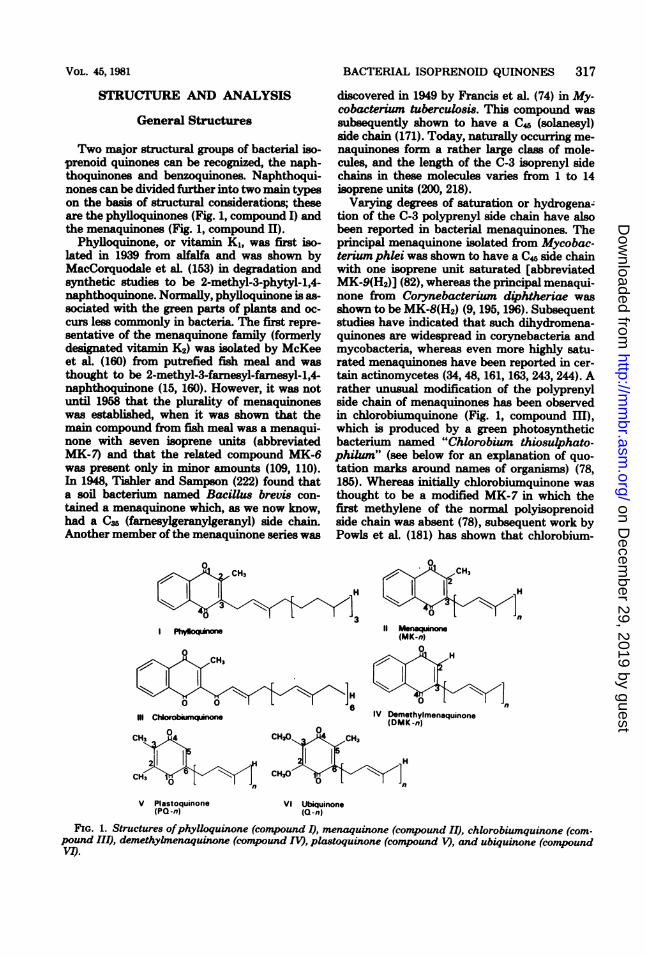
Two major structural groups of bacterial iso-prenoid quinones can be recognized, the naph-thoquinones and benzoquinones. Naphthoqui-nones can be divided further into two main typeson the basis of structural considerations; theseare the phylloquinones (Fig. 1, compound I) andthe menaquinones (Fig. 1, compound II).
Phylloquinone, or vitamin K1, was first iso-lated in 1939 from alfalfa and was shown byMacCorquodale et aL (153) in degradation andsynthetic studies to be 2-methyl-3-phytyl-1,4-naphthoquinone. Normally, phylloquinone is as-sociated with the green parts of plants and oc-curs less commonly in bacteria. The first repre-sentative of the menaquinone family (formerlydesignated vitamin K2) was isolated by McKeeet aL (160) from putrefied fish meal and wasthought to be 2-methyl-3-farnesyl-farnesyl-1,4-naphthoquinone (15, 160). However, it was notuntil 1958 that the plurality of menaquinoneswas established, when it was shown that themain compound from fish meal was a menaqui-none with seven isoprene units (abbreviatedMK-7) and that the related compound MK-6was present only in minor amounts (109, 110).In 1948, Tishler and Sampson (222) found thata soil bacterium named Bacillus brevis con-tained a menaquinone which, as we now know,had a C35 (farnesylgeranylgeranyl) side chain.Another member of the menaquinone series was
I -
III Ctoobxq
BACTERIAL ISOPRENOID QUINONES 317
discovered in 1949 by Francis et al. (74) in My-cobacterium tuberculosis. This compound wassubsequently shown to have a C45 (solanesyl)side chain (171). Today, naturally occurring me-naquinones form a rather large class of mole-cules, and the length of the C-3 isoprenyl sidechains in these molecules varies from 1 to 14isoprene units (200, 218).Varying degrees of saturation or hydrogena-
tion of the C-3 polyprenyl side chain have alsobeen reported in bacterial menaquinones. Theprincipal menaquinone isolated from Mycobac-terium phlei was shown to have a C4 side chainwith one isoprene unit saturated [abbreviatedMK-9(H2)] (82), whereas the principal menaqui-none from Corynebacterium diphtheriae wasshown to be MK-8(H2) (9, 195, 196). Subsequentstudies have indicated that such dihydromena-quinones are widespread in corynebacteria andmycobacteria, whereas even more highly satu-rated menaquinones have been reported in cer-tain actinomycetes (34, 48, 161, 163, 243, 244). Arather unusual modification of the polyprenylside chain of menaquinones has been observedin chlorobiumquinone (Fig. 1, compound III),which is produced by a green photosyntheticbacterium named "Chlorobium thiosulphato-philum" (see below for an explanation of quo-tation marks around names of organisms) (78,185). Whereas initially chlorobiumquinone wasthought to be a modified MK-7 in which thefirst methylene of the normal polyisoprenoidside chain was absent (78), subsequent work byPowls et al. (181) has shown that chlorobium-
0CH3
ItLII M-_
(MK-n)
IV Demethylmenaquinone(DMK -n)
V Plastoquinone VI Ubiquinone(PQ-n) (Q-n)
FIG. 1. Structures ofphylloquinone (compound I), menaquinone (compound II), chlorobiumquinone (com-pound III), demethybnenaquinone (comnpound IV), plastoquinone (comtpound V), and ubiquuione (comnpoundVI).
on Decem
ber 29, 2019 by guesthttp://m
mbr.asm
.org/D
ownloaded from

318 COLLINS AND JONES
quinone is in fact l'-oxomenaquinone with sevenisoprene units (1'-oxomenaquinone- 7). Chloro-biumquinone is the only example of a bacterialpolyisoprenoid quinone containing a side chaincarbonyl group. Demethylmenaquinones (Fig. 1,compound IV), which lack the ring methyl sub-stituent (C-2), have also been isolated from bac-teria (7, 8, 39, 40, 150). To date, demethyl-menaquinones with polyprenyl side chains vary-ing in length from one to nine isoprene unitshave been described (98). An unusual des-methylmenoquinol derivative has been isolatedfrom green photosynthetic bacteria. This com-pound was assigned the structure 4-O-methyl-2-heptaprenyl naphthoquinol by Powls (180).The second major class of bacterial isoprenoid
quinones are the benzoquinones, of which thereare two main types, the plastoquinones (Fig. 1,compound V) and the ubiquinones (Fig. 1, com-pound VI).
Plastoquinone was isolated originally by Ko-fler (135) in 1946 from alfalfa but was not iden-tified. This quinone was rediscovered by Cranein 1959 and in subsequent studies was shown tobe 2,3-dimethyl-5-solanesyl-1,4-benzoquinone(abbreviated PQ-9) (136, 137). Plastoquinone isfound not only in the photosynthetic tissues ofhigher plants but also in red, brown, and greenalgae and in blue-green algae (cyanobacteria)(148, 218, 221). However, it appears to be absentfrom photosynthetic bacteria.The discovery of ubiquinones (formerly called
coenzyme Q) was the result of independent stud-ies by Morton and associates in Liverpool, Eng-land, and Crane and colleagues in the UnitedStates. Ubiquinones contain a 2,3-dimethoxy-5-methyl-1,4-benzoquinone nucleus with a poly-prenyl side chain in position 6 (Fig. 1, compoundVI) (52, 72, 111). Like menaquinones, the ubi-quinones are distributed widely in nature, and awhole range of isoprenologs (Fig. 1, compoundVI; n = 1 to 12) are found in bacteria (168, 218,220, 221). However, in addition to the simple
homologs of ubiquinones, other modifications ofthe side chains, such as hydrogenation (218, 221)and even epoxidation (77), have been discovered.In rhodoquinone, a purple quinone isolated fromRhodospirillum rubrum by Glover and Threlfall(87), the methoxyl group in position 3 of ubiqui-none is replaced by an amino group (Fig. 2,compound VII) (164, 165, 218).
Recently, a rather novel quinone, designatedcaldariellaquinone, was isolated from the ex-tremely thermophilic and acidophilic bacterium"Caldariella acidophila" (61) and was shown tobe 6-(3,7,11,15,19,23-hexamethyltetracosyl)-5-methylthiobenzo-[b]-thiopen-4,7-quinone (Fig.2, compound IX) (59). Caldariellaquinone is theonly sulfur-containing bacterial isoprenoid qui-none that has been isolated to date.
Extraction and PurificationIsoprenoid quinones are susceptible to strong
acid or alkaline conditions and are photooxidizedquite rapidly in the presence of oxygen andstrong light (65, 151). Thus, it is preferable toconduct extraction and subsequent purificationprocedures as rapidly as possible in dim light,avoiding extremes of pH. Isoprenoid quinonesare soluble in the usual lipid solvents; the mostpopular of these are acetone, diethyl ether, chlo-roform, ethanol, and petroleum ether (65). Ad-equate extraction of these components can beachieved with any one of these solvents or amixture of two. any procedures which are usedextensively are direct extraction ofbacterial cellswith acetone-petroleum ether (18, 65) and directextraction with a chloroforn-methanol (2:1, vol/vol) mixture (48, 65). Both of these extractionprocedures yield a complex mixture of lipids plusa small amount of nonlipid material. Isoprenoidquinones can be isolated from this mixture by avariety ofchromatographic procedures; the mostcommon of these are adsorption column chro-matography and adsorption thin-layer chroma-tography (65). Preparative chromatography in
0
0 10
VIl Rhodoquinn VW Epoxyuiino(X.Y-9)
0
IX C_hiIqia
FIG. 2. Structures of rhodoquinone (compound VII), epoxyubiquinone (compound VIII), and caldariella-quinone (compound IX).
MICROBIOL. REV.
on Decem
ber 29, 2019 by guesthttp://m
mbr.asm
.org/D
ownloaded from

VOL. 45, 1981
which layers of Silica Gel HF-254 and solventmixtures such as petroleum ether (boiling point,60 to 80°C)-diethyl ether (85:15, vol/vol) areused provides a particularly simple and rapidmethod for separating menaquinones (Rf - 0.7)and ubiquinones (Rf 0.4) (48). Isoprenoid qui-nones may be detected easily on the resultingchromatograms by brief irradiation with short-wave ultraviolet light (254 nm) and may beeluted with a variety of solvents, such as acetoneor chloroform.
Chromatographic AnalysisThe compositions of purified isoprenoid qui-
none fractions may be investigated further byusing silver ion-impregnated and reverse-phasethin-layer chromatography. The ability of silverions to form reversible complexes with olefinicbonds is well established. The use of Silica GelG impregnated with silver nitrate and develop-ing in nonpolar solvent mixtures, such as meth-anol-benzene (5:95, vol/vol), permit the separa-tion of isoprenoid mixtures according to thenumber of double bonds that the compoundscontain. The incorporation of indicators, such asrhodamine 6G, into the layers permits easy vis-ualization of the separated isoprenologs on de-veloped chromatograms (65). In reverse-phasepartition chromatography a stationary, nonvol-atile hydrocarbon (such as liquid paraffin orhexadecane) and polar developing mixtures(such as acetone-water or dimethyl formamide-water) are used (54, 65, 67). Reverse-phase chro-matography facilitates the separation ofisopren-oid quinone components according to theiroverall physical properties, which dependmainly on their chain lengths and their degreesof unsaturation. Since natural mixtures of iso-prenoid quinones often vary both in degree ofunsaturation and in polyisoprenoid chain length,both of these chromatographic techniquesshould be used to define the composition of anunknown mixture of quinones. The value of sil-ver ions and reverse-phase thin-layer chroma-
BACTERIAL ISOPRENOID QUINONES 319
tography in the analysis of bacterial menaqui-none mixtures has been demonstrated in twoexcellent studies (67, 98).
Physicochemical AnalysisUltraviolet spectrophotometry. Purified
isoprenoid quinones can be analyzed further byusing a variety of physicochemical techniques(207). In particular, ultraviolet spectroscopy pro-vides a simple method for investigating the cat-egory or class to which an unknown isoprenoidquinone belongs. Details of the ultraviolet ab-sorption characteristics of some of the majortypes of bacterial quinones are shown in Table1.The ultraviolet spectra of the majority of the
bacterial naphthoquinones fall into the followingtwo main groups: (i) the 2,3-disubstituted qui-nones, as exemplified by phylloquinone and themenaquinones; and (ii) the monosubstituted (ordemethylated) quinones, such as demethylme-naquinones. Both menaquinones and phylloqui-nones exhibit qualitatively identical ultravioletspectra, with five absorption maxima (XA) at242, 248, 260, 269, and 326 nm and one point ofinflection at 238 nm (Fig. 3 and Table 1). Ab-sorption bands at 242, 248, and 238 nm (shoul-der) are due to benzenoid contributions, whereasbands at 260 and 269 nm are due to quinoneabsorption (65). However, removal ofthe methylgroup from C-2 of the naphthoquinone nucleus(as in demethylmenaquinones and demethyl-phylloquinones) causes a shift in the quinoneabsorption contribution of about 6 nm to shorterwavelengths (Amax 254 and 263 nm), whereas thebenzenoid contributions (Xx, 243 and 248 nmand 238 nm [point of inflection]) remain vir-tually unaltered. Thus, ultraviolet spectropho-tometry provides a simple method for distin-guishing menaquinones and phylloquinonesfrom their demethylated derivatives (Table 1).Chlorobiumquinone (1'-oxomenaquinone-7) iso-lated from green photosynthetic bacteria (78,185) exhibits ultraviolet absorption characteris-
TABLE 1. Ultraviolet absorption characteristics of menaquinones, ubiquinones, and related compoundsCompound Solvent A,,., (nm)a Reference(s)
Phylloquinone Isooctane 242, 248, 260, 269, 326, 238 (inf) 65Menaquinone Isooctane 242, 248, 260, 269, 326, 238 (inf) 65Demethylphylloquinone Isooctane 243, 248, 254, 263, 326, 238 (inf) 65Demethylmenaquinone Isooctane 243, 248, 254, 263, 326, 238 (inf) 65Chlorobiumquinone Ethanol 254, 2g (inf) 78, 181Plastoquinone Isooctane 254, 262 207Ubiquinone Ethanol 275 405 207Rhodoquinone Cyclohexane 251, 280, 320, 500 (inf) 87Rhodoquinone Ethanol 253, 283, 320, 500 (inf) 87Caldariellaquinone Methanol 241, W, 333, 471 59Each major peak is underlined. inf, Point of inflection.
on Decem
ber 29, 2019 by guesthttp://m
mbr.asm
.org/D
ownloaded from

320 COLLINS AND JONES
tics quite distinct from those of menaquinonesand demethylmenaquinones, with A.. at 254rum in ethanol and a point of inflection at 265nm (181).The two commonly encountered benzoqui-
nones, plastoquinones and ubiquinones, can bedistinguished easily by ultraviolet spectropho-tometry. Plastoquinones (Fig. 1, compound V)have X,. at 254 and 262 nm (isooctane) (Fig. 4).In contrast, ubiquinones have a X.. at about270 to 275 nrm and a second absorption band at405 to 407 nm (Fig. 5). The replacement of themethoxyl group in position 3 by an amino group,as in rhodoquinone (2-methyl-3-amino-5-methyl-6-nonaprenyl-1,4-benzoquinone) iso-lated from R. rubrum, causes a marked changein the spectrum; in this case, three X.. (251 to253, 280 to 283, and 500 nm) and one point ofinflection (320 nm) are produced.The rather unusual terpenoid benzo-[b]-thio-
pen-4,7-quinone (designated caldariellaquinone)from "C. acidophila" exhibits a very character-istic ultraviolet spectrum (XA,, 241, 283, 333 and471 nm), which is quite distinct from the spec-trum of any other described bacterial isoprenoidquinone (Table 1).Mass spectrometry. The most precise and
sensitive method for determining isoprenoid qui-none structure is mass spectrometry. Thismethod provides both accurate molecularweights of the isoprenoid quinones and struc-tural information, such as the nature of the ringsystem, the length and degree of saturation ofthe isoprenyl side chain, etc. An excellent reviewon mas spectral analysis of naphthoquinonesand benzoquinones has been published by Som-
Atoo-
SO-
SO-
FIG. 4. Ultraviolet spectrum of PQ-9 (solvent,isooctane).
FIG. 5. Ultraviolet spectrum of Q-10 (solvent,isooctane).
60-
60-
40-
30-
20-
10-
220 240 2.0
FIG. 3. Ultravioletisooctane).
250 300 320 240 nm
spectrum of MK-6 (solvent,
mer and Kofler (207). When subjected to massspectrometry, both menaquinones and ubiqui-nones produce characteristic fagmentation pat-terns (Fig. 6). The base peaks in the mass spectraof menaquinones (including phyiloquinone) oc-cur at m/e 225 (Fig. 7, compounds X and XI)and are derived from the naphthoquinone nu-cleus (Fig. 6). Ubiquinones and plastoquinoneshave corresponding nuclear fragments at mWe235 and 189, respectively (65, 207). Strong peaksin the mass spectra of both menaquinones andubiquinones corresponding to molecular ions(M+) are observed. The pattern offiagmentationof the isoprenoid substituent at the C-3 positionof menaquinones (Fig. 6) is characteristic of the
MICROBIOL. REV.
on Decem
ber 29, 2019 by guesthttp://m
mbr.asm
.org/D
ownloaded from

BACTERIAL ISOPRENOID QUINONES 321
0 23- 37 317-,S 443 s11-
mn 47J 2¢J- L-34" L-2,MM L-M1,2nS L-1^U7 L_4§X10 xI~
us 3 314e 4 S
FIG. 6. Mass spectrum ofMK-6 from B. ochraceus.
cracking pattern exhibited by polyisoprenoidchains in general. Fragmentation of the sidechain under electron impact involves solely dial-lylic bonds, following the rule (M-69) - (68 xN), where N is the number of isoprene units inthe side chain minus one. Thus, using the massspectrum of MK-6 from Bacteroides ochraceusas an illustration (Fig. 6), fragmentation of theside chain involves the loss of a terminal iso-prenyl unit (M-69)+, followed by four successivelosses of 68 mass units. Saturation of an olefinicbond within the side chain of a menaquinonecauses a marked alteration in the cracking pat-tern and provides information on the position ofhydrogenation (5). The fragmentation patternsofthe various bacteril isoprenoid quinones (e.g.,menaquinones, phylloquinones, plastoquinones,ubiquinones) are all very similar. The simplicityof these patterns and the ease of determiningaccurate molecular weights provide an unequiv-ocal means for determining isoprenoid quinonestructures (207).
DISTRIBUTION OF ISOPRENOIDQUINONES AND TAXONOMIC
IMPLICATIONS
Molecular genealogical analyses based upontransfer and ribosomal ribonucleic acid sequencehomologies have revealed that bacteria do notconstitute a phylogenetically monolithic group(237). The kingdom Procaryotae is now consid-ered to contain two phylogenetically distinctgroups, the archaebacteria and the eubacteria,including the mycoplasmas and cyanobacteria(73, 237).Where possible, the names of the bacterial
taxa referred to here are names that are includedin the Approved Lists of Bacterial Names (206).However, although the Approved Lists now de-termine the nomenclature of bacteria, it is nec-essary to refer to strains bearing names notincluded in the Approved Lists. Where this isdone, the names are placed in quotation marks(e.g., "Actinobacillus actinoides"). The nomen-
0
X m/e 225
Xi e/e 225FIG. 7. Nuclear fragments at mWe 225 (compounds
X and XI) derived from the naphthoquinone nuclei ofmenaquinones.
clature of Rippka et al. (187) is used for thecyanobacteria.
ArchaebacteriaThe first organisms recognized as archaebac-
teria were the methanogens, which are fastidiousanaerobes whose metabolism is centered aroundthe reduction of carbon dioxide to methane(237). Detailed studies on the isoprenoid qui-none contents of the methanogens have not beenperformed, although Zeikus and colleagues (249)reported that Methanobacterium thermoauto-trophicum contains neither menaquinones norubiquinones.
Representatives ofthe extreme halophilic taxaHalobacterium and Halococcus have also nowbeen identified as archaebacteria (237). Early
VOL. 45, 1981
on Decem
ber 29, 2019 by guesthttp://m
mbr.asm
.org/D
ownloaded from

322 COLLINS AND JONES
studies indicated that unsaturated MK-8 consti-tuted the sole isoprenoid quinones within theseextreme halophilic taxa (142, 223). However, arecent systematic study of the genera Halobac-terium and Halococcus has shown that dihydro-genated menaquinones are also present in allspecies of these genera (49) (Table 2). The ex-treme thermophilic archaebacterium Thermo-plasma acidophilum also contains menaqui-nones as its sole isoprenoid quinones, with MK-7 predominating (143).An unusual terpenoid, benzo-[b]-thiopen-4,7-
quinone (caldariellaquinone), has been isolatedfrom the extreme acidophile "C. acidophila"(59). Caldarieliaquinone is the only sulfur-con-taining bacterial isoprenoid quinone known. Al-though the present isoprenoid quinone data onthe archaebacteria are fragmentary, several pat-terns are evident (Table 2). These suggest thatfurther quinone studies on these taxa could pro-vide infornation of taxonomic value.
Eubacteria
For clarity of presentation, the groups of theeubacteria are treated, where possible, as theyare in Bergey's Manual of Determinative Bac-teriology, 8th ed. (21).Cyanobacteria. In Bergey's Manual of De-
terminative Bacteriology (21), the cyanobac-teria (blue-green algae) are given little consid-eration. Subsequent studies by Rippka et al.(187) have indicated that these bacteria can bedivided into five major subgroups which contain22 genera. Systematic studies on the distributionofisoprenoid quinones in the cyanobacteria havenot been performed, but preliminary data on thefive taxa (Table 3) examined so far suggest thatsuch an investigation may be rewarding.Cyanobacteria are unusual in that they pos-
sess neither ubiquinones nor menaquinones (Ta-ble 3). They do contain phylloquinone (vitaminK1) and a plastoquinone (PQ-9), which are in-digenous to the plant kingdom but are not nor-mally found in bacteria. Furthernore, membersof the genera Anabaena, Chlorogloeopsis, Fis-cherella, and Nostoc have also been reported tocontain a-tocopherolquinone (Fig. 8, compoundXII), which is normally associated with chloro-plasts in higher plants and algae (218). Thegenus Synechococcus apparently lacks a-toco-pherolquinone but contains a polar naphthoqui-none (probably a monohydroxy derivative ofphylloquinone [144]) not reported in the otherfour species of cyanobacteria that have beenexamined (Table 3). Sun and co-workers (210)also reported the presence of plastoquinones Band C in a strain of the genus Anabaena, al-
TABLE 2. Distribution of isoprenoid quinones in archaebacteria
Taxon Major isoprenolog(s) Minor components Refer-"Amoebobacter morrhuae" MK-8 142"Caldariella acidophila"a Caldariellaquinone 60Halobacterium cutirubrum MK-8 142, 223H. cutirubrum MK-8 MK-8(H2), MK-7(H2), MK-7 49Halobacterium halobium MK-8 142H. halobium NCMB 736, NCMB 764, MK-8, MK-8(H2) MK-7(H2), MK-7 49NCMB 777, and NCMB 2080
Halobacterium saccharovorum MK-8, MK-8(H2) 49Halobacteriumsalinarium MK-8 142H. salinarium MK-8, MK-8(H2) MK-7(H2), MK-7 49"Halobacterium s imoncinii subsp. nea- MK-8, MK-8(H2) MK-7(H2), MK-7 49politanium"
Halobacterium trapanicum MK-8, MK-8(H2) MK-7(H2), MK-7 49Halobacterium volcanii MK-8 MK-8(H2) 167H. vokcanii MK-8, MK-8(H2) MK-7(H2), MK-7 49Halobacterium sp. MK-8, MK-8(H2) MK-7(H2), MK-7 49Halococcus morrhuae MK-8, MK-8(H2) MK-7(H2), MK-7 49"Paracococcus haloxanthus" MK-8 142"Sarcina morrhuae"tb MK-8, MK-8(H2) MK-7(H2), MK-7 49"Sarcina litoralis"b MK-8(H2) MK-8, MK-7(H2), MK-7 49"Sarcina sreenivasani`b MK-8(H2) MK-8, MK-7(H2), MK-7 49Alkalophilic halophiles SP1, SP2, and MK-8, MK-8(H2) MK-7(H2), MK-7 49MS3C
Thermoplasma acidophilum MK-7 106, 143a The archaebacterium (59) "C. acidophila" was described by De Rosa et al. (61).b "S. morrhuae," "S. litoralis," and "S. sreenivasani" are now classified as Halococcus norrhuae.c From alkaline soda lakes.
MICROBIOL. REzV.
on Decem
ber 29, 2019 by guesthttp://m
mbr.asm
.org/D
ownloaded from

BACTERIAL ISOPRENOID QUINONES 323
TABLE 3. Distribution of isoprenoid quinones in the cyanobacteriaTaxon Major quinone typesa Reference(s)
Anabaena ("A. variabilis") PQ-9, K1, a-TQ 22, 30, 31Anabaena ("A. variabilis") PQ-9, K1, a-TQ, PQ-B, PQ-C 210Synechococcus ("Anacystis nidulans") PQ-9, K1, OH-K1b 3, 144, 234Synechococcus ("Anacystis nidulans") PQ-9,.K1, polar naphthoqui- 31, 177, 210
none?Synechococcus ("Anacystis nidulans") PQ-9, OH-K1, polar naphtho- 99
quinone?Synechococcus ("Anacystis nidulans") PQ-9, K1? 148Chlorogloeopsis ("Chlorogloae fritschii") PQ-9, K1, a-TQ 22, 30, 31dFischerella ("Mastigocladus laminosus") PQ-9, K1, a-TQ 30,31Nostoc ("N. muscorum") PQ-9, K1, a-TQ 30, 31Nostoc sp. PQ-9, PQ-C, K1, a-TQ 210Nostoc sp. PQ-9, K1 68
a Abbreviations: K1, phylloquinone; OH-K1, hydroxyphylloquinone; PQ-B, plastoquinone B; PQ-C, plastoqui-none C; a-TQ, a-tocopherolquinone.
b a-Tocopherolquinone was absent (31, 99, 210). The hydroxyphylloquinone has been shown to be 5-monohydroxyphylloquinone (144).
c The presence of phylloquinone was not investigated by Bucke et al. (22).
0
CH, CH,
OH~~~~~
CH,
XII a-Tocopherolquinone (a-TQ)
FIG. 8. Structure of a-tocopherolquinone (com-pound XII).
though this finding awaits confirmation.Mycoplasmas. The distribution ofisoprenoid
quinones within the mycoplasmas is summarizedin Table 4.Apart from an early report of the absence of
isoprenoid quinones in Mycoplasma gaUlisepti-cum (83), all of the mycoplasmas that have beenexamined so far contain menaquinones as theirsole isoprenoid quinones. Strains of Achole-plasma axanthum and Mycoplasma arthritidishave been reported to contain MK-4 as theirmajor isoprenolog (106). To our knowledge, suchshort-chain menaquinones have not been re-ported as major components in any other bac-teria. However, this report must be treated withsome caution, as Hollander et al. (106) used onlyreverse-phase partition thin-layer chromatogra-phy to characterize the quinones. Since naturalmixtures of menaquinones often vary both indegree of unsaturation and in polyisoprenoidchain length, reverse-phase thin-layer chroma-tography by itself is insufficient to define anunknown quinone mixture (see above) (50, 65,67) and should be used in conjunction witheither argentation chromatography or somephysiochemical method, such as mass spectrom-etry. No data are available on the structures ofthe menaquinones isolated from Spiroplasmacitri, although two studies on T. acidophilum
TABLE 4. Distribution of isoprenoid quinones inmycoplasmasa
Taxon Major iso- Refer-prenolog ence(s)
Acholeplasma axanthum MK-4 106Acholeplasma granularum MK-n 106Acholeplasma laidlawii MK-n 106Mycoplasma arthritidis MK-4 106Mycoplasma gallinarum MK-nb 106Mycoplasma homini MK-n 106Mycoplasma neurolyticum MK-n 106Thermoplasma acidophilumc MK-7 106, 143Spiroplasma citri MK-n 106
a Major groupings as in Bergey's Manual of Deter-minative Bacteriology (21).
b Gale et al. (83) did not detect both menaquinonesand ubiquinones in Mycoplasma gallisepticum.
c T. acidophilum may be more appropriatelygrouped with the archaebacteria (see text).
(see above) indicate that MK- 7 predominates inthis species (106, 143). Further structural studieson the isoprenoid quinones of other mycoplas-mas will be necessary to determine the value ofthese compounds in the classification of thisgroup.Gram-negative bacteria. (i) Gram-nega-
tive facultatively anaerobic rods. The dis-tribution of isoprenoid quinones in gram-nega-tive facultative anaerobes is shown in Table 5.Several genera within the family Enterobacte-riaceae (e.g., Escherichia, Klebsiella, and Pro-teus) and certain species of the genera Aero-monas and Erwinia are unusual in that theycontain mixtures of menaquinones, demethyl-menaquinones, and ubiquinones. Structuralstudies have indicated that the lengths of theside chains of the major components of these
VOL. 45, 1981
on Decem
ber 29, 2019 by guesthttp://m
mbr.asm
.org/D
ownloaded from

324 COLLINS AND JONES MICROBIOL. REV.
TABLE 5. Distribution of isoprenoid quinones in gram-negative bacteria: gram-negative facultativelyanaerobic rods'
Taxon Major Mior component(s) Reference(s)isoprenolog(,S)bMiocmrnn() Rfec(s
EnterobacteriaceaeEdwardsiella tarda
Enterobacter aerogenesEnterobacter agglomerans"Enterbacter liquefaciens"Erwinia amylovoraErwinia carotovora
Erwinia carotovora
"Escherichia aurescens"
Escherichia coliE. coliE. coli
E. coli
E. coli
E. coli
E. coli
"Escherichia freundii"
"Kiebsiella aerogenes""K. aerogenes"
Proteus mirabilisP. mirabilis
P. mirabilis
P. mirabilis
Proteus vulgaris
P. vulgarisP. vulgaris
P. vulgarisSerratia marcescensS. marcescens
Yersinia pseudotuberculosis NCTC 1101VibrionaceaeAeromonas hydrophila NCMB 7810
Q-nMK-nQ-8CQ-nQ_8CQ-nQ-nMK-nQ-8MK-8DMK-8Q-8MK-8DMK-8Q-nQ-8CQ-8'
Q-8MK-nQ-8MK-8MK-8DMK-8Q-8MK-8DMK-8Q-8MK-8DMK-8Q-8CQ-8MK-8DMK-8Q-8CMK-8DMK-8Q-nMK-nDMK-nQ-8MK-8DMK-8Q-8MK-8DMK-8MK-ncQ8MK-8Q-9, Q-8, Q-7CfQ-8'Q-8MK-8Q-8
Q-8MK-8DMK-8
Q-7MK-nDMK-nQ-7, Q-6, Q-5, Q-4MK-nDMK-n
Q-7, Q-6, Q-5, Q-4, Q-3,Q-2, Q-1
MK-9, MK-7, MK-6DMK-7Q-7, Q-6, Q-5, Q-4MK-nDMK-nQ-9, Q-7, Q-6, Q-5, Q-4MK-nDMK-n
Q-7, Q-6, Q-5, Q-4MK-nDMK-nQ-7, Q-6, Q-5, Q-4
Q-7, Q-6, Q-5, Q-4MK-nDMK-nQ-7, Q-6MK-nDMK-n
Q-7, Q-6, Q-5, Q-4MK-nDMK-n
139
5810358103103
232d
12458, 174, 18356
148
16, 17
25
232d
232d
132, 133232d
230, 231'20
106
232d
232d
11317, 96
17458, 17414
17
232d
on Decem
ber 29, 2019 by guesthttp://m
mbr.asm
.org/D
ownloaded from

BACTERIAL ISOPRENOID QUINONES 325
TABLE 5-Continued
Taxon i,
Aeromonas punctata
Vibrio alginolyticus
Vibrio costicola
Vibrio succinogenesVibrio sp. NCIB 8250Vibrio sp. strain 01Vibrio sp.
Taxa of uncertain affiliation"Actinobacillus actinoides" ATCC 15900Actinobacillus actinomycetencomitansActinobacillus equuli NCTC 3365Actinobacillus spp. NCTC 10801 and ATCC
27073Actinobacillus equuli NCTC 8529
Actinobacillus lignieresii
"Actinobacillus seminis"
"Aerobacter aerogenes"Cardiobacterium hominis"Chromobacteriumprodigiosum" NCTC 1337Chromobacterium violaceumEikenella corrodens NCTC 10596Flavobacterium aquatileFlavobacterium acidificumFlavobacterium capsulatumFlavobacterium devoransFlavobacterium halmephilumF. halmephilumFlavobacterium sp. ATCC 13945Flavobacterium spp. (six oxidase-positive
strains)Flavobacterium sp. (one oxidase-positive
strain)"Flavobacterium arborescens""Flavobacterium breve"Flavobacterium esteraromaticum"Flavobacterium flavescens"Flavobacterium heparinumFlavobacterium meningosepticumFlavobacterium odoratum"Flavobacterium pectinovorum""Flavobacterium suaveolens""Flavobacterium tirrenicum"Flavobacterium uliginosumFlavobacterium spp. NCTC 10795, NCTC
10796, and NCTC 10797Flavobacterium/Cytophaga app. NCMB 249,NCMB 251, NCMB 275, NCMB 289, NCMB264, NCMB 244, and NCMB 259
Flavobacterium 8p.HaenophilusparasuisHaemophiluspisciumHaemophilus vaginalis ATCC 14018Haemophilus haemoglobinophilus
Majorisoprenolog(s)b
Q-8MK-8Q-nMK-nMK-8Q-8MK-nQ-9Q9CQ-8c
MK-nDMK-nDMK-nDMK-n
Q-nDMK-nQ-nDMK-nQ-nDMK-nQ-8Q-nQ-8Q-8Q-nQ-nQ-nQ-nQ-nQ-nQ-9Q-nQ_8CQ9C
MK-nMK-nMK-nMK-nMK-nMK-nMK-nMK-nMK-nMK-nMK-nMK-n
Minor component(s)
Q-7, Q-6, Q-5, Q-4MK-n
MK-7, MK-6Q-7
Q-10, Q-8, Q-7, Q-6Q-10, Q-8, Q-7, Q-6, Q-5
Q-9, Q-7, Q-6
Q-8, Q-7
MK-n
MK-6cQ-nQ-nQ-nQ-nDMK-n
MK-5
Reference(s)
232d
226
49
14023223158
155155155155
155
155
155
1715517
2321042323232323492358
58'
232323232323232323232323
23
67104104104104
VOL. 45, 1981
on Decem
ber 29, 2019 by guesthttp://m
mbr.asm
.org/D
ownloaded from

TABLE 5-Continued
Taxon iMaoprnjlog(s)b Minor component(s) Reference(s)Haemophilusparainfluenzae NCTC 4101 Q-n 104
DMK-nHaemophilusparagallinarum Q-n 104
DMK-nHaemophilus aegyptius DMK-n 104Haemophilus influenzae DMK-n 104Haemophilusparainfluenzae HIM 170-1 and DMK-n 104HIM 449-8
Haemophilusparahaemolyticus DMK-n 104Haemophilusparaphrophilus DMK-n 104Haemophilus paraphrohaemolyticus DMK-n 104Haemophilusparainfluenzae DMK-6 DMK-7, DMK-5 150Haemophilus parainfluenzae DMK-6 DMK-9, DMK-8, 98
DMK-7, DMK-5,DMK-4, DMK-3,DMK-2, DMK-1
"Pasteurella gallinacum" Q-n 155DMK-n
Pasteurella haemolytica Q-n 155DMK-n
"Pasteurella mastitidis" Q-n 155DMK-n
Pasteurella multocida Q-n 155DMK-n
"Pasteurella piscicida" Q-n 155DMK-n
Pasteurella ureae Q-n 155DMK-n
Pasteurella spp. NCTC 10699 and NCTC Q-n 15511051 DMK-n
Pasteurella spp. NCTC 10547, NCTC 10549, Q-n 155and NCTC 10553 DMK-n
"Pasteurella bettii" DMK-n 155Pasteurella pneumotropica DMK-n 155a Major groupings as in Bergey's Manual ofDeterminative Bacteriology (21).b DMK-8, Demethylmenaquinone containing eight isoprene units.'The possible presence of other respiratory quinones was not investigated.d Minor analogs of menaquinones and demethylmenaquinones were not specified.'Traces of uncharacterized epoxyubiquinones were also present in P. mirabilis and Vibrio sp. (231).fThe major component was not specified (174).' Ubiquinones were reported to be absent from three oxidase-positive and two oxidase-negative Flavobacte-
rium spp. (58).
three quinone structural types are the same(eight isoprene units). However, the relative pro-portions of menaquinones, demethylmenaqui-nones, and ubiquinones can be influenced by thedegree of aeration. Thus, cultivation of Esche-richia coli and other facultative organisms at alow oxygen tension increases the level of mena-quinones but reduces the amount ofubiquinonesproduced (179, 233). Numerous taxa (e.g., En-terobacter agglomerans, Erwinia amylovora,Yersinia pseudotuberculosis, and Vibrio suc-cinogenes) have been shown to contain onlyubiquinones, although further studies will benecessary to confirn the absence of naphtho-quinones.
The taxonomic significance of respiratory qui-nones within the genera Actinobacillus, Cyto-phaga, Flavobacterium, Haemophilus, andPasteurella has been studied extensively byMannheim and associates (23, 104, 155). Invari-ably, typical strains of species within the generaActinobacillus and Pasteurella contain de-methylmenaquinones (155), and some of theseorganisms are capable of synthesizing ubiqui-nones in addition to demethylmenaquinones(Table 5). However, Actinobacillus actinomy-cetemcomitans, Actinobacillus suis, "Pasteu-rella bettii," and Pasteurella pneumotropicastrains produce only demethylmenaquinones,whereas "Actinobacillus actinoides" ATCC
326 COLLINS AND JONES MICROBIOL. REV.
on Decem
ber 29, 2019 by guesthttp://m
mbr.asm
.org/D
ownloaded from

VOL. 45, 1981
15900 apparently produces menaquinones as itssole respiratory quinones. This last taxon is grampositive and is probably related to the actino-mycetes. Members of the genus Haemophilusare heterogeneous on the basis of the types ofrespiratory quinones produced (Table 5). Thetype species of Haemophilus, Haemophilus in-fluenzae, and Haemophilus aegyptius, Haemo-philus paraphrophilus, Haemophilus parahae-molyticus, and Haemophilus paraphrohaemo-lyticus produce demethylmenaquines as theirsole isoprenoid quinones. Haemophilus para-gallinarum, Haemophilus haemoglobinophilus,and Haemophilus parasuis produce ubiqui-nones in addition to demethylmenaquines. Hae-mophilus vaginalis, which contains ubiquinonesas its sole respiratory quinones, has been trans-ferred recently to a new genus, Gardnerella, asGardnerella vaginalis (95). Members of thegenera Cytophaga and Flavobacterium can bedivided into two broad groups on the basis oftheir isoprenoid quinones (23). Menaquinonesare produced by the majority of the flavobac-teria and the typical Cytophaga strains, whereasubiquinones have been shown to be present inFlavobacterium aquatile, Flavobacterium aci-dificum, Flavobacterium capsulatum, Flavo-bacterium devorans, and Flavobacterium hal-mephilium (Table 5). Mannhein and colleaguesdid not investigate the detailed structures (e.g.,isoprene numbers, possible hydrogenation, etc.)of the various quinone classes (Table 5). How-ever, preliminary structural information on thequinones of members of the genus Haemophilusand on the quinones of flavobacteria suggeststhat such an approach will be rewarding for thepurposes of taxonomy.
(ii) Gram-negative aerobic rods. The ma-jority of the gram-negative aerobic rods whichhave been examined contain ubiquinones exclu-sively (Table 6). Members of the genus Pseu-domonas generally contain ubiquinones withnine isoprene units (abbreviated Q-9) as theirmajor components. However, strains labeled"Pseudomonas denitrificans" and some un-named Pseudomonas spp. have been shown topossess major amounts of Q-10 (Table 6). Re-cently, a Pseudomonas sp. (strain M16) has beendescribed which accumulates significant quan-tities of Q-11 in addition to Q-10 (168). Thisreport is of considerable interest, as ubiquinoneisoprenologs with more than 10 isoprene unitshave been found previously only in traceamounts in certain photosynthetic bacteria (218,220, 221).The ubiquinone contents of the acetic acid
bacteria have been investigated systematicallyby Yamada and colleagues (238-240, 245, 246).
BACTERIAL ISOPRENOID QUINONES 327
Members of the genus Acetobacter contain Q-9as their major component, whereas members ofthe genus Gluconobacter and strains labeled"Acetobacter xylinum" possess Q-10 as theirmajor isoprenolog. A number of so-called aceticacid intermnediate strains of uncertain taxonomicposition have been shown to contain Q-8 as theirmajor component (Table 6).Members of the genus Azotobacter contain
ubiquinones as their respiratory quinones. Theearly report by Lester and Crane (148) of anunidentified menaquinone in a strain of Azoto-bacter vinelandii is probably in error.
Studies on the ubiquinone compositions ofother gram-negative aerobic rods have indicatedthat there is relatively little structural variationand that Q-10 predominates in Agrobacterium(182, 232) and Brucella (186, 215), Q-9 predom-inates in Alcaligenes (232), and Q-8 predomi-nates in "Achromobacter" (17), "Comamonas"(232), "Hydrogomonas" (148, 214), and "Meth-ylomonas" (64).Members of the thermophilic genus Thermus
are unusual among the aerobic, gram-negativerods in that they contain menaquinones as theirsole isoprenoid quinones. Thermus aquaticusand "Thermus thermophilus" contain MK-8 astheir major component (Table 6). It is worthnoting that members of the genus Thermus arealso unusual in that they possess citrate syn-thases with the sizes and regulatory propertiesofthe citrate synthases of gram-positive bacteria(229).
(iii) Aerobic gram-negative cocci andcoccobacilli. Data on the isoprenoid quinonecontents of aerobic gram-negative cocci and coc-cobacilli are rather fragmentary (Table 7). All ofthe Acinetobacter, Branhamella, Moraxellaand Neisseria species which have been exam-ined contain Q-8 or Q-9 or both as their majorcomponents. Jones and Weitzman (122) foundthat "Brevibacterium leucinophagum" wasgram negative and proposed the transfer of thisorganism to the genus Acinetobacter. The re-covery of major amounts of Q-9 in "B. leucino-phagum" supports this assignment (34).
Paracoccus denitrificans contains majoramounts of Q-10 and on this basis can be distin-guished from all Acinetobacter, Moraxella, andNeisseria species which have been examined.The taxonomic position of "Paracoccus halox-anthus" remains equivocal. This taxon is notrecognized in Bergey's Manual ofDeterminativeBacteriology (21). "P. haloxanthus" lacks ubi-quinones and contains menaquinones (142), andthis organism may be more appropriately clas-sified alongside the genera Halobacterium andHalococcus in the archaebacteria.
on Decem
ber 29, 2019 by guesthttp://m
mbr.asm
.org/D
ownloaded from

TABLE 6. Distribution of isoprenoid quinones in gram-negative bacteria: gram-negative aerobic rodsa
Taxon Majorieo- Minor component(s) Reference(s)
Acetobacter aceti Q-9 Q-1O, Q-8 239A. aceti Q-9 Q-8 240"Acetobacter acetosus" Q-9 Q-8 240"Acetobacter albuminosus" Q-9 Q-8 240"Acetobacter ascendens" Q-9 Q-8 240"Acetobacter dioxyacetonicus" Q-9 Q-8 240"Acetobacter kutzingianus" Q-9 Q-8 240Acetobacterpasteurianus Q-9 Q-8 240"Acetobacter rancens" Q-9 Q-8 240"Acetobacter xylinum" Q-10 12, 240, 245"A. xylinum" Q-9 Q-8 240Acetobacter spp. A21 to A23 Q-9 Q-8 240"Acetic acid intermediate strains"Peritrichously flagellated strains IAM 1834 Q-10 Q-9, Q-8 239and AJ 2881
Polar flagellated strain IFO 3246 Q-8 Q-7 239Polar flagellated strains Q-8 246"Achromobacter hartlebii" Q-8 17"Achromobacter" sp. marine strain 60-20- Q-n 141A5
Agrobacterium tumefaciens Q-n 124A. tumefaciens Q-1O 182,183A. tumefaciens Q-10 Q-9, Q-8 232"Alcaligenes viscolactis" Q-9 Q-8, Q-7 232"Azotobacter agilis" Q-n 124Azotobacter chroococcum Q-8 17Azotobacter vinelandii Q-n 156A. vielandii Q-8 119,148,149,174,183bBordetellapertussis Q-8 216Brucella abortus Q-10 186,215Brucella melitensis Q-10 216"Comamonas percolans" ATCC 8461 Q-8 Q-7, Q-6, Q-5, Q-4 232"Gluconobacter albidus" Q-1O 240"Gluconobacter capsulatus" Q-1O 240"Gluconobacter cerinus" Q-1O 238"Gluconobacter dioxyacetonicus" Q-1O 240"Gluconobacter gluconicus" Q-10 240"Gluconobacter industrius" Q-10 240"Gluconobacter melanogenus" Q-10 240"Gluconobacter monoxygluconicus" Q-1O 240Gluconobacter oxydans Q-1O 240"Gluconobacter roseus" Q-10 240"Gluconobacter rubiginosus" Q-10 240"Gluconobacter seleroideus" Q-10 240"Gluconobacter suboxydans" Q-10 Q-9, Q-8 239, 240"Gluconobacter turbidans" Q-9 240"Hydrogenomonas eutropha" Q-8 239"Hydrogenomonas" 8p. Q-8 148,214"Methylomonas" sp. 2B36-P11 Q-8 64Pseudomonas aeruginosa Q-9 17,174Pseudomonas beijerinkii Q-9 Q-8, Q-7 49"Pseudomonas denitrificans" Q-1o 174"Pseudomonas desmolytica" Q-9 224Pseudomonas facilis Q-n 217Pseudomonas fluorescens Q-9 148P. fluorescens Q-9 Q-8, Q-7, Q-6 232P. fluorescens Q-9, Q-8 174Pseudomonas fragi Q-9 174P. fragi Q-9 Q-8, Q-7, Q-6 232Pseudomonas geniculata Q-9 174
328 COLLINS AND JONES MICROBIOL. REV.
on Decem
ber 29, 2019 by guesthttp://m
mbr.asm
.org/D
ownloaded from

BACTERIAL ISOPRENOID QUINONES 329
TABLE 6-Continued
Taxon Major iso- Minor component(s) Reference(s)
"Pseudomonas maltophilia" Q-8 107"Pseudomonas mildenbergii" Q-9 174"Pseudomonas ovalis" Q-9 108"P. ovalis" Q-9 Q-8, Q-7, Q-6 230, 232"P. ovalis" Q-9 Q-8, Q-7, Q-6, Q-5, Q-4 231Pseudomonasputida Q-9 174P. putida Q-9 Q-8, Q-7, Q-6 232Pseudomonas sp. Q-9 183Pseudomonas sp. strain N 842 Q-1o 169Pseudomonas sp. strain M16 Q-10 Q-11 168"Protaminobacter ruber" Q-1O Q-9, Q-8 34"P. ruber" Q-10 168Rhizobiumjaponicum Q-10 55Thermus aquaticus MK-8 MK-7 50"Thermus thermophilus" MK-8 MK-7 Collins and Jones,
unpublished dataXanthomonas campestris Q-8 Q-7, Q-6, Q-5, Q-4 232X. campestris Q-8 107"Xanthomonas citri" Q-8 107"Xanthomonas oryzae" Q-8 107
'Major groupings as in Bergey's Manual ofDeterminative Bacteriology (21).b Lester and Crane (148) also detected an uncharacterized menaquinone in a strain of A. vinelandii.
TABLE 7. Distribution of isoprenoid quinones ingram-negative bacteria: gram-negative cocci and
coccobacillia
Taxon Major iso- Refer-prenolog(s) ence
"Acinetobacter anitratum" Q-9 58(seven strains)
"A. anitratum" (one strain) Q-8 58"A. anitratum" (two strains) Q-9, Q-8b 58Acinetobacter lwoffi Q-9, Q_8b 58Acinetobacter ap. Q-9 154Branhamella catarrhalis Q-n 11"Moraxella kingii" Q-n 155'Moraxella urethralis Q-n 106Moraxella sp. NCIB 8250 Q_9d 15"Neisseria catarrhalis" Q-n 10"N. catarrhalis" Q-8 17Neisseria gonorrhoeae Q-n 11Paracoccus denitrificans Q-10 197Paracoccus halodenitrificans Q-9e 49
a Major groupings as in Bergey's Manual of Deter-minative Bacteriology (21).
b Q-8 and Q-9 were present in comparable amounts(58).
c Unpublished data cited by Mannheim et al. (155).d Minor amounts of Q-10, Q-8, Q-7, and Q-6 were
also present in MoraxeUa sp. NCIB 8250 (15).'Minor amounts of Q-8 and Q-7 were also reported
(49).
(iv) Gram-negative obligate anaerobes.Relatively few studies have been perforned onthe isoprenoid quinone compositions of gram-negative obligate anaerobes (Table 8). With the
exception of Bacteroides furcosus, Bacteroidesnodosus, and Bacteroidespreacutus, which lackboth menaquinones and ubiquinones, membersof the genus Bacteroides generally possess me-naquinones as their sole isoprenoid quinones(159, 189, 190, 200, 201). Reports concerning themenaquinone contents of "Bacteroides mela-ninogenicus subsp. levii" are confusing (189,190, 200). "B. melaninogenicus subsp. levii" hasbeen shown to have a defect in menaquinonebiosynthesis above the level of shikimic acid andis normally grown with phylloquinone (vitaminK1) or menadione (2-methyl-1,4-naphthoqui-none). Under these growth conditions, this or-gani8m has been reported to produce both MK-9 and MK-10 (159, 190). However, Shah andCollins (200) failed to detect menaquinones intwo strains of "B. melaninogenicus subsp. levii"when they were grown in the presence of men-adione.The menaquinone components ofBacteroides
asaccharolyticus strains can be used to dividethese strains into two subgroups, containingMK-9 and MK-10, respectively (200). Thesedata are in accord with the existence of twocenters of variation within the taxon B. asac-charolyticus, as determined by enzyme patterns,deoxyribonucleic acid base composition, and de-oxyribonucleic acid reassociation data (200, 201,203). Bacteroides melaninogenicus subsp. inter-medius strains contain MK-11 as their majorisoprenolog and on this basis can be distin-guished from Bacteroides melaninogenicus
VOL. 45, 1981
on Decem
ber 29, 2019 by guesthttp://m
mbr.asm
.org/D
ownloaded from

TABLE 8. Distribution of isoprenoid quinones in gram-negative bacteria: gram-negative obligateanaerobesa
Taxon Major isoprenolog(s) Minor component(s) Reference(s)
Bacteroides asaccharolyticus strains W50and W83
B. asaccharolyticus strains B536 and B537
Bacteroides distasonis
Bacteroides eggerthii
Bacteroides fragilis
B. fragilisBacteroides melaninogenicus strain CR2A1Bacteroides melaninogenicus subsp. leviiB. melaninogenicus subsp. leviicBacteroides melaninogenicus subsp. inter-medius
Bacteroides melaninogenicus subsp. mela-ninogenicusd
Bacteroides ovatus
Bacteroides oralis strains 5540, G9a-C2, and7880
B. oralis strains 1210 and 1221
B. oralis strains VP1 8906D and HS4
Bacteroides ruminicolaBacteroides ruminicola subsp. brevis oral
strains NP333, 75J1, J1, and WP H61B. ruminicola subsp. brevis rumen strainsB5 and 118B
Bacteroides ruminicola subsp. ruminicolarumen strain R2
Bacteroides splanchnicusBacteroides thetaiotaomicron
B. thetaiotaomicronBacteroides vulgatus
Capnocytophaga gingivalisdCapnocytophaga ochraceadCapnocytophaga sputigenadDesulfovibrio desulfuricansDesulfovibrio gigasDesulfovibrio vulgaris
MK-9 MK-10, MK-8, MK-7 200, 201
MK-1O
MK-1O
MK-11, MK lOb
MK-11, MK-1Ob
MK-11, MK-lObMK-7MK-9MK-9MK-11
MK-11, MK-1Ob
MK-11, MK-1Ob
MK-11, MK-1Ob
MK-12, MK-Ilb
MK-13, MK-12b
MK-nMK-13, MK-12b
MK-12, MK-Ilb
MK-12, MK-11 b
MK-9MK-11, MK-lob
MK-11, MK-1OMK-11, MK-lob
MK-6MK-6MK-6MK-6MK-6MK-6
MK-11, MK-9, MK-8,MK-7
MK-11, MK-9, MK-8,MK-7
MK-12, MK-9, MK-8,MK-7
MK-12, MK-9, MK-8,MK-7
MK-9MK-9, MK-8, MK-6MK-1OMK-1O, MK-8MK-12, MK-1O, MK-9,MK-8, MK-7
MK-12, MK-9, MK-8,MK-7
MK-12, MK-9, MK-8,MK:7
MK-12, MK-9, MK-8,MK-7
MK-13, MK-1O, MK-9,MK-8
MK-14, MK-11, MK-1O,MK-9, MK-8, MK-7
MK-14, MK-11, MK-1O,MK-9, MK-8
MK-13, MK-1O, MK-9,MK-8
MK-13, MK-1O, MK-9,MK-8
MK-1O, MK-8, MK-7MK-12, MK-9, MK-8,MK-7
MK-9MK-12, MK-9, MK-8,MK-7
MK-5MK-5MK-5
a Major groupings as in Bergey's Manual ofDeterminative Bacteriology (21).b Isoprenologs were present in comparable amounts.Originally described as Fusiformis nigrescens (159).
d Capnocytophaga as described by Leadbetter et al. (145).
subsp. melaninogenicus strains, which containcomparable amounts of both MK-1O and MK-11(200).Rumen strains of Bacteroides ruminicola
subsp. brevis and Bacteroides ruminicola subsp.ruminicola have been reported to contain majoramounts of unsaturated MK-11 and MK-12 (Ta-ble 8) (200). However, strains of B. ruminicolasubsp. brevis from oral sources have been shown
to contain comparable amounts of unsaturatedMK-12 and MK-13.The menaquinone patterns exhibited by mem-
bers of the "Bacteroides fragilis" group (118)show relatively little structural variation (Table8). Bacteroides splanchnicus possesses MK-9 asits major component, whereas MK-1O predomi-nates in Bacteroides distasonis (Table 8). How-ever, representative strains of Bacteroides eg-
200, 201
206
201
201, 202
50189190159200, 201
200,201
201
200, 201
200, 201
200, 201
166200, 201
200, 201
200, 201
201200, 201
50@200, 201
20250, 200, 202
202157157, 228228
330 COLLINS AND JONES MICROBIOL. REV.
on Decem
ber 29, 2019 by guesthttp://m
mbr.asm
.org/D
ownloaded from
BACTERIAL ISOPRENOID QUINONES 331
gerthii, B. fragilis, Bacteroides ovatus, Bacte-roides thetaiotaomicron, and Bacteroides vul-gatus contain comparable amounts of MK-10and MK-11 (Table 8).The retention of the species B. ochraceus
within the genus Bacteroides has been ques-tioned by several workers (102, 200). This speciescan be differentiated clearly from other Bacte-roides spp. by its microaerophilic properties andby its resistance of metronidazole. Recently, anew genus, Capnocytophaga (145), has beenproposed to accommodate B. ochraceus and re-lated strains. The recovery of unusually shortmenaquinones (MK-6) in B. ochraceus supportsthe recognition of the genus Capnocytophaga(200, 201, 202).Members of the strictly anaerobic genus De-
sulfovibrio also contain menaquinones as theirsole isoprenoid quinones. Desulfovibrio gigasand Desulfovibrio vulgaris possess MK-6 astheir sole menaquinone (157, 228).
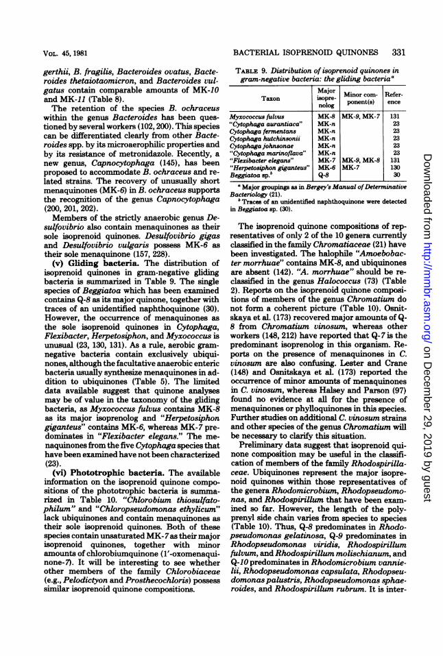
(v) Gliding bacteria. The distribution ofisoprenoid quinones in gram-negative glidingbacteria is smmarized in Table 9. The singlespecies of Beggiatoa which has been examinedcontains Q-8 as its major quinone, together withtraces of an unidentified naphthoquinone (30).However, the occurrence of menaquinones asthe sole isoprenoid quinones in Cytophaga,Flexibacter, Herpetosiphon, and Myxococcus isunusual (23, 130, 131). As a rule, aerobic gram-negative bacteria contain exclusively ubiqui-nones, although the facultative anaerobic entericbacteria usually synthesize menaquinones in ad-dition to ubiquinones (Table 5). The limiteddata available suggest that quinone analysesmay be of value in the taxonomy of the glidingbacteria, as Myxococcus fulvus contains MK-8as its major isoprenolog and "Herpetosiphongiganteus" contains MK-6, whereas MK-7 pre-dominates in "Flexibacter elegans." The me-naquinones from the five Cytophaga species thathave been examined have not been characterized(23).
(vi) Phototrophic bacteria. The availableinformation on the isoprenoid quinone compo-sitions of the phototrophic bacteria is summa-rized in Table 10. "Chlorobium thiosulfato-philum" and "Chloropseudomonas ethylicum"lack ubiquinones and contain menaquinones astheir sole isoprenoid quinones. Both of thesespecies contain unsaturated MK-7as their majorisoprenoid quinones, together with minoramounts of chlorobiumquinone (l'-oxomenaqui-none-7). It will be interesting to see whetherother members of the family Chlorobiaceae(e.g., Pelodictyon and Prosthecochloris) possesssimilar isoprenoid quinone compositions.
TABLE 9. Distribution of isoprenoid quinones ingram-negative bacteria: the gliding bacteriaa
Taxon Major Minor com- Refer-nolog ponent(s) ence
Myxococcus fulvus MK-8 MK-9, MK-7 131"Cytophaga aurantiaca" MK-n 23Cytophaga fermentans MK-n 23Cytophaga hutchinsonii MK-n 23Cytophagajohnsonae MK-n 23"Cytophaga marinoflava" MK-n 23"Flexibacter elegans" MK-7 MK-9, MK-8 131"Herpetosiphon giganteus" MK-6 MK-7 130Beggiatoa sp.bI Q-8 30
a Major grouping as in Bergey's Manual ofDeterminativeBacteriology (21).
'Traces of an unidentified naphthoquinone were detectedin Beggiatoa sp. (30).
The isoprenoid quinone compositions of rep-resentatives of only 2 of the 10 genera currentlyclassified in the family Chromatiaceae (21) havebeen investigated. The halophile "Amoebobac-ter morrhuae" contains MK-8, and ubiquinonesare absent (142). "A. morrhuae" should be re-classified in the genus Halococcus (73) (Table2). Reports on the isoprenoid quinone composi-tions of members of the genus Chromatium donot form a coherent picture (Table 10). Osnit-skaya et al. (173) recovered major amounts of Q-8 from Chromatium vinosum, whereas otherworkers (148, 212) have reported that Q-7 is thepredominant isoprenolog in this organism. Re-ports on the presence of menaquinones in C.vinosum are also confusing. Lester and Crane(148) and Osnitskaya et al. (173) reported theoccurrence of minor amounts of menaquinonesin C. vinosum, whereas Halsey and Parson (97)found no evidence at all for the presence ofmenaquinones or phylloquinones in this species.Further studies on additional C. vinosum strainsand other species of the genus Chromatium willbe necessary to clarify this situation.
Preliminary data suggest that isoprenoid qui-none composition may be useful in the classifi-cation of members of the family Rhodospirilla-ceae. Ubiquinones represent the major isopre-noid quinones within those representatives ofthe genera Rhodomicrobium, Rhodopseudomo-nas, and Rhodospirillum that have been exam-ined so far. However, the length of the poly-prenyl side chain varies from species to species(Table 10). Thus, Q-8 predominates in Rhodo-pseudomonas gelatinosa, Q-9 predominates inRhodopseudomonas viridis, Rhodospirillumfulvum, and Rhodospirillum molischianum, andQ-10 predominates in Rhodomicrobium vannie-lii, Rhodopseudomonas capsulata, Rhodopseu-domonas palustris, Rhodopseudomonas sphae-roides, and Rhodospirillum rubrum. It is inter-
VOL. 45, 1981
on Decem
ber 29, 2019 by guesthttp://m
mbr.asm
.org/D
ownloaded from

TABLE 10. Distribution of isoprenoid quinones in gram-negative bacteria: phototrophic bacteriaa
Taxon Major iso Minor component(s) Reference(s)prenolog"Chlorobium thiosulfatophilum" MK-7 1'-oxo-MK-7b 78, 181, 185"Chloropseudomonas ethylicum'c MK-7 1'-oxo-MK-7 181,185Chromatium vinosum strain D Q-7 MK-n 148C. vinosum strain D Q-7 212C. vinosum Q-8 MK-8 173C. vinosum Q-nd 97Chromatium sp. 8379 Q-10 MK-n 94Ectothiorhodospira halophila Q-8 Q-7 49Rhodomicrobium vannielii Q-10 28, 29, 158Rhodopseudomonas capsulata Q-10 28, 29, 158Rhodopseudomonas gelatinosa Q-8 29R. gelatinosa Q-8 MK-8 158R. gelatinosa Q-9 192Rhodopseudomonaspalustris Q-10 28,29,129,158Rhodopseudomonas sphaeroides Q-10 13,28,29,158Rhodopseudomonas viridis Q-9 MK-9 158Rhodopseudomonas op. FRNY Q-10 158Rhodospirillum fudvum Q-9 MK-9 158RhodpilUM molischianum Q-9 MK-9 158Rhodospirillun rubrwn Q-9 148R. rubrum Q-10 28,29, 101, 172, 219R. rubrum Q-10 Q-9, Q-8, Q-7, Q-6, Q- 56
5, Q-4, Q-3, Q-2, Q-1R. rubrum Q-10 RQ-1O' 87,158R. rubrum Q-10 Q-n, RQ-IO, RQ-nf 170, 235R. rubrum Q-10 Epoxy-Q-10' 77Rhodospirillum sp. 2761 Q-8 MK-8 158
a Major groupings as in Bergey's Manual ofDetermtinative Bacteriology (21).b l'-oxo-MK-7, Chlorobiumquinone (1'-oxomenaquinone-7).c "C. ethylicum" has been shown to be a mixt of two organisms (93).d Halsey and Parson (97) did not detect menaquinone in C. vinosum.'RQ-1O, Rhodoquinone with 10 isoprene units.fSome unspecified minor analogs of ubiquinone and rhodoquinone were also detected (170, 235).' Five new epoxy derivatives Qf Q-10 (epoxy-Q-10) were detected in R. rubrum (77). The concentrations of
the epoxyquinones were about 1 to 2% of the concentration of Q-10 (77).
esting that those species which contain Q-8 andQ-9 also contain menaquinones with side chainsof the same length (i.e., MK-8 and MK-9, re-spectively) (Table 10). On the other hand, spe-cies that contain Q-1O appear to lack menaqui-nones. Minor amounts of rhodoquinone andepoxides ofubiquinones have also been reportedto occur in R. rubrum (77, 164, 165, 219). Thetaxonomic relevance of these observationsawaits further study.Gram-positive bacteria. (i) Endospore-
forming rods and cocci. The distribution ofisoprenoid quinones in endospore-forming gram-positive rods and cocci is m zed in Table11.As presently constituted, the genus Bacillus
is heterogeneous (86). However, despite this het-erogeneity the vast majority of taxa that havebeen examined have remarkably uniform me-naquinone profiles, with unsaturated MK-7 pre-dominating (Table 11). However, Hess et al.(100) have reported recently the presence of
major amounts of MK-9 in Bacillus lentus andof MK-8 in Bacillus pantothenticus.Members of the genus Sporolactobacillus
possess characteristics intermediate betweenthose of the genera Bacillus and Lactobacillusin that they are gram-positive, rod-shaped, mi-croaerophilic, catalase-negative organismswhich form endospores. However, whereasmembers of the genus Lactobacillus generallylack isoprenoid quinones (Table 12) (38, 65),Sporolactobacillus inulinus and strains pres-ently called "SporolactobaciUus laevus" and"Sporolactobacillus racemicus" synthesize un-saturated MK-7 (38). The presence of similarmenaquinone patterns in sporolactobacilli andmembers of the genus Bacillus is in accord withthe similarities in peptidoglycan structures (123,194) and fatty acid profiles (225) among thesebacteria. However, as Collins and Jones havepointed out (38), it would be premature to sug-gest reclassification of S. inulinus or other spo-rolactobacilli into the genus Bacillus as that
332 COLLINS AND JONES MICROBIOL. REV.
on Decem
ber 29, 2019 by guesthttp://m
mbr.asm
.org/D
ownloaded from

BACTERIAL ISOPRENOID QUINONES 333
genus is presently constituted. When the taxon-omy of the genus Bacillus is clarified, it is veryprobable that one of the new taxa will containboth the sporolactobacilli and certain bacteriapresently designated Bacillus species (e.g., "Bat-cillus laevolacticus," "Bacillus racemilacti-cus," "Bacillus myxolactis," and "Bacillus dec-trolacticus").Sporosarcina ureae possesses many proper-
ties in common with the genera Micrococcusand Pkanococcus (194). This species is also sim-ilar to members of the genus Bacillus in itsability to form endospores. The presence of un-saturated MK-7 in S. ureae reinforces its rela-tionship to members of the genus Bacillus butclearly distinguishes it from planococci (whichcontain unsaturated MK-8) and from micrococci(which generally contain partially hydrogenatedmenaquinones) (242). The distinctness of thegenera Sporosarcina and Bacillus is probablyjustified considering their differences in pepti-doglycan structure, mode of cell division, shape,and ability to utilize urea. However, certain ba-cilli, such as Bacillus pasteurii, will probably beclassified with S. ureae when the taxonomy ofthe genus Bacillus is clarified (242).
Early studies indicated that members of thegenus Clostridium lacked respiratory quinones(17, 83, 85, 148). This is in accord with thestrictly anaerobic nature of clostridia and withtheir inability to synthesize heme compoundsand cytochromes. However, recently Gottwaldet al. (92) demonstrated the presence of cyto-chromes and an uncharacterized menaquinonein Clostridium formicoaceticum and Clostrid-ium thermoaceticum. Therefore, although it isgenerally conceded that quinones are absentfrom most clostridia, further systematic studieswill be necessary before the potential of isopre-noid quinone analyses in the taxonomy of thisgroup can be appreciated.
(ii) Lactic acid bacteria. Members of thegenus Lactobacillus generally lack isoprenoidquinones (38, 65). Hess et al. (100) recently re-ported low levels of an uncharacterized mena-quinone in a single strain of Lactobacillusbrevis. Lactobacillus mali and "Lactobacillusyamanashiensis" also contain menaquinones astheir sole isoprenoid quinones, with MK-8, MK-9, and MK-1O constituting the major compo-nents (33). The presence of closely related me-naquinone profiles within these latter taxa sup-ports the hypothesis that L. mali and "L. ya-manashiensis" are the same species (27). Mem-bers of the genus Listeria also contain mena-quinones as their sole isoprenoid quinones. Lis-teria monocytogenes (including the organismsreferred to in the literature as "Listeria inno-
cua" [199] and L. monocytogenes Ivanov sero-type 5 [112]), Listeria grayi, and Listeria mur-rayi all contain unsaturated MK-7 (Table 12),thereby reinforcing the relatedness among thesetaxa (33, 46, 121). The presence of unsaturatedMK-9 in Listeria denitrificans supports the hy-pothesis that this taxon should be removed fromthe genus Listeria (33, 46, 120, 121, 209). Themenaquinone composition of the genus Bro-chothrix is controversial. Collins and associatesrecovered major amounts of MK-7 from severalstrains of Brochothrix thermosphacta (46, 121).However, Hess and colleagues reported the pres-ence of a MK-9 in a single strain of B. thermos-phacta (100). The results of Hess et al. (100)must be treated with caution, as only reverse-phase partition thin-layer chromatography wasused for the menaquinone characterization. Asmentioned above, reverse-phase thin-layer chro-matography carnot be used by itself to resolvea menaquinone mixture (see above) (67). How-ever, Collins et al. used mass spectrometry aswell as reverse-phase thin-layer chromatogra-phy. Molecular ions (M+) were found at m/e648, thereby confirming the presence of MK-7(46). The latter result also confirms the taxo-nomic relationship between the genera Bro-chothrix and Listeria, as determined by numer-ical phenetic studies (120, 236).Most members of the genus Streptococcus
lack isoprenoid quinones, although menaqui-nones have been reported in a few streptococciof serological groups D and N (7, 8, 39, 40, 100).Streptococcus faecalis contains demethylmena-quinones and on this basis can be distinguishedfrom all other members of the family Strepto-coccaceae (39, 40). Streptococcus faecium subsp.casseliflavus, Streptococcus faecium subsp.mobilis, Streptococcus cremoris, Streptococcuscremoris subsp. alcatosus, Streptococcus lactis,and Streptococcus lactis subsp. diacetylactiscontain fully substituted menaquinones, withMK-8 and MK-7 predominating in the first twotaxa and MK-9 predominating in the latter taxa.The presence of menaquinones in S. lactis andS. lactis subsp. diacetylactis is in accord withthe reports of cytochrome-mediated electrontransport in strains of these taxa when they aregrown in the presence of preformed heme (188).To our knowledge, cytochrome-mediated elec-tron transport in S. cremoris, S. faecium subsp.casseliflavus, and S. faecium subsp. mobilis hasnot been described, although the menaquinonedata suggest that such an investigation might berewarding. Representatives of other lactic acidtaxa (e.g., Aerococcus, Erysipelothrix, Gemella,Leuconostoc, and Pediococcus) generally lackisoprenoid quinones (39, 40, 46, 100), thereby
VOL. 45, 1981
on Decem
ber 29, 2019 by guesthttp://m
mbr.asm
.org/D
ownloaded from

334 COLLINS AND JONES
TABLE 11. Distribution of isoprenoid quinones in gram-positive bacteria: endospore-forming rods andcocci
Taxon Major Minor component(s) Reference(s)isoprenolog(s)Bacillus alcalophilus MK-7 MK-3 100Bacillus alvei MK-7 100, 227"Bacillus aminovorans" MK-7 MK-2 100"Bacillus amyloliquefaciens" MK-7 MK-2 100"Bacillus aneurinolyticus" MK-7 100Bacillus anthracis MK-n 100Bacillus badius MK-7 MK-3 100Bacillus brevis MK-7 81, 100, 227B. brevis MK-n 206Bacillus cereus MK-7 100, 113, 237Bacillus circulans MK-7 100, 227"Bacillus cirroflagellosus" MK-n 100Bacillus coagulans MK-7 227B. coagulans MK-7 MK-6, MK-5 38B. coagulans MK-7 MK-3 100"Bacillus dextrolacticus" MK-7 MK-6 38"Bacillus epiphytus" MK-7 MK-3 100"Bacillus filicolonicus" MK-7 100Bacillus firmus MK-7 MK-3 100B. firmus MK-7 227"Bacillus freudenreichii" MK-7 MK-6, MK-5, MK-4 50"B. freudenreichii" MK-7 100Bacillus globisporus MK-7 MK-3 100Bacillus insolitus MK-7 MK-3 100"Bacillus laevolacticus" MK-7 MK-6 38"B. laevolacticus" MK-7 100Bacillus larvae MK-7 100Bacillus laterosporus MK-7 MK-3 100Bacillus lentus MK-9 MK-3 100B. lentus MK-7 227Bacillus licheniformis MK-7 91, 191B. licheniformis MK-7 MK-3 100Bacillus macerans MK-7 MK-3 100B. macerans MK-7 227Bacillus macquariensis MK-7 MK-3 100"Bacillus maroccanus" MK-7 MK-3 100"Bacillus macroides" MK-7 MK-6 38"Bacillus medusa" MK-7 MK-3 100Bacillus megaterium MK-7 17, 20, 100, 137, 191,
;7lz"Bacillus mesentericus""B. mesentericus""Bacillus myxolactis"Bacillus pantothenticusB. pantothenticusBacillus pasteuriiB. pasteuriiBacillus polymyxaBacillus popilliae"Bacillus psychrophilus""Bacillus psychrosaccharolyticus""Bacillus pulvifaciens"Bacillus pumilus"Bacillus racemilacticus""B. racemilacticus"Bacillus sphaericusB. sphaericusB. sphaericusBacillus stearothermophilus
MK-7MK-nMK-7MK-8MK-7MK-7MK-7MK-7MK-7MK-7MK-7MK-7MK-7MK-7MK-7MK-7MK-7MK-7MK-7
MK-6MK-2
MK-6, MK-5, MK-4MK-2
MK-3MK-3MK-2
MK-6
MK-6, MK-5MK-3
691483810022750100100, 227100100100100100, 227381003810084, 22767, 100, 227
MICROBIOL. REV.
on Decem
ber 29, 2019 by guesthttp://m
mbr.asm
.org/D
ownloaded from

BACTERIAL ISOPRENOID QUINONES 335
TABLE 11-Continued
Taxon Major Minor component(s) Reference(s)isoprenolog(s) Miocopnn()Rfrce)
Bacillus subtilis MK-7 17, 63, 71, 100, 113,191, 227
"Bacillus thiaminolyticus" MK-7 MK-2 100Bacillus thuringiensis MK-8, MK-7 MK-3 100Clostridium formicoaceticumb MK-n 92Clostridium thermoaceticum MK-n 92Desulfotomaculum nigrificans MK-7 221Sporolactobacillus inulinus MK-7 MK-6 38S. inulinus MK-7 100"Sporolactobacillus laevus" MK-7 MK-6 38Sporolactobacillus racemicus MK-7 MK-6 38Sporosarcina ureae MK-7 242
a Major groupings as in Bergey's Manual ofDeterminative Bacteriology (21).b Menaquinones and ubiquinones have been reported to be absent in Clostridium butyricum (100), Clostrid-
ium chauvoei (100), Clostridium histolyticum (85, 100), "Clostridium oedematiens" (100), Clostridium para-putrificum (100), Clostridiumperfringens (148), Clostridium sporogenes (17), and Clostridium sticklandii (83).
TABLE 12. Distribution of isoprenoid quinones in gram-positive bacteria: gram-positive, asporogenous rod-shaped bacteria containing menaquinones as their sole isoprenoid quinonesaTaxonb Major isoprenolog(s) Minor component(s) Reference(s)
Brochothrix thermosphacta MK-7 MK-6, MK-5 33, 46, 121B. thermosphacta MK-9c 100Caryophanon latum MK-6 50"Caryophanon tenue" MK-6 MK-5 46Lactobacillus casei subsp. rhamnosus MK-n 100Lactobacillus mali MK-8, MK_9d MK-10, MK-7 33,50"Lactobacillusyamanashiensis" MK-8, MK-9 MK-10, MK-7 33Listeria denitrificans MK-9 MK-8, MK-7 33, 46, 121Listeria grayi MK-7 MK-6, MK-5 33, 46, 121"Listeria innocua" MK-7 MK-6, MK-5 33Listeria murrayi MK-7 MK-6, MK-5 33, 46,121Listeria monocytogenes (including MK-7 MK-6, MK-5 33, 46, 121
"Listeria bulgarica")a Major groupings as in Bergey's Manual ofDeterminative Bacteriology (21).b Gram-positive, asporogenous rod-shaped bacteria that lack both menaquinones and ubiquinones include
Erysipelothrix rhusiopathiae (46), Lactobacillus acidophilus (100), Lactobacillus brevis (33, 100), Lactobacil-lus casei subsp. casei (17, 38), Lactobacillus casei subsp. rhamnosus (38), Lactobacillus delbrueckii (38),Lactobacillus hilgardii (38), "Lactobacillus odontolyticus" (100), Lactobacillus plantarum (38), and Lacto-bacillus salivarius (38).
' Hess et al. (100) used only reverse-phase partition thin-layer chromatography for quinone characterizations.d MK-8 and MK-9 were present in comparable amounts in L. mali and "L. yamanashiensis" (33).
reinforcing their affinity with the majority of thestreptococci (Table 13).
(iii) Micrococcaceae The distribution of me-naquinones within the members of the familyMicrococcaceae is summarized in Table 13. Allmembers of the genera Planococcus and Staph-ylococcus that have been examined possess un-saturated manaquinones as their sole respiratoryquinones. Planococcus citreus and Staphylococ-cus aureus contain MK-8 as their major com-ponent, although MK-7 is also present at sub-stantial levels (114-117, 242). However, the co-agulase-negative staphylococci (Staphylococcuscapitis, Staphylococcus cohnii, Staphylococcus
epidermidis, Staphylococcus haemolyticus,Staphylococcus hominis, Staphylococcus sap-rophyticus, Staphylococcus simulans, andStaphylococcus warneri) contain MK-7 as theirmajor isoprenolog (Table 13). Staphylococcussciuri subsp. sciuri and Staphylococcus sciurisubsp. lentus contain MK-6 as their major me-naquinone component and on this basis can bedifferentiated from all of other staphylococcithat have been examined (Table 13).Members of the genus Micrococcus are het-
erogeneous with respect to menaquinone com-position. Micrococcus varians and Micrococcusagilis are characterized by the presence ofmajor
VOL. 45, 1981
on Decem
ber 29, 2019 by guesthttp://m
mbr.asm
.org/D
ownloaded from

336 COLLINS AND JONES
TABLE 13. Distribution of isoprenoid quinones in gram-positive bacteria: gram-positive cocci containingmenaquinones or demethylmenaquinones as their sole isoprenoid quinonesa
Taxonb Major Minor component(s) Reference(s)
MicrococcaceaeMicrococcus agilis ATCC 966Micrococcus agilis strains ATCC
966, CCM 2390, CCM 2539, CCM2687, and CCM 2688
"Micrococcus cyaneus""Micrococcus eucinetus" CCM 2387d"M. eucinet4s"Micrococcus halobiusMicrococcus luteus strains CCM 134and CCM 840
M. luteus strains CCM 810 andATCC 398
M. luteus CCM 132M. luteus strains CCM 169 andATCC 4698
M. luteus strains CCM 149 andATCC 272
M. luteus strains CCM 810 andATCC 398
"Micrococcus lysodeikticus"g
"M. lysodeikticus"g"M. lysodeikticus"g"Micrococcus morrhuae"^"Micrococcus radiodurans""M. radiodurans""Micrococcus radiophilus""M. radiophilus""Micrococcus radioproteolyticus""M. radioproteolyticus"Micrococcus variansM. varians CCM 132Planococcus citreusPlanococcus spp. strains CCM 1849,CCM 2069, CCM 2104, CCM 2414,CCM 2415, and CCM 2416
Staphylococcus aureus HS653
S. aureusS. aureus"Staphylococcus albus"Staphylococcus capitis
Staphylococcus cohniiStaphylococcus epidermnidisStaphylococcus haemolyticusStaphylococcus hominisStaphylococcus saprophyticusS. saprophyticusStaphylococcus simulansStaphylococcus app. acetoin-negative
bovine strainsStaphylococcus warneriStaphylococcus sciuri subsp. IentusStaphylococcus sciuri subsp. sciuri
Peptococcaceae"Peptostreptococcus magnus""Sarcina flava"
MK-9(H.)cMK-9(H2)
MK-9(H.)MK-8MK-8, MK-7'MK-8MK-8(HX)
MK-9(H,)
MK-7(H.)MK-8
MK-8(H2)
MK-9(H2)
MK-4, MK-3
MK-8MK-9MK-8MK-8MK-8MK-8MK-8MK-8MK-8MK-7(H2)MK-7(H.)MK-8MK-8
MK-7'
MK-8MK-8MK-7MK-7
MK-7MK-7MK-7MK-7MK-7MK-7MK-7MK-6
MK-7MK-6MK-6
MK-nMK-n
MK-8(Hs)
MK-8(H.)MK-7, MK-6
MK-7, MK-6MK-9(H.), MK-7(H.)f
MK-8(Hx)
MK-8(HX), MK-6(H.)MK-7
MK-8(H2), MK-8, MK-5
MK-7, MK-6
MK-7, MK-6
MK-7, MK-6
MK-7, MK-6
MK-8(H.), MK-6(H.)MK-7MK-7
MK-8, MK-6, MK-5,MK-4
MK-7MK-9, MK-7k
MK-8, MK-6, MK-5
MK-8, MK-6, MK-5MK-8, MK-6, MK-5MK-8, MK-6, MK-5MK-8, MK-6, MK-5MK-8, MK-6, MK-5MK-8, MK-6MK-8, MK-6, MK-5MK-8, MK-7
MK-8, MK-6, MK-5
114, 115, 116242
114, 11511511549115
114, 115
114,115242
242
242
67
22716, 79,152, 191
114247Colins'247Collins'247Collinsi115115242242
117
24275, 98,114,115,11617
Collins and Goodfel-low,
Collins and GoodfeUow'Collins and Goodfellow'Collins and Goodfeliow'Collins and Goodfeilow'Collins and Goodfeliow'114Colins'114,115
ColinsCollins'50
85113
MICROBIOL. REV.
on Decem
ber 29, 2019 by guesthttp://m
mbr.asm
.org/D
ownloaded from

BACTERIAL ISOPRENOID QUINONES 337
TABLE 13-ContinuedMajo
Taxonb Mojor Minor component(s) Reference(s)
"Sarcina lutea" MK-8 17"S. lutea" MK-9 85
StreptococcaceaeLeuconostoc lactis MK-n 106Streptococcus cremoris MK-9 MK-8, MK-7, MK-6 39Streptococcus cremoris subsp. alac- MK-9 MK-8, MK-7, MK-6 39
tosusStreptococcus faecalis DMK-9m DMK-8, DMK-7 7,8S. faecalis DMK-n 100,106Streptococcus faecalis subsp. faecais DMK-9 DMK-8, DMK-7, 39,40
DMK-6nStreptococcus faecalis subsp. lique- DMK-9 DMK-8, DMK-7, 39,40
faciens DMK-6MStreptococcus faecalis subsp. zymo- DMK-9 DMK-8, DMK-7, 39,40genes DMK-6"
Streptococcus faecium subsp. casse- MK-8 MK-7, MK-6 39,40liflavus
Streptococcus faecium subsp. mobilis MK-8 MK-7, MK-6 39,40Streptococcus lactis MK-9 MK-8, MK-7, MK-6 39Streptococcus lactis subsp. diacety- MK-9 MK-8, MK-7, MK-6 39
lactis
'Major groupings as in Bergey's Manual ofDeterminative Bacteriology (21).b Gram-positive cocci that lack both menaquinones and ubiquinones include the following members of the
Streptococcaceae. "Aerococcus catalaswus" (39), Aerococcus viridans (39, 100), Gemella haemolysans (39, 40,100), Leuconostoc cremoris (100), Leuconostoc dextranicum (39), Leuconostoc mesenteroides (39), Leuconostocparamesenteroides (100), Pediococcus acidilactici (39), Pediococcus parvulus (39), Pediococcus pentosaceus(39), Streptococcus agalactiae (39, 40), "Streptococcus avium" (39, 40), Streptococcus bovis (39, 40), "Strepto-coccus durans" (39), "Streptococcus dysgalactiae" (39, 40), Streptococcus equi (39), Streptococcus equinus (39,40), "Streptococcus equisimilis" (39,40), Streptococcus faecalis subsp. malodoratus (39), Streptococcus faecium(39), "Streptococcus milleri" (39), Streptococcus mitis (85), Streptococcus mutans subsp. sorbrinus (39),Streptococcus pneumoniae (39), Streptococcus pyogenes (39), Streptococcus raffinolactis (39), Streptococcussalivarius (39), Streptococcus sanguis (39, 40), "Streptococcus suis" (39, 40), Streptococcus sp. group E (39),Streptococcus sp. strain 77007/71 (100), and Streptococcus uberis (39, 40).
(H.) indicates that the degree of hydrogenation was not determined.d"M. eucinetus" is now clasified as Planococcus citreus.MK-8 and MK-7 were present in comparable amounts.
fTraces of hydrogenated MK-5 and MK-6 were also found in M. luteus CCM 134 (114).' Now clified as Micrococcus luteus.h "M. morrhuae" is now classified as Halococcus morrhuae.'M. D. Collins, unpublished data.i'Major amounts of MK-7 were present in an atypical S. aureus strain (117).* Trace amounts of smaller isoprenologs were detected in Streptococcus aureus (75, 98).'M. D. Collins and M. Goodfellow, unpublished data.mDMK-9, Demethylmenaquinone containing nine isoprene units.'Traces of demethylmenaquinone containing 10 isoprene units were found in S. faecalis subap. faecalis, S.
faecalis subsp. liquefaciens, and S. faecalis subsp. zymogenes (39, 40).
amounts of dihydrogenated MK-7 [MK-7(H2)]and MK-9(H2), respectively (242). Three mena-quinone patterns have been reported in strainsof Micrococcus luteus. Strain ATCC 4698, theneotype ofM. luteus (134), contains unsaturatedMK-8. However, M. luteus strains AJ 1081 andATCC 398 possess MK-8(H2) and MK-9(H2),respectively (242). The radiation-resistant taxa"Micrococcus radiodurans," "Micrococcus ra-diophilus," and "Micrococcus radioproteolyti-
cus" all possess closely related menaquinoneprofiles, with MK-8 predominating (247). Thesethree taxa should be removed from the genusMicrococcus.
Little information is available on the isopre-noid quinone contents of the Peptococcaceae.Gibbons and Engle (85) detected an uncharac-terized menaquinone in "Peptostreptococcusmagnus" by using a microbiological assay. Dataon the menaquinone composition of "Sarcina
VOL. 45, 1981
on Decem
ber 29, 2019 by guesthttp://m
mbr.asm
.org/D
ownloaded from

338 COLLINS AND JONES
lutea" are confusing. Bishop et al. (17) recoveredmajor amounts of MK-8 from "S. lutea,"whereas Salton and Schmitt (191) reported thepresence of MK-9 in the same species.
(iv) Coryneform bacteria and actinomy-cetes. In Bergey's Manual of DeterminativeBacteriology (21), the coryneform group of bac-teria contains the genera Arthrobacter, Cellu-lomonas, and Corynebacterium, and Brevibac-terium and Microbacterium are described asgenera incertae sedis. The genus Kurthia is alsoincluded as a placement of convenience. Thegenera Bacterionema and Curtobacteriumshould also be added to the coryneform group.The genus Arthrobacter is heterogeneous, and
Arthrobacterglobiformis and related strains canbe distinguished readily from Arthrobacter sim-plex (36, 126, 127, 194). The menaquinone dataunderline this division, since A. globiformis, Ar-throbacter atrocyaneus, Arthrobacter aures-cens, Arthrobacter crystallopoietes, Arthrobac-terpascens, Arthrobacter ramosus, and Arthro-bacter ureafaciens contain major amounts ofMK-9(H2), whereas A. simplex possesses majorquantities of MK-8(H4) (Table 14). A. simplexcontains a peptidoglycan based on LL-diamino-pimelic acid (194) and is probably more closelyrelated to certain actinomycetes. The phyto-pathogen Cornebacterium ilicis possesses manyproperties in common with true Arthrobacterspp., and the presence of MK-9(H2) within thistaxon underlines this relationship. However, themenaquinones cast serious doubt on the reten-tion of Arthrobacter nicotianae within the ge-nus Arthrobacter. A. nicotianae contains unsat-urated MK-8 and MK-9 (241). Major amountsof MK-8 and MK-9 have also been described instrains of "Brevibacterium fuscum" and Brevi-bacterium protophormiae (50, 241). All three ofthese taxa possess peptidoglycans based on ly-sine (structural variation A4a) (194) and mayconstitute the nucleus of an as-yet-unrecognizedtaxon (36). The menaquinone data are not inaccord with the suggestion of Keddie and Jones(127) that the taxon "Brevibacterium sulfu-reum" be included in the genus Arthrobacter."B. sulfureum" contains major amounts of MK-9, and further studies should be directed to acomparison of this taxon with A. nicotianae, "B.fuscum," and B. protophormiae.
In Bergey's Manual of Determinative Bacte-riology (21), the genus Brevibacterium is de-scribed as a taxon incertae sedis. However, thisgenus has been redefined recently, with Brevi-bacterium linens as the type species (47). B.linens possesses MK-8(H2) as its major compo-nents (34, 47, 241). A similar menaquinone pat-tem has been found in the gram-negative taxon"Chromobacterium iodinum" (Table 14). Other
chemotaxonomic and numerical phenetic datahave led to the recommendation that this speciesshould be reclassified in the genus Brevibacter-ium as Brevibacterium iodinum (47), an obser-vation in accord with the menaquinone data.
Chemical and numerical phenetic data indi-cate that the genus Microbacterium should beretained for the type species Microbacteriumlacticum (34, 120, 126, 194). M. lacticum is char-acterized by the presence of an unusual pepti-doglycan based upon lysine (structural variationBla) (194). Similar cell wall structures basedupon lysine have also been reported in Brevi-bacterium imperiale and "Corynebacterium lae-vaniformans" (variations B1,8 and Bla, respec-tively) (194). The presence of unusually longunsaturated MK-11 and MK-12 within the taxaM. lacticum, B. imperiale, and "C. laevanifor-mans" supports this apparent relationship.
In Bergey's Manual of Determinative Bacte-riology (21), the monospecific genus Kurthia istreated with the coryneforn group of bacteria,although its members do not have a coryneformmorphology. Kurthia zopfii possesses majoramounts of MK-7 and on this basis can be dif-ferentiated from all coryneforn bacteria thathave been examined, with the exception ofBrev-ibacterium acetylicum ATCC 953 and Brevibac-teriurn halotolerans (34, 49). B. acetylicumshould be compared further with K. zopfii,whereas B. halotolerans is probably moreclosely related to Brochothrix thermosphacta(Table 12).The genus Cellulomonas is a homogeneous
taxon. All of the cellulomonads that have beenexamined possess tetrahydrogenated MK-9[MK-9(H4)] (34, 241). The nocardioform genusOerskovia shares many biochemical propertieswith the cellulomonads (34, 48, 120, 162, 163).The recovery of MK-9(H4) from Oerskovia spp.reinforces this relationship (34, 48, 241). Repre-sentatives of the taxa Brevibacterium fermen-tans, Brevibacterium lyticum, "Cellulomonascartalyticum", "Corynebacterium manihot,"and Nocardia cellulans also possess majoramounts of MK-9(H4) (Table 14). Numericalphenetic and chemical data indicate that thesetaxa should be reclassified in the genus Oersko-via (120, 162).The genus Curtobacterium is heterogeneous
with respect to menaquinone composition (Ta-ble 14). Collins et al. (35) have suggested that"true curtobacteria" should be restricted tothose taxa (e.g., Curtobacterium albidum, Cur-tobacterium citreum, Curtobacterium luteum,Corynebacterium betae, Corynebacterium oor-tii, Corynebacterium flaccumfaciens, and Cory-nebacterium poinsettiae) which contain MK-9as their major isoprenolog. However, Curtobac-
MICROBIOL. REV.
on Decem
ber 29, 2019 by guesthttp://m
mbr.asm
.org/D
ownloaded from

VOL. 45, 1981
terium saperdae and Curtobacterium testaceumcontain major amounts of MK-11 and MK-12and should be removed from the genus Curto-bacterium sensu stricto (35, 241). Cell wall dataindicate that "Corynebacterium barkeri" and"Microbacterium liquefaciens" may be relatedto C. saperdae (194). The recovery of MK-11and MK-12 from "C. barkeri" and "M. liquefa-ciens" is in accord with this view. C. saperdae,C. testaceum, "C. barkeri," and "M. liquefa-ciens" should be compared further to determinewhether they constitute the nucleus of a newgenus.
Relatively few coryneform bacteria possesspeptidoglycans based upon diaminobutyric acid(194). Most of these bacteria have been placedin the genus Corynebacterium, and they includesix plant pathogens (Corynebacterium insi-diosum, "Corynebacterium isanicum," Coryne-bacterium michiganense, Corynebacterium ne-braskense, "Corynebacterium tritici," and Cor-ynebacterium sepedonicum), one insect patho-gen ("Corynebacterium okanaganae"), and afew saprophytic species ("Corynebacteriumaquaticum," "Corynebacterium mediolanum,"and "Flavobacterium dehydrogenans"). Otherthan to note that none of these belongs to thegenus Corynebacterium, little comment can bemade on their generic positions. Recent studieshave shown the diaminobutyric acid-containingtaxa can be divided into four groups on the basisof menaquinone patterns (41). C. insidiosum, C.michiganense, C. nebraskense, and C. sepedon-icum possess MK-9; "C. iranicum" and "C. tri-tici" possess MK-10; "C. aquaticum" possessesMK-10 and MK-11; and "C. mediolanum" and"F. dehydrogenans" possess MK-11 and MK-12(41).There is now general agreement that the ge-
nus Corynebacterium sensu stricto (127, 163)should be restricted to those taxa which containa peptidoglycan based upon meso-diaminopi-melic acid (variation Al-y) (194), the wall sugarsarabinose and galactose (126, 194), a deoxyribo-nucleic acid base composition of 51 to 59 mol%guanine plus cytosine (127), and relatively short-chain mycolic acids (approximately 22 to 36carbon atoms) (37, 163). On the basis of mena-quinone patterns, "true corynebacteria" can bedivided into two broad groups (Table 15). Themajority of animal-associated corynebacteria(e.g., Corynebacterium diphtheriae, Corynebac-terium kutscheri, Corynebacterium pseudotu-berculosis, Corynebacterium renale, "Coryne-bacterium ulcerans") possess major amounts ofMK-8(H2), whereas many saprophytic species(e.g., Corynebacterium glutamicum) and occa-sional animal-associated taxa (e.g., Corynebac-terium bovis and "Corynebacterium minutissi-
BACTERIAL ISOPRENOID QUINONES 339
mum") contain predominantly MK-9(H2) (34,48, 241). Bacterionema matruchotii also pos-sesses a peptidoglycan based upon meso-diami-nopimelic acid, a guanine-plus-cytosine contentof 55 to 57 mol%, and a relatively short-chainmycolic acid (30 to 36 carbon atoms) (163). Therecovery of major amounts of MK-8(H2) andMK-9(H2) from B. matruchotii (50) reinforcesthis relationship between Bacterionema spp.and Corynebacterium spp. and suggests that B.matruchotii should be reclassified in the genusCorynebacterium sensu stricto.The distribution of menaquinones within the
mycolic acid-coantaining actinomycetes and co-ryneforms is shown in Table 15. The menaqui-none data support the exclusion of strains of therhodochrous complex from the genus Nocardia.Primarily on the basis ofchemical and numericalphenetic data, the rhodochrous complex and thegenus Gordona have been merged to form thegenus Rhodococcus (88). The rhodococci can bedivided into two groups on the basis of theirmenaquinone pattems (48, 161). Rhodococcuserythropolis, Rhodococcus coprophilus, Rho-dococcus rhodochrous, and Rhodococcus rubercontain MK-8(H2) as their major isoprenolog,whereas MK-9(H2) predominates in strains ofRhodococcus bronchialis and Rhodococcus cor-allinus. In complete contrast, representatives ofNocardia sensu stricto have MK-8(H4) as theirmajor component. A strain of Micropolysporabrevicatena has also been reported to containMK-8(H4) (48). M. brevicatena also containsmycolic acids similar to those found in strains ofNocardia (48). It will be interesting to seewhether other studies support this apparentlyclose relationship. Members of the genus Myco-bacterium and the species Nocardia amaraeand Nocardia farcinica contain major amountsof MK-9(H2) (Table 15).Numerical phenetic and chemical studies in-
dicate that strains currently called "Cordonaaurantiaca" form a distinct taxon worthy ofgeneric rank (90). The presence of majoramounts of MK-9 within "G. aurantiaca" sup-ports this view (90). It is worth noting that, withthe sole exception of Corynebacterium pauro-metabolum, all of the mycolic acid-containingtaxa that have been examined possess partiallyhydrogenated menaquinones (Table 15).A variety of menaquinone patterns are found
in strains of actinomycetes which lack mycolicacids (Table 16). Promicromonospora citrea,Propionibacterium acnes, "Propionibacteriumarabinosum," and "Propionibacterium sher-manii" contain major amounts ofMK-9(H4) (50,198, 208). Members of the genera Actinomaduraand Streptomyces are unusual in that they con-tain very complex mixtures of partially hydro-
on Decem
ber 29, 2019 by guesthttp://m
mbr.asm
.org/D
ownloaded from

TABLE 14. Distribution of isprenoid quinones in gram-positive bacteria: coryneform bacteria lackingmycolic acidsa
Taxon Major isopre- Minor component(s) Reference(s)nolog(s)Arthrobacter atrocyaneus MK-9(H2) MK-9, MK-8(H2), MK-7(H2) 36Arthrobacter aurescens MK-9(H2) MK-9, MK-8(H2), MK-7(H2) 36Arthrobacter citreus MK-9(H2) 241A. citreus MK-9(H2) MK-9, MK-8(H2), MK-7(H2) 36Arthrobacter crystallopoietes MK-9(H2) MK-9, MK-8(H2), MK-7(H2)b, 34, 36Arthrobacter globiformis MK-9(H2) 241A. globiformis MK-9(H2) MK-9, MK-8(H2), MK-7(H2)b 34, 36Arthrobacter nicotianae MK-8 MK-9 241A. nicotianae MK-8 MK-9, MK-7 36Arthrobacter oxydans MK-9(H2) 241A. oxydans MK-9(H2) MK-9, MK-8(H2), MK-7(H2) 36Arthrobacterpolychromogenes MK-9(H2) MK-9, MK-8(H2), MK-7(H2) 34Arthrobacter ramosus MK-9(H2) MK-9, MK-8(H2), MK-7(H2) 36Arthrobacter simplex MK-7 2A. simplex MK-8(H4) 241A simplex MK-n 1A. simplex MK-8(H4) MK-8(H2), MK-7(H4) 34, 36Arthrobacter tumescens MK-8(H4) MK-8(H2), MK-7(H4) 34, 36Arthrobacter ureafaciens MK-9(H2) 241A. ureafaciens MK-9(H2) MK-9, MK-8(H2), MK-7(H2) 34Arthrobacter sp. A4 MK-1O MK-11, MK-9, MK-8, MK-7 41Brevibacterium acetylicum ATCC 953 MK-7 MK-6 Collins, unpublished
dataBrevibacterium fermentans MK-9(H4) MK-9(H2), MK-8(H4), MK-
7(H4)"Brevibacterium fuscum" MK-9 MK-8 241Brevibacterium halotolerans MK-7 MK-6 49"Brevibacterium helvolum" ATCC 11822 MK-9(H2) 241Brevibacterium imperiale MK-12, MK-11 MK-13, MK-1O, MK-9, MK-8 Collins, unpublished
dataBrevibacterium iodinum MK-8(H2) MK-8, MK-7(H2) 47Brevibacterium linens MK-8(H2) MK-8, MK-7(H2) 34, 47B. linens MK-8(H2) 241"Brevibacterium lipolyticum" MK-8(H4) 241, 243"B. lipolyticum" MK-8(H4) MK-8(H2), MK-7(H4) 36Brevibacterium lyticum MK-9(H4) MK-9(H2), MK-8(H4), MK- 44
7(H4)Brevibacterium protophormiae MK-9, MK-8 MK-7 50"Brevibacterium sulfureum" MK-9 MK-1O 241"B. sulfureum" MK-9 MK-1O, MK-8, MK-7 34Cellulomonas biazotea MK-9(H4) 241C. biazotea MK-9(H4) MK-9(H2), MK-8(H4), MK- 34
7(H4)"Cellulomonas cartalyticum" MK-9(H4) MK-9(H2), MK-8(H4), MK- 34
7(H4)Celulomonas fimi MK-9(H4) 241C. fimi MK-9(H4) MK-9(H2), MK-8(H4), MK- 34
7(H4)Cellulomonas flavigena MK-9(H4) MK-9(H2), MK-8(H4), MK- 34
7(H4)"Cellulononas subalbus" M-9(H4) MK-9(H2), MK-8(H4), MK- 34
7(H4)"Corynebacterium aquaticum" MK-11, MK-1O 241"C. aquaticum" MK-11, MK-IO MK-12, MK-9, MK-8, MK-7 34, 41, 50"Corynebacterium barkeri" MK-11, MK-12 MK-13, MK-1O, MK-9, MK-8 Collins, unpublished
dataCorynebacterium betae MK-9 MK-8, MK-7d 34, 35Corynebacterium flaccumfaciens MK-9 241C. flaccumnfaciens MK-9 MK-8, MK-7 34, 35Corynebacterium ilicis MK-9(H2) MK-9, MK-8(H2), MK-7(H2) Collins, unpublished
dataCorynebacterium insidiosum MK-9 MK-1O, MK-8, MK-7 41"Corynebacterium iranicum" MK-1O MK-11, MK-9, MK-8, MK-7 41"Corynebacterium manihot" MK-9(H4) MK-9(H2), MK-8(H4), MK- 34
7(H4)"Corynebacterium mediolanum" MK-12, MK-11 MK-13, MK-IO, MK-9, MK-8, 41,50
MK-7
340 COLLINS AND JONES MICROBIOL. REV.
on Decem
ber 29, 2019 by guesthttp://m
mbr.asm
.org/D
ownloaded from

BACTERIAL ISOPRENOID QUINONES 341
TABLE 14-Continued
Taxon Major isopre- Minor component(s) Reference(s)nolog(s)
Corynebacterium michiganense MK-9 MK-10, MK-8, MK-7 41, 50Corynebacterium nebraskense MK-9 MK-10, MK-8, MK-7 34, 35, 41Corynebacterium oortii MK-9 MK-8 35, 50"Corynebacterium okanaganae" MK-12, MK-11 MK-10, MK-9, MK-8 42Corynebacterium poinsettiae MK-9 241C. poinsettiae MK-9 MK-8, MK-7 34, 35Corynebacterium sepedonicum MK-9 MK-10, MK-8, MK-7 41"Corynebacterium tritici" MK-10 MK-11, MK-9, MK-8, MK-7 41Curtobacterium albidum MK-9 241Curtobacterium citreum MK-9 MK-8, MK-7 34, 35C. citreum MK-9 241Curtobacterium luteum MK-9 MK-8, MK-7 34, 35C. iuteum MK-9 241Curtobacterium saperdae MK-12, MK-11e MK-13, MK-10, MK-9, MK-8 35Curtobacterium testaceum MK-12, MK-11' MK-13, MK-10, MK-9, MK-8 35C. testaceum MK-11 241Microbacterium lacticum MK-12, MK-11' MK-13, MK-10, MK-9, MK-8 Collins and Jones, un-
published data"Microbacterium liquefaciens" MK-12, MK-11 MK-13, MK-10, MK-9, MK-8 Collins and Jones, un-
published dataNocardia ceUulansh MK-9(H4) MK-9(H2), MK-8(H4), MK- 35
7(H4)Oerskovia turbata MK-9(H4) 241,243,2440. turbata MK-9(H4) MK-9(H2), MK-8(H4) 48Oerskovia xanthineolytica MK-9(H4) MK-9(H2), MK-8(H4), MK- 35
7(H4)0. xanthineolytica MK-9(H4) 241
a Mycolic acids are a-alkyl-fi-hydroxy long-chain fatty acids (37, 163).'Traces of MK-1O(H2) were also present (34).c Kostiw et al. (138) reported the presence of ubiquinones in a strain of A. crystallopoietes.d Traces of MK-10 were present in some strains (34).e MK-12 in excess of MK-11 (35).fMK-II in excess of MK-12 (35).' In an earlier investigation, MK-10 and MK-11 were reported as the major components in M. lacticum (35). Recently
however, the major components have been shown to be MK-12 and MK-11 (Collins and Jones, unpublished data).h N. cellulans should be reclassified in the genus Oerskovia (162).
genated menaquinones. Thus, Actinomaduramedurae, Actinomadura pelletieri, Strepto-myces olivaceus, and Streptomyces somaliensiscontain major amounts of tetra-, hexa-, and oc-tahydrogenated MK-9 (5, 6, 48, 161, 163, 248).Partially hydrogenated MK-1O have been re-ported in Actinomyces israelii, Actinomyces vis-cosus, and Nocardiopsis dassonvilki (48, 248).Gram-positive bacteria synthesize either me-
naquinones or demethylmenaquinones, or theylack isoprenoid quinones. Ubiquinones are notformed. However, Kostiw et al. (138) reportedthe presence of an uncharacterized ubiquinonein a strain ofArthrobacter crystallopoietes. Sub-sequent studies on the same strain ofA. crystal-lopoietes have shown that only menaquinonesare present (34, 36). The report of Kostiw et al.(138) of ubiquinones in A. crystallopoietes isprobably in error. Recently, during a systematicinvestigation of the isoprenoid quinone contentsof certain actinomycetes and coryneform bacte-ria, ubiquinones were detected in strains labeled"Brevibacterium leucinophagum," "Corynebac-terium autotrophicum," "Corynebacterium ne-phridii," "Mycobacterium flavum," and "My-coplana rubra" (Table 17) (34). These taxa are
misclassified and should all be reclassified in avariety of gram-negative genera. A cautionarynote on the dangers of analyzing the ubiquinonecontents of bacteria by mass spectrometry aloneis necessary. In the mass spectra of ubiquinonesfrom the above-described taxa, Collins and as-sociates (34) detected peaks attributable to un-saturated and dihydrogenated isoprenologs. Ina recent study in which reverse-phase partitionchromatography was used, dihydrogenated iso-prenologs were shown to be absent (42; Collins,unpublished data). Peaks in the mass spectraattributable to molecular ions of dihydroubiqui-nones were in fact artifacts due to the formationof (M + 2)+ ions. Furthermore, these (M + 2)+ions increased in number with time until theywere approximately the same intensity as themolecular ions (M)+ (Collins, unpublished data).
FINAL REMARKSAlthough the information on the distribution
of isoprenoid quinone structural types in bacte-ria is still fragmentary, as mentioned above, theinformation which is available indicates thatthere is an encouraging correlation between clas-sifications based on other criteria and the type
VOL. 45, 1981
on Decem
ber 29, 2019 by guesthttp://m
mbr.asm
.org/D
ownloaded from

TABLE 15. Distribution of isoprenoid quinones in gram-positive bacteria: actinomycetes and coryneformbacteria possessing mycolic acidsa
Taxon
"Arthrobacter albidus""Arthrobacter roseoparaffineus"Arthrobacter variabilisBacterionema matruchotiiBrevibacterium ammoniagenesB. ammoniagenesBrevibacterium divaricatum"Brevibacterium flavum""Brevibacterium immariophilum""Brevibacterium lactofermentum""Brevibacterium paraffinolyticum""Brevibacterium roseum""Brevibacterium saccharolyticum"Brevibacterium stationis
"Brevibacterium sterolicum""Brevibacterium thiogenitalis"Brevibacterium vitarumenCaseobacterpolymorphus"Corynebacterium acetoacidophilum"Corynebacterium bovisCorynebacterium callunaeCorynebacterium diphtheriaeC. diphtheriaeC. diphtheriaeCorynebacterium equiC. equiCorynebacterium fasciansC. fasciansCorynebacterium flavescens
C. flavescens
Corynebacterium flavidumCorynebacterium glutamicumC. glutamicum"Corynebacterium herculis"Corynebacterium hoagii"Corynebacterium hydrocarboclastus"Corynebacterium kutscheriCorynebacterium liliumC. lilium"Corynebacterium melassecola""Corynebacterium minutissimum"
"Corynebacterium mycetoides"
Corynebacterium paurometabolum
Corynebacterium pseudotuberculosisCorynebacterium pseudodiphtheriticumCorynebacterium renale"Corynebacterium rubrum""C. rubrum""Corynebacterium ulcerans"Corynebacterium xerosisC. xerosis"Gordona aurantiaca""Microbacterium amnwniaphilum"Micropolyspora brevicatenaMycobacterium aviumM. aviumMycobacterium bovisMycobacterium farcinogenesMycobacterium fortuitumMycobacterium intracellulare"Mycobacterium johnei"
Major isopre-nolog(s)
MK-9(H2)MK-8(H2)MK-9(H2)MK-9(H2)MK-9(H2)MK-9(H2)MK-9(H2)MK-9(H2)MK-9(H2)MK-9(H2)MK-8(H2)MK-9(H2)MK-9(H2)MK-9(H2),MK-8(H2)
MK-8(H2)MK-9(H2)MK-8(H2)MK-9(H2)MK-9(H2)MK-9(H2)MK-9(H2)MK-9MK-8(H2)MK-8(H2)MK-8(H2)MK-8(H2)MK-8(H2)MK-8(H2)MK-8(H2),MK-9(H2)
MK-8(H2),MK-9(H2)'
MK-8(H2)MK-9(H2)MK-9(H2)MK-9(H2)MK-8(H2)MK-8(H2)MK-8(H2)MK-9(H2)MK-9(H2)MK-9(H2)MK-9(H2)'
MK-9(H2),MK-8(H2)
MK-9
MK-8(H2)MK-8(H2)MK-8(H2)MK-8(H2)MK-8(H2)MK-8(H2)MK-8(H2)MK-9(H2)MK-9MK-9(H2)MK-8(H4)MK-9(H2)hMK-9(H2)MK-9(H2)MK-9(H2)MK-9(H2)MK-9(H2)MK-9(H2)
Minor component(s) Reference(s)
MK-9, MK-8(H2), MK-7(H2)MK-9(H2), MK-8, MK-7(H2)MK-8(H2),b MK-8, MK-7(H2)MK-8(H2),b MK-7(H2)MK-9, MK-8(H2), MK-7(H2)
MK-9, MK-8(H2), MK-7(H2)MK-9, MK-8(H2), MK-7(H2)MK-9, MK-8(H2), MK-7(H2)MK-9, MK-8(H2), MK-7(H2)MK-9(H2), MK-8, MK-7(H2)MK-9, MK-8(H2), MK-7(H2)MK-9, MK-8(H2), MK-7(H2)MK-9, MK-8, MK-7(H2)
MK-8, MK-7(H2)MK-8(H2)MK-9(H2)MK-8(H2),b MK-7(H2)MK-9, MK-8(H2), MK-7(H2)MK-9, MK-8(H2), MK-7(H2)cMK-9, MK-8(H2), MK-7(H2)
MK-8, MK-7(H2)
MK-8, MK-7(H2)d
MK-8, MK-7(H2)
MK-8, MK-7(H2)
MK-9(H2), MK-7(H2)
MK-9, MK-8(H2)MK-9, MK-8(H2), MK-7(H2)MK-8, MK-7(H2)MK-8, MK-7(H2)MK-8, MK-7(H2)
MK-9, MK-8(H2), MK-7(H2)MK-9, MK-8(H2), MK-7(H2)MK-9, MK 8(H2),b MK-8, MK-
7(H2)MK-9, MK-8, MK-7(H2)c
MK-1O, MK-8, MK-7
MK-8, MK-7(H2)MK-8, MK-7(H2)MK-8, MK-7(H2)d
MK-8, MK-7(H2)MK-8, MK-7(H2)MK-8, MK-7(H2)d
MK-8, MK-7'MK-9, MK-8(H2), MK-7(H2)MK-8(H2), MK-7(H4), MK-6(H4)
MK-9, MK-8(H2)MK-9, MK-8(H2), MK-8MK-9, MK-8(H2)
MK-9, MK-8(H2)MK-9, MK-8(H2)
3434345034
2413434343434343434
34125125503434, 4834174, 91, 96,241
34, 4824134, 48
24134, 48
241
34
482414834343434
241343434
34
Collins, unpublisheddata
4834
34, 489343434, 48
2419034484,9
4848489
4848
342 COLLINS AND JONES MICROBIOL. REV.
on Decem
ber 29, 2019 by guesthttp://m
mbr.asm
.org/D
ownloaded from

BACTERIAL ISOPRENOID QUINONES 343
Taxon
Mycobacterium kansasiiMycobacteriumphleiM. phleiM. phleiM. phleiMycobacterium smegmatisMycobacterium tuberculosisM. tuberculosis
M. tuberculosisMycobacterium spp.Nocardia amaraeNocardia asteroides
N. asteroidesNocardia brasiliensis
N. brasiliensis"Nocardia caviae"
Nocardia farcinica ATCC 3318Nocardia transvalensis
Nocardia vaccinii
Rhodococcus bronchialisRhodococcus coprophilus
Rhodococcus coraUinus ("Gordona" rubra)Rhodococcus erythropolisRhodococcus rhodochrous'Rhodococcus rhodnii
Rhodococcus ruberRhodococcus spp.Rhodococcus terrae
TABLE 15-
Major isopre-nolog(s)
MK-9(H2)iMK-9(H2)MK-9(H2)MK-9(H2)MK-9(H2)MK-9(H2)MK-9(H2)MK-9(H2)
MK-9(H2)MK-9(H2)MK-9(H2)MK-8(H4)
MK-8(H4)MK-8(H4)
MK-8(H4)MK-8(H4)
MK-8(H4)MK-8(H4)
MK-8(H4)
MK-9(H2)MK-8(H2)
MK-9(H2)MK-8(H2)MK-8(H2)MK-8(H2)
-Continued
Minor component(s) Reference(s)
MK-9, MK-8(H2Y
MK-IO(H2), MK-8(H2)MK-8(H2)MK-9, MK-8(H2YMK-9, MK-8(H2YMK-9, MK-8(H2), MK-8, MK-
7(H2)
MK-9, MK-8(H2)MK-9, MK-8(H2), MK-7(H2)MK-8(H2), MK-7(H4), MK-
6(H4) k
MK-8(H2), MK-7(H4), MK-6(H4)k
MK-8(H2), MK-7(H4), MK-6(H4)k
MK-8(H4), MK-7(H4), MK-6(H4)k
MK-8(H2), MK-7(H4), MK-6(H4)k
MK-9, MK-8(H2)MK-8, MK-7(H2)
MK-9, MK-8(H2)MK-8, MK-7(H2)MK-8, MK-7(H2)MK-8, MK-7(H2)
MK-8(H2) MK-8, MK-7(H2)
MK-9(H2) MK-9, MK-8(H2)
4484, 9, 82, 241
2467484867
9, 241578948
241, 24448
241, 24448
24448
48
48Collins, unpublished
data.484848
Collins, unpublisheddata
48
Collins, unpublisheddata
"Nocardia butanica"i MK-8(H2) 244"Nocardia calcarea"J MK-8(H2) 244"Nocardia coraUina"' MK-8(H2) 244"Nocardia erythropolis"m MK-8(H2) 244"Nocardia globerula"" MK-8(H2) 244"Nocardia lutea"m MK-8(H2) 244"Nocardia opaca?"" MK-8(H2) 244"Nocardia restricta"m MK-8(H2) 2,244"Nocardia rubra"t MK-8(H2) 2,244"Nocardia rubropertincta"' MK-9(H2) 2,244"Nocardia rugosa""' MK-8(H2) 2,244
Mycolic acids are a-alkyl-f,-hydroxy long-chain fatty acids (38, 163).Component concentration more than 50% of the major isoprenolog concentration.
'Trace amounts of MK-IO(H2) are also present in some strains (34, 48).d Small amounts of MK-9(H2) are also present in some strains (34).'Reverse-phase thin-layer chromatography indicates that MK-9(H2) and MK-8(H2) are present in comparable amounts in
C. flavescens (Collins, unpublished data).f Reverse-phase thin-layer chromatography indicates that MK-9(H2) is present in greater amounts than MK-8(H2) (Collins,
unpublished data).' Trace amounts of MK-1O are present in some strains (90).' Beau et al. (9) reported the presence of small amounts of a new benzoquinone (designated mavioquinone) in M. avium.
Mavioquinone has been shown to be 5-methoxy-2-methyl-3-(9,11,13,15-tetramethylheptadecyl)-1,4-benzoquinone (193).' Unpublished data cited by Azerad and Cyrot-Pelletier (4).Uncharacterized menaquinones have been reported in M. phlei (113), M. smegmatis (148), and M. tuberculosis (148).'MK-6(H4) was reported to be present in substantial amounts (48). Recently, as determined by reverse-phase thin-layer
chromatography, this isoprenolog has been shown to be present in only trace amounts (Collins, unpublished data).'Silva and Ioneda (205) reported major amounts of MK-8(H2) in Rhodococcus (Nocardia) rhodochrous.' These taxa should be classified in the genus Rhodococcus (88).
VOL. 45, 1981
on Decem
ber 29, 2019 by guesthttp://m
mbr.asm
.org/D
ownloaded from

3-44 COLLINS AND JONES
TABLE 16. Distribution of isoprenoid quinones in gracu
Taxon Major isopre-nolog(s)
Actinomadura madurae MK-9(Hs),MK-9(H6)
A. madurae MK-9(H6)Actinomadura pelletieri MK-9(H8)
A. pelletieriActinomyces bovisActinomyces israeliiActinomyces viscosusActinopolyspora halophilaDermatophilus congolensisElytrosporangium spirale
Micropolyspora faeni
"Nocardia leishmani"
Nocardiopsis dassonvillei strainsATCC 3218 and ATCC 3219
Nocardiopsis dassonvillei strainsIMRU 575 and N3255
Nocardiopsis dassonvillei
Promicromonospora citreaPropionibacterium acnes'
"Propionibacterium arabinosum"Proprionibacterium arabinosum
Proprionibacterium shermaniiStreptomyces albus
Streptomyces gardneriStreptomyces griseusStreptomyces olivaceus
Streptomyces platensis
Streptomyces somaliensis
Streptomyces spp. strains S6 andS49
Streptomyces ap. strain S1
Streptomyces app.
Streptomyces sp.
MK-9(H6)MK-1ObMK-IO(HI)MK-IO(H4)MK-9(H4)MK-9bMK-9(H8),MK-9(H6)
MK-9(H4)
MK-9(H6),MK-9(HI)
MK-IO(H4),MK-1O(H5),MK-IO(H2)'
MK-1O(H4),MK-IO(H6)
MK-IO(H2),MK-1O
MK-9(H4)MK-9(H4)
MK-9(H4)MK-9(H4)
MK-9(H4)MK-9(Hs),MK-9(H5)Y
MK-8(H4)MK-nMK-9(H8),MK-9(H6)
MK-9(H8),MK-9(He)k
MK-9(Hs),MK-9(He)'
MK-9(Hs),MK-9(H6)1
MK-9(H8),MK-9(H2o),MK-9(H6)m
MK-9(H6)
MK-9(H6)
Minor component(s)
MK-9(H4), MK-9(H2), MK-8(Hs),MK-8(Hs), MK-8(H4)
MK-9(Hs), MK-9(H4)MK-9(Hio), MK-9(H6), MK-9(H4),MK-9(H2), MK-8(H5), MK-8(H16)
MK-9(H8), MK-9(H4)
MK-1O(H2), MK-9(H4), MK-9(H2)MK-1O(H2), MK-9(H4), MK-9(H2)MK-8(H4), MK-7(H4)d
MK-9(HT), MK-9(H2)
MK-9(Hs), MK-9(H6), MK-9(H2),MK-8(Hs), MK-8(H6), MK-8(H4)f
MK-1O, MK-9(Hs), MK-9(H4),MK-9(H2), MK-8(H6), MK-8(H4), MK-8(H2)
MK-lO(H5), MK-9(H6), MK-9(H4)
MK-1O(H4)h
MK-8(H4)MK-9(H2), MK-8(H4), MK-7(H4)
MK-9(H2), MK-8(H4), MK-7(H4)
MK-9(H4), MK-9(H2), MK-8(Hs),MK-8(H6), MK-8(H4), MK-8(H2)
MK-9(HI), MK-9(H2)
MK-9(H4), MK-9(H2), MK-8(H6),MK-7(H8), MK-7(H6), MK-7(H4)
MK-9(H4), MK-9(H2), MK-8(Hs)
MK-9(H4)
MK-9(Hs), MK-9(H4), MK-9(H2),MK-9
MK-9(H8), MK-9(H4), MK-9(H2),
MICROBIOL. REV.
-am-positive bacteria: actinomycetes lacking mycolicdsa
Reference(s)
48
244, 24848
248looc48484910050
48
244
48
248
248
50M. D. Collinsand M.Goodfellow,unpublisheddata
208M. D. Collinsand M.Goodfellow,unpublisheddata
19826
2441485,6
50
48
48
50
67
178
on Decem
ber 29, 2019 by guesthttp://m
mbr.asm
.org/D
ownloaded from

BACTERIAL ISOPRENOID QUINONES 345
TABLE 16-Continued
Taxon Major isopre- Minor component(s) Reference(s)nolog(s)MK-9, MK-8(H6), MK-8(H4),MK-8(H2)
Thermoactinomyces sacchari MK-n 100a Mycolic acids are a-alkyl-,8-hydroxyl long-chain fatty acids (37, 163).b Possible hydrogenation was not investigated (100).Hess et al. (100) reported that Actinomyces odontolyticus lacks respiratory quinones.
d Traces of MK-9(H2) are also present (49).MK-9(H4) is the major isoprenolog, although MK-9(H5) and MK-9(Hs) are also present in substantial
amounts.fTrace amounts of MK-1O(H4) and MK-1O(H6) are also present.' MK-1O(H4) is the major component (48).A'Minor amounts of MK-1O(Hs) and MK-1O(H6) are present in some strains (248).'De Vries et al. (62) reported the presence of uncharacterized menaquinones in Propionibacterium freuden-
reichii and "Propionibacterium pentosaceum."Ten unspecified minor menaquinone isoprenologs have been reported to be present in S. albus (26).
k MK-9(H8) in excess of MK-9(Hs) (50).'MK-9(H6) in excess of MK-9(H8) (48).' MK-9(l8) in excess of MK-9(H1O) and MK-9(H6) (50).
TABLE 17. Distribution of ubiquinones inactinomycetes and coryneform bacteria of uncertain
affiliationMajor Minor Refer-
Taxon isopre- compo- efenolog nent(s) ence
Arthrobacter crystallopoietes Q-na 138"Brevibacterium leucinophagum" Q-9 Q-8 34"Celulomonas rossica" Q-9 Q-8 43Corynebacterium beticola Q-8b Q-7 45"Corynebacterium autotrophicum" Q-n 217"Corynebacterium autotrophicum" Q-10 Q-9c 34"Corynebacterium nephridii" Q-10 Q-9, Q- 34
8e"Mycobacterium flavum" Q-8 70"M. flavum" Q-n 105"AM. flavum" Q-10 Q 9' 34"Mycoplana rubra" Q-1O Q 9c 34"Protaminobacter ruber" Q-10 Q-9, Q- 34
8'a Collins et al. (34) reported the presence of menaquinones
in A. crystalopoietes. Ubiquinones were not detected (34).bC. beticola also contains menaquinones (MK-8) (45).c Dihydrogenated ubiquinones were reported to be present.
Subsequent investigations have shown that only unsaturatedisoprenologs are present (43; Collins, unpublished data).
of isoprenoid quinones found in bacterial cells.In the archaebacteria, the presence of a novel
sulfur-containing isoprenoid quinone in the ex-treme acidophilic taxon "Caldariella" (61) isvery interesting in the context of the physiologyand metabolism of these bacteria. It will beinteresting to see whether other acidophilic taxa,such as Sulfolobus, possess similar respiratoryquinones. The apparent absence of isoprenoidquinones in the fastidious, strictly anaerobic spe-cies Methanobacterium thermoautotrophicumis in keeping with the situation most commonly,but not invariably, encountered in other strictlyanaerobic bacteria. The presence of dihydrogen-
ated menaquinones in the halophilic taxa Hal-ococcus and Halobacterium is also unusual. Par-tially saturated menaquinones have been de-scribed previously only in gram-positive eubac-teria.The presence of phylloquinones and plasto-
quinones in the cyanobacteria reinforces theimilarity between these organisms and the chlo-roplasts of higher plants and algae and raisesinteresting evolutionary questions. In addition,preliminary data indicate that the distributionofcertain structural types ofisoprenoid quinonesin the cyanobacteria could yield valuable taxo-nomic information.
Generally, members of the gram-negativefamily Enterobacteriaceae contain ubiquinones,menaquinones, or demethylmenaquinones or acombination of these compounds, whereas mem-bers ofthe generaActinobacillus, Haemophilus,and Pasteurella and many taxa in the familyVibrionaceae produce ubiquinones or de-methylmenaquinones or both. The majority ofthe strictly aerobic gram-negative chemoorgan-otrophs synthesize only ubiquinones; exceptionsto this are the Cytophaga spp. and Myxobacterspp., which, like the anaerobic gram-negativebacteria (e.g., Bacteroides), appear to produceonly menaquinones. Members of gram-positivetaxa synthesize either menaquinones or, in cer-tain cases, demethylmenaquinones (e.g., Strep-tococcus faecalis) or lack respiratory quinones.Some misclassified strains contain ubiquinonesbut should no longer be classified with gram-positive bacteria. Although the isoprenoid qui-none contents of many gram-positive taxa havenot been investigated systematically, prelimi-nary data on micrococci, staphylococci, coryne-form bacteria, and certain actinomycetes suggest
VOL. 45, 1981
on Decem
ber 29, 2019 by guesthttp://m
mbr.asm
.org/D
ownloaded from

346 COLLINS AND JONES
that these compounds will be of considerablevalue in classification. From the available dataon procaryotes in general, it appears that me-naquinones have a far greater discriminatoryvalue than ubiquinones. Menaquinones possessnot only a greater range of isoprenologs, butadditional modifications, such P's ring demeth-ylation and partial hydrogenaation of the poly-prenyl side chain, are found.
All evidence is grist for the taxonomic mill,and there is every indication from the availableinformation that the distribution of the struc-tural types of respiratory quinones can provideextra material for elucidating the taxonomic re-lationships among many bacterial groups. Thewide distribution of isoprenoid quinones in bac-teria, their structural variation within differenttaxonomic groups, and the relative ease withwhich they can now be isolated and character-ized make these compounds ideal chemotaxo-nomic markers.
LITERATURE CITED1. Abul-Hajj, Y. J. 1972. Possible role of vitamin
K in microbial dehydrogenations. Lloydia 35:464.
2. Abul-Hajj, Y. J. 1978. Isolation of vitaminK2(35) from Nocardia restrictus and Coryne-bacterium simplex. A natural electron acceptorin microbial steroid ring A dehydrogenations.J. Biol. Chem. 253:2356-2360.
3. Allen, C. F., H. Franke, and 0. Hirayama.1967. Identification of a plastoquinone and twonaphthoquinones in Anacystis nidulans byn.m.r. and mass spectroscopy. Biochem. Bio-phys. Res. Commun. 26:252-268.
4. Azerad, R., and M. 0. Cyrot-Pelletier. 1973.Structure and configuration of the polyisopre-noid side chain of dihydromenaquinones frommyco- and corynebacteria. Biochimie 55:591-603.
5. Batrakov, S. G., and L. D. Bergelson. 1978.Lipids of the streptomycetes. Structural inves-tigation and biological interrelation. Chem.Phys. Lipids 21:1-29.
6. Batrakov, S. G., A. G. Panosyan, B. V. Ro-synov, L. V. Konova, and L. D. Bergelson.1976. Menaquinones ofActinomyces olivaceus:on the structures of MK-9(H6), MK-9(H8),MK-8(H6) and MK-8(H,). Bioorg. Khim. 2:1538-1546.
7. Baum, R. H., and M. L. Dolin. 1963. Isolation ofa new naphthoquinone from Streptococcus fae-calis 10CL. J. Biol. Chem. 238:PC4109-PC4111.
8. Baum, R. H., and M. I. Dolin. 1965. Isolation of2-solanesyl-1,4-naphthoquinone from Strepto-coccus faecalis. J. Biol. Chem. 240:3425-3433.
9. Beau, P. S., R. Azerad, and E. Lederer. 1966.Isolement et caracterisation des dihydro-me-naquinones des myco- et corynebacteries. Bull.Soc. Chim. Biol. 48:569-581.
10. Beebe, J. L. 1974. Lipid composition of Neis-
seria catarrhalis. Abstr. Annu. Meet. Am. Soc.Microbiol. 1974, P45, p. 152.
11. Beebe, J. L., and T. J. Wlodkowski. 1976.Lipids of Branhamella catarrhalis and Neis-seria gonorrhoeae. J. Bacteriol. 127:168-178.
12. Benziman, M., and H. Goldhamer. 1968. Therole of ubiquinone in the respiratory chain ofAcetobacter xylinum. Biochem. J. 108:311-316.
13. Beugeling, T., L. Slooten, and P. G. M. M.Barel-ds-Van De Beek. 1972. Thin-layerchromatography of pigments from reactioncentre particles of Rhodopseudomonas sphe-roides. Biochim. Biophys. Acta 283:328-333.
14. Bezborodov, A. M., and T. S. Chermenskaya.1969. Biosynthesis of 8-ubiquinone and vitaminK2(40) by Serratia marcescens strain 42. Prikl.Biokhim. Mikrobiol. 5:620-623.
15. Binkley, S. B., R. W. McKee, S. A. Thayer,and E. A. Doisy. 1940. The constitution ofvitamin K2. J. Biol. Chem. 133:721-729.
16. Bishop, D. H. L., and H. K. King. 1962. Ubi-quinone and vitamin K in bacteria. II. Intra-cellular distribution in Escherichia coli andMicrococcus lysodeikticus. Biochem. J. 85:550-554.
17. Bishop, D. H. L., K. P. Pandya, and H. K.King. 1962. Ubiquinone and vitamin K in bac-teria. Biochem. J. 83:606-614.
18. Booth, V. H. 1959. The extraction of pigmentsfrom plant material. Analyst (London) 84:464-465.
19. Brodie, A. F., and T. Watanabe. 1966. Modeof action of vitamin K in microorganisms. Vi-tam. Horm. (N.Y.) 24:447-463.
20. Brown, B. S., G. R. Whistance, and D. R.Threlfall. 1968. Studies on a-naphthol as aprecursor of microbial menaquinone. FEBSLett. 1:323-325.
21. Buchanan, R. E., and N. E. Gibbons (ed.).1974. Bergey's manual of determinative bacte-riology, 8th ed. The Williams & Wilkins Co.,Baltimore.
22. Bucke, C., R. M. Leech, M. Hallaway, and R.A. Morton. 1966. The taxonomic distributionof plastoquinone and tocopherolquinone andtheir intracellular distribution in leaves of Vi-cia faba L. Biochim. Biophys. Acta 112:19-34.
23. Callies, E., and W. Mannheim. 1978. Classifi-cation of the Flavobacterium-Cytophaga com-plex on the basis of respiratory quinones andfumarate respiration. Int. J. Syst. Bacteriol.28:14-19.
24. Campbell, I. M., and R. Bentley. 1968. Inho-mogeneity of vitamin K2 in Mycobacteriumphlei. Biochemistry 7:3323-3327.
25. Campbell, I. M., and R. Bentley. 1969. Inho-mogeneity of vitamin K2 in Escherichia coli.Biochemistry 8:4651-4655.
26. Campbell, I. M., D. J. Robins, M. Kelsey, andR. Bentley. 1971. Biosynthesis of bacterialmenaquinones (vitamin K2). Biochemistry 10:3069-3078.
27. Carr, J. G., P. A. Davies, F. Dellaglio, M.Vescovo, and R. A. D. Williams. 1977. The
MICROBIOL. REV.
on Decem
ber 29, 2019 by guesthttp://m
mbr.asm
.org/D
ownloaded from

VOL. 45, 1981
relationship between Lactobacillus mali fromcider and Lactobacillus yamanashiensis fromwine. J. Appl. Bacteriol. 42:219-228.
28. Carr, N. G. 1964. Ubiquinone concentrations inAthiorhodaceae. Biochem. J. 91:28P.
29. Carr, N. G., and G. Exell. 1965. Ubiquinoneconcentrations in Athiorhodaceae grown un-der various environmental conditions. Bio-chem. J. 96:688-692.
30. Carr, N. G., G. Exell V. Flynn, M. Hallaway,and S. Talukdar. 1967. Minor quinones ofsome Myxophyceae. Arch. Biochem. Biophys.120:503-507.
31. Carr, N. G., and M. Hallaway. 1965. The pres-ence of a-tocopherolquinone in blue-green al-gae. Biochem. J. 97:9C-10C.
32. Cawthorne, M. A., L. R. Jeffries, M. Harris,S. A. Price, A. T. Diplock, and J. Green.1967. Menaquinone-4 and -5 in a bacterium.Biochem. J. 104:35C-36C.
33. Collins, M. D., S. Feresu, and D. Jones. Chem-ical studies on Listeria and possibly relatedtaxa. J. Gen. Microbiol., in press.
34. Collins, M. D., M. Goodfeilow, and D. E. Min-nikin. 1979. Isoprenoid quinones in the classi-fication of coryneform and related bacteria. J.Gen. MicrobioL 110:127-136.
35. Collins, M. D., M. Goodfellow, and D. E. Min-nikin 1980. Fatty acid, isoprenoid quinone andpolar lipid composition in the classification ofthe genus Curtobacterium and possibly relatedbacteria. J. Gen. Microbiol. 118:29-37.
36. Collins, M. D., M. Goodfellow, and D. E. Min-nikin. Fatty acid, isoprenoid quinone and polarlipid composition in the classification of Ar-throbacter and possibly related taxa. J. Appl.Bacteriol., in press.
37. Collins, M. D., M. Goodfellow, and D. E. Min-nikin. Mycolic acids in the classification of thegenus Corynebacterium and related taxa. J.Gen. Microbiol., in press.
38. Collins, M. D., and D. Jones. 1979. Isoprenoidquinone composition as a guide to the classifi-cation of Sporolactobacillus and possibly re-lated bacteria. J. Appl. Bacteriol. 47:293-297.
39. Collins, M. D., and D. Jones. 1979. The distri-bution of isoprenoid quinones in streptococciof serological groups D and N. J. Gen. Micro-biol. 114:27-33.
40. Collins, M. D., and D. Jones. 1979. The distri-bution of isoprenoid quinones in streptococciof serological group D, p. 249-250. In M. T.Parker (ed.), Pathogenic streptococci. Proceed-ings of the 7th Symposium on Streptococci andStreptococcal Diseases, 1978. Reedbooks, Sur-rey, England.
41. Collins, M. D., and D. Jones. 1980. Lipids inthe classification and identification of coryne-form bacteria containing peptidoglycans basedon 2,4-diaminobutyric acid (DAB). J. Appl.Bacteriol. 48:459-470.
42. Collins, M. D., and D. Jones. 1981. Lipid com-position of the entomopathogen Corynebacte-rum okanaganae (Luthy). FEMS Microbiol.Lett. 10:157-159.
BACTERIAL ISOPRENOID QUINONES 347
43. Collins, M. D., and D. Jones. A note on theseparation of natural mixtures of bacterial ubi-quinones using reverse phase partition thin-layer and high pressure-liquid chromatogra-phy. J. Appl. Bacteriol., in press.
44. Collins, M. D., and D. Jones. Lipid compositionof Brevibacterium lyticum (Takayama, Uda-gawa and Abe). FEMS Lett., in press.
45. Collins, M. D., and D. Jones. Taxonomic stud-ies on Corynebacterium beticola (Abdou). J.AppL Bacteriol., in press.
46. Collins, M. D., D. Jones, M. Goodfeliow, andD. E. Minnikin 1979. Isoprenoid quinonecomposition as a guide to the classification ofListeria, Brochothrix, Erysipelothrix and Car-yophanon. J. Gen. Microbiol. 111:453-457.
47. Collins, M. D., D. Jones, R M. Keddie, and P.H. A. Sneath. 1980. Reclasification of Chro-mobacterium iodinum (Davis) in a redefinedgenus Brevibacterium (Breed) as Brevibacte-rium iodinwn nom. rev.; comb. nov. J. Gen.MicrobioL 120:1-10.
48. Collins, M. D., T. Pirouz, M. Goodfellow, andD. E. Minnikim. 1977. Distribution of mena-quinones in actinomycetes and corynebacteria.J. Gen. Microbiol. 100:221-230.
49. Collins, M. D., H. N. M. Ross, B. J. Tindall,and W. D. Grant. Distribution of isoprenoidquinones in halophilic bacteria. J. Appl. Bac-teriol., in press.
50. Collins, M. D., H. N. Shah, and D. E. Minni-kin. 1980. A note on the separation of naturalmixtures of bacterial menaquinones using re-verse phase thin-layer chromatography. J.Appl. Bacteriol. 48:277-282.
51. Cook, T. M., and W. W. Umbrett. 1963. Theoccurrence of cytochrome and coenzyme Q inThiobacillus thiooxidans. Biochemistry 2:194-196.
52. Crane, F.L 1961. Isolation and characterisationof the coenzyme Q (ubiquinone) group andplastoquinone, p. 36-75. In G. E. W. Wolsten-holme and C. M. O'Connor (ed.), Ciba Foun-dation Symposium on Quinones in ElectronTransport. Churchill, London.
53. Crane, F. L 1965. Distribution of quinones, p.183-206. In R. A. Morton (ed.), Biochemistryof quinones. Academic Press, Inc., London.
54. Crane, F. L, and R. Barr. 1971. Determinationof ubiquinones. Methods Enzymol. 18:137-165.
55. Daniel, R M. 1979. The occurrence and role ofubiquinone in electron transport to oxygen andnitrite in aerobically, anaerobically and sym-biotically grown Rhizobium japonicum. J.Gen. Microbiol. 110:333-337.
56. Daves, G. D., RK F. Muraca, J. S. Whittick, P.Friis, and K. Folkers. 1967. Discovery ofubiquinones-1, -2, -3 and -4 and the nature ofbiosynthetic isoprenylation. Biochemistry 6:2861-2866.
57. De Bont, J. A. M., S. B. Primrose, M. D.Collins, and D. Jones. 1980. Chemical studieson some bacteria which utilize gaseous unsat-urated hydrocarbons. J. Gen. Microbiol. 117:
on Decem
ber 29, 2019 by guesthttp://m
mbr.asm
.org/D
ownloaded from

348 COLLINS AND JONES
97-102.58. Denis, F. A., P. A D'Oultrement, J. J. De-
bacq, J. M. ChereL and J. Brisou. 1975.Distribution des ubiquinones (coenzyme Q)chez les bacilles a gram negatif. C. R. Soc. Biol.169:380-383.
59. De Rosa, M., S. De Rosa, A. Gambacorta,and J. D. Bu'lock. 1980. Structure of calditol,a new branched-chain nonitol, and of the de-rived tetraether lipids in thermoacidophilic ar-chaebacteria of the Caldariella group. Phyto-chemistry 19:249-254.
60. De Rosa, M., S. De Rosa, A. Gambacorta, L.Minale, R. H. Thomson, and R. D. Worth-ington. 1977. Caldariellaquinone, a uniquebenzo-b-thiopen-4,7-quinone from Cakdariellaacidophila, an extremely thermophilic andacidophilic bacterium. J. Chem. Soc. PerkinTrans. 1, p. 653-657.
61. De Rosa, M., A. Gambacorta, G. Millonig,and J. D. Bu'lock. 1974. Convergent charac-ters of extremely thermophilic acidophilic bac-teria. Experientia 30:866-868.
62. De Vries, W., M. L H. Aleem, A. Hamrika-Wagner, and A. IL Stouthamer. 1977. Thefunctioning of cytocbrome b in the electrontransport to fumarate in Propionibacteriumfreudenreichii and Propionibacterium pento-saceum. Arch. Microbiol. 112:271-276.
63. Downey, R. J. 1964. Vitamin K-mediated elec-tron transfer in Bacillus subtilis. J. Bacteriol.88:904-911.
64. Drabikowska, A. K. 1977. The respiratorychain of a newly isolated Methylomonas P11.Biochem. J. 168:171-178.
65. Dunphy, P. J., and A. F. Brodie. 1971. Thestructure and function of quinones in respira-tory metabolism. Methods Enzymol. 18:407-461.
66. Dunphy, P. J., D. L Gutnick, P. G. Phillips,and A. F. Brodie. 1968. A new natural naph-thoquinone in Mycobacterium phlei: cis-dihy-dromenaquinone-9. Structure and function. J.Biol. Chem. 243:398407.
67. Dunphy, P. J., P. G. Phillips, and A. F. Bro-die. 1971. Separation and identification ofmenaquinones from microorganisms. J. LipidRes. 12:442-449.
68. Egger, K. 1965. Die Verbreitung von VitamineK und Plastochinone in Pflanzen. Planta 64:41-61.
69. Egorov, N. S., V. L. Ushakova, and G. A.Kalistratov. 1974. Isolation and identificationof vitamin K2 synthetized by the culture ofBacillus mesentericus. Prikl. Biokhim. Mikro-biol. 10:64-67.
70. Erickson, S. K. 1971. The respiratory system ofthe aerobic, nitrogen-fixing, gram-positive bac-terium Mycobacterium flavum 301. Biochim.Biophys. Acta 245:63-69.
71. Farrand, S. K., and H. W. Taber. 1974.Changes in menaquinone concentration duringgrowth and early sporulation in Bacillus sub-tilis. J. Bacteriol. 117:324-326.
72. Folkers, K., C. H. Shunk, B. 0. Linn, N. R.
Trenner, D. E. Wolf, C. H. Hoffman, C. H.Page, and F. R. Koninzy. 1961. CoenzymeQ. XXIII. Organic and biological studies, p.100-126. In G. E. W. Wostenholme and C. M.O'Connor (ed.), Ciba Foundation Symposiumon Quinones in Electron Transport. Churchill,London.
73. Fox, G. E., E. Stackebrandt, R. B. Hespell, J.Gibson, J. Maniloff, T. A. Dyer, R. S.Wolfe, W. E. Bach, R. S. Tanner, L J.Magrum, L. B. Zablen, R. Blakemore, R.Gupta, L Bonen, B. J. Lewis, D. A. Stahl,K. R. Luehrsen, K. N. Chen, and C. R.Woese. 1980. The phylogeny of prokaryotes.Science 209:457-463.
74. Francis, J. J., J. Madinaveitia, H. M. Mac-Turc, and G. A. Snow. 1949. Isolation fromacid-fast bacteria of a growth factor from My-cobacteriumjohnii and a precursor of phthio-col. Nature (London) 163:365-366.
75. Frerman, F. E., and D. C. White. 1967. Mem-brane lipid changes during formation of a func-tional electron transport system in Staphylo-coccus aureus. J. Bacteriol. 94:1868-1874.
76. Friis, P., G. D. Daves, and K. Folkers. 1966.Isolation of ubiquinone-5, a new member ofubiquinone group. Biochem. Biophys. Res.Commun. 24:252-256.
77. Friis, P., G. D. Daves, and K. Folkers. 1967.New epoxyubiquinones. Biochemistry 6:3618-3624.
78. Frydman, B., and H. Rapaport 1963. Non-chlorophyllous pigments of Chlorella thiosul-fatophilum in chlorobium-quinone. J. Am.Chem. Soc. 85:823-825.
79. Fujita, M., S. Ishikawa, and N. Shimazono.1966. Respiratory chain and phosphorylationsite of the sonicated membrane fragments ofMicrococcus lysodeikticus. J. Biochem. (To-kyo) 59:104-114.
80. Fuller, R. C., R. M. Smillie, N. Rigopoulos,and V. Yount. 1961. Comparative studies ofsome quinones in photosynthetic systems.Arch. Biochem. Biophys. 95:197-202.
81. Fynn, G. H., D. V. Thomas, and B. Seddon.1972. On the role of menaquinone in the re-duced nicotinamide adenine dinucleotide oni-dative pathway of BaciUus brevis. J. Gen. Mi-crobiol. 70:271-275.
82. Gale, P. H., B. H. Arison, N. R. Trenner, A.C. Page, and K. Folkers. 1963. Characteri-zation of vitamin K,(H) from Mycobacteriumphlei. Biochemisty 2:200-203.
83. Gale, P. H., R. E. Erickson, A. C. Page, andK. Folkers. 1964. New data on the distributionof coenzyme Q in nature. Arch. Biochem. Bio-phys. 104:169-172.
84. Gale, P. H., A. C. Page, T. H. Stoudt, and K.Folkers. 1962. Identification of vitamin K2(35),an apparent cofactor of a steroidal 1-dehydro-genase of Bacillus sphaericus. Biochemistry1:788-792.
85. Gibbons, RK J., and L. P. Engle. 1964. VitaminK compounds in bacteria that are obligate an-aerobes. Science 146:1307-1309.
MICROBIOL. REV.
on Decem
ber 29, 2019 by guesthttp://m
mbr.asm
.org/D
ownloaded from

BACTERIAL ISOPRENOID QUINONES 349
86. Gibson, T., and R. E. Gordon. 1974. Bacillus,p. 529-550. In R. E. Buchanan and N. E. Gib-bons (ed.), Bergey's manual of determinativebacteriology, 8th ed. The Williams & WilkinsCo., Baltimore.
87. Glover, J., and D. R. Threlfall. 1962. A newquinone (rhodoquinone) related to ubiquinonein the photosynthetic bacterium Rhodospiril-lum rubrum. Biochem. J. 85:14P-15P.
88. Goodfellow, M., and G. Alderson. 1977. Theactinomycete-genus Rhodococcus a home forthe "rhodochrous" complex. J. Gen. Microbiol.100:99-122.
89. Goodfellow, M., D. E. Minnikin, and M. D.Collins. Chemical and numerical taxonomy ofstrains received as Nocardia amarae. J. Gen.Microbiol., in press.
90. Goodfellow, M., P. A. B. Orlean, M. D. Col-lins, L Alshamaony, and D. E. Minnikin.1978. Chemical and numerical taxonomy ofstrains received as Gordona aurantiaca. J.Gen. Microbiol. 109:57-68.
91. Goodman, S. R., B. L. Marrs, S. Bose, and R.E. Olsen. 1975. Occurrence of squalene andmenaquinone-7 in Bacillus licheniformis.Abstr. Annu. Meet. Am. Soc. Microbiol. 1975,K65, p. 157.
92. Gottwald, M., J. R. Andreesen, J. Le Gall,and L G. Ljungdabl. 1975. Presence of cyto-chrome and menaquinone in Clostridium for-micoaceticum and Clostridium thermoaceti-cum. J. Bacteriol. 122:325-328.
93. Gray, B. H., C. F. Fowler, N. A. Nugent, N.Rigopoulos, and R. C. Fuller. 1973. Re-eval-uation of Chloropseudomonas ethylica strain2-K. Int. J. Syst. Bacteriol. 23:256-264.
94. Green, J., S. A. Price, and L. Gare. 1959.Tocopherols in microorganisms. Nature (Lon-don) 184:1339.
95. Greenwood, J. RK, and M. J. Pickett. 1980.Transfer of Haemophilus vaginalis Gardnerand Dukes to a new genus, Gardnerella: G.vaginalis (Gardner and Dukes) comb. nov. Int.J. Syst. Bacteriol. 30:170-178.
96. Guerin, M., M. M. Lederer, and R. G. Azerad.1970. Biosyntheses du noyau naphthoquino-nique des menaquinones bacteriennes. Eur. J.Biochem. 15:421-427.
97. Halsey, Y. D., and W. W. Parson. 1974. Iden-tification of ubiquinone as the secondary elec-tron acceptor in the photosynthetic apparatusof Chromatium vinosum. Biochim. Biophys.Acta 347:404 416.
98. Hammand, R. K., and D. C. White. 1969. Sep-aration of vitamin K2 isoprenologues by re-verse-phase thin-layer chromatography. J.Chromatogr. 45:446-452.
99. Henninger, M. D., H. N. Bhagavan, and F. L.Crane. 1965. Comparative studies on plasto-quinones. I. Evidence for three quinones in theblue-gree alga, Anacystis nidulans. Arch. Bio-chem. Biophys. 110:69-74.
100 Hess, A., R. Hollander, and W. Mannheim.1979. Lipoquinones of some spore-formingrods, lactic acid bacteria and actinomycetes. J.
Gen. Microbiol. 115:247-252.101. Higuti, T., T. Erabi, T. Kakuno, and T. Horio.
1975. Role of ubiquinone-10 in electron trans-port system of chromatophores from Rhodo-spirillum rubrum. J. Biochem. (Tokyo) 78:51-56.
102. Holbrook, W. P., B. L. Duerden, and A. G.Deacon. 1977. The classification of Bacte-roides melaninogenicus and related species. J.Appl. Bacteriol. 42:259-273.
103. Hollander, R. 1978. Erwinia: taxonomy by useof quinones and fumarate metabolism. Abstr.12th Int. Congr. Microbiol., Munchen, C. 23, p.90.
104. Hollander, R., and W. Mannheim. 1975. Char-acterization of hemophilic and related bacteriaby their respiratory quinones and cytochromes.Int. J. Syst. Bacteriol. 25:102-107.
105. Hollander, R., and G. Vobis. 1979. The asso-ciation of Mycobacterium flavum 301 withgram-negative bacteria: ultrastructure and bio-chemical evidence. Antonie van LeeuwenhoekJ. Microbiol. Serol. 45:605-611.
106. Holander, R., G. Wolf, and W. Mannheim.1977. Lipoquinones of some bacteria and my-coplasmas, with considerations on their func-tional significance. Antonie van LeeuwenhoekJ. Microbiol. Serol. 43:177-185.
107. Ikemoto, S., K. Suzuki, T. Kaneko, and K.Komagata. 1980. Characterization of strainsof Pseudomonas maltophilia which do not re-quire methionine. Int. J. Syst. Bacteriol. 30:437-447.
108. Imamoto, S., and S. Senoh. 1967. Two nearmetabolites, 2-nonaprenylphenol and 2-nona-prenyl-3-ethyl-6-methoxy-1,4-benzoquinone,from Pseudomonas ovalis. Tetrahedron Lett.13:1237-1240.
109. Isler, O., R. Ruegg, L H. Chopard-dit-Jean,A. Winterstein, and 0. Wiss. 1958. Syntheseund Isolierung von Vitamin K und Isopreno-logen Verbindungen. Helv. Chim. Acta 41:786-807.
110. Isler, O., R. Ruegg, L H. Chopard-dit-Jean,A. Winterstein, and 0. Wiss. 1958. Synthesevon Vitamin K und Isoprenologer, Verbindun-gen. Chimia 12:69.
111. Isler, O., R. Ruegg, A. Langemann, P. Schu-del, G. Ryser, and J. Wursch. 1961. Chem-istry of ubiquinone and related compounds, p.79-96. In G. E. W. Wolstenholme and C. M.O'Connor (ed.), Ciba Foundation Symposiumon Quinones in Electron Transport. Churchill,London.
112. Ivanov, L. 1975. Establishment of non-motilestrains of Listeria monocytogenes type 5, p.18-26. In M. Woodbine (ed.), Problems oflisteriosis. Leicester University Press, Leices-ter, England.
113. Jacobsen, B. K., and H. Dam. 1960. Vitamin Kin bacteria. Biochim. Biophys. Acta 40:211-216.
114. Jeffries, L. 1969. Menaquinones in the classifi-cation of Micrococcaceae, with observationson the application of lysozyme and novobiocin
VOL. 45, 1981
on Decem
ber 29, 2019 by guesthttp://m
mbr.asm
.org/D
ownloaded from

350 COLLINS AND JONES
sensitivity tests. Int. J. Syst. Bacteriol. 19:183-187.
115. Jeffries, L, M. A. Cawthorne, M. Harris, B.Cook, and A. T. Diplock. 1969. Menaquinonedetermination in the taxonomy of Micrococ-caceae. J. Gen. Microbiol. 54:365-380.
116. Jeffries, L., M. A. Cawthorne, M. Harris, A.T. Diplock, J. Green, and S. A. Price. 1967.Distribution of menaquinones in aerobic Mi-crococcaceae. Nature (London) 215:257-259.
117. Jeffries, L., M. Harris, and S. A. Price. 1967.Atypical menaquinone pattern in a strain ofStaphylococcus aureus. Nature (London) 216:808-809.
118. Johnson, J. L 1978. Taxonomy of the Bacte-roides. I. Deoxyribonucleic acid homologiesamong Bacteroides fragilis and other saccha-rolytic Bacteroides species. Int. J. Syst. Bac-teriol. 28:257-268.
119. Jones, C. W., and E. R. Redfearn. 1966. Elec-tron transport in Azotobacter vinelandii.Biochim. Biophys. Acta 113:467-481.
120. Jones, D. 1975. Numerical taxonomic study ofcoryneform and related bacteria. J. Gen. Mi-crobiol. 87:52-96.
121. Jones, D., M. D. Collins, M. Goodfellow, andD. E. Minikn. 1980. Chemical studies in theclassification ofthe genus Listeria and possiblyrelated bacteria. In I. Ivanov (ed.), Proceedingsof the 7th International Symposium on theProblems of Listeriosis, Varna, Bulgaria.
122. Jones, D., and P. D. J. Weitznan. 1974. Re-classification of Brevibacterium leucino-phagum Kinney and Werkman as a gram-neg-ative organism, probably in the genus Acine-tobacter. Int. J. Syst. Bacteriol. 24:113-117.
123. Kandler, 0. 1967. Taxonomie und technolo-gische Bedeutung der Gattung LactobacillusBeijerinck. Zentralbl. Bakteriol. Parasitenkd.Infektionskr. Hyg. Abt. 1 Suppl. 2:139-164.
124. Kaneshiro, T., and A. G. Marr. 1962. Phospho-lipids of Azotobacter agilis, Agrobacteriumtumefaciens and Escherichia coli. J. Lipid Res.3:184-189.
125. Kanzakl, T., Y. Sugiyama, K. Kitano, Y. Ash-ida, and I. Iada. 1974. Quinones of Brevi-bacterium. Biochim. Biophys. Acta 348:162-165.
126. Keddie, R. M., and G. L Cure. 1978. Cell wallcomposition of coryneform bacteria, p. 47-84.In I. J. Bousfield and A. G. Callely (ed.), Co-ryneform bacteria. Academic Press, Inc., Lon-don.
127. Keddie, R. M., and D. Jones. 1980. Saprophyticarobic coryneform bacteria. In M. P. Starr, H.Stolp, H. G. Truper, A. Balows, and H. G.Schlegel (ed.), The prokaryotes: handbook onhabits, isolation and identification of bacteria.Springer-Verlag, New York.
128. Kehr, W., and 0. N. Thiele. 1971. Neutrallipidevon Brucella abortus. Z. AUg. Mikrobiol. 11:241-243.
129. King, M. T., and G. Drews. 1973. The functionand localization of ubiquinone in the NADH
and succinate oxidase system of Rhodopseu-domonas palustbis. Biochim. Biophys. Acta305:230-248.
130. Kleinig, H., and H. Reichenbach. 1977. Carot-enoid glucosides and menaquinones from thegliding bacterium Heipetos4phon giganteusHpa2. Arch. Microbiol. 112:307-310.
131. Kleinig, H., H. Reichenbach, N. Theobald,and H. Achenbach. 1974. Flexibacter elegansand Myxococcus fulvus: aerobic gram-negativebacteria containing menaquinones as the soleisoprenoid quinones. Arch. Microbiol. 101:91-93.
132. Knook, D. L, and R. J. Planta. 1971. Functionof ubiquinone in electron transport from re-duced nicotinamide adenine dinucleotide to ni-trate and oxygen in Aerobacter aerogenes. J.Bacteriol. 105:483-488.
133. Knook, D. L., and R. J. Planta. 1973. Thefunction of ubiquinone in Kiebsiella aero-genes. Arch. Microbiol. 93:13-22.
134. Kocur, M., Z. Pacova, and T. Martinec. 1972.Taxonomic status of Micrococcus luteus(Schroeter, 1872), Cohn, 1872, and designationof the neotype strain. Int. J. Syst. Bacteriol.22:218-233.
135. Kofler, M. 1946. Uber ein pflanzliches Chinon,p. 199-212. In Festschrift Emil ChristoffBarell.Hoffmann-La Roche.
136. Kofler, M., A. Langemun, R. Ruegg, L. H.Chopard-dit-Jean, A. Rayroud, and 0. Is-ler. 1959. Die Strukter eines pfanzichen Chi-nons mit isoprenoider Seitenkette. Helv. Chim.Acta 42:1283-1292.
137. Kofler, M., A. gemann, R. Ruegg, U.Gloor, U. Schweiter, J. Wursch, 0. Wiss,and 0. Isler. 1959. Struktur und Partial-synthese des pflanzlichen Chinons mit isopre-noider Seitenkette. Helv. Chim. Acta 42:2252-2254.
138. Kostiw, L. L, C. W. Boylen, and B. J. Tyson.1972. Lipid composition of growing and starv-ing cells of Arthrobacter crystallopoietes J.Bacteriol. 111:103-111.
139. Kroger, A. 1977. Phosphorylative electrontransport with fumarate and nitrate as terminalhydrogen acceptors, p. 61-93. In B. A. Haddockand W. A. Hamilton (ed.), Microbial energetics.Cambridge University Press, Cambridge.
140. Kroger, A., and A. Innerhofer. 1976. The func-tion of menaquinone, covalently bound FADand iron-sulfur protein in the electron trans-port from formate to fumarate of Vibrio suc-cinogenes. Eur. J. Biochem. 69:487-495.
141. Kunimoto, M., K. Zuma, and H. Igarashi.1975. Lipids of marine bacteria. I. Lipid com-position of marine Achromobacter species.Bull. Fac. Fish. Hokkaido Univ. 25:332-341.
142. Kushwaha, S. C., M. B. Gochnauer, D. J.Kushner, and M. Kates. 1974. Pigments andisoprenoid compounds in extremely and mod-erately halophilic bacteria. Can. J. Microbiol.20:241-245.
143. Langworthy, T. A., P. F. Smith, and W. R.Mayberry. 1972. Lipids of Thernoplasma
MICROBIOL. REV.
on Decem
ber 29, 2019 by guesthttp://m
mbr.asm
.org/D
ownloaded from
BACTERIAL ISOPRENOID QUINONES 351
acidophilum. J. Bacteriol. 112:1193-1200.144. Law, A., G. Thomas, and D. R. ThrelfalL 1973.
5'-Monohydroxyphylloquinone from Anacystisand Eugkna. Phytochemistry 12:1999-2004.
145. Leadbetter, E. R., S. C. Holt, and S. S. So-cran8ky. 1979. Capnocytophaga: new genusof gram-negative gliding bacteria. I. Generalcharacteristics, taxonomic considerations andsignificance. Arch. Microbiol. 122:9-16.
146. Lechevalier, M. P. 1977. Lipids in bacterial tax-onomy-a taxonomists's view. Crit. Rev. Mi-crobioL p. 109-210.
147. Leistner, E., J. H. Schmitt, and M. H. Zenk.1967. a-Naphthol, a precursor of vitamin K2.Biochem. Biophys. Res. Commun. 28:846850.
148. Lester, R. L, and F. L. Crane. 1959. The nat-ural occurrence of coenzyme Q and relatedcompounds. J. BioL Chem. 234:2169-2175.
149. Lester, R. L, F. L. Crane, and Y. Hatefi. 1958.Coenzyme Q: a new group of quinones. J. Am.Chem. Soc. 80:4751-4752.
150. Lester, R. L., D. C. White, and S. L. Smith.1964. The 2-demethyl vitamin K2's. A new
group of naphthoquinones isolated from Hae-mophilus parainfluenzae. Biochemistry 3:949-954.
151. Links, J. 1960. The conversion of ubiquinone toubichrQmenol. Biochim. Biophys. Acta 38:193-194.
152. Lukoyanova, M. A., and V. V. Koroleva. 1975.Menaquinone function in the respiratory chainof Micrococcus lysodeikticus. Biokhimiya 40:1154-1162.
153. MacCorquodale, D. W., L. C. Cheney, S. B.Binkley, W. F. Holcomb, R. W. McKee, S.A. Thayer, and E. A. Doisy. 1939. The con-stitution and synthesis of vitamin Ki. J. Biol.Chem. 131:357-370.
154. Makula, R. A., P. J. Lockwood, and W. R.Finnerty. 1975. Comparative analysis of thelipids of Acinetobacter species grown on hex-adecane. J. BacterioE. 121:250-258.
155. Mannheim, W., W. Stieler, G. Wolf, and R.ZabeL 1978. Taxonomic significance of respi-ratory quinones and fumarate respiration inActinobacillw and PasteureUa. Int. J. Syst.Bacteriol. 28:7-13.
156. Marcus, L, and T. Kaneshiro. 1972. Lipid com-position of Azotobacter vineiandii in whichthe internal membrane network is induced or
repressed. Biochim. Biophys. Acta 288:296-303.
157. Maroc, J., R. Azerad, D. Kamen, and J. LGaiL 1970. Menaquinone (MK-6) in the sul-fate-reducing obligate anaerobes, Desulfovi-brio. Biochim. Biophys. Acta 197:87-89.
158. Maroc, J., H. De Klerk, and M. D. Kamen.1968. Quinones of Athiorhodaceae. Biochim.Biophys. Acta 162:621-623.
159. Martius, C., and W. Leuzinger. 1964. Uber dieUmwandlung von K-Vitaminen in einem K-heterotrophen Anaerobier (Fusiformis nigres-cens). Biochem. Z. 340:304-315.
160. McKee, R. W., S. V. Binkley, D. W. Mac-Corquodale, S. A. Thayer, and E. A. Doisy.
1939. Constitution and synthesis of vitamin K1.J. Am. Chem. Soc. 61:1295.
161. Minnikin, D. E., M. D. Collins, and M. Good-fellow. 1978. Menaquinone patterns in theclaification of nocardioform and related bac-teria. Zentralbl. BakterioL Parasitenkd. Infek-tionskr. Hyg. Abt. 1 Suppl. 6:85-90.
162. Mnnilin, D. E., M. D. Coin, and M Good-fellow. 1979. Fatty acid and polar lipid com-position in the clification of Cellulomonas,Oerskovia and related taxa. J. Appl. Bacteriol.47:87-95.
163. Minnikin, D. E., M. Goodfellow, and M. D.Collins. 1978. Lipid composition in the classi-fication and identification of coryneform andrelated taxa, p. 85-160. In I. J. Bousfield andA. G. Callely (ed.), Coryneform bacteria. Aca-demic Press, Inc., London.
164. Moore, H. W., and K. Folkers. 1966. Structureof rhodoquinone. J. Am. Chem. Soc. 88:567-570.
165. Moore, H. W., and K. Folkers. 1966. CoenzymeQ. LXVI. New method for structural assign-ments of hydroxy analogs of coenzyme Q. J.Am. Chem. Soc. 88:564-567.
166. Mountford, D. O., and A. K Roberton. 1977.The role of menaquinone and b-type cyto-chrome in anaerobic reduction to fumarate byNADH in membrane preparations from Bac-teroide8 ruminicola strain B4. J. Gen. Micro-biol. 100:309-317.
167. Mullakhanbhai, M. F., and G. W. Francis.1972. Bacterial lipids. I. Lipid constituents of amoderately halophilic bacterium. Acta Chem.Scand. 26:1399-1410.
168. Natori, Y., and T. Nagasakd. 1979. Formationof coenzyme Q,, by Pseudomonas M16, a mu-tant. Agric. BioL Chem. 43:797-01.
169. Natori, Y., T. Nagasaki, A. Kobayashi, andH. Fukawa. 1978. Production ofcoenzyme Qloby Pseudomonas N842. Agric. Biol. Chem. 42:1799-1800.
170. Nilsson, J. 1L G., T. M. Farley, and K. Folk-era. 1968. Determination of biosynthetic pre-cursors from p-hydroxybenzoic acid-U-14C toubiquinone. Anal. Biochem. 23:422-428.
171. Noli, H., A. Ruegg, G. Gloor, G. Ryser, and0. Isler. 1960. Die Strktur einer Vitamin-K2-verbindung aus Tuberkelbazillen und dieSynthese hoherer Isoprenologe der Vitamin-K2-reihe. Helv. Chin. Acta 43:433-438.
172. Oelze, J., W. Pahlke, and S. Bohm. 1975.Ubiquinone-10 formation in Rhodospirillumrubrum under different culture conditions.Arch. MicrobioL 102:6569.
173. Osnitskaya, L K., D. R. Threlfall, and T. W.Goodwin. 1964. Ubiquinone-40 and vitaminK2(40) in Chromatium vinosum. Nature (Lon-don) 204:80-81.
174. Page, A. C., P. Gale, H. Wallick, R. B. Walton,L E. McDaniel, H. B. Woodruff, and K.Folkers. 1960. Coenzyme Q. XVII. Isolation ofcoenzyme Qlo from bacterial fermentations.Arch. Biochem. Biophys. 89:318-321.
175. Parson, W. W., and H. Rudney. 1964. The
VoL. 45, 1981
on Decem
ber 29, 2019 by guesthttp://m
mbr.asm
.org/D
ownloaded from

352 COLLINS AND JONES
biosynthesis of benzoquinone ring of ubiqui-none from p-hydroxybenzaldehyde and p-hy-droxybenzoic acid in rat kidney, Azotobactervinelandii, and baker's yeast. Proc. Natl. Acad.Sci. U.S.A. 51:444-450.
176. Pennock, J. F. 1966. Occurrence of vitamin Kand related quinones. Vitamin. Horm. (N.Y.)24:307-329.
177. Peschek, G. A. 1980. Rescoration of respiratoryelectron transport reactions in quinone de-pleted particle preparations from Anacystisnidulans. Biochem. J. 186:515-524.
178. Phillips, P. G., P. J. Dunphy, K. L Servis,and A. F. Brodie. 1971. A new menaquinoneseries differing in the degree of unsaturation ofthe side chain. Biochemistry 8:2856-2861.
179. Polglase, W. J., W. T. Pun, and J. Withaar.1966. Lipoquinones of Escherichia coli.Biochim. Biophys. Acta 118:425-426.
180. Powls, R. 1970. A desmethylmenoquinol deriv-ative isolated from green photosynthetic bac-teria. FEBS Lett. 6:40-42.
181. Powls, R., E. R. Redfearn, and S. Trippett.1968. The structure of chlorobiumquinone.Biochem. Biophys. Res. Commun. 33:408-411.
182. Ramakrishna, C. K., C. D. Vaidyanathan,and T. Ramasarma. 1966. NADH oxidasesystem of Agrobacterium tumefaciens. Arch.Biochem. Biophys. 113:548-553.
183. Raman, T. S., B. V. S. Sharma, J. Jayara-man, and T. Ramasarma. 1965. Biosynthesisof coenzyme Q in microorganisms. Arch. Bio-chem. Biophys. 110:75-84.
184. Redfearn, E. R. 1966. Mode of action of ubiqui-nones (coenzyme Q) in electron transport sys-tems. Vitam. Horm. (N.Y.) 24:465-488.
185. Redfearn, E. R., and R. Powls. 1968. The qui-nones of green photosynthetic bacteria. Bio-chem. J. 106:50P.
186. Rest, R. F., and D. C. Robertson. 1974. Elec-tron transport in Brucella abortus. Abstr.Annu. Meet. Am. Soc. Microbiol. 1974, P154,p. 170.
187. Rippka, R., J. Deruelles, J. B. Watterbury,M. Herdman, and R. Y. Stanier. 1979. Ge-neric assignments, strain histories and proper-ties of pure cultures of cyanobacteria. J. Gen.Microbiol. 111:1-61.
188. Ritchey, T. W., and H. W. Seeley. 1976. Dis-tribution of cytochrome-like respiration instreptococci. J. Gen. Microbiol. 93:195-203.
189. Rizza, V., A. N. Tucker, and D. C. White.1970. Lipids of Bacteroides melaninogenicus.J. Bacteriol. 101:84-91.
190. Robins, D. J., R. B. Yee, and R. Bentley. 1973.Biosynthetic precursors ofvitamin K as growthpromoters for Bacteroides melaninogenicus.J. Bacteriol. 116:965-971.
191. Salton, M. R. J., and M. D. Schmitt. 1967.Effects of diphenylamine on carotenoids andmenaquinones in bacterial membranes.Biochim. Biophys. Acta 135:196-207.
192. Sasaki, K., and S. Nagai. 1979. The optimumpH and temperature for the aerobic growth ofRhodopseudomonas gelatinosa, and vitamin
B12 and ubiquinone formation on a starch me-dium. J. Ferment. Technol. 57:383-386.
193. Scherrer, F., H. A. Anderson, and R. Azerad.1976. Mavioquinone. A new quinone from My-cobacterium avium. J. Chem. Soc. D, p. 127-130.
194. Schleifer, K. H., and 0. Kandler. 1972. Pepti-doglycan types of bacterial cell walLs and theirtaxonomic implications. Bacteriol. Rev. 36:407-477.
195. Scholes, P. B., and H. K. King. 1964. A mena-quinone (vitamin K2) from Corynebacteriumdiphtheriae. Biochem. J. 91:9P.
196. Scholes, P. B., and H. K. King. 1965. Isolationof a naphthoquinone with partly hydrogenatedside chain from Corynebacterium diphtheriae.Biochem. J. 97:766-768.
197. Scholes, P. B., and L. Smith. 1968. Compositionand properties of the membrane-bound respi-ratory system of Micrococcus denitrificans.Biochim. Biophys. Acta 153:363-375.
198. Schwartz, A. C. 1973. Terpenoid quinones ofthe anaerobic Propionibacterium shermanii.I. (II,III)-Tetrahydromenaquinone-9. Arch.Microbiol. 91:273-279.
199. Seeliger, H. P. R., and M. Schoofs. 1979. Se-rological analysis of nonhaemolyzing Listeriastrains belonging to a species different fromListeria monocytogenes, p. 24-28. In I. Ivanov(ed.), Problems of listeriosis. National Agroin-dustrial Union, Center for Scientific Informa-tion, Sofia.
200. Shah, H. N., and M. D. Collins. 1980. Fattyacid and isoprenoid quinone composition in theclassification of Bacteroides melaninogenicusand related taxa. J. Appl. Bacteriol. 48:75-87.
201. Shah, H. N., and M. D. Collins. The genusBacteroides-a chemotaxonomical view. J.Appl. Bacteriol., in press.
202. Shah, H. N., and M. D. Collins. Chemical stud-ies on the genus Capnocytophaga. J. Appl.Bacteriol., in press.
203. Shah, H. N., R. A. D. Williams, G. H. Bowden,and J. M. Hardie. 1976. Comparisons of thebiochemical properties of Bacteroides mela-ninogenicus from human dental plaque andother sites. J. Appl. Bacteriol. 41:473-492.
204. Short, S. A., D. C. White, and M. L H. Aleem.1969. Phospholipid metabolism in Ferrobacil-lus ferrooxidans. J. Bacteriol. 99:142-150.
205. Silva, C. L., and T. Ioneda. 1977. Isolation andidentification of menaquinone and acylglycer-ols in the acetone-soluble lipids from Nocardiarhodochrous. Rev. Microbiol. 8:39-42.
206. Skerman, V. B. D., V. McGowan, and P. H.A. Sneath. 1980. Approved lists of bacterialnames. Int. J. Syst. Bacteriol. 30:225-420.
207. Sommer, P., and M. Kofler. 1966. Physiochem-ical properties and methods of analysis of phyl-loquinones, menaquinones, ubiquinones, plas-toquinones, menadione and related com-pounds. Vitam. Horm. (N.Y.) 24:349-399.
208. Sone, N. 1974. Isolation of a novel menaquinonewith a partially hydrogenated side chain fromPropionibacterium arabinosum. J. Biochem.
MICROBIOL. REV.
on Decem
ber 29, 2019 by guesthttp://m
mbr.asm
.org/D
ownloaded from

BACTERIAL ISOPRENOID QUINONES 353
(Tokyo) 76:133-136.209. Stuart, S. E., and H. J. Welshimer. 1974. Tax-
onomic re-examination of Listeria Pirie andtransfer of Listeria grayi and Listeria murrayito a new genus, Murraya. Int. J. Syst. Bacte-riol. 24:177-185.
210. Sun, E., R. Barr, and F. L Crane. 1968. Com-parative studies on plastoquinones. IV. Plas-toquinones in algae. Plant Physiol. 43:1935-1940.
211. Taber, H. 1980. Function of vitamin K2 in micro-organisms, p. 177-187. In J. W. Suttie (ed.),Vitamin K metabolism and vitamin K depend-ent proteins. University Park Press, Baltimore.
212. Takamiya, K., and MA Nishimura. 1975. Dualroles of ubiquinones as primary and secondaryelectron acceptors in light induced electrontransfer in chromatophores of Chromatium D.Plant Cell Physiol. 16:1061-1072.
213. Takamiya, K., M. Nishimura, and A. Taka-miya. 1967. Distribution of quinones in somephotosynthetic bacteria and algae. Plant CellPhysiol. 8:79-86.
214. Thiele, 0. W., J. Dreysel, and D. Hermann.1972. The free lipids of two different strains ofhydrogen-oxidising bacteria in relation to theirgrowth phases. Eur. J. Biochem. 29:224-236.
215. Thiele, 0. W., and W. Kehr. 1969. Die freienLipide aus Brucella abortus Bang. Uber dieNeutrallipide. Eur. J. Biochem. 9:167-175.
216. Thiele, 0. W., and G. Schwinn 1973. The freelipids of BruceUa melitensis and Bordetellapertussis. Eur. J. Biochem. 34:333-344.
217. Thiele, 0. W., and C. Thiele. 1977. Lipid pat-terns of various hydrogen oxidising bacterialspecies. Biochem. Syst. Ecol. 5:1-6.
218. Thomson, R. H. 1971. Naturally occurring qui-nones. Academic Press, Inc., London.
219. ThrelfalL D. R., and J. Glover. 1969. Thebiosynthesis and metabolism of uniformly "C-labelled ubiquinone-50. Proc. Biol. Soc, p. 14P.
220. Threlfall, D. R., and G. R. Whistance. 1970.Biosynthesis of ubiquinone-a search forpolyprenol phenol and quinone precursors.Phytochenistry 9:355-359.
221. ThrelfalL D. R., and G. R. Whistance. 1971.Biosynthesis of isoprenoid quinones and chro-manols, p. 357-404. In T. W. Goodwin (ed.),Aspects of terpernoid chemistry and biochem-istry. Academic Press, Inc., London.
222. Tishler, M., and W. L. Sampson. 1948. Isola-tion of vitamin K2 from cultures of a spore-forming soil bacillus. Proc. Soc. Exp. Biol. Med.68:136-137.
223. Tornabene, T. G., M. Kates, E. Gelpi, and J.Oro. Occurrence of squalene, di- and tetrahy-drosqualenes, and vitamin MKa in an ex-tremely halophilic bacterium, Halobacteriumcutirubrum. J. Lipid Res. 10:294-303.
224. Uchida, K., and K. Aida 1972. Incorporationof molecular oxygen during the biosynthesis ofubiquinone in an aerobic bacterium, Pseudom-onas desmolytica. Biochem. Biophys. Res.Commun. 46:130-135.
225. Uchida, K., and K. Mogi. 1973. Cellular fatty
acid spectra of Sporolactobacillus and someother Bacillus-Lactobacillus intermediates asa guide to their taxonomy. J. Gen. Appl. Mi-crobiol. 19:129-140.
226. Unemoto, T., and M. Hayashi. 1979. NADH:quinone oxidoreductase as a site of Na+-de-pendent activation in the respiratory chain ofmarine Vibrio alginolyticus. J. Biochem. (To-kyo) 85:1461-1467.
227. Watanuki, M., and K. Aida. 1972. Significanceof quinones in the clasification of bacteria. J.Gen. Appl. Microbiol. 18:469-472.
228. Weber, M. M., J. T. Matachiner,and H. D.Peck. 1970. Menaquinone-6 in the strict an-aerobes Desulfovibrio vulgaris and Desulfovi-brio gigas. Biochem. Biophys. Res. Commun.38:197-204.
229. Weitzman, P. D. J. 1980. Citrate synthase andsuccinate thiokinase in classification and iden-tification, p. 107-125. In M. Goodfellow and R.G. Board (ed.), Microbiological classificationand identification. Academic Press, Inc., Lon-don.
230. Whistance, G. R., B. S. Brown, and D. R.Threlfall. 1969. Isolation of possible ubiqui-none precursors from nonphotosynthetic gram-negative bacteria. Biochim. Biophys. Acta 176:895-897.
231. Whistance, G. R., B. S. Brown, and D. R.Threlfall. 1970. Biosynthesis of ubiquinone innonphotosynthetic gram-negative bacteria.Biochem. J. 117:119-128.
232. Whistance, G. R., J. F. Dillon, and D. R.Threlfall. 1969. The nature, intergeneric dis-tribution and biosynthesis of isoprenoid qui-nones and phenols in gram-negative bacteria.Biochem. J. 111:461-472.
233. Whistance, G. R., and D. R. Threlfall. 1968.Effect of anaerobiosis on the concentration ofdemethylmenaquinone, menaquinone and ubi-quinone in Escherichia freundii, Proteus mi-rabilis and Aeromonas punctata. Biochem. J.108:505-507.
234. Whistance, G. R., and D. R. Threlfall. 1970.Biosynthesis of phytoquinones. Homogenetisicacid: a precursor of plastoquinones, tocoph-erols and a-tocopherolquinone in higher plants,green algae and blue-green algae. Biochem. J.117:593-600.
235. Whistance, G. R., D. R. ThrelfalL and T. W.Goodwin. 1966. Incorporation of p-hy-droxy(U-14C)benzoic acid into ubiquinone andprenyl phenols by Rhodospirillum rubrum.Biochem. J. 101:5P-6P.
236. Wildinson, B. J., and D. Jones. 1977. A nu-merical taxonomic survey of Listeria and re-lated bacteria. J. Gen. Microbiol. 98:399-421.
237. Woese, C. R., L. G. Magrum, and G. E. Fox.1978. Archaebacteria. J. MoL Evol. 11:245-252.
238. Yamada, Y., K. Aida, and T. Uemura. 1968.Coenzyme Q10 in the respiratory chain linkedto fructose dehydrogenase of Gluconobactercerius. Agric. Biol. Chem. 32:532-534.
239. Yamada, Y., K. Aida, and T. Uemura. 1968.Distribution of ubiquinone-10 and -9 in acetic
VOL. 45, 1981
on Decem
ber 29, 2019 by guesthttp://m
mbr.asm
.org/D
ownloaded from

354 COLLINS AND JONES
acid bacteria and its relation to the classifica-tion of genera Gluconobacter and Acetobacter,especially the so-called intermediate strains.Agric. Biol. Chem. 32:786-788.
240. Yamada, Y., K. Aida, and T. Uemura. 1969.Enzymatic studies on the oxidation of sugarand sugar alcohol. V. Ubiquinone of acetic acidbacteria and its relation to clasification ofgenera Gluconobacter and Acetobacter, espe-cially of the so-called intermediate strains. J.Gen. AppL Microbiol. 15:181-196.
241. Yamada, Y., G. Inouye, Y. Tahara, and K.Kondo. 1976. The menaquinone system in theclassification of coryneform and nocardioformbacteria and related organims. J. Gen. Appl.Microbiol. 22:203-214.
242. Yamada, Y., G. Inouye, Y. Tahara, and K.Kondo. 1976. The menaquinone system in theclassification of aerobic gram-positive cocci inthe genera Micrococcus, Staphylococcus,Planococcus and Sporosarcina. J. Gen. Appl.Microbiol. 22:227-236.
243. Yamada, Y., G. Inouye, Y. Tahara, and K.Kondo. 1977. The structure of the menaqui-nones with a tetrahydrogenatedisoprenoid sidechain. Biochim. Biophys. Acta 488:280-284.
244. Yamada, Y., T. Ishikawa, Y. Tahara, and K.Kondo. 1977. The menaquinone system in theclassification of the genus Nocardia. J. Gen.Appl. Microbiol. 23:207-216.
245. Yamada, Y., E. Nakazawa, A. Nosaki, and K.Kondo. 1976. Characterization of Acetobacterxylinum by ubiquinone system. J. Gen. Appl.MicrobioL 22:285-292.
246. Yamada, Y., Y. Okada, and K. Kondo. 1976.Isolation and characterization of polarly flag-ellated intermediate strains in acetic acid bac-teria. J. Gen. Appl. Microbiol. 22:237-245.
247. Yamada, Y., H. Takinami, Y. Tahara, and K.Kondo. 1977. The menaquinone system in theclassification of radiation-resistant micrococci.J. Gen. AppL MicrobioL 23:105-108.
248. Yamada, Y., ML Yamashita, Y. Tahara, andK. Kondo. 1977. The menaquinone system inthe classification of the genus Actinomadura.J. Gen. AppL MicrobioL 23:331-335.
249. Zeikus, J. G., G. Fuchs, W. Kenealy, and R.K. Thauer. 1977. Oxidoreductae involved incell carbon synthesis of Methanobacteriumthermoautotrophicum. J. Bacteriol. 132:604-613.
MICROBIOL. REV.
on Decem
ber 29, 2019 by guesthttp://m
mbr.asm
.org/D
ownloaded from


![[Clarinet_Institute] Quinones 15 Duets](https://static.fdocuments.in/doc/165x107/577cda4a1a28ab9e78a548de/clarinetinstitute-quinones-15-duets.jpg)