cognitive planning in humans: neuropsychological, neuroanatomical
Upload
phamkhuongCategory
view
214download
0
Distribution of the Octopamine Receptor AmOA1 in theHoney Bee BrainIrina Sinakevitch., Julie A. Mustard., Brian H. Smith*
Arizona State University, School of Life Sciences, Tempe, Arizona, United States of America
Abstract
Octopamine plays an important role in many behaviors in invertebrates. It acts via binding to G protein coupled receptorslocated on the plasma membrane of responsive cells. Several distinct subtypes of octopamine receptors have been found ininvertebrates, yet little is known about the expression pattern of these different receptor subtypes and how each subtypemay contribute to different behaviors. One honey bee (Apis mellifera) octopamine receptor, AmOA1, was recently clonedand characterized. Here we continue to characterize the AmOA1 receptor by investigating its distribution in the honey beebrain. We used two independent antibodies produced against two distinct peptides in the carboxyl-terminus to study thedistribution of the AmOA1 receptor in the honey bee brain. We found that both anti-AmOA1 antibodies revealed labeling ofcell body clusters throughout the brain and within the following brain neuropils: the antennal lobes; the calyces,pedunculus, vertical (alpha, gamma) and medial (beta) lobes of the mushroom body; the optic lobes; the subesophagealganglion; and the central complex. Double immunofluorescence staining using anti-GABA and anti-AmOA1 receptorantibodies revealed that a population of inhibitory GABAergic local interneurons in the antennal lobes express the AmOA1receptor in the cell bodies, axons and their endings in the glomeruli. In the mushroom bodies, AmOA1 receptors areexpressed in a subpopulation of inhibitory GABAergic feedback neurons that ends in the visual (outer half of basal ring andcollar regions) and olfactory (lip and inner basal ring region) calyx neuropils, as well as in the collar and lip zones of thevertical and medial lobes. The data suggest that one effect of octopamine via AmOA1 in the antennal lobe and mushroombody is to modulate inhibitory neurons.
Citation: Sinakevitch I, Mustard JA, Smith BH (2011) Distribution of the Octopamine Receptor AmOA1 in the Honey Bee Brain. PLoS ONE 6(1): e14536.doi:10.1371/journal.pone.0014536
Editor: Bjorn Brembs, Freie Universitaet Berlin, Germany
Received June 9, 2009; Accepted December 1, 2010; Published January 18, 2011
Copyright: � 2011 Sinakevitch et al. This is an open-access article distributed under the terms of the Creative Commons Attribution License, which permitsunrestricted use, distribution, and reproduction in any medium, provided the original author and source are credited.
Funding: This research was funded by the National Institutes of Health National Center for Research Resources grant RR014166 and National Institute onDeafness and Other Communication Disorders grant DC007997 to BHS; National Institute on Drug Abuse grant DA017694 to JAM; and Fondation pour RechercheMedicale grant UFM20060306548 to IS. The funders had no role in study design, data collection and analysis, decision to publish, or preparation of themanuscript.
Competing Interests: The authors have declared that no competing interests exist.
* E-mail: [email protected]
. These authors contributed equally to this work.
Introduction
The biogenic amine octopamine acts as a neurotransmitter,
neuromodulator and neurohormone in the nervous system of
invertebrates [1,2]. Numerous functions have been assigned to
octopamine including regulation of lipid versus carbohydrate
metabolism in insect flight muscle [3,4], increasing levels of
arousal [5–7], modulation of sensory perception [8–12], aggres-
sion [13,14], control of locomotion [15–17] and signaling the
presence of reward in appetitive olfactory learning [18–21]. In the
honey bee, octopamine has also been linked with social behaviors
such as nestmate recognition [22], hygienic behavior [23] and
division of labor [24,25]. In order to understand the specificity of
the action of octopamine in so many different types of behaviors, it
will be necessary to describe not only where octopamine is released
but also the distribution of different octopamine receptor subtypes
in the brain.
Several studies have investigated the sites of octopamine release
in the insect central and peripheral nervous system [26–33]. In the
honey bee brain, different clusters of octopaminergic neurons
release octopamine in distributed areas of the brain. For example,
many areas receive input from the octopaminergic ventral
unpaired median neurons (central VUM neurons) identified in
part by cell bodies that lie on the median (midline) ventral part of
the subesophageal ganglion [30,32,34,35]. Two of these neurons,
VUMmx1 and VUMmd1, have a primary neurite that projects
through the midline tract of the subesophageal ganglion and gives
rise to two symmetrical secondary axons that project collaterals to
the antennal lobes, lateral horn, lateral protocerebrum and to the
lip and basal ring of the mushroom body calyces [34,35].
Collectively, these neuropils process many of the different types
of sensory stimuli that honey bees are capable of associating with
reinforcement.
Octopamine also plays an important role in modulating activity
in the visual neuropil and the central complex of the honey bee
brain. There is a group of six neurons, group G3a [32], located in
the medial part of the tritocerebrum that send their axons through
the posterior optic tract to provide extensive arborizations in the
lobula, medulla and lamina of the compound eyes. Some of these
neurons send their collaterals into the optic lobe, the ocelli and
central complex. Octopamine application in the optic lobes
enhances directional sensitivity of the antennal reflex to visual
stimuli [8], and it has modulatory effects on motion-sensitive
neurons in the lobula [36]. In addition, the central complex
PLoS ONE | www.plosone.org 1 January 2011 | Volume 6 | Issue 1 | e14536
receives octopaminergic innervation from neurons located in the
posterior of the brain. These neurons connect the protocerebral
bridge with the ellipsoid body and lateral protocerebrum [32].
Octopamine acts via binding to different G protein coupled
receptors that regulate intracellular levels of cyclic AMP (cAMP)
or calcium [37,38]. Four distinct octopamine receptor subtypes
have been cloned and characterized from Drosophila [39–41], and
homologs of these receptors are found in the honey bee genome
sequence [38,42]. The characterized fruit fly octopamine receptors
and their putative honey bee orthologs cluster into two classes
[38]. One class contains OAMB, and receptors in this group
probably act to regulate intracellular calcium levels [39,43]. The
other octopamine receptors are closely related to each other and
cluster together in a second class. Octopamine receptors in the
second group act through the cAMP second messenger pathway,
as stimulation with octopamine results in an increase in cAMP
[38].
To date, only one octopamine receptor (AmOA1) has been
cloned and characterized from honey bee [43]. When expressed
in HEK cells, activation of AmOA1 receptors by octopamine
leads to oscillations of intracellular Ca2+ levels and a relatively
small increase in cAMP levels [43]. AmOA1 is the ortholog of
the fruit fly OAMB (also known as DmOA1A or DmOcta)
receptor [40,41]. Although OAMB was originally believed to
regulate cAMP, recent evidence suggests that, as for AmOA1,
activation of this receptor leads to increases in Ca2+ [39]. Down
regulation of the expression of AmOA1 via RNA interference
significantly reduces olfactory learning [18], suggesting that
AmOA1 receptors are an important part of the OA reinforce-
ment pathway.
Previous work on the distribution of Amoa1 transcript in the
honey bee brain suggests that this biogenic amine receptor is
widely expressed in many somata throughout the brain [43].
While these studies have provided a wealth of important
information, using antibodies against the AmOA1 receptor for
immunolabeling can reveal more about the exact locations of
the receptor protein thereby suggesting a role for a receptor in
specific neuroanatomical pathways. In the present study, we use
two polyclonal antibodies generated (in rabbit and goat) against
two different peptide sequences in the C-terminus of AmOA1 to
examine the distribution of the AmOA1 receptor in the honey
bee brain. We describe AmOA1 immunolabeling specifically in
the antennal lobes, mushroom body, central complex, optic
lobes and subesophageal neuropils of forager honey bees.
Furthermore, double immunofluorescence staining using anti-
GABA and anti-AmOA1 receptor antibodies revealed that the
receptor is expressed in the GABAergic local interneurons in the
antennal lobe and in the GABAergic feedback neurons in the
mushroom body. Thus we present here for the first time
evidence that the AmOA1 receptor is expressed in the inhibitory
pathways in the olfactory learning and memory neuropils of the
bee brain.
Materials and Methods
AnimalsHoney bees (Apis mellifera) used in this study were adult New
World Carniolan pollen foragers from colonies maintained at
Arizona State University. We used fruit fly stocks to analyze
specificity of our antibodies. Fly stocks and crosses were maintained
at 25uC on standard corn meal-yeast-agar medium. The following
strains were used: wild-type Canton-S; oamb96 mutant with a
deletion in the OAMB locus [44] kindly provided by Dr. B. Dickson
(Institute of Molecular Pathology, Vienna, Austria).
Primary antiseraRabbit polyclonal antibodies against AmOA1 (Ranti-AmOA1)
were generated against a 15 amino acid peptide (NH2-
DFRFAFKSIICKCFC-OH) conjugated to keyhole limpet hemo-
cyanin (KLH) via glutaraldehyde by Alpha Diagnostic Interna-
tional (San Antonio, TX). AmOA1 antiserum from rabbit was
purified by preabsorption with glutaraldehyde treated KLH. The
purified antiserum from rabbit 65-4 was used for all the
immunocytochemical analyses reported below.
Goat polyclonal antibodies against AmOA1 (Ganti-AmOA1)
were generated against a different AmOA1 peptide sequence. In
this case the synthetic peptide acetyl-AMRNDRSPSYSMQVP
QQGC-amide was used, which corresponds to amino acids 547–
564 of AmOA1 (21st Century Biochemicals, Inc., Marlborough,
MA). The peptide was analyzed by HPLC and nanospray MS and
the sequence confirmed by CID MS/MS (MS CheckTM, 21st
Century Biochemicals). The peptide was conjugated to KLH using
MBS and dialyzed prior to injection. The fourth bleeds were tested
by ELISA and the antibodies were affinity purified using the above
peptide covalently attached to cross-linked agarose beads.
Octopamine antiserum was obtained from rabbit immunized
with octopamine conjugated to bovine serum albumin via
glutaraldehyde (BSA) [45]. Its specificity to octopamine has been
described [46]. This octopamine antiserum has been used
previously to characterize octopamine-like immunoreactivity in
the honey bee brain [32].
GABA antiserum (GEMAC; Talence, France) was raised in
rabbit using GABA conjugated with glutaraldehyde to BSA,
bovine hemoglobin, or poly-L-lysine. The antiserum specificity has
been described elsewhere [47–49], and it has already been used to
characterize GABA in the honey bee visual system [48].
ImmunohistochemistryHoney bee brains and fly brains were removed in fixative
containing 2.5% paraformaldehyde (EMS), 1.5% glutaraldehyde
(EMS) in 0.1 M sodium cacodylate buffer (pH 7.0), with 1%
sodium metabisulfite (SMB, Sigma) and were incubated in the
same solution overnight at 4uC. Fixation with glutaraldehyde
produces a background of blade marks in agarose sections [50,51].
However, the blade marks produce a regular pattern that is
differentiable from the neuropil staining. We found that fixative
containing glutaraldehyde worked best for staining tissue sections,
probably because the AmOA1 antibody was generated with
peptide conjugated to carrier protein via glutaraldehyde.
After fixation, whole brains were incubated for 15 minutes in
Tris-SMB buffer (0.05 M Tris-HCl, pH 7.5; 0.45% SMB)
containing 0.5% NaBH4 to saturate double bonds. After washing
(4610 min) in Tris-SMB buffer, brains were embedded in agarose
and cut into 60 mm sections.
Immunohistochemistry with rabbit primary antibodies:Ranti-AmOA1, anti-octopamine and anti-GABA
Sections were washed (6610 min) in Tris-SMB buffer with
0.5% of Triton X100 (Tris-SMB-TX), then preincubated with 5%
Normal Swine Serum (Dakopatts a/s, Glostrup, Denmark) for one
hour. Next, the brain sections were incubated overnight with
primary antibodies: Ranti-AmOA1 at 1:1000, or anti-octopamine
or anti-GABA at 1:100 diluted in Tris -SMB-TX. After washing
(6610 min) in 0.05 M Tris, pH 7.5, 0.5% Triton X100 (Tris -
TX), sections were incubated overnight in goat anti-rabbit IgG
antibodies conjugated to either Alexa 488 (Molecular Probes,
Eugene, OR) or Cy5 (Jackson Laboratories, West Grove). After a
final wash in 0.05 M Tris-HCl buffer pH 7.5, sections were
AmOA1 in the Honey Bee Brain
PLoS ONE | www.plosone.org 2 January 2011 | Volume 6 | Issue 1 | e14536
embedded in mounting medium and collected on a Zeiss LSM 510
confocal microscope (Zeiss, Oberkochen, Germany).
To examine the specificity of immunostaining with the Ranti-
AmOA1 serum, sections were incubated with the secondary
antibody in the absence of primary antiserum (not shown), or they
were immunoassayed with Ranti-AmOA1 serum (Fig. 1A) or
preimmune serum (Fig. 1B). Furthermore, sections were incubated
with antibody that had been preadsorbed with glutaraldehyde
treated KLH (KLH-G) alone (Fig. 1C) or KLH-G conjugated to
the AmOA1 peptide used to make antibodies in rabbit (Fig. 1D).
To prepare the peptide conjugates for preadsorbtion, 100 mM of
peptide was conjugated to KLH via gluteraldehyde [45].
For double immunofluorescence staining with Ranti-AmOA1 and
with anti-GABA, sections labeled with Ranti-AmOA1 were
detached from slides and washed in 0.05 M Tris-HCl, pH 7.5.
They were then postfixed in 4% paraformaldehyde in PBS with 1%
SMB for 20 minutes in order to deactivate the antibodies of the first
sequence of staining. Then sections were processed for GABA
immunostaining using the same protocol as described above. To
compare octopamine (or GABA) immunostaining with anti-AmOA1
staining, we stained alternating sections from the same brain. For
control of double immunostaining (Figure S1A) we used Tris-HCl
buffer with 5% swine serum instead of GABA antiserum (Fig. S1B)
or AmOA1 antiserum (Fig. S1C) on two consecutive sections. In
these controls we did not observe any interaction between the
reagents of the first and the second sequences of staining.
To test the specificity of GABA immunostaining, working
dilutions of GABA antibodies were preincubated overnight with
conjugate containing 100 mM hapten conjugated GABA-G-BSA.
After preadsorption of the primary antiserum with GABA-G-BSA,
the staining was abolished.
Immunohistochemistry with primary antiserum fromgoat (Ganti-AmOA1)
After reduction of double bonds with NaBH4, sections were
washed (6610 min) in PBS buffer with 0.5% of Triton X100 (PBS-
TX), then preincubated with 5% Normal Donkey Serum (Jackson
ImmunoResearch) for one hour. The brain sections were then
incubated overnight with affinity purified anti-AmOA1antibodies
from goat (Ganti-AmOA1) diluted 1:100 in PBS-TX. After
washing (6610 min) in PBS-TX, sections were incubated
overnight in F(ab’) 2 fragments of donkey anti-goat IgG antibodies
conjugated to either Cy2 or Cy3 (Jackson Laboratories, West
Grove). After a final wash in PBS buffer, sections were embedded
in mounting medium and data were collected on a Zeiss LSM 510
confocal microscope (Zeiss, Oberkochen, Germany).
To examine the specificity of the immunostaining with the
Ganti-AmOA1 antibodies, sections were incubated with the
secondary antibody in the absence of primary antiserum (not
shown) or they were immunoassayed with Ganti-AmOA1 (Fig. 1E)
or preimmune serum (Fig. 1F). Furthermore, sections were
incubated with antibody that had been preadsorbed with KLH
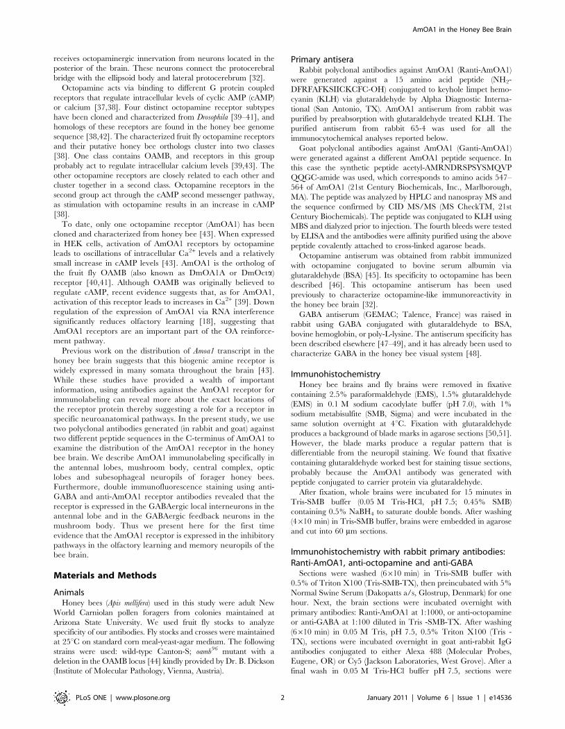
Figure 1. Anti-AmOA1 antibodies are specific for the AmOA1 receptor. Pre- and post-adsorption controls of immunolabeling with anti-AmOA1 antibodies from rabbit (A–D) and from goat (E–G). Consecutive sections of the mushroom body calyx stained with Ranti-AmOA1 serum (A)and pre-immune serum taken from the rabbit before immunization and used at the same dilution as the Ranti-AmOA1 serum (B). The Ranti-AmOA1serum revealed staining in Kenyon cell bodies (K) with different intensity and in processes of the calyx lip and collar (col). C: Section of the antennallobe stained with Ranti-AmOA1 that was preadsorbed with KLH treated with glutaraldehyde (KLH-G) alone. D: A section from the same brain stainedwith Ranti-AmOA1 that was preadsorbed with the AmOA1 peptide conjugated to KLH (KLH-G-OA1pep). E: Sections of the antennal lobe stained withaffinity purified anti-AmOA1 antibodies from goat (Ganti-AmOA1). F: pre-immune serum from the goat before immunization used at the samedilution as the Ganti-AmOA1 serum. G: Specific staining from cell bodies and their processes disappeared after pretreatment with the KLHconjugated to the AmOA1 peptide. The confocal images in D, F and G were adjusted to a higher level of intensity then stained sections in C, E toshow the image of the antennal lobe. Scale bars: 50 mm.doi:10.1371/journal.pone.0014536.g001
AmOA1 in the Honey Bee Brain
PLoS ONE | www.plosone.org 3 January 2011 | Volume 6 | Issue 1 | e14536

conjugated to AmOA1 peptide that was used to make antibodies
in goat (Fig. 1G). To prepare the preadsorbed antibody, 400 mM
of peptide was conjugated to KLH via glutaraldehyde and
incubated with working dilution of Ganti-AmOA1.
For double immunofluorescence staining with Ganti-AmOA1
and GABA, the agarose brain sections were incubated simulta-
neously with both antibodies, then after a thorough wash in PBS,
the secondary antibodies F(ab’) 2 fragments of donkey anti-Goat
IgG Cy2 and F(ab’) 2 fragments of donkey anti-Rabbit IgG Cy5
were added in dilution 1:200 for incubation overnight. For control
of staining, both of the secondary antibodies were incubated with
sections that had only one of the primary antibodies, and the
staining did not show any cross-reaction between the antibodies
(Fig. S1D–F).
Procedures with all antibodies were performed at room tem-
perature. Images of sections treated with antibodies preincubated
with peptide were taken with the intensity of fluorescence gain
equal to or greater than images of antibody treated sections. For
comparison, images were collected at the same level of gain and
intensity. In Fig. 1D, F, G the image was collected at a higher gain
because confocal collection at the same gain as shown in
Figures 1C and D resulted in such low intensity that the image
was not visible.
Results
Specificity of the anti-AmOA1 antibodiesTwo different polyclonal antibodies were used in our studies,
one raised in rabbit (Ranti-AmOA1) and another raised in goat
(Ganti-AmOA1) against two different peptides corresponding to
different regions in the cytoplasmic carboxyl terminus of the
AmOA1 receptor. Double labeling experiments revealed that
these antibodies labeled the same cells and processes in the
neuropil of the bee brain (an example of double staining in the
antennal lobe is in Figure S1, G1-3). For this reason we do not
specify the R or G version of the antibody in the results. Unless
otherwise noted in the legends, figures show data from the Ranti-
AmOA1 serum.
Both antibodies labeled cell body clusters and many processes in
the antennal lobe, mushroom body (calyces, pedunculus, alpha,
beta and gamma lobes), optic lobe, subesophageal ganglion and
central complex (Fig. 1A, C, E, 2). The immunostained cell body
clusters and processes were repeatable from one animal to the next
when we compared brains of forager bees caught at the entrance of
the same colony. Staining in cell bodies and processes ranged from
high to low intensity (Fig. 1A,C,E). This variation is consistent with
data from in situ hybridization where differences in gene expression
in cell bodies were previously reported [43]. Preadsorption of either
antibody with the corresponding peptide-G-KLH (concentration of
the peptide was 100 or 400 mM) abolished specific labeling
(Fig. 1D,G). Furthermore, the rabbit or goat preimmune serum
used on adjacent sections at the same concentration as AmOA1
antiserum did not show any staining (Fig. 1B,F).
The AmOA1 peptide used for immunization in rabbit shows
87% sequence identity to the fruit fly OAMB receptor isoform A
peptide sequence (NH2-DFRFAFKRIICRCFC-OH; amino acids
578–592 of DmOA1 A; AAF55798). Therefore, we used the
Drosophila oamb96 mutant line, which contains a deletion spanning
exons 2 and 3 and does not produce the DmOA1 A protein in the
brain [44], as a control for antibody specificity. We used Ranti-
AmOA1 to label sections from brains of wild type Canton S (CS)
and oamb96 flies (Figure S1H–K). As expected [40], the Ranti-
AmOA1 antibodies labeled cells and neuropils in the mushroom
body alpha prime and beta prime lobes as well in the antennal
lobes of CS female flies (Fig. S1H,J). However, specific labeling in
the mushroom body lobe and antennal lobe was absent from the
oamb96 sterile female flies (Fig. S1I,K). These data show that the
antibodies specifically recognized the Drosophila OAMB receptor
isoform A.
Neuroanatomical distribution of the AmOA1immunostaining in the honey bee brain
Our description below of AmOA1 distribution in the brain is
based on immunostaining of ten honey bee forager brains
processed with Ranti-AmOA1 and five honey bee brains
processed with Ganti-AmOA1. A schematic view of the overall
AmOA1 immunoreactivity of cell bodies and processes in the
honey bee brain is shown in Figure 2. This drawing was made
from confocal images of frontal sections of bee brain. It
summarizes the most typical labeled cell bodies and processes in
the central complex, mushroom bodies, antennal lobes, lateral
protocerebrum, optic lobes and subesophageal ganglion. We begin
each section below with a brief description of the anatomy of that
area of the brain.
AmOA1 staining in the antennal lobesThe primary olfactory neuropil is the antennal lobe, which
consists of approximately 160 spherical glomeruli that surround a
coarse neuropil in the core (Fig. 2, 3A–C, 5). The principal inputs
of the glomeruli are olfactory receptor neurons from the antennal
nerve. Glomeruli are also invaded by processes from at least three
cell types: (1) two types of projection neurons; uniglomerular
projection neurons, which connect the antennal lobe with the
mushroom body calyx and the lateral horn via the lateral or
medial antennocerebral tract (the l- and m-ACT), and multi-
glomerular projection neurons that connect the antennal lobe with
the protocerebral lobe and lateral horn via the medio-lateral tract
(mlACT); (2) two or more types of local interneurons with axons in
the coarse neuropil and dendrites in the glomeruli; and (3)
multiglomerular interneurons that express biogenic amines and
have cell bodies outside of the antennal lobe, for example
VUMmx1 [34,52–54]. All but the latter type of interneuron have
cell bodies in one of three groups that lie lateral (LG), dorsal (DG)
or mediodorsal (MDG) to the antennal lobe (Fig. 3). Each group
(LG, DG, MDG) contains the cell bodies of two or more types of
antennal lobe neurons.
AmOA1 immunoreactivity was present in all three cell body
groups (MDG, DG, and LG) that surround the glomerular
neuropil as well as in glomeruli and the coarse (central) area of the
antennal lobe neuropils (Fig. 3A, B, C). The olfactory receptor
neurons enter from the antennal nerve via four tracts (T1–T4), but
only T1 and T2 enter the coarse area in the center of the antennal
lobe (Fig. 3A). The olfactory receptor neuron axons were not
stained with the AmOA1 antibodies, except for a few scattered
beaded processes, which may also be glial cells, in the T1–T2
tracts where they enter the coarse area (Fig. 3A arrow). The
glomeruli, as well as the coarse neuropil of antennal lobes, were
strongly labeled with AmOA1 antibodies (Fig. 3A–C).
Among the different areas of the antennal lobe, labeling in the
glomeruli was the most variable both within and between
preparations. Immunostaining in some glomeruli revealed a very
strongly labeled core region with absence of staining in the
surrounding cortex (Fig. 3A double arrow) where most of the
olfactory receptor neurons from sensory tracts T1–T3 send their
input to the glomeruli [55]. Some glomeruli exhibited an almost
complete absence of AmOA1 immunoreactivity, or staining was
scattered throughout the entire glomerulus, (Fig. 3B). In other
AmOA1 in the Honey Bee Brain
PLoS ONE | www.plosone.org 4 January 2011 | Volume 6 | Issue 1 | e14536
glomeruli the cortex area was stained with higher intensity
(Fig. 3C).
One type of AmOA1 labeled process in the coarse neuropil
corresponds to axons from a large group of cells located laterally
(LG; Fig. 3B). Among the types of antennal neurons, the LG
contains cell bodies of local interneurons [53]. A large fraction of
neurons from cluster LG were strongly labeled by AmOA1
antiserum in the cell bodies and axons in the coarse neuropil area.
Cell compartments stained with different intensity. The nucleus
was not labeled. Bright staining observed in the cytoplasm could
be due to labeling of receptors during translation, in the process of
being transported to the plasma membrane, during receptor
recycling and/or receptors targeted for degradation.
The DG and MDG (dorsal and medio-dorsal groups respec-
tively, Fig. 3B, C) also contained AmOA1 immunopositive
neurons. These neurons may correspond to multi-glomerular
projection neurons given the presence of staining in the medial-
lateral tract (not shown), which connects the antennal lobes with
the protocerebral lobe [54]. AmOA1 staining is absent in the
axons of the projection neurons of the m- and l-ACT. In our
preparations staining in the target neuropil of these neurons in the
lip and inner basal ring zones of the calyx of the mushroom bodies
has a low intensity. We have not been able to conclusively assign
this weak staining to PN axon endings or to Kenyon cell dendrites
(Fig. 3D, H). Double staining experiments with labeled PNs need
to be performed in order to confirm these observations.
AmOA1 staining in the mushroom bodiesIn honey bees, each mushroom body consists of paired calyces
(lateral and median) connected to a stalk-like structure that forms a
peduncle with two (medial and vertical) lobes (Fig. 2). The major
components of the mushroom body are intrinsic neurons known as
the Kenyon cells [56]. Their axons form the pedunculus and lobes
and their dendrites make up the calyces. There are two broad
classes of Kenyon cells. The Class I Kenyon cells have cell bodies
contained within the calycal cups and their dendrites extend into
the inner wall of the calyx. Their axons project through the
pedunculus and give rise to two branches, one in each lobe. Class
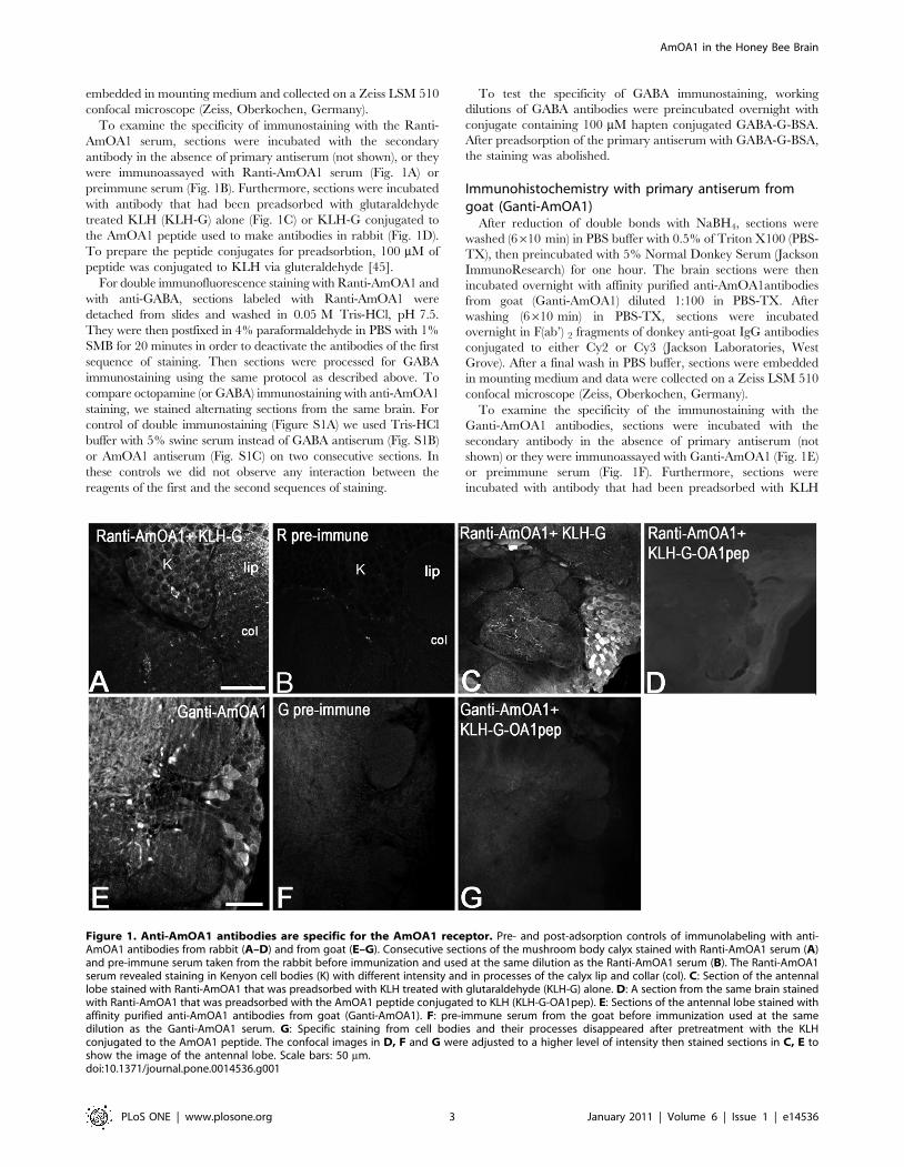
Figure 2. Schematic drawing of AmOA1 immunoreactive neurons and processes in the brain and subesophageal ganglion of thebee made from confocal images of agarose sections of bee brain stained with anti-AmOA1 antibodies. The right hemisphere shows afrontal view of the bee brain with the optic lobe neuropils (Re, retina; La, lamina; Lo, lobula), antennal lobe (al) and summarized mushroom bodyneuropil; the left hemisphere of the brain shows a more posterior frontal view at the level of the dorsal lobe (dl), pedunculus (ped) and calyces of themushroom bodies. AN, antennal nerve; DG, dorsal group of antennal lobe neurons; LG, lateral group of antennal lobe neurons; MDG, medial group ofantennal lobe neurons. Some neurons that express the AmOA1 receptor could be identified as: 1, PCT (or feedback neuron group); 2, lobula andmedulla mushroom body neurons. The cells in this cluster have fibers in the anterior superior optical tract (asot). The arrow shows axons (only theending of the tract) from these neurons as they enter the medulla serpentine layer. A few AmOA1 positive fibers enter through the lobula opticaltract (lot) that connects the lobula and mushroom bodies.; 3, photoreceptor cells in the retina; 4, monopolar cell bodies in the lamina; 5, medullacolumnar neurons; 6, photoreceptor cells in the ocelli; 7, a median group of subesophageal intrinsic neurons; 8, ellipsoid body neurons, 9, a group ofmedian neurosecretory cells (MNC) in the pars intercerebralis. Abbreviations: K, Kenyon cell bodies; Ca, calyx of the mushroom bodies; o.w.br, theouter wedge of the basal ring; co, collar; br, basal ring; ped, pedunculus; vl, vertical lobe of the mushroom bodies; ml, medial lobe of the mushroombodies; c, gamma lobe of the mushroom bodies; PCT, protocerebral-calycal tract; EB, ellipsoid body; FB, fanshaped body; o.Me, outer medulla; i. Me,inner Medulla; es, esophagus; SEG, subesophageal ganglion. Scale bar: 250 mm.doi:10.1371/journal.pone.0014536.g002
AmOA1 in the Honey Bee Brain
PLoS ONE | www.plosone.org 5 January 2011 | Volume 6 | Issue 1 | e14536
II Kenyon cells, also referred to as ‘‘clawed’’ Kenyon cells, have
soma that lie outside the calyces. Their neurites extend through
the outer wall of the calyx where they produce distinctive
‘‘clawed’’ dendrites. Class II Kenyon cells have axons which
supply the ventral part, or c division, of the vertical lobe and may
also bifurcate to supply the vertical and medial lobes [57–60].
The calyces are divided into modality specific zones – lip, collar
and basal ring - that receive input from different sensory
modalities [54,60–63]. The lip receives input from olfactory
projection neurons, the collar receives input from visual, tactile
and gustatory processing neuropils, and the basal ring receives
input from olfactory, gustatory, tactile and visual neuropils (see
schematic in Fig. 3G).
It is important to note here that Kenyon cell bodies were labeled
with different intensity across the calyx (Figures 3D,H, 7B, D2),
which is in accordance with in situ analysis [43]. The AmOA1
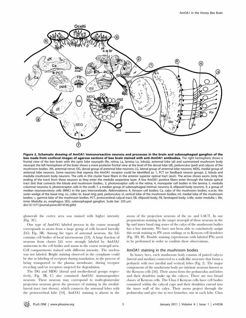
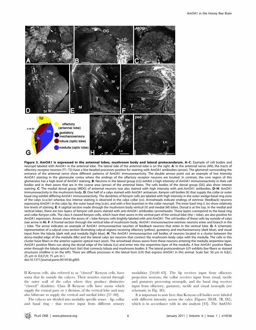
Figure 3. AmOA1 is expressed in the antennal lobes, mushroom body and lateral protocerebrum. A–C: Example of cell bodies andneuropil labeled with AmOA1 in the antennal lobe. The lateral side of the antennal lobe is on the right. A: In the antennal nerve (AN), the tracts ofolfactory receptor neurons (T1–T2) have a few beaded processes positive for staining with AmOA1 antibodies (arrow). The glomeruli surrounding theentrance of the antennal nerve show different patterns of AmOA1 immunoreactivity. The double arrows point out an example of low intensityAmOA1 staining in the glomerular cortex where the endings of the olfactory receptor neurons are located. In contrast, the core region of thisglomerulus has a high level of AmOA1 staining. B: Neurons in the lateral group (LG) exhibit a high intensity of AmOA1 immunoreactivity in their cellbodies and in their axons that are in the coarse area (arrow) of the antennal lobes. The cells bodies of the dorsal group (DG) also show intensestaining. C: The medial dorsal group (MDG) of antennal neurons was also stained with high intensity with anti-AmOA1 antibodies. D–H: AmOA1immunoreactivity in the mushroom body. D: One half of a calyx stained with AmOA1 antiserum. Kenyon cell bodies (K) that supply the collar or outerbasal ring exhibit differing AmOA1 immunoreactivity. The dendrites of Kenyon cells are labeled with high intensity in the outer wedge basal ring zoneof the calyx (o.w.br) whereas less intense staining is observed in the calyx collar (co). Arrowheads indicate endings of extrinsic (feedback) neuronsexpressing AmOA1 in the calyx lip, the outer basal ring (o.br), and with a few branches in the collar neuropil. The inner basil ring (i. br) show relativelylow levels of staining. E: A sagittal section made through the mushroom body vertical (V) and medial (M) lobes. Dorsal is at the top. In the medial andvertical lobes, there are two layers of Kenyon cell axons stained with anti-AmOA1 antibodies (arrowheads). These layers correspond to the basal ringand collar Kenyon cells. The class II clawed Kenyon cells, which have their axons in the ventral part of the vertical lobe (the c lobe), are also positive forAmOA1 expression. Arrows show the axons of c lobe Kenyon cells brightly labeled with anti-AmOA1. The cell bodies of these cells lay outside of calyx(see arrow in H). F: A frontal section through the vertical lobe of mushroom body. AmOA1 immunoreactive extrinsic neurons enter and branch in thec lobe. The arrow indicates an example of AmOA1 immunoreactive neurites of feedback neurons that enter in the vertical lobe. G: A schematicrepresentation of a calycal cross section illustrating calycal regions receiving olfactory (yellow), gustatory and mechanosensory (dark blue), and visualinput from the lobula (dark red) and medulla (light blue). H: The AmOA1 immunoreactive cell bodies of neurons located in a cluster between thedorso-medial edge of the medulla (Me) and the lateral calyx are neurons that connect the mushroom body calyx with the medulla. The cells in thiscluster have fibers in the anterior superior optical tract (asot). The arrowhead shows axons from these neurons entering the medulla serpentine layer.AmOA1 positive fibers run along the dorsal edge of the lobula (Lo) and enter into the serpentine layer of the medulla. A few AmOA1 positive fibersenter through the lobula optical tract (lot) that connects lobula and mushroom bodies. I: The lateral protocerebrum (LP) exhibits few fibers or stainedstructures (midline is on the left). There are diffuse processes in the lateral horn (LH) that express AmOA1 in this animal. Scale bar: 50 mm in A,B,C;25 mm in D,E,F,H; 75 mm in I.doi:10.1371/journal.pone.0014536.g003
AmOA1 in the Honey Bee Brain
PLoS ONE | www.plosone.org 6 January 2011 | Volume 6 | Issue 1 | e14536

antiserum labels a subpopulation of class I Kenyon cell bodies with
higher intensity. It is possible to follow their stained axons and
dendrites in the outer basal ring zone (Fig. 3D,H); the area at the
edge of the dorsal basal ring region is strongly labeled in all
preparations. The Kenyon cell dendrites in the collar are less
intensely stained than dendrites in the outer basal ring (Fig. 3D,H).
The edge of the outer basal ring area receives visual input from
axons from the medulla, whereas the collar region receives input
from both the medulla and lobula of the compound eyes. The
Kenyon cells with dendrites in the collar and basal ring express
AmOA1 protein in their cell bodies, dendrites and in their axons
that make up the layers in the vertical lobe and medial lobe
(Fig. 3E, F).
The class II Kenyon cells also exhibited strong immunoreac-
tivity in their cell bodies and their axons in the ventral part, or cdivision, of the vertical lobe (Fig. 3E, H). Some of these
AmOA1expressing class II cells have their cell bodies outside of
the mushroom body calyces (arrow in Fig. 3H) and their dendrites
spread throughout the calyx into all sensory input zones. Their
axons are brightly labeled in the c division of the vertical lobe
(Fig. 3E).
Apart from the Kenyon cells, other types of neurons in the
mushroom body lobes also express the AmOA1 receptors. These
are extrinsic neurons with large AmOA1 antiserum positive axons
in the c division of the vertical lobe. The neurons that enter the
vertical lobe just above the c division (arrow in Fig. 3F, also 7F,
7H2) correspond to the feedback neurons that connect mushroom
body calyces with mushroom body lobes.
The areas of the calyx that receive inputs from the antennal lobe
(lip and inner basal ring) stained with somewhat different levels of
intensity in different animals (Fig. 1A, 3D, 3H, 7B, 7D2). For
example, the lip is more intensely labeled in figure 3H than in 3D.
In most preparations, the lip and inner basal ring were less
intensely stained compared to subdivisions of the calyx that receive
input from visual processing areas (collar and outer basal ring;
Fig. 3D, 3G, 7B, 7D2). Especially brightly stained was the edge
area in the outer zone of the basal ring (o.w.br in Fig. 3D, 7B).
This area corresponds to rosette-shaped Kenyon cells that receive
input from the ventral medulla [60,61]. The stained processes in
this outer basal ring area may correspond to the endings of
afferents from the medulla (Fig. 3G,H). Neurons that connect the
medulla to the mushroom body calyx via the anterior superior
optical tract (asot) are located in the posterior edge between the
mushroom body calyx and the visual neuropil (Fig. 2, arrowhead
in 3H). Some of the neurons from this group showed strong
AmOA1 immunoreactivity in cell bodies and axons in the asot
(Fig. 2), processes in the serpentine layer of the medulla (Fig. 4C),
and in the visual input area in the outer basal ring (Fig. 3D,H).
There is also staining in some neurons that connect the mushroom
bodies with the lobula through the lobula optic tract (lot; Fig. 2).
In most preparations, the protocerebral lobe is stained with
AmOA1 antibodies at a lower intensity compared the antennal
lobe and mushroom body neuropil (Fig. 3I). The few positively
stained processes are scattered through the lateral protocerebrum
neuropil and in the lateral horn.
AmOA1 expression in the optic lobesThe optic lobes are neuropilar structures adjacent to the
compound eyes. They receive and process input from photore-
ceptors originating in the retina (Fig. 2). The optic lobe consists of
the lamina, the medulla (the outer and the inner separated by the
serpentine layer; Fig. 2, 3H, 4A–C), and the lobula (Fig. 3H).
Some photoreceptor cells exhibit intense staining with the
AmOA1 receptor antibodies (Fig. 2, cluster 3; arrow in Fig. 4A).
Lamina monopolar cells express the AmOA1 receptor as
illustrated in figure 4A where a cluster of monopolar cell bodies
is brightly stained with AmOA1 antibodies (Fig. 2, cluster 4;
Fig. 4A arrowhead). The first and second optic chiasmata between
the lamina and the medulla, and between the medulla and the
lobula, respectively, have AmOA1 immunoreactive fibers (Fig. 4B,
C). In the medulla there are columnar cells labeled with AmOA1
antiserum (Fig. 2, cluster 5; Fig. 4C) that may either be intrinsic to
the medulla or that may be transmedullary and connect the
medulla with the lobula via the 2nd optic chiasma. The AmOA1
antiserum stained processes in the medulla serpentine layer may
belong to the cells that connect the medulla to the mushroom body
calyces (Fig. 2, cluster 2; Fig. 3H), the cell bodies of which are
located between the base of the lateral calyx and the dorso-medial
edge of the medulla. Likewise, stained processes in the lobula may
belong to cells that connect the optic lobula with mushroom body
lobula cells (Fig. 2).
AmOA1 staining in the subesophageal ganglionThe subesophageal ganglion of the honey bee is composed of
the fused mandibular, maxillary and labial neuromeres. These
neuromeres receive gustatory sensory information and are
involved in the motor control of the mouthparts [64]. There are
clusters of small cell bodies located in the median ventral and
lateral parts of the subesophageal ganglion that stained strongly
with AmOA1 antibodies (Fig. 2, cluster 7; Fig. 4D–F). The VUM
cells, characterized by large cell bodies in the median part of the
subesophageal ganglion, are not stained in either the cell bodies or
in the primary neurites in the median and lateral tracts (Fig. 4D–
F). Scattered processes staining with AmOA1 antiserum are
present in the subesophageal ganglion neuropil and may belong to
the descending and ascending neurons as well as to local
interneurons (Fig. 4D–F). Staining was absent in the mandibular
labial and maxillary nerves (data not shown).
AmOA1 staining in the central complexThe central complex is a prominent structure located in the
central brain between the two protocerebral hemispheres [65,66].
It comprises four neuropilar regions: the ellipsoid body (the lower
division of the central body), the fan shaped body (the upper
division of the central body), the paired noduli and the
protocerebral bridge. These structures are interconnected by sets
of columnar neurons that form a regular projection pattern.
Anti-AmOA1 staining in the honey bee central complex is
illustrated in Figures 2 and 4G. A group of AmOA1 expressing
cells located above the dorsal lobe in the medial protocerebrum
(Fig. 2, cluster 8) send their axons to terminate in the ellipsoid
body (Fig. 4G), and these axons have a very high level of AmOA1
immunoreactivity. These axons can be seen entering the base of
the ellipsoid body in Fig. 4G, and we traced them back to the cell
bodies located near the dorsal lobe (Fig. 2, cluster 8; confocal
image not shown). The fan shaped body neuropil has a lower level
of AmOA1 immunoreactivity relative to the high intensity staining
in the ellipsoid body (Fig. 4G).
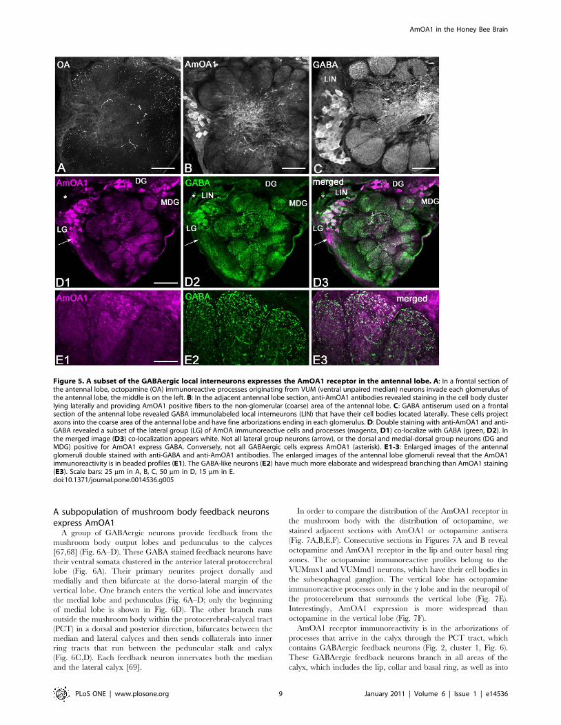
GABAergic local interneurons in the antennal lobes labelwith AmOA1 antibodies
In order to compare the distribution of octopamine and the
distribution of AmOA1 in the antennal lobes, we labeled adjacent
sections of the same brain with octopamine (Fig. 5A) and AmOA1
antisera (Fig. 5B). The octopamine-like immunoreactive processes
from VUM neurons enter in the coarse center of the antennal
lobes from the ventro-posterior deutocerebral area and give rise to
AmOA1 in the Honey Bee Brain
PLoS ONE | www.plosone.org 7 January 2011 | Volume 6 | Issue 1 | e14536

very fine branches to supply each glomerulus of the antennal
lobe. Staining of the adjacent section with AmOA1 antiserum
reveals cell bodies clustered in the lateral rind of the antennal
lobe with AmOA1 positive profiles in the central coarse area and
fine distribution of AmOA1 positive varicosities in each
glomerulus (Fig. 5B). These data suggest that octopamine is
released parasynaptically, rather than presynaptically to each
profile. The cell bodies in the lateral cluster of the antennal lobe
labeled with AmOA1 antibodies (Fig. 5B,D1) belong to local
interneurons that are known to be GABA positive [67]
(Fig. 5C,D2). The anti-GABA antibody stained local interneu-
rons in the cell bodies, processes in the coarse area of the
antennal lobe, and highly packed processes in each glo-
merulus (Fig. 5C, D2). Comparison between the distribution of
AmOA1 (Fig. 5B, D1) and GABA (Fig. 5C,D2) revealed that
GABAergic local interneurons express the AmOA1 receptor. All
co-localized cell bodies and processes are white in the merged
image (Fig. 5D3). At higher magnification, the GABA-like
immunoreactive neurons have much more elaborate and wide
spread branching than AmOA1 staining (Fig. 5E), which could
reflect very localized expression of the AmOA1receptor on
GABAeric cell processes.
In our preparations, the majority of the GABAergic neurons
express AmOA1 receptors with a range of staining intensity.
However, there are some GABAergic neurons where the staining
for AmOA1 is very low or absent (e.g. Fig. 5D, where a group of
cells labeled with an asterisk have very low AmOA1 immunore-
activity). Comparisons of GABAergic with AmOA1 staining in the
cell bodies and processes reveal that not all GABA neurons express
the receptors in those areas of the cell (Fig. 5E1-3, Figure S1D–F).
Conversely, some AmOA1 positive neurons are not GABAer-
gic. The arrow in figure 5D indicates AmOA1 receptor positive
neurons in the lateral cluster that are not stained with the GABA
antibody. In addition, the AmOA1 receptor is expressed in cell
bodies of the medial cluster (DG and MDG) that do not show
GABA-like immunoreactivity.
Figure 4. AmOA1 immunoreactivity is observed in the visual neuropil (A–C), central complex (D) and subesophageal (SEG)ganglion (E–G). A: In the lamina (La), AmOA1 immunoreactivity was observed in the ends of some photoreceptor cells that originate in the retina(Re; arrow) and in a subset of the monopolar cells (arrowhead). B: AmOA1 expression is seen in processes in the first chiasma (1 o ch) and the laminaneuropil. C: AmOA1 immunoreactive processes are organized in discrete strata in the medulla (Me). The columnar intrinsic neurons are intenselystained. The serpentine layer, which separates the outer medulla (o. Me) from the inner medulla (i. Me), has staining in tangential elements that maybelong to medulla neurons that are connected to the calyx of the mushroom bodies. The second optic chiasma (2 o.ch) between the medulla andlobula also contains AmOA1 immunoreactive processes. D: A sagittal section made through the midline of the subesophageal ganglion where theVUM neurons are located in clusters in the maxillar (mx) and mandibular (md) neuromeres is shown. Anterior (a) is to the left and dorsal (d) is on thetop. AmOA1 staining is observed in neurons with small cell bodies located in the clusters with the VUM cells; the VUM cells themselves are notstained with anti- AmOA1 antibodies. E, F: Frontal sections of the subesophageal ganglion from two different bee preparations. AmOA1 staining isobserved in small cell bodies located in the midline of the ventral part of the subesophageal ganglion. ln, lateral neurite tract. G: The fan-shaped (FB)body is less intensely stained with anti-AmOA1 than the ellipsoid body. Scale bars: 50 mm in A; 25 mm in B,C,G; 70 mm in D,E,F.doi:10.1371/journal.pone.0014536.g004
AmOA1 in the Honey Bee Brain
PLoS ONE | www.plosone.org 8 January 2011 | Volume 6 | Issue 1 | e14536
A subpopulation of mushroom body feedback neuronsexpress AmOA1
A group of GABAergic neurons provide feedback from the
mushroom body output lobes and pedunculus to the calyces
[67,68] (Fig. 6A–D). These GABA stained feedback neurons have
their ventral somata clustered in the anterior lateral protocerebral
lobe (Fig. 6A). Their primary neurites project dorsally and
medially and then bifurcate at the dorso-lateral margin of the
vertical lobe. One branch enters the vertical lobe and innervates
the medial lobe and pedunculus (Fig. 6A–D; only the beginning
of medial lobe is shown in Fig. 6D). The other branch runs
outside the mushroom body within the protocerebral-calycal tract
(PCT) in a dorsal and posterior direction, bifurcates between the
median and lateral calyces and then sends collaterals into inner
ring tracts that run between the peduncular stalk and calyx
(Fig. 6C,D). Each feedback neuron innervates both the median
and the lateral calyx [69].
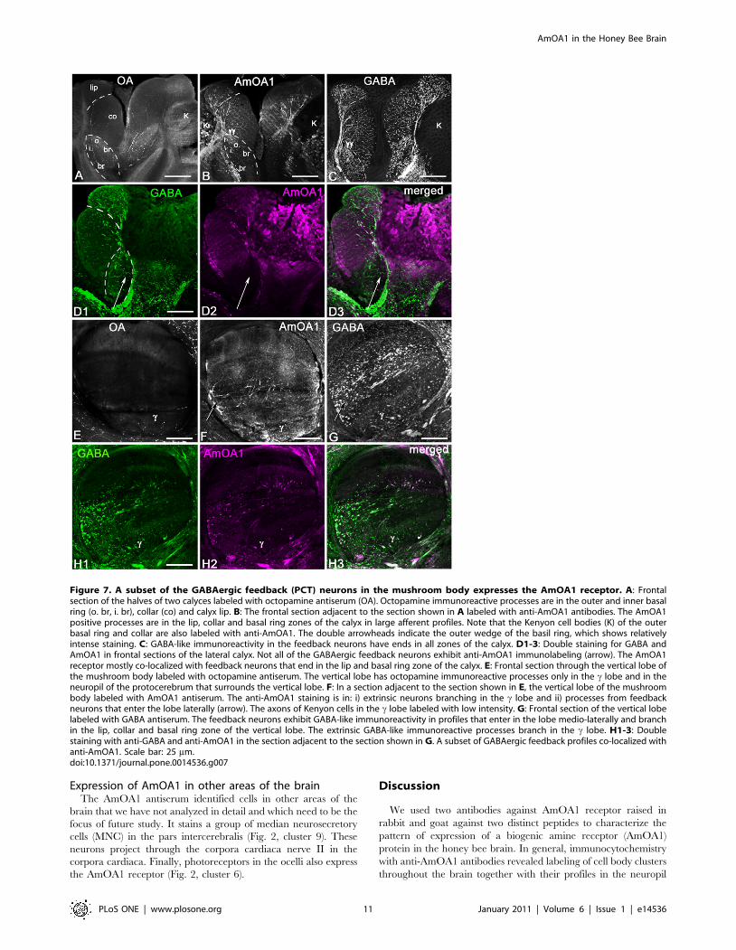
In order to compare the distribution of the AmOA1 receptor in
the mushroom body with the distribution of octopamine, we
stained adjacent sections with AmOA1 or octopamine antisera
(Fig. 7A,B,E,F). Consecutive sections in Figures 7A and B reveal
octopamine and AmOA1 receptor in the lip and outer basal ring
zones. The octopamine immunoreactive profiles belong to the
VUMmx1 and VUMmd1 neurons, which have their cell bodies in
the subesophageal ganglion. The vertical lobe has octopamine
immunoreactive processes only in the c lobe and in the neuropil of
the protocerebrum that surrounds the vertical lobe (Fig. 7E).
Interestingly, AmOA1 expression is more widespread than
octopamine in the vertical lobe (Fig. 7F).
AmOA1 receptor immunoreactivity is in the arborizations of
processes that arrive in the calyx through the PCT tract, which
contains GABAergic feedback neurons (Fig. 2, cluster 1, Fig. 6).
These GABAergic feedback neurons branch in all areas of the
calyx, which includes the lip, collar and basal ring, as well as into
Figure 5. A subset of the GABAergic local interneurons expresses the AmOA1 receptor in the antennal lobe. A: In a frontal section ofthe antennal lobe, octopamine (OA) immunoreactive processes originating from VUM (ventral unpaired median) neurons invade each glomerulus ofthe antennal lobe, the middle is on the left. B: In the adjacent antennal lobe section, anti-AmOA1 antibodies revealed staining in the cell body clusterlying laterally and providing AmOA1 positive fibers to the non-glomerular (coarse) area of the antennal lobe. C: GABA antiserum used on a frontalsection of the antennal lobe revealed GABA immunolabeled local interneurons (LIN) that have their cell bodies located laterally. These cells projectaxons into the coarse area of the antennal lobe and have fine arborizations ending in each glomerulus. D: Double staining with anti-AmOA1 and anti-GABA revealed a subset of the lateral group (LG) of AmOA immunoreactive cells and processes (magenta, D1) co-localize with GABA (green, D2). Inthe merged image (D3) co-localization appears white. Not all lateral group neurons (arrow), or the dorsal and medial-dorsal group neurons (DG andMDG) positive for AmOA1 express GABA. Conversely, not all GABAergic cells express AmOA1 (asterisk). E1-3: Enlarged images of the antennalglomeruli double stained with anti-GABA and anti-AmOA1 antibodies. The enlarged images of the antennal lobe glomeruli reveal that the AmOA1immunoreactivity is in beaded profiles (E1). The GABA-like neurons (E2) have much more elaborate and widespread branching than AmOA1 staining(E3). Scale bars: 25 mm in A, B, C, 50 mm in D, 15 mm in E.doi:10.1371/journal.pone.0014536.g005
AmOA1 in the Honey Bee Brain
PLoS ONE | www.plosone.org 9 January 2011 | Volume 6 | Issue 1 | e14536

the vertical and medial lobes (Fig. 7C, D1, G, H1). In our
immunofluorescence double labeling with anti-GABA and anti-
AmOA1 antibodies, subsets of GABAergic PCT neurons co-
localized with the AmOA1 receptor (Fig. 7D,H).
The feedback neurons that express AmOA1 immunoreactivity in
the calyx are in the lip, collar and outer basal ring area (Fig. 7B, D2).
There is low intensity staining in the inner basal ring area, with high
intensity of staining at the edge of basal ring zone (double
arrowheads in Fig. 7B). The arrows in figure 7F,G show where
feedback neurons enter the vertical lobe (the midline of the brain is
on the left). Double staining on the same sections with anti-GABA
and anti-AmOA1 revealed only a small subpopulation of GABAer-
gic feedback neurons in the calyx and vertical lobes co-stained
(Fig. 7D3,H3). These subpopulations of GABA and AmOA1
immunoreactive feedback neurons have their endings mostly in the
lip, collar and outer layer of the basal ring (Fig. 7D3) while the inner
basal ring zone is not stained with AmOA1 antibodies. In the
vertical lobe, the axons of feedback neurons that express AmOA1
receptors are mostly in the region occupied by the axons of Kenyon
cells with dendrites in the collar and lip zones of the calyx. In
addition, there are extrinsic GABAergic neurons that enter the clobe that also co-stain with anti-AmOA1 (Fig. 7F, H2, H3).
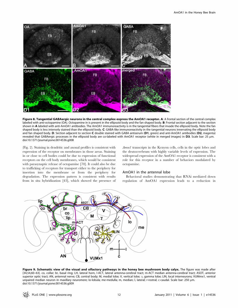
The tangential GABAergic neurons of the centralcomplex express AmOA1
We used octopamine antiserum to compare the distribution of
octopamine labeling with AmOA1 receptor staining in the
central complex of the honey bee on two adjacent sections
(Fig. 8A, B). The origin of the octopamine innervations in the
fan shaped body and ellipsoid body has several sources: neurons
from clusters G4 and G2, and possibly some fibers from the
VUM neuron(s) located in the subesophageal ganglion [32].
The section adjacent to the section shown in figure 8A was
labeled with AmOA1 antibodies (Fig. 8B), and shows high
intensity staining in the ellipsoid body with less intense staining
in the fan shaped body. The neurons that stained for AmOA1
belong to the tangential GABAergic neurons whose cells bodies
are located in cluster 8 (Fig. 2) and that send their input into the
ellipsoid body and fine branches into the fan shaped body
(Fig. 8C). The expression of AmOA1 in these GABAergic
neurons was confirmed using double immunofluorescence
staining with anti-GABA and anti-AmOA1 (Fig. 8D1, 2). The
overlap of staining for GABA and AmOA1 confirms that
AmOA1 receptors are expressed in the GABAerigic tangential
neurons (Fig. 8D3).
Figure 6. GABAergic feedback neurons in the protocerebro-calycal tract (PCT) are shown. A: GABA-like immunoreactive neurons withlarge cell bodies (Cb) are located anteriorly and laterally between the lateral protocerebrum and optic lobes. Each cell sends its primary neuritethrough the lateral protocerebrum towards the vertical lobe of the mushroom body forming the large GABA-like immunoreactive fiber tract (arrow inA, B). B: After reaching the vertical lobe of the mushroom body, each primary neurite divides into two branches. One branch enters the vertical lobeproducing fine arborizations in the dorso-lateral margin of the vertical lobe (vl, arrowhead in A). The other branch follows the GABA-likeimmunoreactive fibers (arrowheads in B, C) through the protocerebro-calycal tract (PCT) to enter the calyces (C, D). C: Each calyx receives GABA-likeimmunoreactive fibers that produce fine arborizations in the lip, collar (co) and basal ring (br). D: GABAergic feedback neurons innervate the median(M Ca) and the lateral (L Ca) calyces, pedunculus (ped) and medial lobe (ml). The insert in C illustrates the specificity of the GABA antibodies. Thissection shows the lack of staining when the GABA antiserum was preadsorbed with 0.01 mM GABA conjugated to carrier protein (BSA) prior to useon the section. Scale bar: 100 mm in A, B; 50 mm in C, D.doi:10.1371/journal.pone.0014536.g006
AmOA1 in the Honey Bee Brain
PLoS ONE | www.plosone.org 10 January 2011 | Volume 6 | Issue 1 | e14536
Expression of AmOA1 in other areas of the brainThe AmOA1 antiserum identified cells in other areas of the
brain that we have not analyzed in detail and which need to be the
focus of future study. It stains a group of median neurosecretory
cells (MNC) in the pars intercerebralis (Fig. 2, cluster 9). These
neurons project through the corpora cardiaca nerve II in the
corpora cardiaca. Finally, photoreceptors in the ocelli also express
the AmOA1 receptor (Fig. 2, cluster 6).
Discussion
We used two antibodies against AmOA1 receptor raised in
rabbit and goat against two distinct peptides to characterize the
pattern of expression of a biogenic amine receptor (AmOA1)
protein in the honey bee brain. In general, immunocytochemistry
with anti-AmOA1 antibodies revealed labeling of cell body clusters
throughout the brain together with their profiles in the neuropil
Figure 7. A subset of the GABAergic feedback (PCT) neurons in the mushroom body expresses the AmOA1 receptor. A: Frontalsection of the halves of two calyces labeled with octopamine antiserum (OA). Octopamine immunoreactive processes are in the outer and inner basalring (o. br, i. br), collar (co) and calyx lip. B: The frontal section adjacent to the section shown in A labeled with anti-AmOA1 antibodies. The AmOA1positive processes are in the lip, collar and basal ring zones of the calyx in large afferent profiles. Note that the Kenyon cell bodies (K) of the outerbasal ring and collar are also labeled with anti-AmOA1. The double arrowheads indicate the outer wedge of the basil ring, which shows relativelyintense staining. C: GABA-like immunoreactivity in the feedback neurons have ends in all zones of the calyx. D1-3: Double staining for GABA andAmOA1 in frontal sections of the lateral calyx. Not all of the GABAergic feedback neurons exhibit anti-AmOA1 immunolabeling (arrow). The AmOA1receptor mostly co-localized with feedback neurons that end in the lip and basal ring zone of the calyx. E: Frontal section through the vertical lobe ofthe mushroom body labeled with octopamine antiserum. The vertical lobe has octopamine immunoreactive processes only in the c lobe and in theneuropil of the protocerebrum that surrounds the vertical lobe. F: In a section adjacent to the section shown in E, the vertical lobe of the mushroombody labeled with AmOA1 antiserum. The anti-AmOA1 staining is in: i) extrinsic neurons branching in the c lobe and ii) processes from feedbackneurons that enter the lobe laterally (arrow). The axons of Kenyon cells in the c lobe labeled with low intensity. G: Frontal section of the vertical lobelabeled with GABA antiserum. The feedback neurons exhibit GABA-like immunoreactivity in profiles that enter in the lobe medio-laterally and branchin the lip, collar and basal ring zone of the vertical lobe. The extrinsic GABA-like immunoreactive processes branch in the c lobe. H1-3: Doublestaining with anti-GABA and anti-AmOA1 in the section adjacent to the section shown in G. A subset of GABAergic feedback profiles co-localized withanti-AmOA1. Scale bar: 25 mm.doi:10.1371/journal.pone.0014536.g007
AmOA1 in the Honey Bee Brain
PLoS ONE | www.plosone.org 11 January 2011 | Volume 6 | Issue 1 | e14536
(Fig. 2). Staining in dendritic and axonal profiles is consistent with
expression of the receptor on membranes in those areas. Staining
in or close to cell bodies could be due to expression of functional
receptors on the cell body membranes, which would be consistent
with parasynaptic release of octopamine [70]. It could also be due
to trafficking of receptors for transport either to the periphery for
insertion into the membrane or from the periphery for
degradation. The expression pattern is consistent with results
from in situ hybridization [43], which showed the presence of
Amoa1 transcripts in the Kenyon cells, cells in the optic lobes and
the deutocerebrum with highly variable levels of expression. The
widespread expression of the AmOA1 receptor is consistent with a
role for this receptor in a number of behaviors modulated by
octopamine.
AmOA1 in the antennal lobeBehavioral studies demonstrating that RNAi mediated down
regulation of AmOA1 expression leads to a reduction in
Figure 8. Tangential GABAergic neurons in the central complex express the AmOA1 receptor. A: A frontal section of the central complexlabeled with anti-octopamine (OA). Octopamine in is present in the ellipsoid body and the fan shaped body. B: Frontal section adjacent to the sectionshown in A labeled with anti-AmOA1 antibodies. The AmOA1 immunoreactivity is in the tangential fibers that invade the ellipsoid body. Note the fanshaped body is less intensely stained than the ellipsoid body. C: GABA-like immunoreactivity in the tangential neurons innervating the ellipsoid bodyand fan shaped body. D. Section adjacent to section C double stained with GABA antiserum (D1, green) and anti-AmOA1 antibodies (D2, magenta)revealed that GABAergic processes in the ellipsoid body are co-labeled with AmOA1 receptor (white in merged images) in D3. Scale bar: 25 mm.doi:10.1371/journal.pone.0014536.g008
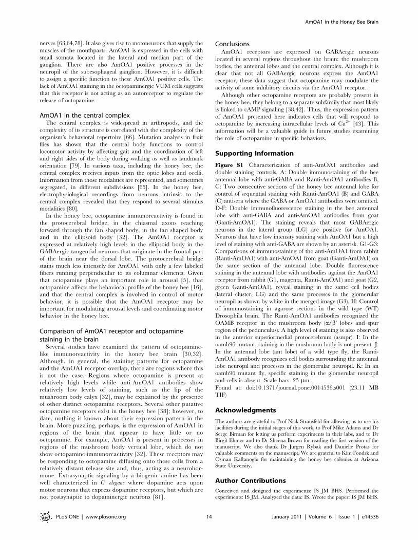
Figure 9. Schematic view of the visual and olfactory pathways in the honey bee mushroom body calyx. The figure was made after[34,54,60–63]. co, collar; br, basal ring; LH, lateral horn, l-ACT, lateral antenna-cerebral tract; m-ACT median antenna-cerebral tract; ASOT, anteriorsuperior optic tract; AN, antennal nerve; CB, central body; M, medial lobe; V, vertical lobe; c, gamma lobe; LIN, local interneurons; VUMmx1, ventralunpaired median neuron in maxillary neuromere; lo-lobula; me-medulla; m, median; l, lateral; r-rostral; c-caudal. Scale bar: 250 mm.doi:10.1371/journal.pone.0014536.g009
AmOA1 in the Honey Bee Brain
PLoS ONE | www.plosone.org 12 January 2011 | Volume 6 | Issue 1 | e14536

olfactory associative learning in the honey bee [18] suggest that
this receptor is an important part of the downstream targets of
the associative reinforcement pathway mediated by VUMmx1.
One way that AmOA1 may play a role in associative learning is
through alteration of interactions in the antennal lobe network
when odor is paired with sucrose [71]. In the antennal lobes,
antibodies against AmOA1 stain three groups of neurons that are
located laterally and dorsally. Double immunostaining with anti-
AmOA1 and anti-GABA showed that these AmOA1 positive
cells are a subpopulation of the GABAergic neurons. The
neurons that stained with anti-AmOA1 but not with GABA
might belong to subset of histaminergic local interneurons [72].
Local interneurons in the antennal lobes (LIN; Fig. 9) act to
transform olfactory sensory information received from the
antennae before it is sent via projection neurons to other areas
of the brain. Expression of AmOA1 in the local interneurons
provides a mechanism for linking the odor representation with
the occurrence of the sucrose reward via release of octopamine
from VUMmx1.
It is difficult to identify the subtype of neurons from the other
two cell clusters in the antennal lobe (the dorsal and dorso-medial
cell clusters) that stain with AmOA1. However, they are probably
not the uniglomerular projection neurons because staining is
absent from their axons and terminals in the lip and basal ring
region of the mushroom body calyx, which are the targets of these
projection neurons. (This observation will need to be confirmed by
AmOA1 staining of uniglomerular PNs that have been intracel-
lularly filled, as the receptor might be expressed in the dendrites of
these neurons). On the other hand, AmOA1 immunoreactivity
was present in the medio-lateral ACT (m-l ACT) and in the
posterior lateral protocerebrum. This pattern suggests that some of
the labeled neurons from the dorsal and dorso-medial groups may
be multiglomerular projection neurons that receive input from
several glomeruli [54].
Therefore, AmOA1 may not only affect odor representation
via modulation of the local interneurons, but may also act upon
the multiglomerular projection neurons that carry odor infor-
mation from the antennal lobes to the lateral horn. This
partitioning of AmOA1 mediated sensitivity to octopamine fits
with a recent hypothesis that proposes different signaling roles
for the two different olfactory tracts in the honey bee antennal
lobes [54,73]. In the fruit fly, a set of projection neurons that
carry olfactory information directly from the antennal lobes to
the lateral protocerebrum appear to be important for more
stereotypical ‘‘experience-independent’’ behaviors [74]. Given
the expression of AmOA1 in the m-l ACT, it would be
interesting to determine if this receptor is involved in
experience-independent olfactory behavior as well as playing a
role in associative learning.
AmOA1 expression in the mushroom bodiesImmunocytochemical studies suggest that octopamine is likely
released in the mushroom body calyces and the c division of the
vertical lobe [30,32]. The calyces are innervated by the VUMmx1
and VUMmd1 neurons, which have fine arborizations mostly in
the lip and basal ring area, and a few branches are also present in
the collar region [30,32,34,35]. AmOA1 immunoreactivity
overlapped substantially with VUM-based octopaminergic inputs
in the mushroom body calyces.
The clawed Kenyon cells express AmOA1 in their cell
bodies, dendrites and axons. These cells are of interest for
several reasons. First, the axons of clawed Kenyon cells from
across the entire calyx converge on the c lobe of the mushroom
body, thereby integrating input from different sensory
modalities [60]. Second, different octopaminergic modulatory
pathways, which may carry different types of information,
target different regions of clawed Kenyon cells. Octopamine
released from VUM neurons in the lip and inner basal ring
likely targets dendrites of the different types of Kenyon cells.
Octopamine released from VUM neurons probably also targets
afferent neurons from visual neuropils and GABAergic
neurons in the PCT since these neurons express AmOA1
receptors. In addition, the octopamineric VCBN neuron
(nomenclature of [75]; LV from [32]) has a cell body located
in the VUM cluster in the subesophageal ganglion, and its
axons target axons of the clawed Kenyon cells in the c lobe of
the mushroom body. The fact that different types of sensory
information from a number of different brain regions converge
on clawed Kenyon cells, and that octopamine has been found
to be necessary for appetitive olfactory learning [18–21],
suggests that the clawed Kenyon cells should be an important
subject for future studies.
A comparison of GABA immunoreactivity with the localization
of staining for AmOA1 suggests that AmOA1 receptors are
expressed in a subset of GABAergic feedback neurons (PCT). In
the honey bee, most of the GABA immunoreactivity in the calyces
arises from a group of feedback or recurrent neurons innervating
both input and output areas of the mushroom bodies [68,69].
These neurons change their properties in response to odors after
olfactory conditioning and are likely to be involved in memory
formation [69]. Our data provide the first evidence that the
octopamine released in the mushroom body calyx and lobe may
target GABAergic feedback neurons.
Possible roles for AmOA1 in visual processingIn most preparations, the regions of the mushroom body
associated with the visual calyx neuropil (collar and outer basal
ring) showed relatively intense staining for AmOA1. We found
small populations of class I Kenyon cells [60] that strongly
express AmOA1 immunoreactivity in their cell bodies and in
axons in both the vertical and medial lobes. These Kenyon cells
have their dendrites in the visual calyx neuropil while their
axons project through defined tracts in both the medial and
vertical lobes to give rise to outputs from the mushroom bodies
that represent the basal ring and collar regions. The edge area
in the outer zone of the basal ring was especially brightly stained
with AmOA1 antibodies. This area corresponds to rosette-
shaped Kenyon cells that receive input from the ventral medulla
[60,61].
Octopamine has been shown to play various roles in visual
processing in insects. In the honey bee, application of
octopamine to the lobula enhances the directional antennal
response to a moving striped pattern [8]. Furthermore, field
potentials in the lobula showed increases in amplitude in the
presence of exogenous octopamine [36]. In locusts, the release of
octopamine by the PM4 cells (homologs of the honey bee G3A
cells) is stimulated by multimodal input from the central brain
[6]. This release of octopamine has been implicated in
dishabituation of output neurons from the optic lobe [6,76,77].
Our results show that octopamine has multiple targets in the
optic lobes, which suggests that the dishabituation may result
from broad-based effects on the neural networks in the optic
lobes.
AmOA1 in the subesophageal ganglionThe subesophageal ganglion of the honey bee receives sensory
projections from receptors located on the respective mouthparts
through the corresponding mandibular, maxillary, and labial
AmOA1 in the Honey Bee Brain
PLoS ONE | www.plosone.org 13 January 2011 | Volume 6 | Issue 1 | e14536
nerves [63,64,78]. It also gives rise to motoneurons that supply the
muscles of the mouthparts. AmOA1 is expressed in the cells with
small somata located in the lateral and median part of the
ganglion. There are also AmOA1 positive processes in the
neuropil of the subesophageal ganglion. However, it is difficult
to assign a specific function to these AmOA1 positive cells. The
lack of AmOA1 staining in the octopaminergic VUM cells suggests
that this receptor is not acting as an autoreceptor to regulate the
release of octopamine.
AmOA1 in the central complexThe central complex is widespread in arthropods, and the
complexity of its structure is correlated with the complexity of the
organism’s behavioral repertoire [66]. Mutation analysis in fruit
flies has shown that the central body functions to control
locomotor activity by affecting gait and the coordination of left
and right sides of the body during walking as well as landmark
orientation [79]. In various taxa, including the honey bee, the
central complex receives inputs from the optic lobes and ocelli.
Information from those modalities are represented, and sometimes
segregated, in different subdivisions [65]. In the honey bee,
electrophysiological recordings from neurons intrinsic to the
central complex revealed that they respond to several stimulus
modalities [80].
In the honey bee, octopamine immunoreactivity is found in
the protocerebral bridge, in the chiasmal axons reaching
forward through the fan shaped body, in the fan shaped body
and in the ellipsoid body [32]. The AmOA1 receptor is
expressed at relatively high levels in the ellipsoid body in the
GABAergic tangential neurons that originate in the frontal part
of the brain near the dorsal lobe. The protocerebral bridge
stains much less intensely for AmOA1 with only a few labeled
fibers running perpendicular to its columnar elements. Given
that octopamine plays an important role in arousal [5], that
octopamine affects the behavioral profile of the honey bee [16],
and that the central complex is involved in control of motor
behavior, it is possible that the AmOA1 receptor may be
important for modulating arousal levels and coordinating motor
behavior in the honey bee.
Comparison of AmOA1 receptor and octopaminestaining in the brain
Several studies have examined the pattern of octopamine-
like immunoreactivity in the honey bee brain [30,32].
Although, in general, the staining patterns for octopamine
and the AmOA1 receptor overlap, there are regions where this
is not the case. Regions where octopamine is present at
relatively high levels while anti-AmOA1 antibodies show
relatively low levels of staining, such as the lip of the
mushroom body calyx [32], may be explained by the presence
of other distinct octopamine receptors. Several other putative
octopamine receptors exist in the honey bee [38]; however, to
date, nothing is known about their expression pattern in the
brain. More puzzling, perhaps, is the expression of AmOA1 in
regions of the brain that appear to have little or no
octopamine. For example, AmOA1 is present in processes in
regions of the mushroom body vertical lobe, which do not
show octopamine immunoreactivity [32]. These receptors may
be responding to octopamine diffusing onto these cells from a
relatively distant release site and, thus, acting as a neurohor-
mone. Extrasynaptic signaling by a biogenic amine has been
well characterized in C. elegans where dopamine acts upon
motor neurons that express dopamine receptors, but which are
not postsynaptic to dopaminergic neurons [81].
ConclusionsAmOA1 receptors are expressed on GABAergic neurons
located in several regions throughout the brain: the mushroom
bodies, the antennal lobes and the central complex. Although it is
clear that not all GABAergic neurons express the AmOA1
receptor, these data suggest that octopamine may modulate the
activity of some inhibitory circuits via the AmOA1 receptor.
Although other octopamine receptors are probably present in
the honey bee, they belong to a separate subfamily that most likely
is linked to cAMP signaling [38,42]. Thus, the expression pattern
of AmOA1 presented here indicates cells that will respond to
octopamine by increasing intracellular levels of Ca2+ [43]. This
information will be a valuable guide in future studies examining
the role of octopamine in specific behaviors.
Supporting Information
Figure S1 Characterization of anti-AmOA1 antibodies and
double staining controls. A: Double immunostaining of the bee
antennal lobe with anti-GABA and Ranti-AmOA1 antibodies B,
C: Two consecutive sections of the honey bee antennal lobe for
control of sequential staining with Ranti-AmOA1 (B) and GABA
(C) antisera where the GABA or AmOA1 antibodies were omitted.
D-F: Double immunofluorescence staining in the bee antennal
lobe with anti-GABA and anti-AmOA1 antibodies from goat
(Ganti-AmOA1). The staining reveals that most GABAergic
neurons in the lateral group (LG) are positive for AmOA1.
Neurons that have low intensity staining with AmOA1 but a high
level of staining with anti-GABA are shown by an asterisk. G1-G3:
Comparisons of immunostaining of the anti-AmOA1 from rabbit
(Ranti-AmOA1) with anti-AmOA1 from goat (Ganti-AmOA1) on
the same section of the antennal lobe. Double fluorescence
staining in the antennal lobe with antibodies against the AmOA1
receptor from rabbit (G1, magenta, Ranti-AmOA1) and goat (G2,
green Ganti-AmOA1), reveal staining in the same cell bodies
(lateral cluster, LG) and the same processes in the glomerular
neuropil as shown by white in the merged image (G3). H: Control
of immunostaining in agarose sections in the wild type (WT)
Drosophila brain. The Ranti-AmOA1 antibodies recognized the
OAMB receptor in the mushroom body (a/b’ lobes and spur
region of the pedunculus). A high level of staining is also observed
in the anterior superiormedial protocerebrum (asmpr). I: In the
oamb96 mutant, staining in the mushroom body is not present. J:
In the antennal lobe (ant lobe) of a wild type fly, the Ranti-
AmOA1 antibody recognizes cell bodies surrounding the antennal
lobe neuropil and processes in the glomerular neuropil. K: In an
oamb96 mutant fly, specific staining in the glomerular neuropil
and cells is absent. Scale bars: 25 mm.
Found at: doi:10.1371/journal.pone.0014536.s001 (23.11 MB
TIF)
Acknowledgments
The authors are grateful to Prof Nick Strausfeld for allowing us to use his
facilities during the initial stages of this work, to Prof Mike Adams and Dr
Serge Birman for letting us perform experiments in their labs, and to Dr
Birgit Ehmer and to Dr Sheena Brown for reading the first version of the
manuscript. We also thank Dr Jurgen Rybak and Danielle Protas for
valuable comments on the manuscript. We are grateful to Kim Fondrk and
Osman Kaftanoglu for maintaining the honey bee colonies at Arizona
State University.
Author Contributions
Conceived and designed the experiments: IS JM BHS. Performed the
experiments: IS JM. Analyzed the data: IS. Wrote the paper: IS JM BHS.
AmOA1 in the Honey Bee Brain
PLoS ONE | www.plosone.org 14 January 2011 | Volume 6 | Issue 1 | e14536
References
1. Roeder T (1999) Octopamine in invertebrates. Prog Neurobiol 59: 533–561.
2. Verlinden H, Vleugels R, Marchal E, Badisco L, Pfluger HJ, et al. (2010) Therole of octopamine in locusts and other arthropods. J Insect Physiol 56: 854–867.
3. Candy DJ (1978) The regulation of locust flight muscle metabolism by
octopamine and other compounds. Insect Biochem 8: 177–181.
4. Mentel T, Duch C, Stypa H, Wegener G, Muller U, et al. (2003) Centralmodulatory neurons control fuel selection in flight muscle of migratory locust.
J Neurosci 23: 1109–1113.
5. Davenport AP, Evans PD (1984) Stress-induced changes in the octopaminelevels of insect hemolymph. Insect Biochem 14: 135–143.
6. Stern M (1999) Octopamine in the locust brain: cellular distribution and
functional significance in an arousal mechanism. Microsc Res Tech 45:135–141.
7. Wicher D (2007) Metabolic regulation and behavior: how hunger produces
arousal - an insect study. Endocr Metab Immune Disord Drug Targets 7:304–310.
8. Erber J, Kloppenburg P (1995) The modulatory effects of serotonin and
octopamine in the visual system of the honey bee (Apis mellifera L.) I. Behavioral
analysis of the motion-sensitive antennal reflex. J Comp Physiol A 176: 111–118.
9. Matheson T (1997) Octopamine modulates the responses and presynaptic
inhibition of proprioceptive sensory neurones in the locust Schistocerca gregaria.
J Exp Biol 200: 1317–1325.
10. Scheiner R, Pluckhahn S, Oney B, Blenau W, Erber J (2002) Behaviouralpharmacology of octopamine, tyramine and dopamine in honey bees. Behav
Brain Res 136: 545–553.
11. Widmer A, Hoger U, Meisner S, French AS, Torkkeli PH (2005) Spiderperipheral mechanosensory neurons are directly innervated and modulated by
octopaminergic efferents. J Neurosci 25: 1588–1598.
12. Zhukovskaya MI, Kapitsky SV (2006) Activity modulation in cockroachsensillum: the role of octopamine. J Insect Physiol 52: 76–86.
13. Dierick HA (2008) Fly fighting: octopamine modulates aggression. Curr Biol 18:
R161–163.
14. Hoyer SC, Eckart A, Herrel A, Zars T, Fischer SA, et al. (2008) Octopamine inmale aggression of Drosophila. Curr Biol 18: 159–167.
15. Brembs B, Christiansen F, Pfluger HJ, Duch C (2007) Flight initiation and
maintenance deficits in flies with genetically altered biogenic amine levels.J Neurosci 27: 11122–11131.
16. Fussnecker BL, Smith BH, Mustard JA (2006) Octopamine and tyramine
influence the behavioral profile of locomotor activity in the honey bee (Apis
mellifera). J Insect Physiol 52: 1083–1092.
17. Saraswati S, Fox LE, Soll DR, Wu CF (2004) Tyramine and octopamine have
opposite effects on the locomotion of Drosophila larvae. J Neurobiol 58: 425–441.
18. Farooqui T, Robinson K, Vaessin H, Smith BH (2003) Modulation of earlyolfactory processing by an octopaminergic reinforcement pathway in the
honeybee. J Neurosci 23: 5370–5380.
19. Hammer M, Menzel R (1998) Multiple sites of associative odor learning as
revealed by local brain microinjections of octopamine in honeybees. Learn Mem5: 146–156.
20. Schwaerzel M, Monastirioti M, Scholz H, Friggi-Grelin F, Birman S, et al.
(2003) Dopamine and octopamine differentiate between aversive and appetitiveolfactory memories in Drosophila. J Neurosci 23: 10495–10502.
21. Unoki S, Matsumoto Y, Mizunami M (2005) Participation of octopaminergic
reward system and dopaminergic punishment system in insect olfactory learningrevealed by pharmacological study. Eur J Neurosci 22: 1409–1416.
22. Robinson GE, Heuser LM, Le Conte Y, Lenquette F, Hollingworth RM (1999)
Neurochemicals aid bee nestmate recognition. Nature 399: 534–535.
23. Spivak M, Masterman R, Ross R, Mesce KA (2003) Hygienic behavior in thehoney bee (Apis mellifera L.) and the modulatory role of octopamine. J Neurobiol
55: 341–354.
24. Schulz DJ, Barron AB, Robinson GE (2002) A role for octopamine in honey beedivision of labor. Brain Behav Evol 60: 350–359.
25. Schulz DJ, Robinson GE (1999) Biogenic amines and division of labor in honey
bee colonies: behaviorally related changes in the antennal lobes and age-related
changes in the mushroom bodies. J Comp Physiol A 184: 481–488.
26. Busch S, Tanimoto H (2010) Cellular configuration of single octopamine
neurons in Drosophila. J Comp Neurol 518: 2355–2364.
27. Braunig P, Burrows M, Morris OT (2004) Properties of descending dorsal
unpaired median (DUM) neurons of the locust suboesophageal ganglion. ActaBiol Hung 55: 13–19.
28. Dacks AM, Christensen TA, Agricola HJ, Wollweber L, Hildebrand JG (2005)
Octopamine-immunoreactive neurons in the brain and subesophageal ganglionof the hawkmoth Manduca sexta. J Comp Neurol 488: 255–268.
29. Konings PN, Vullings HG, Geffard M, Buijs RM, Diederen JH, et al. (1988)
Immunocytochemical demonstration of octopamine-immunoreactive cells in thenervous system of Locusta migratoria and Schistocerca gregaria. Cell Tissue Res 251:
371–379.
30. Kreissl S, Eichmuller S, Bicker G, Rapus J, Eckert M (1994) Octopamine-likeimmunoreactivity in the brain and subesophageal ganglion of the honeybee.
J Comp Neurol 348: 583–595.
31. Monastirioti M (1999) Biogenic amine systems in the fruit fly Drosophila
melanogaster. Microsc Res Tech 45: 106–121.
32. Sinakevitch I, Niwa M, Strausfeld NJ (2005) Octopamine-like immunoreactivityin the honey bee and cockroach: comparable organization in the brain and
subesophageal ganglion. J Comp Neurol 488: 233–254.
33. Sinakevitch I, Strausfeld NJ (2006) Comparison of octopamine-like immunore-
activity in the brains of the fruit fly and blow fly. J Comp Neurol 494: 460–475.
34. Hammer M (1993) An identified neuron mediates the unconditioned stimulus in
associative olfactory learning in honeybees. Nature 366: 59–63.
35. Schroter U, Malun D, Menzel R (2007) Innervation pattern of suboesophageal
ventral unpaired median neurones in the honeybee brain. Cell Tissue Res 327:
647–667.
36. Kloppenburg P, Erber J (1995) The Modulatory Effects of serotonin and
octopamine in the visual-system of the honey-bee (Apis-Mellifera L) .2.Electrophysiological analysis of motion-sensitive neurons in the lobula. J Comp
Physiol A 176: 119–129.
37. Blenau W, Baumann A (2001) Molecular and pharmacological properties of
insect biogenic amine receptors: lessons from Drosophila melanogaster and Apis
mellifera. Arch Insect Biochem Physiol 48: 13–38.
38. Evans PD, Maqueira B (2005) Insect octopamine receptors: a new classification
scheme based on studies of cloned Drosophila G-protein coupled receptors. InvertNeurosci 5: 111–118.
39. Balfanz S, Strunker T, Frings S, Baumann A (2005) A family of octopaminereceptors that specifically induce cyclic AMP production or Ca2+ release in
Drosophila melanogaster. J Neurochem 93: 440–451.
40. Han KA, Millar NS, Davis RL (1998) A novel octopamine receptor with
preferential expression in Drosophila mushroom bodies. J Neurosci 18:3650–3658.
41. Maqueira B, Chatwin H, Evans PD (2005) Identification and characterization of
a novel family of Drosophila beta-adrenergic-like octopamine G-protein coupledreceptors. J Neurochem 94: 547–560.
42. Hauser F, Cazzamali G, Williamson M, Blenau W, Grimmelikhuijzen CJ (2006)A review of neurohormone GPCRs present in the fruitfly Drosophila melanogaster
and the honey bee Apis mellifera. Prog Neurobiol 80: 1–19.
43. Grohmann L, Blenau W, Erber J, Ebert PR, Strunker T, et al. (2003) Molecular
and functional characterization of an octopamine receptor from honeybee (Apis
mellifera) brain. J Neurochem 86: 725–735.
44. Lee HG, Seong CS, Kim YC, Davis RL, Han KA (2003) Octopamine receptorOAMB is required for ovulation in Drosophila melanogaster. Dev Biol 264:
179–190.
45. Geffard M, Buijs RM, Seguela P, Pool CW, Le Moal M (1984) Firstdemonstration of highly specific and sensitive antibodies against dopamine.
Brain Res 294: 161–165.
46. Sinakevitch I, Geffard M, Pelhate M, Lapied B (1994) Octopamine-like
immunoreactivity in the dorsal unpaired median (DUM) neurons innervatingthe accessory gland of the male cockroach Periplaneta americana. Cell & Tissue Res
276: 15–21.
47. Seguela P, Geffard M, Buijs RM, Le Moal M (1984) Antibodies against gamma-
aminobutyric acid: specificity studies and immunocytochemical results. Proc
Natl Acad Sci U S A 81: 3888–3892.
48. Sinakevitch I, Douglass JK, Scholtz G, Loesel R, Strausfeld NJ (2003) Conserved
and convergent organization in the optic lobes of insects and isopods, withreference to other crustacean taxa. J Comp Neurol 467: 150–172.
49. Sinakevitch IG, Geffard M, Pelhate M, Lapied B (1996) Anatomy and targets ofDorsal Unpaired Median neurones in the Terminal Abdominal Ganglion of the
male cockroach Periplaneta americana L. J Comp Neurol 367: 147–163.
50. Brown SM, Strausfeld NJ (2006) Development-dependent and -independent
ubiquitin expression in divisions of the cockroach mushroom body. J Comp
Neurol 496: 556–571.
51. Sinakevitch I, Farris SM, Strausfeld NJ (2001) Taurine-, aspartate- and
glutamate-like immunoreactivity identifies chemically distinct subdivisions ofKenyon cells in the cockroach mushroom body. J Comp Neurol 439: 352–367.
52. Abel R, Rybak J, Menzel R (2001) Structure and response patterns of olfactoryinterneurons in the honeybee, Apis mellifera. J Comp Neurol 437: 363–383.
53. Flanagan D, Mercer AR (1989) An atlas and 3-D reconstruction of the antennallobes in the worker honey bee, Apis mellifera (Hymenoptera, Apidae). Int J Insect
Morph Embyrol 18: 145–159.
54. Kirschner S, Kleineidam CJ, Zube C, Rybak J, Grunewald B, et al. (2006) Dualolfactory pathway in the honeybee, Apis mellifera. J Comp Neurol 499: 933–952.
55. Arnold G, Masson C, Budharugsa S (1985) Comparative study of the antennallobes and their afferent pathways in the worker bee and the drone (Apis mellifera).
Cell Tissue Res 242: 593–605.
56. Kenyon F (1896) The brain of the bee. A preliminary contribution to the
morphology of the nervous system of the Arthropoda. J Comp Neurol 6:133–210.
57. Farris SM, Abrams AI, Strausfeld NJ (2004) Development and morphology of
class II Kenyon cells in the mushroom bodies of the honey bee, Apis mellifera.J Comp Neurol 474: 325–339.
58. Mobbs P (1982) The brain of the honey bee Apis mellifera I. The connections andspatial organization of the mushrrom bodies. Philos Trans R Soc Lond B 298:
309–354.
59. Rybak J, Menzel R (1993) Anatomy of the mushroom bodies in the honey bee
brain: the neuronal connections of the alpha-lobe. J Comp Neurol 334:444–465.
AmOA1 in the Honey Bee Brain
PLoS ONE | www.plosone.org 15 January 2011 | Volume 6 | Issue 1 | e14536

60. Strausfeld NJ (2002) Organization of the honey bee mushroom body:
representation of the calyx within the vertical and gamma lobes. J CompNeurol 450: 4–33.
61. Ehmer B, Gronenberg W (2002) Segregation of visual input to the mushroom
bodies in the honeybee (Apis mellifera). J Comp Neurol 451: 362–373.62. Gronenberg W (2001) Subdivisions of hymenopteran mushroom body calyces by
their afferent supply. J Comp Neurol 435: 474–489.63. Schroter U, Menzel R (2003) A new ascending sensory tract to the calyces of the
honeybee mushroom body, the subesophageal-calycal tract. J Comp Neurol 465:
168–178.64. Rehder V (1989) Sensory pathways and motoneurons of the proboscis reflex in
the suboesophageal ganglion of the honey bee. J Comp Neurol 279: 499–513.65. Homberg U (2008) Evolution of the central complex in the arthropod brain with
respect to the visual system. Arthropod Struct Dev 37: 347–362.66. Loesel R, Nassel DR, Strausfeld NJ (2002) Common design in a unique midline
neuropil in the brains of arthropods. Arthropod Struct Dev 31: 77–91.
67. Schafer S, Bicker G (1986) Distribution of GABA-like immunoreactivity in thebrain of the honeybee. J Comp Neurol 246: 287–300.
68. Ganeshina O, Menzel R (2001) GABA-immunoreactive neurons in themushroom bodies of the honeybee: an electron microscopic study. J Comp
Neurol 437: 335–349.
69. Grunewald B (1999) Morphology of feedback neurons in the mushroom body ofthe honeybee, Apis mellifera. J Comp Neurol 404: 114–126.
70. Watson AH (1984) The dorsal unpaired median neurons of the locustmetathoracic ganglion: neuronal structure and diversity, and synapse distribu-
tion. J Neurocytol 13: 303–327.71. Smith BH, Wright GA, Daly KC (2006) Learning-based recognition and
discrimination of floral odors. In: Dudareva N, Pichersky E, eds. The Biology
and Chemistry of Floral Scents. New York: CRC Press. pp 263–295.
72. Bornhauser BC, Meyer EP (1997) Histamine-like immunoreactivity in the visual
system and brain of an orthopteran and a hymenopteran insect. Cell Tissue Res
287: 211–221.
73. Yamagata N, Schmuker M, Szyszka P, Mizunami M, Menzel R (2009)
Differential odor processing in two olfactory pathways in the honeybee. Front
Syst Neurosci 3: 16.
74. Heimbeck G, Bugnon V, Gendre N, Keller A, Stocker RF (2001) A central
neural circuit for experience-independent olfactory and courtship behavior in
Drosophila melanogaster. Proc Natl Acad Sci U S A 98: 15336–15341.
75. Rybak J, Jenett A, Brandt R, Malun D (2001) Cellular localization of
octopamine-like immunoreactivity in the honeybee brain. Sixth International
Congress of Neuroethology; 29 July - 3 August; Bonn, Germany.
76. Bacon JP, Thompson KS, Stern M (1995) Identified octopaminergic neurons
provide an arousal mechanism in the locust brain. J Neurophysiol 74:
2739–2743.
77. Rowell CHF (1971) Variable responsiveness of a visual interneurone in the free-
moving locust, and its relation to behaviour and arousal. J Exp Biol 55: 727–747.
78. Rehder V (1988) A neuroanatomical map of the suboesophageal and
prothoracic ganglia of the honey bee (Apis mellifera). Proc R Soc Lond B235:
179–202.
79. Strauss R (2002) The central complex and the genetic dissection of locomotor
behaviour. Curr Opin Neurobiol 12: 633–638.
80. Homberg U (1985) Interneurons of the central complex in the bee brain (Apis
mellifera, L.). J Insect Physiol 31: 251–264.
81. Chase DL, Pepper JS, Koelle MR (2004) Mechanism of extrasynaptic dopamine
signaling in Caenorhabditis elegans. Nat Neurosci 7: 1096–1103.
AmOA1 in the Honey Bee Brain
PLoS ONE | www.plosone.org 16 January 2011 | Volume 6 | Issue 1 | e14536