Distribution of 28 kDa Calbindin-Immunopositive Neurons in the … · 2017-04-13 · Merkulyeva et...
13
ORIGINAL RESEARCH published: 28 January 2016 doi: 10.3389/fnana.2015.00166 Distribution of 28 kDa Calbindin-Immunopositive Neurons in the Cat Spinal Cord Natalia Merkulyeva 1,2 , Aleksandr Veshchitskii 1 , Felix Makarov 1 , Yury Gerasimenko 3 and Pavel Musienko 2,3,4 * 1 Laboratory of Neuromorphology, Pavlov Institute of Physiology RAS, Saint Petersburg, Russia, 2 Laboratory of Neuroprosthetics, Institute of Translational Biomedicine, Saint Petersburg State University, Saint Petersburg, Russia, 3 Laboratory of Motor Physiology, Pavlov Institute of Physiology RAS, Saint Petersburg, Russia, 4 Laboratory of Neurophysiology and Experimental Neurorehabilitation, Children’s Surgery and Orthopedic Clinic, Department of Non-pulmonary Tuberculosis, Research Institute of Phthysiopulmonology, Saint Petersburg, Russia Edited by: Shawn Mikula, Max Planck Institute for Neurobiology, Germany Reviewed by: Nobuaki Tamamaki, Kumamoto University, Japan Ursula H. Winzer-Serhan, Texas A&M Health Science Center, USA *Correspondence: Pavel Musienko [email protected] Received: 7 October 2015 Accepted: 21 December 2015 Published: 28 January 2016 Citation: Merkulyeva N, Veshchitskii A, Makarov F, Gerasimenko Y and Musienko P (2016) Distribution of 28 kDa Calbindin-Immunopositive Neurons in the Cat Spinal Cord. Front. Neuroanat. 9:166. doi: 10.3389/fnana.2015.00166 The distribution of vitamin D-dependent calcium-binding protein (28 kDa calbindin) was investigated in cat lumbar and sacral spinal cord segments (L1-S3). We observed specific multi-dimensional distributions over the spinal segments for small immunopositive cells in Rexed laminae II-III and medium-to-large cells of varying morphology in lamina I and laminae V-VIII. The small neurons in laminae II-III were clustered into the columns along the dorsal horn curvature. The medium-to- large cells were grouped into four assemblages that were located in (1) the most lateral region of lamina VII at the L1-L4 level; (2) the laminae IV-V boundary at the L5-L7 level; (3) the lamina VII dorsal border at the L5-L7 level; and (4) the lamina VIII at the L5-S3 level. The data obtained suggest that the morphological and physiological heterogeneity of calbindin immunolabeling cells formed morpho- functional clusters over the gray matter. A significant portion of the lumbosacral enlargement had immunopositive neurons within all Rexed laminae, suggesting an important functional role within and among the spinal networks that control hindlimb movements. Keywords: spinal cord, gray matter, interneurons, Ca 2+ -binding proteins, calbindin-28 kDa INTRODUCTION Calcium ions (Ca 2+ ) play an important regulatory and structural role in a wide range of biological processes (Nordin, 1988; Pochet et al., 2000; Sabatini et al., 2001; Franks and Sejnowski, 2002). As a second messenger, Ca 2+ is involved in the regulation of intracellular functions (Samoilov, 1999). Maintaining the stable neuronal Ca 2+ transmembrane gradient is an important aspect for growth of neuronal processes and synaptic transmission (Heizmann, 1993). Ca 2+ signal transduction into intracellular response is provided by Ca 2+ -binding proteins. Some of these proteins, including 28 kDa calbindin, parvalbumin and calretinin, are members of the EF-hand family, which is characterized by two alpha helices linked by a short loop region that usually binds calcium ions (Grabarek, 2006; Schmidt, 2012). Interneurons in the central nervous system (CNS) highly express specific Ca 2+ -binding proteins and can be used as a specific markers for cell functional types (Hof and Nimchinsky, 1992; Heizmann, 1993; Baizer and Baker, 2005); 28 kDa calbindin plays a major role as an intracellular Ca 2+ buffer (Nägerl et al., 2000). Its high concentration has been observed in the Frontiers in Neuroanatomy | www.frontiersin.org 1 January 2016 | Volume 9 | Article 166
Transcript of Distribution of 28 kDa Calbindin-Immunopositive Neurons in the … · 2017-04-13 · Merkulyeva et...

ORIGINAL RESEARCHpublished: 28 January 2016
doi: 10.3389/fnana.2015.00166
Distribution of 28 kDaCalbindin-Immunopositive Neuronsin the Cat Spinal CordNatalia Merkulyeva 1,2, Aleksandr Veshchitskii 1, Felix Makarov 1, Yury Gerasimenko 3
and Pavel Musienko 2,3,4*
1 Laboratory of Neuromorphology, Pavlov Institute of Physiology RAS, Saint Petersburg, Russia, 2 Laboratory ofNeuroprosthetics, Institute of Translational Biomedicine, Saint Petersburg State University, Saint Petersburg, Russia,3 Laboratory of Motor Physiology, Pavlov Institute of Physiology RAS, Saint Petersburg, Russia, 4 Laboratory ofNeurophysiology and Experimental Neurorehabilitation, Children’s Surgery and Orthopedic Clinic, Department ofNon-pulmonary Tuberculosis, Research Institute of Phthysiopulmonology, Saint Petersburg, Russia
Edited by:Shawn Mikula,
Max Planck Institute forNeurobiology, Germany
Reviewed by:Nobuaki Tamamaki,
Kumamoto University, JapanUrsula H. Winzer-Serhan,
Texas A&M Health Science Center,USA
*Correspondence:Pavel [email protected]
Received: 7 October 2015Accepted: 21 December 2015Published: 28 January 2016
Citation:Merkulyeva N, Veshchitskii A,
Makarov F, Gerasimenko Y andMusienko P (2016) Distribution of28 kDa Calbindin-Immunopositive
Neurons in the Cat Spinal Cord.Front. Neuroanat. 9:166.
doi: 10.3389/fnana.2015.00166
The distribution of vitamin D-dependent calcium-binding protein (28 kDa calbindin)was investigated in cat lumbar and sacral spinal cord segments (L1-S3). Weobserved specific multi-dimensional distributions over the spinal segments for smallimmunopositive cells in Rexed laminae II-III and medium-to-large cells of varyingmorphology in lamina I and laminae V-VIII. The small neurons in laminae II-IIIwere clustered into the columns along the dorsal horn curvature. The medium-to-large cells were grouped into four assemblages that were located in (1) the mostlateral region of lamina VII at the L1-L4 level; (2) the laminae IV-V boundary atthe L5-L7 level; (3) the lamina VII dorsal border at the L5-L7 level; and (4) thelamina VIII at the L5-S3 level. The data obtained suggest that the morphologicaland physiological heterogeneity of calbindin immunolabeling cells formed morpho-functional clusters over the gray matter. A significant portion of the lumbosacralenlargement had immunopositive neurons within all Rexed laminae, suggesting animportant functional role within and among the spinal networks that control hindlimbmovements.
Keywords: spinal cord, gray matter, interneurons, Ca2+-binding proteins, calbindin-28 kDa
INTRODUCTION
Calcium ions (Ca2+) play an important regulatory and structural role in a wide range of biologicalprocesses (Nordin, 1988; Pochet et al., 2000; Sabatini et al., 2001; Franks and Sejnowski, 2002).As a second messenger, Ca2+ is involved in the regulation of intracellular functions (Samoilov,1999). Maintaining the stable neuronal Ca2+ transmembrane gradient is an important aspectfor growth of neuronal processes and synaptic transmission (Heizmann, 1993). Ca2+ signaltransduction into intracellular response is provided by Ca2+-binding proteins. Some of theseproteins, including 28 kDa calbindin, parvalbumin and calretinin, are members of the EF-handfamily, which is characterized by two alpha helices linked by a short loop region that usually bindscalcium ions (Grabarek, 2006; Schmidt, 2012).
Interneurons in the central nervous system (CNS) highly express specific Ca2+-bindingproteins and can be used as a specific markers for cell functional types (Hof and Nimchinsky,1992; Heizmann, 1993; Baizer and Baker, 2005); 28 kDa calbindin plays a major role as anintracellular Ca2+ buffer (Nägerl et al., 2000). Its high concentration has been observed in the
Frontiers in Neuroanatomy | www.frontiersin.org 1 January 2016 | Volume 9 | Article 166

Merkulyeva et al. Calbindin Neurones in Spinal Cord
cerebellar Purkinje cells, in hippocampal granule cells(Baimbridge et al., 1992; Schwaller et al., 2002), and thalamicand cortical ‘‘matrix cells’’ that are characterized by spreadinginterneuronal connections that synchronize thalamocorticalelements into coherent active networks (Jones, 2001).
Neurons expressing 28 kDa calbindin have also been revealedin the spinal cord (Anelli and Heckman, 2005; Porseva et al.,2014). It has been suggested that calbindin is a specificprotein of the excitatory amino acid neuron subpopulation(Antal et al., 1991). It was found that calbindin 28 kDa ispredominantly distributed in lamina I and ependymal cells.Numerous calbindin-immunopositive cells are abundant in thesubstantia gelatinosa (Antal et al., 1991; Ren and Ruda, 1994).A group of calbindin-positive neurons belong to the Renshaw cellpopulation in rodents, primates and cats (Arvidsson et al., 1992;Sanna et al., 1993).
Because Ca2+-binding proteins are expressed in neuronalpopulations with specific functional features, we initiated studiesto determine the 3D distribution and selected features ofcalbindin-positive cells among the lumbosacral spinal cordsegments and laminae. We hypothesize that such comprehensiveneuromorphological evidence, as part of multidimentional spinalinfrastructure characterization, is necessary and useful forongoing and future morphological/functional studies of theneuronal networks participating in different types of spinalfunctions, such as sensorimotor activity (locomotor and posturalcontrol; Deliagina et al., 1983; Orlovsky et al., 1999; Gerasimenkoet al., 2008; Musienko et al., 2010, 2014) and visceral control(bladder function; Horst et al., 2011, 2013).
MATERIALS AND METHODS
Animals and PerfusionAll experimental procedures were approved by the EthicsCommission of the Pavlov Institute of Physiology. Experimentswere performed in accordance with the requirements of CouncilDirective 2010/63EU of the European Parliament regarding theprotection of animals used for experimental and other scientificpurposes. Five normal pigmented adult cats of both sexes [fourmales, K7, K11 (only segment L4 was used), K12, and K15; andone female, K8] were used for this investigation. Under deepanesthesia (Isoflurane) all animals were perfused transcardiallywith 0.9% NaCl (2.0 L) in 0.1 M phosphate-buffered saline (PBS)at pH = 7.4, followed by 4% paraformaldehyde (2.0 L) in 0.1 MPBS at pH = 7.4. After perfusion, the spinal cord was removedfrom the spine and stored in 20 and 30% sucrose until it sank.The lumbosacral cord was divided into segments based uponthe grouping of the dorsal rootlets. The L1-S3 segments werecut on a freezing microtome into 50 µm frontal sections, or40 µm horizontal sections. Sections were collected in 0.1 M PBS,pH = 7.4.
Calbindin 28 kDa ImmunohistochemicalStainingSlices were processed as free floating. Between all procedures,the slices were washed 3 × 5 min in 0.01 M PBS. To unmask
any antigens, sections were processed in 1% NaBH4 for30 min; endogenous peroxidase activity was blocked byincubating the sections in 0.3% H2O2 for 15 min. Thesections were then incubated for 1 h in 10% normalgoat serum (NGS, Vector Labs) in PBS to block non-specific staining. Triton X-100 (0.3%) was added forthis and subsequent incubations to enhance antibodypenetration.
The sections were incubated for 70 h at room temperaturein monoclonal mouse primary antibodies to 28 kDa calbindin(C9848, Sigma-Aldrich, St. Louis, MO, USA, 1:3000 dilution,1% NGS with 0.3% Triton and 0.1% NaN3 were added). Then,the slices were incubated in secondary antibodies (biotinylatedanti-mouse IgG, BA-9200, Vector Laboratories, Peterborough,UK, 1:400 dilution, 0.1% NaN3 was added) for 1 day, followedby incubation in avidin-biotin horseradish-peroxidase complex(ABC Elite system, Vector Labs) for 1 h. The peroxidase reactionwas visualized with a mixture of diaminobenzidine (DAB), NiCland 0.3%H2O2 (Vectastain DAB kit, Vector Labs). After washingin distH2O, sections were mounted, dehydrated, cleared andplaced under coverslips.
Antibody CharacterizationThe mouse monoclonal antibody against 28 kDa calbindinfrom bovine kidney calbindin-D was characterized bythe manufacturer with an immunoblotting analysis. Themanufacturer guarantees that the antibody does not react withother members of the EF-hand family such as calbindin-D-9K,calretinin, myosin light chain, parvalbumin, S-100a, S-100b,S100A2 (S100 L) and S100A6 (calcyclin). The 28 kDa calbindinantibody stained neuronal somas and processes and allowedthe characterization of features of the spinal cord by calbindinlabeling, such as a dark stripe corresponding to laminae II-IV,which contain small dark labeled neurons, as demonstrated inprevious publications (Antal et al., 1991; Anelli and Heckman,2005). As a control for antibody specificity, sections wereprocessed in NGS alone. No staining was observed in this case.
Image Acquisition and Data AnalysisImages were acquired with an Olympus microscope (OlympusCorporation, Tokyo, Japan, a 10× objective) using a Nikonphoto camera (Nikon Corporation, Tokyo, Japan) mountedon the microscope. All images were then processed withAdobe Photoshop (Adobe Systems Inc., San Jose, CA, USA),adjusting for brightness, contrast, and sharpness to makecontoured images. While counting images, small-cells werelabeled with small dots, and medium-to-large cells were labeledwith large dots (Figures 1A,B). For quantitative analysis forimmunopositive neuron distribution in the spinal cord graymatter in the lumbar and sacral segments (L1-S3), all cellswere counted separately in all laminae in all segments. Forevery segment, three slices were used (in caudal, mediumand rostral part of segment), and the total number of cellswas represented in appropriate tables and figures. Data fromthe right and left spinal halves were averaged because nonotable differences were detected between them. The area
Frontiers in Neuroanatomy | www.frontiersin.org 2 January 2016 | Volume 9 | Article 166

Merkulyeva et al. Calbindin Neurones in Spinal Cord
FIGURE 1 | Labeled cells analysis. (A) Scheme for frontal and horizontal slice preparation; (B) Gray matter at the frontal slice; small labeled cells are marked bysmall dots, medium-to-large cells are marked by gross dots; shaded rectangle—the part of the dorsal horn containing laminae II-III. (C) A labeled area assessment inshaded rectangle at (B); Upper image—an initial 2-D pattern of labeled cells in laminae II/III, low image—a blurred and contrasted 2-D pattern of labeled cells inlaminae II/III. (D) Diagram of labeled cell distributions in different laminae (summed by all segments). X axis—laminae, Y axis—number of cells. R, C, D, V—rostral,caudal, dorsal and ventral directions.
of labeled neurons was measured with ImageJ software. Thearea of the small neurons in laminae II-IV was measuredsemi-automatically based upon the differences in cell andbackground brightness; the medium and large cell areas inall laminae were measured manually. In any case, we triedto measure an area only for the least damaged neurons.The average of their areas ± a 95% confidence level (CL,for normally distributed values for small cells in laminaeII-IV) or ± a standard error of the mean (SEM, forabnormally distributed values for medium and large cells)were calculated. All diagrams were created in MicrosoftExcel.
Because the area of segment’s gray matter in a givensegment can influence the number of immunopositive cellsthat were visualized, we assessed the number, cell densityand laminar distribution of immunopositive cells. Because thecells were dispersed in laminae II-IV non-homogeneously,we assessed a density of distribution with regard to areaof a locus containing immune-positive cells. The area ofthese loci was calculated semi-automatically using a spatialfrequency filtration algorithm (Merkulyeva and Nikitina, 2012).Briefly, the region containing labeled neurons in each imagewas contoured (Figure 1C) to assess the area and celldensity.
RESULTS
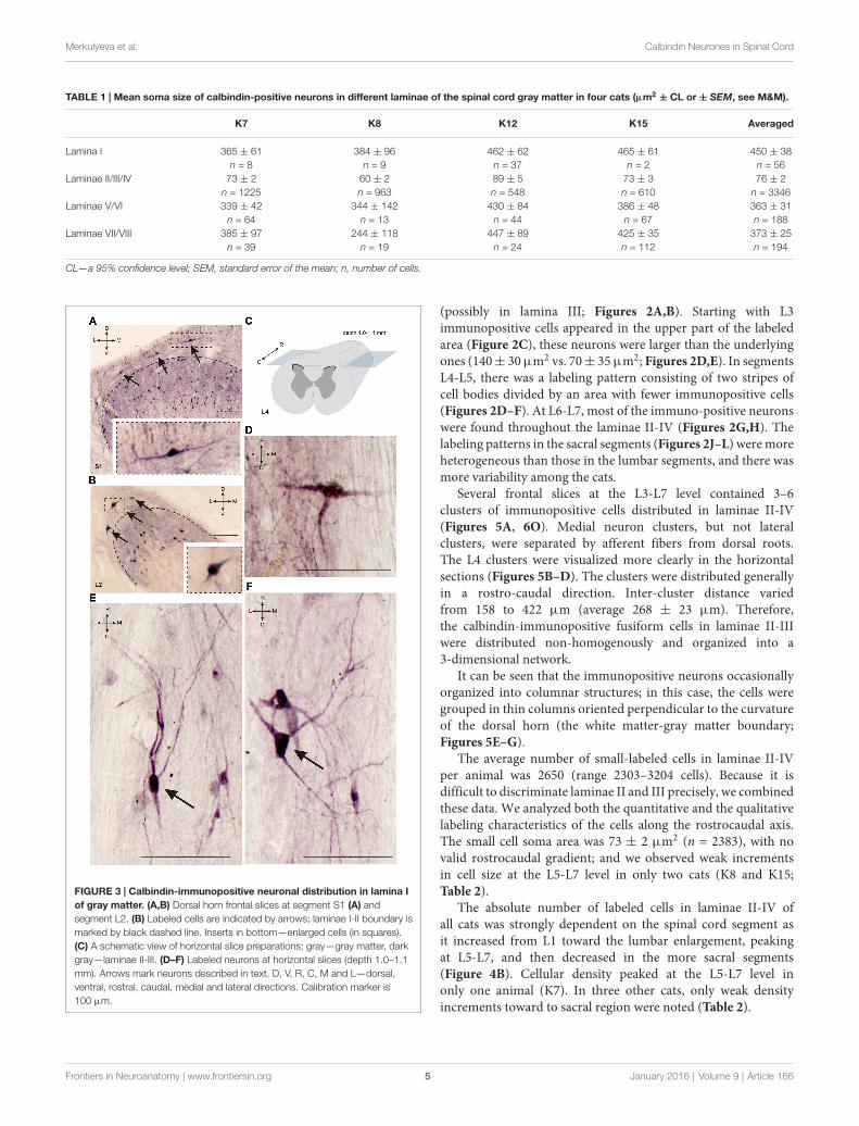
The largest numbers of 28 kDa calbindin-immunopositiveneurons were observed in laminae II and III of the dorsalhorn (77% from total labeled cells). The amount of calbindinneurons in lamina IV was 11%; in lamina I, 3.5%; in laminaeV-VI, 3%; and in laminae VII-VIII, 4%. Labeled neurons inlaminae IX and X were rare (Figure 1D).
Lamina IIt was difficult to distinguish lamina I (a marginal layer)from overlying white matter. We attributed this to neuronslying above the upper boundary of the intensive laminae II-IIIlabeling belonging to lamina I (Figure 2). Three main typesof large neurons (averaged body area 280–515 µm2, Table 1)were visualized in lamina I. In the frontal sections, all hadoval-to-triangle bodies, and only one type of cell differedfrom the other cells: bipolar neurons oriented parallel tothe curvature of dorsal horn (Figures 3A,B). It was obviousin the horizontal slices that these bipolar cells also had athird short dendrite, which was oriented in a caudal direction(Figures 3C,D). The second and third types of immunopositiveneurons could be separated only in horizontal slices; andone of them had an elongated soma and long dendrites
Frontiers in Neuroanatomy | www.frontiersin.org 3 January 2016 | Volume 9 | Article 166

Merkulyeva et al. Calbindin Neurones in Spinal Cord
FIGURE 2 | Calbindin-immunopositive neuron distribution in different segments of the dorsal horn. (A–L) Dorsal horn frontal slices; the laminae I-IIboundary is marked by a black dashed line. (E) Enlarged area of labeling (in rectangle at D). Gross labeled cells in d are indicated by thick arrows. (I) Enlarged area oflabeling (in rectangle at H). Lamina I immunopositive cells are marked by thin arrows. D, V, M and L—dorsal, ventral, medial and lateral directions. Calibration markeris 100 µm.
(up to 650 µm) oriented rostro-caudally (Figure 3E). The othershad plural dendrites oriented without any particular direction(Figure 3F). Scattered pale cells with small oval shaped soma,similar to cells in laminae II-III, were rarely visualized inlamina I.
The cell count was undertaken from frontal sections, soall types of large neurons were intermingled. The maximalnumber of large neurons cord in all cats (K7—segmentsS1-S2; K8—segments L6-S2; K12—segments L5-L6 and S2;K15—segments L5-S1 and S3; Figure 4A).
Laminae II-IVLaminae II-III looked like a darkly stained strip and containedlabeled neuronal somas and neuropil (processes orientedprimarily perpendicular to the gray and white matter boundary,Figure 2I). Calbindin-immunopositive neurons were also spreadthroughout lamina IV. Most of the labeled cells in laminae II-IVhad fusiform somas.
Neuronal distribution within laminae II-IV depended uponthe spinal cord level: most of the cells in the L1-L2 segmentswere localized in the lower part of the labeled region
Frontiers in Neuroanatomy | www.frontiersin.org 4 January 2016 | Volume 9 | Article 166
Merkulyeva et al. Calbindin Neurones in Spinal Cord
TABLE 1 | Mean soma size of calbindin-positive neurons in different laminae of the spinal cord gray matter in four cats (µm2 ± CL or ± SEM, see M&M).
K7 K8 K12 K15 Averaged
Lamina I 365 ± 61 384 ± 96 462 ± 62 465 ± 61 450 ± 38n = 8 n = 9 n = 37 n = 2 n = 56
Laminae II/III/IV 73 ± 2 60 ± 2 89 ± 5 73 ± 3 76 ± 2n = 1225 n = 963 n = 548 n = 610 n = 3346
Laminae V/VI 339 ± 42 344 ± 142 430 ± 84 386 ± 48 363 ± 31n = 64 n = 13 n = 44 n = 67 n = 188
Laminae VII/VIII 385 ± 97 244 ± 118 447 ± 89 425 ± 35 373 ± 25n = 39 n = 19 n = 24 n = 112 n = 194
CL—a 95% confidence level; SEM, standard error of the mean; n, number of cells.
FIGURE 3 | Calbindin-immunopositive neuronal distribution in lamina Iof gray matter. (A,B) Dorsal horn frontal slices at segment S1 (A) andsegment L2. (B) Labeled cells are indicated by arrows; laminae I-II boundary ismarked by black dashed line. Inserts in bottom—enlarged cells (in squares).(C) A schematic view of horizontal slice preparations; gray—gray matter, darkgray—laminae II-III. (D–F) Labeled neurons at horizontal slices (depth 1.0–1.1mm). Arrows mark neurons described in text. D, V, R, C, M and L—dorsal,ventral, rostral, caudal, medial and lateral directions. Calibration marker is100 µm.
(possibly in lamina III; Figures 2A,B). Starting with L3immunopositive cells appeared in the upper part of the labeledarea (Figure 2C), these neurons were larger than the underlyingones (140± 30µm2 vs. 70± 35µm2; Figures 2D,E). In segmentsL4-L5, there was a labeling pattern consisting of two stripes ofcell bodies divided by an area with fewer immunopositive cells(Figures 2D–F). At L6-L7, most of the immuno-positive neuronswere found throughout the laminae II-IV (Figures 2G,H). Thelabeling patterns in the sacral segments (Figures 2J–L) weremoreheterogeneous than those in the lumbar segments, and there wasmore variability among the cats.
Several frontal slices at the L3-L7 level contained 3–6clusters of immunopositive cells distributed in laminae II-IV(Figures 5A, 6O). Medial neuron clusters, but not lateralclusters, were separated by afferent fibers from dorsal roots.The L4 clusters were visualized more clearly in the horizontalsections (Figures 5B–D). The clusters were distributed generallyin a rostro-caudal direction. Inter-cluster distance variedfrom 158 to 422 µm (average 268 ± 23 µm). Therefore,the calbindin-immunopositive fusiform cells in laminae II-IIIwere distributed non-homogenously and organized into a3-dimensional network.
It can be seen that the immunopositive neurons occasionallyorganized into columnar structures; in this case, the cells weregrouped in thin columns oriented perpendicular to the curvatureof the dorsal horn (the white matter-gray matter boundary;Figures 5E–G).
The average number of small-labeled cells in laminae II-IVper animal was 2650 (range 2303–3204 cells). Because it isdifficult to discriminate laminae II and III precisely, we combinedthese data. We analyzed both the quantitative and the qualitativelabeling characteristics of the cells along the rostrocaudal axis.The small cell soma area was 73 ± 2 µm2 (n = 2383), with novalid rostrocaudal gradient; and we observed weak incrementsin cell size at the L5-L7 level in only two cats (K8 and K15;Table 2).
The absolute number of labeled cells in laminae II-IV ofall cats was strongly dependent on the spinal cord segment asit increased from L1 toward the lumbar enlargement, peakingat L5-L7, and then decreased in the more sacral segments(Figure 4B). Cellular density peaked at the L5-L7 level inonly one animal (K7). In three other cats, only weak densityincrements toward to sacral region were noted (Table 2).
Frontiers in Neuroanatomy | www.frontiersin.org 5 January 2016 | Volume 9 | Article 166
Merkulyeva et al. Calbindin Neurones in Spinal Cord
FIGURE 4 | Calbindin-labeled neuronal distribution in different Rexed laminae of the L1-S3 segments. (A) Diagrams of the distribution of medium-to-largecells in lamina I; (B) Diagrams of the distribution of small cells in laminae II-IV; (C) Diagrams of the distribution of medium-to-large cells in laminae V-VIII. (A–C) leftmost, upper—frontal slices, regions of interest are shaded. (A–C), left most, bottom—averaged from all cat data (TOT); (A–C) Medium and rightmostfigures—diagrams for individual cats (K7, K8, K12 and K15). X axis—segments, Y axis—number of labeled cells. (B) Light gray—lamina IV, dark gray—laminae II-III;(C) Light gray—laminae V-VI, dark gray—laminae VII-VIII.
Laminae V-VIIIA range of 26–67 calbindin-immunopositive cells per animalwere observed in laminae V–VI, and 38–112 immunopositivecells per animal were visualized in laminae VII-VIII. Most ofthe labeled cells were multipolar or pyramidal (but multipolarcells can be visualized as a pyramidal under specific tissuecutting angels); these cells were characterized by arborizingdendrites. These cells peaked at segments L5–L7 in all cats(Figures 4C, 6A–P, 7A–L, 8A–L). A single immunopositive cellswere observed at the gray and white matter boundary in the
medial portion of laminae V–VI; the somas and polar processesof these cells were oriented strongly along this boundary(Figures 7M,O).
Some of the labeled cells in laminae V-VIII were distributedwithout a clear order, but many neurons formed groupssymmetrically relative to the central canal. These relatively largeor small groups were divided to four regions: (1) the most lateralregion of lamina VII at the L1-L4 level; (2) the laminae IV-Vboundary at the L5-L7 level; (3) the dorsal border of lamina VIIat the L5-L7 level; and (4) lamina VIII at the sacral level.
Frontiers in Neuroanatomy | www.frontiersin.org 6 January 2016 | Volume 9 | Article 166

Merkulyeva et al. Calbindin Neurones in Spinal Cord
FIGURE 5 | Calbindin-immunopositive neuronal distribution in the frontal (A) and horizontal (C,D) sections of the gray matter of the dorsal horn.(A–D) Clusters of calbindin-immunopositive cells in segment L4, laminae II-IV; clusters are indicated by arrows. (A) Frontal slice, (B) Schematic view of horizontalslice preparations, depths of 1.4, 1.5 and 1.6 mm from the pial surface. (C,D) Horizontal slices; clusters are indicated by arrows. (E–G) Columns ofcalbindin-immunopositive cells in laminae II-III. (F,G) Enlarged regions in rectangles. D, V, R, C, M and L—dorsal, ventral, rostral, caudal, medial and lateral directions.Calibration marker is 100 µm.
The first group consisted of closely-packed immunopositivecells, the number of which peaked at the L3-L4 level(Figure 6). These dark stained cells had long thickdendrites. In the frontal sections, some of them lookedfusiform, but a comparison of the frontal and horizontalsections revealed that some also had multipolar morphology(Figures 6Q–T).
The second and third groups were co-localized within thesame slices at the L5-L7 level (Figure 7); in one animal,these groups were visualized also in rostral S1 (Figure 8A).The second group was located at the laminae IV-V boundaryand consisted of small-to-mid-sized multipolar cells (363 ±31 µm2, n = 180; Figures 7M,N). The third group waslocated at the dorsal border of lamina VII and was subdividedinto two clusters: a medial cluster located near the laminaeVII-X boundary consisting of medium-sized cells (330 ±36 µm2, n = 45) with weak arborized dendrites and alateral cluster consisting of larger neurons (483 ± 60 µm2,n = 56) with long ramified dendrites (Figures 7M,P,Q,R).Figure 7S shows a reconstruction of these cell groups in oneanimal; we combined six slices and centered them relativeto the central canal. Dot size in Figure 7S reflects relativeneuronal soma size, demonstrating that the cells in the lateralcluster were indeed larger than the cells in the medial cluster(Figure 7S).
In the caudal spinal segments in lamina VIII, many labeledneurons with thin long dendrites were evident (Figure 8). Thesecells generally appeared at the L5-L7 (Figures 7D,H,J,K,L),and became more numerous in the more sacral segments(Figures 8E–L). These immunopositive cells had fusiform ortriangle-shaped somas with a mean area of 625 µm2 (±46), witha range of 220–1040 µm2. In some tissue sections, the labeledcells were tightly grouped (Figure 8O, right side; Figure 8R)but some labeled cells were observed outside of the graymatter.
Laminae IX and XOnly scattered pale immunopositive neurons were observedin laminae IX-X (3–5 per animal), so no statistical data areavailable. The immunopositive cells in the most ventral portionof the gray matter (possibly lamina IX) had a large soma size(1100–3200 µm2) and were thus most likely motor neurons(Figures 8M–O, gray arrows).
DISCUSSION
Neuronal control of movement is organized by heterogeneouspopulation of interneurons. Interneurons expressing specificCa2+-binding proteins have different discharge patterns (HofandNimchinsky, 1992; Heizmann, 1993; Kawaguchi and Kubota,
Frontiers in Neuroanatomy | www.frontiersin.org 7 January 2016 | Volume 9 | Article 166

Merkulyeva et al. Calbindin Neurones in Spinal Cord
FIGURE 6 | Calbindin-immunopositive neuronal distribution in the graymatter of L1-L4 segments. (A–P) Contoured images of frontal slices insegments L1 (A–D), L2 (E–H), L3 (I–L) and L4 (M–P) of cat K7 (A,E,I,M), K8(B,F,J,N), K12 (C,G,K,O) and K15 (D,H,L,P). Immunopositive neurons inlaminae II-IV are marked by small dots; those in laminae V-VIII—by gross dots;laminae VI-VII boundary is marked by a dashed line. Rexed laminae areindicated by Roman numerals. Lateral groups of labeled cells are shaded inblue. (Q–T) Labeled cells groups in lamina VII, lateral image. (Q,S) Frontal slice(segment L3), (R,T) horizontal slice (segment L4, depth 2.2 mm, only lefthemisphere is shown). The laminae III/IV, IV/V and VI-VII boundaries aremarked by dashed lines. Rexed laminae are indicated by Roman numerals. D,V, M and L—dorsal, ventral, medial and lateral directions. Calibration marker is100 µm.
1993; Markram et al., 2004; Baizer and Baker, 2005), soimmunolabeling can be used as a functional cell type marker.In the present work, we present a detailed description ofcalbindin-immunopositive neurons that are distributed amongdifferent Rexed laminae over the lumbar and sacral segments ofthe cat spinal cord.
Lamina IIt is known that special types of neurons are located inlamina I (Watson et al., 2008): (1) fusiform cells responding tonociception and expressing GABA and dynorphin; (2) pyramidalcells responding to thermal sensation and expressing encephalin;(3) multipolar cells responding to polymodal stimulation andexpressing GABA; (4) flatted cells expressing substance P anddynorphin; and (5) T-shaped cells (Lima and Coimbra, 1986;Prescott and Koninck, 2002).
We found three main types of calbindin-immunoreactivecells in lamina I: elongated cells with principal dendritesoriented rostro-caudally, oval-shaped curved cells with dendritesprimarily oriented in medio-lateral direction, and cells withdendrites extended without any particular direction. The firsttype possibly corresponds to fusiform cells, the second one,to pyramidal cells, and the third one, to multipolar cells(Lima and Coimbra, 1986; Watson et al., 2008). Lamina Ineurons play a crucial role in the conduction of nociceptiveand temperature signals to supraspinal structures (Craig et al.,2002; Spike et al., 2003). It was shown by Craig et al.(1994, 2002) that in primates, 28 kDa calbindin is labeled inlamina I cells that send spinothalamic axons to the VMponucleus (posterior part of thalamic ventromedial nucleus) inresponse to the perception of temperature and pain. Theyalso showed an absence of any correspondence betweenprojecting neurons and particular morphological cell types inlamina I.
The staining of 28 kDa calbindin in lamina I cells waspreviously shown in both rat and cat spinal cord (Yoshida et al.,1990; Gamboa-Esteves et al., 2001; Anelli and Heckman, 2005).Such neurons have been assumed to be fusiform, flattened andpyramidal in rats (Gamboa-Esteves et al., 2001), or multipolar,in cats (Anelli and Heckman, 2005). As demonstrated previouslyin cats, lamina I large fusiform cells were calretinin-labeled butwere not calbindin immunopositive (Anelli andHeckman, 2005).Overall, 39.4% of the fusiform cells in lamina I were calbindin-positive in the rat spinal cord (Gamboa-Esteves et al., 2001). Ourstudy confirms that the lamina I large spindle-soma cells can becalbindin-positive. We also revealed an increasing of lamina Icells in the lumbosacral enlargement.
Laminae II-IIILaminae II-III interneurons are responsible for the transmissionand modulation of somatosensory information. The neuronalorganization of these laminae is heterogeneous and includesfunctional groups that were determined with electrophysiologicaland neuromorphological data (Yasaka et al., 2010). Four mainneuronal types are found in laminae II-III: islet cells and centralcells with dendrites expanded in the rostrocaudal directionand radial and vertical cells with dendrites expanded in thedorsoventral direction (Grudt and Perl, 2002). Accordingly, mostof the calbindin-immunopositive neurons labeled in our studyhad dendrites oriented dorsoventrally, which presumably wasindicative of radial or vertical cells.
In both cats and in rats, the spinal neurons inlamina III are larger than the corresponding cells inlamina II (Rexed, 1954; Scheibel and Scheibel, 1968;
Frontiers in Neuroanatomy | www.frontiersin.org 8 January 2016 | Volume 9 | Article 166

Merkulyeva et al. Calbindin Neurones in Spinal Cord
TABLE 2 | Number, density and soma size (µm2 ± CL) of 28 kDa calbindin-positive neurons in laminae II-IV of the lumbo-sacral spinal segments in fourcats.
K7 K8 K12 K15
L1 Count 49 57 53 65Density, (cell/µm2)∗1000 0.32 0.38 0.35 0.50Area, µm2 76 ± 10 52 ± 7 74 ± 17 53 ± 7
L2 Count 41 65 71 75Density, (cell/µm2)∗1000 0.28 0.44 0.48 0.49Area, µm2 69 ± 6 57 ± 7 86 ± 19 69 ± 7
L3 Count 63 63 57 77Density, (cell/µm2)∗1000 0.40 0.42 0.38 0.55Area, µm2 88 ± 7 60 ± 5 70 ± 8 54 ± 6
L4 Count 82 77 56 71Density, (cell/µm2)∗1000 0.55 0.52 0.36 0.46Area, µm2 69 ± 5 51 ± 4 62 ± 12 67 ± 11
L5 Count 137 71 66 55Density, (cell/µm2)∗1000 0.92 0.47 0.44 0.37Area, µm2 78 ± 6 45 ± 4 118 ± 16 80 ± 7
L6 Count 118 75 60 62Density, (cell/µm2)∗1000 0.79 0.51 0.40 0.41Area, µm2 74 ± 3 59 ± 5 108 ± 14 84 ± 9
L7 Count 157 76 55 66Density, (cell/µm2)∗1000 1.0 0.51 0.36 0.42Area, µm2 59 ± 4 65 ± 6 95 ± 13 82 ± 6
S1 Count 93 109 60 52Density, (cell/µm2)∗1000 0.57 0.73 0.39 0.37Area, µm2 60 ± 6 72 ± 7 75 ± 14 88 ± 12
S2 Count 79 95 68 56Density, (cell/µm2)∗1000 0.53 0.62 0.45 0.38Area, µm2 64 ± 7 54 ± 5 94 ± 10 52 ± 10
S3 Count 96 96 111 65Density, (cell/µm2)∗1000 0.63 0.64 0.74 0.43Area, µm2 72 ± 13 54 ± 5 74 ± 13 71 ± 10
CL—a 95% confidence level.
Anelli and Heckman, 2005; Watson et al., 2008). Our data,however, show that the immunopositive cells in lamina IIIwere equal to or even smaller in size than the ones in laminaII (Figures 2D–F). In cats, the sizes of the calbindin-positiveneurons in laminae II-III were comparable across differentrostrocaudal levels.
The present work demonstrates that the laminae II-IIIimmunopositive cells occurred in clusters, predominantly in theL3-L5 segments. As shown by Scheibel and Scheibel (1968) usingGolgi technique, neurons in laminae II-III tended to be arrangedin a columnar fashion in frontal sections, although they werethinner than the clusters observed in the present study. It wasshown by Rivero-Melián and Grant (1990) and Takahashi et al.(2007) that the central projections from the lumbar dorsal rootganglia formed patches in laminae II-III in the mediolateral androstrocaudal directions. The location, size and numbers of thesepatches suggest that the calbindin-clusters observed in laminaeII-III may correspond to hindlimb dermatomes and dorsal horncutaneous somatotopic organization. In this case, one functionalrole of these cells may be multisensory integration.
Laminae IV-VIIIThe present study confirms that heterogeneous populations ofcalbindin-labeled neurons are distributed in laminae IV-VIII(Anelli and Heckman, 2005; Porseva et al., 2014). The first group
of calbindin-immunopositive cells was visualized in the mostcaudal portion of the intermediate gray matter (dorsal part oflamina VII) in segments L1-L4, which were especially clear inthe horizontal slices (Figure 6R). The localization of this groupand its disappearance near the L5 segment suggest that it possiblybelongs to the nucleus intermediolateral (IML), which containspreganglionic sympathetic neurons. A gradient of calbindin-containing sympathetic cells in the rat IML has been reported,with most of the cells in the rostral and caudal portions ofthe IML and fewer cells in the central portion (Grkovic andAnderson, 1997).
Among the calbindin D28k-expressing neurons in theventral-most zone of lamina VII, a functionally discrete subtypealso expresses gephyrin (Sanna et al., 1993). These smallcells (10–20 µm2) are likely Renshaw interneurons, whichare responsible for recurrent inhibition of motor neurons(Renshaw, 1946). In contrast to rats, only 47% of the neuronsclassified as Renshaw cells in cats also have calbindin D28k-immunoreactivity (Carr et al., 1998). Carr et al. (1998) reportedthat there were approximately 750 cells per ventral horn presentin the L6 segment of the cat. This may explain why only scatteredsmall immunopositive cells were seen in the present study alongthe ventral margin of lamina VII. These cells, however, cannotbe confidently classified as Renshaw cells based only on theirmorphology.
Frontiers in Neuroanatomy | www.frontiersin.org 9 January 2016 | Volume 9 | Article 166

Merkulyeva et al. Calbindin Neurones in Spinal Cord
FIGURE 7 | Calbindin-immunopositive neuronal distribution in the graymatter on L5-L7 segments. (A–L) Contoured images of frontal slices insegments L5 (A–D), L6 (E–H) and L7 (I–L) of cat K7 (A,E,I), K8 (B,F,J), K12(C,G,K) and K15 (D,H,L). Immunopositive neurons in laminae II-IV are markedby small dots; those in laminae V-VIII—by gross dots; laminae VI-VII boundaryis marked by a dashed line. Rexed laminae are indicated by Roman numerals.Groups of labeled cells are shaded in blue. (M–S) Labeled cells in laminaeV-VIII. (M) Dorsal horn frontal slice (segment L7); (N–R) Enlarged cells (indashed squares). (S) Bubble histogram of labeled cells located at the LaminaeVI-VII boundary (as a sum of three frontal slices). Slices were centered apartfrom the central canal (CC, white circle). Cell area is marked by bubble size.Cell clusters are indicated by arrows (thick arrows—clusters of larger cells, thinarrows—clusters of smaller cells). D, V, M and L—dorsal, ventral, medial andlateral directions. Rexed laminae are indicated by Roman numerals.Calibration marker is 100 µm.
Two elongated groups of labeled cells, which weresymmetrically placed relative to the central canal, were revealedat the laminae IV-V and VI-VII boundaries in the L5-L7segments. These groups were located in similar regions, in which
FIGURE 8 | Calbindin-immunopositive neuronal distribution in the graymatter on the S1-S3 segments. (A–L) Contoured images of frontal slices insegments S1 (A–D), S2 (E–H) and S3 (I–L) of cat K7 (A,E,I), K8 (B,F,J), K12(C,G,K) and K15 (D,H,L). Immunopositive neurons are marked by dots;laminae VI-VII and VII-VIII boundaries are marked by dashed lines. Rexedlaminae are indicated by Roman numerals. (M,N) Frontal slices of segmentsS2 (M) and S3 (N). Labeled cells are indicated by dark arrows; motor neuronsare indicated by light gray arrows. (O–R) Enlarged cells (in dashed squares).Rexed laminae are indicated by Roman numerals. Calibration marker is100 µm.
fibers from the alpha-ventral portion of the gigantocellularreticular formation arborize in the mouse spinal cord (Lianget al., 2015). It is important that calbindin labeling have beenobserved in the medial portion of this nucleus (Celio, 1990). Atwo-stripe pattern (in lamina V and the dorsal portion of laminaVII) was also evident for both the lateral corticospinal fibersystem and the primary afferent collaterals system (Scheibel andScheibel, 1966).
The group of labeled cells in lamina VIII of the sacral segmentcould be related to the spinal nucleus of the bulbocavernosus,which is more prominent in males than in females (Sakamoto,2014). This was the case in our study, based on data fromonly one female and four male cats (Figure 8K). One otherstructure possibly corresponding to this group of cells is the
Frontiers in Neuroanatomy | www.frontiersin.org 10 January 2016 | Volume 9 | Article 166
Merkulyeva et al. Calbindin Neurones in Spinal Cord
nucleus comissuralis. N.comissuralis is also located in ventralhorn, and has maximal expression in the second and thirdsacral segments (Rexed, 1954). In the caudal lumbar segments,two main subpopulations of commissural interneurons werefound, those with monosynaptic input from reticulospinal,vestibulospinal neurons and group I afferents; and those withmonosynaptic input from group II muscle afferents (Jankowskaet al., 2005a,b,c). These cells may be linked to or part of thelocomotor networks (Kiehn, 2006; Jankowska, 2008).
In general, it is important to note that total amount oflabeled cells peaks in the lumbar enlargement and that aparticular pattern appears in this region. Because there isevidence that calbindin is a specific marker of thalamic andcortical interneurons that synchronize specific and nonspecificelements of the thalamocortical network into coherent activity(Jones, 2001) and can also modulate activity levels within allcortical layers (Rausell et al., 1992), it can be hypothesizedthat similar neuronal networks exist in the spinal cord. Wecan suppose their distribution in the spinal areas that areinvolved in (1) multisensory integration (in laminae II-IV); (2) connections with particular supraspinal structures(e.g., reticular formation); or (3) commissural connections.According to our data, the candidates for functionally similarneurons can be medium-to-large calbindin-positive cellsrevealed at the laminae IV-V and VI-VII boundaries andin lamina VIII, as well as small cells in laminae II-IV. Thata significant portion of the immunopositive neurons arelocated in the lumbosacral enlargement (segments L5-L7) invarious Rexed laminae suggests their important functional rolewithin and among the spinal networks that control hindlimbmovements.
As for laminae II-IV, regular clusters of calbindin-positiveneurons are of particular interest to researchers. It is wellknown that one of main principles for some brain structuresis modular organization (Krasnoshchekova, 2007). Neuronsinside of modules are grouped according to their responses,molecular features and connecting properties; visual cortexmodules are an example of this (Hubel and Wiesel, 1977;Hübener et al., 1997; Kaas, 2012). In the cortex, we can seemodules of several orders, and the highest order modulesallow the processing of complex information regarding smallportions of the visual space. Thesemodules contain 1–2 calbindinclusters. Modules in any part of brain are based initiallyupon thin dendritic and axonal bundles. In the spinal cord,thin dendritic and axonal bundles were discovered by Ramóny Cajal (1909), and connection clusterization has also beendemonstrated (Rivero-Melián and Grant, 1990; Takahashi et al.,
2007), but molecular clusterization has not been performed.Therefore, we demonstrated the molecular clusterization oflaminae II-IV of the dorsal horns. Features of calbindinneurons allow us to hypothesize that these clusters can bepart of spinal sensorimotor and somatovisceral integrationnetworks.
Calbindin is one possible method of characterizing thespinal neuronal infrastructure. The current paper is specificallydevoted to describing the detailed distribution of calbindin28 kDa-immunopositive neurons, their morphology and their3D distribution in the lumbar and sacral segments of the catspinal cord. There are other neuromorphological approaches thatmay be used to accomplish the above objectives. We believe thatsuch comprehensive descriptive neuromorphological analysis isnecessary and high useful as a framework for ongoing andfuture morphological/functional studies of the spinal neuronalnetworks participating in different types of sensorimotor activity(locomotor and postural function), as well as spinal neurocontrolof visceral systems (bladder function).
AUTHOR CONTRIBUTIONS
All authors had full access to all the data in the study andtake responsibility for the integrity of the data and the accuracyof the data analysis. Study concept and design: NM, PM.Acquisition of data: NM. Analysis and interpretation of data:NM, AV. Drafting of the manuscript: NM, PM. Critical revisionof the manuscript for important intellectual content: NM, PM,YG, FM. Statistical analysis: NM. Obtained funding: NM, AV.Administrative, technical, and material support: NM, AV, PM.Study supervision: NM, PM.
ACKNOWLEDGMENTS
This study was supported by the Russian Foundation for BasicResearch (RFBR grant No. 13-04-12030 OFI-m); partial supportfor the data analysis was provided to PM, YG, NM from theRussian Science Foundation (RSF grant No. 14-15-00788). Theauthors thank Dr. Reggie Edgerton for his critical reading ofthe manuscript and helpful suggestions. The authors thankSchkorbatova P., Pavlova N. and Bazenova E. for their assistancewith the animals and the tissue preparation. The authors thankDr. Rüdiger Veh (Medizin und Pflegewissenschaften Charitéund BSPH Berlin) for assisting with the immunohistochemicalprotocol. The authors thank the Center for Molecular and CellTechnologies, Research Park, St. Petersburg State University, forsupporting this research.
REFERENCES
Anelli, R., and Heckman, C. J. (2005). The calcium binding proteins calbindin,parvalbumin and calretinin have specific patterns of expression in the graymatter of cat spinal cord. J. Neurocytol. 34, 369–385. doi: 10.1007/s11068-006-8724-2
Antal, M., Polgár, E., Chalmers, J., Minson, J. B., Llewellyn-Smith, I.,Heizmann, C. W., et al. (1991). Different populations of parvalbumin-and calbindin-D28k-immunoreactive neurons contain GABA and accumulate
3H-D-Aspartatei in the dorsal horn of the rat spinal cord. J. Comp. Neurol. 314,114–124. doi: 10.1002/cne.903140111
Arvidsson, U., Ulfhake, B., Cullheim, S., Ramírez, V., Shupliakov, O., andHökfelt, T. (1992). Distribution of calbindin D28k-like immunoreactivity (LI)in the monkey ventral horn: do Renshaw cells contain calbindin D28k-LI?J. Neurosci. 12, 718–728.
Baimbridge, K. G., Cello, M. R., and Rogers, J. H. (1992). Caldum-binding proteinsin the nervous system. Trends Neurosci. 15, 303–308. doi: 10.1016/0166-2236(92)90081-I
Frontiers in Neuroanatomy | www.frontiersin.org 11 January 2016 | Volume 9 | Article 166

Merkulyeva et al. Calbindin Neurones in Spinal Cord
Baizer, J. S., and Baker, J. F. (2005). Immunoreactivity for calcium-bindingproteins defines subregions of the vestibular nuclear complex of the cat. Exp.Brain Res. 164, 78–91. doi: 10.1007/s00221-004-2211-8
Carr, P. A., Alvarez, F. J., Leman, E. A., and Fyffe, R. E. (1998). Calbindin D28kexpression in immunohistochemically identified Renshaw cells. Neuroreport 9,2657–2661. doi: 10.1097/00001756-199808030-00043
Celio, M. R. (1990). Calbindin D-28k and parvalbumin in the rat nervous system.Neuroscience 35, 375–475. doi: 10.1016/0306-4522(90)90091-h
Craig, A. D., Bushnell, M. C., Zhang, E. T., and Blomqvist, A. (1994). A thalamicnucleus specific for pain and temperature sensation. Nature 372, 770–773.doi: 10.1038/372770a0
Craig, A. D., Zhang, E. T., and Blomqvist, A. (2002). Association of spinothalamiclamina I neurons and their ascending axons with calbindin-immunoreactivityin monkey and human. Pain 97, 105–115. doi: 10.1016/s0304-3959(02)00009-x
Deliagina, T. G., Orlovsky, G. N., and Pavlova, G. A. (1983). The capacity forgeneration of rhythmic oscillations is distributed in the lumbosacral spinal cordof the cat. Exp. Brain Res. 53, 81–90. doi: 10.1007/bf00239400
Franks, K. M., and Sejnowski, T. J. (2002). Complexity of calciumsignaling in synaptic spines. Bioessays 24, 1130–1144. doi: 10.1002/bies.10193
Gamboa-Esteves, F. O., Lima, D., and Batten, T. F. (2001). Neurochemistry ofsuperficial spinal neurones projecting to nucleus of the solitary tract thatexpress c-fos on chemical somatic and visceral nociceptive input in the rat.Metab. Brain Dis. 16, 151–164. doi: 10.1023/A:1012536910214
Gerasimenko, Y. P., Roy, R. R., and Edgerton, V. R. (2008). Epidural stimulation:comparison of the spinal circuits that generate and control locomotion in rats,cats and humans. Exp. Neurol. 209, 417–425. doi: 10.1016/j.expneurol.2007.07.015
Grabarek, Z. (2006). Structural Basis for diversity of the EF-hand calcium-binding proteins. J. Mol. Biol. 359, 509–525. doi: 10.1016/j.jmb.2006.03.066
Grkovic, I., and Anderson, C. R. (1997). Calbindin D28K-immunoreactivityidentifies distinct subpopulations of sympathetic pre- and postganglionicneurons in the rat. J. Comp. Neurol. 386, 245–259. doi: 10.1002/(sici)1096-9861(19970922)386:2<245::aid-cne6=3.3.co;2-u
Grudt, T. J., and Perl, E. R. (2002). Correlations between neuronal morphology andelectrophysiological features in the rodent superficial dorsal horn. J. Physiol.540, 189–207. doi: 10.1113/jphysiol.2001.012890
Heizmann, C. W. (1993). Calcium signaling in the brain. Acta Neurobiol. Exp.(Wars) 53, 15–23.
Hof, P. R., and Nimchinsky, E. A. (1992). Regional distribution of neurofilamentand calcium-binding proteins in the cingulate cortex of the macaque monkey.Cereb. Cortex 2, 456–467. doi: 10.1093/cercor/2.6.456
Horst, M., Heutschi, J., van den Brand, R., Andersson, K. E., Gobet, R., Sulser, T.,et al. (2013). Multisystem neuroprosthetic training improves bladder functionafter severe spinal cord injury. J. Urol. 189, 747–753. doi: 10.1016/j.juro.2012.08.200
Horst, M., van den Brand, R., Heutschi, J., Musienko, P., Gobet, R., Sulser, T., et al.(2011). Multisystem neurorehabilitation improves bladder function after spinalcord injury. J. Urol. 185:e171. doi: 10.1016/j.juro.2011.02.514
Hubel, D. H., and Wiesel, T. N. (1977). Ferrier lecture. Functional architecture ofmacaquemonkey visual cortex. Proc. R. Soc. Lond. B Biol. Sci. 198, 1–59. doi: 10.1098/rspb.1977.0085
Hübener, M., Shoham, D., Grinvald, A., and Bonhoeffer, T. (1997). Spatialrelationships among three columnar systems in cat area 17. J. Neurosci. 17,9270–9284.
Jankowska, E. (2008). Spinal interneuronal networks in the cat: elementarycomponents. Brain Res. Reviews. 57, 46–55. doi: 10.1016/j.brainresrev.2007.06.022
Jankowska, E., Cabaj, A., and Pettersson, L. G. (2005a). How to enhance ipsilateralactions of pyramidal tract neurons. J. Neurosci. 25, 7401–7405. doi: 10.1523/jneurosci.1838-05.2005
Jankowska, E., Edgley, S. A., Krutki, P., and Hammar, I. (2005b). Functionaldifferentiation and organization of feline midlumbar commissuralinterneurones. J. Physiol. (Lond.) 565, 645–658. doi: 10.1113/jphysiol.2005.083014
Jankowska, E., Krutki, P., and Matsuyama, K. (2005c). Relative contribution ofIa inhibitory interneurones to inhibition of feline contralateral motoneurones
evoked via commissural interneurones. J. Physiol. (Lond.) 568, 617–628. doi: 10.1113/jphysiol.2005.088351
Jones, E. G. (2001). The thalamic matrix and thalamocortical synchrony. TrendsNeurosci. 24, 595–601. doi: 10.1016/s0166-2236(00)01922-6
Kaas, J. H. (2012). Evolution of columns, modules, and domains in the neocortexof primates. Proc. Natl. Acad. Sci. U S A 109, 10655–10660. doi: 10.1073/pnas.1201892109
Kawaguchi, Y., and Kubota, Y. (1993). Correlation of physiological subgroupingsof nonpyramidal cells with parvalbumin- and calbindin D28k-immunoreactiveneurons in layer V of rat frontal cortex. J. Neurophys. 70, 387–396.
Kiehn, O. (2006). Locomotor circuits in the mammalian spinal cord.Annu. Rev. Neurosci. 29, 279–306. doi: 10.1146/annurev.neuro.29.051605.112910
Krasnoshchekova, E. I. (2007). Modular Organization of Neuronal Centers. Saint-Petersburg: Saint-Petersburg University Press.
Liang, H., Watson, C., and Paxinos, G. (2015). Terminations of reticulospinalfibers originating from the gigantocellular reticular formation in the mousespinal cord. Brain Struct. Funct. 331, 1–11. doi: 10.1007/s00429-015-0993-z
Lima, D., and Coimbra, A. (1986). A Golgi study of the neuronal population of themarginal zone (lamina I) of the rat spinal cord. J. Comp. Neurol. 244, 53–71.doi: 10.1002/cne.902440105
Markram, H., Toledo-Rodriguez, M., Wang, Y., Gupta, A., Silberberg, G., andWu, C. (2004). Interneurons of the neocortical inhibitory system. Nat. Rev.Neurosci. 5, 793–807. doi: 10.1038/nrn1519
Merkulyeva, N., and Nikitina, N. (2012). A method for the detection andquantitative analysis of two-dimensional distribution patterns of labeledneurons in the cerebral cortex. Neurosci. Behav. Physiol. 42, 48–51. doi: 10.1007/s11055-011-9531-5
Musienko, P. E., Deliagina, T. G., Gerasimenko, Y. P., Orlovsky, G. N., andZelenin, P. V. (2014). Limb and trunk mechanisms for balance control duringlocomotion. J. Neurosci. 34, 5704–5716. doi: 10.1523/JNEUROSCI.4663-13.2014
Musienko, P. E., Zelenin, P. V., Orlovsky, G. N., and Deliagina, T. G. (2010).Facilitation of postural limb reflexes with epidural stimulation in spinal rabbits.J. Neurophysiol. 103, 1080–1092. doi: 10.1152/jn.00575.2009
Nägerl, U. V., Novo, D., Mody, I., and Vergara, J. L. (2000). Binding kinetics ofcalbindin-D28k determined by flash photolysis of caged Ca2+. Biophys. J. 79,3009–3018. doi: 10.1016/s0006-3495(00)76537-4
Nordin, B. E. C. (1988). Calcium in Human Biology. New York: Springer.Orlovsky, G. N., Deliagina, T. G., and Grillner, S. (1999). Neuronal Control of
Locomotion. From Mollusc to Man. Oxford: Oxford University Press.Pochet, R., Donato, R., Haiech, J., Heizmann, C. W., and Gerke, V. (2000).
Calcium: The Molecular Basis of Calcium Action in Biology and Medicine.Dordrecht: Kluwer Academic Publishers.
Porseva, V. V., Shilkin, V. V., Strelkov, A. A., and Masliukov, P. M. (2014).Subpopulation of calbindin-immunoreactive interneurons in the dorsal hornof the mice spinal cord. Tsitologiia 56, 612–618.
Prescott, S. A., and Koninck, Y. D. (2002). Four cell types with distinctivemembrane properties and morphologies in lamina I of the spinal dorsal hornof the adult rat. J. Physiol. 539, 817–836. doi: 10.1113/jphysiol.2001.013437
Ramón y Cajal, S. (1909). Histologie du Système Nerveux de l’Homme et desVertébrés (French edition reviewed and updated by the author, translated fromSpanish by L. Azoulay), Paris: Hachette Livre-Bnf.
Rausell, E., Bae, C. S., Viñuela, A., Huntley, G. W., and Jones, E. G.(1992). Calbindin and parvalbumin cells in monkey VPL thalamic nucleus:distribution, laminar cortical projections and relations to spinothalamicterminations. J. Neurosci. 12, 4088–4111.
Ren, K., and Ruda, M. A. (1994). A comparative study of the calcium-bindingproteins calbindin-D28K, calretinin, calmodulin and parvalbumin in the ratspinal cord. Brain Res. Brain Res. Rev. 19, 163–179. doi: 10.1016/0165-0173(94)90010-8
Renshaw, B. (1946). Central effects of centripetal impulses in axons of spinalventral roots. J. Neurophysiol. 9, 191–204.
Rexed, B. A. (1954). Cytoarchitectonic atlas of the spinal cord in the cat. J. Comp.Neurol. 100, 297–379. doi: 10.1002/cne.901000205
Rivero-Melián, C., and Grant, G. (1990). Distribution of lumbar dorsal rootfibers in the lower thoracic and lumbosacral spinal cord of the rat studiedwith choleragenoid horseradish peroxidase conjugate. J. Comp. Neurol. 299,470–481. doi: 10.1002/cne.902990407
Frontiers in Neuroanatomy | www.frontiersin.org 12 January 2016 | Volume 9 | Article 166

Merkulyeva et al. Calbindin Neurones in Spinal Cord
Sabatini, B. L., Maravall, M., and Svoboda, K. (2001). Ca2+ signaling in dendriticspines. Curr. Opin. Neurobiol. 11, 349–356. doi: 10.1016/S0959-4388(00)00218-X
Sakamoto, H. (2014). Sexually dimorphic nuclei in the spinal cord control malesexual functions. Front. Neurosci. 8:184. doi: 10.3389/fnins.2014.00184
Samoilov, M. O. (1999). Brain and Adaptation: Molecular and CellularMechanisms. Saint-Petersburg: Pavlov Institute of Physiology.
Sanna, P. P., Celio, M. R., Bloom, F. E., and Rendeu, M. (1993). PresumptiveRenshaw cells contain decreased calbindin during recovery from sciatic nervelesions. Proc. Natl. Acad. Sci. U S A 90, 3048–3052. doi: 10.1073/pnas.90.7.3048
Scheibel, M. E., and Scheibel, A. B. (1966). Terminal axonal patterns in cat spinalcord. I. the lateral commissural tract. Brain Res. 2, 333–350. doi: 10.1016/0006-8993(66)90003-5
Scheibel, M. E., and Scheibel, A. B. (1968). Terminal axonal patterns in catspinal cord. II. The dorsal horn. Brain Res. 9, 32–58. doi: 10.1016/0006-8993(68)90256-4
Schmidt, H. (2012). Three functional facets of calbindin D-28k. Front. Mol.Neurosci. 5:25. doi: 10.3389/fnmol.2012.00025
Schwaller, B., Meyer, M., and Schiffmann, S. (2002). ‘New’ functions for ‘old’proteins: the role of the calciumbinding proteins calbindin D-28k, calretininand parvalbumin, in cerebellar physiology. Studies with knockout mice.Cerebellum 1, 241–258. doi: 10.1080/147342202320883551
Spike, R. C., Puskár, Z., Andrew, D., and Todd, A. J. (2003). A quantitativeand morphological study of projection neurons in lamina I of the rat lumbarspinal cord Europ. J. Neurosci. 18, 2433–2448. doi: 10.1046/j.1460-9568.2003.02981.x
Takahashi, Y., Aoki, Y., and Doya, H. (2007). Segmental somatotopic organizationof cutaneous afferent fibers in the lumbar spinal cord dorsal horn in rats. Anat.Sci. Int. 82, 24–30. doi: 10.1111/j.1447-073x.2006.00164.x
Watson, C., Paxinos, G., Heise, C., and Kayialoglou, G. (2008). The Spinal Cord,A Christopher and Dana Reeve Foundation Text and Atlas. San Diego, CA:Elsevier.
Yasaka, T., Tiong, S. Y. X., Hughes, D. I., Riddell, J. S., and Todd, A. J. (2010).Populations of inhibitory and excitatory interneurons in lamina II of theadult rat spinal dorsal horn revealed by a combined electrophysiological andanatomical approach. Pain 151, 475–488. doi: 10.1016/j.pain.2010.08.008
Yoshida, S., Senba, E., Kubota, Y., Hagihira, S., Yoshiya, I., Emson, P. C., et al.(1990). Calcium-binding proteins calbindin and parvalbumin in the superficialdorsal horn of the rat spinal cord.Neuroscience 37, 839–848. doi: 10.1016/0306-4522(90)90113-i
Conflict of Interest Statement: The authors declare that the research wasconducted in the absence of any commercial or financial relationships that couldbe construed as a potential conflict of interest.
Copyright © 2016 Merkulyeva, Veshchitskii, Makarov, Gerasimenko and Musienko.This is an open-access article distributed under the terms of the Creative CommonsAttribution License (CC BY). The use, distribution and reproduction in other forumsis permitted, provided the original author(s) or licensor are credited and that theoriginal publication in this journal is cited, in accordance with accepted academicpractice. No use, distribution or reproduction is permitted which does not complywith these terms.
Frontiers in Neuroanatomy | www.frontiersin.org 13 January 2016 | Volume 9 | Article 166