Distal Radial Fracture Management With an … ARTICLE Distal Radial Fracture Management With an...
8
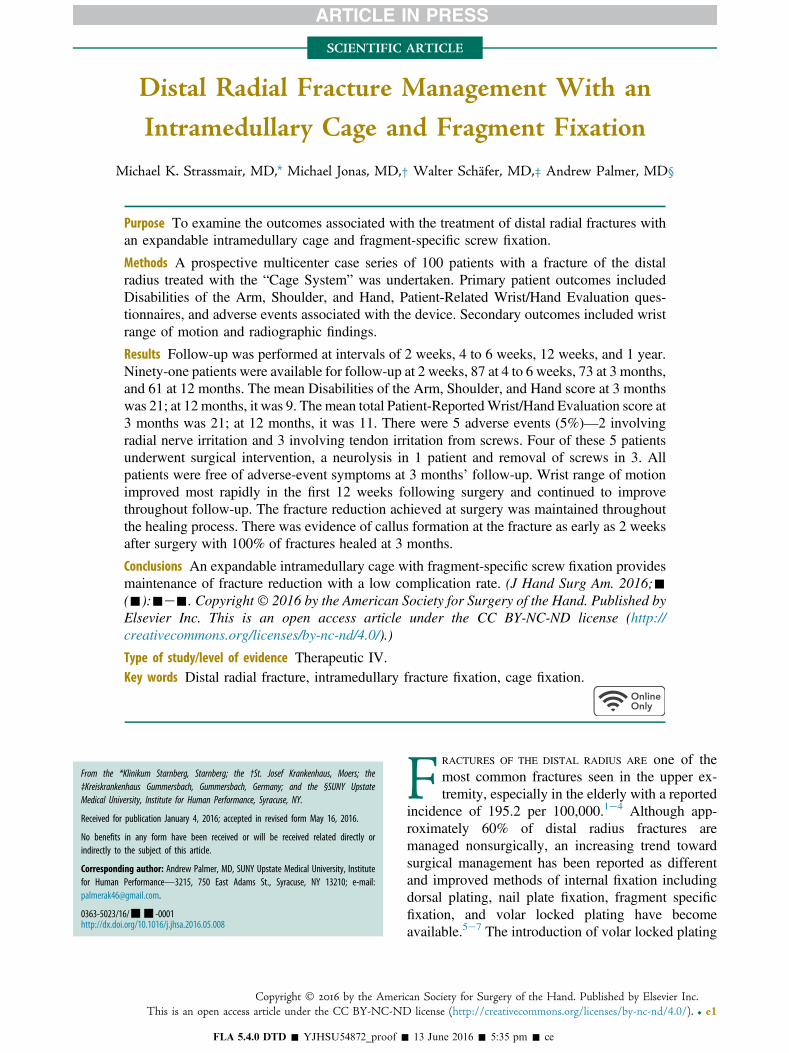
SCIENTIFIC ARTICLE Distal Radial Fracture Management With an Intramedullary Cage and Fragment Fixation Michael K. Strassmair, MD, * Michael Jonas, MD,† Walter Schäfer, MD,‡ Andrew Palmer, MD§ Purpose To examine the outcomes associated with the treatment of distal radial fractures with an expandable intramedullary cage and fragment-specific screw fixation. Methods A prospective multicenter case series of 100 patients with a fracture of the distal radius treated with the “Cage System” was undertaken. Primary patient outcomes included Disabilities of the Arm, Shoulder, and Hand, Patient-Related Wrist/Hand Evaluation ques- tionnaires, and adverse events associated with the device. Secondary outcomes included wrist range of motion and radiographic findings. Results Follow-up was performed at intervals of 2 weeks, 4 to 6 weeks, 12 weeks, and 1 year. Ninety-one patients were available for follow-up at 2 weeks, 87 at 4 to 6 weeks, 73 at 3 months, and 61 at 12 months. The mean Disabilities of the Arm, Shoulder, and Hand score at 3 months was 21; at 12 months, it was 9. The mean total Patient-Reported Wrist/Hand Evaluation score at 3 months was 21; at 12 months, it was 11. There were 5 adverse events (5%)—2 involving radial nerve irritation and 3 involving tendon irritation from screws. Four of these 5 patients underwent surgical intervention, a neurolysis in 1 patient and removal of screws in 3. All patients were free of adverse-event symptoms at 3 months’ follow-up. Wrist range of motion improved most rapidly in the first 12 weeks following surgery and continued to improve throughout follow-up. The fracture reduction achieved at surgery was maintained throughout the healing process. There was evidence of callus formation at the fracture as early as 2 weeks after surgery with 100% of fractures healed at 3 months. Conclusions An expandable intramedullary cage with fragment-specific screw fixation provides maintenance of fracture reduction with a low complication rate. (J Hand Surg Am. 2016;- (-):-e-. Copyright Ó 2016 by the American Society for Surgery of the Hand. Published by Elsevier Inc. This is an open access article under the CC BY-NC-ND license (http:// creativecommons.org/licenses/by-nc-nd/4.0/).) Type of study/level of evidence Therapeutic IV. Key words Distal radial fracture, intramedullary fracture fixation, cage fixation. F RACTURES OF THE DISTAL RADIUS ARE one of the most common fractures seen in the upper ex- tremity, especially in the elderly with a reported incidence of 195.2 per 100,000. 1e4 Although app- roximately 60% of distal radius fractures are managed nonsurgically, an increasing trend toward surgical management has been reported as different and improved methods of internal fixation including dorsal plating, nail plate fixation, fragment specific fixation, and volar locked plating have become available. 5e7 The introduction of volar locked plating From the *Klinikum Starnberg, Starnberg; the †St. Josef Krankenhaus, Moers; the ‡Kreiskrankenhaus Gummersbach, Gummersbach, Germany; and the §SUNY Upstate Medical University, Institute for Human Performance, Syracuse, NY. Received for publication January 4, 2016; accepted in revised form May 16, 2016. No benefits in any form have been received or will be received related directly or indirectly to the subject of this article. Corresponding author: Andrew Palmer, MD, SUNY Upstate Medical University, Institute for Human Performance—3215, 750 East Adams St., Syracuse, NY 13210; e-mail: [email protected]. 0363-5023/16/---0001 http://dx.doi.org/10.1016/j.jhsa.2016.05.008 Copyright Ó 2016 by the American Society for Surgery of the Hand. Published by Elsevier Inc. This is an open access article under the CC BY-NC-ND license (http://creativecommons.org/licenses/by-nc-nd/4.0/). r e1 FLA 5.4.0 DTD ĸ YJHSU54872_proof ĸ 13 June 2016 ĸ 5:35 pm ĸ ce
Transcript of Distal Radial Fracture Management With an … ARTICLE Distal Radial Fracture Management With an...
From the *Klinikum Starnberg, Starnberg; the †St. Josef Krank‡Kreiskrankenhaus Gummersbach, Gummersbach, Germany; andMedical University, Institute for Human Performance, Syracuse, NY.
Received for publication January 4, 2016; accepted in revised form
No benefits in any form have been received or will be receiveindirectly to the subject of this article.
Corresponding author: Andrew Palmer, MD, SUNY Upstate Medicafor Human Performance—3215, 750 East Adams St., Syracuse,[email protected].
0363-5023/16/---0001http://dx.doi.org/10.1016/j.jhsa.2016.05.008
CopyrightThis is an open access article unde
FLA 5.4.0 DTD � Y
SCIENTIFIC ARTICLE
Distal Radial Fracture Management With an
Intramedullary Cage and Fragment Fixation
Michael K. Strassmair, MD,* Michael Jonas, MD,† Walter Schäfer, MD,‡ Andrew Palmer, MD§
Purpose To examine the outcomes associated with the treatment of distal radial fractures withan expandable intramedullary cage and fragment-specific screw fixation.
Methods A prospective multicenter case series of 100 patients with a fracture of the distalradius treated with the “Cage System” was undertaken. Primary patient outcomes includedDisabilities of the Arm, Shoulder, and Hand, Patient-Related Wrist/Hand Evaluation ques-tionnaires, and adverse events associated with the device. Secondary outcomes included wristrange of motion and radiographic findings.
Results Follow-up was performed at intervals of 2 weeks, 4 to 6 weeks, 12 weeks, and 1 year.Ninety-one patients were available for follow-up at 2 weeks, 87 at 4 to 6 weeks, 73 at 3 months,and 61 at 12 months. The mean Disabilities of the Arm, Shoulder, and Hand score at 3 monthswas 21; at 12 months, it was 9. The mean total Patient-ReportedWrist/Hand Evaluation score at3 months was 21; at 12 months, it was 11. There were 5 adverse events (5%)—2 involvingradial nerve irritation and 3 involving tendon irritation from screws. Four of these 5 patientsunderwent surgical intervention, a neurolysis in 1 patient and removal of screws in 3. Allpatients were free of adverse-event symptoms at 3 months’ follow-up. Wrist range of motionimproved most rapidly in the first 12 weeks following surgery and continued to improvethroughout follow-up. The fracture reduction achieved at surgery was maintained throughoutthe healing process. There was evidence of callus formation at the fracture as early as 2 weeksafter surgery with 100% of fractures healed at 3 months.
Conclusions An expandable intramedullary cage with fragment-specific screw fixation providesmaintenance of fracture reduction with a low complication rate. (J Hand Surg Am. 2016;-(-):-e-. Copyright� 2016 by the American Society for Surgery of the Hand. Published byElsevier Inc. This is an open access article under the CC BY-NC-ND license (http://creativecommons.org/licenses/by-nc-nd/4.0/).)
Type of study/level of evidence Therapeutic IV.Key words Distal radial fracture, intramedullary fracture fixation, cage fixation.
enhaus, Moers; thethe §SUNY Upstate
May 16, 2016.
d related directly or
l University, InstituteNY 13210; e-mail:
� 2016 by the Amerir the CC BY-NC-ND
JHSU54872_proof �
F RACTURES OF THE DISTAL RADIUS ARE one of themost common fractures seen in the upper ex-tremity, especially in the elderly with a reported
incidence of 195.2 per 100,000.1e4 Although app-roximately 60% of distal radius fractures aremanaged nonsurgically, an increasing trend towardsurgical management has been reported as differentand improved methods of internal fixation includingdorsal plating, nail plate fixation, fragment specificfixation, and volar locked plating have becomeavailable.5e7 The introduction of volar locked plating
can Society for Surgery of the Hand. Published by Elsevier Inc.license (http://creativecommons.org/licenses/by-nc-nd/4.0/). r e1
13 June 2016 � 5:35 pm � ce

e2 INTRAMEDULLARY FRACTURE FIXATION
in the early 2000s represented a major innovation inthe surgical repair of distal radius fractures.8 Today,volar locking plates are used to treat the majorityof surgically managed distal radial fractures.8 Mostexisting procedures are relatively invasive, andhardware-related complications including tendon irri-tation and/or rupture, hardware failure, neurologicalinjury, loss of reduction, delayed or nonunion, andinfection are relatively common to all procedures(16%e27%).9e13 Therefore, the need still exists for asurgical solution that minimizes soft tissue traumaand hardware-related complications, addresses a broadrange of fracture patterns, and promotes rapid returnto function. The objective of this study was to assessthe functional and radiographic outcomes for a seriesof patients treated with an expandable metallic implantthat is deployed into the medullary canal of the distalradius providing a scaffold to which bone fragmentscan be stabilized using fragment specific screws.
MATERIALS AND METHODSA multicenter nonsequential study was undertaken atmultiple health centers in Germany after the studyprotocol was approved by the Ethics Committee ineach participating center. The protocol conformed tothe ethical guidelines of the 1975 Declaration ofHelsinki. Required exclusion criteria for this study, inaccordance with Conformité Européene markedproduct labeling, included mental illness, alcoholism,foreign body sensitivity, AO class C3 fractures, andopen growth plates.
Participating centers were provided financial com-pensation by the implant manufacturer for completionof required study follow-up only. No payments weremade to physicians or patients.
Follow-up comprising completion of the Disabilitiesof the Arm, Shoulder, and Hand (DASH) and Patient-Related Wrist/Hand Evaluation (PRWHE) forms, mea-surement of range ofmotion, and assessment for adverseeventswasperformedat 2weeks, 4 to6weeks, 12weeks,and 1 year by the principal investigators (M.S., M.J., andW.S.), subinvestigators, and/or site coordinators. Of the100 patients initially admitted to the study, 91 patientswere available for follow-up at 2 weeks, 87 at 4 to 6weeks, 73 at 12 weeks, and 61 at 1 year.
Patients were lost to follow-up for a number ofreasons including voluntary withdrawal by 26 in-dividuals because they were satisfied with the resultsfrom their surgery and they, therefore, requested tonot have to return for additional follow-up. Sevenpatients were lost from 1 site because the principalinvestigator changed practice location. Six patients, at
FLA 5.4.0 DTD � YJHSU54872_proof �
J Hand Surg Am. r V
multiple sites, were excluded because they missedfollow-up appointments.
X-rays were obtained before surgery, immediatelyafter surgery, and at intervals up to 1 year. Follow-upx-rays were available on 63 patients at 12 weeks and48 patients at 1 year. X-rays were taken in compli-ance with each site’s standard of care, which meant,in many instances, x-rays were not allowed after thefracture was judged to be healed out of concern forunnecessary radiation exposure to the patient. X-rays,when available, were evaluated in final follow-up bya board certified orthopedic hand surgeon (A.P.) inregards to radial inclination, volar tilt, ulnar variance,coronal shift, and evidence of fracture healing.Fracture union was judged to be present if osseousbridging across the fracture site was seen in 2 of 3views as recommended by Rosental et al.14
Surgical technique
All surgical procedures were performed through astandard radial approach. After a reduction of thefracture was obtained by the surgeon, using eitherclosed or open methods and held in place via longi-tudinal finger trap traction or axially placed K-wires,the technique and instrumentation unique to the CageDistal Radius System (Conventus Orthopedics,Maple Grove, MN) was utilized. The technique in-volves entering the radial aspect of the radius usingdrill bits through a 2- to 3-cm radial incision that islocated approximately 3 cm proximal to the articularsurface. This is followed by preparation of the site forthe implant with the system’s reaming device in thesubcortical region of the distal radius. The implant isthen deployed into the prepared space of the distalradius just proximal to the fracture. The device islocked in its expanded position and fixed to the radialborder of the radius with a small side plate andscrews. The final step is the introduction of fragment-specific, cannulated screws through the device, acrossthe fracture and into the far cortex.
Postoperative immobilization varied according tothe surgeon’s preference and the site’s standard of carefor distal radial fractures. Fifty-five patients under-went no immobilization, 40 utilized a commercialorthosis for 2 weeks, and in 5 patients, a cast was usedfor 2 weeks. No patients received formal therapy butwere instructed to begin range of motion exercises astolerated.
RESULTSEighty-nine percent of patients were female with amean age of 68 years. Ninety-three percent wereright-handed, although the right and left wrists were
13 June 2016 � 5:35 pm � ce
ol. -, - 2016
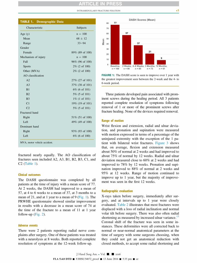
TABLE 1. Demographic Data
Characteristic Subjects
Age (y) n ¼ 100
Mean 68 � 12
Range 33e94
Gender
Female 89% (89 of 100)
Mechanism of injury n ¼ 100
Fall 96% (96 of 100)
Sports 2% (2 of 100)
Other (MVA) 2% (2 of 100)
AO classification
A2 27% (27 of 101)
A3 37% (38 of 101)
B1 6% (6 of 101)
B2 5% (5 of 101)
B3 1% (1 of 101)
C1 19% (19 of 101)
C2 5% (5 of 101)
Fractured hand
Right 51% (51 of 100)
Left 49% (49 of 100)
Dominant hand
Right 93% (93 of 100)
Left 6% (6 of 100)
MVA, motor vehicle accident.
77
57
37
21
9
0
10
20
30
40
50
60
70
80
Baseline, n = 100
2 Weeks, n = 90
4–6 Weeks, n = 85
3 Months, n = 73
12 Months, n = 61
Mea
n
DASH Scores (Mean)
FIGURE 1: The DASH score is seen to improve over 1 year withthe greatest improvement seen between the 2-week and the 4- to6-week period.
INTRAMEDULLARY FRACTURE FIXATION e3
fractured nearly equally. The AO classification offractures seen included A2, A3, B1, B2, B3, C1, andC2 (Table 1).
Clinical outcomes
The DASH questionnaire was completed by allpatients at the time of injury with a mean score of 77.At 2 weeks, the DASH had improved to a mean of57, at 4 to 6 weeks to a mean of 37, at 3 months to amean of 21, and at 1 year to a mean of 9 (Fig. 1). ThePRWHE questionnaire showed similar improvementin results with a decrease in a mean score of 74 atthe time of the fracture to a mean of 11 at 1 yearfollow-up (Fig. 2).
Adverse events
There were 2 patients reporting radial nerve com-plaints after surgery. One of these patients was treatedwith a neurolysis at 8 weeks. Both reported completeresolution of symptoms at the 12-week follow-up.
FLA 5.4.0 DTD � YJHSU54872_proof �
J Hand Surg Am. r V
Three patients developed pain associated with prom-inent screws during the healing period. All 3 patientsreported complete resolution of symptoms followingremoval of 1 or more of the prominent screws afterfracture healing. None of the devices required removal.
Range of motion
Wrist flexion and extension, radial and ulnar devia-tion, and pronation and supination were measuredwith motion expressed in terms of a percentage of theuninjured extremity with the exception of the 1 pa-tient with bilateral wrist fractures. Figure 3 showsthat, on average, flexion and extension measuredabout 50% of normal at 2 weeks and had improved toabout 75% of normal by 12 weeks. Radial and ulnardeviation measured close to 60% at 2 weeks and hadimproved to 78% by 12 weeks. Pronation and supi-nation improved to 88% of normal at 2 weeks and95% at 12 weeks. Range of motion continued toimprove up to 1 year, but the majority of improve-ment was seen in the first 12 weeks.
Radiographic evaluation
X-rays taken before surgery, immediately after sur-gery, and at intervals up to 1 year were closelyevaluated. Table 2 illustrates that most fractures weredisplaced with a loss of radial inclination and normalvolar tilt before surgery. There was also often radialshortening as measured by increased ulnar variance.15
Coronal shift of the fracture was seen in some in-stances. These deformities were all corrected back tonormal or near-normal anatomical parameters at thetime of surgery with some surgeons choosing, whenthey could not get an anatomical reduction withclosed methods, to accept some radial shortening and
13 June 2016 � 5:35 pm � ce
ol. -, - 2016
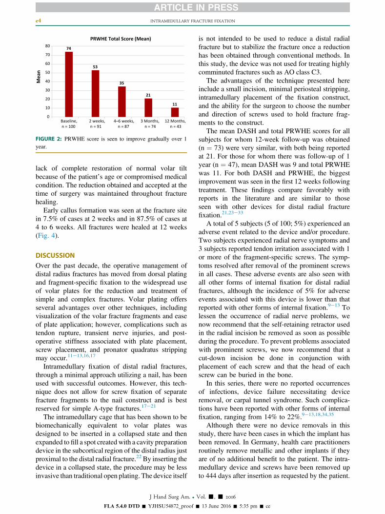
74
53
35
21
11
0
10
20
30
40
50
60
70
80
Baseline, n = 100
2 weeks, n = 91
4–6 weeks, n = 87
3 Months, n = 74
12 Months, n = 43
PRWHE Total Score (Mean)M
ean
FIGURE 2: PRWHE score is seen to improve gradually over 1year.
e4 INTRAMEDULLARY FRACTURE FIXATION
lack of complete restoration of normal volar tiltbecause of the patient’s age or compromised medicalcondition. The reduction obtained and accepted at thetime of surgery was maintained throughout fracturehealing.
Early callus formation was seen at the fracture sitein 7.5% of cases at 2 weeks and in 87.5% of cases at4 to 6 weeks. All fractures were healed at 12 weeks(Fig. 4).
DISCUSSIONOver the past decade, the operative management ofdistal radius fractures has moved from dorsal platingand fragment-specific fixation to the widespread useof volar plates for the reduction and treatment ofsimple and complex fractures. Volar plating offersseveral advantages over other techniques, includingvisualization of the volar fracture fragments and easeof plate application; however, complications such astendon rupture, transient nerve injuries, and post-operative stiffness associated with plate placement,screw placement, and pronator quadratus strippingmay occur.11e13,16,17
Intramedullary fixation of distal radial fractures,through a minimal approach utilizing a nail, has beenused with successful outcomes. However, this tech-nique does not allow for screw fixation of separatefracture fragments to the nail construct and is bestreserved for simple A-type fractures.17e21
The intramedullary cage that has been shown to bebiomechanically equivalent to volar plates wasdesigned to be inserted in a collapsed state and thenexpanded to fill a spot created with a cavity preparationdevice in the subcortical region of the distal radius justproximal to the distal radial fracture.22 By inserting thedevice in a collapsed state, the procedure may be lessinvasive than traditional open plating. The device itself
FLA 5.4.0 DTD � YJHSU54872_proof �
J Hand Surg Am. r V
is not intended to be used to reduce a distal radialfracture but to stabilize the fracture once a reductionhas been obtained through conventional methods. Inthis study, the device was not used for treating highlycomminuted fractures such as AO class C3.
The advantages of the technique presented hereinclude a small incision, minimal periosteal stripping,intramedullary placement of the fixation construct,and the ability for the surgeon to choose the numberand direction of screws used to hold fracture frag-ments to the construct.
The mean DASH and total PRWHE scores for allsubjects for whom 12-week follow-up was obtained(n ¼ 73) were very similar, with both being reportedat 21. For those for whom there was follow-up of 1year (n ¼ 47), mean DASH was 9 and total PRWHEwas 11. For both DASH and PRWHE, the biggestimprovement was seen in the first 12 weeks followingtreatment. These findings compare favorably withreports in the literature and are similar to thoseseen with other devices for distal radial fracturefixation.21,23e33
A total of 5 subjects (5 of 100; 5%) experienced anadverse event related to the device and/or procedure.Two subjects experienced radial nerve symptoms and3 subjects reported tendon irritation associated with 1or more of the fragment-specific screws. The symp-toms resolved after removal of the prominent screwsin all cases. These adverse events are also seen withall other forms of internal fixation for distal radialfractures, although the incidence of 5% for adverseevents associated with this device is lower than thatreported with other forms of internal fixation.9e13 Tolessen the occurrence of radial nerve problems, wenow recommend that the self-retaining retractor usedin the radial incision be removed as soon as possibleduring the procedure. To prevent problems associatedwith prominent screws, we now recommend that acut-down incision be done in conjunction withplacement of each screw and that the head of eachscrew can be buried in the bone.
In this series, there were no reported occurrencesof infections, device failure necessitating deviceremoval, or carpal tunnel syndrome. Such complica-tions have been reported with other forms of internalfixation, ranging from 14% to 22%.9e13,18,34,35
Although there were no device removals in thisstudy, there have been cases in which the implant hasbeen removed. In Germany, health care practitionersroutinely remove metallic and other implants if theyare of no additional benefit to the patient. The intra-medullary device and screws have been removed upto 444 days after insertion as requested by the patient.
13 June 2016 � 5:35 pm � ce
ol. -, - 2016
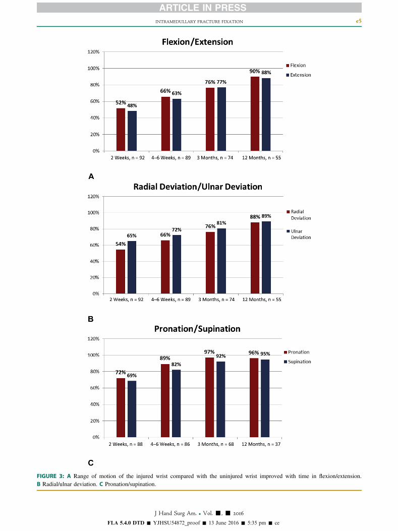
FIGURE 3: A Range of motion of the injured wrist compared with the uninjured wrist improved with time in flexion/extension.B Radial/ulnar deviation. C Pronation/supination.
FLA 5.4.0 DTD � YJHSU54872_proof � 13 June 2016 � 5:35 pm � ce
INTRAMEDULLARY FRACTURE FIXATION e5
J Hand Surg Am. r Vol. -, - 2016
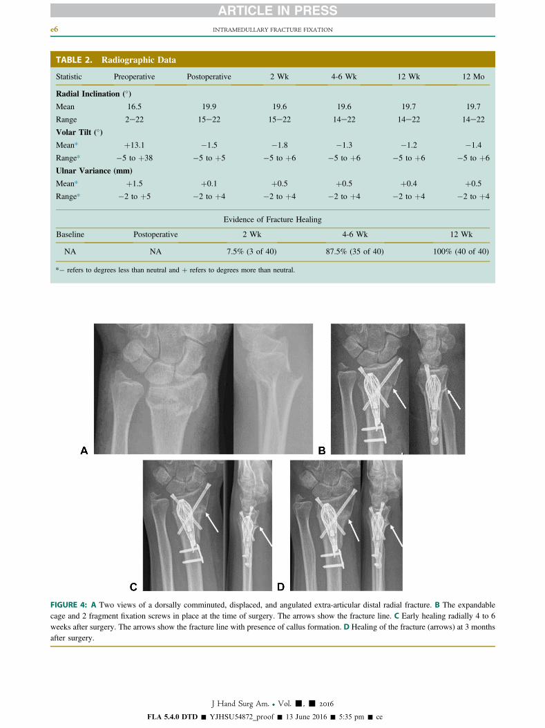
TABLE 2. Radiographic Data
Statistic Preoperative Postoperative 2 Wk 4-6 Wk 12 Wk 12 Mo
Radial Inclination (�)
Mean 16.5 19.9 19.6 19.6 19.7 19.7
Range 2e22 15e22 15e22 14e22 14e22 14e22
Volar Tilt (�)
Mean* þ13.1 �1.5 �1.8 �1.3 �1.2 �1.4
Range* �5 to þ38 �5 to þ5 �5 to þ6 �5 to þ6 �5 to þ6 �5 to þ6
Ulnar Variance (mm)
Mean* þ1.5 þ0.1 þ0.5 þ0.5 þ0.4 þ0.5
Range* �2 to þ5 �2 to þ4 �2 to þ4 �2 to þ4 �2 to þ4 �2 to þ4
Evidence of Fracture Healing
Baseline Postoperative 2 Wk 4-6 Wk 12 Wk
NA NA 7.5% (3 of 40) 87.5% (35 of 40) 100% (40 of 40)
*� refers to degrees less than neutral and þ refers to degrees more than neutral.
FIGURE 4: A Two views of a dorsally comminuted, displaced, and angulated extra-articular distal radial fracture. B The expandablecage and 2 fragment fixation screws in place at the time of surgery. The arrows show the fracture line. C Early healing radially 4 to 6weeks after surgery. The arrows show the fracture line with presence of callus formation. D Healing of the fracture (arrows) at 3 monthsafter surgery.
FLA 5.4.0 DTD � YJHSU54872_proof � 13 June 2016 � 5:35 pm � ce
e6 INTRAMEDULLARY FRACTURE FIXATION
J Hand Surg Am. r Vol. -, - 2016
INTRAMEDULLARY FRACTURE FIXATION e7
In each case, the device was able to be removedwithout causing additional damage to surroundingtissues.
Radiographic follow-up in this study revealed thatthe intramedullary device and fragment-specific screwfixation resulted in a stable fracture construct as evi-denced by minimal or no change in position of thepostoperative reduction throughout healing (Table 2).Full healing of the fracturewas seen in 100%of cases at12 weeks as well as early signs of healing in 87% ofcases at 4 to 6 weeks. Because cavity preparation forthe implant essentially creates a bone slurry from theintramedullary bone while removing no bone, it ispossible that an osteogenic milieu is created that con-tributes to rapid fracture healing. Further study of this isrequired.
The study shows that an expandable intramedu-llary implant with fragment-specific screw fixationhas been associated with satisfactory results and alow complication rate when used for treating distalradius fractures, with the exception of AO class C3fractures.
REFERENCES
1. Singer BR, McLauchlan GJ, Robinson CM, Christie J. Epidemiologyof fractures in 15000 adults: the influence of age and gender. J BoneJoint Surg Br. 1998;80(2):243e248.
2. Court-Brown CM, Caesar B. Epidemiology of adult fractures: areview. Injury. 2006;37(8):691e697.
3. Chung KC, Spilson SV. The frequency and epidemiology of handand forearm fractures in the United States. J Hand Surg Am.2001;26(5):908e915.
4. Lofthus CM, Frihagen F, Meyer HE, Nordsletten L, Melhuus K,Falch JA. Epidemiology of distal forearm fractures in Oslo. Norway.Osteoporos Int. 2008;19(6):781e786.
5. Chung KC, Shauver MJ, Birkmeyer JD. Trends in the United Statesin the treatment of distal radius fractures in the elderly. J Bone JointSurg Am. 2009;91(8):1868e1873.
6. Koval KJ, Harrast JJ, Anglen JO, Weinstein JN. Fractures of thedistal radius. the evolution of practice over time. Where’s the evi-dence? J Bone Joint Surg Am. 2008;90(9):1855e1861.
7. Jupiter JB. Fractures of the distal end of the radius. J Bone Joint SurgAm. 1991;73(3):461e469.
8. Orbay JL, Fernandez DL. Volar fixed-angle plate fixation for un-stable distal radius fractures in the elderly patient. J Hand Surg Am.2004;29(1):96e102.
9. Arora R, Lutz M, Hennerbichler A, Krappinger D, Espen D, Gabl M.Complications following internal fixation of unstable distal radiusfracture with a palmar locking plate. J Orthop Trauma. 2007;21(5):316e322.
10. Jupiter JB, Marent-Huber M, LCP Study Group. Operative man-agement of distal radial fractures with 2.4-millimeter locking plates: amulticenter prospective case series. J Bone Joint Surg Am.2009;91(1):55e65.
11. Williksen JH, Husby T, Hellund JC, Kvernmo HD, Rosales C,Frihagen F. External fixation and adjuvant pins versus volar lockingplate fixation in unstable distal radius fractures: a randomized,controlled studywith a 5-year follow-up. J Hand Surg Am. 2015;40(7):1333e1340.
FLA 5.4.0 DTD � YJHSU54872_proof �
J Hand Surg Am. r V
12. Knudsen R, Bahadirov Z, Damborg F. High rate of complicationsfollowing volar plating of distal radius fractures. Dan Med J.2014;61(10):A4906.
13. Matzon JL, Kenniston J, Beredjiklian PK. Hardware-related com-plications after dorsal plating for displaced distal radius fractures.Orthopedics. 2014;37(11):e978ee982.
14. Rozental TD, Blazar PE, Franko OI, Chacko AT, Earp BE, Day CS.Functional outcomes for unstable distal radial fractures treated withopen reduction and internal fixation or closed reduction and percu-taneous fixation. A prospective randomized trial. J Bone Joint SurgAm. 2009;91(8):1837e1846.
15. Graham TJ. Surgical Correction of malunited fractures of the distalradius. J Am Acad Orthop Surg. 1997;5(5):270e281.
16. Roh YH, Lee BK, Baek JR, Noh JH, Gong HS, Baek GH.A randomized comparison of volar plate and external fixation forintra-articular distal radius fractures. J Hand Surg Am. 2015;40(1):34e41.
17. Gunther SB, Lynch TL. Rigid internal fixation of displaced distalradius fractures. Orthopedics. 2014;37(1):e34ee38.
18. Gradl G, Mielsch N, Wendt M, et al. Intramedullary nail versus volarplate fixation of extra-articular distal radius fractures. Two year re-sults of a prospective randomized trial. Injury. 2014;45(Suppl 1):S3eS8.
19. Tan V, Bratchenko W, Nourbakhsh A, Capo J. Comparative analysisof intramedullary nail fixation versus casting for treatment of distalradius fractures. J Hand Surg Am. 2012;37(3):460e468.
20. Safi A, Hart R, Te�kne�d�zjan B, Kozák. Treatment of extra-articularand simple articular distal radial fractures with intramedullary nailversus volar locking plate. J Hand Surg Eur Vol. 2013;38(7):774e779.
21. Schønnemann JO, Hansen TB, Søballe K. Randomised study of non-bridging external fixation compared with intramedullary fixation ofunstable distal radial fractures. J Plast Hand Surg. 2011;45(4e5):232e237.
22. van Kampen RJ, Thoreson AR, Knutson NJ, Hale JE, Moran SL.Comparison of a new intramedullary scaffold to volar plating fortreatment of distal radius fractures. J Orthop Trauma. 2013;27(9):535e541.
23. Capo JT, Hashem J, Orillaza NS, Tan V, Warburton M, Bonilla L.Treatment of extra-articular distal radial malunions with an intra-medullary implant. J Hand Surg Am. 2010;35(6):892e899.
24. Egol K, Walsh M, Tejwani N, McLaurin T, Wynn C, Paksima N.Bridging external fixation and supplementary Kirschner-wire fixationversus volar locked plating for unstable fractures of the distal radius:a randomised, prospective trial. J Bone Joint Surg Br. 2008;90(9):1214e1221.
25. Ilyas AM, Thoder JJ. Intramedullary fixation of displaced distalradius fractures: a preliminary report. J Hand Surg Am. 2008;33(10):1706e1715.
26. Marcheix PS, Dotzis A, Benkö PE, Siegler J, Arnaud JP,Charissoux JL. Extension fractures of the distal radius in patientsolder than 50: a prospective randomized study comparing fixationusing mixed pins or a palmar fixed-angle plate. J Hand Surg Eur Vol.2010;35(8):646e651.
27. McFadyen I, Field J, McCann P, Ward J, Nicol S, Curwen C. Shouldunstable extra-articular distal radial fractures be treated with fixed-angle volar-locked plates or percutaneous Kirschner wires? A pro-spective randomised controlled trial. Injury. 2011;42(2):162e166.
28. van Manen CJ, Dekker ML, van Eerten PV, Rhemrev SJ, vanOlden GD, van der Elst M. Bio-resorbable versus metal implants inwrist fractures: a randomised trial. Arch Orthop Trauma Surg.2008;128(12):1413e1417.
29. Wei DH, Raizman NM, Bottino CJ, Jobin CM, Strauch RJ,Rosenwasser MP. Unstable distal radial fractures treated withexternal fixation, a radial column plate, or a volar plate. A pro-spective randomized trial. J Bone Joint Surg Am. 2009;91(7):1568e1577.
13 June 2016 � 5:35 pm � ce
ol. -, - 2016

e8 INTRAMEDULLARY FRACTURE FIXATION
30. WilckeMK,AbbaszadeganH,Adolphson PY.Wrist function recoversmore rapidly after volar locked plating than after external fixation butthe outcomes are similar after 1 year. Acta Orthop. 2011;82(1):76e81.
31. Hunsaker FG, Cioffi DA, Amadio PC, Wright JG, Caughlin B. TheAmerican Academy of Orthopaedic Surgeons Outcome Instruments.Normative values from the general population. J Bone Joint Surg Am.2002;84(2):208e215.
32. Lauder A, Agnew S, Bakri K, Allan CH, Hanel DP, Huang JI.Functional outcomes following bridge plate fixation for distal radiusfractures. J Hand Surg Am. 2015;40(8):1554e1562.
FLA 5.4.0 DTD � YJHSU54872_proof �
J Hand Surg Am. r V
33. Costa ML, Achten J, Parsons NR, et al. Volar locking-plate andKirschner-wire fixation did not differ in terms of functional outcomesafter dorsally displaced distal radial fracture. J Bone Joint Surg Am.2015;97A(10):859.
34. McKay SD, MacDermid JC, Roth JH, Richards RS. Assessmentof complications of distal radius fractures and developmentof a complication checklist. J Hand Surg Am. 2001;26(5):916e922.
35. Davis DI, Baratz M. soft tissue complications of distal radius frac-tures. Hand Clin. 2010;26(2):229e235.
13 June 2016 � 5:35 pm � ce
ol. -, - 2016M-4420-July-16