DISSERTATION SUBMITTED TO UNIVERSITY OF … Subramanyam.pdf · deforming forces due to peculiar...
85
1 A STUDY OF MANAGEMENT OF SUBTROCHANTERIC FRACTURE FEMUR BY PROXIMALFEMORAL NAILING A STUDY DONE AT ALLURI SITARAMA RAJU ACADEMY OF MEDICAL SCIENCES ELURU DISSERTATION SUBMITTED TO UNIVERSITY OF SEYCHELLES AMERICAN INSTITUTE OF MEDICINE IN PARTIAL FULFILLMENT OF THE REQUIREMENTS FOR THE DEGREE M.Ch (Orthopaedic Surgery) Submitted By Dr. SURBRAMANYAM YADLAPALLI M.S (ORTHO), MRCS (UK) DECEMBER 2011
Transcript of DISSERTATION SUBMITTED TO UNIVERSITY OF … Subramanyam.pdf · deforming forces due to peculiar...

1
A STUDY OF MANAGEMENT OF
SUBTROCHANTERIC FRACTURE FEMUR BY
PROXIMALFEMORAL NAILING
A STUDY DONE AT A L L U R I SI T A R A M A RA J U
A C A D E M Y OF M E D I C AL S C I E N CE S
ELURU
DISSERTATION SUBMITTED TO
UNIVERSITY OF SEYCHELLES
AMERICAN INSTITUTE OF MEDICINE
IN PARTIAL FULFILLMENT OF THE REQUIREMENTS FOR THE DEGREE
M.Ch (Orthopaedic Surgery)
Submitted By
Dr. SURBRAMANYAM YADLAPALLI
M.S (ORTHO), MRCS (UK)
DECEMBER 2011

2
CONTENTS
PART I
1. Introduction ………………………………………………. 1
2. Aim & Objective ………………………………………………. 2
3. Review of literature …………………………………………… 3
4. Applied anatomy of subtrochanteric region of femur …………. 7
5. Biomechanics of hip and proximal femur …………………….. 8
6. Classification of Subtrochanteric fractures ……………………10
7. Proximal femoral nailing system …………………………….. 12
8. Operative procedure …………………………………………. 16
9. Assessment of results ……………………………………….. 19
PART II
1. Materials and methods ………………………………………. 22
2. Data entry card ………………………………………………. 24
3. Case sheets of patients ...………………………………….. 27
4. Observations ………………………………………………… 64
5. Discussion …………………………………………………… 76
PART III
1. Summary & Conclusions ……………………………………. 79
2. Bibliography ………………………………………………... 80
3. Master chart of patients ………………………………………84

3
INTRODUCTION
Civilization and modernization has brought certain problems along
with its own benefits to the citizens of the world over. Fast life styles, rapid
and high speed transportation, risky infrastructural projects and modern
commando warfare activities have brought in with them morbidity and
mortality due to trauma in general and fractures of femur in particular.
Among the femoral shaft injuries upper femoral fractures present a
peculiar problem of securing effective neutralization of deforming forces.
The mechanical stresses at this level are very high, as they occur at the
junction between the trabecular and cortical zone and also because of the
deforming forces due to peculiar muscle insertion to the proximal and distal
fragments. These factors have made subtrochanteric fractures demand
special consideration in orthopaedic trauma, because defective union of this
fracture can lead to high disability levels for an individual and thereby loss
of valuable man days
Orthopaedic fraternity is always on the lookout for an effective and
suitable method to treat the upper femoral fractures in the best possible way.
In this process surgical management of these fractures and the surgical
implants used have also gone through an array of changes in their procedures
and designs. Various upper femoral devices like dynamic condylar screw,
dynamic hip screw with barrel plate, gamma nail, proximal femoral nail etc
are being used by various centres and each centre claims reasonably
satisfactory results with each type of device. The present study was conducted
to assess the utility and effectiveness of Proximal Femoral Nail evolved by
AO-ASIF in 1997, for various types of upper femoral fractures

4
AIMS & OBJECTIVES
To evaluate the results of internal fixation of Subtrochanteric fractures
of the femur with PROXIMAL FEMORAL NAIL – AO Type Design

5
REVIEW OF LITERATURE
In 1902 HIBBS1
treated subtrochanteric fractures
conservatively in the position of flexion, abduction and external rotation
stating that it improves the reduction by bringing distal fragment into
alignment with proximal fragment. SARMIENTO2 in 1960‘s initially
attempted to treat subtrochanteric fractures with femoral cast bracing but
later advised against it because of poorer results. MOONEY’S, in 1975 also
noted poor results with cast brace in terms of varus angulation. Observing
the high rate of complications associated with operative treatment of type
IIIA subtrochanteric fractures, SEINSHEIMER3 in 1978 suggested
conservative treatment of these fractures. Couple of years later JC DE
LEE, T.O.CLANTON & C.A.ROCKWOOD4 also conservatively treated
these fractures with preliminary traction followed by ambulatory long leg
quadrilateral cast bracing with pelvic band and reported good results with
this technique. In 1981 DE LEE4 etal reported good results with 90-90
traction followed by a single hip spica and recommended this for patients
with inoperable or open fractures. WADDEL5, emphasized the role of
traction treatment in extensively comminuted subtrochanteric fractures
analyzing that it worked well as the deforming muscle forces are dissipated.
However satisfactory results are observed in only 50% of displaced
fractures. Difficulty in neutralizing the deforming forces on fracture
fragments leading to malunion and complications of prolonged bed rest
have lead surgeons to favour internal fixation in most of the subtrochanteric
fractures.
6
Initial attempts to treat proximal femoral fractures by operative
intervention were made by DELBET6
in 1910 with a thick screw with
higher pitch that purchased better into the bone. CLEVELAND7 in 1947,
and EVANS8 in 1951 used Moore-Blount plate, Neufled plate and Lorenzo
screw respectively, but reported high rates of implant failure. In 1942
G.KUNTSCHER9, used Cloverleaf Nail for the treatment of
subtrochanteric fractures. In 1940‘s and 1950‘s Jewett nail was well
popularized by BOYD & GRIFFITH10
, KRIK WATSON &
CAMPBELL11
. Though Jewett nail initially showed promising results,
TEITGE FIELDING & MAGILATO15
noted 35-55% failure rates
depending upon the type of subtrochanteric fracture. The AO group in 1969
designed angle plate with ‗U‘ profile and fixed angle of 95 and 135 degrees.
ARONOFF12
in 1971, DISTEFANO13
in 1972, CECH14
in 1974 and
FEILDING15
used these plates and reported high rates of complications in
terms of varus and rotational deformities, non union, implant failure and
medial shift of distal fragment. AO blade plate became a popular device for
subtrochanteric fractures in late 1970‘s and in 1978 HANSON &
TULLOS16
reported 87.5% union rates with this device. WADDEL5 in
1979 used sliding hip screw for subtrochanteric fractures and stated that it is
a better implant for type I and II fractures. Couple of years later Dynamic
Hip Screw designed by AO/ASIF group was popularised in 1992 by
SCHLEMNINGER etal, CLAWSON and MASSIE for selected
subtrochanteric fractures, as they noted complication rates of 32% with AO
blade plate. WADDEL5 later reported 10% failure rates with this device.
Intramedullary device with an inbuilt screw was introduced by
ZICKEL17
in 1966 and he stated that it provided excellent strength and
good control of varus and rotation of proximal fragment, but it lacked
rotational control over distal fragment as there was no facility for distal
locking.
7
The effectiveness of this implant for subtrochanteric fractures was
further analyzed by WADDEL5 in 1979 and he noted shortening frequently
and also observed that it required additional fixation for grossly
comminuted fractures. Closed nailing techniques started to gain importance
in early 1980‘s and attempts to treat subtrochanteric fractures with this
technique has shown high rates of union, low rate of infection than previous
implants. In 1986 the development of 3rd
generation interlocking
intramedullary nails took place. RUSSEL TAYLOR NAIL was introduced
in late 1980s & in 1990, HALDER introduced GAMMA NAIL.
HALDER S C18
in his study on 421 patients reported that Gamma nail
transmits weight closer to the calcar than Dynamic Hip Screw and had less
operative complications in unstable pertrochanteric fractures. However few
complications including the fracture of base of greater trochanter and
fractures of shaft of femur at the distal end of the nail have been reported. A
search for a new implant for addressing these issues have lead to the
development of PROXIMAL FEMORAL NAIL by AO – ASIF GROUP.
PROXIMAL FEMORAL NAIL was designed in 1997 to
overcome implant related complications and facilitate the operative
treatment of unstable peritrochanteric fractures. HUBER SM, HEINING
SM, EULER E19
studied the biomechanics of Proximal Femoral Nail and
showed a significant reduction of distal stress and an increased stability
compared with the Gamma Nail. SIMMERMACHER RK, BOSCH20
in
1999 and A.HERRERA21
in their respective studies on Proximal Femoral
Nail showed a relatively low percentage of complications and low incidence
of implant failure as compared to Gamma nail. In 2002, SUDAN M,
SADOWSKI C22
in their prospective randomized study on 206 patients,
compared Dynamic Hip Screw with Proximal Femoral Nail and stated the
advantages of this intramedullary nail.

8
A year later CHRISTIAN BOLDIN etal23
in his study concluded that
proximal Femoral Nail is a good minimally invasive implant for unstable
proximal femoral fractures. In 2005, DANIEL F.A. etal24
also suggested
Proximal Femoral Nail as a useful treatment option for subtrochanteric
factures because of low rates of femoral shaft fractures and failure of
fixation associated with this implant. A comparative study by WOO-KIE
MIN etal25
in 2007, on Proximal Femoral Nail and Gamma Nail for
Reverse oblique trochanteric fractures reported better biomechanical results
with PFN group, in terms of less sliding of lag screw and less change of
neck shaft angle. In 2008, MSG BALLAL26
emphasized that good
reduction with minimal dissection, use of appropriate length of nail and
proper positioning of the nail and screws are necessary to avoid failure or
revision with Proximal Femoral Nail and in the same year SI YONG
PARK27
etal concluded that lesser trochanteric fragment and posteromedial
defect played an important role in the stability after intramedullary hip
nailing and attributed the fixation failures in the PFN group excessive
sliding of the femoral neck screw.
9
APPLIED ANATOMY OF SUBTROCHANTERIC REGION
SUBTROCHANTERIC REGION OF THE FEMUR is
defined as the region between lesser trochanter and junction of proximal
and middle thirds of femur. In this region femur is covered
circumferentially by well vascularised muscle groups. Anteriorly and
laterally it is covered by vastus medialis, vastus intermedius, Vastus
Lateralis and medially by adductor brevis and adductor longus and
posteriorly by vastus Lateralis and Adductor brevis.
Unbalanced muscle pull following a closed subtrochanteric fracture
is the main cause of fracture fragment displacement. The iliopsoas, with its
insertion to the lesser trochanter, typically causes the proximal fragment to
flex and externally rotate and the short abductors that insert onto the greater
trochanter cause the abduction of the proximal fragment. Distal fragment
because of the unopposed pull from the adductor magnus, always displaces
it medially and further aggravates the deformity.

10
BIOMECHANICS OF HIP AND PROXIMAL FEMUR
Forces applied to the hip during ambulation produces stresses in
the proximal femur because of combined effects of axial, bending and
torsional loads. Normally the proximal femur is loaded so that the medial
cortex is compressed and the lateral cortex is under tension. Major
compressive stresses in the femur are greatest in the medial cortex 1 to 3
inches below the lesser trochanter, i.e. the subtrochanteric region and this
region is considered to be one of the highly stressed region in the body.
Tensile stresses of about 25% less occur at the lateral cortex slightly
proximally.
Following the fracture, unbalanced muscle pull results in
displacement of the fracture and this displacement is difficult to neutralize.
The iliopsoas, with its insertion to the lesser trochanter, typically causes the
proximal fragment to flex and externally rotate and the short abductors that
insert onto the greater trochanter cause the abduction of the proximal
fragment. Distal fragment because of the unopposed pull from the adductor
magnus, always displaces it medially and further aggravates the deformity.
In addition comminution of the medial cortex further adds to the injury. In
addition comminution of the medial cortex further adds to the insult of this
highly stressed area.
Cephalomedullary nails are able to provide necessary bending and
torsional stability to combat the displacement of the fracture fragments.
Proximal femoral nail being an intra medullary device is a load sharing
device and has the inherent advantage of shorter lever arm, thereby
decreasing the tensile strain on the implant29

11
The hip screw and the anti rotational screw proximally provide increased
rotational stability of the head-neck fragment. The two distal locking screws
control the rotational stability of the distal fragment. A biomechanical
analysis by TENCER etal30
on various implants used for subtrochanteric
fracture have found that bending stress, torsional stress, load to axial failure
are superior in cephalomedullary implants than all the other implants.
Another biomechanical evaluation done by PAUL R.T. KUZYK etal31
in
2009, on reverse oblique trochanteric fractures concluded that
intramedullary devices were significantly stiffer and had a greater load to
failure than the 135 degree and 95 degree constructs, especially with a gap
between the bony fragments. Indirect fracture reduction, preserving the
fracture hematoma, less soft tissue dissection, decreased amount of blood
loss add to the decreased overall morbidity.
12
CLASSIFICATION OF SUBTROCHANTERIC FRACTURES
1. FIELDING AND MAGLIATO15
Devised a three part anatomical
classification in 1966.
TYPE 1: Fracture at the level of lesser trochanter
TYPE 2: Fracture within 1 inch below lesser trochanter
TYPE 3: Fracture within 1 to 2 inches below lesser trochanter
2. RUSSEL TAYLOR classification32, 38
: This classification is based on
current techniques and principals of closed intramedullary nailing and
continuity of lesser trochanter and extension of fracture lines into greater
trochanter (or) posteriorly into pyriform fossa. It disregards the degree of
comminution.
TypeI: Fracture does not extend into pyriform fossa.
TypeIA: Comminution and fracture line extend from below
lesser trochanter to femoral isthmus
TypeIB: Fracture line and comminution involve area of lesser
trochanter to isthmus.
TypeII: Fracture extends into Pyriform fossa.
TypeIIA. No significant comminution (or) fracture of lesser
trochanter is seen.
TypeIIB. Comminution of medical cortex and loss of continuity
of lesser trochanter are seen.
13
3. In 1978 SEINSHEIMER3 developed a classification based on fracture
pattern. Significance of this classification is that it identified fractures with
loss of medial cortex stability, which is known to have a higher rate of
implant failure
TYPE I: Undisplaced (or) Less than 2mm displacement
TYPE II: Two Part Fracture.
TYPE IIA. Transverse fracture
TYPE IIB. Spiral Fracture with lesser trochanter attached
to proximal fragment.
TYPE IIC. Spiral Fracture with lesser trochanter attached to
distal fragment
TYPE III: Three Part Fracture.
TYPE IIIA: Three part spiral fracture with lesser trochanter
as a part of third fragment.
TYPE IIIB: Three part spiral fracture with third part a
butterfly fragment.
TypeIV: Comminuted fracture with four (or) more fragments
TypeV: Subtrochanteric-Intertrochanteric configuration.
In this study we have followed SEINSHEIMER’s classification,
as in this classification subtrochanteric fractures are classified according to
the number of major fragments and the location and shapes of the fracture
lines3 . This helps us to identify specific type of fracture patterns that are
associated with high complication rates.
14
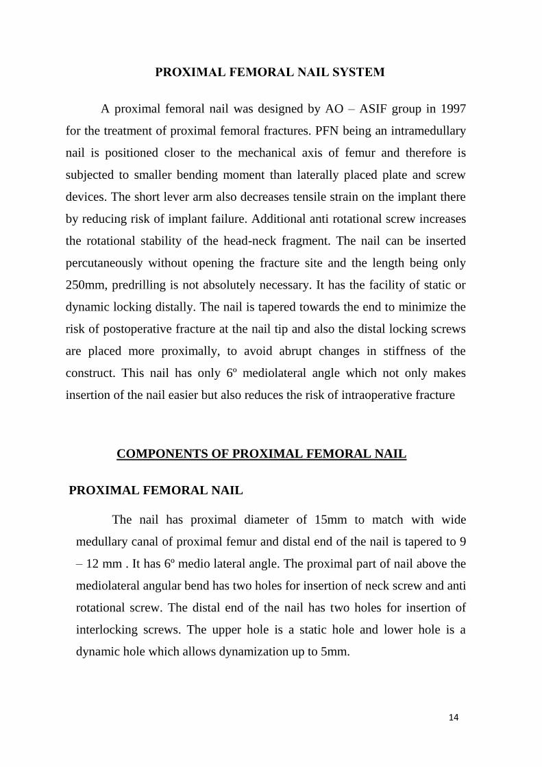
PROXIMAL FEMORAL NAIL SYSTEM
A proximal femoral nail was designed by AO – ASIF group in 1997
for the treatment of proximal femoral fractures. PFN being an intramedullary
nail is positioned closer to the mechanical axis of femur and therefore is
subjected to smaller bending moment than laterally placed plate and screw
devices. The short lever arm also decreases tensile strain on the implant there
by reducing risk of implant failure. Additional anti rotational screw increases
the rotational stability of the head-neck fragment. The nail can be inserted
percutaneously without opening the fracture site and the length being only
250mm, predrilling is not absolutely necessary. It has the facility of static or
dynamic locking distally. The nail is tapered towards the end to minimize the
risk of postoperative fracture at the nail tip and also the distal locking screws
are placed more proximally, to avoid abrupt changes in stiffness of the
construct. This nail has only 6º mediolateral angle which not only makes
insertion of the nail easier but also reduces the risk of intraoperative fracture
COMPONENTS OF PROXIMAL FEMORAL NAIL
PROXIMAL FEMORAL NAIL
The nail has proximal diameter of 15mm to match with wide
medullary canal of proximal femur and distal end of the nail is tapered to 9
– 12 mm . It has 6º medio lateral angle. The proximal part of nail above the
mediolateral angular bend has two holes for insertion of neck screw and anti
rotational screw. The distal end of the nail has two holes for insertion of
interlocking screws. The upper hole is a static hole and lower hole is a
dynamic hole which allows dynamization up to 5mm.

15
The nail is available in angles of 125º, 130º, 135º to match with various
femoral neck – shaft angles and diameters of 9,10,11,12 mm sizes and the
total length of nail is 250mm. The proximal end of the nail also has threads
for insertion of end cap which prevents in growth of bone into the nail
FEMORAL NECK SCREW
This is an 8.0mm screw which bears 80-90% of load under axial
loading and gives main stability in the proximal fragment for fracture
fixation the screw is available in lengths from 70-110mm
ANTI ROTATION HIP SCREW
This is a 6.4 mm stabilization screw, which bears 10-20% of load and
provides the rotational stability for the proximal fragment and the screw is
available in lengths from 70-110mm.
DISTAL LOCKING SCREWS :
These are 4.9 mm screws inter locking screws
COMPONENTS OF PROXIMAL FEMORAL NAIL SYSTEM
INSERTION HANDLE
It is used for insertion of nail along with conical locking bolt and
locking nut. The lugs on the handle must engage the positioning notches at
the upper end of nail for insertion. It is used for insertion of proximal neck
screws and distal locking screws. The holes in the insertion handle position
the locking instruments.
THREADED CONICAL BOLT AND CONICAL NUT
The threaded bolt is screwed by hand into the nail and assembled with
insertion handle. Once the lugs of the handle have engaged in notches, firm
tightening is achieved with wrench.
16
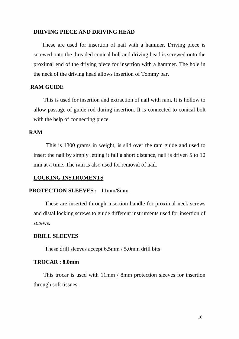
DRIVING PIECE AND DRIVING HEAD
These are used for insertion of nail with a hammer. Driving piece is
screwed onto the threaded conical bolt and driving head is screwed onto the
proximal end of the driving piece for insertion with a hammer. The hole in
the neck of the driving head allows insertion of Tommy bar.
RAM GUIDE
This is used for insertion and extraction of nail with ram. It is hollow to
allow passage of guide rod during insertion. It is connected to conical bolt
with the help of connecting piece.
RAM
This is 1300 grams in weight, is slid over the ram guide and used to
insert the nail by simply letting it fall a short distance, nail is driven 5 to 10
mm at a time. The ram is also used for removal of nail.
LOCKING INSTRUMENTS
PROTECTION SLEEVES : 11mm/8mm
These are inserted through insertion handle for proximal neck screws
and distal locking screws to guide different instruments used for insertion of
screws.
DRILL SLEEVES
These drill sleeves accept 6.5mm / 5.0mm drill bits
TROCAR : 8.0mm
This trocar is used with 11mm / 8mm protection sleeves for insertion
through soft tissues.

17
DRILL BITS: 6.5mm, 5.0mm, and 4.0mm.
The 6.5 mm drill bit and 5.0mm drill bit are used to drill holes for
8.0mm femoral neck screw and 6.4 mm anti rotation hip screw respectively.
These two drill bits are cannulated for drilling over a guide wire and are
marked to know the length of screws to be inserted. The 4.0mm drill bit is
used to drill hole for 4.9mm distal locking bolts.
DEPTH GAUZE FOR LOCKING BOLTS
This depth gauze measures up to 115mm. It has a long neck allowing
measuring for locking bolts through distal locking holes in insertion handle
HEXAGONAL SCREW DRIVER
This large hexagonal screw driver is used for insertion of 8.0mm
femoral neck screw, 6.4mm anti rotational hip screw and 4.9mm distal
locking bolts.

18
OPERATIVE PROCEDURE
PATIENT POSITIONING
Patient lying supine on Albee‘s fracture table allows good
roentgenographic control and enable manipulation of leg and application of
traction.
REDUCTION OF FRACTURE
After positioning the anaesthetised patient supine on fracture table, taking
care to avoid undue pressure or tension on any part of the body, closed
reduction of fracture is performed. The Uninjured limb is held in well leg
holder so that it remains out of the way by putting it in 90 – 90 º leg holder.
Reduction is achieved by aligning distal fragment to flexed and externally
rotated proximal fragment by rotating the foot of effected extremity. If
Reduction is not achieved with ease, a unicortical 5mm threaded joystick is
used to control proximal fragment after draping the patient. If closed
reduction is not successful or not acceptable an open reduction is performed
PROCEDURE
A Slightly curved lateral incision is made from the level of trochanter
proximally for about 6 to 9cm. The length of incision varies with the size of
the patient. Under fluoroscopic guidance, a 3.2mm pin is inserted into the
tip of greater trochanter, taking care to centre it on both antero posterior and
lateral views. The pin is then driven 5cm into proximal femur. An
alternative to this method is to use an awl, under fluoroscopic guidance to
provide the opening. The awl should be inserted up to the point of largest
outer diameter under fluoroscopic guidance and then removed. A guide wire
is then inserted into proximal fragment.
19
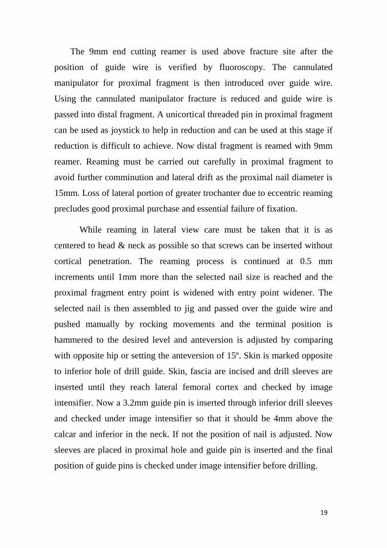
The 9mm end cutting reamer is used above fracture site after the
position of guide wire is verified by fluoroscopy. The cannulated
manipulator for proximal fragment is then introduced over guide wire.
Using the cannulated manipulator fracture is reduced and guide wire is
passed into distal fragment. A unicortical threaded pin in proximal fragment
can be used as joystick to help in reduction and can be used at this stage if
reduction is difficult to achieve. Now distal fragment is reamed with 9mm
reamer. Reaming must be carried out carefully in proximal fragment to
avoid further comminution and lateral drift as the proximal nail diameter is
15mm. Loss of lateral portion of greater trochanter due to eccentric reaming
precludes good proximal purchase and essential failure of fixation.
While reaming in lateral view care must be taken that it is as
centered to head & neck as possible so that screws can be inserted without
cortical penetration. The reaming process is continued at 0.5 mm
increments until 1mm more than the selected nail size is reached and the
proximal fragment entry point is widened with entry point widener. The
selected nail is then assembled to jig and passed over the guide wire and
pushed manually by rocking movements and the terminal position is
hammered to the desired level and anteversion is adjusted by comparing
with opposite hip or setting the anteversion of 15º. Skin is marked opposite
to inferior hole of drill guide. Skin, fascia are incised and drill sleeves are
inserted until they reach lateral femoral cortex and checked by image
intensifier. Now a 3.2mm guide pin is inserted through inferior drill sleeves
and checked under image intensifier so that it should be 4mm above the
calcar and inferior in the neck. If not the position of nail is adjusted. Now
sleeves are placed in proximal hole and guide pin is inserted and the final
position of guide pins is checked under image intensifier before drilling.
20
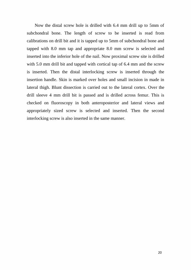
Now the distal screw hole is drilled with 6.4 mm drill up to 5mm of
subchondral bone. The length of screw to be inserted is read from
calibrations on drill bit and it is tapped up to 5mm of subchondral bone and
tapped with 8.0 mm tap and appropriate 8.0 mm screw is selected and
inserted into the inferior hole of the nail. Now proximal screw site is drilled
with 5.0 mm drill bit and tapped with cortical tap of 6.4 mm and the screw
is inserted. Then the distal interlocking screw is inserted through the
insertion handle. Skin is marked over holes and small incision in made in
lateral thigh. Blunt dissection is carried out to the lateral cortex. Over the
drill sleeve 4 mm drill bit is passed and is drilled across femur. This is
checked on fluoroscopy in both anteroposterior and lateral views and
appropriately sized screw is selected and inserted. Then the second
interlocking screw is also inserted in the same manner.
21
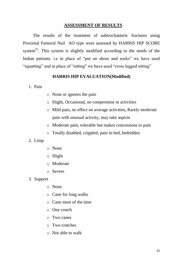
ASSESSMENT OF RESULTS
The results of the treatment of subtrochanteric fractures using
Proximal Femoral Nail AO type were assessed by HARRIS HIP SCORE
system33
. This system is slightly modified according to the needs of the
Indian patients. i.e in place of ―put on shoes and socks‖ we have used
―squatting‖ and in place of ―sitting‖ we have used ―cross legged sitting‖
HARRIS HIP EVALUATION(Modified)
1. Pain
o None or ignores the pain
o Slight, Occasional, no compromise in activities
o Mild pain, no effect on average activities, Rarely moderate
pain with unusual activity, may take aspirin
o Moderate pain, tolerable but makes concessions to pain
o Totally disabled, crippled, pain in bed, bedridden
2. Limp
o None
o Slight
o Moderate
o Severe
3. Support
o None
o Cane for long walks
o Cane most of the time
o One crutch
o Two canes
o Two crutches
o Not able to walk
22
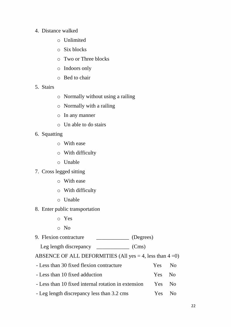
4. Distance walked
o Unlimited
o Six blocks
o Two or Three blocks
o Indoors only
o Bed to chair
5. Stairs
o Normally without using a railing
o Normally with a railing
o In any manner
o Un able to do stairs
6. Squatting
o With ease
o With difficulty
o Unable
7. Cross legged sitting
o With ease
o With difficulty
o Unable
8. Enter public transportation
o Yes
o No
9. Flexion contracture ____________ (Degrees)
Leg length discrepancy ____________ (Cms)
ABSENCE OF ALL DEFORMITIES (All yes = 4, less than 4 =0)
- Less than 30 fixed flexion contracture Yes No
- Less than 10 fixed adduction Yes No
- Less than 10 fixed internal rotation in extension Yes No
- Leg length discrepancy less than 3.2 cms Yes No
23
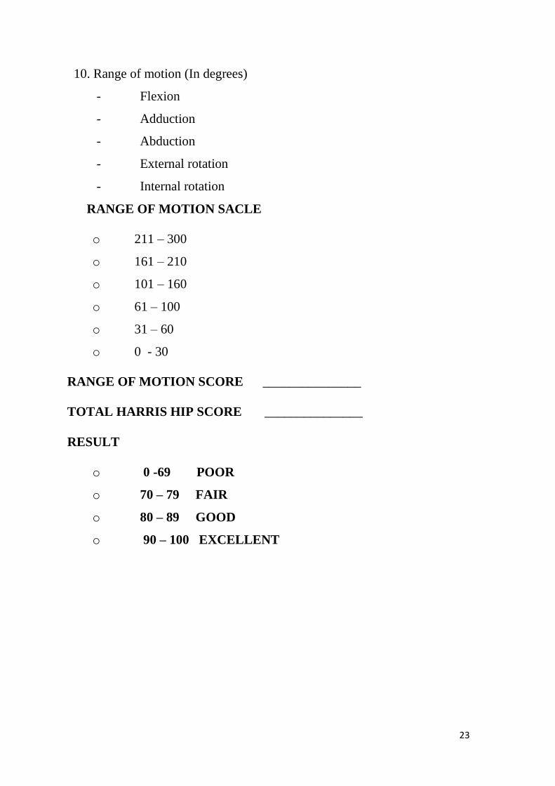
10. Range of motion (In degrees)
- Flexion
- Adduction
- Abduction
- External rotation
- Internal rotation
RANGE OF MOTION SACLE
o 211 – 300
o 161 – 210
o 101 – 160
o 61 – 100
o 31 – 60
o 0 - 30
RANGE OF MOTION SCORE _______________
TOTAL HARRIS HIP SCORE _______________
RESULT
o 0 -69 POOR
o 70 – 79 FAIR
o 80 – 89 GOOD
o 90 – 100 EXCELLENT
24
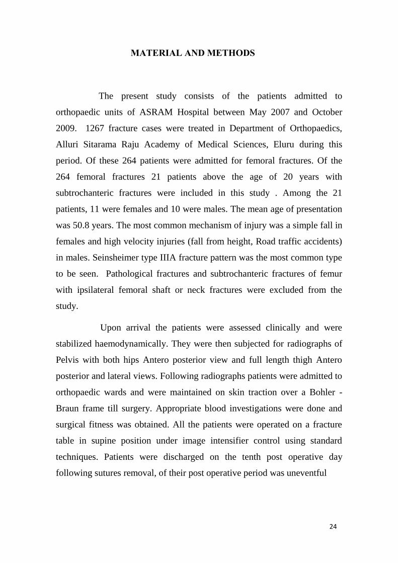
MATERIAL AND METHODS
The present study consists of the patients admitted to
orthopaedic units of ASRAM Hospital between May 2007 and October
2009. 1267 fracture cases were treated in Department of Orthopaedics,
Alluri Sitarama Raju Academy of Medical Sciences, Eluru during this
period. Of these 264 patients were admitted for femoral fractures. Of the
264 femoral fractures 21 patients above the age of 20 years with
subtrochanteric fractures were included in this study . Among the 21
patients, 11 were females and 10 were males. The mean age of presentation
was 50.8 years. The most common mechanism of injury was a simple fall in
females and high velocity injuries (fall from height, Road traffic accidents)
in males. Seinsheimer type IIIA fracture pattern was the most common type
to be seen. Pathological fractures and subtrochanteric fractures of femur
with ipsilateral femoral shaft or neck fractures were excluded from the
study.
Upon arrival the patients were assessed clinically and were
stabilized haemodynamically. They were then subjected for radiographs of
Pelvis with both hips Antero posterior view and full length thigh Antero
posterior and lateral views. Following radiographs patients were admitted to
orthopaedic wards and were maintained on skin traction over a Bohler -
Braun frame till surgery. Appropriate blood investigations were done and
surgical fitness was obtained. All the patients were operated on a fracture
table in supine position under image intensifier control using standard
techniques. Patients were discharged on the tenth post operative day
following sutures removal, of their post operative period was uneventful

25
Patients were assessed clinically and radiologically on the 2nd
post
operative day, at 6 weeks, 3 months and then between 6 months to 1 year
depending upon the fracture union. These findings are documented
according to a protocol that was developed. Healing was judged by both
clinical (pain & motion at fracture site and radiological (bridging callus
filling the fracture site or trabeculations across the fracture site) criteria and
functional outcome was reviewed according to the Harris Hip score
(modified)33
.

26
CASE STUDY PROFORMA
1. General Data
- Name -
- Age - Sex –
- Occupation -
- Address -
- IP No. -
2. Chronological Data
- Date of injury -
- Date of Admission -
- Date of Surgery -
- Date of Discharge -
3. Mode of Injury
- RTA Fall Others
- Details of Injury -
4. Pre Existing systemic Illness -
5. Examination –
- Side – Unilateral – Right Left Bilateral
- Type of Injury – Open Closed
- Distal Neurovascular status
- Associated Injuries
6. Radiographs –
- Seinsheimer type –
- Associated Osteoporosis – Present Absent
7. Management
Primary Management
- Traction – Skin Skeletal
- If open - Debridement -

27
Definitive Management
- Procedure - Closed Open
- Details of implant –
Nail - Length - Diameter -
Hip screw - Position - length -
Anti rotational screw Position - length -
Distal screws – No. - Size -
- Reduction –
Post Operative Management
- Antibiotics
- Suture removal
- Physiotherapy – Quadriceps strengthening exercises
- Hip / knee Bending exercises -
- Mobilization
Non weight bearing -
Partial weight bearing -
Full weight bearing -
Postoperative Complications
Early complications –
- Infection – Superficial- Deep -
- Wound gaping -
- Epidermal necrosis -
- Seroma -
- Haematoma -
- Decubitus ulcer -

28
Late complications -
- Cutting out of screws -
- Z‘ effect of screws -
- Reverse Z effect of screws -
- Varus collapse -
- Nail breakage -
- Diaphyseal fracture -
- Limb length discrepancy -
- Hip stiffness -
- Delayed union -
- Non union -
Secondary treatment if any
- Debridement
- Bone grafting
- Revision surgery
FOLLOW UP
1st FOLLOW UP
2nd
FOLLOW UP
3rd
FOLLOW UP
ASSESSMENT AT FINAL FOLLOW UP
HARRIS HIP SCORE - __________
RESULT - __________
29
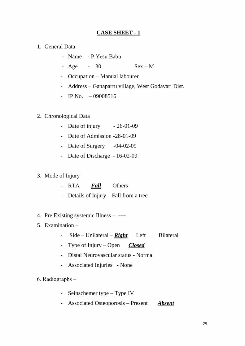
CASE SHEET - 1
1. General Data
- Name - P.Yesu Babu
- Age - 30 Sex – M
- Occupation – Manual labourer
- Address – Ganaparru village, West Godavari Dist.
- IP No. – 09008516
2. Chronological Data
- Date of injury - 26-01-09
- Date of Admission -28-01-09
- Date of Surgery -04-02-09
- Date of Discharge - 16-02-09
3. Mode of Injury
- RTA Fall Others
- Details of Injury – Fall from a tree
4. Pre Existing systemic Illness – ----
5. Examination –
- Side – Unilateral – Right Left Bilateral
- Type of Injury – Open Closed
- Distal Neurovascular status - Normal
- Associated Injuries - None
6. Radiographs –
- Seinschemer type – Type IV
- Associated Osteoporosis – Present Absent

30
7. Management
Primary Management
- Traction – Skin Skeletal
Definitive Management – Proximal Femoral Nail – AO type
- Procedure - Closed Open
- Details of implant –
Nail - Length – 25cm Diameter – 10mm
Hip screw - Position – Inf/cent length –90mm
Anti rotational screw Position – Sup/Cent, length – 85mm
Distal screws –No. - 2 Size – 36mm/34mm
- Reduction – Good
Post Operative Management
Post operatively patient was on intravenous antibiotics for 3 days.
Check x ray was done 48 hours after the surgery and it was found to be
satisfactory. Quadriceps strengthening exercises, hip and knee bending
exercises were taught on 2nd
post operative day. Suture removal was done on
10th post operative day and the patient was discharged the following day
with advice, not to weight bear on the right leg
FOLLOW UP
1st Follow up
Patient turned up for the first time on 30/03/09. He complained
of mild pain in the right thigh and on examination minimal tenderness was
noted in the proximal one third of thigh. Patient had 0-110 degrees of
flexion in the hip and 0-120 degrees of flexion in the knee.

31
No limb length discrepancy was noted. Radiograph of the right hip with
femur showed that the union was in progress. Patient was advised to
continue hip and knee mobilization exercises in bed with non weight bearing
on the right leg.
2nd
Follow up
Patient attended the Ortho OPD on 20-05-09 for the second
time. He had no pain at the fracture site and also full range of movements in
the right hip and knee. Check x ray showed that union is satisfactory and the
position of the implant was good. Patient was advised to weight bear on the
right lower limb.
3rd
Follow up
Nearly 9 months after the surgery patient turned up for the last
time. Patient had no pain at the fractures site and is walking comfortably for
long distances. He is also squatting and sitting cross legged with ease. He
has near full range of movements of the hip and knee. Check x ray showed
that fracture has united well.
HARRIS HIP SCALE(MODIFIED) SCORE – 100
RESULT - EXCELLENT

32
CASE SHEET - 2
6. General Data
- Name - B.Srinivas
- Age - 25 Sex – M
- Occupation – Manual labourer
- Address – Eluru
- IP No. – 09008153
7. Chronological Data
- Date of injury - 21-03-09
- Date of Admission -23-03-09
- Date of Surgery -28-03-09
- Date of Discharge - 09-04-09
8. Mode of Injury
- RTA Fall Others
- Details of Injury – Motor cycle passenger hit by an auto
9. Pre Existing systemic Illness – Nil
10. Examination –
- Side – Unilateral – Right Left Bilateral
- Type of Injury – Open Closed
- Distal Neurovascular status - Normal
- Associated Injuries - None
6. Radiographs –
- Seinschemer type – Type V
- Associated Osteoporosis – Present Absent

33
7. Management
Primary Management
- Traction – Skin Skeletal
Definitive Management – Long Proximal femoral nail – AO type
- Procedure - Closed Open
- Details of implant –
Nail - Length – 36cm Diameter – 9mm
Hip screw - Position – Inf/Post length –85 mm
Anti rotational screw Position – Sup/Post, length -85mm
Distal screws – No. – 1(Dynamic slot) Size – 40 mm
- Reduction – Poor – Proximal fragment is flexed
Post Operative Management
Post operatively patient was on intravenous antibiotics for 2 days.
Check x ray was done 48 hours after the surgery and it was satisfactory.
Patient was in the hospital for 10 days and was discharged following suture
removal. His post operative period was uneventful.
Follow up
1st Follow up
Patient came for his follow up for the first time on 25/5/09. He
was pain free and on examination his operative wound was healthy and he
had 0-110 degrees of flexion in the hip and 0-120 degrees of flexion in the
knee. Limb length discrepancy of 1 cm was seen. As the radiograph showed
no callus formation patient was advised to continue non weight bearing on
the right leg.
34
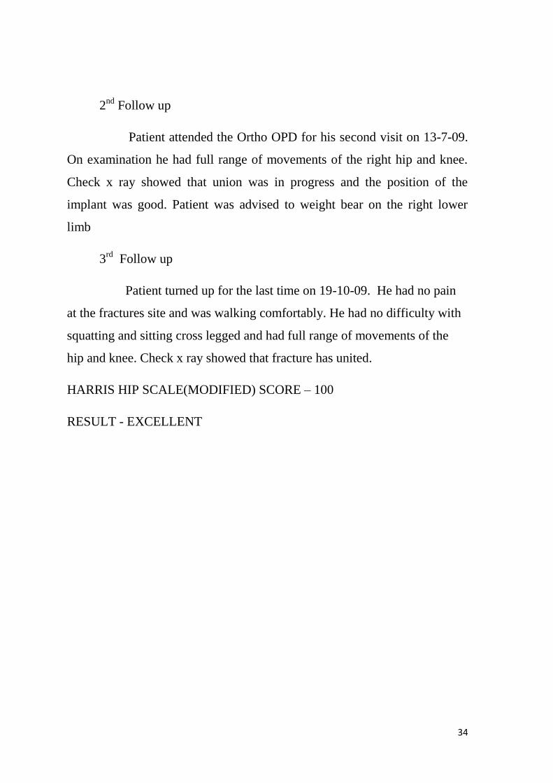
2nd
Follow up
Patient attended the Ortho OPD for his second visit on 13-7-09.
On examination he had full range of movements of the right hip and knee.
Check x ray showed that union was in progress and the position of the
implant was good. Patient was advised to weight bear on the right lower
limb
3rd
Follow up
Patient turned up for the last time on 19-10-09. He had no pain
at the fractures site and was walking comfortably. He had no difficulty with
squatting and sitting cross legged and had full range of movements of the
hip and knee. Check x ray showed that fracture has united.
HARRIS HIP SCALE(MODIFIED) SCORE – 100
RESULT - EXCELLENT
35
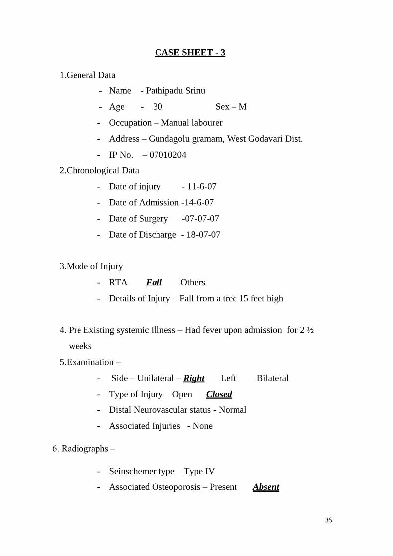
CASE SHEET - 3
1.General Data
- Name - Pathipadu Srinu
- Age - 30 Sex – M
- Occupation – Manual labourer
- Address – Gundagolu gramam, West Godavari Dist.
- IP No. – 07010204
2.Chronological Data
- Date of injury - 11-6-07
- Date of Admission -14-6-07
- Date of Surgery -07-07-07
- Date of Discharge - 18-07-07
3.Mode of Injury
- RTA Fall Others
- Details of Injury – Fall from a tree 15 feet high
4. Pre Existing systemic Illness – Had fever upon admission for 2 ½
weeks
5.Examination –
- Side – Unilateral – Right Left Bilateral
- Type of Injury – Open Closed
- Distal Neurovascular status - Normal
- Associated Injuries - None
6. Radiographs –
- Seinschemer type – Type IV
- Associated Osteoporosis – Present Absent
36
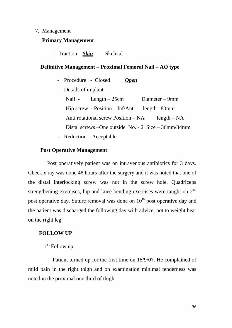
7. Management
Primary Management
- Traction – Skin Skeletal
Definitive Management – Proximal Femoral Nail – AO type
- Procedure - Closed Open
- Details of implant –
Nail - Length – 25cm Diameter – 9mm
Hip screw - Position – Inf/Ant length –80mm
Anti rotational screw Position – NA length – NA
Distal screws –One outside No. - 2 Size – 36mm/34mm
- Reduction – Acceptable
Post Operative Management
Post operatively patient was on intravenous antibiotics for 3 days.
Check x ray was done 48 hours after the surgery and it was noted that one of
the distal interlocking screw was not in the screw hole. Quadriceps
strengthening exercises, hip and knee bending exercises were taught on 2nd
post operative day. Suture removal was done on 10th post operative day and
the patient was discharged the following day with advice, not to weight bear
on the right leg
FOLLOW UP
1st Follow up
Patient turned up for the first time on 18/9/07. He complained of
mild pain in the right thigh and on examination minimal tenderness was
noted in the proximal one third of thigh.

37
Patient had 0-90 degrees of flexion in the hip and 0-100 degrees of flexion
in the knee. Shortening of 2 cms was noted in the right lower limb.
Radiograph of the right hip with femur showed no callus formation at the
fracture site. Patient was advised to continue hip and knee mobilization
exercises in bed with non weight bearing on the right leg.
2nd
Follow up
Patient attended the Ortho OPD on 26-10-07 for the second
time. He had no pain at the fracture site and also full range of movements
were noted in the right hip and knee. Check x ray showed that union was in
progress and the position of the implant was good. Patient was advised to
weight bear on the right lower limb and shoe raise of 2 cms was also given.
3rd
Follow up
Patient turned up in our department for the third time on 30-11-
07. Check x ray done showed that the fracture is uniting and the patient was
advised to weight bearing.
4th
Follow up
Nearly 1 year after the surgery patient turned up for the last time.
Patient had no pain at the fractures site and is walking comfortably for long
distances. He is also squatting and sitting cross legged with ease. He has
near full range of movements of the hip and knee. Check x ray showed that
fracture has united.
HARRIS HIP SCALE(MODIFIED) SCORE – 97
RESULT - EXCELLENT

38
CASE SHEET - 4
11. General Data
- Name - M.Rama Mani
- Age - 40 Sex – F
- Occupation –House wife
- Address – Subba Raju peta, Nidadavolu
- IP No. – 07021905
12. Chronological Data
- Date of injury - 04-07-07
- Date of Admission -07-07-07
- Date of Surgery -12-07-07
- Date of Discharge - 23-07-07
13. Mode of Injury
- RTA Fall Others
- Details of Injury – Fall from a table 7 feet high
14. Pre Existing systemic Illness – Nil
15. Examination –
- Side – Unilateral – Right Left Bilateral
- Type of Injury – Open Closed
- Distal Neurovascular status - Normal
- Associated Injuries - None
6. Radiographs –
- Seinschemer type – Type III A
- Associated Osteoporosis – Present Absent

39
7. Management
Primary Management
- Traction – Skin Skeletal
- If open - Debridement - None
Definitive Management - Proximal femoral nail – AO type
- Procedure - Closed Open
- Details of implant –
Nail - Length – 25cm Diameter – 10mm
Hip screw - Position – Inf/central length –90 mm
Anti rotational screw Position – Sup/Cent, length -80mm
Distal screws – No. – 1(Dynamic slot) Size – 32 mm
- Reduction – Good
Post Operative Management
Post operatively patient developed pyrexia on the 2nd
post op day.
Intravenous antibiotics were continued for 4 days and fever eventually
subsided. Check x ray was done 48 hours after the surgery and it was
satisfactory. Active exercises of the hip and knee were started on the 5th
post
operative day. Patient was discharged on 10th post operative day with
advice of strict non weight bearing on the left leg
FOLLOW UP
1st Follow up
Patient came for her follow up for the first time on 11/10/08. She started
weight bearing with out medical advice as her pain started improving.

40
On examination her operative wound was healthy and she had 0-130 degrees
of flexion in the hip and 0-120 degrees of flexion in the knee. No limb
length discrepancy was noted. Check x ray showed that fracture has united.
2nd
Follow up
Patient attended the Ortho OPD for the second visit on 26-6-
08.She was pain free and she got back to her house hold duties. She was able
to squat and sit cross legged comfortably. She was walking long distances
without any aid and was climbing stairs comfortably. On examination she
had full range of movements of the left hip and knee. Check x ray showed
that the fracture has united well and the position of the implant was also
good.
HARRIS HIP SCALE(MODIFIED) SCORE – 100
RESULT - EXCELLENT
41
CASE SHEET - 5
1.General Data
- Name - Saidu Venkata Rao
- Age - 35 Sex – M
- Occupation – Electrician
- Address – Bimadolu mandalam, West Godavari Dist.
- IP No. – 08031476
2. Chronological Data
- Date of injury - 15-10-08
- Date of Admission - 15-10-08
- Date of Surgery - 21-10-08
- Date of Discharge - 01-11-08
3.Mode of Injury
- RTA Fall Others
- Details of Injury – Fall from a electric pole
4.Pre Existing systemic Illness – Nil
5.Examination –
- Side – Unilateral – Right Left Bilateral
- Type of Injury – Open Closed
- Distal Neurovascular status - Normal
- Associated Injuries - None
6.Radiographs –
- Seinschemer type – Type III B
- Associated Osteoporosis – Present Absent

42
7.Management
Primary Management
- Traction – Skin Skeletal
Definitive Management – Proximal Femoral Nail – AO type
- Procedure - Closed Open
- Details of implant –
Nail - Length – 25cm Diameter – 10mm
Hip screw - Position – Cent/Post length –95mm
Anti rotational screw Position – Sup/Post, length – 85mm
Distal screws –One outside No. - 2 Size – 34mm/34mm
- Reduction – Acceptable
Post Operative Management
Post operatively patient was on intravenous antibiotics for 2 days.
Check x ray was done 48 hours after the surgery and it was found
satisfactory. Quadriceps strengthening exercises, hip and knee bending
exercises were taught on 2nd
post operative day. Suture removal was done on
10th post operative day and the patient was discharged the same day with
advice, not to weight bear on the right leg
Follow up
1st Follow up
Patient turned up for the first time on 15/12/08. He complained
of moderate pain in his right thigh and on examination minimal tenderness
was noted in the proximal one third of thigh. Patient had 0-100 degrees of
flexion in the hip and 0-110 degrees of flexion in the knee.
43
No limb length discrepancy was noted. Radiograph of the right hip with
femur showed no callus formation at the fracture site. Patient was advised to
continue hip and knee mobilization exercises in bed with non weight bearing
on the right leg.
2nd
Follow up
Patient attended the Ortho OPD for his second visit on 29-01-08
for the second time. He had significant improvement in pain at the fracture
site> He had full range of movements of the right hip and knee. Check x ray
showed that the fracture has united but back out of the Anti rotational hip
screw was noted. Patient was advised to weight bear on the right lower limb.
3rd
Follow up
Patient turned up for the last time on 24-10-09. He was
absolutely pain free and he got back to his job. On examination he had full
range of movements of the hip and knee. He was squatting and sitting cross
legged comfortably. Check x ray done showed that the fracture has united
well.
HARRIS HIP SCALE(MODIFIED) SCORE – 100
RESULT - EXCELLENT
44
CASE SHEET - 6
1.General Data
- Name - B.Vijay
- Age - 24 Sex – M
- Occupation – Shop keeper
- Address – Eluru, West Godavari Dist.
- IP No. – 07021704
2.Chronological Data
- Date of injury - 06-10-07
- Date of Admission -07-10-07
- Date of Surgery -12-10-07
- Date of Discharge - 25-10-07
3.Mode of Injury
- RTA Fall Others
- Details of Injury – Driver of a motor bike, hit by a car
4.Pre Existing systemic Illness – Nil
5.Examination –
- Side – Unilateral – Right Left Bilateral
- Type of Injury – Open Closed
- Distal Neurovascular status - Normal
- Associated Injuries - None
6.Radiographs –
- Seinschemer type – Type III A
- Associated Osteoporosis – Present Absent

45
7.Management
Primary Management
- Traction – Skin Skeletal
Definitive Management
- Procedure - Closed Open
- Details of implant –
Nail - Length – 25cm Diameter – 10mm
Hip screw - Position – Inf/Post length –95mm
Anti rotational screw Position – Sup/Post, length –90 mm
Distal screws – No. - 2 Size – 30mm/34mm
- Reduction – Good
Post Operative Management
Post operatively patient was on intravenous antibiotics for 2
days. Patient was taught quadriceps and hip & knee bending exercises on 1st
post operative day. Check x ray was done 48 hours after the surgery and it
was satisfactory. Patient was discharged on 10th
post operative day following
suture removal. His post operative period was uneventful.
FOLLOW UP
1st Follow up
Patient attended the OPD for the first time on 03/12/07. He had
no pain at the fracture site and on examination his operative wound was
healthy and he had full range of movements of the hip and knee. No limb
length discrepancy was seen and the radiograph that were taken showed that
the fracture union was in progress and the patient was advised partial weight
bearing.
46
2nd
Follow up
Patient came back for his check up on 07-01-08. He was
mobilizing full weight bearing without and had full range of movements of
the left hip and knee. Check x ray showed that fracture has united well.
There was no limb length discrepancy as well.
3rd
Follow up
Patient turned up for the last time on 11-09-08. He was walking
comfortably and he got back to his work as well. He had no difficulty with
squatting and sitting cross legged and had full range of movements of the
hip and knee. Check x ray showed that fracture has united well.
HARRIS HIP SCALE(MODIFIED) SCORE – 100
RESULT - EXCELLENT
47
CASE SHEET - 7
1.General Data
- Name - M.Rama Krishna
- Age - 42 Sex – M
- Occupation – Carpenter
- Address – Bimadolu, West Godavari District
- IP No. – 09014853
2.Chronological Data
- Date of injury - 15-01-09
- Date of Admission - 16-01-09
- Date of Surgery - 19-01-09
- Date of Discharge - 30-01-09
3.Mode of Injury
- RTA Fall Others
- Details of Injury – Passenger in a auto. Auto got toppled
4.Pre Existing systemic Illness – Nil
5.Examination –
- Side – Unilateral – Right Left Bilateral
- Type of Injury – Open Closed
- Distal Neurovascular status - Normal
- Associated Injuries - Intra articular fracture of distal end
radius
6. Radiographs –
- Seinschemer type – Type II A
- Associated Osteoporosis – Present Absent
48
7.Management
Primary Management
- Traction – Skin Skeletal
Definitive Management – Short Proximal femoral nail – AO type
- Procedure - Closed Open
- Details of implant –
Nail - Length – 25cm Diameter – 9mm
Hip screw - Position – Inf/Post length –100 mm
Anti rotational screw Position – Not applied
Distal screws – No. – 2, Size – 34mm/32 mm
- Reduction – Good
Post Operative Management
Post operatively patient was on intravenous antibiotics for 2 days.
Quadriceps strengthening exercises and hip & knee bending exercises were
started on 1st post operative day. Check x ray was done on the 2
nd post
operative day and it was found to be satisfactory. Patient‘s post operative
period was uneventful and he was discharged on the 10th post operative day
following suture removal.
FOLLOW UP
1st Follow up
Patient attended the OPD for the first time after surgery on
13/03/09. He was pain free and on examination his operative wound was
healthy and he had near full range of movements of the hip and knee. There
was no limb length discrepancy. Radiograph showed satisfactory progress of
the union and the patient was advised to partially weight bear on his left leg.
49
2nd
Follow up
Patient attended the Ortho OPD for his second visit only after 7
months on 21-08-09. He had no pain and even got back to his job. On
examination he had full range of movements of the left hip and knee. There
was no limb length discrepancy. Check x ray showed that the fracture is well
united and the position of the implant was good.
HARRIS HIP SCALE(MODIFIED) SCORE – 100
RESULT - EXCELLENT
50
CASE SHEET - 8
1.General Data
- Name - T.Agnesamma
- Age - 65 Sex – F
- Occupation – House wife
- Address – Arjavari gudem, Bimadolu
- IP No. – 08012217
2.Chronological Data
- Date of injury - 14-01-08
- Date of Admission – 17-01-09
- Date of Surgery - 02-02-08
- Date of Discharge - 18-02-08
3.Mode of Injury
- RTA Fall Others
- Details of Injury – Fall at home
4.Pre Existing systemic Illness – Hypertensive
5.Examination –
- Side – Unilateral – Right Left Bilateral
- Type of Injury – Open Closed
- Distal Neurovascular status - Normal
- Associated Injuries - Nil
6.Radiographs –
- Seinschemer type – Type II B
- Associated Osteoporosis – Present Absent
51
7.Management
Primary Management
- Traction – Skin Skeletal
- If open - Debridement - Nil
Definitive Management
- Procedure - Closed Open
- Details of implant –
Nail - Length – 25mm Diameter – 12mm
Hip screw - Position –Cent/ Cent length – 85mm
Anti rotational screw Position - NA length –
Distal screws - No. - 2 Size – 36mm & 34mm
- Reduction – Good
Post Operative Management
Post operatively in the post operative ward patient developed angina
and was ventilated shifted to intensive care unit for 5 days. Following that
she recovered well from the 4th
post operative day. She was on intravenous
antibiotics for 6 days. Check x ray was done on the 4th post operative day
and it was satisfactory. She started doing Quadriceps strengthening exercises
and hip & knee bending exercises. Patient was in the hospital for 2 weeks
and was discharged on 16th
post operative day.
Follow up
1st Follow up
Patient came for her follow up for the first time on 31/3/08. She
had moderate pain and on examination her operative wound was healthy.

52
She had 0-100 degrees of flexion in the hip and 0-100 degrees of flexion in
the knee. No limb length discrepancy was noted. Check X ray showed
satisfactory progress of fracture union and patient was advised to partially
weight bearing on the right leg.
2nd
Follow up
Patient attended the Ortho OPD for his second visit on 19-05-
08.her pain at the operative site improved but still had mild pain. On
examination she had good range of movements of the right hip and knee.
Check x ray showed that has united and the position of the implant was
good. Patient was advised full weight bearing on the right lower limb
3rd
Follow up
Patient turned up for the last time on 02-03-09. She had slight
pain at the fracture site on and off and was walking with the help of a stick.
She had difficulty with squatting and sitting cross legged and had good
range of movements of the hip and knee. Check x ray showed that fracture
has united well.
HARRIS HIP SCALE(MODIFIED) SCORE – 75
RESULT - FAIR

53
CASE SHEET - 9
1.General Data
- Name - V.Satyavathi
- Age - 70 Sex – F
- Occupation – House wife
- Address – Kovalli, Denduluru mandalam.
- IP No. –07024163
2.Chronological Data
- Date of injury - 02-12-08
- Date of Admission - 03-12-08
- Date of Surgery - 15-12-08
- Date of Discharge - 26-12-08
3.Mode of Injury
- RTA Fall Others
- Details of Injury – Fall at home
4. Pre Existing systemic Illness – Known diabetic. Underwent open
reduction and internal fixation for Right distal end femur fracture
5. Examination –
- Side – Unilateral – Right Left Bilateral
- Type of Injury – Open Closed
- Distal Neurovascular status - No
- Associated Injuries - No
6. Radiographs –
- Seinschemer type – Type III A
- Associated Osteoporosis – Present Absent
54
7. Management
Primary Management
- Traction – Skin Skeletal
- If open - Debridement - None
Definitive Management
- Procedure - Closed Open
- Details of implant –
Nail - Length – 25cm Diameter –10mm
Hip screw - Position – Inf/Cent, length –85mm
Anti rotational screw Position – NA length –
Distal screws – No. - 1 Size – 32mm
- Reduction – Good
Post Operative Management
Post operatively patient‘s rehabilitation was poor because her
previous surgery for distal end femur fracture. She also had uncontrolled
blood sugar levels for few days. She was on intravenous antibiotics for 3
days. Check x ray was done on 2nd
post operative day and it was satisfactory.
Quadriceps strengthening exercises were started on 5th
post operative day.
Hip & knee bending exercises were delayed till the 10th
post operative day.
Good control of her blood sugar levels and satisfactory range of movements
of the hip & knee were achieved by 15th post operative day and she was
discharged the following day.
55
FOLLOW UP
1st Follow up
Patient turned up for the first time on 03/03/08. She had
moderate pain but was mobilizing non weight bearing till date. On
examination her operative wound was healthy and she had no tenderness
noted at the fracture site. She had a flexion of 90 degrees and full abduction
and adduction of the hip. But she had minimal restriction of internal
rotation. She had only 80 degrees of flexion at the knee and limb length
discrepency of 2 cms was noted. Radiograph taken showed good union of
the fracture and the patient was advised to weight bear
2nd
Follow up
Patient attended our OPD for last time on 03-11-09. She had
moderate pain at the fractures site and was walking wit the support of a cane
most of the time. She was unable to squat and sit cross legged and had 0-100
degrees of flexion at the hip. She had full abduction and adduction but some
restriction of internal rotation was noted. She had only 0-90 degrees of
flexion at the knee. Check x ray showed that fracture has united completely
and the implant is well in situ.
HARRIS HIP SCALE(MODIFIED) SCORE – 50
RESULT - POOR

56
CASE SHEET - 10
1. General Data
- Name - G.Chittamma
- Age - 62 Sex – F
- Occupation – House wife
- Address – Lingapalem, West Godavari Dist.
- IP No. – 08011681
2. Chronological Data
- Date of injury - 05-05-08
- Date of Admission -06-05-08
- Date of Surgery -13-05-08
- Date of Discharge - 24-05-08
3. Mode of Injury
- RTA Fall Others
- Details of Injury – Fall after tripping over a step.
4. Pre Existing systemic Illness – Nil
5. Examination –
- Side – Unilateral – Right Left Bilateral
- Type of Injury – Open Closed
- Distal Neurovascular status - Normal
- Associated Injuries - None
6. Radiographs –
- Seinschemer type – Type III A
- Associated Osteoporosis – Present Absent

57
7. Management
Primary Management
- Traction – Skin Skeletal
- If open - Debridement - None
Definitive Management
- Procedure - Closed Open
- Details of implant –
Nail - Length – 25cm Diameter – 10mm
Hip screw - Position – Cent/Post length –105mm
Anti rotational screw Position – Sup/Post, length –95 mm
Distal screws – No. - 1 Size – 38mm
- Reduction – Poor
Post Operative Management
Post operatively patient was on intravenous antibiotics for 2
days. Check x ray was done on the 2nd
post operative day and it was noted
that the entry point of the nail was a bit distal but the reduction was
satisfactory. Post operative period was uneventful and the patient was
discharged on the 11th post operative day with advice to continue quadriceps
strengthening exercises and hip & knee bending exercises in bed.
FOLLOW UP
1st Follow up
Patient turned up in our out patient department for the first time
on 14/07/08. She had moderate pain at the fracture site. On examination her
operative wound was healthy and he had 0-80 degrees of flexion in the hip
contd…
58
and 0-90 degrees of flexion in the knee. There was no limb length
discrepancy and Check x ray showed that fracture has union is progressing
and position of the implant was also satisfactory. Patient was advised to
continue non weight bearing on the left leg and to continue bending
exercises of the hip and knee.
2nd
Follow up
Patient attended the OPD for the 2nd
time on 28-08-08. She had
moderate at the fracture site. She was doing her hip and knee bending
exercises and on examination she had 0-110 degrees of flexion, 30 degrees
of abduction, 20 degrees of adduction, 30 degrees of external rotation and 10
degrees of internal rotation. No limb length discrepancy was noted. Check x
ray showed satisfactory union of the fracture and the patient was advised to
weight bear on the left limb
3rd
Follow up
Patient came for her final follow up 07-05-09. She was
complaining of moderate amount of pain because of which she was unable
to walk long distances. On examination her range of movements of the hip
were same as her previous visit. She had 0-100 degrees of flexion in her hip.
She had difficulty squatting and sitting cross legged but was able to manage
the stairs with support. No limb length discrepancy was noted. Check x ray
showed that the fracture has united well.
HARRIS HIP SCALE(MODIFIED) SCORE – 41
RESULT - POOR

59
CASE SHEET - 11
1. General Data
- Name - P.Kamakshamma
- Age - 63 Sex – F
- Occupation – House wife
- Address – Ponangipunta, Eluru
- IP No. – 08031305
2. Chronological Data
- Date of injury - 16-10-08
- Date of Admission – 17-10-08
- Date of Surgery - 22-10-08
- Date of Discharge - 07-11-08
3. Mode of Injury
- RTA Fall Others
- Details of Injury – Fall at home
4. Pre Existing systemic Illness – Hypertension, Anaemia
5. Examination –
- Side – Unilateral – Right Left Bilateral
- Type of Injury – Open Closed
- Distal Neurovascular status - Normal
- Associated Injuries - Nil
6. Radiographs –
- Seinschemer type – Type III A
- Associated Osteoporosis – Present Absent

60
7. Management
Primary Management
- Traction – Skin Skeletal
- If open - Debridement - Nil
Definitive Management
- Procedure - Closed Open
- Details of implant –
Nail - Length – 25mm Diameter – 9mm
Hip screw - Position –Sup/ Ant length – 85mm
Anti rotational screw Position – Sup/Ant, length – 85mm
Distal screws - No. - 1 Size – 34mm
- Reduction – Unsatisfactory – Proximal fragment abducted
Post Operative Management
Post operatively patient had localised redness and warmth at
operative site which settled with parentral antibiotics and was on
intravenous antibiotics for 3 days. She was started on quadriceps
strengthening exercises; hip and knee bending exercises were taught on 2nd
post operative day. The same day patient developed confusion due to
electrolyte imbalance and sustained a fall from bed as she was trying to get
out. On examination movements of the hip were painful and shortening of 3
cms was noted. Check x ray was done and it showed cut out of the anti
rotational hip screw. Patient refused removal of the cut out screw. Suture
removal was done on 10th post operative day and the patient was discharged
the following day with advice, not to weight bear on the right leg
61
FOLLOW UP
1st Follow up
Patient came back to the OPD for the first time on 19/12/08. She
complained of marked pain in the left hip and was on regular analgesics. On
examination minimal tenderness was noted in the proximal one third of
thigh and movements of the hip were painful and restricted. Shortening of 3
cms was noted in the left lower limb. Radiograph of the left hip with femur
showed cut of the anti rotational screw and no callus formation was seen at
the fracture site. Patient was admitted and the cut out screw was removed.
2nd
Follow up
Patient attended the Ortho OPD on 29-01-09 for the second
time. She had moderate pain at the fracture site and also her range of
movements were restricted. Check x ray showed that fracture was uniting.
Patient was advised to weight bear on the right lower limb and was
encouraged to do hip and knee bending exercises.
3rd
Follow up
Patient turned up for the last time on 02-11-09. Patient still had
moderate pain at the fractures site and is walking with the support of a
crutch. She was finding it tough to go out doors and was unable to squat and
sit cross legged. She had a flexion of 90 degrees, abduction of 30 degrees,
adduction of 20 degrees, external rotation of 20 degrees and 10 degrees of
internal rotation at her hip. Check x ray showed that fracture has united and
the implant was well in situ.
HARRIS HIP SCALE(MODIFIED) SCORE – 29
RESULT - POOR

62
CASE SHEET - 12
1.General Data
- Name - M.Bullama
- Age - 70 Sex – F
- Occupation – House wife
- Address – Unguturu mandalam , West Godavari District.
- IP No. –09003166
2. Chronological Data
- Date of injury - 07-01-09
- Date of Admission - 07-01-09
- Date of Surgery - 12-01-09
- Date of Discharge - 23-01-09
3. Mode of Injury
- RTA Fall Others
- Details of Injury – Fall at home
4. Pre Existing systemic Illness – Known Hypertensive on treatment.
5. Examination –
- Side – Unilateral – Right Left Bilateral
- Type of Injury – Open Closed
- Distal Neurovascular status - No
- Associated Injuries - No
6. Radiographs –
- Seinschemer type – Type III A
- Associated Osteoporosis – Present Absent

63
7.Management
Primary Management
- Traction – Skin Skeletal
- If open - Debridement - None
Definitive Management
- Procedure - Closed Open
- Details of implant –
Nail - Length – 25 cm Diameter –10mm
Hip screw - Position – Inf/Cent, length –85mm
Anti rotational screw Position – NA length –
Distal screws – No. - 2 Size – 32/ 34mm
- Reduction – Good
Post Operative Management
Post operatively patient‘s rehabilitation was good. She had 1 unit
of blood transfused and was on parentral antibiotics for 2 days. She also had
uncontrolled blood sugar levels for few days. Check x ray was done on 2nd
post operative day and it was satisfactory. Quadriceps strengthening
exercises were started on 3rd
post operative day. Hip & knee bending
exercises were started the following day. There was satisfactory range of
movements of the hip & knee by 10th day. Suture removal was done on 10
th
day and she was discharged the following day with advice not to weight bear
on the right leg.

64
Follow up
1st Follow up
Patient turned up for the first time on 09/03/09. She had mild
pain and was mobilizing non weight bearing till date. On examination her
operative wound was healthy and she had no tenderness noted at the fracture
site. She had a flexion of 110 degrees and full abduction and adduction of
the hip. But she had minimal restriction of internal rotation. She had 110
degrees of flexion at the knee and no limb length discrepancy was noted.
Radiograph taken showed good progress of fracture union. She was advised
to continue non weight bearing on her right leg
2nd
Follow up
Patient attended our OPD for second time on 04/05/09. She had
mild pain at the fractures site and was having good range of movements in
her hip and knee. No limb length discrepancy was noted. Check x ray
showed that fracture has united well and the implant is well in situ. She was
advised to weight bear on her right leg.
3rd
Follow up
Patient came to our OPD on 25-07-09 for the third time. She had
mild pain and was using a cane for mobilization. She had a moderate limp
and had some discomfort in climbing stairs. she had difficulty in squatting
and sitting cross legged also. On examination she had good range of
movements of the hip and knee and no limb length discrepancy was noted.
She had a Harris hip score of 65 and her functional outcome at6 months was
not satisfactory. She was asked to come for her next follow up 6 months
later.

65
4th
follow up
This time patient presented to the emergency department 06-08-
09 with history of another fall onto her right side. She had severe pain in her
right thigh. On examination deformity of the thigh was noted and crepitus
could be elicited in the upper femur. Radiographs were taken and was found
that there is a fracture in the subtrochanteric region corresponding to the
previous fractures site with the nail broken at the distal interlocking screw.
She was admitted and 1 week later she was subjected for a repeat surgery,
where the broken nail was removed and the fractures was stabilized with an
interlocking intramedullary nail.
5th
Follow up
She came for her follow up on 22/10/09 and she was
mobilizing non weight bearing. She had good range of movements in her hip
and in knee she had 0 – 100 degrees of flexion. Check x ray showed that the
union is in progress and she is due for her follow up on 28/11/09
66
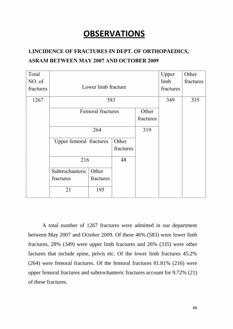
OBSERVATIONS
1.INCIDENCE OF FRACTURES IN DEPT. OF ORTHOPAEDICS,
ASRAM BETWEEN MAY 2007 AND OCTOBER 2009
A total number of 1267 fractures were admitted in our department
between May 2007 and October 2009. Of these 46% (583) were lower limb
fractures, 28% (349) were upper limb fractures and 26% (335) were other
factures that include spine, pelvis etc. Of the lower limb fractures 45.2%
(264) were femoral fractures. Of the femoral fractures 81.81% (216) were
upper femoral fractures and subtrochanteric fractures account for 9.72% (21)
of these fractures.
Total
NO. of
fractures
Lower limb fracture
Upper
limb
fractures
Other
fractures
1267
583 349 335
Femoral fractures Other
fractures
264 319
Upper femoral fractures Other
fractures
216 48
Subtrochanteric
fractures
Other
fractures
21 195
67
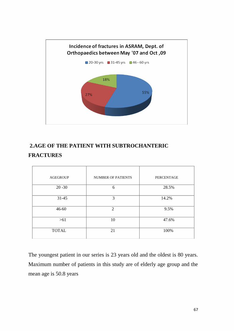
2.AGE OF THE PATIENT WITH SUBTROCHANTERIC
FRACTURES
The youngest patient in our series is 23 years old and the oldest is 80 years.
Maximum number of patients in this study are of elderly age group and the
mean age is 50.8 years
AGEGROUP
NUMBER OF PATIENTS
PERCENTAGE
20 -30 6 28.5%
31-45 3 14.2%
46-60 2 9.5%
>61 10 47.6%
TOTAL 21 100%

68
3. SEX AND TYPE OF INJURY
SEX NUMBER OFPATIENTS PERCENTAGE
Male 10 47.6%
Female 11 52.3%
Total 21 100%
In the present study, it is seen that subtrochanteric fractures are
slightly more common in females than males.

69
In the present series 80% (8) males sustained this injury because of
high velocity injury. Where as in females they are most often caused by low
velocity injury compared to their counter parts. In this study 90.9% (10)
females sustained injury because of low velocity injury.
Average age in males is 36 years and in females is 64.27 years.
This also signifies that female patients are older than male patients and so
were more predisposed to low velocity trauma.
SEX HIGH VELOCITY INJURY LOW VELOCITY INJURY /
CONVENTIONAL TRAUMA
Male 8 (80%) 2(20%)
Female 1 (9.1%) 10 (90.9%)
Total 9 12

70
4. ADMISSION – OPERATION INTERVAL
Mean
PRESENT SERIES
6.6 Days
The mean injury operation interval in the current series is 6.6 days. This
increased interval is mainly due to uncontrolled pre existing illness at the
time of presentation.
5. INCIDENCE BASED ON SEINSCHEMER’S CLASSIFICATION
Percentage
TYPE II TYPE III TYPE IV TYPE V
A B C A B
9.52% 19.04% 0 42.85% 9.52% 14.23% 4.76%
Most commonly seen fractures pattern in this study is
Seinschemer‘s type III A.

71
6. TYPE OF REDUCTION
PERCENTAGE
CLOSED OPEN
95.23% 4.76%
Intra operatively reduction of the fracture was achieved through closed
means in 95.23% (20) of cases. Open reduction was performed in only 1
patient who had high injury operation interval

72
7. RESULT OF REDUCTION
Percentage
GOOD
ACCEPTABLE
POOR
76.01% 4.76% 19.04%
Reduction was good in 76% (16) of the cases. Poor reduction was
noted in 4 patients. Three of them were of elderly age group and had poor
outcome at final follow up.
8. FRACTURE UNION
AVERAGE TIME FOR UNION
PRESENT STUDY
3.04 months

73
9. COMPLICATIONS
In the present series, 4.76% (1) of cases had superficial infection and
no deep infections were recorded. Cut of the anti rotational screw was noted
in 1 patient. Fracture of the shaft with breakage of the nail was was noted
4.76% (1) of patients.
10. ASSESSMENT AT FINAL FOLLOW UP
A. Pain
QUALITY OF PAIN NO. OF PATIENTS PERCENTAGE
None or ignores it 8 38.09%
Slight occasional 7 33.33%
Mild 3 14.28%
Moderate 2 9.52%
Totally disabled 1 4.76%
Majority of the patients (71.42%) in this study had either no pain or slight
pain which did not effect their activities. Onle one patient had severe pain.
14.28% (3) of patients had mild pain which was relieved with analgesics.
S NO. COMPLICATION PERCENTAGE
1. Superficial infection 4.76%
2. Deep infection 0
3. Cut out of screw 4.76%
4. Reverse ‗Z‘ effect of hip
screws
0
5. ‗Z‘ effect of hip screws 0
6. Shaft fracture 4.76%

74
b. Limp
NO. OF PATIENTS PERCENTAGE
None 8 38.09%
Slight 10 47.61%
Moderate 3 14.28%
Severe 0 --
In the current study majority of patients had no or slight limp that
did not effect their activities. 14.28% (3) had moderate limp which was
mainly due to shortening.
c. Walking Ability
NO. OF PATIENTS PERCENTAGE
None 10 47.6%
Cane for long walks 6 28.56
Cane most of the time 4 19.04%
Crutch 1 4.76%
Not able to walk 0 0
In our study 47.6% (10) patients did not require any support for
walking and 28.56% (6) of patients cane for long walks. Only 1 patient was
mobilizing with the help of crutch. The requirement of the cane is primarily
because of old age of the patients and associated osteoarthritis
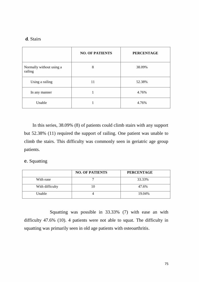
75
d. Stairs
NO. OF PATIENTS PERCENTAGE
Normally without using a
railing
8 38.09%
Using a railing 11 52.38%
In any manner 1 4.76%
Unable 1 4.76%
In this series, 38.09% (8) of patients could climb stairs with any support
but 52.38% (11) required the support of railing. One patient was unable to
climb the stairs. This difficulty was commonly seen in geriatric age group
patients.
e. Squatting
NO. OF PATIENTS PERCENTAGE
With ease 7 33.33%
With difficulty 10 47.6%
Unable 4 19.04%
Squatting was possible in 33.33% (7) with ease an with
difficulty 47.6% (10). 4 patients were not able to squat. The difficulty in
squatting was primarily seen in old age patients with osteoarthritis.

76
f. Sitting cross legged
NO. OF PATIENTS PERCENTAGE
With ease 7 33.33%
With difficulty 10 47.6%
Unable 4 19.04%
In our study, squatting was possible in 80.93% (17) of the patients, but
47.6% (10) of these patients had some difficulty while doing so. 4 elderly
patients were unable to sit cross legged and this was primarily because of
knee osteoarthritis.
g. Limb length discrepancy
NO. OF PATIENTS PERCENTAGE
2 cms or more 2 9.52%
Less than 2 cms 1 4.76%
Of the 21 patients in this series, 2 patients had shortening of more
than 2 cms which required shoe raise. 1 patient had less than 2 cms of
shortening and it did not require any treatment.
11. OVERALL RESULT BASED ON HARRIS HIP SCORE
Harris Hip score
Mean score
80.76
In the current series, the mean Harris Hip score was 80.76 and it
was ranging from 100 - 29.

77
12. Overall result based on the age group of the patient
In this series all the patients between 20 – 30 years had excellent result
irrespective of the type of fracture. Older age group patients had relatively
poor results and 50% (5) of them had poor results and another 50% (5) had
good to fair results. In this study excellent outcome was not seen in the older
age group patients.
78
DISCUSSION
Fractures of the long bones are a major social and economic problem.
Of the long bone fractures Subtrochanteric fractures of the femur have
peculiar anatomic and mechanical characteristics which poses problems in
their management. Closed intramedullary devices have a mechanical
advantage that effectively addresses these factors. The benefit of minimal
surgical exposure, more efficient load transfer through calcar femorale and
decreased tensile strain on the implant because of its shorter lever arm makes
proximal Femoral Nail a good choice of implant for subtrochanteric fractures
of the femur. Various studies have considered Proximal Femoral Nail as an
acceptable minimally invasive implant for Subtrochanteric fracture.
The incidence of subtrochanteric fracture is relatively low. In
our study 21 subtrochanteric fractures accounted for 9.7% of all proximal
femoral fractures. In other studies 7% - 34% of all femur fractures occurred
in the subtrochanteric region 36, 37.
Most of our patients were of the elderly
age group, the average age being 50.8 years. This is significantly lower
compared to that quoted by other authors in literature , BOLDIN ET AL23
73 yrs, I.B.SCHIPPER SERIES13
82.2 years. Slight female preponderance
of 52.3% was noted in our patients and it was also reported by BOLDIN ET
AL23
(70%) and I.B.SCHIPPER34
( 82%). 61.9% of the subtrochanteric
fractures involved the right femur in this series as compared to 52% in
I.B.SCHIPPER series34
and 38.09% involved the left as compared to 48%
in I.B.SCHIPPER series34
. In 57.14% of patients fracture is a result of
trivial fall and majority of them are elderly age group patients especially
females. High velocity injuries like road traffic accidents and fall from
heights accounted for 42.85% of these fractures and most of them were
males.

79
In W.M.GADEGONE’s series35
75% of the fractures were due to
domestic falls and this can be explained by the higher mean age group of the
patients in this study. Vehicular accidents resulted in the remaining 25% of
subtrochanteric fractures in their study. Fractures were classified according
to Seinsheimer‘s classification and type III A fracture pattern constituted
the highest percentage 42.85% (9) of all fracture patterns.
SEINSHEIMER3 in his original study also noted high incidence of type III
A fracture pattern (38.29%) than other fracture patterns. Admission –
operation interval in our study varied from 3 – 26 days. Mean interval was
high in our series. It was 6.6 days which is more when compared to
I.B.SCHIPPER’s series34
where it was 2 days. Most of the patients with
delayed injury – operation interval had pre existing uncontrolled medical
problems. These medical co morbidities especially in elderly age group
patients with associated degenerative joint disease of the knee significantly
affected their final functional outcome.
The mean duration of hospital stay in our series was 19.09 days
which is at par with I.B.SCHIPPER’s series34
19 days). Intra operatively
fracture reduction was achieved by closed means in 94% (20) of patients and
1 patient with delayed injury – operation interval required open reduction.
The result of the reduction was considered good in 76% (16) of the patients
and acceptable in 4.76% (1) of patients. Poor reduction was noted in 19.04%
(4) of patients and it was associated with poor outcome. In
I.B.SCHIPPER’s series34
reduction was good to acceptable in 96.2% of
their patients and poor reduction was seen only in 2.9% of their patients. Post
operatively 1 patient in this study had superficial infection(4.76%) and this
settled with parentral antibiotics. I.B.Schipper noted 4.1% superficial
infections and 2.5% deep infections.
80
We did not encounter any deep infections in our series. Cut out of hip screw
was noted in 1 patient (4.76%) and it was following a fall in the post
operative period. 6% of patients in I.B.SCHIPPER’s series had this
problem. 1 patient had fracture shaft of femur with breakage of the nail noted
at the distal locking screw hole that was not locked. I.B.Schipper noted this
problem in 2% of his patients . The average time for radiological union was
3 months in the present study whereas in I.B.Schipper series it was
approximately 4 months. 71.42% (15) of the patients in our series had no or
slight pain that did not effect their activities. Only 1 patient who had cut out
of the anti rotational screw had severe pain that restricted her activity
significantly. 85.7% (18) of these patients had no or slight limp. 47.6% (10)
of the patients mobilized without any walking aids. Cane was required for
long walks in 28.56% (6) of patients and most of the time in 19.04% (4) of
patients. Only one patient required crutch for mobilization. Difficulty
squatting and sitting cross legged noted in 47.6% (10) of patients. Most of
these patients were of geriatric age group who had associated degenerative
disease of the knee. Limb length discrepancy was noted in 3 patients of
which 2 of them had shortening of more than 2 cms. They were patients with
Seinsheimer‘s type IV and type V fracture patterns. Final outcome was
excellent to good in 61.9% (13) of patients. It was fair in 14.28% (3) of
patients and poor in 23.8% (5) of patients. Younger age group patients
irrespective of their fracture pattern had excellent outcome in our series.
Most of the poor results were seen in the elderly age group patients with
associated Osteoarthritis of the knee. The mean Harris Hip score was in our
series was 80.76% which was higher than I.B.Schipper series34
where the
mean was 77.6.

81
SUMMARY AND CONCLUSION
The incidence of Subtrochanteric fractures of the femur is on the raise
because of fast and high speed automobiles and modern lifestyles and
increased life expectancy of the elderly age group patients. The deforming
forces, high mechanical stresses and morbidity of the fractures in this region
have always challenged the ingenuity and skills of the orthopaedic surgeon.
Various devices have evolved in an attempt to effectively neutralize these
forces. Closed insertion technique, shorter lever arm decreasing the tensile
strain on the implant and increased purchase of the proximal fragment are
the added advantages of Cephalomedullary nails over other fixation devices
in subtrochanteric fractures. This study was conducted to analyze the results
of Subtrochanteric fractures treated with this Proximal Femoral Nail – AO
Type
In our series of 21 cases of Subtrochanteric fractures treated
with Proximal Femoral Nail, 14 patients had Excellent to good outcome at
their final follow up. Poor outcome was seen in 4 patients. 3 of these 4
patients had poor reduction intraoperatively. All these patients also belonged
to geriatric age group who had associated degenerative joint disease of the
knee affecting the final functional outcome. The mean Harris Hip score at
their final follow up was 80.76 which is comparable to international
publications in the literature.
From this sample study, we conclude that Proximal Femoral
Nail is a good implant for the treatment of Subtrochanteric fractures of
femur provided optimal reduction of the fracture and good positioning of the
nail and screws are achieved.
82
BIBLIOGRAPHY
1.Hibbs, R.A : The Management of the Tendency of the Upper Fragment to
Tilt Forwards in Fractures of the upper third of femur. New York,
Med.J.,75: 177-179, 1902
2. Sarmiento, Augusto: Functional Bracing of Tibial and Femoral shaft
Fractures. Clin Orthop. , 82: 2 – 13, 1972
3. Seinsheimer, F ., III: Subtrochanteric fractures of the femur. J.Bone Jt.
Surg., 60-A: 300-306, 1978
4. JC DeLee, TO Clanton and CA Rockwood : Closed treatment of
subtrochanteric fractures of the femur in a modified cast-brace ., J Bone
Joint Surg Am. 1981 :63 : 773-779
5. . WADDELL, J. P.: Subtrochanteric Fractures of the Femur: A Review
of 130 Patients. J. Trauma, 19: 582-592, 1979.
6. Delbet P. Les fractures du femur. Paris librarie alcan., 1920
7. Cleveland, M.Bosworth, D.M. and Thompson, F.R. Intertrochanteric
fractures of the femur: A survey of treatment in traction and by internal
fixation J. Bone & Joint Surg.,29: 1049 – 1067. ., , 1947
8. Evans, E. M. : The treatment of trochanteric fractures of the femur. J.
Bone Jt. Surg., 31-B: 190-203, 1949
9. Baumgaertal J. Operative treatment of experimental comminuted
subrtrochanteric fractures
10. Boyd, H. B and Griffin, L.L. classification and treatment of trochanteric
fractures.. Arch. Surgery., 1949; 88; 58
11. Watson HK, Campbell RD, Wade PA : Classification, treatment and
complications of Subtrochanteric fracture. J Trauma 4 : 457 – 480, 1964
12. Aronoff, P. M.; Davis, P. M., JR.; and Wickstrom,J. K.:Intramedullary
Nail Fixation as Treatment of Subtrochanteric Fractures of the Femur. J.
Trauma, 11: 637-650, 1971.
13. Distefano, V.J.Nixon.; and Klein, K.S: Stable fixation of the difficult
Subtrochanteric fracture. J.Trauma, 12: 1066 – 1070, 1972
83
14. Cech, 0., and Sosna, A.: Principles of the Surgical Treatment of
Subtrochanteric Fractures. Orthop. Clin. North America, 5: 65 1-662, 1974
15. Fielding, J. W., and Magiliato, H. J.: Subtrochanteric Fractures. Surg.,
Gynec. and Obstet., 122: 555-560, 1966
16. Hanson, G. W. , and Tullos, H. S. : Subtrochanteric Fractures of the
Femur Treated with Nail-Plate Devices. A Retrospective Study.
Clin.Orthop., 131: 191-194, 1978.
17. Zickel R. E. : An Intramedullary Fixation Device for the Proximal Part
of the Femur. Nine Years‘ Experience. J. Bone and Joint Surg. , 58-A:866-
872, Sept. 1976.
18. Halder S C.: The Gamma Nail for peritrochanteric fractures.; JBJS (Br)
1992. 74: 340 – 344
19. Huber S M. Heining. : Pertrochanteric fracture fixation. Photoelastic
stress measurement company DHS, Gamma nail & PFN. ; JBJS (Br) 1997;
79 B: 166
20. Simmermacher R K, Bosch A M.; The AO Proximal femoral nail - A
new device for unstable proximal femoral fractures. ; Injury 1999; 30 : 327 –
332
21. A. Herrera, L.J. Domingo, A.Calvo, A.Martinez, J.Cuenca. : A
comparative study of trochanteric fractures treated with the Gamma nail or
the proximal femoral nail.; International Orthopaedics 2002; 26: 365 – 369
22. Sudan M, Sadowski C etal. : Peritrochanteric fractures. Is there an
advantage of intramedullary nail? ; J Orthop Trauma 2002; 16 : 386 – 393
23. Christian Boldin, Franz J Seibert, Florian Fankhauser.: etal.: "The
proximal femoral nail (PFN)—-a minimal invasive treatment of unstable
proximal femoral fractures . Acta Orthop Scand 2003; 74(1): 53 - 58.
24. Daniel F.A. Menezes, Axel Gamulin etal. : Is the Proximal femoral nail
a suitable implant of all the trochanteric fractures? : CORR; 2005; 439 : 221
– 227
25. Woo-kie Min, Shin – Yoon etal. :Proximal femoral nail for the treatment
of Reverse obliquity intertrochanteric fractures compared with Gamma nail.
: J of Trauma ; 2007; 73 : 1054 – 1060

84
26. MSG Ballal, N Emms, G Thomas. : Proximal femoral nail failures in
extra capsular fractures of the hip. : J of Orthopaedic Surgery 2008; 16(2) :
146 -9
27. Si Yong Park, Kyu Hyun Yang etal. : The treatment of Reverse obliquity
intertrochanteric fractures with the intramedullary nail. : J of trauma; 2008;
65 : 852 – 857.
28. Currey J. : The biomechanical properties of bone.: Clin Orthop 1970; 73:
203 -231
29. Schipper I B etal. : Biomechanical evaluation of Proximal femoral nail. :
CORR 2002; 405: 277 – 286
30. Tencer, A. F.; JOHNSON, K. D.; Johnston, D. W. C. ; and Gill, K.: A
Biomechanical Comparison of Various Methods of Stabilization of
Subtrochanteric Fractures of the Femur. J. Orthop. Res. , 2: 297-305, 1984.
31. Paul R.T. Kuzyk, Joel Lobo etal. : Biomechanical evaluation of
extramedullary versus intramedullary fixation for Reverse obliquity
Intertrochanteric fractures. ; J of Orthop Trauma 2009 ; 23; 1. : 31 - 38
32. Fractures in adults – Rockwood and Greens; 6th
Edition
33. Harris W H (modified) JBJS 1969; 51:1
34. Schipper I B etal Treatment of Unstable trochanteric fractures :JBJS
2004; 86 B : 86 – 94
35 . Bergman GD, Winquist RA, Mayo KA etal. : Subtrochanteric fractures
of the femur fixation using the Zickel Nail. : J Bone Joint Surg 1987; 69:
1032 – 1040
36.Velasco RU, Comfort T. : Analysis of treatment problems in
Subtrochanteric fractures of the femur. ; J of Trauma 1978: 18 : 513 - 22
37. W.M. Gadegone, Y.S. Salphale. : Proximal femoral nail – An analysis of
100 cases of Proximal femoral fractures with an average follow up of 1
year.; International Orthopaedics 2007; 31 : 403 – 408
38. Russel TA, Taylor JC. : Subtrochanteric fractures of the femur. In :
Browner BD, Jupiter JB, Levine AM. : Skeletal trauma 2nd
Edition,
Philadelphia, PA: WB Saunders; 1992 p. 1832 – 78

85
LIST OF ABBREVIATION USED
# : Fracture
A – OP : Admission – operation interval
F FT : Fall from height
RTA : Road traffic accident
S & F : Slip and fall
R : RIGHT
LF : Left
S CLASS : Excellent
DM : Diabetas mellitus
HT : Hypertension
SA : Spinal anaesthesia
S : Short
L : Long
Y : Yes
ARS : Anti rotational screw
NB : Nail breakage
W E : With ease
W D : With difficulty
E X : Excellent
F : Female
M : Male
MOI : Mode of injury
AO : The Arbeitsgemeinschaft fur osteosyntheses
ASIF : Association of study of internal fixation