DISSERTATION SUBMITTED FOR M.S (BRANCH III)...
85
SPECTRUM OF ORBITAL TUMORS IN A TERTIARY EYE CARE CENTRE DISSERTATION SUBMITTED FOR M.S (BRANCH III) OPHTHALMOLOGY THE TAMIL NADU Dr. M.G.R. MEDICAL UNIVERSITY CHENNAI MARCH 2006
Transcript of DISSERTATION SUBMITTED FOR M.S (BRANCH III)...

SPECTRUM OF ORBITAL TUMORS IN A TERTIARY
EYE CARE CENTRE
DISSERTATION SUBMITTED FOR M.S (BRANCH III) OPHTHALMOLOGY
THE TAMIL NADU Dr. M.G.R. MEDICAL UNIVERSITY CHENNAI
MARCH 2006
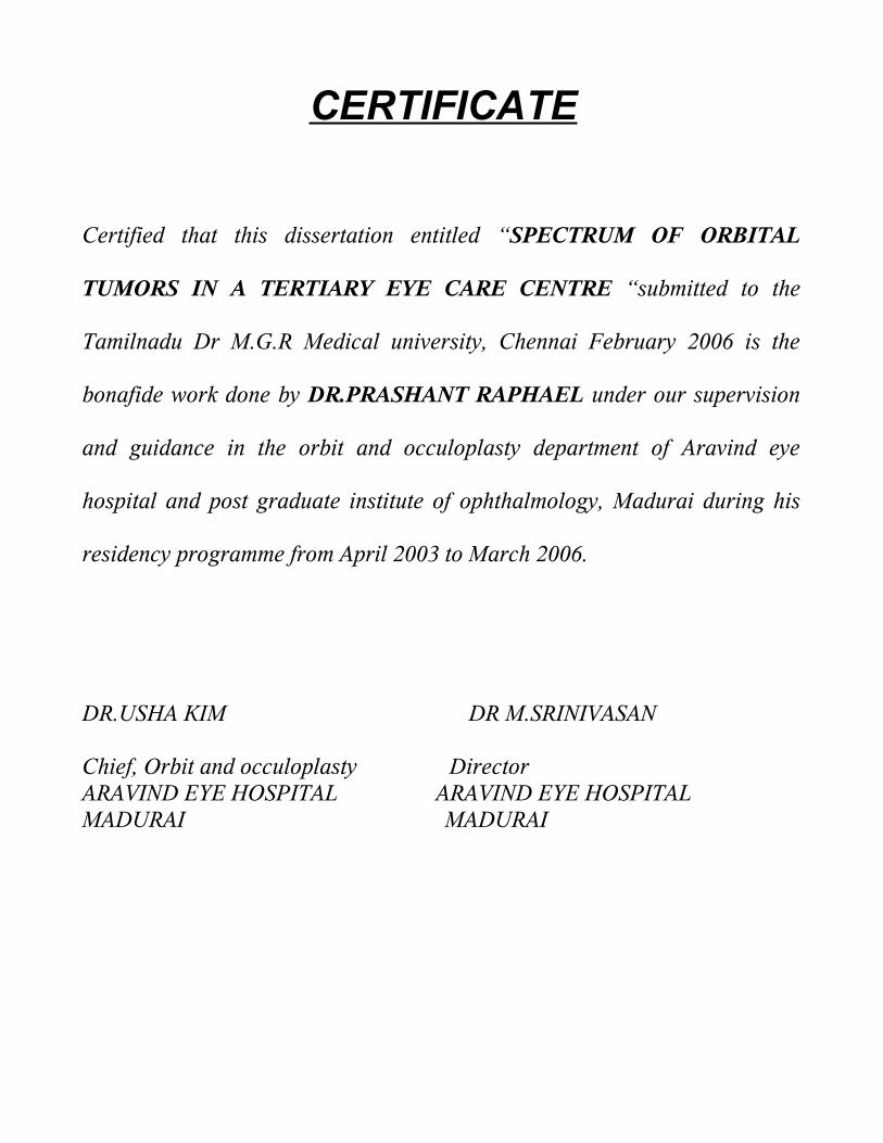
CERTIFICATE
Certified that this dissertation entitled “SPECTRUM OF ORBITAL
TUMORS IN A TERTIARY EYE CARE CENTRE “submitted to the
Tamilnadu Dr M.G.R Medical university, Chennai February 2006 is the
bonafide work done by DR.PRASHANT RAPHAEL under our supervision
and guidance in the orbit and occuloplasty department of Aravind eye
hospital and post graduate institute of ophthalmology, Madurai during his
residency programme from April 2003 to March 2006.
DR.USHA KIM DR M.SRINIVASAN
Chief, Orbit and occuloplasty DirectorARAVIND EYE HOSPITAL ARAVIND EYE HOSPITALMADURAI MADURAI

ACKNOWLEDGEMENT
I acknowledge with sincere thanks all the people without whom this thesis
could not have been a success.
I am grateful to our Chairman Dr. G.Venkataswamy, and
Dr .M .Srinivasan whose perseverance and single minded approach to work
has been a great source of inspiration.
My sincere and heartfelt gratitude to my guide Dr.Usha Kim, Chief of Orbit
and Occuloplasty services, Aravind Eye Hospital for her unfailing support
and critical evaluation of my work .Her constant appraisal and constructive
suggestions at all times has brought together this study.
I am also thankful to Dr. N.V.Prajna, Director, Residency programme for
his constant support during my residency programme.
My sincere thanks to Dr. Hadi whose help was invaluable in completion of
this thesis.
I will forever remain indebted and acknowledge my sincere thanks to all my

study patients without whom this work could not have been possible.
My special thanks to the paramedical staff of the orbit clinic, medical
records department and the library staff for their untiring support.
I would like to thank my parents and my brother for their unwavering
support. I remain grateful to my friends and colleagues for their support at
all times.
I thank God for making this all happen.
DR.PRASHANT RAPHAEL

CONTENTS Page
INTRODUCTION 1
I. REVIEW OF LITERATURE 3
II. PERSPECTIVE OF ORBITAL TUMORS
a)CLASSIFICATION OF ORBITAL TUMORS 7
b)ORBITAL TUMORS 11
III. AIMS AND OBJETIVES 40
IV. PATIENTS AND METHODOLOGY 41
V. RESULTS 42
VI. DISCUSSION 62
VII. CONCLUSION 67
VIII. ANNEXURE 69
IX. BIBLIOGRAPHY 73

INTRODUCTION
The orbit contains variety of tissues including bone, extra ocular muscles,
peripheral nerves, cranial nerves, fibro vascular adipose tissue and cartilage and a
variety of systemic disorders may manifest in the orbit of which orbital tumors
constitute the major category. Diseases of the orbit create some of the most complex and
perplexing problems in ophthalmology The initial clinical evaluation of the patient with
an orbital lesion is frequently inconclusive .After taking the patient’s history and then
examining the patient there invariably remains enough uncertainties to require
consultation with other specialists and referral for radiological and ultrasonographic
studies . Very often, a desirable end result of the workup is the localization of the lesion
to a particular area of the orbit, thereby facilitating the surgical exposure of the lesion
and permitting its excision. Such a complex subject deserves a precise table of
organization; unfortunately none exists. The small confines of the orbit and the anterior
location of the eye ball limit direct observation or palpation of orbital tumors. Even
knowledge of the approximate size, location and consistency of the lesion is not
effective means of sorting out the various lesions of the orbit. Additional information
regarding the direction degree and rate of progression of proptosis and other information

does not facilitate the formulation of a workable classification. Classifications of the
orbital disease derived from the use of ultrasonography and computerized tomography
have become very helpful; however such data are usually not available at the initial
evaluation. Perhaps the most commonly used classification is one based on the review of
histopathologic material 1. The reported incidence of orbital tumors varies widely from
series to series depending on the material studied .One finds a considerable difference in
the incidence of various lesions in series derived from biopsy material as compared with
those that include clinical diagnosis without histopathological verification .Series from
large ophthalmic departments have inherent bias depending on the referral patterns
special interests of the ophthalmologist and other factors 2. This study intends to study
the spectrum of orbital tumors in a tertiary eye care centre.

REVIEW OF LITERATURE
The reported incidence of orbital tumors varies from series to series depending primarily on the source of the material reviewed .Variation in frequency of different tumors in different series can have relationship to national ,regional and other socioeconomic factors. Biopsy proven series is recognized for the certainty and prognostic aspect. A clinical series represents a wider practical correlation and may include cases not likely to be biopsied.
Daniel Silva M.D published a study in 1968 on orbital tumors which included 300 patients, all of whom underwent surgery at least for biopsy .Pseudo tumors were the most common group followed by mucocoeles and orbital expansion of retinoblastoma. The unusually high incidence of retinoblastomas with orbital involvement was explained by the fact that the hospital was an oncology hospital where only children referred for radiotherapy or chemotherapy were seen.3
O.P.Kureshtra et al presented a study consisting of 104 cases seen over the last 20 years .Benign primary tumors consisted 48 ,malignant (primary)22, malignant (metastatic)3,extensions from paranasal sinuses malignant 25,benign 1,extension from the cranial fossa .4
Robert Kennedy reviewed 820 clinical orbital cases over a 34 year period, which include biopsy proven (450) and other clinical cases .These were patients seen by ophthalmologists practicing in an average community area rather than a large referral center and thus may be truly representative of what might be encountered in practices. This study included cases which were biopsy proven and also cases which were diagnosed without biopsy when there was strong medical, xray, or computerized tomography evidence Trauma, lymphoma, dermoid and idiopathic orbital inflammation were the most common diagnoses. 5
Jerry Shields et al reviewed 645 consecutive biopsies of orbital lesions performed at a major ophthalmic hospital during a 20 year period .Their series of 645 specimens included dermoid, dacryoadenitis, reactive lymphoid hyperplasia and lymphoma as the most common lesions. This study included a group of primary orbital melanocytic tumors which were absent in the other studies.6
Between 1977 and 1984, 84 cases of orbital tumors with histological basis were analyzed at Chang Gung memorial hospital, Taiwan. There were cases with primary orbital tumors (70.2%), secondary orbital tumors (27.4%), and metastatic orbital tumors (2.4%).The most common primary orbital tumor was the dermoid cyst and hemangioma .Adenexal squamous cell carcinoma, retinoblastoma, and mucocoele were the secondary tumors .Nasopharyngeal cancer in secondary orbital tumor was also common.7
Vijayalakshmi Ammal reviewed 212 cases of orbital tumors of which 84 cases were neurofibromas and 64 were lacrimal gland tumors in 1990.8
Sylvia et al reviewed cases of histopathologically verified orbital tumors in children to

determine the distribution of various pathological processes and trends over time .The medical records and pathology specimens from 340 patients aged 18 years or younger who under went biopsy from orbital mass in 1932 to 1991 were reviewed. The most common tumors were cysts, vascular lesions optic nerve and meningeal neoplasms. The overall frequency of malignancy was 18.2%of which 11.5%were primary tumors and 6.8 % were secondary. 9
Henderson summarized his clinical experience at the Mayo clinic .He had a total of
1376 cases over a time period of 18 years ,and his cases were from surgically excised
specimens and angiographic findings in case of vascular malformations. Mucocoele,
lymphoma, squamous cell carcinoma, and meningioma were the most frequent lesions in
his review.10
Wilson et al reviewed 312 orbital lesions accessioned in their laboratory between 1942 and 1993 .Orbital invasion of uveal melanoma, idiopathic orbital inflammation and dermoid were the most common lesions in their series.11
Demirci, Hakan et al in 2002 reviewed approximately 950 cases of orbital space occupying lesions in the older adult population. The orbital tumor was unilateral in 183 patients and bilateral in 17 patient’s .The most common clinical features included mass proptosis and pain .The most common diagnosis were malignant lymphoma, idiopathic orbital inflammation and cavernous hemangioma.12
Jerry shields et al reviewed 1264 consecutive patients with orbital tumors and stimulating lesions referred to an ocular oncology center .In this series 64% were benign and 36% were malignant. The most common diagnosis was lymphoid tumor, idiopathic orbital inflammation and cavernous hemangioma.13

PERSPECTIVE OF ORBITAL TUMORS

CLASSIFICATION OF ORBITAL TUMORS
CLASSIFICATION OF ORBITAL TUMORSAccording to Grossniklaus, the orbital disorders can be classified into (I) Systemic (II)
Inflammatory (III) Trauma (IV) Congenital (V) Primary neoplasms (VI) Secondary neoplasms (VII) Metastasis (VIII) Vascular (IX) Others. Tumors were classified into primary, secondary and metastatic. Primary tumors were further classified into 2
EPITHELIAL
BENIGN MIXED TUMOR
MALIGNANT MIXED
ADENOID CYSTIC CARCINOMA
SPINDLE CELL CARCINOMA
ADENO CARCINOMA
MUCOEPIDERMOID CARCINOMA
LYMPHOEPITHELIOMA
CONJUNCTIVAL CYST
SUDORIFEROUS CYST
FIBROUS CONNECTIVE TISSUE
FIBROMA
FIBROSARCOMA
FIBROUS HISTIOCYTOMA
MYXOMA
FIBROMATOSIS

FIBROOSSEOUS
OSTEOMA
OSTEOSARCOMA
FIBROUS DYSPLASIA
OSSIFYING FIBROMA
PAGET’S DISEASE
ANEURYSMAL BONE CYST
FIBROOSTEOMA
EWING’S SARCOMA
CARTILAGENOUS
CHONDROMA
CHONDROSARCOMA
ADIPOSE
LIPOMA
LIPOSARCOMA
VASCULAR
CAPILLARY HEMANGIOMA
CAVERNOUS HEMANGIOMA
LYMPHANGIOMA
HEMANGIOPERICYTOMA
HEMANGIOENDOTHELIOMA
ANGIOLEIOMYOMA
FIBROANGIOMA

GLOMANGIOMA
MUSCLE
RHABDOMYOMA
RHABDOMYOSARCOMA
NEURAL
MENINGIOMA
NEUROFIBROMA
GLIOMA
NEURILEMMOMA
MALIGNANT PERIPHERAL NERVE SHEATH TUMOR
ASTROCYTOMA
EPENDYMOMA
GRANULAR CELL MYOBLASTOMA
MALIGNANT LYMPHOMAS
LYMPHOID HYPERPLASIA
WELL DIFFRENTIATED LYMPHOCYTIC TYPE
POORLY DIFFRETIATED LYMPHOCYTIC TYPE
HODGKINS DISEASE
RETICULUM CELL TYPE
OTHER TUMORS OF RETICULUM CELL PROGENY
HISTIOCYTOSIS
INFANTILE XANTHOGRANULOMA

PLASMA CELL DYSCRASIAS
LEUKEMIA
SECONDARY NEOPLASMS
INVADE THE ORBIT FROM ADJACENT STRUCTRES INCLUDING
THE EYE AND OCULAR ADENEXA, PARANASAL SINUSES, ORAL
CAVITY AND CRANIUM.
METASTATIC TUMORS
TUMORS METASTAZISING TO THE ORBIT

ORBITAL TUMORS
VASCULAR

CAPILLARY HEMANGIOMA23
DEFINITION : Common benign hamartoma (mass of disorganized but mature specialized cells or tissue indigenous to the particular site) of orbital & periorbital areas in childhood. INCIDENCE: 1-2% of orbital tumor. AGE/SEX: Usually presents in first few months of life (95% by 6 months of age), but never at birth with female preponderance. CLINICAL FEATURES : Mainly of 3 types being Superficial type presenting as “strawberry nevus”, deep variety in orbit giving rise to proptosis and combined variety. HISTOLOGY: It is composed of endothelial & capillary vessel proliferations with benign endothelial cells surrounding the small capillarised vascular spaces. RADIOLOGY: On CT-Scan, appears fairly well marginated or poorly marginated, irregular enhancing lesions. On MRI, appears hypo or slightly hyper intense to brain in T1 weighted images and hyper intense in T2.MANAGEMENT : Indicated in cases of amblyopia, optic nerve compression, exposure keratitis or cosmetic blemish with necrosis. It includes intralesional steroids, systemic steroids, alpha-interferon, local resection & low dose radiotherapy.
CAVERNOUS HEMANGIOMA24
DEFINITION: Type of developmental polymorphic vascular hamartoma commonly
seen in adults. . Most are located within the muscle cone.
INCIDENCE: 1-2% of orbital tumour.AGE GROUP: 1st to 8th decade. Mainly in middle aged women (M: F=1:3) .CLINICAL PRESENTATION: Unilateral, painless, gradually progressive axial proptosis usually asymptomatic. Can cause decreased vision due to optic nerve compression or induced hyperopia, amaurosis due to intermittent optic nerve compression. HISTOLOGY : Well-circumscribed, well encapsulated, mulberry like, spongy , reddish brown colored mass On cut-section—large blood-filled spaces. RADIOLOGY: CT-scan: Well-defined, rounded, intraconal mass with smooth margin, with uniform homogenous enlargement on contrast. They do not deform the globe. Serial dynamic MRI shows connecting point of tumor to the feeder vessel in early stage (50 sec). In T-1image: iso intense with muscles. In T-2 image, hyper intense to fat and muscles. MANAGEMENT: Surgical excision in symptomatic patients and the common site being intraconal, lateral orbitotomy is the surgery of choice.
LYMPHANGIOMA25
DEFINITION : Benign vascular malforma tions, which are abortive and nonfunctional. . This lesion is seen most frequently in young children.This lesion may wax and wane in size, especially when the child has an upper respiratory infection(reflecting the proliferating lymphoid elements in the connective tissue trabeculae of the tumor). INCIDENCE: Less than 7% of patients with childhood orbital tumors AGE/SEX : Mainly in 1st two decades of life, M: F=1:3.CLINICAL FEATURES : If anterior in orbit, multiple small bluish masses in superonasal quadrant of orbit. If posterior, it causes slowly progressive proptosis ,which spontaneously become painful, due to hemorrhage in tumor it will form “chocolate cysts” which will resolve with time. MICROSCOPY: Non encapsulated networks of endothelially lined channels, separated by thin septae and filled with pale-staining lymph. RADIOLOGY : Poorly circumscribed heterogeneous masses of increased density in extra & intraconal space. Fluid-fluid levels related to hemorrhages of various stages are best seen in MRI MANAGEMENT: Persistent sight-threatening chocolate cysts should be drained or removed sub totally by controlled vaporization of CO2

laser, otherwise conservative treatment is gold standard.
HEMANGIOPERICYTOMA/HEMAGIOENDOTHELIOMA26
DEFINITION: Group of tumors with rich vasculature. In Haemangiopericytomas, where
capillaries consisting of endothelial channels surrounded by proliferation of contractile
pericytes of Zimmerman. In Haemangioendothelioma there are both locally aggressive
and capable of metastasis where it is called malignant
haemangioendothelioma.INCIDENCE : About 15-30% of hemangiopericytomas occur
in the head and neck region. haemangioendothelioma compose less than 1% of all soft
tissue sarcoma .AGE / SEX : The tumor can appear in persons of any age, with 80-95% of
patients older than 20 years. ). Approximally 10% of hemangiopericytomas are found in
children .Males=Females.CLINICAL PRESENTATION : About 50% cases are malignant
having potential for distant spread. In orbit, generally presents as non tender, irreducible
and non-pulsatile proptosis, superior orbit and intraconal location being common.
Capable of metastasizing to distant organs .In striking contrast to cavernous
hemangioma, these cause puffiness of the eye lids with bluish discoloration of the ocular
adenexa.PATHOLOGY: Histopathologically it may assume one of 3
configurations:1)A prominent vascular pattern of sinusoidal spaces,2)An ostensibly
solid pattern in which the vasculature may not be seen.3)A combination of the two.
Vascular pericytes are of mesenchymal origin that spiral around capillaries and
postcapillary venules. The tightly packed cells around the thin-walled blood vessels are
lined by flattened endothelial cells. Hemangiopericytomas contain periodic acid schift
and reticulin-staining basement membrane material.RADIOLOGY: Orbital CT Scan

documents a round to oval mass usually located in the superior orbit.The well-
encapsulated lesion,with its large arterial feeding vessels,exhibits dramatic enhancement
on injection of contrast material. Slightly less distinct margins is due to invasion of
adjacent tissues.Erosion of underlying bone and marked contrast enhancement are key to
diagnosis.On MRI ,these well-delineated tumors are hypointense to fat on T1-weighted
images and hyperintense to fat and isointense to vitreous on T2-weighted images.
MANAGEMENT : Optimal treatment is wide excision with negative margins. Tumor
cells may be left behind with enucleation. For unresectable or incompletely excised
tumors, radiotherapy has an important role.
FIBROUS CONNECTIVE TISSUEFIBROMA28
DEFINITION : It is a true benign neoplasm of bone originating in the mesenchymal tissue of periosteum. INCIDENCE: 4%-6% AGE/SEX: Younger patient,7-18 years (average 13) CLINICALPRESENTATION: Slowly progressive painless proptosis. Orbital plate of frontal and spheno ethmoidal bone commoly affected. It consists of units of lamellar bone which show clear-cut lines or lamellae on polarization. Osteoblastic activity is present.HISTOPATHOLOGY: Usually tan-gray colored whitish red tissues with soft fibrous texture having grittiness depending on osteoid formation. Highly cellular fibroblastic stroma interspersed with bony spicules forming osseous trabeculae rimmed by osteoid which is again rimmed by osteoblasts.Typically shows ”zonation phenomenon” showing more mature osteoids towards periphery.RADIOLOGY: CT shows a mass with less radiodense centre with scattered calcifications with sclerotic margin, more delimited than fibrous dysplasia. MANAGEMENT: Wide local excision.
FIBROUS HISTIOCYTOMA29
DEFINITION- Malignant fibrous histiocytoma (MFH), described by O'Brien and Stout in 1964, is the most common soft tissue sarcoma of late adult life due to fibroblastic proliferation involving fascia, muscles and soft tissues .INCIDENCE- MFH accounts for 20-24% of soft tissue sarcomas, making it the most common soft tissue sarcoma occurring in late adult life.AGE/SEX- The tumor occurs with a peak incidence in the fifth and sixth decades but an age range of 10-90 years is reported. Although the

tumor is rare in children, the angiomatoid subtype is the most frequently occurring variety in patients younger than 20 years. Male-to-female ratio is approximately 2:1. CLINICAL PRESENTATION-Slowly growing proptosis with growth in superonasal quadrant of orbit, with congestion, chemosis, lid retraction, motility disturbances and occasionally compressive neuropathy. PATHOLOGY: The tumor contains both fibroblast like and histiocyte like cells in varying proportions, with spindled and rounded cells exhibiting a storiform arrangement. Five histologic subtypes have been described including (1) storiform/pleomorphic (most common), (2) myxoid, (3) giant cell, (4) inflammatory (usually retroperitoneal), and (5) angiomatoid (often located more superficially than other varieties).RADIOLOGY-CT shows well-circumscribed mixed density tumor with heterogeneous enhancement and intact bones. USG reveals a high tissue reflectivity and heterogeneity. Benign lesions will show compressive bone remodeling. MANAGEMENT-Benign-surgical excision. Malignant- Exentration and radiotherapy /chemotherapy.
OSSEOUSOSTEOMA30
DEFINITION : Osteoid osteoma is a benign skeletal neoplasm of unknown etiology that is composed of osteoid and woven bone. INCIDENCE: Accounted for 12.1% of benign tumors and 2.9% of all tumors 10%-15%AGE/SEX : Three quarters of patients are in age 10-30 years, and more than 90% of patients are aged 5-25 years. The age range in patients is 5-56 years. The male-to-female ratio is 2:1. CLINICAL PRESENTATION : Focal skeletal bone pain, which worsens at night and is frequently relieved with a small dose of aspirin. Nonaxial Proptosis. Frontal and ethmoidal sinuses involvements are the most common presentation in and around orbit. PATHOLOGY: The tumor consists of an ovoid or spherical nidus of osteoid-rich tissue and interconnected bone trabeculae superimposed on a background of highly vascularized connective tissue containing large dilated vascular channels. RADIOLOGY: CT shows hyperdense, rounded or multilobular lesion projecting into the orbit. MANAGEMENT: Surgical excision. ASSOCIATIONS: Turcot’s syndrome , Gardner’s syndrome.
CHONDROMA31
DEFINITION : Enchondromas are benign cartilaginous neoplasms in bone. . In orbit it envolves trochlear cartilage. INCIDENCE: Enchondromas account for 12-14% of benign bone neoplasms and 3-10% of osseous neoplasms in general. AGE/SEX: Solitary enchondromas most often are discovered in those aged 20-40 years. Enchondromas occur equally in males and females.CLINICAL PRESENTATION: Slowly progressive painless proptosis. Typically located in superonasal quadrant and attached to medial orbital wall. HISTOLOGY: Enchondromas are ectopic hyaline cartilage rests in intramedullary bone hypocellular population of cells of hyaline cartilage that are widely dispersed singly in lacunae. RADIOLOGY: The lesions replace normal bone with mineralized or unmineralized hyaline cartilage, thereby generating a lytic pattern on radiographs or a lytic area containing rings and arcs of chondroid calcifications. MANAGEMENT: Wide local excision
OSSIFYING FIBROMA28
DEFINITION: Osteofibrous dysplasia which occurs exclusively in adults commonly is referred to as ossifying fibroma. INCIDENCE: Osteofibrous dysplasia usually is first diagnosed in children with a peak incidence occurring in children aged 1-5 years. AGE/SEX: The age range at the time of

diagnosis has been variable in the literature.No significant sex preponderance consistently has been reported. CLINICAL PRESENTATION: Slowly progressive painless proptosis. Orbital plate of frontal and sphenoethmoidal bone most commonly affected.
PATHOLOGY: Highly cellular fibroblastic stroma containing interspersed with bony
spicules forming osseous trabeculae rimmed by osteoid which is again rimmed by
osteoblasts. Typically shows,”Zonation phenomenon” showing more mature osteoids
towards periphery.RADIOLOGY: Osteofibrous dysplasia is an eccentric, intracortical,
osteolytic lesion. CT shows a mass with less radiodense centre with scattered
calcifications with sclerotic margin..MANAGEMENT: Due to the high recurrence rate,
nonoperative treatment of the lesion until after skeletal maturity is reached, at which
time marginal resection and bone grafting may be performed without increased risk of
recurrence.
MUSCLERHABDOMYOSARCOMA32
DEFINITION: Rhabdomyosarcoma (RMS) (from Greek, rhabdo, meaning rod shape, and myo, meaning muscle) is the most common soft tissue sarcoma in children which arises from undifferentiated mesenchyme rather than differentiated striated muscle.Pre-existent radiotherapy, germ line mutation of p53 suppressor gene(Li-Fraumeni Syndrome), translocation (2;13), (q35;q14).INCIDENCE: 6:1,00,000 per year. 5% of childhood malignancies, 50% of all paediatric sarcomas and 15% of all paediatric solid tumors. 44% in head and neck- 25%-60% of these originate in orbit. AGE/SEX: Approximately 87% of patients are younger than 15 years, and 13% of patients are aged 15-21 years. RMS rarely affects adults. In patients with orbit RMS, 42% are aged 5-9 years Bimodal age distribution, with higher .prevalence in children, average age being 7 years (Embryonal & Alveolar) whereas Pleomorphic type is common in adults. Overall, the male-to-female ratio is 1.2-1.4:1. Differences exist according to the site of primary disease.In orbit: The male-to-female ratio is 0.88:1. CLINICAL PRESENTATION : Acute onset of painless, rapidly progressive proptosis. Swelling and ptosis of upper lid caused by a palpable mass usually in upper quadrant. PATHOLOGY : RMS is one of the small, round, blue cell tumors of childhood. RMS cells may demonstrate positive immunohistochemistry results for muscle-specific markers, such as myoglobin, actin, and desmin. Cells from the RMS subtypes have the following distinctive features: (1) Botryoid: The cambium layer is characteristic, containing a condensation of loose tumor cells below an epithelial surface. (2) Alveolar: Cells line up

along membranes that may be imperceptibly thin or may be obvious collagen bands, resembling the lung alveoli. Alveolar has worst prognosis (3) Undifferentiated: Usually, no evidence of myogenesis differentiation is present. RADIOLOGY : CT shows enhancing mass within the orbit with bone destruction in larger tumors. Its usually isointense in relation to normal muscles, appears homogenous, well-defined soft tissue masses without bone damage. MRI: T1 weighted appears isointense or slightly hypointense compared to brain and appears hyperintense on fat suppression. T2 images shows increased signal intensity.MANAGEMENT: Incision biopsy followed by chemotherapy and /or radiotherapy.
NEURALMENINGIOMA33
DEFINITION : Tumor arising from meningothelial cap cells of arachanoid matter of optic nerve. INCIDENCE: It is approximately 20% of all intracranial tumors; population-based studies indicate an overall incidence of 2.3 cases per 100,000.AGE/SEX: In children, the male-to-female ratio is 2:1, with an average age at presentation of 10.1 years (range 1-16 y). The incidence of meningiomas increases with age, 2-7 cases per 100,000 in women and 1-5 cases per 100,000 in men. Peak incidence is in the seventh decade in women and in the eighth decade in men. Meningiomas rarely occur in infants.CLINICAL PRESENTION : Profound visual loss evolves over decades & early loss apparent only with threshold perimetry. Occasionally patient may present with proptosis. MICROSCOPY : The macroscopic appearance of meningiomas may be hemispheric, bun-shaped, or globular. They usually are attached to the dura and invaginate into adjacent neural structures. Enveloped in a thin capsule derived from the adjacent meninges, they remain extraaxial and are separated easily from the brain or the spinal cord. RADIOLOGY: CT shows two morphologic types: En plaque- a focal exophytic tumor along the course of intraorbital segment with enhancement. Fusiform- tumor is denser than normal nerve & shows “rail-road track” sign produced by calcification. ONSM is confined to the dura mater; therefore, it often appears as a distinct, fusiform thickening of the optic nerve. This enlargement may appear as localized or as an eccentric expansion of the optic nerve, and it occurs most commonly at the orbital apex.On MRI, tumor is hyperintense on T1 & T2 weighted images. Although an MRI most readily shows the soft tissue tumor characteristics of ONSM, a CT scan better displays any associated bony hyperostosis.MANAGEMENT: Patients with ONSM can be observed if there is no evidence of intracranial extension, vision loss, or visual field loss. Treatment with primary radiation or radiation following surgical removal has been associated with a better chance of visual improvement. Chemotherapy is reserved for patients with unresectable, recurrent, or previously irradiated meningiomas. Combination treatment with 5-fluroouracil, folate, and levamisole, or a combination of intraarterial cisplatin with intravenous doxorubicin, may be beneficial.
OPTIC NERVE GLIOMA34
DEFINITION : They are juvenile pilocytic astrocytoma, fusiform, ordinarily benign enlargement of optic nerve contained within an intact dural sheath. INCIDENCE : Optic nerve gliomas represent 4% of orbital tumors, 4% of intracranial gliomas, 2% of intracranial tumors and two thirds of all primary optic nerve tumors.AGE/SEX: In the pediatric population, the median age of patients is 5 years, and 80% of patients present prior to age 15 years . In adult patients, age ranges from 22-79 years, with a mean age of 52 years . In pediatric patients, a slight female predominance is found, while in adult patients, a slight male predominance is found.CLINICAL FEATURES : The presenting symptom is painless proptosis. Optic atrophy is common When the lesions are large, local compressive effects may

result in optic chiasm symptoms such as nystagmus. Hypothalamic symptoms, such as changes in appetite or sleep, also may occur. When lesions are massive, compression of the third ventricle may occur,resulting in obstructive hydrocephalus with headache,nausea, and vomiting.HISTOPATHOLOGY: consists of spindle cells with frequent mucinous changes.rosenthal fibres are pathognomic with fibrillary astrocytes positive for phosphotungstic acid hematoxylin and glial fibrillary acidic protein. RADIOLOGY: In children, unenhanced CT typically reveals a marked diffuse enlargement of the optic nerve, with characteristic kinking or bending. The enlargement may be tubular, fusiform, or excrescent. Areas of lucency may be observed as a result of mucinous or cystic changes. Approximately 50% of the lesions demonstrate enhancement, and enhancement is more common with intracranial (especially retrochiasmatic) extension. Calcifications are rare. MRI shows hypotense or isotense lesion on T1 & hyperintense on T2 weighted images. Enlargement of optic canal. MRI shows hypotense or isotense lesion on T1 & hyperintense on T2 weighted images. Intracranial extension better appreciated on MRI. MANAGEMENT: Guided by location of the tumor & visual potential. Usually observation with monitoring of optic nerve function & disc edema 3 monthly .Surgery is the main stay of treatment.
NEUROFIBROMA35
DEFINITION: Orbital neurofibroma is usually an isolated tumor of nerve cells, and is not associated with neurofibromatosis. Orbital neurofibroma that is associated with neurofibromatosis also occurs, and this type is typically the plexiform neurofibroma, which begins in the eyelid. Consits of a mixture of Schwann’s cells, peripheral nerve axons, endoneural fibroblasts & perineural cells. ssociated with neurofibriomatosis-1 which is an autosomal dominant inherited trait. INCIDENCE : 0.5%-2.4% of orbital tumors. AGE/SEX : Localised neurofibroma in young to middle-aged patient and diffuse and plexiform neurofibroma make their appearance within first decade of life.CLINICAL PRESENTATION: Three types: Isolated neurofibromas, plexiform, diffuse. Isolated NF presents a painless of mildly painful proptosis caused by an orbital soft tissue mass in superior quadrant commonly. It’s a non-encapsulated-has compression pseudocapsule-white to tan colored tumor. In Plexiform and Diffuse NF patient presents with massive overgrowth of lids along with similar infiltration of orbit and facial tissues, In the first there is vermiform collection of molluscusm,Lisch’s nodule,axillary freckle etc. In Plexiform, tissues are expanded by recognizable neural units whereas Diffuse form observes a monotonous spindle cells proliferation. MICROSCOPY : Consists of wavy cells typically dispersed in small bundles. In Plexiform and Diffuse neurofibromas, patient presents with massive overgrowth of lids along with similar infiltration of orbit and facial tissues, in the first there is vermiform collection of individual units on palpation not in the later. Other features are café-au-lait spots, fibroma molluscusm, Lisch nodule, axillary freckling. In Plexiform, tissues are expanded by recognizable neural units whereas diffuse form observes a monotonous spindle cell proliferation. RADIOLOGY: In isolated form CT reveals a homogenous well-circumscribed soft tissue density that has uniform contrast medium enhancement. MRI shows lesions hypointense to isointense on T1 and hyperintense on T2 weighted images. In plexiform neurofibromas, CT shows moderately enhancing soft tissue density without clearly defined borders. May show absence of sphenoid wing..MANAGEMENT : Surgical excision in localized form. In plexiform and diffuse , cosmetic debulking using CO2 laser.
NEURILEMOMA36
DEFINITION: A pure proliferation of Schwann’s cells. Orbital schwannomas are usually isolated lesions, except when they are associated with neurofibromatosis type 2 (NF2), a rare autosomal dominant disorder occurring in approximately 1 live birth in 50,000. NF2 is also called the multiple inherited schwannomas, meningiomas, and ependymomas (MISME) syndrome. It arise from the nerve

sheath ,consist of Schwann cells in a collagenous matrix.INCIDENCE: Schwannomas account for 6-8% of intracranial neoplasmsAccounts for 1% of all orbital tumors. Are found in 1.5% of patients with NF1. AGE/SEX: Young to middle aged adults. No sex predilection has been described.CLINICAL PRESENTATION : Produce slowly progressive painless proptosis. HISTOPATHOLOGY: They are extremely encapsulated, have smooth surface & friable. Usually appears yellowish-tan colored, solid, encapsulated with varicose, violaceous tumor vessels on surface. They have both solid and myxoid areas alternate with each other in varying proportion. Spindle-cells oriented in fascicles, with long axis parallel to each other called Antoni Type A., in type B it is haphazardly arranged. RADIOLOGY: CT shows a homogenous well-circumscribed soft tissue mass. On nonenhanced CT scans, most schwannomas are isoattenuating relative to brain parenchyma. Calcification or areas of hemorrhage are rare. Long standing lesions may produce remodeling or frank erosion of bone. MRI shows a discrete,homogenous soft tissue density that is hypointense on T1 and hyperintense on T2 weighted images. The cystic spaces can result in high signal intensity on T2-weighted MRIs. Gadolinium enhancement is typically homogeneous, although larger schwannomas can show areas of cystic degeneration and heterogeneous signal intensity; these findings are based on increased numbers of areas with Antoni type B histologic features. MANAGEMENT: Stereotactic radiosurgery (ie, gamma knife radiosurgery) largely has replaced surgical resection for the treatment of vestibular schwannomas, particularly when the lesions do not compress the brainstem. Lesions should be smaller than 3 cm. Studies have demonstrated rates of tumor control (ie, lesion stabilizes or shrinks) of greater than 95% and a rate of hearing preservation of approximately 70%. Although less well studied, other CN schwannomas also can be treated with radiosurgery
MALIGNANT LYMPHOMAS 37 DEFINITION : These are solid tumors of immune system composed of monoclonal B-
cells. It mimics Non-Hodgkin’s lymphoma. The lacrimal gland is the most frequent site
of involvement in the orbit. It may be either due to systemic disease(75%) or may
indicate primary site. Bilateral involvement can also be seen. 2 parts in orbit having
lymphoid tissue, i.e. substantia propria of conjunctiva & lacrimal glands,account for
lymphoreticuloses of orbit .Of all the patients of orbital lymphoma,75% will have
systemic disease. INCIDENCE ; Almost up to 10-15% of orbital masses are extranodal
NHL.AGE / SEX : Rarely in children.Mainly in 6-7 decade of life with slight female
preponderance. CLINICAL PRESENTATION: They usually occur in anterior orbit
with pink, fleshy, sub conjunctival tumefaction (salmon patch), tends to mould to the
shape of globe. On palpation they are nodular with well-defined margins and slightly

friable texture with rich vascularity intra operatively. Roughly 75% lesions are
unilateral. It produces painless progressive non axial proptosis, visual disturbances &
lacrimal gland enlargement Any pt of orbital lymphoma should be subjected to systemic
evaluation to rule out systemic lympho proliferative disorder. PATHOLOGY:The cells
are monotonous with a high nuclear-to-cytoplasmic ratio.Nuclei often are convoluted
with finely stippled chromatin; nucleoli usually are visible but not prominent.
Immunohistochemical analysis reveals T-cell markers,including CD5 and
CD7.RADIOLOGY: They are homogenous masses with relatively high density &
sharp margins, mainly in anterior orbit, superior orbit or retrobulbar area. It shows putty
like molding of tumor to preexisting structures without eroding the bone or expanding
the orbit. Lacrimal gland lymphoma displaces the globe medially & forward. The optic
nerve lymphomatous infiltration shows tram-like (axial) and ring (coronal) enhancement
after IV contrast which is also noticed in meningiomas and sarcoidosis.
MANAGEMENT: Low dose radiotherapy and chemotherapy for systemic cases.
TUMORS OF RETICULUM CELL ORIGIN 37 Histiocytosis X or Langerhans’ cell histiocytosis
DEFINITION + CLASSIFICATION: A group of histiocytic proliferative disorders derived from dendritic types of cells derived from bone marrow, epidermis, brain & oral mucosa.Mainly classified into 3 groups [1].Eosinophilic granuloma (self limited, benign) [2].Hand-Schuller-Christian Disease (Multi focal with systemic involvement) [3].Letterer-Sewe Disease( Acute , disseminated disease ).INCIDENCE: Comprises 7% of orbital tumors in children and <1% of total orbital tumors. AGE/SEX : Mainly in early childhood , 1 to 3 years of age with M:F=3:1.CLINICAL PRESENTATION: [1]. Eosinophilic granuloma - comprises 70% of all histiocytosis, mainly solitary, orbital mass with painful ,tender, erythematous swelling at anterior & superotemporal orbit causing proptosis. Skull bones are the commonly involved with frontal & parietal being commonest. [2]. Hand-Schuller-Christian Disease - comprises 20% of all histiocytosis, with equal sex distribution. It presents as multifocal bony lesions with hepatosplenomegaly, lymphadenopathy, malaise, anorexia, fever & classical triad of exophthalmos, diabetes insipidus & bony destructions.[3]. Letterer-Sewe Disease —

comprises 10% of histiocytosis, with acute, disseminated histiocytosis in infants<2yrs.It shows hemorrhagic papules of skin, spleenic & hepatic involvement, pancytopenia with poorest 5 year survival (<50%).Orbital involvement is very rare here. HISTOPATHOLOGY:Usually the tumors are soft, friable, hemorrhagic & tan-yellow colored. Microscopy shows large Langerhans histiocytic cells (15-25 microns) with round to indented nuclei and marked nuclear folds resembling coffee beans. Typical rod-shaped, tubular, tennis racquet shaped cytoplasmic inclusions called Birbeck granules are diagnostic on electron microscopy. Immuno histochemical markers are nuclear membrane S-100 protein & cell-surface and cytoplasmic marker called CD1a (OKT-6). RADIOLOGY: Bone involvement is characterized by irregular lucent patches with cortical destruction but without any sclerosis. In orbit mainly the superotemporal part shows pathology. MANAGEMENT: For localized disease, localized curettage with intralesional steroids followed by low dose radiation and systemic steroids. For disseminated diseases, chemotherapy is a must.
Granulocytic sarcoma (Chloroma)DEFINITION: Extra medullary form of Acute Myeloblastic Leukemia INCIDENCE: It occurs in about 3% of all AML patients.AGE/SEX: Mean age of onset is 7 years with Male preponderance (3:2) and non-Caucasian population. CLINICAL FEATURES: As orbit being the site of initial presentation showing involvement of soft tissues, lacrimal glands & bones. The lesion typically involves subperiosteal space of lateral orbital wall with extension into temporal fossa. HISTOPATHOLOGY: Typical greenish hue of tumor is due to enzyme of tumor cells. Cells of myeloid origin are best seen by Leder stain for chloroacetate esterase. RADIOLOGY: Depending on area of infiltration, mainly subperiosteal space of superotemporal quadrant with proptosis. MANAGEMENT Radiotherapy and chemotherapy.
INTRINSIC NEOPLASMS OF THE LACRIMAL GLAND 38
PLEOMORPHIC ADENOMADEFINITION : Is a benign mixed-cell tumor and most common epithelial tumor of lacrimal gland. It is derived from ducts, stroma, and myoepithelial elements.INCIDENCE: It is the most common epithelial tumor of lacrimal gland. Epithelial neoplasms account for only 4% of all lacrimal gland lesions. AGE/SEX: Second to Fifth decade of life. M=F.CLINICAL PRESENTATION: Slowly progressive swelling in upper outer quadrant of more than 1 year duration and may cause non-axial proptosis. HISTOPATHOLOGY: firm, grayish-white bosselated and solitary masses. Mixture of epithelium and connective tissue with pleomorphism. Pseudocapsule is present showing microscopic nodular extensions into pseudocapsule in 60% cases. RADIOLOGY: CT shows a round or oval smooth outline with excavation of lacrimal gland fossa with destruction with moderate to marked enhancement on CT and MRI.MANAGEMENT: Complete surgical excision without violating the capsule. Incisional biopsy of these lesions is contraindicated because, although histologically benign, incomplete excision often leads to repeated recurrences (as high as 30% in some studies) and malignant transformation.
ADENOID CYSTIC CARCINOMADEFINITION: Most common malignant epithelial tumor of lacrimal gland. INCIDENCE: Adenoid cystic carcinoma is the most common malignant lacrimal gland tumor, comprising 50% of malignant tumors of lacrimal gland and 25% of all lacrimal gland-tumors.AGE/SEX: 4th to 6th decade.CLINICAL

PRESENTATION: Proptosis of shorter duration (<10 months) and with pain associated with parasthesiae. HISTOPATHOLOGY: Derived from duct cells, and they form spaces into which basement membrane like material is deposited. This confers a cribriform or "Swiss cheese" appearance to the tissue. Five histologic patterns have been observed in these lesions, as follows: (1) cribriform (the most common subtype) (2) sclerosing, (3) basaloid, (4) comedo, and (5) ductal. The basaloid type has the worst prognosis.RADIOLOGY: CT shows large tumors, calcification with bony erosions having destruction of walls and pressure changes having expansion and lytic lesions of lacrimal fossa with micro serrations. MRI is useful to see perineural infiltration, cavernous sinus
involvement.MANAGEMENT: Wide local excision followed by radiotherapy or brachytherapy
SECONDARY NEOPLASMS 39 DEFINITION: Involves the orbit by direct extension and can arise in any of adjacent structures. Para nasal sinus, Nasopharyngeal, Lacrimal, Conjunctival, Eyelid, Intraocular, Intracranial tumors. INCIDENCE: 48%. AGE/SEX: Most patients are diagnosed in their fifth or sixth decadeCLINICAL PRESENTATION: Patient presents may with chronic sinusitis, nasal obstruction, pain, non axial displacement of the globe, lower lid swelling, diplopia, blurred vision, epiphora. On examination, patient may have proptosis and displacement of the globe, congestion, chemosis, strabismus, decreased acuity, hypoesthesia, nasal mass. Squamous cell carcinoma is the most common sinus malignancy to involve the orbit these tumors arise from the maxillary sinus in most cases. Nasopharyngeal carcinoma may spread to the orbit through the inferior orbital fissure Retinoblastoma is the most common intra ocular tumor of child hood .The tumor reaches the orbit through the loose connective tissue surrounding the vessels and the nerves .On reaching the orbit, the tumor cells grow quickly as undifferentiated neuroblastic cells without Flexner Winter Steiner rosettes.
TYPES OF SECONDARY ORBITAL TUMOURS:
Paranasal Sinus Tumours: Benign-Mucocele/Osteoma/Inverted papilloma
Malignant-SCC/ADC/ACC/SNUC/Melanoma/Esthesioneuroblastoma
Nasopharyngeal Tumours: SCC/Lymphoepithelioma
Lacrimal Sac Tumours: Epithelial tumours/Melanoma
Conjunctival Tumours: SCC/Melanoma
Eyelid Tumours: BCC/SCC/Seb cell Carcinoma
Intraocular Tumours: Uveal melanoma/Retinoblastoma
IntracranialTumours:Meningioma/Glioblastoma/Chordoma
RADIOLOGY: CT scan of the paranasal sinuses with contrast is particularly important in the initial assessment of sinus malignancies due to the superior capability of the CT scan to demonstrate bony involvement.MRI of the paranasal sinuses with and without gadolinium.MRI also is very useful in determining the extent of sinus disease.MRI can be used to differentiate between inspissated secretions and tumor. MRI is useful in evaluating subtle soft tissue changes. MANAGEMENT: According to primary site and type of tumor. Can be wide surgical excision, exenteration, chemotherapy and radiotherapy.

METASTATIC TUMORS 39 DEFINITION: Invade the orbit by metastasizing from the primary site via heamatogenous or lymphatic spread.ETIOLOGY: In adults, breast -42%, lungs -11%, prostate -08%,melanoma-05%, unknown-11%.Paediatric tumors: Embryonal or undifferentiated sarcoma, Neuroblastoma, Ewing’s sarcoma, Wilm’s tumor. INCIDENCE: 10%-12%.CLINICAL PRESENTAION: In the child orbit is more frequently involved and the globe less often. Whereas in adults 70% of metastasis are ocular and only 30% are orbital. Orbital metastatic lesion usually presents with an abrupt onset of proptosis, external ophthalmoplegia and orbital pain. Metastases to the orbit most often have indistinct boundaries and are diffusely infiltrating. RADIOLOGY: CT shows mass lesion and bone involvement. Osteolytic lesions caused by thyroid and renal cell carcinoma. Osteoblastic lesion caused by prostatic carcinoma.
MANAGEMENT: Observation, systemic hormonal or chemotherapy, radiation,surgical excision, exenteration.

AIMS AND OBJECTIVES
The aims of the study is to
1. Study the demographic distribution of orbital tumors –age wise, sex wise and
state wise.
2. Study the presenting complaints, the presenting eye and the duration of the
symptoms.
3. Study the history of the associated symptoms and the presence of past and
family history
4. Study the clinical features like the visual acuity, puppilary reactions and
fundus of the affected eye.

5. Study the laterality of the tumor, type of proptosis, and degree of proptosis.
6. Study the associated clinical features of the affected eye.
7. Study the type of investigative modality used in the diagnosis of the tumor.
8. Study the treatment modality used in the treatment of the tumor.
9. Study the classification of the tumor, the nature of the tumor and type of
tumor.
10. To compare the results got with that of international studies.

PATIENTS AND METHODOLOGY
This prospective descriptive study has enrolled patients with orbital tumors, coming to the orbit clinic of a tertiary eye care centre from September 2003 to March
2005.Any case of orbital tumor that presented for the first time to the hospital was included
in the study.
Follow up cases were excluded from the study thereby avoiding repetition.
The orbital tumors were diagnosed based on the proforma given below. The
diagnosis was based on clinical history and eye examination .Laboratory investigations
as well as radiological investigations like X-ray, USG, CT scan were undergone by the
patient as required .Histopathological examination using light microscopic examination
of H and E stained slides after fixation, paraffin embedding and sectioning were done.
Treatment of orbital tumors was also studied.
This study includes orbital tumors diagnosed both by clinical methods and by
investigations.

RESULTS

0
10
20
30
40
PERCENTAGE
AGE GROUP
AGE GROUP
Percent 22 27.3 34 16.7
0-19 20-39 40-59 60-89
SEX DISTRIBUTION
MALE, 49.3FEMALE, 50.7MALE
FEMALE
0
100
PERCENTAGE
STATE
STATE WISE DISTRIBUTION
STATE WISEDISTRIBUTION
68 21.3 8.7 2
TN KE AP OT
Age Group

Frequency Percent0-1920-3940-5960-89Total
33415125150
22.027.334.016.7100
The age wise distribution showed 20%in the 0-19 year age group, 27.3%in 20-39 year age group, 34% in the 40-59 year age group and 16.7% in the 60-89 year age group.
The sex wise distribution of orbital tumors showed that in 49.3%of cases males were involved and in 50.7 of cases females were involved.
The state wise distribution of the cases showed that 68% of cases were from Tamilnadu, 21.3% from Kerala, 8.7%from Andra Pradesh and 2% from other states.
Sex
74 49.3
76 50.7
150 100.0
Male
Female
Total
Frequency Percent

AFFECTED EYE
RIGHT47%LEFT
50%
BOTH3%
PRESENTING COMPLAINTS
22
3.3 2.712
62
32
010203040506070
DEF VIS
ION
DOUBLE
VIS
ION
PTOSIS
PAIN
PROPTO
SIS
MASS
TYPE
PE
RC
EN
TA
GE

The affected eye was the right eye in 47.3% of cases and the left eye in
50% of cases .Both the eyes were involved in 2.7% of cases.
Defective vision
33 22.0
117 78.0
150 100.0
Present
Absent
Total
Frequency Percent
Double Vision
5 3.3
145 96.7
150 100.0
Present
Absent
Total
Frequency Percent
Drooping of eyelids
4 2.7
146 97.3
150 100.0
Present
Absent
Total
Frequency Percent
Pain
18 12.0
132 88.0
150 100.0
Present
Absent
Total
Frequency Percent
Protrusion of eye ball
93 62.0
57 38.0
150 100.0
Present
Absent
Total
Frequency Percent
Mass
48 32.0
102 68.0
150 100.0
Present
Absent
Total
Frequency Percent
Defective vision was present in 22% of cases, double vision in 3.3%, drooping of
eyelids in 2.7%, pain in 12%, proptosis in 62% and a mass was felt in 32 %of cases.
Affected Eye
71 47.3
75 50.0
4 2.7
150 100.0
Right Eye
Left Eye
Both Eye
Total
Frequency Percent

Descriptive Statistics
32 .2 72.0 11.005 16.0309
5 1.0 12.0 4.000 4.6368
4 1.0 12.0 6.625 6.2099
93 .2 120.0 17.147 24.3630
19 1.0 72.0 14.579 25.8442
48 .2 72.0 14.327 16.0925
0
Defective vision duration
Double Vision duration
Drooping of eyelidsduration
Protrusion of eye ballduration
Pain duration
Mass duration
Valid N (listwise)
N Minimum Maximum Mean Std. Deviation
The duration of symptoms was a minimum of 2 weeks for defective vision, protrusion of eyeball and mass respectively while it was 1 month for pain, double vision and drooping of eyelids. The maximum duration of symptoms 12 months for double vision and drooping of eyelids, 6 years for defective vision, pain and mass while it was 10 years for protrusion of eyeball.
VISUAL ACUITY
54.5
13.6
8.3
24.1
6/6-6/18
6/24-6/36
6/60-3/60
<3/60
VIS
UA
L A
CU
ITY
PERCENTAGE

EYELIDS
0 10 20 30 40 50 60 70 80NO
RM
AL
PTOSIS
LAGOPHTHALMOSO
THERS
NO
T EXAM
INED
EY
EL
IDS
PERCENTAGE
Group visual acuityFrequency Percent
6/6 – 6/186/24--6/366/60—3/60
<3/60TOTAL
79191235
145
54.513.18.3
24.1100
54.5%of patients had visual acuity between 6/6-6/18, 13.1% between 6/24-6/36,8.3% between 6/60-3/60, while 24.1%had vision less than 3/60. 5 patients could not be examined because of their age.

Eye lids were normal in 72.7%of cases while it showed ptosis in 10% of cases, lagophthalmos in 2.7% of cases and other presentation in 13.3% of cases .2 cases could not be examined because one had a squamous cell carcinoma involving the orbit and the other had a basal cell carcinoma involving the orbit.
Conjunctiva was normal in 79.3% of cases while it was congested in 8% of cases ,chemosed in 8% of cases .Salmon patch was present in 1.3%of cases .5 cases could not be examined because of conjuntival involvement by the tumor.
Pupil reaction was normal in 72.7% of cases, absent in 4.7% of cases, RAPD was present in 18.7% of cases.6 cases could not be examined because of the involvement of the cornea by the tumor or the cornea was hazy.
Eye lids
109 72.7 73.615 10.0 10.14
2.7 2.720 13.3 13.5
148 98.7 100.02
1.3150 100.0
NormalPtosisLagopthalmosOthersTotal
Valid
SystemMissingTotal
Frequency Percent Valid Percent
Conjunctiva
119 79.3 82.1
12 8.0 8.3
12 8.0 8.32
1.3 1.4
145 96.7 100.05
3.3
150 100.0
Normal
Congested
Chemosis
Salmon patch
Total
Valid
SystemMissing
Total
Frequency Percent Valid Percent
Pupil
109 72.6 75.77
4.7 4.9
28 18.7 19.4
144 96.0 100.06
4.0
150 100.0
Present
Absent
RAPD
Total
Valid
SystemMissing
Total
Frequency Percent Valid Percent

Fundus was normal 71.3% of cases and abnormal in 22% of cases .6.7% of cases could not be examined because of no view of the fundus due to cataract, involvement of the eye by the tumor or hazy cornea.
Fundus
107 71.3 76.4 76.4
33 22.0 23.6 100.0
140 93.3 100.0
10 6.7
150 100.0
Normal
Abnormal
Total
Valid
SystemMissing
Total
Frequency Percent Valid PercentCumulative
Percent

LATERALITY
U/L, 96
B/L, 4,
U/L
B/L
37.340.7
22
05
1015202530354045
PERCENTAGE
AXIAL ECCENTRIC NOPROPTOSIS
TYPE OF PROPTOSIS
TYPE OF PROPTOSIS
LATERALITYFREQUENCY PERCENTAGE

UNILATERALBILATERAL
TOTAL
1446
150
964
100
Unilateral presentation was in 96% of cases and bilateral in 4% of cases
Axial proptosis was present in 37.3% of case and eccentric proptosis in 40.7% of cases. Proptosis was absent in 22% of cases.
Degree of proptosis
1 1.8
6 10.7
8 14.3
16 28.6
2 3.6
12 21.4
3 5.4
3 5.4
1 1.8
3 5.4
1 1.8
56 100.0
1.00
2.00
3.00
4.00
5.00
6.00
7.00
8.00
9.00
10.00
17.00
Total
Frequency Valid Percent
Degree of proptosis ranged from a minimum of 1mm to a maximum of 17 mm.
Globe displacement
56 37.3 47.9
61 40.7 52.1
117 78.0 100.0
33 22.0
150 100.0
Axial
Eccentric
Total
Valid
NO PROPTOSIS
Total
Frequency Percent Valid Percent

MASS FELT
PRESENT, 44
NOT FELT, 54
NOT EXAMINED, 2
PRESENT
NOT FELT
NOT EXAMINED
OCULAR MOVEMENTS
FULL, 40
RESTRICTED, 58
NOT EXAMINED, 2
FULL
RESTRICTED
NOT EXAMINED

Examination of mass
66 44.0 44.9
81 54.0 55.1
147 98.0 100.03
2.0
150 100.0
Mass felt
Mass not felt
Total
Valid
SystemMissing
Total
Frequency Percent Valid Percent
Warmth
51 34.0 34.9
95 63.3 65.1
146 97.3 100.04
2.7
150 100.0
Present
Absent
Total
Valid
SystemMissing
Total
Frequency Percent Valid Percent
Tenderness
18 12.0 12.2
129 86.0 87.8
147 98.0 100.03
2.0
150 100.0
Present
Absent
Total
Valid
SystemMissing
Total
Frequency Percent Valid Percent
Pulsation
147 98.0 100.03
2.0
150 100.0
AbsentValid
SystemMissing
Total
Frequency Percent Valid Percent

Finger insinuation
45 30.0 30.6
102 68.0 69.4
147 98.0 100.03
2.0
150 100.0
Possible
Not Possible
Total
Valid
SystemMissing
Total
Frequency Percent Valid Percent
Resistance to retropulsion
94 62.7 63.9 63.9
53 35.3 36.1 100.0
147 98.0 100.03
2.0
150 100.0
Present
Absent
Total
Valid
SystemMissing
Total
Frequency Percent Valid PercentCumulative
Percent
Variation with posture
1.7 .7
146 97.3 99.3
147 98.0 100.03
2.0
150 100.0
Present
Absent
Total
Valid
SystemMissing
Total
Frequency Percent Valid Percent

Mass was felt in 44% of cases and was not felt in 54% of cases .2% of cases could not be examined because of involvement of the eyeball by the tumor. Warmth was felt in 34% of cases and was not felt in 63.3% of cases .2.7% of cases could not be examined. Tenderness was present in 12% of cases while it was absent in 86% of cases .2% of cases could not be examined because of involvement by the tumor. Pulsation was absent in all the cases that could be examined (98%).Finger insinuation was possible in 30% of cases and was not possible in 68% of cases. Resistance to retropulsion was present in 62.7% of cases and was absent in 35.3% cases. Variation with posture was present in only.7% of cases and was absent in 97.3% of cases. Variation with valsalva manouevre was absent in all the cases that were examined. Bruit was absent in all the cases examined. Ocular movements were restricted in 58% of cases and were normal in 40% of cases .2% of cases could not be examined because of involvement of the eyeball by the tumor.
Valsalva maneuver
147 98.0 100.03
2.0
150 100.0
AbsentValid
SystemMissing
Total
Frequency Percent Valid Percent
Bruit over the mass
147 98.0 100.03
2.0
150 100.0
AbsentValid
SystemMissing
Total
Frequency Percent Valid Percent
Ocular movements
60 40.0 40.8
87 58.0 59.2
147 98.0 100.03
2.0
150 100.0
Full
Restricted
Total
Valid
SystemMissing
Total
Frequency Percent Valid Percent

INVESTIGATIONS
INVESTIGATIONS
1.3 1.3 0.7
14
78
88
0
10
20
30
40
50
60
70
80
90
100
HEM
ATO
LOGIC
AL
X-R
AY P
NS
X-R
AY O
RBIT
USG
CT S
CAN
HIS
TOPATH
OLO
GY
INVESTIGATIONS
PE
RC
EN
TA
GE
Haematological
21.3 1.3
148 98.7 98.7
150 100.0 100.0
Done
Not Done
Total
ValidFrequency Percent Valid Percent
X-Ray
147 98.0 98.02
1.3 1.31
.7 .7
150 100.0 100.0
Not Done
PNS
ORBIT
Total
ValidFrequency Percent Valid Percent
USG B Scan
21 14.0 14.0
129 86.0 86.0
150 100.0 100.0
Done
Not Done
Total
ValidFrequency Percent Valid Percent
CT Scan
117 78.0 78.0
33 22.0 22.0
150 100.0 100.0
Done
Not Done
Total
ValidFrequency Percent Valid Percent
Histopathology
132 88.0 88.0
18 12.0 12.0
150 100.0 100.0
Done
Not Done
Total
ValidFrequency Percent Valid Percent

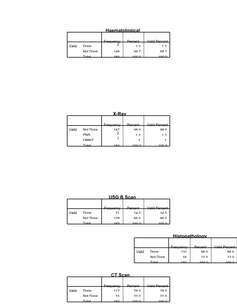
Hematological investigation and X-ray PNS was done in 1.3% and X-ray orbits in .7%of cases, USG Scan in 14% of cases, CT scan in 78% of cases and histopathological examination was done in 88% of cases.
CT Scan
117 78.0 78.0
33 22.0 22.0
150 100.0 100.0
Done
Not Done
Total
ValidFrequency Percent Valid Percent
Histopathology
132 88.0 88.0
18 12.0 12.0
150 100.0 100.0
Done
Not Done
Total
ValidFrequency Percent Valid Percent

MANAGEMENT
6.7
48.7
67.3 6.7
21.3
2.70.7
0
10
20
30
40
50
60
MED
Sx CT RT
Sx+CT
Sx+RT
REF
ABSCONDED
MANAGEMENT
PE
RC
EN
TA
GE
MANAGEMENT
FREQUENCY PERCENTAGE

MEDICAL 10 6.7
SURGERY 73 48.7
CHEMOTHERAPY 9 6.0
RADIOTHERAPY 11 7.3
SX +CT 10 6.7
SX+RT 32 21.3
REF 4 2.7
TOTAL 149 99.3
ABSCONDED 1 .7
TOTAL 150 100
Sxsurgery; CTchemotherapy; RT radiotherapy;REFreferred for further management
Medical therapy was instituted in 6.7% of cases. Surgery was done in 48.7% of cases,
chemotherapy in 6%, radiotherapy in 7.3%, surgery and hemotherapy in 6.7%, surgery
and radiotherapy in 21.3% of cases and 2.7% of the patients were referred. 1 patient
absconded.

TUMOR TYPE
PRIMARY , 82
SECONDARY, 14.7
METASTASIS, 3.3
PRIMARY
SECONDARY
METASTASIS
BENIGN OR MALIGNANT
BENIGN 51%
MALIGNANT49%
DIAGNOSIS

Primary tumors constituted 82% of the cases, secondary 22% and metastasis constituted 3.3% of cases.
BENIGN OR MALIGNANT
76 50.7
74 49.3
150 100.0
Benign
Malignant
Total
Frequency Percent
50.7% of the cases were benign tumors and 49.3% of cases were malignant tumors
Tumor Type
123 82.0
22 14.75
3.3
150 100.0
PRIMARY
SECONDARY
METASTASIS
Total
Frequency Percent

FREQUENCY OF PRIMARY TUMORS
0 10 20 30
EPITHELIAL
FIBROUS
FIBROSSEOUS
CARTILAGE
ADIPOSE
VASCULAR
LYMPHOID/LEUKEMIC
LANGERHANS
NEURAL
STRIATED MUSCLET
YP
E O
F T
UM
OR
FREQUENCY
PERCENTAGE

Primary tumors included–epithelial -19.5%, fibrous -6.5%, fibrosseus-4.8%, cartilaginous - .8%, adipose-1.6%, vascular 23.5%, lymphoid and leukemic-15.4%, langerhans cell type -1.6%, neural -22.7% and striated muscle-3.5%.
TUMOR CLASS
24 19.58
6.56
4.81
.82
1.6
29 23.5
19 15.42
1.6
28 22.74
3.5
123 100.0
Epithelial
Fibrous
Fibroosseus
Cartilage
Adipose
Vascular
Lymphoid/Leukemic
Langerhan's
Neural
Striated muscle
Total
Frequency Percent

05
1015202530354045
PERCENTAGE
P A A C A C C CA P C C SQ P
TYPE
EPITHELIAL TUMORS
25
12.5
50
12.5
0 10 20 30 40 50
PERCENTAGE
FIB HIS
FIBROMA
FIBRMATOSIS
FIB SARC
TY
PE
FIBROUS TUMORS
EPITHELIAL
FREQUENCY PERCENTAGEPLEOMORPHIC ADENOMA 8 33.3

ADENOCARCINOMA
ADENOIDCYSTICCARCINOMA
CA FROM PLEOMORPHIC
CONJUNCTIVAL CYST
SQUAMOUS PAPPILOMA
3
10
1
1
1
12.5
41.6
4.2
4.2
4.2Pleomorphic adenoma constituted 33.3% of the epithelial tumors,adenocarcinoma12.5%, adenoid cystic carcinoma 41.6%,and carcinoma from pleomorphic adenoma conjunctival cyst and squamous pappiloma being 4.2% each.
FIBROUS
FREQUENCY PERCENTAGE
FIBROUS HISTIOCYTOMA
FIBROMA
FIBROMATOSIS
FIBROSARCOMA
TOTAL
2
1
4
1
8
25
12.5
50
12.5
100
Fibromatosis (50%) was the most common tumor of the fibrous variety followed by fibrous histiocytoma (25%) and fibroma and fibrosarcoma being 12.5% each.
Ossifying fibroma was 66.7%of the osteoid tumors while osteoid osteoma and pagets disease with osteosarcoma was16.7% each.
Lipoma constituted 100% of the adipose tissue tumors.
FIBROOSSEOUS
466.7
116.7
116.7
6100.0
OSSIFYING FIBROMA
OSTEOID OSTEOMA
PAGETS DISEASE
Total
Frequency Percent
.
ADIPOSE
2100.0LIPOMA
Frequency Percent

Chondrosarcoma was the only tumor of the cartilaginous variety encountered.
VASCULAR TUMORS
20.7
62.1
6.9
3.4
3.4
3.4CAPILLARY
CAVERNOUS
HEE
HEP
KS
L
CARTILAGE
1100.0CHONDROSARCOMA
Frequency Percent

LEUKEMIC /LYMPHOID TUMORS
ALLAML
NHL
PLASALL
AML
NHL
PLAS
Cappillary hemangioma constituted 20.7% , cavernous hemangioma 62.1%, hemangioendothelioma6.9%, hemangiopericytoma ,kaposis sarcoma and lymphangioma having3.4% eachof vascular tumors
LYMPHOID /LEUKEMIC
FREQUENCY PERCENTAGE
VASCULAR
620.7
18 62.12
6.91
3.41
3.41
3.4
29 100.0
CAPILLARYHEMANGIOMACAVERNOUSHEMANGIOMAHEMANGIOENDOOMAHEMANGIOPERICYTOMA
KAPOSI'S SARCOMA
LYMPHANGIOMA
Total
Frequency Percent
.

ALL
AML
NHL
PLASMOCYTOMA
TOTAL
1
3
14
1
19
5.3
15.8
73.7
5.3
100
Non Hodgkins lymphoma was the most common leukemic/lymphoid tumor followed by acute myeloid leukemia. Acute lymphoid leukemia and plasmocytoma constituted 5.3% of the cases.
Eosinophilic granuloma was the only tumor encountered in the Langerhans cell variety.
FREQUENCY PERCENTAGE
GRANULAR CELL MYOBLASTOMA
RHABDOMYOSARCOMA
TOTAL
1
3
4
25
75
100
Granular cell myoblastoma 25% and rhabdomyosarcoma 75% were the tumors of the striated muscle variety
LANGERHANS
2100.0
2100.0
EOSINOPHILICGRANULOMA
Total
Frequency Percent

0 10 20 30 40
PERCENTAGE
GLIOMA
MENING
NEUROFI
SCHWAN
PO NEUROT
YP
E
NEURAL TUMORS
SECONDARIES
36.6
13.54.518.1
13.6
4.5
9.1 SINUS
RETINO
BCC
MGC
SCC
MELAN
ESTHESIO
NEURAL
FREQUENCY PERCENTAGE
OPTIC NERVE GLIOMA
MENINGIOMA
5
6
17.9
21.4

SCHWANNOMA
NEUROFIBROMA
PRIMARYORBITALNEUROEPITHI
TOTAL
6
10
1
28
21.4
35.7
3.6
100
Neurofibroma was the most common tumor of the neural variety 35.7%,meningioma and schwannoma being 21.4% each ,glioma 17.9%and primary orbital neuroepithelioma was 3.6%.
SECONDARIES
FREQUENCY PERCENTAGE
SINUS MALIGNANCY
BASAL CELL CARCINOMA
MALIGNANT MELANOMA
MEIBOMIAN GLAND CA
ESTHESIONEUROBLASTOMA
RETINOBLASTOMA
SQUAMOUS CELL CA
TOTAL
8
1
1
4
2
3
3
22
36.6
4.5
4.5
18.1
9.1
13.6
13.6
100
Secondaries were common from the sinuses 36.6%,meibomian gland carcinoma 18.1% ,13.6% each retinoblastoma and squamous cell carcinoma esthesioneuroblastoma9.1% and basal cell carcinoma and malignant melanoma 4.5% each.
METASTASIS
FREQUENCY PERCENTAGE
PRIMARY UNKNOWN
BREAST
CLEAR CELL CA
CERVIX
TOTAL
2
1
1
1
5
40
20
20
20
100
Metastasis were from breast, cervix and clear cell were encountered but majority of the primary was unknown

DISCUSSION

A spectrum of tumors can involve the orbit .Several publications have addressed the
incidence of space occupying lesions in the orbit. However the reported incidence of
orbital lesions show great variation .Most reported series of the frequency of orbital
tumors are biased by various factors like specialty of the reviewer, histopathological
confirmed lesions, age range of patients and geographic area of the patients. The present
study differs from the other studies in that it includes patients seen in a tertiary care
centre and also on clinical and pathologically proven cases and so reflects the true
incidence of orbital tumors seen in ophthalmic practices
Our study is compared to the five major studies done by Shield, Kennedy, Rootman,
Henderson and Wilson. Robert Kennedy’s series was based on the tumor registry,
Shields on the pathology specimens, Henderson summarized his clinical experience,
Rootman tabulated his orbital practice and Wilson summarized the orbital lesions
submitted to them. This study includes tumors encountered in a tertiary eye care centre
and includes both the clinically and the pathologically proven cases.
Kennedy collected his cases in a time period of 34 years, Shields in 20 years,
Henderson in 34 years, Rootman in 10 years and Wilson in 51 years while in this study
the cases have been collected in a time frame of 18 months.
Type shields kennedy hender rootman wilson Our
studyprimary 188 284 662 244 76 123

(69%) (78.5%) (63.5%) (74.8%) (42%) (82%)secondary 70
(26%)
51
(14%)
269
(25.8%)
44
(13.5%)
90
(50%)
22
(14.7%)metastatic 16
(5%)
27
(7.5%)
111
(10.7%)
38
(11.7%)
15
(8%)
5
(3.3%)
This table shows the distribution of tumors in the six studies compared with each
other .Our study shows that the primary tumors were more in number compared to the
other studies while secondaries were of same value and metastasis were comparatively
less in number
DISTRIBUTION OF PRIMARY TUMORS
Type Shield Kennedy Hender Rootm Wilson StudyEpith 11.1 6.3 10.2 6.1 6.57 19.5Fibrous 5.85 0.7 3.02 5.3 2.63 6.5Fibrooseus 4.25 9.85 4.68 8.6 3.9 4.8Cartilage 1.06 0.35 1.05 0.8 _ 0.8Adipose 1.06 _ 1.05 _ 3.9 1.6Vascular 20.2 17.9 18.8 22.9 21 23.5Neural 12.2 23.5 33.9 29 21 22.7Striated
Muscle
4.25 2.8 5.7 2.04 15.7 3.5
Lymph/Leu
k
38.8 37.6 19.3 22.5 21 15.4
Langerhans 0.53 1.05 1.66 1.6 3.9 1.6Others 1.06 _ 0.6 0.8 _ _

Compared to the other studies, more malignant epithelial tumors than pleomorphic
adenoma were noted and this may be due to the fact that ours is a tertiary care centre and
the malignant lacrimal gland tumors were referred for further management.
In the other studies, fibrous histiocytoma and fibrosarcoma were the most common
while fibromatosis was the most common lesion in oour study. All the lesions were
confirmed by histopathology.
Ossifying fibroma was the most common tumor in the fibrosseous group which was
unlike that of the other studies in which osteoma and fibrous dysplasia were more
common .Primary osteosarcoma of orbital bones were rare.
Primary neoplasms in cartilage and adipose tissue were infrequent in all the studies.
Vascular tumors showed capillary and cavernous hemangioma to be the most
common tumors .Shields study was based on pathology specimens and so may have
underestimated the frequency of these lesions and so may have some basis against these
tumors .Our study, as being one including both histological and clinically diagnosed
lesions eliminates this basis and hence shows a higher number of vascular tumors.
Optic nerve glioma, neurofibroma and meningioma were the most common tumors
of neural origin in all the six studies.
Lymphoid and leukemic tumors were relatively common .The most common
lymphoid tumor was Non Hodgkins Lymphoma except in the study done by Shields
where lymphoid hyperplasia was more common.
All the tumors of the myogenic variety were malignant in all the six studies.

The three most common primary tumors in all the studies put together were
malignant lymphoma, cavernous hemangioma and meningioma while in our study it was
malignant lymphoma, cavernous hemangioma, neurofibroma and adenoid cystic
carcinoma. All the studies show that the vascular, neural and the lymphoid type of
tumors were the most common tumor type. In my study epithelial tumors were found to
be more common than in the other studies.
In the secondary neoplasm’s invading the orbit the uveal melanoma was found to be
the most common ocular tumor invading the orbit while in our study retinoblastoma was
the more common one .This may be due to the fact that the uveal melanoma is more
common in blue /grey iris than in brown iris. Squamous cell carcinoma was the most
common adnexal tumor involving the orbit in the other studies while in our study it was
meibomian gland carcinoma. Sinus tumors were also encountered in our study. Other
rare tumors like esthesioneuroblastoma were also reported in our study.
In keeping with touch with the prior studies the most frequent metastatic neoplasms
have been from the breast .In our study, we had 2 out of the 5 metastatic lesions, in
which the primary was unknown
All the studies had different inclusion criteria and the time interval for the six studies
were different, which are considered to be the limitations of the study

CONCLUSION

The age wise distribution in the study showed that the most common age group was the
40-59 year age group with the mean age being 39.6 years
In this study females were more commonly involved than males
In this study the most common state from where the patients presented was Tamil nadu and
the second being from Kerala
In this study the left eye was more commonly involved than the right eye
The most common presenting symptom was proptosis followed by the presence of a mass.
All patients who had the disease before had taken treatment for it before. Family history was
positive only in 2% of cases.
The visual acuity was between 6/6 -6/18 in most of the patients
The eye lids and the conjunctiva were normal in most of the cases. The most common
presentation in the eyelids were ptosis and the chemosis and congestion and chemosis were
present in all the other cases .Pupil reaction was normal in most of the cases and the fundus
was normal in most of the cases examined.
Unilateral presentation was more common than bilateral and eccentric proptosis was more
common than axial proptosis
Mass felt, warmth ,tenderness ,resistance to retropulsion ,pulsation ,variation with posture
and valsalva manouvre and bruit were not felt in most of the cases .Ocular movements were
restricted in most of the cases.
Histopathological examination and CT scan were the two most common investigations done
Surgery was the most common modality of treatment followed by a combination of surgery

and radiotherapy.
Benign tumors were more common than the malignant ones.
Vascular, neural and epithelial tumors were the most common tumors encountered.
The most common primary tumors were malignant lymphoma ,cavernous hemangioma
,neurofibroma and adenoid cystic carcinoma.

ANNEXURE

PROFORMA
NAME AGE SEX
MRNO STATE TAMIL NADU—1 KERALA—2 ANDHARAPRADESH—3
KARNATAKA—4OTHERS—5
PRESENTING COMPLAINTS
AFFECTED EYE RE1 LE 2 BE3
SYMPTOMS PRESENT—> 1
ABSENT—> 2
IF PRESENT DURATION
DEFECTIVE VISION
DOUBLE VISION
DROOPING OF EYELIDS
PROTRUSION OF EYEBALL
PAIN
MASS
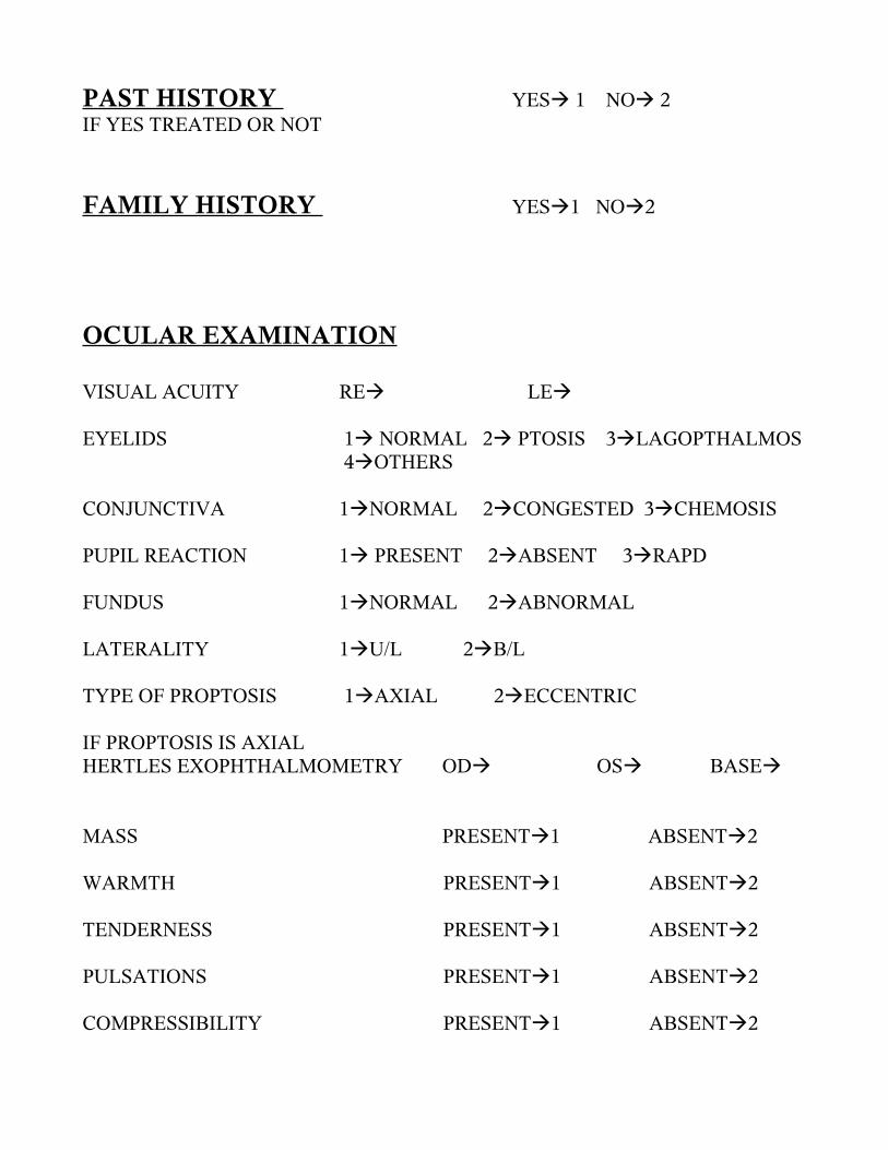
PAST HISTORY YES 1 NO 2IF YES TREATED OR NOT
FAMILY HISTORY YES1 NO2
OCULAR EXAMINATION
VISUAL ACUITY RE LE
EYELIDS 1 NORMAL 2 PTOSIS 3LAGOPTHALMOS 4OTHERS
CONJUNCTIVA 1NORMAL 2CONGESTED 3CHEMOSIS
PUPIL REACTION 1 PRESENT 2ABSENT 3RAPD
FUNDUS 1NORMAL 2ABNORMAL
LATERALITY 1U/L 2B/L
TYPE OF PROPTOSIS 1AXIAL 2ECCENTRIC
IF PROPTOSIS IS AXIALHERTLES EXOPHTHALMOMETRY OD OS BASE
MASS PRESENT1 ABSENT2
WARMTH PRESENT1 ABSENT2
TENDERNESS PRESENT1 ABSENT2
PULSATIONS PRESENT1 ABSENT2
COMPRESSIBILITY PRESENT1 ABSENT2

FINGER INSINUATION POSSIBLE1 NOT POSSIBLE2
RESISTANCE TO RETROPULSION PRESENT1 ABSENT2
VARIATIONS WITH POSTURE PRESENT1 ABSENT2
VALSALVA MANOUVRE PRESENT1 ABSENT2
BRUIT OVER THE MASS PRESENT1 ABSENT2
OCULAR MOVEMENTS FULL1 RESTRICTED2
INVESTIGATIONS
HEMATOLOGICAL 1 DONE 2NOT DONE
X-RAY 1NOT DONE 2PNS 3ORBIT 4OPTIC FORAMEN 5SKULL
CT SCAN 1DONE 2NOT DONE
USG 1DONE 2NOT DONE
HISTOPATHOLOGY 1DONE 2NOT DONE
OTHERS 1DONE 2NOT DONE
MANAGEMENT
1 MEDICAL
2 SURGERY
3 CHEMOTHERAPY

4 RADIOTHERAPY
5 SURGERY AND CHEMOTHERAPY
6 SURGERY AND RADIOTHERAPY
7 REFFERED FOR FURTHER MANAGEMENT
DIAGNOSIS
TYPE OF TUMOR 1PRIMARY 2SECONDARY 3METASTASIS
CLASS OF TUMOR 1-EPITHELIAL 2-FIBROUS 3-FIBROOSSEUS4-CARTILAGE 5-ADIPOSE 6-VASCULAR7-LYMPHOID/LEUKEMIC 8-LANGERHANSCELL 9-NEURAL 10-STRIATED MUSCLE11-SECONDARY 12-METASTATIC
NATURE OF TUMOR 1BENIGN 2MALIGNANT

BIBLIOGRAPHY

1. PRINCIPLES AND PRACTICES OF OPHTHALMOLOGY VOLUME 3
PEYMAN ,SANDERS ,GOLDBERG FIRST INDIAN EDITION 1987.
2. OPHTHALMOLOGY CLINCS OF NORTH AMERICA DEC 1996 HANS
E.GROSSNIKLAUSAND JEFFREY A.NERAD ORBITAL DISEASE Pg 539
3. AMERICAN JOURNAL OF OPHTHALMOLOGY,VOL 65 1968 MAR
ORBITAL TUMORS SILVA DANIEL PG.318
4. INDIAN JOURNAL OF OPHTHALMOLOGY KURESHTRA O P ET AL
VOL 31 1983 JUL Pg.313 EXPERIENCES WITH ORBITAL TUMORS
5. TRANS AM OPHTHALMOL SOC VOL 82 1984 Pg.134 -155 KENNEDY R E
AN EVALUATION OF 820 ORBITAL TUMORS
6. ARCHIVES OF OPHTHALMOLOGY SHIELDS JA ,BAKEWELL B,
AUGSBURGER JJ,ET AL VOL 102 1984 1606 -1611 CLASSIFICATION
AND INCIDENCE OF SPACE OCCUPYING LESIONS OF THE ORBIT
7. AFRO ASIAN JOURNAL OF OPHTHALMOLOGY KAO, LING ET AL
VOL 4 1985 DEC Pg. 139 -144 ORBITAL TUMORS -- A STUDY OF 84
CASES
8. KSOS VIJAYALAKSHMI AMMAL D VOL 2 1990 JAN P19 PRIMARY
ORBTAL TUMORS PREVALENT IN KERALA

9. AMERICAN JOURNAL OF OPHTHALMOLOGY KODSI, SYLVIA R ET AL
VOL 117 1994 FEB Pg.177-182 A REVIEW OF 340 ORBITAL TUMORS IN
CHILDREN DURING A 60 YEAR PERIOD
10. HENDERSON J ED 3 NEW YORK , BRIAN C .DECKER, 1994 P43-51
ORBITAL TUMORS
11. ORBIT WILSON MW ,BUGGAGE RR ,GROSSINIKLAUS HE VOL 15
1996 P.17-24 ORBITAL LESIONS IN THE SOUTHASTERN UNITED STATES
12. TRANS AMERICAN ACADEMY OF OPHTHALMOLOGY &
OTOLARYNGOLOGY DEMIRCI , HAKAN ET AL VOL 109 2002 FEB
PG.243 -248 ORBITAL TUMORS IN OLDER ADULT POPULATION
13. TRANS AMERICAN ACADEMY OF OPHTHALMOLOGY &
OTOLARYNGOLOGY SHIELDS ET AL VOL 111 2004 MAY P997 1008
SURVEY OF 1264 PATIENTS WITH ORBITAL TUMORS AND STIMULATING
LESIONS :THE 2002 MONTGOMERY LECTURE , PART 1
14. JOURNAL OF TAMIL NADU OPHTHALMIC ASSOSCIATION VOLXXXIII
No 2 JUNE 1995 PROPTOSIS SUJATHA Pg 1
15. USG OF THE EYE AND THE ORBIT –COLMAN ,LIZZI ,JACK Pg 157
16. AMERICAN ACADEMY OF OPHTHALMOLOGYBASIC AND CLINICAL
SCIENCE COURSE ORBIT ,EYELIDS AND LACRIMAL SYSTEM 2000 -2001

17. OPHTHALMOLOGY CLINICS OF NORTH AMERICA VOL 111 NO 3 SEP 1998
A COMPARISON OF COMPUTED TOMOGRAPHY AND MAGNETIC
RESONANCE IMAGING Pg 381
18. JACOBIEC VOLUME 4
19. ROBBINS PATHOLOGY
20. HARRISON’S PRINCIPLES OF INTERNAL MEDICINE VOLUME 1
PRINCIPLES OF CHEMOTHERAPY Pg 523
21. WASHINGTON MANUAL OF MEDICAL THERAPEUTICS 30TH
EDITIONMEDICAL MANAGEMENT OF MALIGNANT DISEASE Pg 429
22. API TEXTBOOK OF MEDICINE 7TH EDITION Pg 175
23. REFRENCES ON CAPILLARY HEMANGIOMA
• Barrett G.Haik et al,”Capillary Hemangioma (Infantile periocular
hemangioma),”Survey Ophth, March-April, 1994, Vol: 38(5), p.no-399-420.
• Jack Rootman,”Diseases Of The Orbit:A Multidisciplinary Approach,”2nd
Edition,2003,Lippincott Williams and Williams,Ch:13,p.no-539-43
• Peter M. Som,Hugh D. Curtin,”Head and Neck Imaging.”4th
Edition,2003,Mosby,Vol:2,Ch:9,p.no.-613-14
24. REFERENCES ON CAVERNOUS HEMANGIOMA
• Atlas SW et al. The Orbit, Cra Com Tomo and MRI 1987; 3:124C. Z
• Ian Hood,et al.,Archieves,Vol:83(1),Jan.1970,p.no-49-55
• Jerry Shields,et al.,Ophthalmology,Vol:105,June 1987,p.no-8534-

25. REFERENCES ON LYMPHANGIOMA
• Gerald J.Harris,et al,”An analysis of 30 cases of Orbital
Lymphangioma,”Ophthalmology,December 1990,Vol 97(12),p.no.-1583-92.
• Jack Rootman,”Diseases Of The Orbit:A Multidisciplinary Approach,”2nd
Edition,2003,Lippincott Williams and Williams,Ch:13,p.no-526-28
26. REFERENCES ON HEMANGIOENDOTHELIOMA AND
HEMANGIOPERICYTOMA
• David Friendly,et al, ”Hemangioendothelioma of frontal bone,” AJO, April
1983,Vol:93,p.no-482-90.
• David Lyon,et al, ”Epithelial Hemangioendothelioma of orbital
bones,”Ophthal,Dec,1992,Vol 99(12),p.no-1773-78
27. REFRENCES ON DERMATOFIBRSARCOMA PROTUBERANS
• Gloster HM Jr: Dermatofibrosarcoma protuberans. J Am Acad Dermatol 1996
Sep; 35(3 Pt 1): 355-74; quiz 375-6.
• Gutierrez G, Ospina JE, de Baez NE, et al: Dermatofibrosarcoma protuberans. Int
J Dermatol 1984 Jul-Aug; 23(6): 396-401.
28. REFRENCES ON OSSIFYING FIBROMA
• Kempson RL: Ossifying fibroma of the long bones. A light and electron
microscopic study. Arch Pathol 1966 Sep; 82(3): 218-33.
29. REFRENCES ON FIBROUS HISTIOCYTOMA

• Gibbs JF, Huang PP, Lee RJ, et al: Malignant fibrous histiocytoma: an
institutional review. Cancer Invest 2001; 19(1): 23-7
• Kearney MM, Soule EH, Ivins JC: Malignant fibrous histiocytoma: a
retrospective study of 167 cases. Cancer 1980 Jan 1; 45(1): 167-78
30. REFRENCES ON OSTEOID OSTEOMA
• Jack Rootman,”Diseases Of The Orbit:A Multidisciplinary Approach,”2nd
Edition,2003,Lippincott Williams and Williams,Ch:13,p.no-526-28
• Davies AM, Wellings RM: Imaging of bone tumors. Curr Opin Radiol 1992; 4(6):
32-8.
31. .REFRENCES ON CHONDROMA /CHONROSARCOMA
• Healey JH, Lane JM: Chondrosarcoma. Clin Orthop 1986 Mar; (204): 119-29.
• Peiper M, Zornig C: Chondrosarcoma / enchondroma. Arch Orthop Trauma Surg
1997; 116(4): 246-8. 1
32. REFRENCES ON RHABDOMYOSARCOMA
• Arndt CA, Crist WM: Common musculoskeletal tumors of childhood and
adolescence. N Engl J Med 1999 Jul 29; 341(5): 342-52.
• Stout AP: Rhabdomyosarcoma of the skeletal muscles. Ann Surg 1946; 123:
447-72.
• Dagher R, Helman L: Rhabdomyosarcoma: an overview. Oncologist 1999; 4(1):
34-44.

• Pappo AS, Shapiro DN, Crist WM: Rhabdomyosarcoma. Biology and treatment.
Pediatr Clin North Am 1997 Aug; 44(4): 953-72.
33. REFRENCES ON MENINGIOMA
• Akeyson EW, McCutcheon IE: Management of benign and aggressive intracranial
meningiomas. Oncology (Huntingt) 1996 May; 10(5): 747-56; discussion 756-9
• Cushing H, Eisenhardt L: Meningiomas: their classification, regional behaviour,
life history, and surgical end results. Springfield, Ill: Charles Thomas; 1938.
• Jakobiec FA, Depot MJ, Kennerdell JS, et al: Combined clinical and computed
tomographic diagnosis of orbital glioma and meningioma. Ophthalmology 1984
Feb; 91(2):137-55.
• Maroon JC, Kennerdell JS: Optic nerve sheath meningioma. Arch Neurol 1981
May; 38(5)
• Sibony PA, Krauss HR, Kennerdell JS, et al: Optic nerve sheath meningiomas.
Clinical manifestations. Ophthalmology 1984 Nov; 91(11): 1313-26
34. REFRENCES ON GLIOMA
• Millar WS, Tartaglino LM, Sergott RC, et al: MR of malignant optic glioma .
AJNR Am J Neuroradiol 1995Sep;16(8):1673-6.

• Thiagalingam S, Flaherty M, Billson F,North K: Neurofibromatosis type 1 and
optic pathway gliomas: follow-up of 54 patients. Ophthalmology 2004 Mar;
111(3): 568-77.
• Atlas S, ed: Magnetic Resonance Imaging of the Brain and Spine. 2nd
ed.Lippincott-Raven; 1996: 781-6.
35.REFRENCES ON NEUROFIBROMA
• Dachman AH: CT in neurofibromatosis type I. AJR Am J Roentgenol 2000 Dec;
175(6): 1748.
• Jain R, Sawhney S, Berry M: Mesenteric plexiform neurofibroma: computed
tomography appearance. Australas Radiol 1996 May; 40(2): 158-9.
• Lin J, Martel W: Cross-sectional imaging of peripheral nerve sheath tumors:
characteristic signs on CT, MR imaging, and sonography. AJR Am J Roentgenol
2001 Jan; 176(1): 75-82..
• Jack Rootman,”Diseases Of The Orbit:A Multidisciplinary Approach,”2nd
Edition,2003,Lippincott Williams and Williams,Ch:9,p.no.-241-250
• Peter M. Som,Hugh D. Curtin,”Head and Neck Imaging.”4th
Edition,2003,Mosby,Vol:2,Ch:9,p.no.-624-28.
36.REFRENCES ON SCHWANNOMA
• Eld OP, Gab TO, JacEA: Images in schwannomas . AJNR Am J Neuroradiol
2000Jun-Jul; 21(6):1139-4.

• Gilmer-Hill HS, Kline DG: Neurogenic tumors in literature. Neurosurgery 2000
Jun; 46(6): 1498-503.
• Gin F, Pey RG, Rose WS, Dra AJ: Schwannoma: MR - CT . J Com Ass Tom 1988
May-Jun; 12(3): 482-
37..REFRENCES ON LYMPHOMA/LEUKEMIA
• Shad A, Magrath I: Malignant non-Hodgkin's lymphomas in children. In:
Principles and Practice of Pediatric Oncology. 1997: 545-87.
• Atlas SW et al. The Orbit, Cra Com Tomo and MRI 1987; 3: 149-150
• JackKanski,”ClinicalOphthalmology:ASystematicApproach,”5thEdition,2003,But
terworth
• Jack Rootman,”Diseases Of The Orbit:A Multidisciplinary Approach,”2nd
Edition,2003,Lippincott Williams and Williams,Ch:10,p.no.-391-96.
38. REFRENCES ON LACRIMAL GLAND TUMORS
• Forrest AW: Pathologic criteria for effective management of epithelial lacrimal
gland tumors. Am J Ophthalmol 1971 Jan; 1(1 Part 2): 178-92.
• Jakobiec FA, Yeo JH, Trokel SL, et al: Combined clinical and computed
tomographic diagnosis of primary lacrimal fossa lesions. Am J Ophthalmol 1982
Dec; 94(6): 785-807-
• Jones IS: Surgical considerations in the management of lacrimal gland tumors.
Clin Plast Surg 1978 Oct; 5(4): 561-9

• Mafee MF, Edward DP, Koeller KK, Dorodi S: Lacrimal gland tumors and
simulating lesions. Clinicopathologic and MR imaging features. Radiol Clin
North Am 1999 Jan; 37(1): 219-39, xii
• Forrest AW: Pathologic criteria for effective management of epithelial lacrimal
gland tumors. Am J Ophthalmol 1971 Jan; 1(1 Part 2): 178-92
• Gamel JW, Font RL: Adenoid cystic carcinoma of the lacrimal gland: the clinical
significance of a basaloid histologic pattern. Hum Pathol 1982 Mar; 13(3):
219-25.
• Mafee MF, Edward DP, Koeller KK, Dorodi S: Lacrimal gland tumors and
simulating lesions. Clinicopathologic and MR imaging features. Radiol Clin
North Am 1999 Jan; 37(1): 219-39,
• xiiStewart WB, Krohel GB, Wright JE: Lacrimal gland and fossa lesions: An
approach to diagnosis and management. Ophthalmol 1979; 86: 886
• Wright JE, Stewart WB, Krohel GB: Clinical presentation and management of
lacrimal gland tumors. Br J Ophthalmol 1979 Sep; 63(9): 600-6
39.REFRENCES ON SECONDARYAND METASTATIC TUMORS
• Hesselink JR, Davis KR, Weber AL, Davis JM. Taveras JM: Radiological
evaluation of orbital metastasis with emphasis on computed tomography, Radiol
1980; 137:363-366.
• Hesselink JR, Weber AL: Pathways of orbital extension of extraorbital
neoplasma. J Comput Assist Tomogr 182 Jun: 6(3): 593-597.

• Weisberg LA: The computed tomographic findings in intracranial metastasis due
to breast carcinoma. Comput Radiol 1986; 10(6): 297-306.
• Jack Rootman,”Diseases Of The Orbit:A Multidisciplinary Approach,”2nd
Edition,2003,Lippincott Williams and Williams,Ch:9,p.no.-330-343
• Jack Kanski,”Clinical Ophthalmology:A Systematic Approach,” 5th
Edition,2003,Butterworth Heinemann,Ch:17,P.No.-587.