Endoscopic retrograde cholangiopancreatography (ERCP) in Norway
Upload
makoto-itoCategory
view
212download
0
Journal of Infection (1991) 23, 77-80
CASE REPORT
Dis seminated Candida tropical is infect ion fo l lowing endoscopic retrograde cho langiopancreatography
Makoto Ito,*~ Tadashi Kato,*t Kenji Sano* and Masao Hotchi*
* Department of Pathology, Shinshu University School of Medicine, Matsumoto, Nagano, Japan and t Department of Internal Medicine,
Yokohama City University School of Medicine, Yokohama, Kanagawa, Japan
Accepted for publication 3 January 1991
Summary A necropsy case of a 59-year-old man who developed disseminated Candida tropicalis infection following endoscopic retrograde cholangiopancreatography (ERCP) is reported. Septic fungal infection complicating ERCP has not been described previously.
Introduction
Endoscopic retrograde cholangiopancreatography (ERCP) has become an indispensable diagnostic procedure for the evaluation of biliary tract or pancreatic disease. As a consequence of its widespread use, several complic- ations of this procedure have been reported. 1-4 Among them, septic infection represents the most significant and serious problem. The usual causative organisms are members of the oral or intestinal floras such as Escherichia coli, Serratia marcescens, and sometimes anaerobic bacteria such as Bacteroides spp}' 3 A fungus, however, has not been recorded as the responsible organism. We report, therefore, a case of fatal fungaemia due to Candida tropicalis following ERCP. Pathogenesis of this form of infection complicating ERCP is briefly discussed.
Case report
A 59-year-old man, previously in good health, was suspected of having cholelithiasis when given a health screening examination in January 1986. For further evaluation, he underwent ERCP which demonstrated multiple extrahepatic stones in the bile duct. Two days after the examination, pain in the right hypochondrium and fever developed. Soon he became icteric and was transferred to the Department of Surgery, Shinshu University Hospital on io March 1986. On admission, he was febrile and severely icteric. Laboratory examination showed the following abnormalities suggesting obstructive cholangitis : total bilirubin was 215 #tool/l ; serum aspartate aminotransferase 4151 IU/1; alanine aminotransferase 135o IU/l. The peripheral WBC count was 11"5 × IO~/1 with 78 % neutrophils, 12 % lymphocytes, 8 % monocytes and
~: Address correspondence to: Dr Makoto Ito, Department of Pathology, Shinshu University School of Medicine, 3-1-1 Asahi, Matsumoto, Nagano 39o, Japan.
o163-4453/91/o4oo77 +04 $03.00/0 © 1991 The British Society for the Study of Infection
78 M. ITO E T A L .
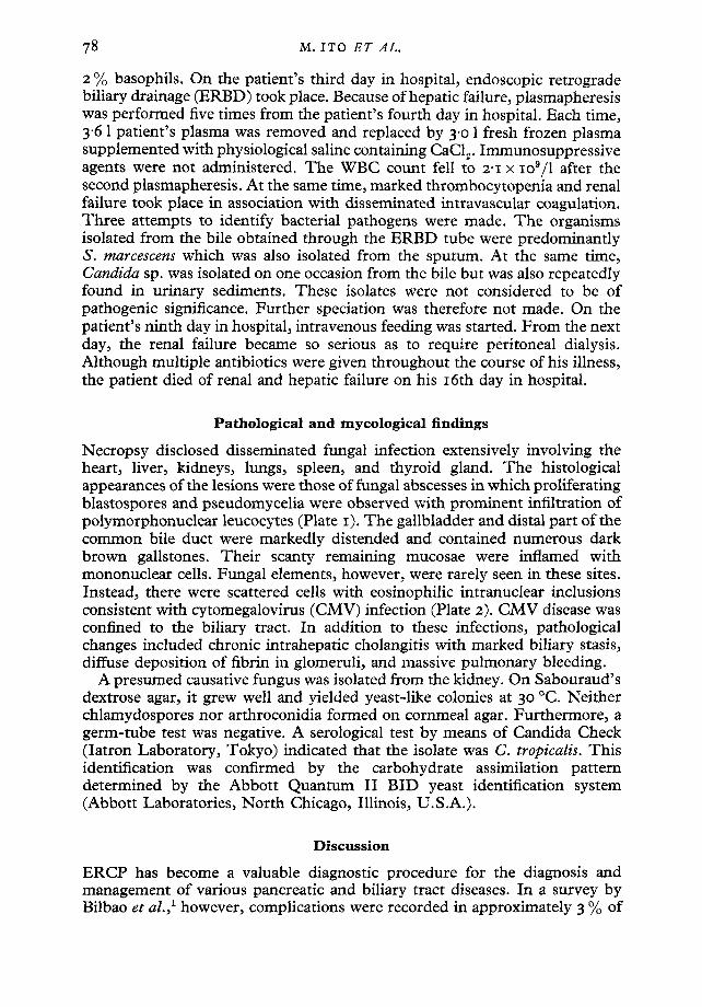
2 % basophils. On the patient's third day in hospital, endoscopic retrograde biliary drainage (ERBD) took place. Because of hepatic failure, plasmapheresis was performed five times from the patient's fourth day in hospital. Each time, 3"6 1 patient's plasma was removed and replaced by 3"0 1 fresh frozen plasma supplemented with physiological saline containing CaCI~. Immunosuppressive agents were not administered. The W B C count fell to 2-I x Io9/1 after the second plasmapheresis. At the same time, marked thrombocytopenia and renal failure took place in association with disseminated intravascular coagulation. Three attempts to identify bacterial pathogens were made. The organisms isolated from the bile obtained through the ERBD tube were predominantly S. marcescens which was also isolated from the sputum. At the same time, Candida sp. was isolated on one occasion from the bile but was also repeatedly found in urinary sediments. These isolates were not considered to be of pathogenic significance. Further speciation was therefore not made. On the patient's ninth day in hospital, intravenous feeding was started. From the next day, the renal failure became so serious as to requ'.'re peritoneal dialysis. Although multiple antibiotics were given throughout the course of his illness, the patient died of renal and hepatic failure on his I6th day in hospital.
Pathological and mycological findings
Necropsy disclosed disseminated fungal infection extensively involving the heart, liver, kidneys, lungs, spleen, and thyroid gland. The histological appearances of the lesions were those of fungal abscesses in which proliferating blastospores and pseudomycelia were observed with prominent infiltration of polymorphonuclear leucocytes (Plate I). The gallbladder and distal part of the common bile duct were markedly distended and contained numerous dark brown gallstones. Their scanty remaining mucosae were inflamed with mononuclear cells. Fungal elements, however, were rarely seen in these sites. Instead, there were scattered cells with eosinophilic intranuclear inclusions consistent with cytomegalovirus (CMV) infection (Plate 2). CMV disease was confined to the biliary tract. In addition to these infections, pathological changes included chronic intrahepatic cholangitis with marked biliary stasis, diffuse deposition of fibrin in glomeruli, and massive pulmonary bleeding.
A presumed causative fungus was isolated from the kidney. On Sabouraud's dextrose agar, it grew well and yielded yeast-like colonies at 3o °C. Neither chlamydospores nor arthroconidia formed on cornmeal agar. Furthermore, a germ-tube test was negative. A serological test by means of Candida Check (Iatron Laboratory, Tokyo) indicated that the isolate was C. tropicalis. This identification was confirmed by the carbohydrate assimilation pattern determined by the Abbott Quantum II BID yeast identification system (Abbott Laboratories, North Chicago, Illinois, U.S.A.).
Discuss ion
ERCP has become a valuable diagnostic procedure for the diagnosis and management of various pancreatic and biliary tract diseases. In a survey by Bilbao et al., 1 however, complications were recorded in approximately 3 % of

Journal of Infection Plates I and 2
Plate I. Blastospores and pseudomycelial forms of Candida tropicalis in a renal abscess. Grocott 's methenamine silver, x 968.
Plate 2. Chronically inflamed mucosa of the gallbladder with a few cytomegalic inclusion cells marked with arrows. Haematoxylin-eosin, x 32o.
M. I. ITo ~T AL. (Facing p. 78)
Septic candidiasis following E R C P 79
patients undergoing the procedure. The 6omplications comprised pancreatitis induced by injection of contrast medium, cholangitic or pancreatic sepsis, drug reactions, instrumental injury and aspiration pneumonia. Among these, the septic complications seemed the most hazardous because of their high mortality. In a review by Botoman et al., 2 the incidence of cul ture-proven bacteraemia following ERCP was estimated at 5"6 %, an incidence relatively higher than that encountered with other gastro-intestinal endoscopic pro- cedures. In a series from Davis et al., ~ Gram-negative bacteria, mainly derived from the intestinal flora, were by far the organisms most often isolated. In our case, at least two pathogens, namely S. marcescens and C. tropicalis, were responsible for the ERCP-associated sepsis. Even though bacterial culture was not done at necropsy, it was evident that the infection with S. marcescens had been almost eliminated by antibiotic therapy because there remained only systemic fungal abscesses in the necropsy specimens. In the retrospective view, the infection with C. tropicalis may explain the persistent septic condition intractable to antibiotic treatment.
The biliary tract is thought to be a rare site for fungi unless some obstruction produces persistent biliary stasis. In fact, meticulous attempts to seek fungal pathogens following endoscopic duodenal papillotomy have failed to recover any fungal pathogen from the duodenal mucosa or aspirated bile. 5 Even so, there have been several case reports in which primary biliary tract infections due to C. albicansfl. 7 C. parapsilosis, 6 or C. glabrata 8 were recorded in otherwise healthy persons. Thus , Candida sp. may potentially become an endogenous pathogen of biliary tract infection. Candida tropicalis seems to be in the minori ty among yeasts colonising human beings but has a pathogenic potential equal to that of C. albicans for producing disseminated infection in immunocompromised patients. 9
Although the pathogenesis of these septic complications is not fully understood, several explanations have been proposed by Davis et al. 3 They suggested that the biliary stasis due to underlying biliary tract disease may provide a favourable med ium for overgrowth with certain micro-organisms. In addition, bacterial contamination of the endoscope is likely to be the most important source of infection. Certainly, the insertion of an endoscope through the oral cavity may involve the risk of not only bacterial but also fungal contamination.
In our patient, the portal of entry of Candida sp. and Serratia sp. is assumed to have been through the mucosa of the biliary tract, some predisposing mucosal damage probably being required to enable these pathogens to invade the systemic circulation. 10 In our case, the gall stones may have caused primary injury to the mucosa. Fur thermore , the superimposed CMV infection may have rendered the mucosa vulnerable to invasion by other micro-organisms, The role of CMV as a pr imary pathogen of the digestive and biliary tracts is disputed. 11 This is especially so when, in immunocompeten t persons, it causes an inapparent infection associated with mucosal ulceration. At least, such a localised form of infection may cause retardation of healing of the mucosal damage.
In order to reduce septic complications following ERCP, this procedure should be restricted to selected patients requiring imminent surgical
8o M. ITO E T AL.
i n t e rven t ion , as p r o p o s e d by N e b e l et al. 4 T h e y also sugges ted tha t ear ly surgical i n t e r v e n t i o n shou ld be c o n s i d e r e d w i th in 24 h i f o b s t r u c t i o n is f o u n d .
T h e p r e s e n t case has a le r ted us to cons ide r funga l as well as bac te r ia l pa thoge ns as aet iological agents o f E R C P - a s s o c i a t e d sepsis.
References I. Bilbao MK, Dotter CT, Lee TG, Katon RM. Complications of endoscopic retrograde
cholangiopancreatography (ERCP). A study of IOOOO cases. Gastroenterology I976; 70: 314-32o.
z. Botoman VA, Surawicz CM. Bacteremia with gastrointestinal endoscopic procedures. Gastrointest Endosc 1986; 32: 342-346.
3. Davis JL, MiUigan FD, Cameron JL. Septic complications following endoscopic retrograde cholangiopancreatography. Surg Gynecol Obstet I975; 14o: 365-367 .
4. Nebel OT, Silvis SE, Rogers G, Sugawa C, Mandelstam P. Complications associated with endoscopic retrograde cholangiopancreatography. Results of the 1974 ASGE survey. Oastrointest Endosc 1975 ; 22: 34-36.
5. Hoechter W, Ottenjann R. Pilzbesiedlung des Ductus choledochus nach endoskopischer Papillotomie. Dtsch Med Wochenschr 1985; IlO: 1473-1474.
6. Gomez-Mateos JM, Sanchez Porto A, Martinez Parra D, Royo Balbontin A, Lesmes Serrano A, Aguilar Luque J. Disseminated candidiasis and gangrenous cholecystitis due to Candida spp. ff Infect Dis 1988 ; x58 : 653-654.
7. Irani M, Truong LD. Candidiasis of the extrahepatic biliary tract. Arch Pathol Lab Med 1986; IIO: 1087-1o90.
8. Valainis GT, Sachitano RA, Pankey GA. Cholecystitis due to Torulopsis glabrata, ff Infect Dis 1987; 156: 244-245.
9. Wingard JR, Merz WG, Saral R. Candida tropicalis. A major pathogen in immuno- compromised patients. Ann Intern Affed 1979; 91 : 539-543.
IO. Wingard JR, Dick JD, Merz WG, Sandford GR, Saral R, Burns WH. Pathogenicity of Candida tropicalis and Candida albicans after gastrointestinal inoculation in mice. Infect Immun 198o; 29: 8o8-813.
Ii . Morson BC, Dawson IMP, Day DW, Jass JR, Price AB, Williams GT. Inflammatory disorders. In: Morson BC, Dawson IMP, Day DW, Jass JR, Price AB, Williams GT, Eds. Afforson and Dawson's gastrointestinal pathology. Oxford: Blackwell Scientific Publications, I99O: 487.

![Journal Name: International Journal of Hepatobiliary and … · 2018. 12. 7. · 96 Endoscopic retrograde cholangiopancreatography [ERCP] ... 98 biopsy and confirm the diagnosis.](https://static.fdocuments.in/doc/165x107/60d37e520da2ff39e45fd202/journal-name-international-journal-of-hepatobiliary-and-2018-12-7-96-endoscopic.jpg)