Dissecting the role of novel EZH2 inhibitors in primary ...
17
RESEARCH Open Access Dissecting the role of novel EZH2 inhibitors in primary glioblastoma cell cultures: effects on proliferation, epithelial- mesenchymal transition, migration, and on the pro-inflammatory phenotype Giulia Stazi 1 , Ludovica Taglieri 2 , Alice Nicolai 2 , Annalisa Romanelli 1 , Rossella Fioravanti 1 , Stefania Morrone 2 , Manuela Sabatino 3 , Rino Ragno 1,3 , Samanta Taurone 4 , Marcella Nebbioso 4 , Raffaella Carletti 5 , Marco Artico 4 , Sergio Valente 1*† , Susanna Scarpa 2*† and Antonello Mai 1*† Abstract Background: Glioblastoma (GBM) is the most lethal and aggressive malignant primary brain tumor in adults. After surgical resection of the tumor, the patient typically should be subjected to chemotherapy (temozolomide, TMZ) and concomitant radiotherapy. Since the TMZ treatment does not lead to complete remission and often develops resistance, the identification of efficacious therapeutics is strongly to pursue. Among the epigenetic players, the H3K27 methyltransferase (MT) EZH2 (enhancer of zeste homologue 2) has been found overexpressed or mutated in several human cancers including gliomas, and its overexpression is associated with poor outcome in GBM. Two EZH2 inhibitors (EZH2i), UNC1999 and GSK343, suppressed GBM growth in vitro and in vivo indicating that EZH2i can be potential drugs against GBM. Results: Two new EZH2i, MC4040 and MC4041, were designed, prepared, and tested by us to determine their effects in primary GBM cell cultures. MC4040 and MC4041 displayed single-digit micromolar inhibition of EZH2, 10-fold less potency against EZH1, and no activity towards other MTs. In primary GBM cells as well as in U-87 GBM cells, the two compounds reduced H3K27me3 levels, and dose- and time-dependently impaired GBM cell viability without inducing apoptosis and arresting the cell cycle in the G0/G1 phase, with increased p21 and p27 levels. In combination with TMZ, MC4040 and MC4041 displayed stronger, but not additive, effects on cell viability. The potent clinical candidate as EZH2i tazemetostat, alone or in combination with TMZ, exhibited a similar potency of inhibition of GBM cell growth when compared to MC4040 and MC4041. At the molecular level, MC4040 and MC4041 reduced the VEGFR1/VEGF expression, reversed the epithelial-mesenchymal transition (EMT), and hampered cell migration and invasion attenuating the cancer malignant phenotype. Treatment of GBM cells with MC4040 and MC4041 also impaired the GBM pro-inflammatory phenotype, with a significant decrease of TGF-β, TNF-α, and IL-6, joined to an increase of the anti-inflammatory cytokine IL-10. (Continued on next page) © The Author(s). 2019 Open Access This article is distributed under the terms of the Creative Commons Attribution 4.0 International License (http://creativecommons.org/licenses/by/4.0/), which permits unrestricted use, distribution, and reproduction in any medium, provided you give appropriate credit to the original author(s) and the source, provide a link to the Creative Commons license, and indicate if changes were made. The Creative Commons Public Domain Dedication waiver (http://creativecommons.org/publicdomain/zero/1.0/) applies to the data made available in this article, unless otherwise stated. * Correspondence: [email protected]; [email protected]; [email protected] † Sergio Valente, Susanna Scarpa and Antonello Mai contributed equally to this work. 1 Department of Chemistry and Technologies of Drugs, Sapienza University of Rome, P.le A. Moro 5, 00185 Rome, Italy 2 Department of Experimental Medicine, Sapienza University of Rome, P.le A. Moro 5, 00185 Rome, Italy Full list of author information is available at the end of the article Stazi et al. Clinical Epigenetics (2019) 11:173 https://doi.org/10.1186/s13148-019-0763-5
Transcript of Dissecting the role of novel EZH2 inhibitors in primary ...
RESEARCH Open Access
Dissecting the role of novel EZH2 inhibitorsin primary glioblastoma cell cultures:effects on proliferation, epithelial-mesenchymal transition, migration, and onthe pro-inflammatory phenotypeGiulia Stazi1, Ludovica Taglieri2, Alice Nicolai2, Annalisa Romanelli1, Rossella Fioravanti1, Stefania Morrone2,Manuela Sabatino3, Rino Ragno1,3, Samanta Taurone4, Marcella Nebbioso4, Raffaella Carletti5, Marco Artico4,Sergio Valente1*†, Susanna Scarpa2*† and Antonello Mai1*†
Abstract
Background: Glioblastoma (GBM) is the most lethal and aggressive malignant primary brain tumor in adults. After surgicalresection of the tumor, the patient typically should be subjected to chemotherapy (temozolomide, TMZ) and concomitantradiotherapy. Since the TMZ treatment does not lead to complete remission and often develops resistance, theidentification of efficacious therapeutics is strongly to pursue. Among the epigenetic players, the H3K27 methyltransferase(MT) EZH2 (enhancer of zeste homologue 2) has been found overexpressed or mutated in several human cancersincluding gliomas, and its overexpression is associated with poor outcome in GBM. Two EZH2 inhibitors (EZH2i), UNC1999and GSK343, suppressed GBM growth in vitro and in vivo indicating that EZH2i can be potential drugs against GBM.
Results: Two new EZH2i, MC4040 and MC4041, were designed, prepared, and tested by us to determine their effects inprimary GBM cell cultures. MC4040 and MC4041 displayed single-digit micromolar inhibition of EZH2, 10-fold less potencyagainst EZH1, and no activity towards other MTs. In primary GBM cells as well as in U-87 GBM cells, the two compoundsreduced H3K27me3 levels, and dose- and time-dependently impaired GBM cell viability without inducing apoptosis andarresting the cell cycle in the G0/G1 phase, with increased p21 and p27 levels. In combination with TMZ, MC4040 andMC4041 displayed stronger, but not additive, effects on cell viability. The potent clinical candidate as EZH2i tazemetostat,alone or in combination with TMZ, exhibited a similar potency of inhibition of GBM cell growth when compared toMC4040 and MC4041. At the molecular level, MC4040 and MC4041 reduced the VEGFR1/VEGF expression, reversed theepithelial-mesenchymal transition (EMT), and hampered cell migration and invasion attenuating the cancer malignantphenotype. Treatment of GBM cells with MC4040 and MC4041 also impaired the GBM pro-inflammatory phenotype, with asignificant decrease of TGF-β, TNF-α, and IL-6, joined to an increase of the anti-inflammatory cytokine IL-10.
(Continued on next page)
© The Author(s). 2019 Open Access This article is distributed under the terms of the Creative Commons Attribution 4.0International License (http://creativecommons.org/licenses/by/4.0/), which permits unrestricted use, distribution, andreproduction in any medium, provided you give appropriate credit to the original author(s) and the source, provide a link tothe Creative Commons license, and indicate if changes were made. The Creative Commons Public Domain Dedication waiver(http://creativecommons.org/publicdomain/zero/1.0/) applies to the data made available in this article, unless otherwise stated.
* Correspondence: [email protected]; [email protected];[email protected]†Sergio Valente, Susanna Scarpa and Antonello Mai contributed equally tothis work.1Department of Chemistry and Technologies of Drugs, Sapienza University ofRome, P.le A. Moro 5, 00185 Rome, Italy2Department of Experimental Medicine, Sapienza University of Rome, P.le A.Moro 5, 00185 Rome, ItalyFull list of author information is available at the end of the article
Stazi et al. Clinical Epigenetics (2019) 11:173 https://doi.org/10.1186/s13148-019-0763-5

(Continued from previous page)
Conclusions: The two novel EZH2i MC4040 and MC4041 impaired primary GBM cell viability, showing even strongereffects in combination with TMZ. They also weakened the aggressive malignant phenotype by reducing angiogenesis, EMT,cell migration/invasion and inflammation, thus they may be considered potential candidates against GBM also forcombination therapies.
Keywords: Epigenetics, EZH2, Histone methylation, Glioblastoma, EMT, Inflammation
IntroductionGlioblastoma (GBM) is the most common and aggres-sive malignant primary brain tumour in adults [1]. Thestandard treatment consists of surgical resection of thetumour, followed by chemotherapy and concomitantradiotherapy [2]. To date, the sole drug in use againstGBM is the alkylating agent temozolomide (TMZ). Des-pite such combined and articulated regimen, the currenttherapeutic strategy is not a cure, and the prognosis forGBM patients remains poor with a median survivalaround 15 months and a 2-year survival rate of about27%. Additionally, the intrinsic or acquired resistance toTMZ reduces the therapeutic success. Several differentfactors obstacle the development of successful treat-ments against GBM, including its intrinsic aggressiveand infiltrative nature, and the high inter- and intra-tumour variability at the histopathological, genetic andepigenetic levels [3, 4]. Therefore, alternative and/orcomplementary therapeutic targets and strategies needto be found. In the last years, efforts have been done tothis aim, and researchers started also to evaluate thepossibility to use immunotherapy; however, no successhas been yet recorded [5]. A growing number of litera-ture evidences prove that epigenetic mechanisms are im-plied in the development and progression of GBM [6–8].The S-adenosyl-L-methionine (SAM)-dependent methyl-transferase EZH2 (enhancer of zeste homologue 2) is thecatalytic subunit of the polycomb repressive complex 2(PRC2), responsible of the methylation of the lysine 27of the histone 3 (H3K27), a mark of gene silencing [9].EZH2 and the PRC2 complex can silence many genes in-volved in cell proliferation, cell-cycle regulation, cell dif-ferentiation, and self-renewal [9]. EZH2 has been foundoverexpressed or mutated in several human cancers [9]including gliomas, where its overexpression has beencorrelated with the glioma grade and poor prognosis[10, 11]. Accordingly, EZH2 depletion by RNA interfer-ence in glioma cells led to cell growth inhibition andcycle arrest in the G0/G1 phase both in vitro and in vivo[10, 12]. Moreover, EZH2 regulates cell stemness andepithelial to mesenchymal transition (EMT) in gliomas[11], and it is involved in the development of multidrugresistance, with its inhibition restoring the normal drugsensitivity in GBM [13].
To date, numerous molecules have been synthesizedand evaluated in preclinical and clinical settings as com-petitive, catalytic EZH2 inhibitors (Fig. 1) [14], and someof them including GSK126, tazemetostat and CPI-1205,are being tested in clinical studies [14]. Moreover, few ofthem (UNC1999 and GSK343, an analogue of GSK126)have been tested against GBM in vitro and in vivo, prov-ing the relevance of EZH2 as target to fight in this dis-order [15, 16].Recently, through a pruning strategy applied on known
indazole-containing EZH2 inhibitors (EZH2i) (such asEPZ005687 and UNC1999, Fig. 1), our group reported thenovel pyrazole-based EZH2i MC3629 (Fig. 1), potent at lowmicromolar doses and able to reduce H3K27me3 levels, toarrest cell proliferation and to induce autophagy in a panelof cancer cell lines (SK-N-BE neuroblastoma, MDA-MB231 breast cancer, and K562 leukaemia cells) [17].When tested in Sonic Hedgehog (SHH) medulloblastoma(MB) stem-like cells, MC3629 impaired cell proliferationand self-renewal inducing apoptosis [18]. In MB xeno-grafted mice, MC3629 showed a significant decrease oftumour volume, a reduction of stemness and cell prolifera-tion, and induction of apoptosis [18]. Mimicking thebenzo-cracking approach followed for MC3629, startingfrom the structures of known indole-based EZH2 inhibitors(such as GSK126, EI1, and CPI-1205, Fig. 1) we designedand synthesized a series of pyrrole dimethylpyridone-containing compounds to be tested against EZH2. Amongthem, MC4040 and MC4041 (Fig. 1) were tested in GBMcells including primary GBM cell cultures, to determinetheir antiproliferative effects alone and in combination withtemozolomide. To dissect the molecular mechanisms ofthese new EZH2 inhibitors, their efficacy in mitigating theGBM malignant phenotype has been determined throughthe reduction of VEGFR1/VEGF expression, reversion ofthe EMT process, inhibition of invasive phenotype, and de-crease of inflammatory cytokines levels.
Materials and MethodsChemistryMelting points were determined on a Buchi 530 meltingpoint apparatus and are uncorrected. 1H NMR (nuclearmagnetic resonance) spectra were recorded at 400MHzon a Bruker AC 400 spectrometer; chemical shifts are
Stazi et al. Clinical Epigenetics (2019) 11:173 Page 2 of 17

reported in δ (ppm) units relative to the internal refer-ence tetramethylsilane (Me4Si). EIMS spectra were re-corded with a Fisons Trio 1000 spectrometer; onlymolecular ions (M+) and base peaks are given. All com-pounds were routinely checked by TLC (thin layer chro-matography), 1H NMR and 13C NMR spectra. TLC wasperformed on aluminium-backed silica gel plates (MerckDC, Alufolien Kieselgel 60 F254) with spots visualized byUV light. All solvents were reagent grade and, when ne-cessary, were purified and dried by standard methods.The concentration of solutions after reactions and ex-tractions involved the use of a rotary evaporator operat-ing at reduced pressure of ca. 20 Torr. Organic solutionswere dried over anhydrous sodium sulphate. Elementalanalysis has been used to determine the purity of the de-scribed compounds, that is > 95%. Analytical results arewithin ± 0.40% of the theoretical values. All chemicalswere purchased from Sigma-Aldrich Chimica, Milan(Italy), or from Alfa Aesar, Karlsruhe (Germany), andwere of the highest purity.
Synthesis of 1-(3-bromophenyl)-2,5-dimethyl-1H-pyrrole(1)A solution of 2,5-hexanedione (8.50mmol, 1.2 eq, 1 mL)in glacial acetic acid (3mL) was prepared in a flame driedsealed tube, and 3-bromoaniline (7.08mmol, 1.0 eq, 1.22g, 0.77mL) was added to the solution. The reaction solu-tion was stirred at 100 °C for 2 h. After this time the reac-tion was complete, the acetic acid was removed in vacuo(azeotrope with toluene) and the residue was purified bysilica gel chromatography, eluting with the n-hexane:chloroform (60:1) mixture. The product was obtained as a
white solid (5.31 mmol, 1.33 g, 75%). m.p. 84–85 °C (n-hexane). 1H-NMR (CDCl3, 400MHz, δ; ppm): δH 1.96(6H, s, C(2)CH3, C(5)CH3), 5.82 (2H, s, C(3)H, C(4)H pyr-role), 7.08–7.11 (1H, ddd, J = 7.6 Hz, 1.8 Hz, 0.8 Hz, aro-matic proton), 7.26 (1H, t, J = 8Hz, aromatic proton), 7.32(1H, t, J = 1.6 Hz, aromatic proton), 7.45-7.48 (1H, ddd, J= 8Hz, 1.8, 1.2 Hz, aromatic proton) ppm. MS (EI) m/z[M]+: 249.02. The reported data are in good agreementwith the literature [19, 20].
General procedure for the synthesis of the intermediates2a,b. Example: Synthesis of 1-(3-(2,5-dimethyl-1H-pyrrol-1-yl)phenyl)piperidine (2b)In a flame dried sealed tube, 1-(3-bromophenyl)-2,5-di-methyl-1H-pyrrole 1 (1.59 mmol, 1.0 eq, 0.40 g), piperi-dine (3.20 mmol, 2 eq, 0.27 g, 0.316 mL), palladium(II)acetate (Pd(OAc)2) (0.024 mmol, 1.5% mol, 5.4 mg), tri-tert-butylphosphonium tetrafluoroborate (PH(tBu)3BF4)(0.0192mmol, 1.2% mol, 5.6 mg), and potassium tert-butoxide (3.59 mmol, 2.25 eq, 0.404 g) were added in se-quence and suspended in dry toluene (3.0 mL). The sys-tem was degassed (N2) and left under stirring at 80 °Cfor 16 h. After this time, the reaction was quenched byadding ethyl acetate, and it was filtered over celite pad.The filtrate was concentrated in vacuo and the crudewas purified by silica gel chromatography eluting withthe n-hexane:ethyl acetate (5:1) mixture, to give the 1-(3-(2,5-dimethyl-1H-pyrrol-1-yl)phenyl)piperidine 2b asa colourless oil (1.27 mmol, 0.32 g, 80%). 1H-NMR (d6-DMSO, 400MHz, δ; ppm): δH 1.59-1.60 (6H, m, piperi-dine protons), 1.97 (6H, s, C(2)CH3, C(5)CH3 pyrrole),3.17-3.20 (4H, m, piperidine protons), 5.70 (2H, s,
Fig. 1 Structures of EZH2 inhibitors studied in a clinical setting and/or in GBM models, including MC4040 and MC4041 used in thepresent research
Stazi et al. Clinical Epigenetics (2019) 11:173 Page 3 of 17
C(3)H, C(4)H pyrrole), 6.56 (1H, dd, J = 7.6 Hz, 2.0 Hz,aromatic proton), 6.69 (1H, t, J = 2.0 Hz, aromatic pro-ton), 6.97 (1H, dd, J = 7.6 Hz, 2.0 Hz, aromatic proton),7.29 (1H, t, J = 7.6 Hz, aromatic proton) ppm. MS (EI)m/z [M]+: 254.18.
Chemical and physical characterization of 4-(3-(2,5-dimethyl-1H-pyrrol-1-yl)phenyl)morpholine (2a): lightyellow oil (yield 82%)1H-NMR (d6-DMSO, 400MHz, δ; ppm): δH 1.97 (6H, s,C(2)CH3, C(5)CH3 pyrrole), 3.16 (4H, t, J = 11.0 Hz,morpholine protons), 3.73 (4H, t, J = 11.0 Hz, morpho-line protons), 5.76 (2H, s, C(3)H, C(4)H pyrrole), 6.63(1H, dd, J = 8.2 Hz, 2.0 Hz, aromatic proton), 6.73 (1H, t,J = 2.0 Hz, aromatic proton), 6.99 (1H, dd, J = 8.2 Hz,2.0 Hz, aromatic proton), 7.32 (1H, t, J = 8.0 Hz, aro-matic proton) ppm. MS (EI) m/z [M]+: 256.16.
General procedure for the synthesis of pyrrole-3-carboxylic acids (3a,b). Example: Synthesis of 2,5-dimethyl-1-(3-morpholinophenyl)-1H-pyrrole-3-carboxylicacid (3a)In a sealed tube, 4-(3-(2,5-dimethyl-1H-pyrrol-1-yl)phenyl)morpholine 2a (0.84 mmol, 1.0 eq, 0.22 g)was dissolved in 1,2-dichloroethane (2 mL), the solu-tion was cooled to 0 °C, and trichloroacetyl chloride(2.53 mmol, 3.0 eq, 0.28 mL) was added dropwise tothe solution. The reaction was stirred at 70 °C for 1h. After this time the reaction was complete, and thevolatiles were removed in vacuo. The residue wasdissolved in an ethanol/tetrahydrofuran (1:1) mix-ture, the solution was cooled to 0 °C, and 2 M aque-ous solution of potassium hydroxide (8.43 mmol, 10eq, 0.47 g) was added dropwise to the solution. Thereaction mixture was stirred at room temperaturefor 1 h and then at 80 °C for 3 h. After this time thereaction was complete, and the organic solvents wereremoved in vacuo. The byproducts were extractedfrom the basic aqueous layer with ethyl acetate (3 ×10 mL). The aqueous layer was then cooled to 0 °Cand acidified till pH 2 by addition of a 2 M aqueoussolution of HCl. The product precipitated, and itwas filtered, rinsed with distilled water and dried inoven (60 °C). Yield 69%. m.p. 173–175 °C (toluene/acetonitrile). 1H-NMR (d6-DMSO, 400 MHz, δ; ppm):δH 1.94 (3H, s, C(5)CH3 pyrrole), 2.23 (3H, s,C(2)CH3 pyrrole), 3.15-3.18 (4H, m, morpholine pro-tons), 3.71–3.74 (4H, m, morpholine protons), 6.21(1H, s, C(4)H pyrrole), 6.76 (1H, s, aromatic proton),7.02 (1H, d, J = 7.6 Hz, aromatic proton), 7.13 ( 1H,d, J = 7.6 Hz, aromatic proton), 7.41 (1H, t, J = 7.6Hz, aromatic proton), 11.66 (1H, bs, COOH) ppm.MS (EI) m/z [M]+: 300.15.
Chemical and physical properties of 2,5-dimethyl-1-(3-(piperidin-1-yl)phenyl)-1H-pyrrole-3-carboxylic acid (3b):m.p. 178–180 °C (toluene/acetonitrile)1H-NMR (d6-DMSO, 400MHz, δ; ppm): δH 1.56 (6H,m, piperidine protons), 1.94 (3H, s, C(5)CH3 pyrrole),2.21 (3H, s, C(2)CH3 pyrrole), 3.19-3.21 (4H, m, piperi-dine protons), 6.20 (1H, s, C(4)H pyrrole), 6.59 (1H, d, J= 7.6 Hz, aromatic proton), 6.75 (1H, bs, aromatic pro-ton), 7.30 (1H, dd, J = 8 Hz, 2 Hz, aromatic proton), 7.33(1H, t, J = 8 Hz, aromatic proton), 11.56 (1H, bs, COOH)ppm. MS (EI) m/z [M]+: 298.17.
General procedure for the synthesis of pyrrole amides(MC4040 and MC4041). Example: Synthesis of N-((4,6-dimethyl-2-oxo-1,2-dihydropyridin-3-yl)methyl)-2,5-dimethyl-1-(3-morpholinophenyl)-1H-pyrrole-3-carboxamide (MC4040)In a flame dried round bottom flask, 2,5-dimethyl-1-(3-mor-pholinophenyl)-1H-pyrrole-3-carboxylic acid (0.33mmol,1.0 eq, 100.0mg) was solved in dry N,N-dimethylformamide(DMF) (1mL) under N2 atmosphere. Triethylamine (2.33mmol, 7.0 eq, 0.24mL), and O-(benzotriazol-1-yl)-N,N,N’,N’-tetramethyluronium tetrafluoroborate (TBTU) (0.40mmol,1.2 eq, 0.13 g) were added to the reaction solution, and thesystem was left under stirring at room temperature for 40min. After this time, 3-(aminomethyl)-4,6-dimethylpyridin-2(1H)-one hydrochloride 4 [21] (0.37mmol, 1.1 eq, 69mg)was added to the solution and the reaction was stirred underN2 at 60 °C for 4 h. After this time, the reaction wascomplete, and it was quenched by addition of 3mL of a satu-rated aqueous solution of NaCl. The product precipitated,and it was filtered and rinsed with distilled water. The solidwas dried and further purified by silica gel chromatographyeluting with the chloroform:methanol (35:1) mixture, to giveMC4040 (0.18mmol, 76.7mg, 53%). m.p. 178-180 °C (aceto-nitrile). 1H NMR (DMSO-d6, 400MHz, δ, ppm): δH 1.92(3H, s, C(5)CH3 pyrrole), 2.12 (3H, s, CH3 dimethylpyridone),2.18 (3H, s, CH3 dimethylpyridone), 2.21 (3H, s, C(2)CH3
pyrrole), 3.16 (4H, t, J = 4.6Hz, morpholine protons), 3.72(4H, t, J = 4.6Hz, morpholine protons), 4.22 (2H, d, J = 5.2Hz, -CH2NHCO-), 5.86 (1H, s, dimethylpyridone CH), 6.26(1H, s, C(4)H pyrrole), 6.64 (1H, d, J = 8Hz, aromatic pro-ton), 6.76 (1H, s, aromatic proton), 7.03 (1H, d, J = 7.2Hz,aromatic proton), 7.34-7.39 (2H, m, aromatic proton and-CH2NHCO-), 11.47 (1H,bs, -NH- pyridone) ppm. MS (EI)m/z [M]+: 434.23.
Chemical and physical characterization of N-((4,6-dimethyl-2-oxo-1,2-dihydropyridin-3-yl)methyl)-2,5-dimethyl-1-(3-(piperidin-1-yl)phenyl)-1H-pyrrole-3-carboxamide (MC4041)1H NMR (DMSO-d6, 400MHz, δ, ppm): δH 1.54–1.60(6H, m, piperidine protons), 1.92 (3H, s, C(5)CH3 pyr-role), 2.11 (3H, s, CH3 dimethylpyridone), 2.18 (3H, s,
Stazi et al. Clinical Epigenetics (2019) 11:173 Page 4 of 17

CH3 dimethylpyridone), 2.21 (3H, s, C(2)CH3 pyrrole),3.18–3.20 (4H, m, piperidine protons), 4.22 (2H, d, J =5.2 Hz, -CH2NHCO-), 5.86(1H, s, dimethylpyridone CH),6.25 (1H, s, C(4)H pyrrole), 6.55 (1H, d, J = 7.6 Hz, aro-matic proton), 6.70 (1H, bs, aromatic proton), 7.01 (1H,dd, J = 2 Hz, 8.4 Hz, aromatic proton), 7.34 (1H, t, J = 8Hz, aromatic proton), 7.40 (1H, t, J = 5.2 Hz,-CH2NHCO-), 11.48 (1H, bs, -NH- pyridone). m.p. 135-137 °C (benzene); Yield = 55%. MS (EI) m/z [M]+:432.25.
BiochemistryEZH2/PRC2 complex assayThe EZH2 substrate (0.05 mg/mL core histone) wasadded in the freshly prepared reaction buffer (50 mMTris-HCl (pH 8.0), 50 mM NaCl, 1 mM EDTA, 1 mMDTT, 1 mM PMSF, 1% DMSO). The PRC2 complex[complex of human EZH2, human EED, human SUZ12,human AEBP2, and human RbAp48] was delivered intothe substrate solution and the mixture was mixed gently.Afterwards, the tested compounds dissolved in DMSOwere delivered into the enzyme/substrate reaction mix-ture by using Acoustic Technology (Echo 550, LabCyteInc. Sunnyvale, CA) in a nanolitre range, and 3H-SAMwas added into the reaction mixture to initiate the reac-tion. The reaction mixture was incubated for 1 h at30 °C and then it was delivered to filter-paper for detec-tion. The data were analysed using Excel and GraphPadPrism software for IC50 curve fits.
EZH1/PRC2 complex, G9a, PRMT1, and DNMT1 assaysThe appropriate methyltransferase (MT) substrate (0.05mg/ml core histone for EZH1 complex, 5 μM histoneH3 (1-21) peptide for G9a, 5 μM histone H4 for PRMT1,and 0.001 mg/ mL poly(dI-dC) for DNMT1) was addedin freshly prepared reaction buffer (50 mM Tris-HCl(pH 8.5), 5 mM MgCl2, 50 mM NaCl, 0.01 % Brij35, 1mM DTT, 1 % DMSO). The MT enzyme was deliveredinto the substrate solution and the mixture was mixedgently. Afterwards, the tested compounds dissolved inDMSO were delivered into the enzyme/substrate reac-tion mixture by using Acoustic Technology (Echo 550,LabCyte Inc. Sunnyvale, CA) in nanolitre range, and1 μM 3H-SAM was also added into the reaction mixtureto initiate the reaction. The reaction mixture was incu-bated for 1 h at 30 °C and then it was delivered to filter-paper for detection. The data were analysed using Exceland GraphPad Prism software for IC50 curve fits.
Molecular modellingMolecular docking simulations were run on MC4041and MC4040 modelled conformation to gather infor-mation on their likely binding modes into EZH2. Tothis, eight experimental structures of human PRC2 in
complex with several inhibitors (pdb entry codes:4w2r, 5ch2, 5ij7, 5ls6, 5ch1, 5hyn, 5ij8 and 6b3w) wereretrieved from PDB [22] (www.rcsb.org). PCR2 co-crystallized complexes were structurally inspected andexperimentally missing residues were modelled bymeans of the programme MODELLER ver 9.19 [23].To avoid insertion of too unfolded parts, only 10 ofthe about 80 initial missing residues were fully mod-elled. The internal gaps were all automatically filledsimilarly as described in the MODELLER manual. Themodelled complexes were then subjected to single-point energy geometry optimization in explicit waterand the minimized complexes. Cross-docking experi-ments were performed with the programme PLANTS[24] using the ChemPlp scoring functions. Chemicalcompounds were parametrized with AM1-BCC [25]partial charges and GAFF v2.1 [26] Lennard-Jones andvalence parameters, using AmberTools 17 antecham-ber and parmchk2 utilities [27, 28]. The OPC [29]water model and AMBER14SB [30] forcefield wereused during complexes minimizations. By means ofmolecular mechanics/generalized Born surface area(MMGBSA) method [31], the protein-ligand bindingfree energies were evaluated. MarvinSketch 17.1.16(http://www.chemaxon.com) was used for drawing thechemical structures, which were then converted in3D-Structures with OpenBabel 2.4.1 [32] employingMMFF94 [33] force field.
In cell studiesCell culture and treatmentsThree human GBM primary cell cultures (GL1, GL2,GL3) were established from surgical tissues of three dif-ferent patients, and in addition, one human normal der-mal fibroblast primary culture (HF) was obtained. Allthe primary cell cultures were prepared as previously de-scribed [34]. Additionally, a human GBM cell line U-87MG (ATCC HTB-14), here indicated as U-87, was uti-lized. The U-87 cell line was grown in EMEM mediumsupplemented with 10% foetal calf serum (FCS), 2 mMglutamine, 50 U/mL penicillin-streptomycin, 1 mM non-essential amino acid, 1 mM sodium pyruvate. GL1, GL2,GL3 and HF were grown in DMEM medium supple-mented with 10% FCS, 2 mM glutamine, 50 U/mLpenicillin-streptomycin. MC 4040 and MC 4041 weresolubilized in dimethylsulfoxide (DMSO) (Sigma-Al-drich) at 10 mM stock solution and utilized to final con-centrations from 10 to 50 μM. Control cells were treatedwith equivalent amounts of DMSO in each experiment.Staurosporine (Sigma-Aldrich) was dissolved in DMSOin a 1 mM stock solution and used at 5 μM for 6 h.Tazemetostat was solubilized in a 10mM DMSO stocksolution and utilized at 25 μM. Temozolomide was
Stazi et al. Clinical Epigenetics (2019) 11:173 Page 5 of 17

solubilized in a 100mM DMSO stock solution and uti-lized at 100 μM.
Cytotoxicity assayTo determine cytotoxicity, sulforhodamine B colorimet-ric assay was performed: 1.5 × 10⁴ cells were plated on96 well plates, grown for 24 h and treated with differentconcentrations of MC4040 and MC4041 (10, 25, and 50μM), with tazemetostat (25M) and/or with temozolo-mide (100 μM) for 48, 72, 96 and 120 h. Control cellswere treated with equivalent amounts of DMSO. Cellswere then fixed with 50% trichloroacetic acid for 1 h at4 °C and stained for 30 min at room temperature with0.4% sulforhodamine B in 1% acetic acid. Excess dye wasremoved by washing four times with 1% acetic acid.Protein-bound dye was dissolved in 10 mM TRIS pH 10,and optical density (OD) was determined at 510 nmusing a microplate reader.
Flow cytometry analysisGL1 and U-87 cells were seeded in 60-mm culture platesand grown for 48 h untreated or treated with 25 μMMC4040 and MC4041. The cells were harvested by tryp-sinization, washed twice with cold PBS, and fixed in 70%ethanol at 4 °C overnight. After fixation, the cells wererinsed with PBS and incubated with 10mg/mL RNaseand 1mg/mL propidium iodide (Sigma-Aldrich) at roomtemperature for 2 h in the dark. The cell cycle distribu-tion was measured and analysed by FACS Calibur flowcytometry using the Cell Quest analysis programme todetect the distribution of subpopulations through thecell cycle. Each experiment was performed in triplicate.
Western blot analysisCell lysates were obtained scraping the cells in lysis buf-fer 1% Triton, 0.1% SDS, 150 mM NaCl, 50 mM TRIS-HCl pH 7.4, 2 mM EDTA plus protease inhibitor cocktailtablet (Roche Applied Sciences) for 30 min at 4 °C. Thelysates were then centrifuged at 12,000 rpm for 15minat 4 °C. The protein concentration was evaluated by Bio-Rad Protein Concentration Assay. Samples of lysate(100 μg) were separated by molecular weight on 10 or12% SDS-PAGE and then transferred into a nitrocellu-lose membrane. Blots were blocked for 1 h at roomtemperature in 5% non-fat dry milk and then incubatedovernight at 4 °C with rabbit polyclonal anti-H3K27me3antibodies (cell signalling technology) diluted 1:1000 in5% BSA, 0.1% Tween 20 TRIS-buffered saline, or alter-natively for 1 h at room temperature with all the otherprimary antibodies in TRIS-buffered saline. The primaryantibodies were the following: mouse anti-Parp1 (1:500diluted) (Santa Cruz Biotechnology); rabbit anti-p21Waf1/Cip1 (1:1000 diluted) (Cell Signaling); mouse anti-p27 (1:500 diluted) (Santa Cruz Biotechnology); rabbit
anti-ß-actin (1:1000 diluted) (Sigma-Aldrich) rabbit anti-E-cadherin (1:1000 diluted) (Gene Tex); rabbit anti-N-cadherin (1:1000 diluted) (Gene Tex); rabbit anti-MMP-2 (1:1000 diluted) (Gene Tex); rabbit anti-MMP-9 (1:500diluted) (Biomol); mouse anti-TGF-ß (1:500 diluted)(Santa Cruz Biotechnology); rabbit anti-TNF-α (1:1000diluted) (Elabscience); mouse anti-IL-6 (1:500 diluted)(Santa Cruz Biotechnology); mouse anti-IL-10 (1:500)(Santa Cruz Biotechnology). Blots were washed in TRIS-buffered saline with 0.1% Tween 20 and then incubatedwith horseradish peroxidase-conjugated anti-rabbit oranti-mouse antibodies (1:5000 diluted) (Sigma-Aldrich).The filters were then developed by enhanced chemilu-minescence (Super Signal West Pico ChemiluminescentSubstrate, Thermo Scientific) using Kodak X-Omatfilms. The densitometry quantitation of the bands wasperformed using Image J software.
ImmunofluorescenceThe cells were grown directly on Labteck chamber slides(Nunc) for 24 h and then used untreated or treated for48 h with 25 μM MC4040 or MC4041. The cells werethen washed with PBS with Ca/Mg and fixed with abso-lute cold methanol for 5 min (only for nestin staining) orwith 4% buffered paraformaldehyde for 20 min at 4 °C(for all other antibodies). The cells were incubated with3% bovine serum albumin for 1 h at room temperatureand then with anti-glial fibrillary acidic protein (GFAP)rabbit antibody (1:200 diluted, Abcam) or anti-vascularendothelial growth factor (VEGF) mouse antibody (1:100diluted, Santa Cruz Biotechnology) or anti-VEGF-receptor1 (VEGFR1) mouse antibody (1:100 diluted,Abcam) for 1 h at room temperature; with anti-nestinrabbit antibody (1:200 diluted, Abcam) overnight at 4 °C.Cells were then washed twice with PBS with Ca/Mg andthen incubated with the secondary anti-rabbit antibodyAlexa Fluor 594 conjugated (Invitrogen, 1:400 diluted) oranti-mouse antibody Alexa Fluor 488 conjugated (Invi-trogen, 1:200 diluted) for 1 h at room temperature. Thecells were finally washed twice with PBS with Ca/Mg,mounted with Prolong Antifade reagent (Life Technolo-gies) and analysed by a fluorescence microscope (Olym-pus BX52); imagine acquisition and processing wereconducted by IAS 2000 software.
Invasion assayInvasion assay was performed with Bio Coat Matrigel In-vasion Chambers (Corning), consisting of inserts with an8 μm pores size membrane that was previously treatedwith Matrigel matrix. For invasion assay 2.5 × 10⁵/mLcells were plated in serum-free medium plus vehicleDMSO or in serum-free medium plus 20 μM MC4040and MC4041 in the insert chamber, the lower chamberinstead contained only complete medium (with FCS).
Stazi et al. Clinical Epigenetics (2019) 11:173 Page 6 of 17

After 48 h of culture at standard conditions, the insertswere washed with PBS with Ca/Mg and fixed by 100%methanol for 20 min at 4 °C, then washed twice withPBS with Ca/Mg and stained for 20 min at roomtemperature with crystal violet. The inserts were thenmounted on a slide with glycerol and the cells that mi-grated through the filter pores to the lower side of themembrane were counted by an optical microscope(Olympus BX52). Imagine acquisition and processingwere conducted by IAS 2000 software.
Statistical analysis and graphic programmesAll results were analysed by ANOVA, and the signifi-cance was evaluated by the Tukey HSD post hoc test(Honestly Significant Difference). All figures were elabo-rated with Adobe Photoshop CS5 and all graphs withGraph Pad Prism 5.0
ResultsSynthesis of MC4040 and MC4041The synthesis of MC4040 and MC4041 started with thepreparation of the pyrrole key intermediate 1 through aPaal-Knorr reaction (Scheme 1). More in detail, 2,5-hexane-dione was treated with 3-bromoaniline in the presence ofacetic acid to afford the 1-(3-bromophenyl)-2,5-dimethyl-1H-pyrrole 1, which underwent a Buchwald-Hartwigreaction by using morpholine or piperidine, palladium (II)acetate (Pd(OAc)2) as catalyst and tri-tert-butylphospho-nium tetrafluoroborate (PH(tBu)3BF4) as ligand, and pro-viding 2a and 2b, respectively, in good yields (Scheme 1)[35]. The subsequent Friedel-Craft acylation performed on2a,b with trichloroacetyl chloride gave the correspondingcrude 3-trichloroacetyl pyrroles, which were directly con-verted into the related 3-pyrrolecarboxylic acids 3a,b(Scheme 1). Further coupling of the acids 3a,b, activatedwith O-(benzotriazole-1-yl)-N,N,N’,N’-tetramethyluroniumtetrafluoroborate (TBTU), with the 3-(aminomethyl)-4,6-
dimethylpyridin-2(1H)-one hydrochloride 4 [21] in thepresence of triethylamine yielded the final compoundsMC4040 and MC4041 (Scheme 1).
Assessment of EZH2 inhibition activity and selectivityagainst other methyltransferasesThe newly synthesized compounds have been screenedin a 10-dose IC50 mode with 2-fold serial dilutionstarting from 200 μM solutions, in an in vitro enzym-atic assay against a human five component PRC2 com-plex, containing EZH2, EED, SUZ12, RbAp48, andAEBP2, to evaluate their ability to inhibit the EZH2catalytic activity. The assay was performed using corehistone as substrate and 3H-SAM as co-substrate. S-Adenosyl-L-homocysteine (SAH), GSK126 and taze-metostat were used as reference compounds. Com-pounds MC4040 and MC4041 proved to inhibit EZH2displaying IC50 values in the low micromolar range(Table 1). Both compounds proved to inhibit also thePRC2-EZH1 complex in vitro, displaying however 10-fold higher IC50 values (Table 1). When tested againstother SAM-dependent methyltransferases such as G9a(another lysine methyltransferase), PRMT1 (an argin-ine methyltransferase), and DNMT1 (a DNA methyl-transferase), no inhibition was recorded for bothcompounds up to 200 μM. Only MC4041 showed aslight PRMT1 inhibition (11.9%) at 200 μM.
Molecular modelling studiesMolecular modelling studies were performed to high-light any difference in the binding mode of the twonovel compounds. Cross-docking experiment revealedthat the programme PLANTS is suitable to performdocking experiments for potential ligands on PRC2 as adocking accuracy of 90% was achieved [36]. MC4040and MC4041 lowest energy docking conformations wereassociated with the PRC2 protein obtained from the
Scheme 1 Reagents and conditions: (a) glacial acetic acid, sealed tube, 100 °C, 1.5 h (35-80%); (b) Pd(OAc)2, PH(tBu)3BF3, potassium tert-butoxide,dry toluene, sealed tube, N2, 80 °C, 4 h (76-80%); (c) trichloroacetyl chloride, dry 1,2-dichloroethane, sealed tube, 0 °C to 70 °C, 1 h; (d) I. 2 N KOH(aq), EtOH, r.t. to 60 °C; II. 2 N HCl (aq) (54-73 %); (e) triethylamine, TBTU, dry DMF, N2, r. t., 4 h (52-75%)
Stazi et al. Clinical Epigenetics (2019) 11:173 Page 7 of 17
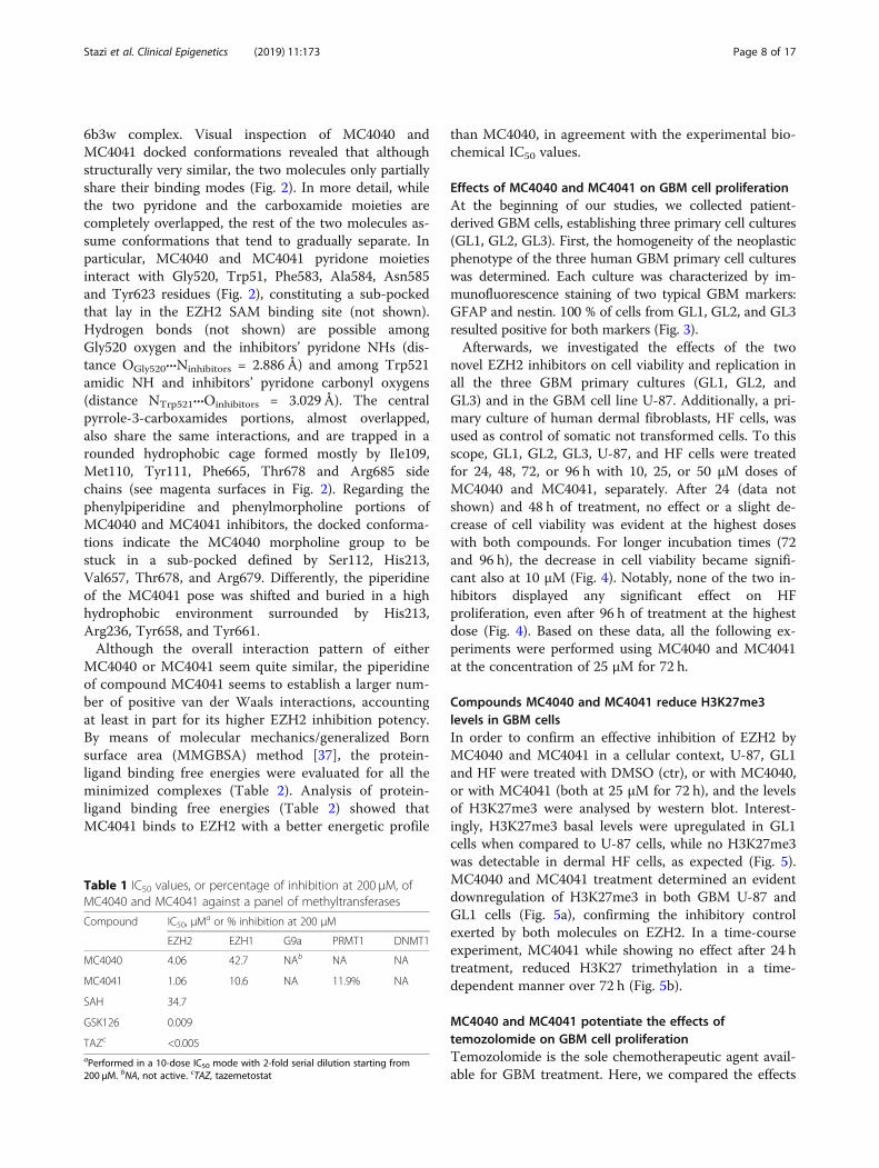
6b3w complex. Visual inspection of MC4040 andMC4041 docked conformations revealed that althoughstructurally very similar, the two molecules only partiallyshare their binding modes (Fig. 2). In more detail, whilethe two pyridone and the carboxamide moieties arecompletely overlapped, the rest of the two molecules as-sume conformations that tend to gradually separate. Inparticular, MC4040 and MC4041 pyridone moietiesinteract with Gly520, Trp51, Phe583, Ala584, Asn585and Tyr623 residues (Fig. 2), constituting a sub-pockedthat lay in the EZH2 SAM binding site (not shown).Hydrogen bonds (not shown) are possible amongGly520 oxygen and the inhibitors’ pyridone NHs (dis-tance OGly520···Ninhibitors = 2.886 Å) and among Trp521amidic NH and inhibitors’ pyridone carbonyl oxygens(distance NTrp521···Oinhibitors = 3.029 Å). The centralpyrrole-3-carboxamides portions, almost overlapped,also share the same interactions, and are trapped in arounded hydrophobic cage formed mostly by Ile109,Met110, Tyr111, Phe665, Thr678 and Arg685 sidechains (see magenta surfaces in Fig. 2). Regarding thephenylpiperidine and phenylmorpholine portions ofMC4040 and MC4041 inhibitors, the docked conforma-tions indicate the MC4040 morpholine group to bestuck in a sub-pocked defined by Ser112, His213,Val657, Thr678, and Arg679. Differently, the piperidineof the MC4041 pose was shifted and buried in a highhydrophobic environment surrounded by His213,Arg236, Tyr658, and Tyr661.Although the overall interaction pattern of either
MC4040 or MC4041 seem quite similar, the piperidineof compound MC4041 seems to establish a larger num-ber of positive van der Waals interactions, accountingat least in part for its higher EZH2 inhibition potency.By means of molecular mechanics/generalized Bornsurface area (MMGBSA) method [37], the protein-ligand binding free energies were evaluated for all theminimized complexes (Table 2). Analysis of protein-ligand binding free energies (Table 2) showed thatMC4041 binds to EZH2 with a better energetic profile
than MC4040, in agreement with the experimental bio-chemical IC50 values.
Effects of MC4040 and MC4041 on GBM cell proliferationAt the beginning of our studies, we collected patient-derived GBM cells, establishing three primary cell cultures(GL1, GL2, GL3). First, the homogeneity of the neoplasticphenotype of the three human GBM primary cell cultureswas determined. Each culture was characterized by im-munofluorescence staining of two typical GBM markers:GFAP and nestin. 100 % of cells from GL1, GL2, and GL3resulted positive for both markers (Fig. 3).Afterwards, we investigated the effects of the two
novel EZH2 inhibitors on cell viability and replication inall the three GBM primary cultures (GL1, GL2, andGL3) and in the GBM cell line U-87. Additionally, a pri-mary culture of human dermal fibroblasts, HF cells, wasused as control of somatic not transformed cells. To thisscope, GL1, GL2, GL3, U-87, and HF cells were treatedfor 24, 48, 72, or 96 h with 10, 25, or 50 μM doses ofMC4040 and MC4041, separately. After 24 (data notshown) and 48 h of treatment, no effect or a slight de-crease of cell viability was evident at the highest doseswith both compounds. For longer incubation times (72and 96 h), the decrease in cell viability became signifi-cant also at 10 μM (Fig. 4). Notably, none of the two in-hibitors displayed any significant effect on HFproliferation, even after 96 h of treatment at the highestdose (Fig. 4). Based on these data, all the following ex-periments were performed using MC4040 and MC4041at the concentration of 25 μM for 72 h.
Compounds MC4040 and MC4041 reduce H3K27me3levels in GBM cellsIn order to confirm an effective inhibition of EZH2 byMC4040 and MC4041 in a cellular context, U-87, GL1and HF were treated with DMSO (ctr), or with MC4040,or with MC4041 (both at 25 μM for 72 h), and the levelsof H3K27me3 were analysed by western blot. Interest-ingly, H3K27me3 basal levels were upregulated in GL1cells when compared to U-87 cells, while no H3K27me3was detectable in dermal HF cells, as expected (Fig. 5).MC4040 and MC4041 treatment determined an evidentdownregulation of H3K27me3 in both GBM U-87 andGL1 cells (Fig. 5a), confirming the inhibitory controlexerted by both molecules on EZH2. In a time-courseexperiment, MC4041 while showing no effect after 24 htreatment, reduced H3K27 trimethylation in a time-dependent manner over 72 h (Fig. 5b).
MC4040 and MC4041 potentiate the effects oftemozolomide on GBM cell proliferationTemozolomide is the sole chemotherapeutic agent avail-able for GBM treatment. Here, we compared the effects
Table 1 IC50 values, or percentage of inhibition at 200 μM, ofMC4040 and MC4041 against a panel of methyltransferases
Compound IC50, μMa or % inhibition at 200 μM
EZH2 EZH1 G9a PRMT1 DNMT1
MC4040 4.06 42.7 NAb NA NA
MC4041 1.06 10.6 NA 11.9% NA
SAH 34.7
GSK126 0.009
TAZc <0.005aPerformed in a 10-dose IC50 mode with 2-fold serial dilution starting from200 μM. bNA, not active. cTAZ, tazemetostat
Stazi et al. Clinical Epigenetics (2019) 11:173 Page 8 of 17

of EZH2 inhibition by MC4040, MC4041, or by the clin-ically studied inhibitor tazemetostat with those of temo-zolomide. Therefore, U-87 and GL1 were treated eitherwith MC4040 (25 μM), MC4041 (25 μM), tazemetostat(25 μM), or temozolomide (100 μM) for 72, 96, and 120h. As shown in Fig. 6, the effect of all the three testedEZH2 inhibitors on cell replication was comparable tothe effect obtained with temozolomide at all the testedincubation times in both the GBM cell lines.To check if EZH2 inhibitors in combination with tem-
ozolomide could potentiate its effects, we also performeda combination study with each of the three EZH2i in U-87 and GL-1 cells in the same time range observed forthe single drug treatment. Pleasingly, the combinationhad much stronger, but not additive, effects on cell via-bility already after 72 h, reaching 60% reduction after 96and 120 h (Fig. 6). Despite the higher potency of tazeme-tostat in biochemical assays, its effects on the viability ofthe tested cell lines were comparable with those of ourtwo inhibitors, also when used in combination withtemozolomide.
MC4040 and MC4041 arrest GBM cell cycle in G0/G1phase without apoptosis inductionIn further investigation, we decided to better define themechanism at the basis of GBM cell proliferation arrestby MC4040 and MC4041. We started by checkingwhether these EZH2 inhibitors induced apoptosis,through the evaluation of poly ADP-ribose polymerase 1(PARP-1) expression. As shown by western blot analysis,nor MC4040 neither MC4041 induced PARP-1 cleavageup to 120 h of treatment in GL1 cells (Fig. 7a). As apositive control, the treatment with staurosporine (ST),a strong inducer of apoptosis, determined typical PARP-1 cleavage in the same cell line. (Figure 7a).Once determined that MC4040 and MC4041 did
not induce apoptosis, we analysed the effects of thesecompounds on two negative regulators of cell cycle,p21 and p27, which are cyclin-dependent kinase in-hibitors. We found that the basal expression of bothp21 and p27 was quite low in GL1 and U-87 cells,while both markers were significantly upregulatedafter MC4040 treatment (25 μM, 72 h) in both celllines (Fig. 7b). Additionally, flow cytometry cell cycleanalysis revealed an accumulation of U-87 and GL1cells in the G0/G1 phase after treatment withMC4040 or MC4041 (25 μM, 72 h) (Fig. 7c). Theseresults suggested that MC4040 and MC4041 arrestcell proliferation at the G0/G1 phase by increasingp21 and p27 levels and do not induce apoptosis. Our
Fig. 2 a MC4040 (carbon atoms in black) docked conformation into PCR2. Only surface (blue) and names of residues within 5 Å are displayed.Close contact surfaces with the pyridone moiety are coloured in pink. Close contact surfaces with the pyrrole-3-carboxamide central group arecoloured in magenta and contact surfaces with the piperidine heterocycle are coloured in yellow. Residue numbering is taken from the 6b3wcomplex. b MC4041 (carbon atoms in dark grey) docked conformation into PCR2. Only surface (blue) and names of residues within 5 Å aredisplayed. Close contact surfaces with the pyridone moiety are coloured in pink. Close contact surfaces with the pyrrole-3-carboxamide centralgroup are coloured in magenta and contact surfaces with the piperidine heterocycle are coloured in orange. Residue numbering are taken fromthe 6b3w complex
Table 2 MMGBSA calculated binding energies (ΔG) for MC4040and MC4041
Ligand IC50 (μM) GBSA ΔG (kcal/mol)
MC4040 4.06 -23.0432
MC4041 1.06 -30.4428
Stazi et al. Clinical Epigenetics (2019) 11:173 Page 9 of 17

Fig. 3 Immunofluorescence of GL1, GL2 and GL3 for GFAP and for nestin
Fig. 4 Cell viability of untreated (ctr) and MC4040- and MC4041-treated cultures expressed as percentage of alive cells. Treatments wereperformed at 48 h (black), 72 h (dark grey), and 96 h (light grey). *p < 0.05 and **p < 0.01
Stazi et al. Clinical Epigenetics (2019) 11:173 Page 10 of 17

findings strengthen the existing literature evidencesdocumenting a G0/G1 cell cycle arrest after EZH2 in-hibition in GBM cells [10, 38].
MC4040 and MC4041 impair GBM aggressive and invasivephenotype by blocking neo-vascularization processes andreverting the EMTAfterwards, we investigated in depth the molecular ef-fects of our described compounds on U-87 and/or GL1cells, to determine whether they could affect GBM ag-gressiveness and invasiveness. GBM is characterized by aproangiogenic and proinflammatory microenvironment[5]. Typically, neoangiogenesis is supported by highlevels of secreted vascular endothelial growth factor(VEGF) [39]. Together with its “classical” functions,VEGF is also increasing the number of infiltrative mac-rophages participating in the establishment of an im-munosuppressive microenvironment through the releaseof inhibitory cytokines [40]. Moreover, it has beenshown that in the blood vessels of high-grade GBM theVEGFR1 levels are higher when compared with lowergrade gliomas [41]. Interestingly, VEGFR1 is expressedby endothelial cells during vessel formation and remod-elling, but it does not have a relevant role in physio-logical angiogenesis in adults [42]. Accordingly, VEGFR1signalling has been related to tumour growth, progres-sion, and metastasis, being involved in apoptosis inhib-ition and chemoresistance induction [43, 44]. In time,the VEGF/VEGFR1 signalling cascade has been studied
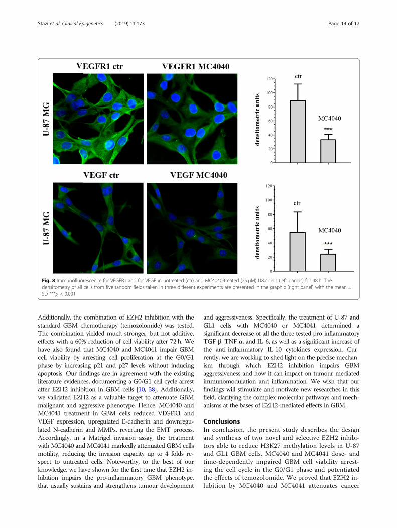
in GBM and it was proven an interesting target to hit.Hence, we questioned ourselves if our EZH2 inhibitorscould indirectly hit also this important pathway. We de-termined the expression of VEGFR1 and VEGF in un-treated and MC4040-treated U-87 cells, expressingdetectable basal protein levels of VEGF and its receptorVEGFR1, and we found that MC4040 was able tostrongly reduce the levels of both proteins (Fig. 8).In parallel, the effect of MC4040 and MC4041 on epi-
thelial to mesenchymal transition (EMT) was analyseddetermining, in GL1 and U87 cells, the levels of E-cadherin as epithelial marker, of N-cadherin as mesen-chymal marker, and of matrix metalloproteinases(MMPs), specifically MMP2, MMP3, and MMP9, basallyupregulated in GL1 and U87 cells, and usually highlyexpressed in the majority of aggressive tumours and re-sponsible for tumour invasiveness [15]. In these assays,MC4040 and MC4041 upregulated E-cadherin anddownregulated N-cadherin, thus reverting the EMTprocess (Fig. 9a). Furthermore, MC4040 and MC4041treatment decreased the expression of all the testedMMPs in U-87 and GL1 cells (Fig. 9a). In order to quan-tify the final readout of the effect of MC4040 andMC4041 on GBM cell invasiveness, we performed aMatrigel invasion assay. In both U-87 and GL1 cell linesthe basal rate of motility was very high. The treatmentwith MC4040 and MC4041 markedly attenuated thismalignant attitude (Fig. 9b, c), reducing the invasioncapacity up to 4-fold. Taken together, our results
Fig. 5 a Western blot of U-87, GL1 and HF treated with DMSO (ctr) or with 25 μM MC4040 and MC4041 for 72 h showing H3K27me3 and β-actinlevels. The means from densitometry quantifications of three different experiments normalized to β-actin are indicated below each band. ND, notdetectable. b Time-course experiment performed treating U-87 and GL1 cells with MC4041 (25 μM) for 24, 48, and 72 h. The levels of H3K27me3are shown (mean from three different experiments) and have been normalized to β-actin
Stazi et al. Clinical Epigenetics (2019) 11:173 Page 11 of 17

demonstrate that our EZH2 inhibitors played an import-ant role in reducing the angiogenic and invasive pheno-type of GBM cells.
MC4040 and MC4041 display anti-inflammatory effects inGBM cellsSeveral different cancer types, including GBM, benefit of achronically active inflammatory microenvironment, provid-ing the proliferative and mutational incentive, necessary tounable the tumour to uncontrolled development. Histonemethylation has been reported to epigenetically control theinnate and adaptive immune responses [45]. Recently, sev-eral studies demonstrated that EZH2 plays a key role in in-flammatory and autoimmune disorders. More in detail,EZH2 was reported to regulate cell adhesion and migration,with possible implications in leukocyte migration and im-mune responses [46], in production of inflammatory cyto-kines in dental pulp cells [47] or in macrophages [48], in
regulation of microglial inflammatory gene expression [49],and in adaptative resistance to tumour immunotherapy[50]. The present evidences surely depict an articulated sce-nario in which EZH2 is a main regulator of inflammatoryand immune processes at various levels.To evaluate if EZH2 inhibition in GBM could impact
on inflammation, and to gain further insight into themolecular mechanism of MC4040 and MC4041, we in-vestigated on the expression patterns of the main inflam-matory and anti-inflammatory cytokines in U-87 andGL1 cells in untreated and in MC4040- and MC4041-treated samples. As expected, both U-87 and GL1expressed basally high levels of the pro-inflammation cy-tokines TGF-β, TNF-α and IL-6, and very low levels ofthe anti-inflammatory cytokine IL-10 (Fig. 10). Thetreatment with either MC4040 or MC4041 determined asignificant decrease of TGF-β, TNF-α, and IL-6 and asignificant increase of IL-10 (Fig. 10). These data show
Fig. 6 Cell viability of U-87 and GL1 untreated (ctr) and treated with 25 μM MC4040 or MC4041 or tazemetostat, and with 100 μM temozolomideas single or combined treatments expressed as percentage of alive cells. Treatments were performed at 72 h (a, dark grey), 96 h (b, light grey)and 120 h (c, white). *p < 0.05 and **p < 0.01 for the comparison between single treatments and untreated cells. #indicates p < 0.05 for thecomparison between each combined treatment and each single treatment with MC4040 and MC4041. §p < 0.05 for the comparison betweeneach combined treatment and the single treatment with temozolomide
Stazi et al. Clinical Epigenetics (2019) 11:173 Page 12 of 17

that U-87 and GL1 cells have a strong pro-inflammatoryphenotype, that could contribute to their aggressivenessand proliferation. Here we provided a first evidence onthe possibility to attenuate this attitude by treatmentwith the EZH2 inhibitors MC4040 and MC4041.
DiscussionSeveral different factors still obstacle the development of aproper cure for GBM, that remains the most common andaggressive malignant primary brain tumour in adults. Todate, the sole chemotherapeutic agent in use against GBMis the alkylating drug temozolomide. The standard GBMtreatment consists of surgical resection of the tumour,followed by chemotherapy and concomitant radiotherapy[2]. It is worth to point out that the current available treat-ment is not a cure. Hence, there is an urgent need for alter-native and/or complementary therapeutic strategies andtargets to hit. The epigenetic mechanisms underlying GBMdevelopment and progression are being elucidated andclarified. The SAM-dependent histone methyltransferaseEZH2 has been found upregulated in gliomas and corre-lated with an aggressive phenotype [10, 11], while its deple-tion by RNA interference resulted in anti-glioma effects[12]. Importantly, some known EZH2 inhibitors have beenalready tested against GBM models in vitro and in vivo,confirming the biological evidences assessing the relevanceof EZH2 as target in this disorder [16]. Here, we report theeffects on GBM, including primary GBM cell cultures, of
two novel EZH2 inhibitors, MC4040 and MC4041, contain-ing a N-phenylpyrrole core decorated with a cyclic amine(morpholine, MC4040, or piperidine, MC4041), oftenpresent in known EZH2i, and carrying the 2-pyridone moi-ety known to be crucial for the EZH2 inhibiting activity[51]. When tested against a human five-component PRC2complex, MC4040 and MC4041 displayed single-digit mi-cromolar IC50 values against EZH2-PRC2 with a 10-fold se-lectivity over EZH1-PRC2. Importantly, the two novelcompounds proved to be selective for EZH2/EZH1 over apanel of SAM-dependent methyltransferases includingDNMT1, G9a, and PRMT1, displaying no inhibition ofthese targets up to 200 μM. Molecular docking andMMGBSA calculations helped us to shed light on the dif-ferences in the binding mode of the two newly synthesizedcompounds, explaining also the difference in activity re-corded in biochemical assays. The two novel compoundshave been tested on the primary GBM cell cultures GL1,GL2, and GL3 as well as on the GBM cell line U-87, dis-playing a dose- and time-dependent reduction of cell viabil-ity, without significant effect on human normal dermalfibroblast (HF) primary culture proliferation. Interestingly,the effects of the two newly synthesized EZH2 inhibitorswere comparable to those of the clinically studied inhibitortazemetostat, tested at the same doses and times. TheMC4040- and MC4041-mediated EZH2 inhibition in U-87and GL1 cells was confirmed by the decrease of theH3K27me3 levels, basally increased in both cell lines.
Fig. 7 a Western blot of GL1 untreated (ctr) and treated for 72 or 120 h with 25 μM MC4040 and MC4041 and for 6 h with 5 μM staurosporine(ST) showing uncleaved and cleaved PARP-1 and actin. b Western blot of GL1 and U-87 untreated (ctr) and treated for 72 h with 25 μM MC4040showing p21 and p27 levels. c Cell cycle analysis of untreated and treated cells
Stazi et al. Clinical Epigenetics (2019) 11:173 Page 13 of 17
Additionally, the combination of EZH2 inhibition with thestandard GBM chemotherapy (temozolomide) was tested.The combination yielded much stronger, but not additive,effects with a 60% reduction of cell viability after 72 h. Wehave also found that MC4040 and MC4041 impair GBMcell viability by arresting cell proliferation at the G0/G1phase by increasing p21 and p27 levels without inducingapoptosis. Our findings are in agreement with the existingliterature evidences, documenting a G0/G1 cell cycle arrestafter EZH2 inhibition in GBM cells [10, 38]. Additionally,we validated EZH2 as a valuable target to attenuate GBMmalignant and aggressive phenotype. Hence, MC4040 andMC4041 treatment in GBM cells reduced VEGFR1 andVEGF expression, upregulated E-cadherin and downregu-lated N-cadherin and MMPs, reverting the EMT process.Accordingly, in a Matrigel invasion assay, the treatmentwith MC4040 and MC4041 markedly attenuated GBM cellsmotility, reducing the invasion capacity up to 4 folds re-spect to untreated cells. Noteworthy, to the best of ourknowledge, we have shown for the first time that EZH2 in-hibition impairs the pro-inflammatory GBM phenotype,that usually sustains and strengthens tumour development
and aggressiveness. Specifically, the treatment of U-87 andGL1 cells with MC4040 or MC4041 determined asignificant decrease of all the three tested pro-inflammatoryTGF-β, TNF-α, and IL-6, as well as a significant increase ofthe anti-inflammatory IL-10 cytokines expression. Cur-rently, we are working to shed light on the precise mechan-ism through which EZH2 inhibition impairs GBMaggressiveness and how it can impact on tumour-mediatedimmunomodulation and inflammation. We wish that ourfindings will stimulate and motivate new researches in thisfield, clarifying the complex molecular pathways and mech-anisms at the bases of EZH2-mediated effects in GBM.
ConclusionsIn conclusion, the present study describes the designand synthesis of two novel and selective EZH2 inhibi-tors able to reduce H3K27 methylation levels in U-87and GL1 GBM cells. MC4040 and MC4041 dose- andtime-dependently impaired GBM cell viability arrest-ing the cell cycle in the G0/G1 phase and potentiatedthe effects of temozolomide. We proved that EZH2 in-hibition by MC4040 and MC4041 attenuates cancer
Fig. 8 Immunofluorescence for VEGFR1 and for VEGF in untreated (ctr) and MC4040-treated (25 μM) U87 cells (left panels) for 48 h. Thedensitometry of all cells from five random fields taken in three different experiments are presented in the graphic (right panel) with the mean ±SD ***p < 0.001
Stazi et al. Clinical Epigenetics (2019) 11:173 Page 14 of 17

Fig. 9 a Western blot of EMT and invasion related proteins: E-cadherin, N-cadherin, MMP-2, MMP-3, and MMP-9. MC4040 and MC4041 were usedat 20 μM for 48 h. The densitometry quantitation is indicated for each band normalized for actin band. b Matrigel invasion assay of U-87 and GL1cells untreated (ctr) and treated with 20 μM MC4040 or MC4041 for 48 h. c Migrated cells were counted in five random fields. Three independentexperiments were performed, and the results are presented with the mean ± SD **p < 0.01
Fig. 10 Western blot of U-87 and GL1 untreated (ctr) and treated with 20 μM MC4040 or MC4041 for 48 h for TGF-ß, TNF-α, IL-6, IL-10, and actin.The densitometry quantification is indicated for each band normalized for actin band
Stazi et al. Clinical Epigenetics (2019) 11:173 Page 15 of 17
malignant phenotype by reducing VEGFR1/VEGF ex-pression, reverting the EMT process and inhibiting in-vasiveness. To the best of our knowledge, here, weshow for the first time that targeting EZH2 in GBMdisplays anti-inflammatory effects, further strengthen-ing the relevance of this target in this cancer type.
AbbreviationsAEBP2: Adipocyte enhancer-binding protein 2; BSA: Bovine serum albumin;DMEM: Dulbecco’s modified Eagle’s medium; DMF: N,N-DimethylformamideDNMT1DNA methyltransferase 1; DTT: Dithiothreitol;EDTA: Ethylenediaminetetraacetic acid; EED: Embryonic ectodermdevelopment; EIMS: Electron ionization mass spectrometry; EMEM: Eagle’sminimum essential medium; EMT: Epithelial-mesenchymal transition;EZH1: Enhancer of zeste homologue 1; EZH2: Enhancer of zeste homologue2; EZH2i: EZH2 inhibitors; FCS: Foetal calf serum; G9a (EHMT2): Euchromatichistone-lysine N-methyltransferase 2; GBM: Glioblastoma; GFAP: Glial fibrillaryacidic protein; H3K27: Lysine 27 of the histone 3; H3K27me3: TrimethylatedH3K27; HF: Human fibroblasts; IL: Interleukin; MB: Medulloblastoma; MDA-MB231: breast cancer cell line; MMGBSA: Molecular mechanics/generalizedBorn surface area; MMP: Matrix metalloproteinase; MT: Methyltransferase;NMR: Nuclear magnetic resonance; OD: Optical density; PARP: Poly ADP-ribose polymerase; PBS: Phosphate-buffered saline; PDB: Protein data bank;PMSF: Phenylmethanesulfonyl fluoride; PRC2: Polycomb repressive complex2; PRMT1: Protein arginine methyltransferase 1; RbAp48: Retinoblastoma-associated protein 48; SAH: S-Adenosyl-L-homocysteine; SAM: S-Adenosyl-L-methionine; SDS: Sodium dodecyl sulphate; SHH: Sonic Hedgehog;ST: Staurosporine; SUZ12: Polycomb repressive complex 2 subunit;TAZ: Tazemetostat; TBTU: O-(Benzotriazole-1-yl)-N,N,N',N'-tetramethyluroniumtetrafluorborate; TGF-β: Transforming growth factor β; TLC: Thin layerchromatography; TMZ: Temozolomide; TNF-α: Tumour necrosis factor α;VEGF: Vascular endothelial growth factor; VEGFR1: Vascular endothelialgrowth factor receptor 1
AcknowledgementsNot applicable.
Authors’ contributionsAM, SV, MA, and SS conceived and designed the experiments; GS, AR, RF, LT,AN, SM, MS, RR, ST, MN, and RC performed the experiments; AM, SV, MA, SS,GSLT, and ST analysed the data; GS, LT, SS, RR, SV, and AM wrote the paper.
FundingThis work was supported by Ricerca Finalizzata 2013 PE-2013-02355271(A.M.), by PRIN 2016 (prot. 20152TE5PK) (A.M.), AIRC 2016 (n. 19162) (A.M.),and NIH (n. R01GM114306) (A.M.) funds.
Availability of data and materialsAll the published data are available. There is no supporting material.
Ethics approval and consent to participateAll the authors approved the described studies and consented to participate.
Consent for publicationAll the authors approved the publication of the reported data.
Competing interestsThe authors declare that they have no competing interests.
Author details1Department of Chemistry and Technologies of Drugs, Sapienza University ofRome, P.le A. Moro 5, 00185 Rome, Italy. 2Department of ExperimentalMedicine, Sapienza University of Rome, P.le A. Moro 5, 00185 Rome, Italy.3Alchemical Dynamics s.r.l, 00125 Roma, Italy. 4Department of Sense Organs,Sapienza University of Rome, P.le A. Moro 5, 00185 Rome, Italy. 5Departmentof Radiologic, Oncological, and Anatomical and Pathological Sciences,Sapienza University of Rome, P.le A. Moro 5, 00185 Rome, Italy.
Received: 5 June 2019 Accepted: 9 October 2019
References1. Ostrom QT, Gittleman H, Fulop J, Liu M, Blanda R, Kromer C, Wolinsky
Y, Kruchko C, Barnholtz-Sloan JS: CBTRUS statistical report: primary brainand central nervous system tumors diagnosed in the United States in2008-2012. Neuro-Oncology 2015, 17 Suppl 4:iv1-iv62. doi: https://doi.org/10.1093/neuonc/nov189.
2. Wick W, Platten M. Understanding and treating glioblastoma. Neurol Clin.2018;36:485–99. https://doi.org/10.1016/j.ncl.2018.04.006.
3. Meyer M, Reimand J, Lan X, Head R, Zhu X, Kushida M, Bayani J, Pressey JC,Lionel AC, Clarke ID, et al. Single cell-derived clonal analysis of humanglioblastoma links functional and genomic heterogeneity. Proc Natl AcadSci U S A. 2015;112:851–6. https://doi.org/10.1073/pnas.1320611111.
4. Patel AP, Tirosh I, Trombetta JJ, Shalek AK, Gillespie SM, Wakimoto H, CahillDP, Nahed BV, Curry WT, Martuza RL, et al. Single-cell RNA-seq highlightsintratumoral heterogeneity in primary glioblastoma. Science. 2014;344:1396–401. https://doi.org/10.1126/science.1254257.
5. Buerki RA, Chheda ZS, Okada H. Immunotherapy of primary braintumors: facts and hopes. Clin Cancer Res. 2018. https://doi.org/10.1158/1078-0432.CCR-17-2769.
6. Ferreira WA, Pinheiro Ddo R, Costa Junior CA, Rodrigues-Antunes S, AraujoMD, Leao Barros MB, Teixeira AC, Faro TA, Burbano RR, Oliveira EH, et al. Anupdate on the epigenetics of glioblastomas. Epigenomics. 2016;8:1289–305.https://doi.org/10.2217/epi-2016-0040.
7. Pangeni RP, Zhang Z, Alvarez AA, Wan X, Sastry N, Lu S, Shi T, Huang T, LeiCX, James CD, et al. Genome-wide methylomic and transcriptomic analysesidentify subtype-specific epigenetic signatures commonly dysregulated inglioma stem cells and glioblastoma. Epigenetics. 2018:1–17. https://doi.org/10.1080/15592294.2018.1469892.
8. Tan SK, Pastori C, Penas C, Komotar RJ, Ivan ME, Wahlestedt C, Ayad NG.Serum long noncoding RNA HOTAIR as a novel diagnostic and prognosticbiomarker in glioblastoma multiforme. Mol Cancer. 2018;17:74. https://doi.org/10.1186/s12943-018-0822-0.
9. Kim KH, Roberts CW. Targeting EZH2 in cancer. Nat Med. 2016;22:128–34.https://doi.org/10.1038/nm.4036.
10. Zhang J, Chen L, Han L, Shi Z, Zhang J, Pu P, Kang C. EZH2 is a negativeprognostic factor and exhibits pro-oncogenic activity in glioblastoma.Cancer Lett. 2015;356:929–36. https://doi.org/10.1016/j.canlet.2014.11.003.
11. Yin Y, Qiu S, Peng Y. Functional roles of enhancer of zeste homolog 2 ingliomas. Gene. 2016;576:189–94. https://doi.org/10.1016/j.gene.2015.09.080.
12. Suva ML, Riggi N, Janiszewska M, Radovanovic I, Provero P, Stehle JC,Baumer K, Le Bitoux MA, Marino D, Cironi L, et al. EZH2 is essential forglioblastoma cancer stem cell maintenance. Cancer Res. 2009;69:9211–8.https://doi.org/10.1158/0008-5472.CAN-09-1622.
13. Fan TY, Wang H, Xiang P, Liu YW, Li HZ, Lei BX, Yu M, Qi ST. Inhibition ofEZH2 reverses chemotherapeutic drug TMZ chemosensitivity inglioblastoma. Int J Clin Exp Pathol. 2014;7:6662–70.
14. Fioravanti R, Stazi G, Zwergel C, Valente S, Mai A. Six years (2012-2018) ofresearches on catalytic EZH2 inhibitors: the boom of the 2-pyridonecompounds. Chem Rec. 2018. https://doi.org/10.1002/tcr.201800091.
15. Yu T, Wang Y, Hu Q, Wu W, Wu Y, Wei W, Han D, You Y, Lin N, Liu N. TheEZH2 inhibitor GSK343 suppresses cancer stem-like phenotypes andreverses mesenchymal transition in glioma cells. Oncotarget. 2017;8:98348–59. https://doi.org/10.18632/oncotarget.21311.
16. Grinshtein N, Rioseco CC, Marcellus R, Uehling D, Aman A, Lun X, MutoO, Podmore L, Lever J, Shen Y, et al. Small molecule epigenetic screenidentifies novel EZH2 and HDAC inhibitors that target glioblastomabrain tumor-initiating cells. Oncotarget. 2016;7:59360–76. https://doi.org/10.18632/oncotarget.10661.
17. Mellini P, Marrocco B, Borovika D, Polletta L, Carnevale I, Saladini S,Stazi G, Zwergel C, Trapencieris P, Ferretti E, et al. Pyrazole-basedinhibitors of enhancer of zeste homologue 2 induce apoptosis andautophagy in cancer cells. Philos Trans R Soc Lond Ser B Biol Sci. 2018;373. https://doi.org/10.1098/rstb.2017.0150.
18. Miele E, Valente S, Alfano V, Silvano M, Mellini P, Borovika D, Marrocco B, PoA, Besharat ZM, Catanzaro G, et al. The histone methyltransferase EZH2 as adruggable target in SHH medulloblastoma cancer stem cells. Oncotarget.2017;8:68557–70. https://doi.org/10.18632/oncotarget.19782.
Stazi et al. Clinical Epigenetics (2019) 11:173 Page 16 of 17

19. Joshi SD, Kumar D, More UA, Yang KS, Aminabhavi TM. Design anddevelopment of pyrrole carbaldehyde: an effective pharmacophore forenoyl-ACP reductase. Med Chem Res. 2016;25:672–89. https://doi.org/10.1007/s00044-016-1517-y.
20. Yonekura K, Oki K, Tsuchimoto T. Indium-Catalyzed Formal N-Arylation andN-Alkylation of Pyrroles with Amines. Adv Synth Catal. 2016;358:2895–902.https://doi.org/10.1002/adsc.201600656.
21. Verma SK, Tian X, LaFrance LV, Duquenne C, Suarez DP, Newlander KA,Romeril SP, Burgess JL, Grant SW, Brackley JA, et al. Identification of Potent,selective, cell-active inhibitors of the histone lysine methyltransferase EZH2.ACS Med Chem Lett. 2012;3:1091–6. https://doi.org/10.1021/ml3003346.
22. Berman H, Henrick K, Nakamura H. Announcing the worldwide Protein DataBank. Nat Struct Biol. 2003;10:980. https://doi.org/10.1038/nsb1203-980.
23. Webb B, Sali A. Protein structure modeling with MODELLER. Methods MolBiol. 2017;1654:39–54. https://doi.org/10.1007/978-1-4939-7231-9_4.
24. Korb O, Stutzle T, Exner TE. Empirical scoring functions for advancedprotein-ligand docking with PLANTS. J Chem Inf Model. 2009;49:84–96.https://doi.org/10.1021/ci800298z.
25. Jakalian A, Jack DB, Bayly CI. Fast, efficient generation of high-quality atomiccharges. AM1-BCC model: II. Parameterization and validation. J ComputChem. 2002;23:1623–41. https://doi.org/10.1002/jcc.10128.
26. Wang J, Wolf RM, Caldwell JW, Kollman PA, Case DA. Development andtesting of a general amber force field. J Comput Chem. 2004;25:1157–74.https://doi.org/10.1002/jcc.20035.
27. Ramsey S, Nguyen C, Salomon-Ferrer R, Walker RC, Gilson MK, Kurtzman T.Solvation thermodynamic mapping of molecular surfaces in AmberTools:GIST. J Comput Chem. 2016;37:2029–37. https://doi.org/10.1002/jcc.24417.
28. Sousa da Silva AW, Vranken WF. ACPYPE - AnteChamber PYthon ParserinterfacE. BMC Res Notes. 2012;5:367. https://doi.org/10.1186/1756-0500-5-367.
29. Izadi S, Anandakrishnan R, Onufriev AV. Building water models: a differentapproach. J Phys Chem Lett. 2014;5:3863–71. https://doi.org/10.1021/jz501780a.
30. Maier JA, Martinez C, Kasavajhala K, Wickstrom L, Hauser KE, Simmerling C.ff14SB: improving the accuracy of protein side chain and backboneparameters from ff99SB. J Chem Theory Comput. 2015;11:3696–713. https://doi.org/10.1021/acs.jctc.5b00255.
31. Genheden S, Ryde U. The MM/PBSA and MM/GBSA methods to estimateligand-binding affinities. Expert Opin Drug Discov. 2015;10:449–61. https://doi.org/10.1517/17460441.2015.1032936.
32. O'Boyle NM, Banck M, James CA, Morley C, Vandermeersch T, Hutchison GR.Open Babel: An open chemical toolbox. J Cheminform. 2011;3:33. https://doi.org/10.1186/1758-2946-3-33.
33. Halgren TA. MMFF VI. MMFF94s option for energy minimization studies. JComput Chem. 1999;20:720–9. https://doi.org/10.1002/(SICI)1096-987X(199905)20:7<720::AID-JCC7>3.0.CO;2-X.
34. Bianchi E, Taurone S, Bardella L, Signore A, Pompili E, Sessa V, Chiappetta C,Fumagalli L, Di Gioia C, Pastore FS, et al. Involvement of pro-inflammatorycytokines and growth factors in the pathogenesis of Dupuytren’scontracture: a novel target for a possible future therapeutic strategy? ClinSci (Lond). 2015;129:711–20. https://doi.org/10.1042/CS20150088.
35. Hartwig JF, Kawatsura M, Hauck SI, Shaughnessy KH, Alcazar-Roman LM.Room-temperature palladium-catalyzed amination of aryl bromides andchlorides and extended Scope of aromatic C-N bond formation with acommercial ligand. J Org Chem. 1999;64:5575–80.
36. Sabatino M, Rotili D, Patsilinakos A, Forgione M, Tomaselli D, Alby F,Arimondo PB, Mai A, Ragno R. Disruptor of telomeric silencing 1-like(DOT1L): disclosing a new class of non-nucleoside inhibitors by means ofligand-based and structure-based approaches. J Comput Aided Mol Des.2018;32:435–58. https://doi.org/10.1007/s10822-018-0096-z.
37. Miller BR 3rd, McGee TD Jr, Swails JM, Homeyer N, Gohlke H, Roitberg AE.MMPBSA.py: an efficient program for end-state free energy calculations. JChem Theory Comput. 2012;8:3314–21. https://doi.org/10.1021/ct300418h.
38. Qi W, Chan H, Teng L, Li L, Chuai S, Zhang R, Zeng J, Li M, Fan H, Lin Y,et al. Selective inhibition of Ezh2 by a small molecule inhibitor blocks tumorcells proliferation. Proc Natl Acad Sci U S A. 2012;109:21360–5. https://doi.org/10.1073/pnas.1210371110.
39. Dubois LG, Campanati L, Righy C, D’Andrea-Meira I, Spohr TC, Porto-CarreiroI, Pereira CM, Balca-Silva J, Kahn SA, DosSantos MF, et al. Gliomas and thevascular fragility of the blood brain barrier. Front Cell Neurosci. 2014;8:418.https://doi.org/10.3389/fncel.2014.00418.
40. Lohr J, Ratliff T, Huppertz A, Ge Y, Dictus C, Ahmadi R, Grau S, Hiraoka N,Eckstein V, Ecker RC, et al. Effector T-cell infiltration positively impacts survival
of glioblastoma patients and is impaired by tumor-derived TGF-beta. ClinCancer Res. 2011;17:4296–308. https://doi.org/10.1158/1078-0432.CCR-10-2557.
41. Baumgarten P, Blank AE, Franz K, Hattingen E, Dunst M, Zeiner P, HoffmannK, Bahr O, Mader L, Goeppert B, et al. Differential expression of vascularendothelial growth factor A, its receptors VEGFR-1, -2, and -3 and co-receptors neuropilin-1 and -2 does not predict bevacizumab response inhuman astrocytomas. Neuro-Oncology. 2016;18:173–83. https://doi.org/10.1093/neuonc/nov288.
42. Roskoski R Jr. VEGF receptor protein-tyrosine kinases: structure andregulation. Biochem Biophys Res Commun. 2008;375:287–91. https://doi.org/10.1016/j.bbrc.2008.07.121.
43. Fischer C, Mazzone M, Jonckx B, Carmeliet P. FLT1 and its ligands VEGFBand PlGF: drug targets for anti-angiogenic therapy? Nat Rev Cancer. 2008;8:942–56. https://doi.org/10.1038/nrc2524.
44. Atzori MG, Tentori L, Ruffini F, Ceci C, Lisi L, Bonanno E, Scimeca M,Eskilsson E, Daubon T, Miletic H, et al. The anti-vascular endothelial growthfactor receptor-1 monoclonal antibody D16F7 inhibits invasiveness ofhuman glioblastoma and glioblastoma stem cells. J Exp Clin Cancer Res.2017;36:106. https://doi.org/10.1186/s13046-017-0577-2.
45. Stender JD, Glass CK. Epigenomic control of the innate immune response. CurrOpin Pharmacol. 2013;13:582–7. https://doi.org/10.1016/j.coph.2013.06.002.
46. Gunawan M, Venkatesan N, Loh JT, Wong JF, Berger H, Neo WH, Li LY, La WinMK, Yau YH, Guo T, et al. The methyltransferase Ezh2 controls cell adhesionand migration through direct methylation of the extranuclear regulatoryprotein talin. Nat Immunol. 2015;16:505–16. https://doi.org/10.1038/ni.3125.
47. Hui T, A P, Zhao Y, Wang C, Gao B, Zhang P, Wang J, Zhou X, Ye L. EZH2, apotential regulator of dental pulp inflammation and regeneration. J Endod.2014;40:1132–8. https://doi.org/10.1016/j.joen.2014.01.031.
48. Zhang W, Liu H, Liu W, Liu Y, Xu J. Polycomb-mediated loss of microRNAlet-7c determines inflammatory macrophage polarization via PAK1-dependent NF-kappaB pathway. Cell Death Differ. 2015;22:287–97. https://doi.org/10.1038/cdd.2014.142.
49. Arifuzzaman S, Das A, Kim SH, Yoon T, Lee YS, Jung KH, Chai YG. Selectiveinhibition of EZH2 by a small molecule inhibitor regulates microglial geneexpression essential for inflammation. Biochem Pharmacol. 2017;137:61–80.https://doi.org/10.1016/j.bcp.2017.04.016.
50. Zingg D, Arenas-Ramirez N, Sahin D, Rosalia RA, Antunes AT, Haeusel J,Sommer L, Boyman O. The histone methyltransferase Ezh2 controlsmechanisms of adaptive resistance to tumor immunotherapy. Cell Rep.2017;20:854–67. https://doi.org/10.1016/j.celrep.2017.07.007.
51. Brooun A, Gajiwala KS, Deng YL, Liu W, Bolanos B, Bingham P, He YA, DiehlW, Grable N, Kung PP, et al. Polycomb repressive complex 2 structure withinhibitor reveals a mechanism of activation and drug resistance. NatCommun. 2016;7:11384. https://doi.org/10.1038/ncomms11384.
Publisher’s NoteSpringer Nature remains neutral with regard to jurisdictional claims inpublished maps and institutional affiliations.
Stazi et al. Clinical Epigenetics (2019) 11:173 Page 17 of 17