diseasesof the aorta1 (1)€¢Detection of peri-aortic and mediastinal bleeding 15 Aortic...
37
1 Diseases of the Aorta: Dissection, Hematoma and Trauma Roberto M. Lang M.D., FASE LVOT Sinus of Valsalva Aortic Valve RCC LCC NCC SAX Long Axis Normal Ao Valve
Transcript of diseasesof the aorta1 (1)€¢Detection of peri-aortic and mediastinal bleeding 15 Aortic...

1
Diseases of the Aorta:
Dissection, Hematoma and Trauma
Roberto M. Lang M.D., FASE
LVOT Sinus of Valsalva
Aortic Valve
RCCLCC
NCC
SAX
Long Axis
NormalAo Valve

2
Identify the Reason for this Emergency TEE
Trauma
Connective tissue generic disordersNon-inflammatory medial diseaseAortitisAtherosclerosis
Connective tissue disorders: Hypertension, Marfan’s
False saccularaneurysm
True diffuse and saccularaneurysms
Dissecting aneurysms
Type of Aneurysm : CausesType of Aneurysm : Causes

3
PATHOPHYSIOLOGY
• Deterioration of medial
collagen and elastin
• A tear in the intimal layer allows blood to
enter the intima-media space
• Blood then propagates down
this new space creating a “true”
and a “false” lumen
Cystic Medial ChangeCystic Medial Change
HypertensionMarfan’s and Ehler-Danlos Coarctation and bicuspid aortic valve Pregnancy Trauma Perforation through an intimal atheromatous plaque

4
Types of Aortic Dissection De Bakey Type I Type II Type IIIStanford Type A Type A Type B
Proximal or Ascending Type A
Hallmark is visualization of mobile dissection flapMotion that is independent of the AortaVisualization on more than one viewClear distinction from reverberations

5
Predicting Death in Patients with Acute Type A Aortic Dissection
• 547 pts; IRAD; Jan 96-Dec 99
• In hospital mortality 32.5%
- Age 70 years
- Abrupt onset of Cx pain
- Hypotension, shock, tamponade
- Kidney failure
- Pulse deficit
- ECG abnormalities
Circulation 2002;105:200-206

6
Distal or descending – Type III Aortic Dissection
Iatrogenic (intra-arterial catheterization) – Type IV
Management of Aortic Dissection
Depends not only on the type but alsoalso on time elapsed between onset and presentation
• Acute < 2 weeks• 24 Hour hyper-acute period (Risk of
rupture approaching 1% per hour• 75% od AD related deaths occur in
first two weeks• Subacute > 2 weeks - 2 months• Chronic > 2 months

7
Clinical Presentation: Physical Exam
A + B Type A Type B P =
AI on exam 32% 44% 12% <.001
Pulse deficit 15% 19% 9.2% .006
CVA 4.7% 6.1% 2.3% .07
CHF 6.6% 8.8% 3.0% .02
Aortic Dissection: Pulse Loss
Due to direct compression
Blockade due to flap of intima

8
Survival Curves for Type A DissectionBy Number of Pulse Deficits on Presentation
Circulation 2003;108:628-635
GOALS OF DIAGNOSTIC IMAGING
• Confirm the diagnosis
• Classify the dissection and determine extent
• Detect extravasation
• Detect and Grade AI
• Confirm the diagnosis
• Classify the dissection and determine extent
• Detect extravasation
• Detect and Grade AI
• Aortography• Spiral CT• MRI• TTE / TEE
• Aortography• Spiral CT• MRI• TTE / TEE

9
Aortic Dissection: Choice of Imaging Technique
• Fine tradeoff in sensitivity and specificity
• Availability of technique
• Experience and expertise at a given institution
• Degree of urgency
• Stability of the patient
Aortic Dissection: Why Multiple Studies?
• Initial study often done at referral siteConfirmation needed or desired
• If CT firstStill need cardiac anatomy, valve status etc
• If echocardiography firstStill need assessment of abdominal aorta in many instances

10
Moore, A. et al. Am J Cardiology, 89:1235-1238, 2002
Procedure Used for the Diagnosis of Aortic Dissection
Diagnostic value of different imaging modalities in acute aortic syndromes
Lesion TTE TEE CT MRI
Ascending aortic dissection ++ +++ +++ +++
Aortic arch dissection + + +++ +++
Descending aortic dissection + +++ +++ +++
Size ++ +++ +++ +++
Mural thrombus + +++ +++ +++
Intramural hematoma + +++ +++ +++
Penetrating aortic ulcer ++ ++ +++ +++
Involvement of aortic branches +² (+) +++ +++
²Can be improved when combined by vascular ultrasound (carotid, subclavian, vertebral, celiac, mesenteric, and renal arteries). +++=excellent; ++=moderate; +=poor,(+)=poor and inconstant; CT=computed tomography; MRI=magnetic resonance imaging,TOE=transesophageal echocardiography; TTE=transthoracic echocardiography.
11
TEE in Aortic Dissection• Hallmark is
visualization of mobile dissection flap
• Motion that is independent of the Aorta
• Visualization on more than one view
• Clear distinction from reverberations
Systolic expansion of the true lumenDiastolic expansion of the false lumen
Aortic Dissection: Intimal Flap and Entry Site

12
Advantages of TEE in Aortic Dissection
• Origin and proximal extent of the dissection flap
• Dimension of the aorta
• Severity of Aortic Insufficiency
• Pericardial effusion
• Coronary involvement
TEE in AD:Disadvantages
• Interposition of the trachea between the ascending aorta and the esophagus impeding visualization of distal ascending aorta and proximal arch
• Brachiocephalic and and LCC artery difficult to visualize
• Celiac trunk and superior mesenteric artery cannot be consistently imaged
• Reverberation artifacts

13
TTE Echo in Aortic DissectionSuprasternal Approach
3D Epicardial Echo in Aortic Dissection

14
• CT first time imaging modality• In 62% of Type A Ad, CT is the first
imaging modality
• Diagnostic accuracy near 100% to exclude Ad
• Evaluation of the entire aorta and branches
• Shortest time to diagnosis
• Disadvantage: Need for iodinated contrast and radiation
Advantages of CT in Aortic Dissection
Information required from imaging in acute aortic dissection
• Visualization of intimal flap
• Extent of the disease (aortic segmentation)
• Identification of the false and true lumens (if present)
• Localization of entry and re-entry tears (if present)
• Identification of severity and mechanism of aortic valve regurgitation
• Involvement of side branches
• Detection of pericardial effusion
• Detection and extent of pleural effusion
• Detection of peri-aortic and mediastinal bleeding

15
Aortic Dissection: Complications
• Aortic regurgitation
• Pericardial effusion (rupture of the false lumen into the pericardium) Echo best for pericardial effusion; CT for pleural effusion and peri- aortic hematoma
• Coronary artery involvement (invagination of intimal flap into the coronary
• Other branch vessel involvement
Aortic Dissection: Mechanism of AR
Dilatation of aortic root.
Pressure from dissecting hematoma may depress one leaflet below line of closure.
Prolapse or flail
–Torn annular support of the
leaflets.
AR occurs in 50% of patients with typeA aortic dissection

16
Aortic Dissection: Mechanism of AR

17
Aortic Dissection: Mechanism of AR
Intimal Flap Prolapse
Aortic Dissection:
Complications

18
Aortic Dissection: Endovascular Repair
• 1,3,6 and 12 months then yearly• Ao diameter and status of the false lumen
• (thrombosed or patent)
• Dilatation of the Ao is predictor of rupture • (Diameter > 60 or annual growth > 5 mm)
• Completely thrombosed false lumen (improved outcomes)• Patent false lumen (risk for expansion and death)
• Entry tear size• Flap confined to ascending Ao (improved outcomes)
Aortic Dissection: Follow-Up

19
Discrete AD withbulging of the Ao wall
Ulceration of Aortic plaque
following plaque rupture
IntramuralHematoma
Iatrogenic or traumatic AD
Classic AD with True and False Lumen
Classification of Acute Aortic Syndrome in Aortic Dissection
Class 1 Class 2
Class 3 Class 4 Class 5
Atypical Aortic Dissection
Intramural HematomaIntramural Hematoma
Penetrating Atherosclerotic Ulcer
Penetrating Atherosclerotic Ulcer

20
• Rupture of the VASA vasorum
Discrete hematoma
Extends for a variable distance by dissecting along the outer media beneath the adventitia
………
Intramural Hematoma
Intramural Hematoma: Diagnosis
Crescentic area along the aortic
wall
Crescentic area along the aortic
wall
MRI CT
TEE
Contained hemorrhage within the
medial layer of the aortic
wall
Contained hemorrhage within the
medial layer of the aortic
wall
21
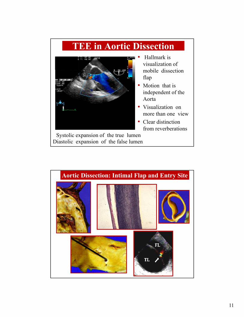
Imaging features of IMH
• IMH represents hemorrhage
into medial layer of aorta
with absence of dissection
flap and false lumen
• Focal aortic wall thickening
(crescentic > concentric)
• Preserved luminal shape
with smooth luminal border
• Echoluscent regions may be
present in the aortic wall
• Central displacement of intimal calcium
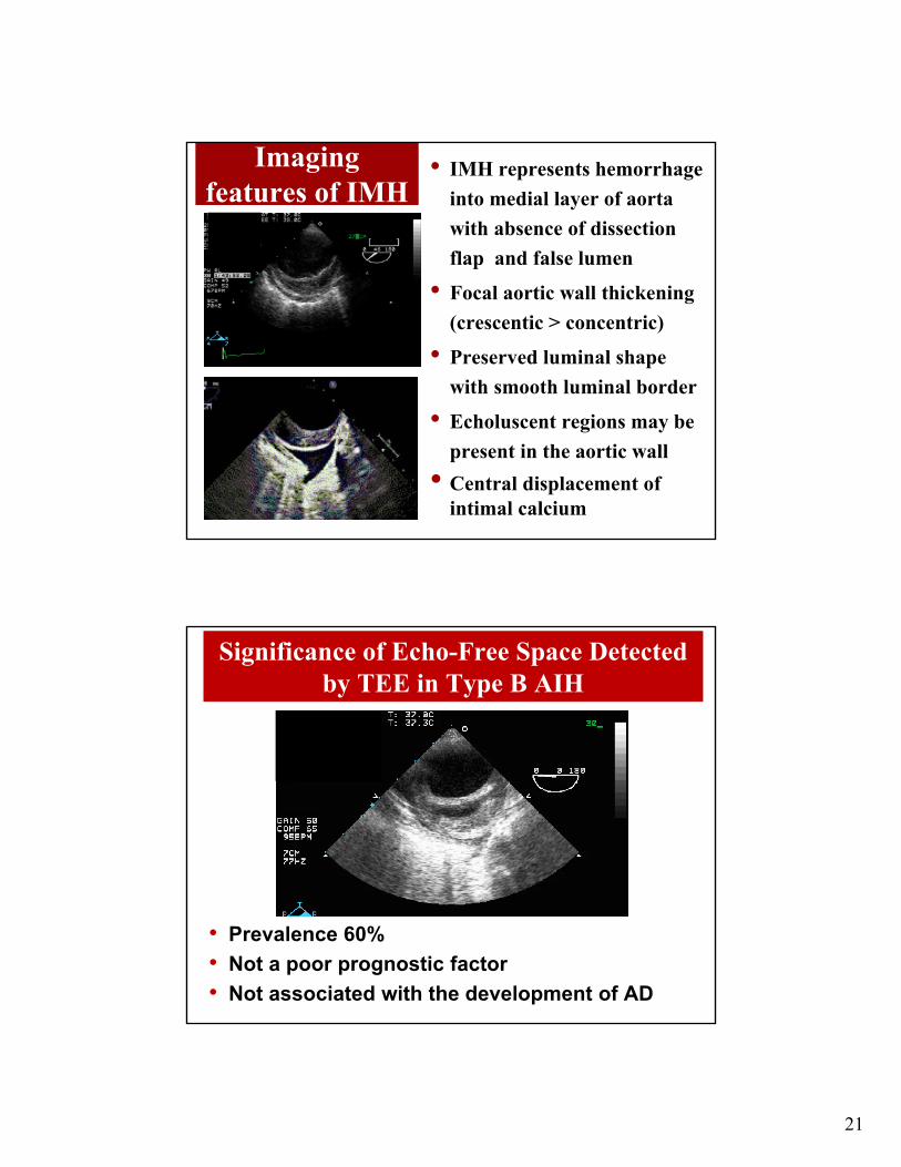
Significance of Echo-Free Space Detected by TEE in Type B AIH
• Prevalence 60%
• Not a poor prognostic factor
• Not associated with the development of AD

22
Differential Diagnosis
Acute and Chronic Complications of IMH
Day 1 Day 7 J Comput Assist Tomogr 2007; 31:435-440

23
Intramural Hematoma: Natural History
Persistence of IMH
Fusiform aneurysmDissection with
longitudinal propagation
Regress
1, 3, 6, 9, and 12 months, then yearly
Localized Dissection
Saccular aneurysm
Pseudo-aneurysm
IMH: Predictors
• Can progress to localized or frank dissection or rupture
• IMH thickness (>10 mm) and maximal aortic diameter (4cm) predict risk for progression
• Peri-aortic hemorrhage or pleural effusion (microperforations or inflammatory exudate)
• Penetrating ulcer or ulcer-like projection secondary to localized dissections in the involved segment

24

25
Penetrating Aortic Ulcer
• Ulceration of an atherosclerotic lesion penetrates the aortic internal elastic lamina into the aortic media
• Disease of the intima
• Mid and distal descending thorcic aorta
Penetrating Atherosclerotic Ulcer
• Almost exclusively in
the descending Ao
• Usually remains
localized
• Chest and back pain
without associated AR
or neurological deficits

26
Details required from imaging in Penetrating Aortic Ulcer
• Localization of the lesion (length and depth)
• Co-existence of intramural hematoma
• Peri-aortic tissue and bleeding
• Thickness of the residual wall
• CT, MRI and TEE
PAU’s: imaging parameters to report
•Lesion location
•Lesion depth of penetration
•Width at entry site
•Axial length of associated intramural hematoma

27
• Natural history is unclear
• No defined strategy
• Surgical repair for
Pseudoaneurysm
Transmural rupture
Hemodynamic instability
Continued pain
Distal embolization
Aneurysmal dilatation
Penetrating Atherosclerotic Ulcer
Differential Diagnosis
Ao Dissection Thrombosed False Lumen Ao atherosclerotic aneurysm Pseudoaneurysm IAH

28
Aortic Root and Ascending Aortic Aneurysm
Stretching of the entire thickness of
the aortaThe majority involve
root and proximal tubular ascending
aorta
• Confirm diagnosis• Maximal diameter• Define longitudinal extent• Involvement of the aortic valve• Involvement of arch vessels• Mural thrombus, dissection, periaortic hematoma
Aortic diameter principal predictor of ruptureincreases significantly >6 cm
CT first lineMRI second lineTTE second lineTEE third line

29
Grading system for severity of aortic atherosclerosis
Grade Severity (atheroma thickness)
Description
1 Normal Intimal thickness <2mm
2 Mild Mild(focal or diffuse) intimal thickening of 2-3 mm
3 Moderate Atheroma >3-5mm (no mobile/ulcerated components)
4 Severe Atheroma >5mm (no mobile/ulcerated components)
5 Complex Grade 2,3, or 4 atheroma plus mobile or ulcerated components
Grading system for severity of aortic atherosclerosis
Intimal thickness
<2mm
Intimal thickness2-3mm
Atheroma3-5mm
No mobile or
ulcerated
Atheroma>5mm
No mobile or
ulcerated
Complex: Grade 2,3, or 4 atheroma plus mobile or ulcerated components
Grade 1 Grade 2
Grade 3 Grade 4

30
High-Speed Deceleration Accident
• Cardiac contusion• Aortic injury• Myocardial valve
injury
Blunt Chest Trauma
Generates shearing forces that act maximally on the aortic isthmus

31
Blunt Aortic-Brachiocephalic Trauma
88%
8%
4%
Fisher et al, 1981 (n=510)
84%
8%
8%
Vignon et al, 1998 (n=25)
Vignon et al. Circulation 1995;92: 2959-68
Aortic Disruption: Anatomical Types
Complete Subtotal
Intimal Tear
PartialTear

32
Complete Transection
Circular flap, separation of media from adventitia along entire circumferenceof the aorta
Elongation of the aorta consistent withpseudoaneurysm formation
Increased distance from the sectorconsistent with hemomediastinum
Turbulent color flow at the site of the tear
Subtotal Transection
Media1 flap involves at least 2/3 of aortic circumference
Spiral effect, small section of intact media and adventitia
Flap is vertically oriented
Tubulent color flow Doppler on both sides of the flap
Oblong shape of the aorta

33
Partial Tear
Localized media1 flap involving arelatively small section of the aorta
Extravasation of blood betweenthe media and adventitia
Usually can define an entry siteinto a pseudoaneurysm
Intimal Tear
Intima is lifted off of the media
Free, highly mobile
No color flow disturbance on Doppler
Unclear prognostic importance

34
A B
C D
Dissection vs. Disruption
TEE Longitudinal View
Dissection Disruption

35

36

37