Diseases of crustaceans Viral diseases—Yellowhead...
6
Sourced from AGDAFF–NACA (2007) Aquatic Animal Diseases Significant to Asia-Pacific: Identification Field Guide. Australian Government Department of Agriculture, Fisheries and Forestry. Canberra. © Commonwealth of Australia 2007 This work is copyright. It may be reproduced in whole or in part subject to the inclusion of an acknowledgment of the source and no commercial usage or sale. PAGE 1 Signs of disease Important: animals with disease may show one or more of the signs below, but disease may still be present in the absence of any signs. Disease signs at the farm level • moribund prawns aggregate near surface at pond edges • infected 5–15 gram prawns begin feeding at abnormally high rate for several days and then cease feeding entirely • mass mortality three days after cessation of feeding Clinical signs of disease in an infected animal • white, yellow or brown gills • yellowing of the cephalothorax and general bleaching of body • yellow, swollen digestive gland makes head appear yellow Diseases of crustaceans Viral diseases—Yellowhead disease Yellowhead disease in giant black tiger prawn (Penaeus monodon). Note yellow heads of infected prawns on left. Prawns on right are normal Source: DV Lightner Disease agent The causative agent of yellowhead disease is yellowhead virus (YHV), a corona-like RNA viruses in the genus Okavirus, family Ronaviridae and order Nidovirales. Host range YHV is highly infectious for most known species of cultivated penaeid prawns. Crustaceans known to be susceptible to yellowhead disease: black tiger prawn* (Penaeus monodon) — primarily Gulf banana prawn* (Penaeus merguiensis)
-
Upload
nguyendien -
Category
Documents
-
view
215 -
download
0
Transcript of Diseases of crustaceans Viral diseases—Yellowhead...

Sourced from AGDAFF–NACA (2007) Aquatic Animal Diseases Significant to Asia-Pacific: Identification Field Guide. Australian Government Department of Agriculture, Fisheries and Forestry. Canberra.
© Commonwealth of Australia 2007 This work is copyright. It may be reproduced in whole or in part subject to the inclusion of an
acknowledgment of the source and no commercial usage or sale.PAGE 1
Signs of disease
Important: animals with disease may show one or more of the signs below, but disease may still be present in the absence of any signs.
Disease signs at the farm level
• moribund prawns aggregate near surface at pond edges
• infected 5–15 gram prawns begin feeding at abnormally high rate for several days and then cease feeding entirely
• mass mortality three days after cessation of feeding
Clinical signs of disease in an infected animal
• white, yellow or brown gills
• yellowing of the cephalothorax and general bleaching of body
• yellow, swollen digestive gland makes head appear yellow
Diseases of crustaceans Viral diseases—Yellowhead disease
Yellowhead disease in giant black tiger prawn (Penaeus monodon). Note yellow heads of infected prawns on left. Prawns on right are normalSource: DV Lightner
Disease agent
The causative agent of yellowhead disease is yellowhead virus (YHV), a corona-like RNA viruses in the genus Okavirus, family Ronaviridae and order Nidovirales.
Host range
YHV is highly infectious for most known species of cultivated penaeid prawns.
Crustaceans known to be susceptible to yellowhead disease:
black tiger prawn* (Penaeus monodon) — primarilyGulf banana prawn* (Penaeus merguiensis)
Sourced from AGDAFF–NACA (2007) Aquatic Animal Diseases Significant to Asia-Pacific: Identification Field Guide. Australian Government Department of Agriculture, Fisheries and Forestry. Canberra.
© Commonwealth of Australia 2007 This work is copyright. It may be reproduced in whole or in part subject to the inclusion of an
acknowledgment of the source and no commercial usage or sale.PAGE 2
northern white shrimp* (Penaeus setiferus)prawn* (Palaemon styliferus)red endeavour prawn* (Metapenaeus ensis)tropical krill* (Acetes spp)blue shrimp (Penaeus stylirostris)northern brown shrimp (Penaeus aztecus)northern pink shrimp (Penaeus duorarum)Pacific white shrimp (Penaeus vannamei)
Until proven otherwise, it should be assumed that most penaeid prawns worldwide are susceptible to infection with yellowhead disease.
Presence in Asia–Pacific
Yellowhead disease has been officially reported from India, Thailand and Vietnam.
Epidemiology
• The tiger prawn suffers acute epidemics, with mortality reaching 100% within 3–5 days from first appearance of the gross signs.
• Transmission is horizontal, direct from the water column and through ingestion of infected material.
• Tiger prawns younger than 15 days postlarvae (PL-15) are fairly resistant to yellowhead disease compared to those from PL-20–25 to subadult, which are highly susceptible.
• Massive mortality usually affects early to late juvenile stages in rearing ponds.
• There appear to be at least four types of virus in the YHV group. Only yellow head virus has been reported to cause yellowhead disease.
• Vectors may include asymptomatic carrier crustaceans.
Gill-associated virus, which has a similar ultrastructure to that of YHV, has been reported from Australia:
Comparison of DNA sequences of reverse-transcription polymerase chain reaction (RT-PCR) products from YHV and GAV suggests that they are closely related, but distinctly different viral strains or species.
Yellowhead disease continued
* naturally susceptible (other species have been shown to be experimentally susceptible)

Sourced from AGDAFF–NACA (2007) Aquatic Animal Diseases Significant to Asia-Pacific: Identification Field Guide. Australian Government Department of Agriculture, Fisheries and Forestry. Canberra.
© Commonwealth of Australia 2007 This work is copyright. It may be reproduced in whole or in part subject to the inclusion of an
acknowledgment of the source and no commercial usage or sale.PAGE 3
Differential diagnosis
The differential diagnostic table and the list of similar diseases appearing at the bottom of each disease page refer only to the diseases covered by this field guide. Gross signs observed might well be representative of a wider range of diseases not included here. Therefore, these diagnostic aids should not be read as a guide to a definitive diagnosis, but rather as a tool to help identify the listed diseases that most closely account for the gross signs.
Similar diseases
Taura syndrome
Sample collection
Because of uncertainty in differentiating diseases using only gross signs, and because some aquatic animal disease agents might pose a risk to humans, you should not try to collect samples unless you have been trained. Instead, you should phone your national hotline number and report your observations. If samples have to be collected, the agency taking the call will advise you on what you need to do. Local or district fisheries/veterinary authorities could advise you on sampling.
Emergency disease hotline
For your national emergency disease hotline number, see Whom to contact if you suspect a disease.
Further reading
http://www.oie.int/aac/eng/cards/en_diseasecard.htm
The currently accepted procedures for a conclusive diagnosis of yellowhead disease are summarised at http://www.oie.int/eng/normes/fmanual/A_00050.htm
These hyperlinks were correct and functioning at the time of publication.
Yellowhead disease continued
Sourced from AGDAFF–NACA (2007) Aquatic Animal Diseases Significant to Asia-Pacific: Identification Field Guide. Australian Government Department of Agriculture, Fisheries and Forestry. Canberra.
© Commonwealth of Australia 2007 This work is copyright. It may be reproduced in whole or in part subject to the inclusion of an
acknowledgment of the source and no commercial usage or sale.PAGE 4
Yellowhead disease continued
Histological images
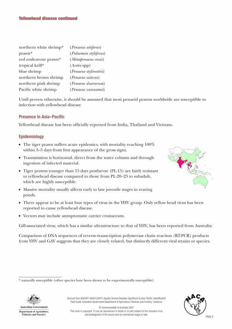
Histological section of the lymphoid organ (LO) of a juvenile giant black tiger prawn (Penaeus monodon) with severe acute yellowhead disease (YHD) at low (525x) and high (1700x) magnification. A generalised, diffuse necrosis of LO cells is shown. Affected cells display pyknotic and karyorrhectic nuclei. Single or multiple perinuclear inclusion bodies, ranging from pale to darkly basophilic, are apparent in some affected cells (arrows). This marked necrosis in acute YHD distinguishes YHD from infections due to Taura syndrome virus, which produces similar cytopathology in other target tissues, but not in the LOSource: DV Lightner
Histological section (1000x) of the gills from a juvenile black tiger prawn with YHD. A generalised, diffuse necrosis of cells in the gill lamellae is shown, and affected cells display pyknotic and karyorrhectic nuclei (arrows). A few large, conspicuous, generally spherical cells with basophilic cytoplasm are present in the section. These cells may be immature haemocytes, released prematurely in response to a YHV-induced haemocytopeniaSource: DV Lightner

Sourced from AGDAFF–NACA (2007) Aquatic Animal Diseases Significant to Asia-Pacific: Identification Field Guide. Australian Government Department of Agriculture, Fisheries and Forestry. Canberra.
© Commonwealth of Australia 2007 This work is copyright. It may be reproduced in whole or in part subject to the inclusion of an
acknowledgment of the source and no commercial usage or sale.PAGE 5
Histological sections of the LO of juvenile white shrimp (P. vannamei) (Fig 4, 1000x) and northern brown shrimp (P. aztecus) (Fig 5, 525x) experimentally infected with YHV. Severe (grade 3–4) diffuse to multifocal necrosis, characterised by cells with increased eosinophilic cytoplasm, pyknotic or karyorrhectic nuclei (arrows) and pale to densely basophilic perinuclear inclusions, is presentSource: DV Lightner
Yellowhead disease continued
Histological images
Histological sections (1000x) of the gills of a juvenile northern pink shrimp (P. duorarum) (Fig 6) and the oesophagus of a white shrimp (Fig 7) experimentally infected with YHV. Severe (grade 4) diffuse to multifocal necrosis, characterised by cells with increased eosinophilic cytoplasm, pyknotic or karyorrhectic nuclei, and pale to densely basophilic perinuclear inclusions, is presentSource: DV Lightner
Sourced from AGDAFF–NACA (2007) Aquatic Animal Diseases Significant to Asia-Pacific: Identification Field Guide. Australian Government Department of Agriculture, Fisheries and Forestry. Canberra.
© Commonwealth of Australia 2007 This work is copyright. It may be reproduced in whole or in part subject to the inclusion of an
acknowledgment of the source and no commercial usage or sale.PAGE 6
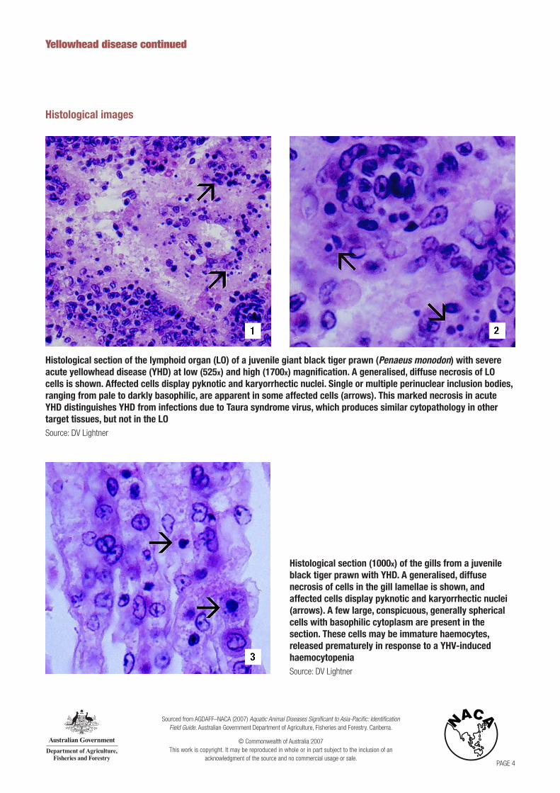
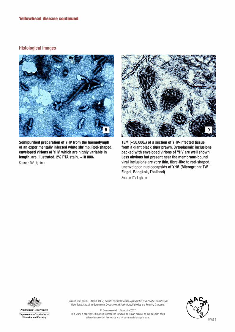
Semipurified preparation of YHV from the haemolymph of an experimentally infected white shrimp. Rod-shaped, enveloped virions of YHV, which are highly variable in length, are illustrated. 2% PTA stain, ~18 000x
Source: DV Lightner
Yellowhead disease continued
Histological images
TEM (~50,000x) of a section of YHV-infected tissue from a giant black tiger prawn. Cytoplasmic inclusions packed with enveloped virions of YHV are well shown. Less obvious but present near the membrane-bound viral inclusions are very thin, fibre-like to rod-shaped, unenveloped nucleocapsids of YHV. (Micrograph: TW Flegel, Bangkok, Thailand)Source: DV Lightner