Diseases affecting pigs: an overview of common bacterial ...
29
Veterinary Diagnostic and Production Animal Medicine Publications Veterinary Diagnostic and Production Animal Medicine 2018 Diseases affecting pigs: an overview of common bacterial, viral and parasitic pathogens of pigs Alejandro Ramirez Iowa State University, [email protected] Follow this and additional works at: hps://lib.dr.iastate.edu/vdpam_pubs Part of the Large or Food Animal and Equine Medicine Commons , Veterinary Infectious Diseases Commons , and the Veterinary Pathology and Pathobiology Commons e complete bibliographic information for this item can be found at hps://lib.dr.iastate.edu/ vdpam_pubs/125. For information on how to cite this item, please visit hp://lib.dr.iastate.edu/ howtocite.html. is Book Chapter is brought to you for free and open access by the Veterinary Diagnostic and Production Animal Medicine at Iowa State University Digital Repository. It has been accepted for inclusion in Veterinary Diagnostic and Production Animal Medicine Publications by an authorized administrator of Iowa State University Digital Repository. For more information, please contact [email protected].
Transcript of Diseases affecting pigs: an overview of common bacterial ...
Veterinary Diagnostic and Production AnimalMedicine Publications
Veterinary Diagnostic and Production AnimalMedicine
2018
Diseases affecting pigs: an overview of commonbacterial, viral and parasitic pathogens of pigsAlejandro RamirezIowa State University, [email protected]
Follow this and additional works at: https://lib.dr.iastate.edu/vdpam_pubs
Part of the Large or Food Animal and Equine Medicine Commons, Veterinary InfectiousDiseases Commons, and the Veterinary Pathology and Pathobiology Commons
The complete bibliographic information for this item can be found at https://lib.dr.iastate.edu/vdpam_pubs/125. For information on how to cite this item, please visit http://lib.dr.iastate.edu/howtocite.html.
This Book Chapter is brought to you for free and open access by the Veterinary Diagnostic and Production Animal Medicine at Iowa State UniversityDigital Repository. It has been accepted for inclusion in Veterinary Diagnostic and Production Animal Medicine Publications by an authorizedadministrator of Iowa State University Digital Repository. For more information, please contact [email protected].

Diseases affecting pigs: an overview of common bacterial, viral andparasitic pathogens of pigs
AbstractRecent events such as the 2009 pandemic influenza outbreak, the continuous spread of African swine fevervirus in Eastern Europe and the introduction of several new pathogens into the United States and their spreadto Canada, Mexico, Central and South America have emphasized the ability of pig diseases to cross bordersrapidly and the importance of global cooperation to improve the health and welfare of pigs. This chaptersummarizes recent research on the causes and epidemiology of major bacteria, viruses and parasites found inpig production, focusing on those with a particular impact on safety and global production.
DisciplinesLarge or Food Animal and Equine Medicine | Veterinary Infectious Diseases | Veterinary Pathology andPathobiology
CommentsThis is a chapter from Wiseman, J. Achieving sustainable production of pig meat: Animal health and welfare, vol. 3.Ed. Julian Wiseman, University of Nottingham, UK. Burleigh Dodds Series in Agricultural Science ; No. 25.Cambridge, UK: Burleigh Dodds Science Publishing (2018). DOI: 10.19103/AS.2017.0013.14. Posted withpermission.
This book chapter is available at Iowa State University Digital Repository: https://lib.dr.iastate.edu/vdpam_pubs/125

http://dx.doi.org/10.19103/AS.2017.0013.14© Burleigh Dodds Science Publishing Limited, 2018. All rights reserved.
Chapter 1
Diseases affecting pigs: an overview of common bacterial, viral and parasitic pathogens of pigsAlejandro Ramirez, Iowa State University, USA
1 Introduction
2 The most common bacterial pathogens in pig production: gram-negative bacteria
3 The most common bacterial pathogens in pig production: gram-positive bacteria
4 The most common viral pathogens in pig production
5 The most common parasitic pathogens in pig production
6 Case studies
7 Summary
8 Future trends
9 Where to look for further information
10 References
1 Introduction
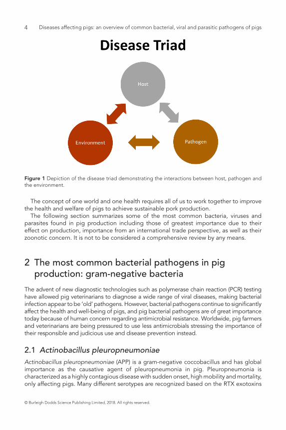
Diseases affecting pigs can be quite complex. It is well recognized that often these conditions are multifactorial, especially as in the case of respiratory diseases, hence the term ‘porcine respiratory disease complex’ (PRDC) has been accepted (Brockeier, 2002). The concept of the disease triad (Fig. 1) emphasizes this complexity and the interaction between not only different pathogens, but the host, pathogen and the environment.
To maintain a sustainable pork production system, we must move away from the idea of one agent–one disease and look at the whole picture from a holistic point of view. Recent world events concerning pigs such as the 2009 pandemic influenza outbreak, the continuous spread of African swine fever virus (ASFv) in Eastern Europe and the introduction of several new pathogens into the United States (porcine epidemic diarrhoea virus and porcine deltacoronavirus) and their spread to Canada (limited), Mexico, Central and South America have emphasized that we live in one world with minimal borders. Diseases of pigs spread rapidly across various countries.
Diseases affecting pigs: an overview of common bacterial, viral and parasitic pathogens of pigs4
© Burleigh Dodds Science Publishing Limited, 2018. All rights reserved.
The concept of one world and one health requires all of us to work together to improve the health and welfare of pigs to achieve sustainable pork production.
The following section summarizes some of the most common bacteria, viruses and parasites found in pig production including those of greatest importance due to their effect on production, importance from an international trade perspective, as well as their zoonotic concern. It is not to be considered a comprehensive review by any means.
2 The most common bacterial pathogens in pig production: gram-negative bacteria
The advent of new diagnostic technologies such as polymerase chain reaction (PCR) testing have allowed pig veterinarians to diagnose a wide range of viral diseases, making bacterial infection appear to be ‘old’ pathogens. However, bacterial pathogens continue to significantly affect the health and well-being of pigs, and pig bacterial pathogens are of great importance today because of human concern regarding antimicrobial resistance. Worldwide, pig farmers and veterinarians are being pressured to use less antimicrobials stressing the importance of their responsible and judicious use and disease prevention instead.
2.1 Actinobacillus pleuropneumoniaeActinobacillus pleuropneumoniae (APP) is a gram-negative coccobacillus and has global importance as the causative agent of pleuropneumonia in pig. Pleuropneumonia is characterized as a highly contagious disease with sudden onset, high mobility and mortality, only affecting pigs. Many different serotypes are recognized based on the RTX exotoxins
Figure 1 Depiction of the disease triad demonstrating the interactions between host, pathogen and the environment.

© Burleigh Dodds Science Publishing Limited, 2018. All rights reserved.
Diseases affecting pigs: an overview of common bacterial, viral and parasitic pathogens of pigs 5
secreted by the organism. Currently, four of these exotoxins are recognized (ApxI, ApxII, ApxIII, ApxIV) and their presence varies among serotypes (Chiers et al., 2010). These toxins cause severe internal pulmonary haemorrhage and cytotoxicity. Apart from these toxins, many other virulence factors have also been identified. The virulence of serotypes, as well (asymptomatic to high mortality) as their prevalence in different geographical regions, is highly variable (Clota et al., 1996; Mittal et al., 1996; Kucerova et al., 2005; Gottschalk, 2003). It is well recognized that isolates from most herds have more than one low-virulent serotype (Gottschalk, 2003).
One of the major challenges with APP is that pigs continue to carry the organism in their lungs and tonsils for several months (Desrosiers, 2004), thus creating an opportunity for repeated outbreaks of this clinical disease, as well as creating a challenging environment at all stages of pig production, from growing pigs until slaughter. Antimicrobials are used to treat the disease with several countries, especially the United States, demonstrating a pattern of resistance to beta-lactams (penicillin, amoxicillin). Antimicrobial treatment helps minimize mortalities during the early stages of an outbreak, but they do not eliminate carrier pigs and Sjölund et al. (2009) have suggested that the use of highly effective antimicrobials prevents good antibody response to the infection, thus leaving pigs susceptible to future re-infections.
Vaccination against APP is challenging due to the various different serotypes. They involve stimulation by several different Apx exotoxins as well as an outer membrane protein (Gottschalk, 2012).
The severity of APP infections is a significant burden on the sustainability and feasibility of an infected herd. This is especially true today with the current emphasis on antimicrobial stewardship, thus infected herds must undergo a de-population and re-population many times with APP-free pigs.
There are no zoonotic concerns regarding APP other than it can lead to extensive antimicrobial use in infected herds.
2.2 Bordetella bronchisepticaBordetella bronchiseptica is a gram-negative rod that is found throughout the world and infects many different mammalian species (Brockmeier et al., 2012). In pigs Bordetella bronchiseptica primarily causes pneumonia and atrophic rhinitis.
Several virulence genes which appear to require co-expression of the BvgAS genes are identified (Beier and Gross, 2008) and are also subject to phase variation. Early BvgAS genes are involved in bacterial attachment followed later (once a large number of bacteria have colonized the area) with gene expression for toxin production (Brockmeier et al., 2012). It is this toxin production (especially dermonecrotic toxin) that contributes to the progression of the disease, especially atrophic rhinitis (nasal turbinate and septal damage).
Vaccination of sows before farrowing can be used in conjunction with antimicrobials to prevent atrophic rhinitis in pigs. The aim of these interventions is to prevent or minimize early colonization by Bordetella bronchiseptica, allowing the pig’s immune system to protect it as it matures as well as preventing the late stages of BvgAS gene expression, which produces the dermonecrotic toxin. Vaccination helps minimize disease but does not prevent infection. Antibiotics can help minimize disease transmission between pigs.
Bordetella bronchiseptica often works in conjunction with toxigenic Pasteurella multocida to cause the more severe disease known as progressive atrophic rhinitis (de Jong and Nielsen, 1990).

Diseases affecting pigs: an overview of common bacterial, viral and parasitic pathogens of pigs6
© Burleigh Dodds Science Publishing Limited, 2018. All rights reserved.
Although human infections can occur with Bordetella bronchiseptica they are rare, and pigs do not appear to be a concern for zoonosis.
2.3 Brachyspira spp.The Brachyspira hyodysenteriae is a spirochetal bacterium that causes swine dysentery (SD). Herds infected with SD incur serious financial losses due to mucoid and bloody diarrhoea resulting in a significant number of deaths as well as poor growth performance.
SD affects the large intestine of grower and finisher pigs but rarely affects weaners (Hampson, 2012). With the advent of PCR technology and genetic sequencing, several new species of Brachyspira have been identified and shown to cause SD lesions in growing pigs (Burrough et al., 2012; Chander et al., 2012). So although technically SD is only associated with Brachyspira hyodysenteriae, today the phenotypic culture characteristics (especially hemolysis) of Brachyspira spp. appear to be a more sensitive indicator of potential to induce dysentery-like disease in pigs (Table 1) than molecular identification alone (Burrough et al., 2012).
There is a limited arsenal of antimicrobials which can be used to treat SD. Mice and rats can serve as an important reservoir for Brachyspira hyodysenteriae (and maybe other Brachyspira spp.), which make it difficult to completely eliminate an infection from a herd. The limited weapons available are expensive antimicrobials that can add a significant cost to SD control. Therefore, de-population and re-population with SD-free pigs may be necessary in conjunction with aggressive cleaning, disinfection of premises and extensive rodent control programmes. Currently there are no effective vaccines against Brachyspira spp.
There are no zoonotic concerns regarding Brachyspira hyodysenteriae other than there may need to be extensive antimicrobial use in infected herds.
2.4 Brucella suisBrucella suis is a gram-negative coccobacillus of zoonotic importance in pigs, usually causing reproductive failure including long-term, non-fatal, granulomatous inflammation in several organs (including joints and reproductive organs such as testes). Its impact on production is mostly noted in acute outbreaks as frequently endemic brucellosis is mild
Table 1 Clinical significance of some Brachyspira spp. in pigs (Burrough 2015, pers. comm.)
Hemolysis Clinical disease in pigs
Brachyspira hyodysenteriae Strong beta-hemolysis Swine dysentery
Brachyspira hampsonii Strong beta-hemolysis Dysentery-like disease
Brachyspira suanatina Strong beta-hemolysis Dysentery-like disease
Brachyspira pilosicoli Weak beta-hemolysis Spirochetal colitis (mild disease)
Brachyspira murdochii Weak beta-hemolysis Mild to non-pathogenic
Brachyspira intermedia Weak beta-hemolysis Mild to non-pathogenic
Brachyspira innocens Weak beta-hemolysis Non-pathogenic
© Burleigh Dodds Science Publishing Limited, 2018. All rights reserved.
Diseases affecting pigs: an overview of common bacterial, viral and parasitic pathogens of pigs 7
enough that it can go undetected. Feral pigs are often found to be infected with Brucella suis throughout the United States (Leuenberger et al., 2007; Leiser et al., 2013).
The long duration of bacteraemia (5 weeks to 34 months) reported by Deyoe (1967 and 1972) suggests that immune response to infection is insufficient to eliminate bacteria from blood and intracellular niche, especially in macrophages (Olsen et al., 2012). Treatment or vaccination is not recommended because of the zoonotic potential of this organism, and the animal should be removed from the herd.
Although human brucellosis is extremely important, swine brucellosis is of lesser importance since milk from pigs is rarely consumed (Pappas et al., 2006). The zoonotic potential for infection due to prolonged bacteraemia and the relatively low infectious dose for humans is especially important due to work-related exposure and wild boar hunters.
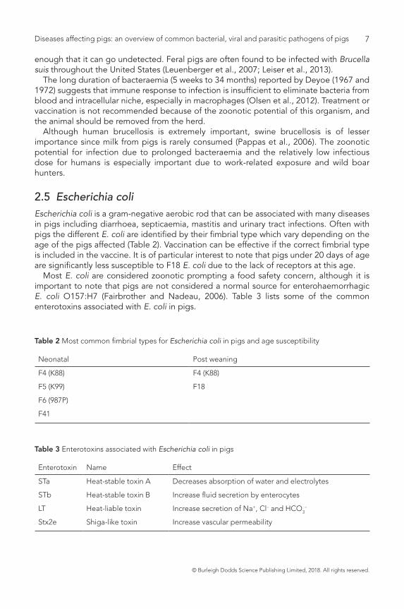
2.5 Escherichia coliEscherichia coli is a gram-negative aerobic rod that can be associated with many diseases in pigs including diarrhoea, septicaemia, mastitis and urinary tract infections. Often with pigs the different E. coli are identified by their fimbrial type which vary depending on the age of the pigs affected (Table 2). Vaccination can be effective if the correct fimbrial type is included in the vaccine. It is of particular interest to note that pigs under 20 days of age are significantly less susceptible to F18 E. coli due to the lack of receptors at this age.
Most E. coli are considered zoonotic prompting a food safety concern, although it is important to note that pigs are not considered a normal source for enterohaemorrhagic E. coli O157:H7 (Fairbrother and Nadeau, 2006). Table 3 lists some of the common enterotoxins associated with E. coli in pigs.
Table 2 Most common fimbrial types for Escherichia coli in pigs and age susceptibility
Neonatal Post weaning
F4 (K88) F4 (K88)
F5 (K99) F18
F6 (987P)
F41
Table 3 Enterotoxins associated with Escherichia coli in pigs
Enterotoxin Name Effect
STa Heat-stable toxin A Decreases absorption of water and electrolytes
STb Heat-stable toxin B Increase fluid secretion by enterocytes
LT Heat-liable toxin Increase secretion of Na+, Cl– and HCO3–
Stx2e Shiga-like toxin Increase vascular permeability
Diseases affecting pigs: an overview of common bacterial, viral and parasitic pathogens of pigs8
© Burleigh Dodds Science Publishing Limited, 2018. All rights reserved.
2.6 Haemophilus parasuisHaemophilus parasuis (HPS) is a gram-negative bacterium which causes Glässer’s disease (fibrinous polyserositis) with at least 15 different serovar groups identified (Kielstein and Rapp-Gabrielson, 1992). There does not appear to be a direct association between serovars and virulence. One of the challenges with HPS is that it tends to be more common in high health status herds. HPS often appears after co-mingling (mixing) pigs from different sources (Wiseman et al., 1989). In acute outbreaks, HPS tends to affect the biggest and healthiest pigs in the group. HPS invades endothelial cells and causes apoptosis and production of pro-inflammatory cytokines creating accumulation of fibrin (Vanier et al., 2006; Bouchet et al., 2008).
Strategic use of antimicrobials can be used to mitigate the sudden effects of HPS by administering it at times of high risk, especially during stressful events (weaning and animal movement). Unfortunately, after killing the bacteria, the antimicrobials do nothing to the fibrin already produced. As this fibrin dries, it becomes fibrous and can affect heart and lung movements, resulting in a chronically ill pig. Effectiveness of HPS vaccination can be variable (Oliveira et al., 2004; Oh et al., 2013).
There are no zoonotic concerns regarding HPS.
2.7 Lawsonia intracellularisLawsonia intracellularis is an obligate intracellular bacterium which grows preferentially in intestinal epithelial cells, causing ileitis in pigs. There are three quite distinct clinical presentations of the disease:
1 the traditional porcine intestinal adenomatosis (thickening of intestine);2 the more chronic form of enteritis with fibrinonecrotic membrane; and 3 the peracute haemorrhagic form resulting in sudden death.
The importance of this disease is its continuous effect on decreasing feed efficiency and average daily gain (McOrist et al., 1997; McOrist, 2005). The peracute form usually affects market-ready pigs, resulting in sudden death with significant consequential economic losses.
Prevention involves the use of vaccines and/or strategic pulsing with a variety of antimicrobials (McOrist et al., 1999; Hammer, 2004; Bak and Rathkjen, 2009).
There are no zoonotic concerns regarding L. intracellularis infection in pigs.
2.8 Pasteurella multocidaPasteurella multocida is a gram-negative rod or coccobacillus which can cause pneumonia and atrophic rhinitis in pigs. Most pig isolates are either serotype A or D, whereas serotypes B, C and E are found in cattle, reindeer and water buffalo. Most P. multocida Type A have a predilection to lung tissue while Type D are usually involved in progressive atrophic rhinitis along with Bordetella bronchiseptica, although either type can be found in the other’s preferred tissue (Carter, 1955; Pijoan et al., 1983; Rimler and Rhoads, 1987).
P. multocida is the most common bacterial infection found in PRDC and it is the primary target for antimicrobial therapy. There are toxigenic and non-toxigenic strains of P. multocida. It is interesting to note that P. multocida by itself cannot cause pneumonia
© Burleigh Dodds Science Publishing Limited, 2018. All rights reserved.
Diseases affecting pigs: an overview of common bacterial, viral and parasitic pathogens of pigs 9
even when heavily inoculated. This suggests that there is a need for a primary co-infection to enable the establishment of P. multocida (Brockmeier et al., 2001). Vaccines containing P. multocida toxin have been effective against progressive atrophic rhinitis in pigs. P. multocida has no food safety concerns but has the potential for being zoonotic.
2.9 Salmonella spp.Salmonella spp. are gram-negative rods known to infect a broad range of hosts. The two most important salmonella in pigs are S. choleraesuis (pigs only) and S. typhimurium (humans and pigs).
Clinical signs of salmonellosis can be variable in pigs depending on the strain. Both S. choleraesuis and S. typhimurium cause diarrhoea in pigs while S. choleraesuis more often tends to be systemic causing cyanosis of the skin as well as an interstitial pneumonia (Schwartz, 1997; Foley and Lynne, 2008; Carlson et al., 2012). There are over 200 virulence factors that have been identified with Salmonella, but few have been fully characterized (Carlson et al., 2012).
Vaccination is quite effective in helping prevent disease and antimicrobial use helps with treatment. As a gram-negative enteric pathogen, drug resistance (via plasmids) is common (Barnes and Sorensen, 1975; Schwartz, 1997). In addition to vaccination and antimicrobial therapy, bio-security with heavy emphasis on cleanliness to minimize faecal-oral exposure is important.
Salmonella is of zoonotic concern impacting on food safety.
3 The most common bacterial pathogens in pig production: gram-positive bacteria
3.1 Clostridium spp.Clostridium spp. are anaerobic gram-positive spore-forming rods with several different species causing different diseases in pigs. Clostridium spp. cause disease via the different toxins they produce. In this chapter we will only discuss the two Clostridium spp. of greatest concern in pigs.
3.1.1 Clostridium difficileIn pigs, the clinical signs usually appear in the first few hours or days of life. The disease is believed to be caused by two toxins (Toxins A and B) and the administration of equine-origin antitoxins can mitigate the effects (Ramirez et al., 2014). Antimicrobial use does not appear to affect the severity of the disease in neonatal pigs (Arruda et al., 2013), which makes sense as the microflora is barely established in newborn piglets immediately after birth. Interestingly, although C. difficile infections are not seen in older pigs (more than 7 days of age), work by Arruda et al. (2013) has shown pigs are still susceptible at 10 days of age. There are currently no effective pig vaccines against C. difficile, which can be found in the faeces of most mammals.
In humans, C. difficile infections can be very serious or even deadly with antimicrobial-associated diarrhoea (Bartlet et al., 1978). In humans it can result in simple diarrhoea,

Diseases affecting pigs: an overview of common bacterial, viral and parasitic pathogens of pigs10
© Burleigh Dodds Science Publishing Limited, 2018. All rights reserved.
colitis, pseudomembranous colitis, ileus, toxic mega colon and even bowel perforation (Kelly et al., 1984). However, there is currently no data to directly link C. difficile infection in pigs to zoonotic issues.
3.1.2 Clostridium perfringensThere are currently five different toxinotypes (Table 4), with only toxinotypes A and C affecting pigs. Enteritis by C. perfringens Type C has been well characterized and pre-farrowing vaccination programmes have been effective in controlling this disease quite well. On the other hand, C. perfringens Type A is still a bit of an enigma. Traditionally, Type A infections have only been associated with alpha toxin production. Unfortunately, as seen in Table 3, all other C. perfringens toxinotypes also produce this same toxin. Several research labs have suggested a role for a beta2 toxin with this disease (Bueschel et al., 2003; Waters et al., 2003), while others more recently question its role (Faranz et al., 2013). Field vaccination with C. perfringens Type A toxoid does not appear to be as effective as Type C vaccination.
There are no zoonotic concerns regarding C. perfringens in pigs. Human food poisoning with C. perfringens is mostly associated with consumption of beef, poultry or gravies.
3.2 TuberculosisTuberculosis continues to be responsible for significant economic losses for pig producers in many countries while others such as the United States have practically eliminated it from their pig population (Thoen, 2012). Pigs are susceptible to Mycobacterium avium complex (MAC) and M. tuberculosis complex amongst other mycobacterial species (Thoen et al., 1975).
Pigs often acquire MAC when reared on ground contaminated by poultry (Schalk et al., 1935) and sometimes even sawdust (Schliesser and Weber, 1973). Most cases in pigs are asymptomatic or non-specific and therefore only diagnosed at slaughter (Thoen, 2012). Slaughter inspection specifically looks for granulomatous lesions in lungs or lymph nodes (granulomatous lymphadenitis). However, current European Union guidelines on pig meat inspection discourage the palpation/incision of such post-mortem lesions during routine slaughter in an effort to minimize bacterial cross-contamination (EUFSA, 2011)
As with most diseases that cause granulomas, the use of antimicrobials is not recommended due to long duration of treatment and poor prognosis. There are no effective vaccines available for pigs.
Table 4 Five Clostridium perfringens toxinotypes and their respective toxin and animals they can infect
Clostridium perfringens
Animals affected Toxins
Pigs Sheep Goats Poultry Cattle Horses Alpha Beta Beta2 Epsilon Iota
Type A X X X X X X
Type B X X X X X X
Type C X X X X X X X X
Type D X X X X X
Type E ? X X X

© Burleigh Dodds Science Publishing Limited, 2018. All rights reserved.
Diseases affecting pigs: an overview of common bacterial, viral and parasitic pathogens of pigs 11
Although MAC can be a significant zoonotic and food safety risk for humans, especially those with immunocompromised immune systems (e.g., elderly, AIDS), pigs and pork have not been implicated as an exposure risk for human infection (Arasteh et al., 2000; Thoen, 2012).
3.3 Mycoplasma spp.Mycoplasmas are a type of bacteria that lack cell walls. There are several mycoplasmas of importance in pigs including Mycoplasma hyopneumoniae (Mhyop), M. hyorhinis (Mhyor), M. hyosynoviae (Mhyos) and M. suis. Because of the large number of different mycoplasmas, it is important to be specific in name and not just refer to them as simply ‘mycoplasma’.
There are no zoonotic concerns regarding any of the Mycoplasmas in pigs.
3.3.1 Mycoplasma hyopneumoniaeMhyop is the aetiologic agent for enzootic pneumonia in pigs, one of the most significant bacterial respiratory pig pathogens worldwide. The strains of Mhyop are antigenically diverse (Frey et al., 1992; Thacker and Minion, 2012). Mhyop is difficult to grow in most laboratories.
Transmission of Mhyop occurs via nose-to-nose contact especially from sow to pig (Calsamiglia and Pijoan, 2000) but can also occur via aerosol up to 3.2 km (Goodwin, 1985) and 9.2 km (Otke et al., 2010). The organism attaches to ciliary epithelium of the respiratory tract and grows slowly. Protein P97 is involved in adherence (Zhang et al., 1994). Mhyop also alters the function of macrophages (Caruso and Ross, 1990) as well as other parts of the immune system (Thacker and Minion, 2012). Mhyop is an important potentiate of other respiratory pathogens in association with PRDC.
Vaccination of growing pigs can be considered ‘standard’ in today’s pig production. Antimicrobials can also be used strategically to mitigate Mhyop as well as other bacterial co-infections.
3.3.2 Mycoplasma hyorhinisMhyor is associated with polyserositis and arthritis in three- to ten-week-old pigs. Current interest in this pathogen has increased due to welfare concerns of lameness in growing pigs. Little is known regarding the virulence and pathogenesis of Mhyor (Thacker and Minion, 2012). With the advent of newer PCR technology, many samples are now being tested for Mhyor, possibly creating a false sense of increased prevalence in recent years. Bacterial culture for Mhyor can be quite easy but requires special media (Ross and Whittlestone, 1983).
There are several antimicrobials which are used to treat Mhyor infection, although efficacy is quite variable (Thacker and Minion, 2012). It is suspected that part of the problem is late diagnosis of the disease. As with any joint infection, early detection is key.
3.3.3 Mycoplasma hyosynoviaeMhyos is mostly associated with arthritis, and is very similar to Mhyor except that Mhyos tends to affect older pigs (3–5 months of age). As is the case with Mhyor, the Mhyos

Diseases affecting pigs: an overview of common bacterial, viral and parasitic pathogens of pigs12
© Burleigh Dodds Science Publishing Limited, 2018. All rights reserved.
bacteria can be found in the tonsils of infected and ‘normal’ pigs (Thacker and Minion, 2012). Bacterial culture for Mhyos can be quite straightforward but requires special media (Friis et al., 1992).
3.3.4 Mycoplasma suisEperythrozoon suis, now renamed Mycoplasma suis, infects red blood cells of pigs causing anaemia (moderate to severe), respiratory distress (Doyle, 1932) and possible reproductive problems. The organism can live in the cytoplasm as well as in membrane-bound vacuoles of erythrocytes (Groebel et al., 2009) which can make it inaccessible to many antimicrobials. Bacterial culture for M suis is not yet possible so diagnosis is currently done via PCR.
3.4 Staphylococcus spp.Staphylococcus spp. are gram-positive cocci that are regarded as normal bacterial flora of adult pig skin (Frana, 2012). There are two primary Staphylococcus of importance in pigs: S. hyicus and methicillin-resistant S. aureus (MRSA).
3.4.1 Staphylococcus aureusStaphylococcus aureus can often be isolated from the skin of pigs as well as from septicaemia, mastitis, metritis and metritis infections. Although S. aureus rarely causes disease, recent attention to a specific type of S. aureus known as MRSA has stimulated interest in this bacteria. In particular, a unique MRSA known as ST398 was first associated with pigs in Europe (Armad-Leferve et al., 2005). This same sequence type does not appear to be as prevalent or important in pig production in the United States (Frana et al., 2013).
MRSA appears to be asymptomatic in pigs and is not considered to be a herd problem. Although on-farm antimicrobial use is suspected in MRSA, no studies have been able to demonstrate this association, which brings into question the ethical legitimacy of using stigmatization as a direct means to achieve public health outcomes (Plough et al., 2015).
Although MRSA is a significant human health concern, outside Denmark, the role of pigs in MRSA zoonosis does not appear to be significant. MRSA is not a food safety concern.
3.4.2 Staphylococcus hyicusStaphylococcus hyicus is the causative agent for greasy pig disease or exudative epidermitis. This condition has worldwide distribution and presents as a skin infection (pyoderma) in young pigs (nursery age or younger). Although S. hyicus is commonly found in pig skin, under the right conditions the bacterium will establish itself in the epidermis via an abrasion in the skin. In severe cases the loss of fluids and electrolytes can lead to dehydration and death. There are several exfoliative toxins that have been identified and are considered the most important virulence factors for greasy pig disease (Amtsberg, 1979).
High humidity in pens, as well as a high number of young gilts farrowing, contributes to higher incidence or acute outbreaks of the disease. Injectable antimicrobials are commonly used to treat affected pigs along with topical treatments (spraying/soaking) which may involve the use of disinfectants (Frana, 2012). Pig farmers often use autogenous vaccines against S. hyicus with variable effectiveness.
There are no zoonotic concerns regarding S. hyicus in pigs.

© Burleigh Dodds Science Publishing Limited, 2018. All rights reserved.
Diseases affecting pigs: an overview of common bacterial, viral and parasitic pathogens of pigs 13
3.5 Streptococcus suisAlthough there are many other Streptococcus spp. that infect pigs, we will only discuss S. suis. Streptococcus suis is a gram-positive encapsulated coccus that can frequently be found in the tonsils, intestines and genital tract of healthy pigs (Gottschalk, 2012). There are 35 serotypes based on capsular polysaccharide but serotype 2 is the most common and most important one in pigs because of its zoonotic potential.
There are a large number of virulence factors identified for S. suis but none are fully understood due to the complexity of multiple factors (Braums and Valentin-Weigand, 2009). Streptococcus suis infection in pigs can be variable, in part due to variations in virulence and may include septicaemia, central nervous signs (meningitis), arthritis, pneumonia, vegetative valvular endocarditis, rhinitis and abortions (Sanford and Tilker, 1982).
Vaccination against S. suis is often ineffective. Beta-lactams and macrolide antimicrobials are commonly used to prevent and treat S. suis infections.
In South-East Asia, S. suis is the most common cause of bacterial meningitis in humans and therefore has been identified as a serious emerging public health threat (Wertheim, 2009). S. suis poses a zoonotic concern including a food safety concern in countries with particular cultural practices and preferences such as drinking uncooked blood from infected pigs and eating organs such as the uterus.
4 The most common viral pathogens in pig production
As a general rule it is helpful to know if the viral pathogen is a DNA or an RNA virus as well as whether it is an enveloped or non-enveloped virus. Compared with DNA viruses, RNA viruses mutate often as they do not have the necessary proofing mechanism when they replicate. Thus there can be great variability between strains and developing vaccines can be more challenging. Non-enveloped viruses tend to be much more resistant to inactivation than enveloped viruses. This means that non-enveloped viruses tend to persist longer in the environment. Clearly there are always exceptions to the rule, but these guiding principles can be very useful when learning about a new virus and its possible behaviour regarding transmission between pigs. Many of these important viruses are listed by the World Organization for Animal Health (OIE) as notifiable diseases due to their importance regarding animal and human health as well as international trade.
4.1 African swine fever virusASFv is an enveloped DNA virus from the family Asfarvideae that causes a highly contagious and haemorrhagic disease in pigs of all ages. ASF is an OIE-listed disease with important international trade consequences. The disease is endemic in sub-Saharan Africa and has now spread to several Eastern European countries. There are many different hosts for the virus as well as significant variation in virulence between strains (De Villeret al., 2010).
ASFv can be transmitted by many means including soft ticks and direct contact with contaminated oral and nasal secretions (Colgrove et al., 1969), consumption of contaminated feed and possibly short distances via aerosol (CFSPH, 2015a). The virus can survive long periods in cured meats (Mebus et al., 1993). ASFv can infect multiple tissues but their primary cells for replication include monocytes and macrophages (Malmquist and Hays, 1960; Minguez et al., 1988). Although ASFv does not induce neutralizing antibodies

Diseases affecting pigs: an overview of common bacterial, viral and parasitic pathogens of pigs14
© Burleigh Dodds Science Publishing Limited, 2018. All rights reserved.
(De Boer, 1967), protective immunity against homologous but not heterologous re-infection still occurs (Ruiz Gonzalvo et al., 1981).
Currently there are no good vaccines against ASFv and treatment is not recommended as disease eradication should be the goal.
ASFv is not zoonotic but the severity of the disease causes significant food security and sustainability concerns.
4.2 Aujeszky’s disease virusAujeszky’s disease, also known as pseudorabies, is caused by the herpes virus, which is an enveloped DNA virus whose only natural host is pigs. Aujeszky’s disease presents with central nervous system indicators, reproductive problems including abortions, respiratory illness and mortality. All other mammals, except humans, are end hosts for the virus, resulting in close to 100% mortality in these species. Its distribution is worldwide, with variations in virulence between strains and is an OIE-reportable disease.
Aujeszky’s disease virus is primarily transmitted between pigs via direct and indirect contact including long-distance aerosol (Christen et al., 1990). Even after recovery, pigs remain infected for the rest of their life (common amongst herpes virus infections) and stress can reactivate viral shedding, helping to spread the disease to other pigs (Wittmann and Rziha, 1989).
Gene-deleted vaccines can be used with DIVA (differentiate vaccinated from infected) capabilities. They are quite effective at protecting against viraemia and clinical signs but unfortunately they do not prevent latent infections. Aujeszky’s disease is not of zoonotic concern.
4.3 Classical swine fever virusClassical swine fever (CSF), also known as hog cholera, is caused by an enveloped RNA Pestivirus that causes generalized systemic disease indistinguishable from many other common, endemic bacterial and viral pig diseases. CSF is an OIE-reportable disease. CSF is endemic in parts of Asia, South and Central America and some Caribbean islands (CFSPH, 2015b).
CSF virus is highly contagious and can cause septicaemia, anorexia, constipation, diarrhoea, lethargy and abortions, to mention just a few. The variable clinical signs and virulence of the virus is dependent on many factors including variation in strains (asymptomatic to high mortality) (Depner et al., 1997; Moenning et al., 2003).
As with natural infections, a combination of cell-mediated immunity and neutralizing antibodies appear to be important in producing sterilizing immunity (Pirou et al., 2003). There are several vaccines, especially live or modified live, that provide good protection against disease including some oral vaccines for wild boars (CFSPH, 2015b). CSF disease is not of zoonotic concern.
4.4 CoronavirusesThere are several different coronaviruses of importance in pigs. For the most part, they have quite similar clinical presentation, primarily causing diarrhoea with similar treatment
© Burleigh Dodds Science Publishing Limited, 2018. All rights reserved.
Diseases affecting pigs: an overview of common bacterial, viral and parasitic pathogens of pigs 15
and control but immunologically are very distinct from each other (i.e. no cross-protection between these different viruses). They are enveloped RNA viruses with spike proteins that give them a crown-like appearance under an electron microscope. None of the pig coronaviruses are of zoonotic concern.
4.4.1 DeltacoronavirusWith the introduction of porcine epidemic diarrhoea virus (PEDv) into the United States in early 2013 a new porcine deltacoronavirus (PDCov) was identified in faeces from pigs with diarrhoea (Wang et al., 2014). Work done by June et al. (2015) demonstrated that this virus is capable of causing disease in gnotobiotic pigs. Anecdotal field evidence suggests PDCov can cause diarrhoea, but is significantly milder and shorter in duration than PEDv. Currently there are no vaccines for PDCov.
4.4.2 Porcine epidemic diarrhoea virusThe importance of PEDv has re-surfaced after its introduction into the United States in May 2013. This introduction highlighted the possible role feed and feed ingredients can play in disease transmission – an area previously ignored most of the time. The clinical presentation for the disease is almost exactly the same as that of transmissible gastroenteritis virus (TGEv). The challenge of any new disease introduced into a naïve population was quite apparent. Disease spread easily from place to place and the infectious dose of the virus was quite low as data to be published in peer-review journals will show. Recent research suggest faecal viral shedding occurs within one day of exposure, peaks around 7 days and can continue for over 28 days (Magstadt et al., 2014). Interestingly, faecal consistency in the same study was clinically normal at 10 days post-inoculation, even though PEDv shedding was still occurring.
Clinical disease is more severe in piglets less than 3 weeks of age, with piglets less than 10 days old usually experiencing 100% mortality in an acute outbreak. Clinical signs can be short in duration to asymptomatic in older pigs. Immunity to PEDv appears to be short term (probably less than 4 months) and both colostral and lactogenic immunity are important in protecting baby pigs (Thomas, 2014). Vaccines can be helpful in herds with previous field exposure to PEDv but do not appear to be as effective when used in naïve animals (Thomas, 2014; Schelkopf et al., 2015). Although currently there are at least three different reported isolates in the United States, research suggests there is still good cross-protection between these different isolates (Zhang et al., 2015). Current vaccines against PEDv appear to be effective when used in animals previously exposed to live virus (Jung and Saif, 2015).
4.4.3 Transmissible gastroenteritis virusThe occurrence of TGEv has significantly decreased over the past decade as the appearance and widespread prevalence of the TGEv respiratory mutant (porcine respiratory coronavirus) spread throughout the United States suggesting possible cross-protection between these two related pathogens (Yaeger et al., 2002).
Clinical presentation, treatment and control options are the same for TGEv and PEDv. Vaccines against TGEv are not very effective and produce partial immunity of short duration (Saif et al., 1994).
Diseases affecting pigs: an overview of common bacterial, viral and parasitic pathogens of pigs16
© Burleigh Dodds Science Publishing Limited, 2018. All rights reserved.
4.5 Foot-and-mouth disease virusFoot-and-mouth disease virus (FMDv) is a member of the Picornaviridae, a family of non-enveloped RNA viruses of significant international importance. It appears that FMDv is one of the best recognized and dreaded livestock diseases causing severe vesicular lesions in cloven-hooved animals and is at the top of the OIE-reportable diseases. FMDv is endemic in large areas of Africa, Asia, the Middle East and South America.
Seven serotypes (O, A, C, SAT 1, SAT 2, SAT 3 and Asia 1) which are important for vaccination are recognized. Serotype O is the most common serotype worldwide (CFSPH, 2014). Foot-and-mouth disease (FMD) can be transmitted via direct, indirect and aerosol means, with all secretions and excretions from infected animals containing the infectious virus (Alexandersen et al., 2012). Unlike ruminants, pigs do not become carriers or harbour FMDv for more than 28 days. FMDv is quite resistant and can remain infectious in the environment, cured meats and dairy products for several weeks (Bachrach, 1968; Cottral, 1969; CFSPH, 2014).
Vesicular lesions from FMD are clinically indistinguishable from any other vesicular disease including swine vesicular disease, vesicular exanthema of swine, vesicular stomatitis and Seneca virus A. Mortality in adults tends to be low, but animals have difficulty eating and moving around, resulting in a welfare concern.
Vaccination requires matching the proper serotype as there is no cross-protection between all seven serotypes. Most FMD vaccination has been focused on cattle and not pigs. There is also a wide range of strains within each serotype, thus complicating vaccine efficacy (Kitching et al., 1989). This requirement to match the strain and serotype of FMDv makes it difficult to react to an emergency outbreak. Vaccine-induced protection is short, lasting only about 4–6 months necessitating at least two doses per year (Domenech et al., 2010). Most of the largest producers are free of FMD and do not vaccinate.
The first OIE/Food and Agriculture Organization of the United Nations Global Conference on FMD led to the development of a Global FMD Control Strategy. This effort is focused on improving FMD control in regions where the disease is still endemic through the use of a Progressive Control Pathways (PCP) tool and is supported by many countries including the European Commission for the Control of Foot-and-Mouth Disease (EUFMD). This PCP offers a structured five-stage approach to FMD control, allowing FMD-endemic countries to become more successful in achieving FMD-free status strategically (OIE and FAO, 2012).
FMDv is not zoonotic, but the severity of the disease and international trade restrictions as an OIE-listed disease causes significant food security and sustainability concerns.
4.6 Influenza A virus in swineInfluenza A viruses in swine (IAv-S) are members of the family Orthomyxoviridae, which are enveloped viruses with segmented RNA, and can cause respiratory infections in most mammals. The segmented genome of IAv-S facilitates the exchange of gene segments between different IAv-S which may infect the same cell. This rearrangement of genes can generate new strains of the virus. Virus replication is limited to the upper and lower respiratory tract (Van Reeth et al., 2012).
The primary IAv-S are H1N1 and H3N2. Within each of these influenza types there are many different groupings of strains and cross-protection between strains, even within one type, can be quite variable. Infection occurs primarily via direct nose-to-nose contact

© Burleigh Dodds Science Publishing Limited, 2018. All rights reserved.
Diseases affecting pigs: an overview of common bacterial, viral and parasitic pathogens of pigs 17
between pigs and between people and pigs but more rarely between pigs and people (Nelson, 2014).
Vaccination continues to be the best means for prevention (Van Reeth et al., 2012). With such large diversity in field strains, matching the correct vaccine isolates to field isolates is extremely challenging, especially in livestock where vaccine regulations limit how quickly vaccine isolates can be changed. It is important to note that maternal antibodies do interfere with vaccine efficacy.
IAv-S are of zoonotic importance but are not a food safety concern. The biggest public health fear is the rearrangement of IAv-S into a novel strain infecting a naïve human population as was the case in the 2009 influenza pandemic.
4.7 Porcine circovirus type 2 virusPorcine circovirus 2 viruses (PCV2v) belong to the family Circoviridae and are non-enveloped DNA viruses of global importance in pigs. PCV2v causes a variety of systemic diseases in pigs including wasting, pneumonia, late-term abortions, stillbirths, porcine dermatitis, nephropathy syndrome and diarrhoea (Harding and Clark, 1997).
PCV2v causes immunosuppression in pigs, making them vulnerable to a wide variety of infections (Chianini et al., 2003). The characteristic case definition for post-weaning multi-wasting syndrome (PMWS) requires three components:
1 lymphoid depletion, 2 large number of PCV2v in the lesion, and3 clinical signs of wasting with a doubling of mortality (Sorden, 2000).
PCV2-infected animals develop good neutralizing antibodies in 10–28 days (Pogtanichniy et al., 2000; Fort et al., 2007). Vaccines are extremely effective in preventing PCV2-associated disease and is a standard part of any vaccination programme for growing pigs.
PCV2v is not of zoonotic concern, but the severity of the disease causes significant food security and sustainability concerns.
4.8 Porcine reproductive and respiratory syndrome virusPorcine reproductive and respiratory syndrome virus (PRRSv) is an enveloped RNA virus from the family Arteriviridae, which affects pigs. PRRSv is present in most pig-producing regions, although there are a few countries with significant pig production where the virus has never been documented. Porcine reproductive and respiratory syndrome (PRRS) is an OIE-reportable disease.
One of the most significant characteristics of PRRSv is its ability to mutate. PRRSv has been reported to have a mutation rate multiple times that of the human immunodeficiency virus. This high mutation rate results in the development of quasi-species (a grouping of more than one genetic sequence related to a common mutation in an animal at the same time) (Rowland et al., 1999; Goldberg et al., 2003). This quasi-species is important because when genetically sequencing a sample from an infected animal, it is actually obtaining the consensus sequence of the different PRRSv viruses present. The high mutation rate also makes it difficult to find a single vaccine isolate that will provide broad cross-protection.
Diseases affecting pigs: an overview of common bacterial, viral and parasitic pathogens of pigs18
© Burleigh Dodds Science Publishing Limited, 2018. All rights reserved.
PRRSv infects macrophages, especially pulmonary macrophages (Thanawongnuwech et al., 1997; Duan et al., 1997). Targeting pulmonary macrophages results in an increased susceptibility to secondary infections in pigs. Protective immunity against PRRSv is slow to develop (4–6 weeks), so frequently requires closure of the herd for around 200+ days (Torremorell et al., 2002). The slow protective immunity along with low transmissibility of the virus has generated sub-populations of animals with different levels of immunity (Dee et al., 1996).
There are many vaccines available in the market with none able to provide universal protection against all strains. With the diversity of PRRSv, cross-protection becomes difficult to predict. Even today with sequencing technology, this genetic information only helps with epidemiological investigations but cannot be used in any way to predict cross-protection. New technology has identified genetic resistance in some pigs (Rowland et al., 2012) as well as the creation in 2015 of the first genetically engineered pigs completely resistant to PRRSv (Basi, 2015).
PRRSv is not of zoonotic concern, but the severity of the disease causes significant food security and sustainability concerns.
4.9 RotavirusesRotaviruses are non-enveloped RNA viruses of the family Reoviridae and are a major cause of diarrhoea in neonatal and young pigs. There are four different porcine rotavirus serogroups identified (A, B, C and E). It was not until the last five to ten years that PCR technology has enabled the detection of serogroups other than type A (Médici et al., 2011). This new technology has resulted in increased detection and awareness of rotavirus B and C as well as the concept of co-infections with more than one rotavirus at a time.
Rotaviruses are highly prevalent throughout the world with some countries demonstrating up to 100% sero-prevalence in adult pigs (Chang et al., 2012).
Rotaviruses replicate predominately in villous epithelium in the small intestine (Buller and Moxley, 1988) as well as the large intestine (Theil et al., 1978) causing villous atrophy and diarrhoea. The high prevalence of rotavirus in the field suggests piglets are constantly being exposed to the virus and are likely to have reduced performance due to infections. Rotavirus infections are well recognized in neonatal pigs but are often ignored post-weaning.
Colostrum and lactogenic immunity play an important role in helping protect neonatal piglets from clinical disease (Saif, 1999; Wagstrom et al., 2000). Currently there are only vaccines against rotavirus type A infections.
Porcine rotaviruses are not considered to be of zoonotic concern.
5 The most common parasitic pathogens in pig production
Modern pig production has moved pigs indoors and thus limited their exposure to many internal and external parasites. Unfortunately, there is a general misconception that indoor pigs do not have internal parasites. Although many indoor facilities are clean and disinfected between groups of pigs, eggs from internal parasites are quite resistant. Personal experience suggests that the discontinued use of anthelmintic in

© Burleigh Dodds Science Publishing Limited, 2018. All rights reserved.
Diseases affecting pigs: an overview of common bacterial, viral and parasitic pathogens of pigs 19
indoor production has allowed some internal parasites to slowly propagate, increasing the exposure dose of growing and breeding pigs.
5.1 AscaridsThere are several ascarids that can infect pigs but Ascaris suum (pig round worm) is the most common and most important one. The life cycle is direct, taking four weeks for eggs passed in faeces to develop infectivity. These ascarid eggs are highly resistant to desiccation allowing them to build up in outdoor as well as indoor facilities (Barrett, 1976).
As infective eggs are consumed by the pig, the larvae will hatch in the intestine and travel through the intestinal wall to the liver, causing the traditional milk spots (scars) The larvae then travel on to the lungs causing verminous pneumonia before being re-swallowed completing the life cycle by developing into adults in the small intestine. This whole migration process causes internal damage reducing pig growth and production. Ascaris suum are zoonotic but are not a food safety concern.
5.2 CysticerciThe adult tapeworm (Taenia solium) produces larvae which are then ingested by pigs and hatch into cysticercus (Cysticercus cellulosae). They are found in the skeletal and cardiac muscle of infected pigs and are referred to as ‘measly pork’ or ‘pork measles’.
Pigs do not appear to have many clinical signs when infected but they can serve as a source of infection for humans (Greve, 2012). Pigs cannot re-infect themselves, infection requires ingestion of human tapeworm eggs. Pigs must not be allowed to ingest human faeces.
Cysticercosis is not a concern for modern pig production farms due to bio-security practices. In 2010 the World Health Organization added cysticercosis as a neglected tropical disease of zoonotic food safety concern, especially in undeveloped countries with free-roaming pigs (WHO, 2016). In humans cysticerci have a predilection for the central nervous system, making this a serious disease.
5.3 CoccidiaCoccidia are obligate intracellular protozoan with two main coccidia in pigs. Pigs can be infected with both Isospora suis or Eimeria spp.
5.3.1 Isospora suisIsospora suis is the primary protozoal disease of neonatal pigs while Eimeria spp. are rarely identified. There appears to be a seasonal incidence of this disease as warmer temperatures and higher humidity favours sporulation of I. suis (Stuart and Lindsay, 1986). I. suis is not affected by the use of traditional coccidiostats as employed in other livestock (decoquinate, Amprolium, sulphas and ionophores). Prevention is focused on increased sanitation.
I. suis is not zoonotic.
5.3.2 Eimeria spp.Eimeria spp. can infect pigs but are rarely identified in them (Lindsay et al., 1987). There are many different species of Eimeria which can be found in pigs worldwide. Although

Diseases affecting pigs: an overview of common bacterial, viral and parasitic pathogens of pigs20
© Burleigh Dodds Science Publishing Limited, 2018. All rights reserved.
clinical disease in pigs is rare there have been sporadic reports of clinical diarrhoea in pigs of different ages (Hill et al., 1985). There are no studies on treatment or control options for Eimeria spp. Prevention should be focused on increased sanitation. Eimeria spp. are not zoonotic.
5.4 Sarcoptes scabieiSarcoptic mange (Sarcoptes scabiei) is the most important external parasite of pigs globally as the mite creates a highly pruritic condition affecting average daily gain, feed efficiency and even reproductive performance in herds (Kessler et al., 2003). The intense itching causes significant property damage with consequential financial losses for the pig farmer.
Eradication programmes can be effective because S. scabiei lives and completes its entire life cycle on the skin of pigs and environmental contamination is fairly trivial (Smith, 1986). Mange is not zoonotic or of food safety concern.
5.5 Trichuris suisThe pig whip worm Trichuris suis occurs primarily in the cecum of pigs and can cause diarrhoea with or without blood and mucous, affecting growth rate and feed efficiency. Its clinical significance is that many anthelmintics used for roundworms are ineffective against whipworms.
5.6 Trichinella spiralisTrichinae in pigs is usually caused by Trichinella spiralis, which have minimal effect on pigs, but have significant health effects on people. Garbage feeding, as well as pig access to infected rodent carcasses or other dead pigs, is the primary means for transmission. Raising pigs indoors with limited access to wildlife along with aggressive rodent control has practically eliminated this disease from commercial pigs in the United States (Greve, 2012). This elimination of Trichinae has also allowed for new lower cooking recommendations for pork in the United Sates (from 71˚C to 63˚C), enabling people to enjoy a tastier (less dry) pork chop (USDA, 2011). Trichinella spiralis is of great zoonotic and food safety concern.
6 Case studies
The evolution and complexity of pig disease can be challenging. Knowledge of diseases continues to change as production practices and pathogens change, requiring veterinarians, nutritionists and animal scientists to be constantly attentive while monitoring the health and well-being of pigs.
PCV2v serves as a perfect case study to demonstrate these points and the complexity of evolution in knowledge and the pathogen itself. PCV2v was first identified in the late 1990s. This new pathogen appeared to be causing post-weaning wasting in several European countries while in the United States, most pigs tested positive for antibodies to the disease but otherwise were unaffected. The development and availability of new diagnostic techniques (antibody detection via ELISA) left veterinarians and pig farmers

© Burleigh Dodds Science Publishing Limited, 2018. All rights reserved.
Diseases affecting pigs: an overview of common bacterial, viral and parasitic pathogens of pigs 21
unsure of how to interpret the results, some even publically mocked the new discovery calling it a ‘Circus’ virus. Then in 2007–8 there was a genotype shift from PCV2a to PCV2b, which resulted in a highly pathogenic strain causing post-weaning wasting in pigs in North America (Carman et al., 2008). This small mutation had now changed the virus from a ‘routine commensal’ to a devastating, highly virulent, systemic wasting disease with herd mortalities at times in excess of 50%. In the same pen one would find pigs starting to waste right next to healthy-looking pigs (Fig. 2). All the pigs were eating, yet those infected pigs would quickly waste until they were euthanized or they would die. There was no halting the process. Blood samples would show that most pigs (healthy and wasting ones) had antibodies and virus in their blood. It was no longer a matter of just knowing they were positive or negative. A new definition had to be developed to help clarify which pigs had PMWS and which did not. A diagnosis of PMWS now required three components:
1 lymphoid depletion,2 large number of PCV2v in the lesion, and 3 clinical signs of wasting with a doubling in mortality (Sorden, 2000).
Then the miracle of vaccination came. Although pigs were being affected by PCV2b, the new vaccine used a PCV2a strain. Initially pig farmers and veterinarians were unsure about vaccinating their pigs against the old ‘less pathogenic’ strain when it was the new variant causing the high mortalities. The structure of the pig industry in the United States facilitated the spread of the PCV2b strain over the entire continental United States within just a few months. There was a new disease with new technology (diagnostics and vaccine), new knowledge and a new industry structure. The disease triad discussed in the introduction of this chapter (Fig. 1) was in full effect. Fortunately, it was quickly realized the new PCV2 vaccine truly was a miracle. The killed bacterin reduced mortalities from more than 25 to between 4 and 6% instantly; vaccinated pigs were now protected. Academically, it appeared the vaccine efficacy sounded too good to be true, but it was. Even herds that did not have the clinical disease and had ‘normal’ productivity who started using the PCV2 vaccine noted slight improvements in the overall health of the herd and lower mortalities.
Figure 2 Size variation appearing in a group of field pigs infected with the new PCV2b variant starting to cause wasting in some of these pigs while others appear to be perfectly normal.

Diseases affecting pigs: an overview of common bacterial, viral and parasitic pathogens of pigs22
© Burleigh Dodds Science Publishing Limited, 2018. All rights reserved.
7 Summary
Hopefully through reading this chapter the complexity of the various diseases has been emphasized. Diseases do not occur in a vacuum. They are impacted directly by a large cohort of factors including environment, nutrition, animal husbandry, genetics and co-infections amongst others.
Ultimately scientists, veterinarians, pig farmers, nutritionists, researchers and pig lovers are all striving to improve the health and well-being of the pigs raised so as to provide a more sustainable, abundant, wholesome, safe, economical and delicious protein for mankind.
8 Future trends
The future of sustainable pig production is positive. Vast amounts of knowledge are being gained rapidly. New technologies in diagnostic surveillance such as spatial-temporal pen sampling with oral fluids, metagenomics and microbiota in pig health are already being developed. Caution must be exercised when applying these new technologies to ensure a better understanding of what is known as well as what is unknown. A point of information overload is being reached as well as at times over-interpretation of the information. To ensure farmers stay focused on the ultimate goal, it is critical to collaborate with others who have expertise in different fields while embracing the clinical significance and implementation of new discoveries.
9 Where to look for further information
There are several sources available for additional information on swine diseases. Diseases of Swine, which is currently in its 10th edition, is recognized as the most comprehensive and authoritative textbook on swine diseases. Additionally the following websites can be consulted for information: The Pig Site (http://www.thepigsite.com/diseaseinfo/), Pig333 (https://www.pig333.com/pig-diseases/), The Merck Veterinary Manual (http://www.merckvetmanual.com/) and American Association of Swine Veterinarians Swine Disease Manual (https://vetmed.iastate.edu/vdpam/about/food-supply/swine/swine-disease-manual).
10 ReferencesAlexandersen, S., Knowles, N. J., Dekker, A., Belsham, G. J., Zhang, Z. and Koenen, F. (2012)
Picornaviruses. In: Zimmerman, J. J., Karriker, L. A., Ramirez, A., Schwartz, K. J. and Stevenson, G. W. (eds), Diseases of Swine, 10th ed. Ames: Wiley-Blackwell Publishing, pp 592.
Amtsberg, G. (1979) Determination of exfoliation triggering substances in cultures of Staphylococcus hyicus in swine and Staphylococcus epidermidis biotype 2 in cattle. Zentralbl Veterinarmed B. 26(4):257–72. German.
Arasteh, K. N., Cordes, C., Ewers, M., Simon, V., Dietz, E., Futh, U. M., Brockmeyer, N. H. and L’Age, P. (2000) Human immunodeficiency virus-related nontuberculous mycobacterial infection: incidence, survival analysis and associated risk factors. Eur. J. Med. Res. 5:424–30.

© Burleigh Dodds Science Publishing Limited, 2018. All rights reserved.
Diseases affecting pigs: an overview of common bacterial, viral and parasitic pathogens of pigs 23
Armand-Lefevre, L., Ruimy, R. and Andremont, A. (2005) Clonal comparison of Staphylococcus aureus isolates from healthy pig farmers, human controls, and pigs. Emerg. Infect. Dis. 11(5):711–4.
Arruda, P. H., Madson, D. M., Ramirez, A., Rowe, E., Lizer, J. T. and Songer, J. G. (2013) Effect of age, dose and antibiotic therapy on the development of Clostridium difficile infection in neonatal piglets. Anaerobe. 22:104–10.
Bachrach, H. L. (1968) Foot-and-mouth disease. Ann. Rev. Microbiol. 22:201–44. Bak, H. and Rathkjen, P. H. (2009) Reduced use of antimicrobials after vaccination of pigs against
porcine proliferative enteropathy in a Danish SPF herd. Acta Vet. Scand. 51:1. Barnes, D. M. and Sorensen, D. K. (1975) Salmonellosis. In: Dunne, H. W. and Leman, A. D. (eds),
Diseases of Swine, 4th ed. Ames: Iowa State University Press, pp. 560–61.Barrett, J. (1976) Studies on the induction of permeability in Ascaris lumbricoides eggs. Parasitology
73:109–21. Bartlett, J. G., Chang, T. W., Gurwith, M., Gorbach, S. L. and Onderdonk, A. B. (1978) Antibiotic-
associated pseudomembranous colitis due to toxin-producing clostridia. N. Engl. J. Med. 298(10):531–4.
Basi, C. (2015) Pigs that are resistant to incurable disease developed at University of Missouri. New Bureau – University of Missouri. [Online] 8 December. Available from: http://munews.missouri.edu/news-releases/2015/1208-pigs-that-are-resistant-to-incurable-disease-developed-at-university-of-missouri/ [Accessed 12 December 2015].
Baums, C. G. and Valentin-Weigand, P. (2009) Surface-associated and secreted factors of Streptococcus suis in epidemiology, pathogenesis and vaccine development. Anim. Health Res. Rev. 10(1):65–83.
Beier, D. and Gross, R. (2008) The BvgS/BvgA phosphorelay system of pathogenic Bordetellae: structure, function and evolution. Adv. Exp. Med. Biol. 631:149–60.
Bouchet, B., Vanier, G., Jacques, M. and Gottschalk, M. (2008) Interactions of Haemophilus parasuis and its LOS with Porcine Brain Microvascular Endothelial Cells. Vet. Res. 39:42.
Brochmeier, S. L., Halbur, P. G. and Thacker, E. L. (2002) Porcine Respiratory Disease Complex. In: Brogden, K. A. and Guthmiller, J. M. (eds). Polymicrobial Diseases. ASM Press:Washington DC.
Brochmeier, S. L., Register, K. B., Nicholson, T. L. and Loving, C. L. (2012) Bordetellosis. In: Zimmerman, J. J., Karriker, L. A., Ramirez, A., Schwartz, K. J. and Stevenson, G. W. (eds), Diseases of Swine, 10th ed. Ames: Wiley-Blackwell Publishing.
Brockmeier, S. L., Palmer, M. V., Bolin, S. R. and Rimler, R. B. (2001) Effects of intranasal inoculation with Bordetella bronchiseptica, porcine reproductive and respiratory syndrome virus, or a combination of both organisms on subsequent infection with Pasteurella multocida in pigs. Am. J. Vet. Res. 62(4):521–5.
Bueschel, D. M., Jost, B. H., Billington, S. J., Trinh, H. T. and Songer, J. G. (2003) Prevalence of cpb2, encoding beta2 toxin, in Clostridium perfringens field isolates: correlation of genotype with phenotype. Vet. Microbiol. 94:121.
Buller, C. R. and Moxley, R. A. (1988) Natural infection of porcine ileal dome M cells with rotavirus and enteric adenovirus. Vet. Pathol. 25:516–17.
Burrough, E. R., Strait, E. L., Kinyon, J. M., Bower, L. P., Madson, D. M., Wilberts, B. L., Schwartz, K. J., Frana, T. S. and Songer, J. G. Comparative virulence of clinical Brachyspira spp. isolates in inoculated pigs. J. Vet. Diagn. Invest. 24(6):1025–34.
Calsamiglia, M. and Pijoan, C. (2000) Colonisation state and colostral immunity to Mycoplasma hyopneumoniae of different parity sows. Vet. Rec. 146:530–2.
Carlson, S. A., Barnhill, A. E. and Griffith, R. W. (2012) Salmonellosis. In: Zimmerman, J. J., Karriker, L. A., Ramirez, A., Schwartz, K. J. and Stevenson, G. W. (eds), Diseases of Swine, 10th ed. Ames: Wiley-Blackwell Publishing.
Carman, S., Cai, H. Y., DeLay, J., Youssef, S. A., McEwen, B. J., Gagnon, C. A., Tremblay, D., Hazlett, M., Lusis, P., Fairles, J., Alexander, H. S. and van Dreumel, T. (2008) The emergence of a new strain of porcine circovirus-2 in Ontario and Quebec swine and its association with severe porcine circovirus associated disease–2004–2006. Can. J. Vet. Res. 72:259–68.

Diseases affecting pigs: an overview of common bacterial, viral and parasitic pathogens of pigs24
© Burleigh Dodds Science Publishing Limited, 2018. All rights reserved.
Carter, G. R. (1955) Studies on Pasteurella multocida: A hemagglutination test for the identification of serological types. Am. J. Vet. Res. 16:481–4.
CFSPH - Center for Food Security and Public Health. (2005) Tanea infections. [Online] Available from: http://www.cfsph.iastate.edu/Factsheets/pdfs/taenia.pdf. [Accessed 31 May 2016].
CFSPH - Center for Food Security and Public Health. (2014) Foot and mouth disease. [Online] Available from: http://www.cfsph.iastate.edu/Factsheets/pdfs/foot_and_mouth_disease.pdf. [Accessed: 17th December 2015].
CFSPH - Center for Food Security and Public Health. (2015a) African Swine Fever. [Online] Available from: http://www.cfsph.iastate.edu/Factsheets/pdfs/african_swine_fever.pdf. [Accessed: 10th January 2016].
CFSPH - Center for Food Security and Public Health. (2015b) Classical Swine Fever. [Online] Available from: http://www.cfsph.iastate.edu/Factsheets/pdfs/classical_swine_fever.pdf. [Accessed: 22nd January 2016].
Chander, Y., Primus, A., Oliveira, S. and Gebhart, C. J. (2012) Phenotypic and molecular characterization of a novel strongly hemolytic Brachyspira species, provisionally designated ‘Brachyspira hampsonii’. J. Vet. Diagn. Invest. 24(5):903–10.
Chang, K.-O., Saif, L. J. and Kim, Y. (2012) Reoviruses (Rotaviruses and Reoviruses). In: Zimmerman, J. J., Karriker, L. A., Ramirez, A., Schwartz, K. J. and Stevenson, G. W. (eds), Diseases of Swine, 10th ed. Ames: Wiley-Blackwell Publishing.
Chianini, F., Majo, N., Segales, J., Dominguez, J. and Domingo, M. (2003) Immunohistochemical characterization of PCV2 associate lesions in lymphoid and non-lymphoid tissues of pigs with natural postweaning multisystemic wasting syndrome (PMWS). Vet. Immunol. Immunopathol. 94:63–75.
Chiers, K., De Waele, T., Pasmans, F., Ducatelle, R. and Haesebrouck, F. (2010) Virulence factors of Actinobacillus pleuropneumoniae involved in colonization, persistence and induction of lesions in its porcine host. Vet. Res. 41(5):65.
Christensen, L. S., Mousing, J., Mortensen, S., Soerensen, K. J., Strandbygaard, S. B., Henriksen, C. A. and Andersen, J. B. (1990) Evidence of long distance airborne transmission of Aujeszky’s disease (pseudorabies) virus. Vet. Rec. 127:471–4.
Clota, J., Foix, A., March, R., Riera, P. and Costa, L. (1996) Caracterización serológica de cepas de Actinobacillus pleuropneumoniae aisladas en Espaa. Med. Vet. 13:17–22.
Colgrove, G. S., Haelterman, E. O. and Coggins, L. (1969) Pathogenesis of African swine fever in young pigs. Am. J. Vet. Res. 30(8):1343–59.
Cottral, G. E. (1969) Persistence of foot-and-mouth disease virus in animals, their products and the environment. Bull.Off. Int. Epizoot. 70:549–68.
De Boer, C. V. (1967) Studies to determine neutralizing antibody in sera from animals recovered from African swine fever and laboratory animals inoculated with African swine fever virus with adjuvants. Arch Gesamte Virusforsch. 20:164–79.
de Jong, M. F. and Nielsen, J. P. (1990) Definition of progressive atrophic rhinitis. Vet. Rec. 126:93.De Villier, E. P., Gallardo, C., Arias, M., Da Silva, M., Upton, C., Martin, R. and Bishop, R. P. (2010)
Phylogenomic analysis of 11 complete African swine fever virus genome sequences. Virol. 400(1):128–36.
Dee, S. A., Joo, H. S., Henry, S., Tokach, L., Park, K., Molitor, T. and Pijoan, C. (1996) Detecting subpopulations after PRRS virus infection in large breeding herds using multiple serologic tests. J. Swine Health Prod. 4(4):181–4.
Depner, K. R., Hinrichs, U., Bickhardt, K., Greiser-Wilke, I., Pohlenz, J., Moennig, V. and Liess, B. (1997) Influence of breed-related factors on the course of classical swine fever virus infection. Vet. Rec. 140:506–7.
Desrosiers, R. (2004) Epidemiology, diagnosis and control of swine diseases. Howard Dunne Memorial Lecture. In Proc. 35th Annu. Meet Am. Assoc. Swine Pract., pp. 9–37.
Domenech, J., Lubroth, J. and Sumption, K. (2010) Immune protection in animals: the examples of rinderpest and foot-and-mouth disease. J. Comp. Pathol. 142 Suppl 1:S120–4.
Doyle, L. (1932) A rickettsia-like or anaplasmos-like disease in swine. J. Am. Vet. Med. Assoc. 8:668–71.

© Burleigh Dodds Science Publishing Limited, 2018. All rights reserved.
Diseases affecting pigs: an overview of common bacterial, viral and parasitic pathogens of pigs 25
Duan, X., Nauwynck, H. J. and Pensaert, M. B. (1997) Virus quantification and identification of cellular targets in the lungs and lymphoid tissues of pigs at different time intervals after inoculation with porcine reproductive and respiratory syndrome virus (PRRSV). Vet. Microbiol. 56(1–2):9–19.
European Union Food Safety Authority. (2011) Scientific Opinion on the public health hazards to be covered by inspection of meat (swine). EFSA Journal. 9(10):2351.
Fairbrother, J. M. and Nadeau, E. (2006) Escherichia coli: on-farm contamination of animals. Rev. Sci. Tech. 25(2):555–69.
Farez, S. and Morley, R. S. (1997) Potential animal health hazards of pork and pork products. Rev. Sci. Tech. 16(1):65–78.
Farzan, A., Kircanski, J., DeLay, J., Soltes, G., Songer, J. G., Friendship, R. and Prescott, J. F. (2013) An investigation into the association between cpb2-encoding Clostridium perfringens type A and diarrhea in neonatal piglets. Can. J. Vet. Res. 77(1):45–53.
Foley, S. L., Lynne, A. M. and Nayak, R. Salmonella challenges: prevalence in swine and poultry and potential pathogenicity of such isolates. J. Anim. Sci. 2008 April; 86(14 Suppl):E149–62. Epub 2 October 2007. Review. PubMed PMID: 17911227.
Fort, M., Olvera, A., Sibila, M., Segalés, J. and Mateu, E. (2007) Detection of neutralizing antibodies in postweaning multisystemic wasting syndrome (PMWS)-affected and non-PMWS-affected pigs. Vet. Microbiol. 125:244–55.
Frana, T. S. (2012) Staphylococcosis. In: Zimmerman, J. J., Karriker, L. A., Ramirez, A., Schwartz, K. J. and Stevenson, G. W. (eds) Diseases of Swine, 10th ed. Ames: Wiley-Blackwell Publishing.
Frana, T. S., Beahm, A. R., Hanson, B. M., Kinyon, J. M., Layman, L. L., Karriker, L. A., Ramirez, A. and Smith, T. C. (2013) Isolation and characterization of methicillin-resistant Staphylococcus aureus from pork farms and visiting veterinary students. PLoS One. 8(1):e53738.
Frey, J., Haldimann, A. and Nicolet, J. (1992) Chromosomal heterogeneity of various Mycoplasma hyopneumoniae field strains. Int. J. Syst. Bacteriol. 42:275–80.
Friis, N. F., Hansen, K. K., Schirmer, A. L. and Aabo, S. (1992) Mycoplasma hyosynoviae in joints with arthritis in abattoir baconers. Acta Vet. Scand. 33:425–9.
Goldberg, T. L., Lowe, J. F., Milburn, S. M. and Firkins, L. D. (2003) Quasispecies variation of porcine reproductive and respiratory syndrome virus during natural infection. Virology. 20; 317(2):197–207.
Goodwin, R. F. (1985) Apparent reinfection of enzootic-pneumonia-free pig herds: search for possible causes. Vet. Rec. 116:690–4.
Gottschalk, M. (2012) Actinobacillosis. In: Zimmerman, J. J., Karriker, L. A., Ramirez, A., Schwartz, K. J. and Stevenson, G. W. (eds), Diseases of Swine, 10th ed. Ames: Wiley-Blackwell Publishing.
Gottschalk, M. (2012) Streptococcosis. In: Zimmerman, J. J., Karriker, L. A., Ramirez, A., Schwartz, K. J. and Stevenson, G. W. (eds), Diseases of Swine, 10th ed. Ames: Wiley-Blackwell Publishing.
Gottschalk, M., Broes, A. and Fittipaldi, N. (2003) Recent developments on Actinobacillus pleuropneumoniae. In Proc. 34th Annu. Meet Am. Assoc. Swine Pract, pp. 387–93.
Greve, J. H. (2012) Internal parasites: Helminths. In: Zimmerman, J. J., Karriker, L. A., Ramirez, A., Schwartz, K. J. and Stevenson, G. W. (eds), Diseases of Swine, 10th ed. Ames: Wiley-Blackwell Publishing.
Groebel, K., Hoelzle, K., Wittenbrink, M. M., Ziegler, U. and Hoelzle, L. E. (2009) Mycoplasma suis invades porcine erythrocytes. Infect. Immun. 77:576–84.
Hammer, J. M. (2004) The temporal relationship of fecal shedding of Lawsonia intracellularis and seroconversion in field cases. J. Swine Health Prod. 12: 29–33.
Hampson, D. (2012) Brachyspiral Colitis. In: Zimmerman, J. J., Karriker, L. A., Ramirez, A., Schwartz, K. J. and Stevenson, G. W. (eds), Diseases of Swine, 10th ed. Ames: Wiley-Blackwell Publishing.
Harding, J. C. and Clark, E. G. (1997) Recognizing and diagnosing postweaning multisystemic wasting syndrome (PMWS). J. Swine Health Prod. 5:201–3.
Hill, J. E., Lomax, L. G., Lindsay, D. S. and Lynn, B. S. (1985) Coccidosis caused by Eimeria scabra in a finishing hog. J. Am. Vet. Med. Assoc. 186(9):981–3.
Jung, K., Hu, H. and Saif, L. J. (2016) Porcine deltacoronavirus induces apoptosis in swine testicular and LLC porcine kidney cell lines in vitro but not in infected intestinal enterocytes in vivo. Vet. Microbiol. 182:57–63.

Diseases affecting pigs: an overview of common bacterial, viral and parasitic pathogens of pigs26
© Burleigh Dodds Science Publishing Limited, 2018. All rights reserved.
Jung, K. and Saif, L. J. (2015) Porcine epidemic diarrhea virus infection: Etiology, epidemiology, pathogenesis and immunoprophylaxis. Vet. J. 204(2):134–43.
Kelly, A. R., Jones, R. J., Gillick, J. C. and Sims, L. D. (1984) Outbreak of botulism in horses. Eq. Vet. J. 16:519–21.
Kessler, E., Matthes, H. F., Schein, E. and Wendt, M. (2003) Detection of antibodies in sera of weaned pigs after contact infection with Sarcoptes scabiei var. suis and after treatment with an antiparasitic agent by three different indirect ELISAs. Vet. Parasitol. 114(1):63–73.
Kielstein, P. and Rapp-Gabrielson, V. J. (1992) Designation of 15 serovars of Haemophilus parasuis on the basis of immunodiffusion using heat-stable antigen extracts. J. Clin. Microbiol. 30(4):862–5.
Kitching, R. P., Knowles, N. J., Samuel, A. R. and Donaldson, A. I. (1989) Development of foot-and-mouth disease virus strain characterisation--a review. Trop. Anim. Health Prod. 21:153–66.
Kucerova, Z., Jaglic, Z., Ondriasova, R. and Nedbalcova, K. (2005) Serotype distribution of Actinobacillus pleuropneumoniae isolated from porcine pleuropneumonia in the Czech Republic during period 2003–2004. Vet. Med. – Czech. 50:355–60.
Leiser, O. P., Corn, J. L., Schmit, B. S., Keim, P. S. and Foster, J. T. (2013) Feral swine brucellosis in the United States and prospective genomic techniques for disease epidemiology. Vet. Microbiol. 166(1–2):1–10.
Leuenberger, R., Boujon, P., Thür, B., Miserez, R., Garin-Bastuji, B., Rüfenacht, J. and Stärk, K. D. (2007) Prevalence of classical swine fever, Aujeszky’s disease and brucellosis in a population of wild boar in Switzerland. Vet. Rec. 160(11):362–8.
Li, G., Chen, Q., Harmon, K. M., Yoon, K. J., Schwartz, K. J. and Hoogland, M. J. (2014) Full-length genome sequence of porcine deltacoronavirus strain USA/IA/2014/8734. Genome Announc. 2:e00278–14.
Lindsay, D. S., Blagburn, B. L. and Boosinger, T. R. (1987) Experimental Eimeria debliecki infections in nursing and weaned pigs. Vet. Parasitol. 25(1):39–45.
Magstadt, D., Madson, D., Arruda, P., Hoag, H., Sun, D., Pillatzki, A., Stevenson, G., Wilberts, B., Brodie, J., Harmon, K., Chong, W. and Main, R. (2014) Porcine epidemic diarrhea: Pathogenesis and viremia in weaned and neonatal pigs. In Proc 22nd Annual Swine Disease Conference for Swine Practitioners, pp. 34–7.McOrist, S. (2005) Defining the full cost of endemic porcine proliferative enteropathy. Vet. J. 170(1):8–9.
McOrist, S., Smith, S. H. and Green, L. J. (1997) Estimate of direct financial losses due to porcine proliferative enteropathy. Vet. Rec. 140: 579–81.
McOrist, S., Smith, S. H. and Klein, T. (1999) Monitored control programme for proliferative enteropathy on British pig farms. Vet. Rec. 144: 202–4.
Mebus, C. A., House, C., Ruiz Gonzalvo, F., Pineda, J. M., Tapiador, J., Pire, J. J., Bergada J., Yedloutschnig, R. J., Sahu, S., Becerra, V. and Sanchez-Vizcaino, J. M. (1993) Survival of foot-and-mouth disease, African swine fever, and hog cholera viruses in Spanish serrano cured hams and Iberian cured hams, shoulders and loins. Food Micro. 10:133–43.
Médici, K. C., Barry, A. F., Alfieri, A. F. and Alfieri, A. A. (2011) Porcine rotavirus groups A, B, and C identified by polymerase chain reaction in a fecal sample collection with inconclusive results by polyacrylamide gel electrophoresis. J. Swine Health Prod. 19(3):146–50.
Mittal, K. R., Higgins, R., Larivière, S. and Nadeau M. (1992) Serological characterization of Actinobacillus pleuropneumoniae strains isolated from pigs in Quebec. Vet. Microbiol. 32(2):135–48.
Moennig, V., Floegel-Niesmann, G. and Greiser-Wilke, I. (2003) Clinical signs and epidemiology of classical swine fever: A review of new knowledge. Vet. J. 165:11–20.
Nelson, M. I. and Vincent, A. L. (2014) Reverse zoonosis of influenza to swine: new perspectives on the human-animal interface. Trends Microbiol. 23(3):142–53.
Oh, Y., Han, K., Seo, H. W., Park, C. and Chae, C. (2013) Program of vaccination and antibiotic treatment to control polyserositis caused by Haemophilus parasuis under field conditions. Can. J. Vet. Res. 77(3): 183–90.
Oliveira, S., Pijoan, C. and Morrison, R. (2004) Evaluation of Haemophilus parasuis control in the nursery using vaccination and controlled exposure. J. Swine Health Prod. 12(3):123–8.

© Burleigh Dodds Science Publishing Limited, 2018. All rights reserved.
Diseases affecting pigs: an overview of common bacterial, viral and parasitic pathogens of pigs 27
Olsen, S. C., Garin-Bastuji, B., Blasco, J. M., Nicola, A. M, and Samartino, L. (2012) Brucellosis In: Zimmerman, J. J., Karriker, L. A., Ramirez, A., Schwartz, K. J. and Stevenson, G. W. (eds), Diseases of Swine, 10th ed. Ames: Wiley-Blackwell Publishing.
Otake, S., Dee, S., Corzo, C., Oliveira, S. and Deen, J. Long-distance airborne transport of infectious PRRSV and Mycoplasma hyopneumoniae from a swine population infected with multiple viral variants. Vet. Microbiol. 145(3–4):198–208.
Pappas, G., Akritidis, N., Bosilkoviski, M. and Tsianos, E. (2005) Brucellosis. N. Engl. J. Med. 352:2325–36.
Pijoan, C., Morrison, R. B. and Hilley, H. D. (1983) Serotyping of Pasteurella multocida isolated from swine lungs collected at slaughter. J. Clin. Microbiol. 17:1074–6.
Piriou, L., Chevallier, S., Hutet, E., Charley, B., Le Potier, M. F. and Albina, E. (2003) Humoral and cell-mediated immune responses of d/d histocompatible pigs against classical swine fever (CSF) virus. Vet. Res. 34:389–404.
Ploug, T., Holm, S. and Gjerris, M. (2015) The stigmatization dilemma in public health policy-the case of MRSA in Denmark. BMC Public Health. 15:640.
Pogranichny, R. M., Yoon, K. J., Harms, P. A., Swenson, S. L., Zimmerman, J. J. and Sorden, S. D. (2000) Characterization of immune response of young pigs to porcine circovirus type 2 infection. Viral. Immunol. 13:143–53.
Ramirez, A., Rowe, E. W., Arruda, P. H. and Madson, D. (2014) Use of equine-origin antitoxins in piglets prior to exposure to mitigate the effects of Clostridium difficile infection – a pilot study. J. Swine Health Prod. 2014, 22(1):29–32.
Rimler, R. B. and Rhoades, K. R. (1987) Serogroup F, a new capsule serogroup of Pasteurella multocida. J. Clin. Microbiol. 25:615–18.
Ross, R. F. and Whittlestone, P. (1983) Recovery of, identification of, and serological response to porcine mycoplasmas. In: Tully, J. G. and Razin, S. (eds), Methods in Mycoplasmology. New York: Academic Press.
Rowland, R. R., Lunney, J. and Dekkers, J. (2012) Control of porcine reproductive and respiratory syndrome (PRRS) through genetic improvements in disease resistance and tolerance. Front. Genet. 3:260.
Rowland, R. R., Steffen, M., Ackerman, T. and Benfield, D. A. (1999) The evolution of porcine reproductive and respiratory syndrome virus: quasispecies and emergence of a virus subpopulation during infection of pigs with VR-2332. Virology. 5, 259(2):262–6.
Ruiz Gonzalvo, F., Carnero, M. E. and Bruyel, V. (1981) Immunological responses of pigs to partially attenuated ASF and their resistance to virulent homologous and heterologous viruses. In: Wilkinson, P. J. (ed.), FAO/CEC Expert Consultation in ASF Research. Rome, pp. 206–16.
Saif, L. J. (1999) Comparative pathogenesis of enteric viral infections of swine. Adv. Exp. Med. Biol. 473:47–59.
Saif, L. J., van Cott, J. L. and Brim, T. A. (1994) Immunity to transmissible gastroenteritis virus and porcine respiratory coronavirus infections in swine. Vet. Immunol. Immunopathol. 43(1–3):89–97.
Sanford, S. E. and Tilker, M. E. (1982) Streptococcus suis type II-associated diseases in swine: observations of a one-year study. J. Am. Vet. Med. Assoc. 181(7):673–6.
Schalk, A. F., Roderick, L. M., Foust, H. L. and Harshfield, G. S. (1935) Avian Tuberculosis: Collected Studies. ND Agric. Exp. Stn. Tech. Bull. 279.
Schelkopf, A. C., Magstadt, D. R., Arruda, B. L. Arruda, P. H. E., Zimmerman, J. J. Wetzell, T., Dee, S. A. and Madson, D. M. (2015) Lactogenic Protection against porcine epidemic diarrhea virus in piglets following homologous challenge. In Proc 23rd Annual Swine Disease Conference for Swine Practitioners, pp. 41–4.
Schliesser, T. and Weber, A. (1973) Untersuchungen über die Tenazitzt von Mykobakterien der Gruppe III nach Runyon in Sagemehleinstreu. Zentralbl Veterinärmed (B) 20:710–14.
Schwartz, K. J. 1997. Salmonellosis. Proc AASP, Quebec City, Quebec.Sjölund, M., de la Fuente, A., Fossum, C. and Wallgren, P. (2009) Responses of pigs to a re-challenge
with Actinobacillus pleuropneumoniae after being treated with different antimicrobials following their initial exposure. Vet Rec. 164:550–5.
Smith, H. J. (1986) Transmission of Sarcoptes scabiei in Swine by Fomites. Can. Vet J. 27(6):252–54.

Diseases affecting pigs: an overview of common bacterial, viral and parasitic pathogens of pigs28
© Burleigh Dodds Science Publishing Limited, 2018. All rights reserved.
Sorden, S. D. (2000) Update on porcine circovirus and postweaning multisystemic wasting syndrome. J. Swine Health Prod. 8:133–6.
Stuart, B. P. and Lindsay, D. S. (1986) Coccidiosis in swine. Vet. Clin. North Am. Food Anim. Pract. 2(2):455–68.
Thacker, E. J. and Minion, F. C. (2012) Mycoplasmosis. In: Zimmerman, J. J., Karriker, L. A., Ramirez, A., Schwartz, K. J. and Stevenson, G. W. (eds), Diseases of Swine, 10th ed. Ames: Wiley-Blackwell Publishing.
Thanawongnuwech, R., Thacker, E. L. and Halbur, P. G. (1997) Effect of porcine reproductive and respiratory syndrome virus (PRRSV) (isolate ATCC VR-2385) infection on bactericidal activity of porcine pulmonary intravascular macrophages (PIMs): in vitro comparisons with pulmonary alveolar macrophages (PAMs). Vet. Immunol. Immunopathol. 59(3–4):323–35.
Theil, K. W., Bohl, E. H., Cross, R. F., Kohler, E. M. and Agnes, A. G. (1978) Pathogenesis of porcine rotaviral infection in experimentally inoculated gnotobiotic pigs. Am. J. Vet. Res. 39:213–20.
Thoen, C. O., Jarnagin, J. L. and Richards, W. D. (1975) Isolation and identification of mycobacteria from porcine tissues: A three-year summary. Am. J. Vet. Res. 36:1383–6.
Thoen, C. O. (2012) Tuberculosis. In: Zimmerman, J. J., Karriker, L. A., Ramirez, A., Schwartz, K. J. and Stevenson, G. W. (eds), Diseases of Swine, 10th ed. Ames: Wiley-Blackwell Publishing.
Thomas, P. (2014) Field experiences using porcine epidemic diarrhea virus (PEDv) vaccine in herds experiencing endemic disease. In Proc 22nd Annual Swine Disease Conference for Swine Practitioners, pp. 38–42.
Torremorrell, M., Moore, C. and Christianson, W. T. (2002) Establishment of a herd negative for porcine reproductive and respiratory syndrome virus (PRRSV) from PRRSV-positive sources. J. Swine Health Prod. 10(4):153–60.
United States Department of Agriculture. (2011) News Release. [Online] Available from: http://www.fsis.usda.gov/wps/portal/fsis/newsroom/!ut/p/a0/04_Sj9CPykssy0xPLMnMz0vMAf GjzOINAg3MDC2dDbwMDIHQ08842MTDy8_YwNtMvyDbUREAzbjixQ!!/?1dmy¤t=true&urile=wcm%3Apath%3A%2FFSIS-Archives-Content%2Finternet%2Fmain%2Fnewsroom%2Fnews-releases-statements-and-transcripts%2Fnews-release-archives-by-year%2Farchives%2FCT_Index919. [Accessed: 31st May 2016].
Vanier, G., Szczotka, A., Friedl, P., Lacouture, S., Jacques, M. and Gottschalk, M. (2006) Haemophilus parasuis invades porcine brain microvascular endothelial cells. Microbiology. 152:135–42.
Wagstrom, E. A., Yoon, K. J. and Zimmerman, J. J. (2000) Immune components in porcine mammary secretions. Viral. Immunol. 13:383–97.
Wang, L., Byrum, B. and Zhang, Y. (2014) Detection and genetic characterization of deltacoronavirus in pigs, Ohio, USA, 2014. Emerg. Infect Dis. 20:1227–30.
Waters, M., Savoie, A., Garmory, H. S., Bueschel, D., Popoff, M. R., Songer, J. G., Titball, R. W., McClane, B. A. and Sarker, M. R. (2003) Genotyping and phenotyping of beta2-toxigenic Clostridium perfringens fecal isolates associated with gastrointestinal diseases in piglets. J. Clin. Microbiol. 41:3584–91.
Wertheim, H. F., Nghia, H. D., Taylor, W. and Schultsz, C. (2009) Streptococcus suis: an emerging human pathogen. Clin. Infect. Dis. 48(5):617–25.
Wiseman, B., Harris, D. L., Glock, R. D. and Wilkins, L. (1989) Management of seedstock that is negative for Haemophilus parasuis. In Proc 20th Annu Meet Am Assoc Swine Pract., pp. 23–5.
Wittmann, G. and Rziha, H. J. (1989) Aujeszky’s disease (pseudorabies) in pigs. In: Wittmann, G. (ed.), Herpesvirus diseases of cattle, horses and pigs. Boston: Kluwer Academic Publishers.
World Health Organization. (2016) Taeniasis/cysticercosis. [Online] Available from: http://www.who.int/mediacentre/factsheets/fs376/en/. [Accessed: 26th May 2016].
World Organization for Animal Health and Food and Agriculture Organization of the United Nations. (2012) The Global Foot and Mouth Disease Control Strategy – Strengthening animal health systems through improved control of major disease. [Online] Available from: http://www.oie.int/doc/ged/D11886.PDF. [Accessed: 26th May 2016].
© Burleigh Dodds Science Publishing Limited, 2018. All rights reserved.
Diseases affecting pigs: an overview of common bacterial, viral and parasitic pathogens of pigs 29
Yaeger, M., Funk, N. and Hoffman, L. (2002) A survey of agents associated with neonatal diarrhea in Iowa swine including Clostridium difficile and porcine reproductive and respiratory syndrome virus. J. Vet. Diagn. Invest. 14(4):281–7.
Zhang, J., Chen, Q., Thomas, J. and Gauger, P. (2015) Characterization of pathogenicity and cross-protective immunity of U. S. PEDVs. In Proc 23rd Annual Swine Disease Conference for Swine Practitioners, pp. 45–9.
Zhang, Q., Young, T. F. and Ross, R. F. (1994) Microtiter plate adherence assay and receptor analogs for Mycoplasma hyopneumoniae. Infect. Immun. 62:1616–22.