Discovery of Small Molecule Inhibitors of Protein−Protein Interactions Using Combined Ligand and...
10
Discovery of Small Molecule Inhibitors of Protein-Protein Interactions Using Combined Ligand and Target Score Normalization Fergal P. Casey, † Emilie Pihan, † and Denis C. Shields* UCD Complex and Adaptive Systems Laboratory, UCD Conway Institute, and School of Medicine and Medical Sciences, University College Dublin, Dublin 4, Ireland Received August 7, 2009 Docking experiments of multiple compounds typically focus on a single protein. However, other targets provide information about relative binding efficiencies that is otherwise lacking. We developed a docking strategy that normalized results in both the ligand and target dimensions. This was applied to dock 287 approved small drugs with 35 peptide-binding proteins, including 15 true positives. The combined docking score was normalized by drug and protein and by incorporating information on contact similarity to the template protein-peptide contacts. The 20 top ranking hits included 6 true positives, and three matches with suggestive evidence in the literature: the cardiac glycoside digitoxin may inhibit WW domain interactions, the 14-3-3 protein may bind negatively charged ligands, and the nuclear receptor coactivator site may bind nuclear receptor agonists. Additionally, the Bcl-2 antiapoptotic protein is predicted to bind pargyline, and the antiapoptic p53 interacting protein MDM2 is suggested to bind clofazimine. These predictions represent starting points for the experimental development of PPI inhibitors based on an existing database of approved drugs and demonstrate that two-dimensional normalization improves docking efficiency. INTRODUCTION Cellular signaling, both extracellularly and intracellularly, relies heavily upon the physical interactions among proteins. Targeting such interactions is emerging as a goal of pharmaceutical research. The interaction interfaces between proteins may provide a greater diversity of targets and may allow more specific regulation than simply inhibiting the major output of a protein, such as its catalytic activity. However, many such interaction interfaces are large and will require larger drugs to inhibit them. An example is the design of a large imidazole molecule for the inhibition of MDM2/ p53 interaction. 1 Computational modeling of potential drug and compound interactions with protein targets will likely be even more challenging than that for traditional drug targets with small deep pockets. Prediction of compounds that disrupt protein interactions with proteins will typically require a priori structural models of the protein interaction complex, often derived by X-ray crystallography. The goal is then to find a compound that replaces one or other surfaces of the proteins, thus preventing their interaction, 2 or alternatively a compound that combines with both proteins to stabilize their interaction. 3 Here, we focus on the former, concentrating on short peptide ligands of peptide-binding domains. In this context, we seek to identify small compounds that resemble the (relatively) short peptide ligands. The peptide binding domains may form a variety of conformations: some provide a deep pocket or groove into which a peptide inserts; in other cases the peptide forms a strand in the sheet of the peptide binding domain. Feasible computational approaches to address this problem include pharmacophore searching 4-6 and compound docking. Previous approaches 6 searched approved drugs with defined pharmacophore queries defined from common peptide bind- ing motifs drawn from the database of eukaryotic linear motifs 7 (ELM) in complex with interacting protein domains. Here, we investigated whether a more computationally intensive approach, molecular docking, might reveal drug- peptide similarities not uncovered in pharmacophore search- ing. In order to evaluate the utility of the technique, we included a set of protein-small molecule complexes which have been structurally characterized by X-ray crystallography or NMR, which act as a true positive set in our docking experiments. In docking, we aim to determine if compounds can establish the same interactions as the native ligands. Docking dynamically places the ligand within the binding site and, unlike pharmacophore matching, is able to incorporate important steric constraints and to approximate desolvation effects. We implemented a combined ligand and target normalization procedure, which we showed to improve the ability to rank true positives more highly and then improved this score further by combining the docking energy score with a score based on the number of similar interactions formed between the compound and the native ligand. We present interesting findings for computational binding of approved drug compounds to peptide binding domains and discuss the potential practical applications of this information. METHODS Selection of True Positives. Fourteen cocrystal structures and one NMR model were selected from the Protein Data Bank (PDB), with the requirements that they have in the complex a protein and a small molecule that mimics a natural peptide. In the set, we sample different classes of proteins: * Corresponding author phone: 00 353 1 7165344; e-mail: denis.shields@ ucd.ie. † These authors contributed equally to this work. J. Chem. Inf. Model. 2009, 49, 2708–2717 2708 10.1021/ci900294x 2009 American Chemical Society Published on Web 12/08/2009
Transcript of Discovery of Small Molecule Inhibitors of Protein−Protein Interactions Using Combined Ligand and...

Discovery of Small Molecule Inhibitors of Protein-Protein Interactions Using CombinedLigand and Target Score Normalization
Fergal P. Casey,† Emilie Pihan,† and Denis C. Shields*
UCD Complex and Adaptive Systems Laboratory, UCD Conway Institute, and School of Medicine andMedical Sciences, University College Dublin, Dublin 4, Ireland
Received August 7, 2009
Docking experiments of multiple compounds typically focus on a single protein. However, other targetsprovide information about relative binding efficiencies that is otherwise lacking. We developed a dockingstrategy that normalized results in both the ligand and target dimensions. This was applied to dock 287approved small drugs with 35 peptide-binding proteins, including 15 true positives. The combined dockingscore was normalized by drug and protein and by incorporating information on contact similarity to thetemplate protein-peptide contacts. The 20 top ranking hits included 6 true positives, and three matcheswith suggestive evidence in the literature: the cardiac glycoside digitoxin may inhibit WW domain interactions,the 14-3-3 � protein may bind negatively charged ligands, and the nuclear receptor coactivator site maybind nuclear receptor agonists. Additionally, the Bcl-2 antiapoptotic protein is predicted to bind pargyline,and the antiapoptic p53 interacting protein MDM2 is suggested to bind clofazimine. These predictionsrepresent starting points for the experimental development of PPI inhibitors based on an existing databaseof approved drugs and demonstrate that two-dimensional normalization improves docking efficiency.
INTRODUCTION
Cellular signaling, both extracellularly and intracellularly,relies heavily upon the physical interactions among proteins.Targeting such interactions is emerging as a goal ofpharmaceutical research. The interaction interfaces betweenproteins may provide a greater diversity of targets and mayallow more specific regulation than simply inhibiting themajor output of a protein, such as its catalytic activity.However, many such interaction interfaces are large and willrequire larger drugs to inhibit them. An example is the designof a large imidazole molecule for the inhibition of MDM2/p53 interaction.1 Computational modeling of potential drugand compound interactions with protein targets will likelybe even more challenging than that for traditional drug targetswith small deep pockets.
Prediction of compounds that disrupt protein interactionswith proteins will typically require a priori structural modelsof the protein interaction complex, often derived by X-raycrystallography. The goal is then to find a compound thatreplaces one or other surfaces of the proteins, thus preventingtheir interaction,2 or alternatively a compound that combineswith both proteins to stabilize their interaction.3 Here, wefocus on the former, concentrating on short peptide ligandsof peptide-binding domains. In this context, we seek toidentify small compounds that resemble the (relatively) shortpeptide ligands. The peptide binding domains may form avariety of conformations: some provide a deep pocket orgroove into which a peptide inserts; in other cases the peptideforms a strand in the � sheet of the peptide binding domain.
Feasible computational approaches to address this probleminclude pharmacophore searching4-6 and compound docking.
Previous approaches6 searched approved drugs with definedpharmacophore queries defined from common peptide bind-ing motifs drawn from the database of eukaryotic linearmotifs7 (ELM) in complex with interacting protein domains.Here, we investigated whether a more computationallyintensive approach, molecular docking, might reveal drug-peptide similarities not uncovered in pharmacophore search-ing. In order to evaluate the utility of the technique, weincluded a set of protein-small molecule complexes whichhave been structurally characterized by X-ray crystallographyor NMR, which act as a true positive set in our dockingexperiments.
In docking, we aim to determine if compounds canestablish the same interactions as the native ligands. Dockingdynamically places the ligand within the binding site and,unlike pharmacophore matching, is able to incorporateimportant steric constraints and to approximate desolvationeffects. We implemented a combined ligand and targetnormalization procedure, which we showed to improve theability to rank true positives more highly and then improvedthis score further by combining the docking energy scorewith a score based on the number of similar interactionsformed between the compound and the native ligand. Wepresent interesting findings for computational binding ofapproved drug compounds to peptide binding domains anddiscuss the potential practical applications of this information.
METHODS
Selection of True Positives. Fourteen cocrystal structuresand one NMR model were selected from the Protein DataBank (PDB), with the requirements that they have in thecomplex a protein and a small molecule that mimics a naturalpeptide. In the set, we sample different classes of proteins:
* Corresponding author phone: 00 353 1 7165344; e-mail: [email protected].
† These authors contributed equally to this work.
J. Chem. Inf. Model. 2009, 49, 2708–27172708
10.1021/ci900294x 2009 American Chemical SocietyPublished on Web 12/08/2009
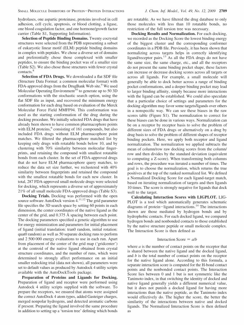
hydrolases, one aspartic proteinase, proteins involved in celladhesion, cell cycle, apoptosis, or blood clotting, a ligase,one blood coagulation factor, and one hormone/growth factorcarrier (Table S1, Supporting Information).
Selection of Peptide Binding Domains. Twenty cocrystalstructures were selected from the PDB representing a subsetof eukaryotic linear motif (ELM) peptide binding domainsin complex with peptides. We chose a diverse set of domainsand preferentially chose those complexed with smallerpeptides, to ensure the binding pocket was of a smaller size(Table S2). We also chose those structures with fewer crystalcontacts.8
Selection of FDA Drugs. We downloaded a flat SDF file(Structure Data Format: a common molecular format) withFDA-approved drugs from the DrugBank Web site.9 We usedMolecular Operating Environment10 to generate up to 50 3Dconformations with the stochastic search option, using theflat SDF file as input, and recovered the minimum energyconformation for each drug based on evaluation of the MerckMolecular Force Field MMFF94. This conformation wasused as the starting conformation of the drug during thedocking procedure. We initially selected FDA drugs that havealready been shown to have common pharmacophore pointswith ELM proteins,6 consisting of 161 compounds, but alsoincluded FDA drugs without ELM pharmacophore pointmatches. We filtered the pharmacophore matched list bykeeping only drugs with rotatable bonds below 10, and byclustering with 70% similarity between molecular finger-prints, and retaining the compound with smallest rotatablebonds from each cluster. In the set of FDA-approved drugsthat do not have ELM pharmacophore query matches, toreduce the data set size further, we reclustered with 40%similarity between fingerprints and retained the compoundwith the smallest rotatable bonds for each new cluster. Intotal, 287 FDA-approved small molecule drugs were selectedfor docking, which represents a diverse set of approximately21% of all small molecule FDA-approved drugs (Table S3).
Docking Trials. Docking was performed with the opensource software AutoDock version 4.11,12 The grid parameterfile specifies the 3D search space by setting 60 points in eachdimension, the center coordinates of the native ligand as thecenter of the grid, and 0.375 Å spacing between each point.The docking parameters specified a genetic algorithm to usefor energy minimization and parameters for initial placementof ligand (initial translation: tran0 random, initial rotation:quat0 random) as well as 50 separate docking runs to performand 2 500 000 energy evaluations to use in each run. Apartfrom placement of the center of the grid map (‘gridcenter’)at the centroid of the native ligand obtained from crystalstructure coordinates, and the number of runs, which weredetermined to strongly affect performance on an initialsmaller redocking trial (data not shown), all parameters wereset to default values as produced by Autodock 4 utility scriptsavailable with the AutoDockTools package.
Preparation of Proteins and Ligands for Docking.Preparation of ligand and receptor were performed usingAutodock 4 utility scripts supplied with the software. Toprepare the receptors, we ensured that atoms were assignedthe correct AutoDock 4 atom types, added Gasteiger charges,merged nonpolar hydrogens, and detected aromatic carbonsif present. Preparing the ligand involved the same procedurein addition to setting up a ‘torsion tree’ defining which bonds
are rotatable. As we have filtered the drug database to onlythose molecules with less than 10 rotatable bonds, norestriction of the full torsion tree was necessary.
Docking Results and Normalization. For each docking,we recorded as the Docking Score the lowest binding energyof the biggest cluster and the corresponding conformercoordinates in a PDB file. Previously, it has been shown thatnormalizing across targets helps in correctly identifyingligand/receptor pairs.13 As all the FDA drugs do not havethe same size, the same charge, etc., and all the receptorsdo not present the same binding pocket shape, these factorscan increase or decrease docking scores across all targets oracross all ligands. For example, a small molecule willgenerally be able to dock better across a range of bindingpocket conformations, and a deeper binding pocket may leadto larger binding affinity, simply because more interactionswith the ligand can be established. We could also speculatethat a particular choice of settings and parameters for thedocking algorithm may favor some targets/ligands over othersin a nonspecific way. We can see these biases in the rawscores table (Figure S1). The normalization to correct forthese biases can be done in various ways. Normalization canbe on a receptor by receptor basis to solve the problem ofdifferent sizes of FDA drugs or alternatively on a drug bydrug basis to solve the problem of different shapes of receptorbinding pockets. Here, we apply both receptor and ligandnormalization. The normalization we applied subtracts themean of column/row raw docking scores from the column/row and then divides by the standard deviation (equivalentto computing a Z-score). When transforming both columnsand rows, the procedure was iterated a number of times. Thegoal is to choose the normalization, which returns the truepositives at the top of the ranked normalized list. We defineda Normalized Docking Score for each ligand-target match,based on iterating normalization of targets and then ligands10 times. The score is strongly negative for ligands that dockwell to the target.
Calculating Interaction Scores with LIGPLOT. LIG-PLOT is a tool which automatically generates schematicdiagrams of protein-ligand interactions.14 The interactionsshown are those mediated by hydrogen bonds and byhydrophobic contacts. For each docked ligand, we comparedhydrogen bonds and nonbonded contacts to those establishedby the native structure peptide or small molecule complex.The Interaction Score is then defined as
Interaction Score ) a/b
where a is the number of contact points on the receptor thatis shared between the native ligand and the docked ligand,and b is the total number of contact points on the receptorfor the native ligand alone. According to this formula, aseparate interaction score is computed for the H-bond contactpoints and the nonbonded contact points. The InteractionScore lies between 0 and 1 but is not symmetric like theTanimoto index, in that switching the identity of docked andnative ligand generally yields a different numerical value,but it does not punish a docked ligand for having moreinteractions than the native peptide, as the Tanimoto indexwould effectively do. The higher the score, the better thesimilarity of the interactions between native and dockedligands. The Normalized Interaction Score is then definedas
SMALL MOLECULE INHIBITORS OF PROTEIN-PROTEIN INTERACTIONS J. Chem. Inf. Model., Vol. 49, No. 12, 2009 2709

(Normalized H-bond Interaction Score +Normalized Nonbonded Interaction Score)/√2
Note that this implicitly gives equal weight to each type ofinteraction.
Combined Docking and Interaction Scores. As both thedocking and interaction scores are separately normalized,they are on the same scale, and we then add the NormalizedDocking Score to the negative of the Normalized InteractionScore (favorable docking scores are more negative; favorableinteraction scores are more positive; the sign change meansthat the favorable combined score is more negative). Thosewith better combined ranking not only show favorablebinding to the domains but establish the same interactionswith the domain residues, making it a better potentialcandidate for mimicking the crystal structure peptide ligandor small molecule. It should be noted that this implicitly givesequal weight to the docking scores and the interaction scores,which may not be optimal. In particular, the native ligandsfor the true positives will always be able to establish thesame interactions in the docked and crystal structures whichsomewhat biases the interaction score to favor true positives.However, by applying the same symmetric normalization tointeraction scores as we used for docking scores, wepresumably remove some of this bias. As a partial confirma-tion of this hypothesis, we allowed some flexibility anddefined a combined score with a free weighting parameter
Combined Score ) w(Normalized Docking Score) +(1 - w)(-Normalized Interaction Score)
and found that optimizing the weight, w, to maximize theranking of the true positives gives w ) 0.4, and the changein ranking is marginal if we choose equal weights (w ) 0.5).For a simpler explanation, we applied equal weights in thefinal ranking.
RESULTS AND DISCUSSION
Importance of Normalization of Docking Scores. Thegoal of normalization is to produce a score which preferen-tially enriches for true positives within the highest rankedcompounds: if we see true positive ligand receptor interac-tions being ranked highly with a normalized score, then weplace more confidence in the highly ranked predictions ofunknown compound receptor interactions. Thus, we exam-ined the number of true positives within the first 1% ofscores. We found that ranking after a symmetric normaliza-tion procedure (normalizing scores iteratively by columnsand then by rows, repeating this procedure a total of 10 times)yields many true positive interactions and removes receptorand ligand biases apparent in the raw scores (Figure S1, S2).We noted that this iterative normalization procedure doesnot converge to a fixed set of values, but 10 iterations wassufficient to yield good results for this particular data setsize and structure. In addition, the order of transformation,row then column or column then row, generally producesdifferent results, but we chose the latter, as it enriched formore true positive interactions (data not shown).
In Figure 1, we show the percentage of true positives‘discovered’ by selecting the top ranked compounds by score.In all, there are 10 570 compound receptor docking scores.The diagonal (bottom line) represents what will happen ifcompound receptor pairs are chosen randomly: there is no
enrichment for the selection of true positive ligand receptorcombinations. All the lines (raw scores and normalizedscores) are above the diagonal which is a positive result forusing docking to identify compounds that genuinely bind toproteins. We note that Normalized Docking Scores outper-form untransformed (raw) Normalized Docking Scores, andthat performance is considerably improved using the com-bined score, utilizing information from both the NormalizedInteraction Score and the Normalized Docking Score.
Moreover, using the Combined Score, we observe that 7/15of the true positive ligands have the lowest normalized scorefor their native receptor, and 4/15 of the true positivereceptors have the lowest normalized score for their nativeligand, a criterion which was used in Vigers and Rizzi.13 InTable 1, we see the top 20 ranked compound-receptormatches. Within the list we recover 6 out of 15 of the truepositive interactions which strengthens the belief in theremaining predicted interactions. We further evaluated theevidence for the predicted drug binders by (1) visualizationof the docked complex versus the original native crystalcomplex, by (2) a literature search for information concerninga known interaction with the target, or with a pathwayinvolving the target or an interaction of a related compound/drug with the target.
HP1 Chromo Shadow Domain. The HeterochromatinProtein 1 (HP1) or Chromobox Homologue is a nuclear-associated protein with histone binding and DNA silencingfunctions. It contains a pair of peptide binding domains(chromo shadow domains) forming a dimer. Short peptiderecognition occurs by sandwiching the extended peptidebetween strands of each monomer domain to form anaugmented � sheet.15 The predicted docked compound, 1,is a HIV-1 protease inhibitor peptidomimetic (Ligand ID:INT16) which establishes all five of the same backboneinteractions as the peptide motif (see Supporting Information,Figure S3) and also carries hydrophobic side-chain mimetics.HIV-1 protease inhibitors such as 1 and protease inhibitorsin general are designed to mimic � strands,17 the usualconformation of peptides that undergo cleavage, so it isplausible that the predicted interaction would indeed occur.The HP1 protein is important in suppressing breast cancer
Figure 1. Percentage of true positive ligand/receptor pairs in aranked list of all pairs based on various scores. Docking Scores:solid line; Normalized Docking Scores: dashed line; CombinedScore (normalized docking score combined with matching nativeligand interactions): dotted line; random expectation: dashed-dottedline.
2710 J. Chem. Inf. Model., Vol. 49, No. 12, 2009 CASEY ET AL.
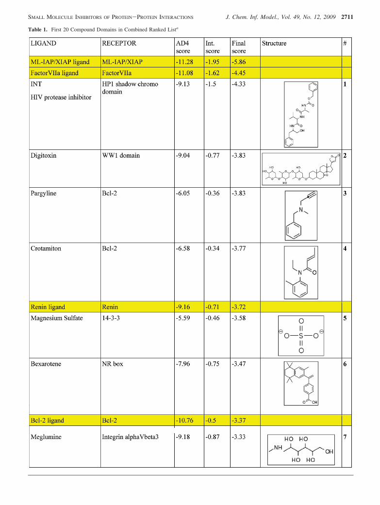
Table 1. First 20 Compound Domains in Combined Ranked Lista
SMALL MOLECULE INHIBITORS OF PROTEIN-PROTEIN INTERACTIONS J. Chem. Inf. Model., Vol. 49, No. 12, 2009 2711

metastasis,18 and modulation of pathways involving it maybe of interest in the future.
WW1 Domain. The WW1 domain, so-called because oftwo conserved tryptophan residues, is a small � sheet structure
that preferentially binds proline rich peptides and can often sharerecognition of peptide sequences with the SH3 domain.19 Thestructure for the WW domain (PDB ID: 1eg420) comes fromhuman dystrophin, an essential muscoskeletal protein, of
Table 1. Continued
a The yellow rows are true positive matches. The ligand column contains the drug name or three-letter ligand code from MSDchem. Thedomain/protein name is in parentheses if the structure came from different PDB entries. AD4 score is untransformed AutoDock4 estimated freeenergy score. Int. Score is the negative of the sum of the Tanimoto scores for H-bond and nonbonded contact similarity: total similarity wouldgive a score of -2. The final score is the sum of the symmetrically normalized AD4 and interaction scores, which we use to rank predictedligand receptor binding. Structures and compound numbers are given for those discussed further in the text.
2712 J. Chem. Inf. Model., Vol. 49, No. 12, 2009 CASEY ET AL.
which impaired function causes muscular dystrophy. We finda cardiac glycoside, 2 (digitoxin), as having very strongaffinity for the domain. Digitoxin is used in the treatment ofcardiac arrythmias, where its apparent mechanism of actionis to modulate the activity of the sodium ATPase transporterpump.21 Digitoxin, being a glycoside, contains many oxygengroups capable of establishing hydrogen bonds. Even thoughthe canonical WW1 domain peptide binding motif PP.Y isnot very obviously charged or polar with respect to side-chain composition, the full sequence bound in the structurehas a backbone H-bond donor interaction with a WWhydroxyl group and two CdO acceptor groups from thepeptide backbone. The glycoside establishes H-bond interac-tions with polar Tyr and Thr residues in the binding site.We note that some evidence exists in the literature thatdystrophic subjects show increased sensitivity to the closelyrelated molecule digoxin,22 and that digitoxin can in fact leadto accelerated muscle wasting,23 consistent with an alreadycompromised dystrophin protein being further inhibited informing complexes. Interestingly, digitoxin was demonstratedto have antiproliferative effects on tumor cell lines.24 Oursecond match against the WW domain, compound 12(etoposide phosphate), is an antineoplastic agent which isthought to interfere with DNA topoisomerase and may havea secondary action in interfering with the JNK stress responsepathway.25 Thus, it is potentially possible for both thesecompounds to have actions via WW signaling. If this is thecase, which WW domains are the most likely mediators ofsuch effects? Transiently activated JNK kinase interacts witha WW domain containing pro-apoptotic protein, WOX1, andappears to block its apoptotic function.26 Second, theantiproliferative effect of digitoxin has been hypothesizedto relate to inhibition of the WOX1-interactor, of TNFreceptor death-associated domain (TRADD), and of TNFreceptor complex formation.27 These clues suggest theWOX1 WW domain as a target worthy of investigation,although other less studied WW domain-containing proteinsmay also be good candidates.
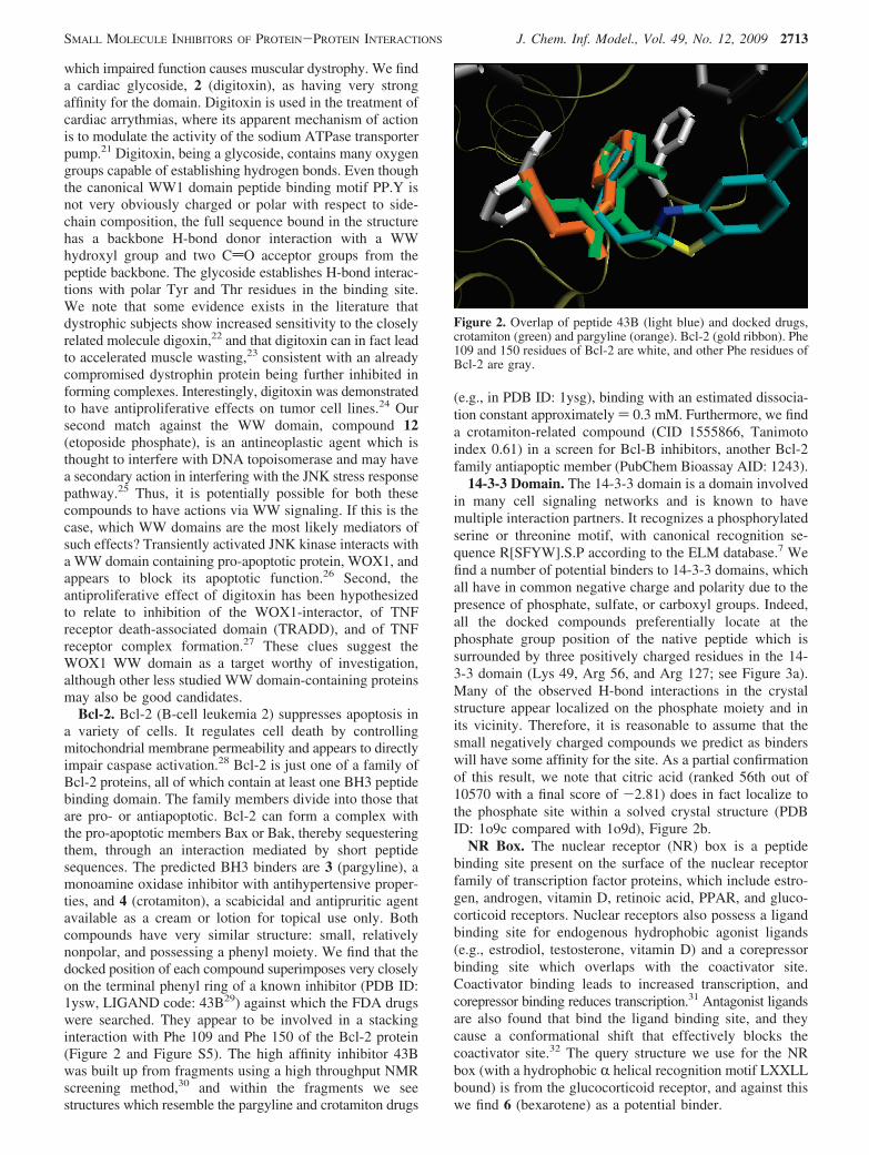
Bcl-2. Bcl-2 (B-cell leukemia 2) suppresses apoptosis ina variety of cells. It regulates cell death by controllingmitochondrial membrane permeability and appears to directlyimpair caspase activation.28 Bcl-2 is just one of a family ofBcl-2 proteins, all of which contain at least one BH3 peptidebinding domain. The family members divide into those thatare pro- or antiapoptotic. Bcl-2 can form a complex withthe pro-apoptotic members Bax or Bak, thereby sequesteringthem, through an interaction mediated by short peptidesequences. The predicted BH3 binders are 3 (pargyline), amonoamine oxidase inhibitor with antihypertensive proper-ties, and 4 (crotamiton), a scabicidal and antipruritic agentavailable as a cream or lotion for topical use only. Bothcompounds have very similar structure: small, relativelynonpolar, and possessing a phenyl moiety. We find that thedocked position of each compound superimposes very closelyon the terminal phenyl ring of a known inhibitor (PDB ID:1ysw, LIGAND code: 43B29) against which the FDA drugswere searched. They appear to be involved in a stackinginteraction with Phe 109 and Phe 150 of the Bcl-2 protein(Figure 2 and Figure S5). The high affinity inhibitor 43Bwas built up from fragments using a high throughput NMRscreening method,30 and within the fragments we seestructures which resemble the pargyline and crotamiton drugs
(e.g., in PDB ID: 1ysg), binding with an estimated dissocia-tion constant approximately ) 0.3 mM. Furthermore, we finda crotamiton-related compound (CID 1555866, Tanimotoindex 0.61) in a screen for Bcl-B inhibitors, another Bcl-2family antiapoptic member (PubChem Bioassay AID: 1243).
14-3-3 Domain. The 14-3-3 domain is a domain involvedin many cell signaling networks and is known to havemultiple interaction partners. It recognizes a phosphorylatedserine or threonine motif, with canonical recognition se-quence R[SFYW].S.P according to the ELM database.7 Wefind a number of potential binders to 14-3-3 domains, whichall have in common negative charge and polarity due to thepresence of phosphate, sulfate, or carboxyl groups. Indeed,all the docked compounds preferentially locate at thephosphate group position of the native peptide which issurrounded by three positively charged residues in the 14-3-3 domain (Lys 49, Arg 56, and Arg 127; see Figure 3a).Many of the observed H-bond interactions in the crystalstructure appear localized on the phosphate moiety and inits vicinity. Therefore, it is reasonable to assume that thesmall negatively charged compounds we predict as binderswill have some affinity for the site. As a partial confirmationof this result, we note that citric acid (ranked 56th out of10570 with a final score of -2.81) does in fact localize tothe phosphate site within a solved crystal structure (PDBID: 1o9c compared with 1o9d), Figure 2b.
NR Box. The nuclear receptor (NR) box is a peptidebinding site present on the surface of the nuclear receptorfamily of transcription factor proteins, which include estro-gen, androgen, vitamin D, retinoic acid, PPAR, and gluco-corticoid receptors. Nuclear receptors also possess a ligandbinding site for endogenous hydrophobic agonist ligands(e.g., estrodiol, testosterone, vitamin D) and a corepressorbinding site which overlaps with the coactivator site.Coactivator binding leads to increased transcription, andcorepressor binding reduces transcription.31 Antagonist ligandsare also found that bind the ligand binding site, and theycause a conformational shift that effectively blocks thecoactivator site.32 The query structure we use for the NRbox (with a hydrophobic R helical recognition motif LXXLLbound) is from the glucocorticoid receptor, and against thiswe find 6 (bexarotene) as a potential binder.
Figure 2. Overlap of peptide 43B (light blue) and docked drugs,crotamiton (green) and pargyline (orange). Bcl-2 (gold ribbon). Phe109 and 150 residues of Bcl-2 are white, and other Phe residues ofBcl-2 are gray.
SMALL MOLECULE INHIBITORS OF PROTEIN-PROTEIN INTERACTIONS J. Chem. Inf. Model., Vol. 49, No. 12, 2009 2713

Bexarotene is an antineoplastic drug that selectively bindsand activates the retinoid X (RXR) subfamily of nuclearreceptors. The RXR receptors can dimerize with other NRreceptors such as vitamin D, retinoic acid receptors (RAR),and PPAR. That a nuclear receptor agonist could also haveaffinity for the coactivator site, i.e., the NR box, as predictedhere has recently been demonstrated experimentally forestrogen receptors, ER R, ER �,33,34 for vitamin D recep-tors,35 and computationally for the PPAR coactivator site.6
We suggest that a similar mechanism may be possible here,although previous studies suggest that bexarotene is selectiveonly for the RXR receptor family. Bexarotene has recentlybeen successfully tested as a topical gel in the treatment ofeczema, normally treated with corticosteroids, implying apossible bexarotene glucocorticoid receptor interaction, butmore likely due to crosstalk from receptor heterodimerizationrather than at the ligand level.36
Integrin rV�3. The RGD tripeptide motif binds integrinsin a binding groove at the interface of the heterodimer madeup of RV and �3 subunits. The RGD motif is present infibrinogen, an extracellular protein whose attachment tointegrin receptors of platelets causes clotting. We see 7(meglumine), a contrast agent, highly ranked as a potentialbinder of integrins. Contrast agents such as diatrizoatemeglumine and ioxaglate can inhibit the formation of clotsin partial thromboplastin time (PTT) assays37 but are notrecorded to have any impact on platelet function. For thisreason, we consider it unlikely that meglumine will bindintegrins in vivo, even though it appears to be able to adopta conformation that mimics many of the hydrogen bonds ofthe bound crystal peptide RGD sequence, including theimportant coordination to the manganese cofactor; seeSupporting Information Figure S7.
Cyclin A. The cyclin A/CDK2 kinase complex is involvedin cell cycle control. A critical interaction occurs with thesubstrates p21 or p27 which are inhibitors of the cyclin/CDKkinase activity and block cell cycle progression at G1 andthus block cell proliferation. Recognition of cyclin/CDKsubstrates occurs with the motif [RK].L.{0,1}[FYLIVMP]binding to a groove in cyclin and a phospho-acceptor site[ST]P.[KR] recognized by CDK2. Many inhibitors have beendeveloped to block the recognition site of CDK2, and crystalstructures are available for CDK/inhibitor complexes, butinhibitors of the cyclin peptide binding domain are not ascommon (however, see ligand IDs: C35 and GVC). Peptidesbased on the p21 or p27 interacting fragments (RXL peptidemimetics) are effective at blocking tumor growth andinducing apoptosis.38 8 (buspirone) is an antianxiety agentwith high affinity for serotonin receptors and moderateaffinity for D2-dopamine receptors.39 It aligns with thebinding groove and effectively mimics the aromatic groupof the peptide motif and hydrophobic leucine contacts, andit establishes a backbone H-bond with Ile 281 (Figure S6),though it shares little similarity to the known cyclin Abinders, C35 and GVC.
MDM2. MDM2 is a ubiquitin ligase anticancer target, asit is a negative regulator of the tumor suppressor transcriptionfactor, p53. The p53/MDM2 interaction is the focus of manycomputational and experimental screens for small moleculeinhibitors.40 9 (clofazimine) is a lipophilic dye used as atreatment of mycobacterial infections, due to a supposedmycobacterial DNA binding function.41 It also appears tohave an anti-inflammatory effect against some of thesymptoms of mycobacterial infection. Clofazimine showsantitumor activity (bioassay AID: 37142,43) and interfereswith the NFAT transcription factor pathway (bioassay AID:
Figure 3. (a) LIGPLOT 14-3-3 peptide and domain, and (b) 14-3-3 peptide phosphate and citric acid superposition (from PDB 1o9c and1o9d). See also Supporting Information, Figure S4.
2714 J. Chem. Inf. Model., Vol. 49, No. 12, 2009 CASEY ET AL.

444) which is directly affected by activated MDM2, althoughthe proposed mechanism for its antiproliferative effects isnot mediated by this interaction.44 It is similar in size to theknown inhibitors of p53/MDM2 association, and it appearsto bind in the high affinity hydrophobic Trp binding pocketidentified in MDM2. The addition of halogen atoms on theTrp residue and on aromatic groups of other MDM2inhibitors is known to increase binding affinity1 because ofincreased shape complementarity within the binding pocket.Clofazimine contains two chlorinated phenyl rings and abranched valine-like terminal group which appear to fill twodeep binding cavities on the surface analogous to the boundinhibitor, ligand ID: IMZ, Figure 4.
Dynein DLC8. Dynein is an ATP-dependent motor proteinthat is responsible for protein transport along microtubulesand maintains Golgi structure.45 10 (flunisolide) is a syntheticcorticosteroid and a glucocorticoid receptor agonist. Thedynein light chain (DLC) peptide binding motif is[KR].TQT,7 and its mode of binding is a � augmentation ofthe DLC8 � sheet, exhibiting many backbone and side-chainoxygen and carboxyl interactions in complex. Flunisolide iscapable of mimicking these interactions but only capturesbackbone interactions of the lysine and first threonine residueof the motif. It has recently been noticed that the dyneinlight chain 8 can inhibit NF-κB transcription, an immuneand inflammatory response pathway, through an interactionwith the I-κB R subunit.46,47 Inhibition of the same pathwayoccurs with administration of glucocorticoids, includingflunisolide, although this is normally considered to occur ina more direct manner by heterodimerization of glucocorticoidreceptor and NF-κB.48
ML-IAP/X-IAP. Melanoma inhibitor of apoptosis (ML-IAP) and X-linked inhibitor of apoptosis (X-IAP) are relatedantiapoptotic proteins which share a BIR-3 peptide bindingdomain and are capable of binding caspases thereby blockingtheir pro-death activities. Overexpression of these proteins
in tumor cell lines has been connected to chemotherapeuticinsensitivity, and conversely inhibitors of the BIR-3 domaininteractions (such as by the Smac protein) cause resensiti-zation of the tumor cells to cytotoxic factors.49 An inhibitorof IAP can be as small as a tetrapeptide with consensus motifA[ITV]P[FWIVY] which has two distinct binding pockets:a small N terminal pocket which is predominantly negativelycharged and a C-terminal pocket that can incorporate ahydrophobic residue.49 The N-terminal alanine also estab-lishes backbone interactions in the first pocket. We found asingle sucrose molecule, compound 11, is able to occupythe first (N-terminal pocket) and mimic the peptide interac-tions, and it forms interactions with Trp 147 protruding fromthe surface of IAP but is unable to reach to the secondbinding pocket. It has partial similarity with an antibiotic(Pubchem CID 6473883, Tanimoto coefficient 0.43) whichis active as an IAP antagonist (Pubchem Bioassay AID 1018).
CONCLUSION AND PERSPECTIVES
Protein-protein interactions regulate many biologicalfunctions. Discovering new interactions between FDA-approved small molecules and proteins involved in importantbiological processes such as apoptosis or cell growthsignaling would have great therapeutic significance, as theclinical safety and pharmacokinetic profiles of these com-pounds is already known. In the present work, we have useda docking algorithm to predict the binding tendencies of 287FDA-approved small molecules against 35 different pocketsof proteins which are of the peptide binding class. Wedeveloped a normalization procedure that both maximizesthe occurrence of true positive compound receptor pairs andensures that the docked compounds mimic a substantialfraction of the native peptide/ligand’s interaction points. Asa byproduct, this tends to remove docked compounds thatdo not bind at the correct position within the domain.
Examination of the scientific literature with respect to thetop 20 predictions of compound receptor pairs, we found anumber of results worth further investigation, outside of theunambiguous true positive receptor ligand set.
Bcl-2 is a known antiapoptotic protein which has beenimplicated in the survival of drug-resistant tumors and whoseinhibition directly reduces cancer cell proliferation. A numberof inhibitors are currently being developed to prevent theformation of Bcl-2 /BAX complexes29,30,50,51 as well assiRNA molecule targeting which prevents its expression.52
Two potential Bcl-2 binding compounds were indicated bythe docking analysis. Given their small size relative to otherhigh affinity compounds, they would appear less likely tobind with high affinity. Nevertheless, if they show sufficientspecificity in particular settings, they could be investigatedas cotherapies with other agents targeting this pathway.
The 14-3-3 � protein which is involved in propagatinggrowth signals and is crucial for the proliferation of tumorcells could potentially be inhibited by small acidic moleculeswhich are predicted to bind strongly to the positively chargedbinding pocket.
The HP1 protein has a DNA silencing and gene regulationfunction and forms noncovalent bonds through � sheetaugmentation of its shadow chromo domain with interactionpartners. We found a very plausible interaction with theextended � strand mimetic of the HIV protease inhibitor,
Figure 4. MDM2 binding groove (PDB ID: 1rv1) with inhibitor(ligand ID: IMZ, thin sticks) bound and the docked structure, 9,clofazimine (thick sticks). The chlorine atom (brown) and thephenazine rings on the drug locate to the bromine atoms (green)on IMZ and fill the same large surface cavities.
SMALL MOLECULE INHIBITORS OF PROTEIN-PROTEIN INTERACTIONS J. Chem. Inf. Model., Vol. 49, No. 12, 2009 2715

TL-3. While TL-3 is not an approved drug (it entered ourstudy as one of the true positive compounds from knownstructures16), it could provide an interesting lead compoundfor the investigation of HP1 and its role in tumor metastasis.18
The WW domain is present in ubiquitin ligases, kinases,and cytoskeletal proteins. It binds sequence motifs inpolyproline rich peptides. The digitoxin cardiac glycosideis a putative binder of the domain, which may suggestalternative modes of action that are related to its antiprolif-erative function.53
We have shown that a systematic docking procedurefollowed by the correct transformations and analysis ofresults can lead to significant enrichment of potential activesin a compound database. The choice of compound andreceptor database in this study was motivated by speedytranslation to clinical development, and tractability for thedevelopment of the search strategy. For a number of the druginteractions with peptide binding domains, suggestive cluesfrom the existing literature regarding drug activity or bindingprovide an additional rationale for prioritizing such matchesfor further experimental validation.
We believe that dual normalization across ligands andreceptors represents an important step in computational drugsearching. As described by Warren and co-workers,54 dock-ing algorithms tend to display widely varying degrees of truepositive enrichment for different target proteins, and the setof target proteins that show good enrichment vary fromalgorithm to algorithm, presumably because of training setselection which affects both protein and compound models.These biases may be mitigated by the post processing ofdocking scores suggested in this work and other work suchas that by Jacobsson.55 The two-dimensional arrays of rawscores can also be analyzed to determine how the biases arise,by classifying score profiles in terms of molecular descriptorsand protein pocket shape and composition. The score profilesthemselves can be used to cluster compounds and receptorsinto similarity classes that may contain more informationthan that of traditional QSAR descriptors, as they capturemodes of interaction rather than static conformations. Selec-tion for the correct mode of interaction is achieved by addinganother term to the scores which rewards configurations thatmimic the native peptide/ligand interaction points, and weshow how that significantly improves the enrichment for truepositives.
A future extension would be to apply the same approachto pharmacophore searching, which allows for greatlyincreased compound database size and would presumablyimprove scoring and ranking, as previous work6 indicatesthat the pharmacophore search is also biased toward certainmolecular features. A selection of top ranked matches fromsuch a pharmacophore search or the most highly scoreddocked poses in this study could be also regarded as initialconfigurations for extensive molecular dynamics simulations,which provide extra information about the stability of theinteraction and explicit solvent effects and allow for moreaccurate free energy predictions.56-58
The computational cost of the scoring procedure remainsrelatively small, in comparison with the computational costof docking. The true cost, therefore, lies in the requirementto dock not only a single target of interest but a series ofother targets. Parallelized computer systems, such as the IrishCentre for High-End Computing used in this study, are
making such computational approaches feasible. For a givendrug library, once the initial investment is made for a diverseseries of targets, additional targets may be added to the resultsdatabase at little extra computational cost.
Abbreviations. Bcl-2, B cell leukemia-2; ELM, eukaryoticlinear motif; FDA, Food and Drug Administration; MOE,molecular operating environment; rmsd, root mean squaredeviation; PDB, protein data bank.
ACKNOWLEDGMENT
This work was supported by the Irish Research Councilfor Science, Engineering and Technology (IRCSET), and byScience Foundation Ireland (SFI). We thank the Irish Centrefor High-End Computing for the use of computing resources.
Supporting Information Available: Additional informa-tion as noted in the text. This material is available free ofcharge via the Internet at http://pubs.acs.org.
REFERENCES AND NOTES
(1) Vassilev, L. T.; et al. In Vivo Activation of the P53 Pathway bySmall-Molecule Antagonists of Mdm2. Science 2004, 303 (5659),844–848.
(2) Arkin, M. R.; Wells, J. A. Small-Molecule Inhibitors of Protein-ProteinInteractions: Progressing towards the Dream. Nat. ReV. Drug DiscoVery2004, 3, 301–317.
(3) Reixach, N.; Adamski-Werner, S. L.; Kelly, J. W.; Koziol, J.;Buxbaum, J. N. Cell Based Screening of Inhibitors of TransthyretinAggregation. Biochem. Biophys. Res. Commun. 2006, 348 (3), 889–897.
(4) Sun, H. Pharmacophore-Based Virtual Screening. Curr. Med. Chem.2008, 15 (10), 1018–1024.
(5) Toba, S.; Srinivasan, J.; Maynard, A. J.; Sutter, J. Using Pharma-cophore Models To Gain Insight Into Structural Binding and VirtualScreening: An Application Study with Cdk2 and Human Dhfr.J. Chem. Inf. Model. 2006, 46, 728–735.
(6) Parathasarathi, L.; Casey, F.; Stein, A.; Aloy, P.; Shields, D. C.Approved Drug Mimics of Short Peptide Ligands from ProteinInteraction Motifs. J. Chem. Inf. Model. 2008, 48, 1943–1948.
(7) Puntervoll, P.; et al. Elm Server: A New Resource for InvestigatingShort Functional Sites in Modular Eukaryotic Proteins. Nucleic AcidsRes. 2003, 31 (13), 3625–3630.
(8) Stein, A.; Aloy, P. Contextual Specificity in Peptide-Mediated ProteinInteractions. PloS ONE 2008, 3 (7), e2524.
(9) Wishart, D. S.; et al. DrugBank: A Comprehensive Resource for inSilico Drug Discovery and Exploration. Nucleic Acids Res. 2006, 34,D668–672.
(10) Chemical Computing Group Inc. Molecular Operating Environment,Version 2008.08. Montreal, Canada.
(11) Huey, R.; Morris, G. M.; Olson, A. J.; Goodsell, D. S. A SemiempiricalFree Energy Force Field with Charge-Based Desolvation. J. Comput.Chem. 2007, 28, 1145–1152.
(12) Morris, G. M.; Goodsell, D. S.; Halliday, R. S.; Huey, R.; Hart, W. E.;Belew, R. K.; Olson, A. J. Automated Docking Using a LamarckianGenetic Algorithm and Empirical Binding Free Energy Function.J. Comput. Chem. 1998, 19, 1639–1662.
(13) Vigers, G. P.; Rizzi, J. P. Multiple Active Site Corrections for Dockingand Virtual Screening. J. Med. Chem. 2004, 47, 80–89.
(14) Wallace, A. C.; Laskowski, R. A.; Thornton, J. M. Ligplot: A ProgramTo Generate Schematic Diagrams of Protein-Ligand Interactions.Protein Eng. 1995, 8, 127–134.
(15) Brasher, S. V.; Smith, B. O.; Fogh, R. H.; Nietlispach, D.; Thiru, A.;Nielsen, P. R.; Broadhurst, R. W.; Ball, L. J.; Murzina, N. V.; Laue,E. D. The Structure of Mouse Hp1 Suggests a Unique Mode of SinglePeptide Recognition by the Shadow Chromo Domain Dimer. EMBOJ. 2000, 19 (7), 1587–1597.
(16) Li, M.; Morris, G. M.; Lee, T.; Laco, G. S.; Wong, C. H.; Olson,A. J.; Elder, J. H.; Wlodawer, A.; Gustchina, A. Structural Studies ofFIV and HIV-1 Proteases Complexed with an Efficient Inhibitor ofFIV Protease. Proteins 2000, 38, 29–40.
(17) Loughlin, W. A.; Tyndall, J. D. A.; Glenn, M. P.; Fairlie, D. P. Beta-Strand Mimetics. Chem. ReV. 2004, 104, 6085–6117.
(18) Norwood, L. E.; et al. A Requirement for Dimerization of Hp1 inSuppression of Breast Cancer Invasion. J. Biol. Chem. 2006, 281,18668–18676.
2716 J. Chem. Inf. Model., Vol. 49, No. 12, 2009 CASEY ET AL.

(19) Wintjens, R.; Wieruszeski, J. M.; Drobecq, H.; Rousselot-Pailley, P.;Buee, L.; Lippens, G.; Landrieu, I. 1H NMR Study on the Binding ofPin1 Trp-Trp Domain with Phosphothreonine Peptides. J. Biol. Chem.2001, 276 (27), 25150–25156.
(20) Huang, X.; Poy, F.; Zhang, R.; Joachimiak, A.; Sudol, M.; Eck, M. J.Structure of a WW Domain Containing Fragment of Dystrophin inComplex with Beta-Dystroglycan. Nat. Struct. Biol. 2000, 7, 634–638.
(21) Bluschke, V.; Bonn, R.; Greeff, K. Increase In the (Na+ + K+)-Atpase Activity in Heart Muscle after Chronic Treatment withDigitoxin or Potassium Deficient Diet. Eur. J. Pharmacol. 1976,37 (1), 189–191.
(22) Saito, K.; Ohkura, H.; Kashima, T.; Tanaka, H. Enhanced Sensitivityto Digoxin in Dystrophic Mice. Jpn. Heart J. 1984, 25, 765–777.
(23) Heyck, H.; Laudahn, G.; Luders, C. J.; Muller-Stephann, H.; Schmidt-Peter, P. Anabolic Steroids and Digitoxin in the Treatment ofProgressive Muscular Dystrophy. Acta Paediatrica 1965, 54 (3), 205–221.
(24) Lopez-Lazaro, M.; Pastor, N.; Azrak, S. S.; Ayuso, M. J.; Austin,C. A.; Cortes, F. Digitoxin Inhibits the Growth of Cancer Cell Linesat Concentrations Commonly Found in Cardiac Patients. J. Nat. Prod2005, 68 (11), 1642–1645.
(25) Anderson, S. M.; et al. Etoposide-Induced Activation of C-JunN-Terminal Kinase (Jnk) Correlates with Drug-Induced Apoptosisin Salivary Gland Acinar Cells. Cell Death Differentiation 1999, 6(5), 454–462.
(26) Chang, N.-S.; et al. Molecular Mechanisms Underlying Wox1 Activa-tion during Apoptotic and Stress Responses. Biochem. Pharmacol.2003, 66 (8), 1347–1354.
(27) Yang, Q.; et al. Cardiac Glycosides Inhibit TNF-R/NF-κB Signalingby Blocking Recruitment of Tnf Receptor-Associated Death Domainto the Tnf Receptor. Proc. Natl. Acad. Sci. U.S.A. 2005, 102 (27),9631–9636.
(28) Allen, R. T.; Cluck, M. W.; Agrawal, D. K. Mechanisms ControllingCellular Suicide: Role of Bcl-2 and Caspases. Cell. Mol. Life Sci. 1998,54, 427–445.
(29) Oltersdorf, T.; et al. An Inhibitor of Bcl-2 Family Proteins InducesRegression of Solid Tumours. Nature 2005, 435, 677–681.
(30) Shuker, S. B.; Hajduk, P. J.; Meadows, R. P.; Fesik, S. W. DiscoveringHigh-Affinity Ligands for Proteins: Sar by NMR. Science 1996, 274,1531–1153.
(31) Horwitz, K. B.; Jackson, T. A.; Bain, D. L.; Richer, J. K.; Takimoto,G. S.; Tung, L. Nuclear Receptor Coactivators and Corepressors. Mol.Endocrinol. 1996, 10, 1167–1177.
(32) Hashimoto, Y.; Miyachi, H. Nuclear Receptor Antagonists DesignedBased on the Helix-Folding Inhibition Hypothesis. Bioorg. Med. Chem.2005, 13 (17), 5080–5093.
(33) Jensen, E. V.; Khan, S. A. A Two-Site Model For AntiestrogenAction. Mechanisms of Ageing and DeVelopment 2004, 125 (10),679–682.
(34) Wang, Y.; Chirgadze, N. Y.; Briggs, S. L.; Khan, S.; Jensen, E. V.;Burris, T. P. A Second Binding Site for Hydroxytamoxifen withinthe Coactivator-Binding Groove of Estrogen Receptor Beta. Proc. Natl.Acad. Sci. U.S.A. 2005, 103 (26), 9908–9911.
(35) Mizwicki, M. T.; et al. Identification of An Alternative Ligand-BindingPocket in the Nuclear Vitamin D Receptor and Its FunctionalImportance in 1Alpha,25(OH)2-Vitamin D3 Signaling. Proc. Natl.Acad. Sci. U.S.A. 2004, 101, 12876–12881.
(36) Kuenzli, S.; Tran, C.; Saurat, J. H. Retinoid Receptors in InflammatoryResponses: A Potential Target for Pharmacology. Current DrugTargets: Inflammation and Allergy 2004, 3 (4), 355–360.
(37) Farrehi, P. M.; Zhu, Y.; Fay, W. P. An Analysis of MechanismsUnderlying the Antifibrinolytic Properties of Radiographic ContrastAgents. J Thrombosis and Thrombolysis 2001, 12 (3), 273–281.
(38) Mendoza, N.; Fong, S.; Marsters, J.; Koeppen, H.; Schwall, R.;Wickramasinghe, D. Selective Cyclin-Dependent Kinase2/Cyclin AAntagonists That Differ from Atp Site Inhibitors Block Tumor Growth.Cancer Res. 2003, 63, 1020–1024.
(39) de Boer, S. F.; Lesourd, M.; Mocaer, E.; Koolhaas, J. M. SelectiveAntiaggressive Effects of Alnespirone in Resident-Intruder Test AreMediated via 5-Hydroxytryptamine1A Receptors: A ComparativePharmacological Study with 8-Hydroxy-2-dipropylaminotetralin, Ip-sapirone, Buspirone, Eltoprazine, and Way-100635. J. Pharmacol. Exp.Ther. 1999, 288 (3), 1125–1133.
(40) Buolamwini, J. K.; Addo, J.; Kamath, S.; Patil, S.; Mason, D.; Ores,M. Small Molecule Antagonists of the Mdm2 Oncoprotein asAnticancer Agents. Curr. Cancer Drug Targets 2005, 5, 57–68.
(41) Morrison, N. E.; Marley, G. M. Clofazimine Binding Studies withDeoxyribonucleic Acid. Int. J. Lepr Other Mycobact Dis 1976, 44(4), 475–481.
(42) Pourgholami, M. H.; Lu, Y.; Wang, L.; Stephens, R. W.; Morris, D. L.Regression of Novikoff Rat Hepatocellular Carcinoma FollowingLocoregional Administration of a Novel Formulation of Clofaziminein Lipiodol. Cancer Lett. 2004, 207 (1), 37–47.
(43) Van Rensburg, C. E.; Joone, G. K.; O’Sullivan, J. F. Clofazimine andB4121 Sensitize an Intrinsically Resistant Human Colon Cancer CellLine to P-Glycoprotein Substrates. Oncol. Rep. 2000, 7 (1), 193–195.
(44) Van Rensburg, C. E. J.; Van Staden, A. M.; Anderson, R. TheRiminophenazine Agents Clofazimine and B669 Inhibit the Proliferationof Cancer Cell Lines in Vitro by Phospholipase A2-Mediated Oxidativeand Nonoxidative Mechanisms. Cancer Res. 1993, 53, 318–323.
(45) Corthesy-Theulaz, I.; Pauloin, A.; Pfeffer, S. R. Cytoplasmic DyneinParticipates in the Centrosomal Localization of the Golgi Complex.J. Cell Biol. 1992, 118 (6), 1333–1345.
(46) Jung, Y.; Kim, H.; Min, S. H.; Rhee, S. G.; Jeong, W. Dynein LightChain Lc8 Negatively Regulates NF-κB through the Redox-DependentInteraction with IκBR. J. Biol. Chem. 2008, 283 (35), 23863–23871.
(47) Crepieux, P.; et al. IκBR Physically Interacts with a Cytoskeleton-Associated Protein through Its Signal Response Domain. Mol. Cell.Biol. 1997, 17 (12), 7375–7385.
(48) Widen, C.; Gustafsson, J. A.; Wikstrom, A. C. Cytosolic GlucocorticoidReceptor Interaction with Nuclear Factor-Kappa B Proteins in RatLiver Cells. Biochem. J. 2003, 372, 211–220.
(49) Zobel, K.; et al. Design, Synthesis, and Biological Activity of a PotentSmac Mimetic That Sensitizes Cancer Cells to Apoptosis by Antago-nizing Iaps. ACS Chem. Biol. 2006, 1 (8), 525–533.
(50) Enyedy, I. J.; et al. Discovery of Small-Molecule Inhibitors of Bcl-2Through Structure-Based Computer Screening. J. Med. Chem. 2001,44, 4313–4324.
(51) Tzung, S.-P.; et al. Antimycin A Mimics a Cell-Death-Inducing Bcl-2Homology Domain 3. Nat. Cell Biol. 2001, 3, 183–191.
(52) Anderson, E. M.; et al. Gene Profiling Study of G3139- and Bcl-2-Targeting Sirnas Identifies a Unique G3139 Molecular Signature.Cancer Gene Ther. 2006, 13, 406–414.
(53) Winnicka, K.; Bielawski, K.; Bielawska, A. Cardiac Glycosides inCancer Research and Cancer Therapy. Acta Pol. Pharm. 2006, 63 (2),109–115.
(54) Warren, G. L.; et al. A Critical Assessment of Docking Programs andScoring Functions. J. Med. Chem. 2006, 49, 5912–5931.
(55) Jacobsson, M.; Karlen, A. Ligand Bias of Scoring Functions inStructure-Based Virtual Screening. J. Chem. Inf. Model. 2006, 46 (3),1334–1343.
(56) Alonso, H.; Bliznyuk, A. A.; Gready, J. E. Combined Docking andMolecular Dynamic Simulations in Drug Design. Med. Res. ReV. 2006,26 (5), 531–568.
(57) Moitessier, N.; Henry, C.; Maigret, B.; Chapleur, Y. CombiningPharmacophore Search, Automated Docking, and Molecular DynamicsSimulation as a Novel Strategy for Flexible Docking. Proof of Concept:Docking of Arginine-Glycine-Aspartic Acid-like Compounds into theRV�3 Binding Site. J. Med. Chem. 2004, 47, 4178–4187.
(58) Rapp, C. S.; Schonbrun, C.; Jacobson, M. P.; Kalyanaraman, C.;Huang, N. Automated site preparation in physics-based rescoring ofreceptor ligand complexes. Proteins 2009, 77, 52–61.
CI900294X
SMALL MOLECULE INHIBITORS OF PROTEIN-PROTEIN INTERACTIONS J. Chem. Inf. Model., Vol. 49, No. 12, 2009 2717