Biologically active secondary metabolites from Actinomycetes

HAL Id: tel-02156725https://tel.archives-ouvertes.fr/tel-02156725
Submitted on 14 Jun 2019
HAL is a multi-disciplinary open accessarchive for the deposit and dissemination of sci-entific research documents, whether they are pub-lished or not. The documents may come fromteaching and research institutions in France orabroad, or from public or private research centers.
L’archive ouverte pluridisciplinaire HAL, estdestinée au dépôt et à la diffusion de documentsscientifiques de niveau recherche, publiés ou non,émanant des établissements d’enseignement et derecherche français ou étrangers, des laboratoirespublics ou privés.
Discovery of active secondary metabolites fromPaenibacillus odorifer, a lichen-associated bacterium
Thi Bach Le Nguyen
To cite this version:Thi Bach Le Nguyen. Discovery of active secondary metabolites from Paenibacillus odorifer, a lichen-associated bacterium. Pharmacology. Université Rennes 1, 2018. English. �NNT : 2018REN1S098�.�tel-02156725�

THESE DE DOCTORAT DE
L'UNIVERSITE DE RENNES 1 COMUE UNIVERSITE BRETAGNE LOIRE
ECOLE DOCTORALE N° 596 Matière Molécules et Matériaux Spécialité : Chimie
Par
Thi Bach Le NGUYEN
Discovery of active secondary metabolites from Paenibacillus odorifer, a
lichen-associated bacterium
Thèse présentée et soutenue à Rennes, le 28 Juin 2018
Unité de recherche : UMR CNRS 6226 Équipe CORINT (Chimie Organique & INTerfaces)
Thèse N° :
Composition du Jury :
Angèle LENGO-MAMBU Professeur à l’Université de
Limoges/rapporteur Véronique EPARVIER Ingénieur de Recherche 1 -
CNRS - HDR /rapporteur
Nicolas PAPON Professeur à l’Université d’Angers /examinateur
Nicolas RUIZ Maître de Conférences à l’Université de Nantes/ examinateur
Sylvain TRANCHIMAND Maître de Conférences à l’ ENSCR/examinateur
Sophie TOMASI Professeur à l’Université de Rennes 1/ Directeur de thèse
Rapporteurs avant soutenance :
Angèle LENGO-MAMBU Professeur à l’Université de Limoges Véronique EPARVIER Ingénieur de Recherche 1 - CNRS -
HDR
ACKNOWLEDGEMENTS
First and foremost, I would like to express my deep gratitude to my supervisor, Pr. Sophie
Tomasi, for providing excellent, both scientific and personal advice during my Ph.D. study. Her
profound guidance, her great patience, her faith and enthusiasm for science encouraged me to go
through all the difficulties and to complete my study and researches. Her kindness led me feel the
warmth of home when I studied in Rennes.
I would like to express my thanks to my committee members for their time and advice on my
thesis, Dr. Véronique Eparvier, Pr. Angèle Lengo-Mambu, for their valuable comments and
evaluation of my work, Pr. Nicolas Papon, Dr. Nicolas Ruiz and Dr. Sylvain Tranchimand for
their acceptance as examiners.
I would like to thank Dr. David Delmail who helps me a lot for biological experiments in the first
stage of my work. I am also grateful to Isabelle Rouaud who gave me a great help in biological
tests. I wish to thank Solenn Ferron for her technical assistance in NMR spectra, to Aurélie
Sauvager for her guidance and support in HPLC, as well as to Marielle Blot for her technical help
in GC-MS.
I would to thank Dr. Jonathan Farjon, Pr. Vincent Ferrières and for their evaluation of my
work in the midterm of my thesis.
I would like to express my gratitude to Pr. Joël Boustie, Dr. Marylène Chollet-Krugler, Dr.
Béatrice Legouin, Dr. Françoise Lohézic-Le Dévéhat and who gave me a lot of care and
encouragement during my study and stay in Rennes. The journeys with them at Lyon, at
Copenhagen and at Angers became wonderful memories in my life. I am glad to be a part of such a
team.
I am especially grateful to Pr. Philippe Uriac and Dr. Arnaud Bondon for their critical
discussion in NMR spectra for the new structures, to Pr. Jean-Pierre Hurvois for his valuable
advice in the biosynthetic pathway, to Dr. Rémy Pedeux for his great help in DNA damage
assays, to Dr. Olivier Delalande for his discussion about NOEs calculations and to Dr. Philipe
Jéhan and members of CRMPO team for HRMS measurement.

I wish to thank my friends, my lab mate, Alice Gadéa, Nathalie Legrave, Pierre Le Pogam,
Alba Noel who gave me their warm welcome, nice atmosphere, and for their helping hand, sharing
ideas and encouragement during my study.
My gratitude to other members of the CORINT team – old and new members- to help me during
my thesis, Patricia Courtel, Dr. Bertrand Carboni, Dr. Michèle David, Maryse Demay,
Vianney Durel, Dr. Nicolas Gouault, Dr. René Grée, Benjamin Guieu, Myriam Le Roch,
Julia Mocquard, Damien Olivier, Dr. Jacques Renault, …
Vietnamese Government and the CORINT team are gratefully acknowledged for the financial
support.
I would like to thank Pr. Nguyen Kim Phi Phung, Dr. Vo Thi Nga, my colleagues and my
friends in Vietnam for their encouragement during my study.
Finally, I would like to thank my family, my parents, my sister and my brother for their love,
encouragement, support and patience throughout my education. My daughter, Quỳnh-Lê, has been
my inspiration over the past years and has helped me to find focus at a time when it was the most
crucial. Last, but not least, I thank my husband, Vinh-Quí, who has been willing to go with me
wherever, who has been a constant source of support, assisting me with everything from preparing
countless hot meals to informatic sources, and formating this final document and for his love
during the past years.
I would like to thank sincerely everyone who has helped me in various ways throughout my study.

TABLE OF CONTENTS
Page i
TABLE OF CONTENTS
TABLE OF CONTENTS ................................................................................................................. I
ABREVIATIONS ............................................................................................................................ V
LIST OF FIGURES ..................................................................................................................... VII
LIST OF TABLES ......................................................................................................................... IX
INTRODUCTION............................................................................................................................ 1
PART 1: STATE OF THE ART ON LICHEN-ASSOCIATED BACTERIA ........................... 6 1.1. INTRODUCTION ON LICHENS .......................................................................................... 6
1.1.1. The lichens ........................................................................................................................ 6 1.1.1.1 Description .................................................................................................................. 6 1.1.1.2 Morphology................................................................................................................. 8 1.1.1.3 Metabolites produced by lichen thallus .................................................................... 10
1.2. BACTERIA ASSOCIATED WITH LICHENS .................................................................... 14 1.2.1. State of art about bacterial communities on lichens ....................................................... 14 1.2.2. The roles of bacteria ....................................................................................................... 26
1.3. METABOLITES FROM THE LICHEN-ASSOCIATED BACTERIA ............................... 27 1.4. FOCUS ON RHIZOCARPON GEOGRAPHICUM ............................................................ 34
1.4.1. Description of Rhizocarpon genus ................................................................................. 34 1.4.2. Morphology of R. geographicum ................................................................................... 36 1.4.3. Studies on R. geographicum ........................................................................................... 38 1.4.4. Bacteria communities of R. geographicum .................................................................... 42
1.5. NATURAL CYTOTOXIC COMPOUNDS FROM BACTERIAL CULTURE .................. 43 1.6. THE OBJECTIVES OF THE WORK .................................................................................. 60
PART II. RESULTS ...................................................................................................................... 63
CHAPTER 1: ISOLATION OF BACTERIAL STRAINS FROM R. GEOGRAPHICUM..... 65
CHAPTER 2: THE SELECTION OF PAENIBACILLUS ODORIFER AS A PROMISING SOURCE OF INTERESTING METABOLITES ....................................................................... 82

TABLE OF CONTENTS
Page ii
CHAPTER 3: OPTIMIZATION OF THE CULTURE OF PAENIBACILLUS ODORIFER
........................................................................................................................................................ 102 3.1. THE FIRST OPTIMIZATION OF PROCESS ................................................................... 102
3.1.1. The selected parameters................................................................................................ 102 3.1.2. Results .......................................................................................................................... 105 3.1.3. The application to bioreactor fermentation .................................................................. 106
3.2. THE SECOND OPTIMIZATION PROCESS .................................................................... 107 3.2.1. The selected parameters................................................................................................ 107 3.2.2. Results .......................................................................................................................... 108 3.2.3. The application of these results for cytotoxic compounds discovery ........................... 113
CHAPTER 4: ISOLATION OF METABOLITES FROM PAENIBACILLUS ODORIFER ... 1 4.1 THE PROCESS FOR THE PRODUCTION OF CRUDE EXTRACTS FROM FERMENTATION ......................................................................................................................... 1 4.2. ISOLATION OF A POLYSACCHARIDE UNIT .................................................................. 4
4.2.1. General presentation of bacterial polysaccharides: structure and properties ................... 4 4.2.2. Production of one polysaccharide fraction from P. odorifer ............................................ 5
4.3. ISOLATION OF TERT-BUTYL PHENOLIC COMPOUNDS ........................................... 20 4.3.1. State of art about tert-butyl compounds ......................................................................... 20 4.3.2. Production of the tert-butyl phenol compounds ............................................................. 22
4.4. THE ISOLATION PROCESS OF A NOVEL ALKALOID ................................................ 37 4.4.1. General presentation of alkaloids ................................................................................... 37 4.4.2 Isolation of alkaloid ......................................................................................................... 39
4.5. DESCRIPTION OF THE OTHER ISOLATED METABOLITES ...................................... 50 4.5.1. Structural elucidation of compounds .............................................................................. 53
CHAPTER 5: CONCLUSIONS AND PERSPECTIVES .......................................................... 64
CHAPTER 6: MATERIALS AND METHODS ......................................................................... 70 6.1. MATERIALS ........................................................................................................................ 70 6.2. METHODS............................................................................................................................ 71
6.2.1. The process for the production of crude extracts from fermentation ............................. 71 6.2.2. Analytical methods used for isolation steps ................................................................... 71
6.2.2.1. Thin layer chromatography (TLC) ........................................................................... 71 6.2.2.2. Classical column chromatography ........................................................................... 71 6.2.2.3. Flash chromatography .............................................................................................. 72 6.2.2.4. HPLC-UV/MS analysis ........................................................................................... 73 6.2.2.5. Semi-preparative HPLC ........................................................................................... 74 6.2.2.6. GC-MS (Gas Chromatography- Mass Spectrometry) .............................................. 74

TABLE OF CONTENTS
Page iii
6.2.3. HRMS (High Resolution Mass Spectrometry) ............................................................... 75 6.2.4. NMR (Nuclear Magnetic Resonance) spectroscopy ...................................................... 75 6.2.5. Optical rotation ............................................................................................................... 75 6.2.6. Fourier transform infra-red (FT-IR) spectroscopy ......................................................... 77 6.2.7. Biological assays ............................................................................................................ 77
6.2.7.1. Cytotoxicity evaluation using MTT assay ............................................................... 77 6.2.7.2. Antioxidant evaluation ............................................................................................. 77
6.2.8. CFU (Colony-forming unit) ............................................................................................ 78 6.3. DESCRIPTIONS OF ISOLATED COMPOUNDS .............................................................. 80
6.3.1. 4-methyl-1-phenylpentane-2,3-diol (1) ..................................................................... 80 6.3.2. 4-methyl-1-phenylhexane-2,3-diol (2) ...................................................................... 81 6.3.3. Methyl 2-propylpentadec-2-enoate (3) ..................................................................... 82 6.3.4. 5-(hydroxymethyl) furan-2-carbaldehyde (4) ........................................................... 83 6.3.5. 4-(5-(hydroxymethyl) furan-2-yl)but-3-en-2-one (5) ............................................... 84 6.3.6. 4-methoxy-3-methylfuran-2(5H)-one (6) ................................................................. 85 6.3.7. 2-((3-hydroxy-2-methylpropanoyloxy)methyl)-2-(hydroxymethyl)butyl methacrylate (7) ........................................................................................................................ 86 6.3.8 Ethyl 1-ethyl-4-methoxy-2-(methoxymethyl)cyclopent-3-enecarboxylate (8) ......... 87 6.3.9. Hexyl 2-hydroxybenzoate (9) ................................................................................... 88 6.3.10. 4,4'-(propane-2,2-diyl)diphenol (10) ......................................................................... 89 6.3.11. 1H-indole-3-carbaldehyde (11) ................................................................................ 90
ANNEXE 1: SUPPORTING INFORMATION FOR THE ARTICLE OF TERT-BUTYLPHENOL COMPOUNDS .............................................................................................. 125
ANNEXE 2: SUPPORTING INFORMATION FOR THE ARTICLE OF ALKALOID COMPOUND................................................................................................................................ 137
ANNEXE 3: NMR SPECTRA OF THE METABOLITES FROM P. ODORIFER .............. 143
ABREVIATIONS
Page v
ABREVIATIONS
ACN Acetonitrile
CHCl3 Chloroform
CoA Coenzyme A
COSY Correlation Spectroscopy
CFU Colony- forming unit
Da Dalton
DAD Diode Array Detector
DCM DiChloroMethane
DMSO DiMethylSulfOxide
OD Optical Density
DPPH DiPhenylPicrylHydrazine
EDTA EthyleDiamine Tetra Acetic acid
ESI ElectroSpray Ionization
EtOAc Ethyl acetate
EtOH Ethanol
FISH Fluorescent In Situ Hybridization
FTIR Fourier Transform Infra-Red
GC Gas Chromatography
HMBC Heteronuclear Multiple Bond Correlation
HPLC High Performance Liquid Chromatography
HRESIMS High Resolution ElectroSpray Ionization
HRMS High Resolution Mass Spectrometry

ABREVIATIONS
Page vi
HSQC Heteronuclear Single Quantum Coherence spectroscopy
IC50 Inhibitory Concentration of 50%
IR Infra-Red
MeOH Methanol
MS Mass Spectrometry
NBT NitroBlueTetrazolium
NI Negative Ionization
NMR Nuclear Magnetic Resonance
NOESY Nuclear Overhauser Effect Spectroscopy
ORF Open Reading Frame
P Para-phenylene diamine
PCB Poly Chlorinated Biphenyl
PCR Polymerase Chain Reaction
PDA Photo Diode Array
PI Positive Ionization
ppm part per million
Rf Retention front
rpm rotation per minute
TLC Thin Layer Chromatography
tR Retention time
UV/Vis UltraViolet/Visible

LIST OF FIGURES
Page vii
LIST OF FIGURES
Figure 1.1 Exchange nutrients between lichen symbiotic partners (R. geographicum as the model) (adapted from Grube et al., 2015). ......................................................................................................7
Figure 1.2 Types of the lichens ...........................................................................................................9
Figure 1.3 Putative pathways of the major groups of lichen metabolites (adapted from Elix J.A. and Stocker-Wörgötter 2008) ...........................................................................................................11
Figure 1.4 Structure of typical lichen products derived from the Acetyl-polymalonyl pathway .....12
Figure 1.5 Structure of typical lichen products derived from the Shikimic acid pathway ...............13
Figure 1.6 Structure of typical lichen products derived from the Mevalonic acid pathway .............14
Figure 1.7 Roles of bacteria in lichens (adapted from Grube et al., 2015) .......................................26
Figure 1.8 Cross section of R. geographicum ...................................................................................35
Figure 1.9: Map of the sites of Rhizocarpon geographicum in France ............................................37
Figure 1.10 Morphology of Rhizocarpon geographicum .................................................................37
Figure 1.11 Stuctures of some compounds from R. geographicum ..................................................38
Figure 2.3.1 The curves of bacterial growth at 15oC and at 25°C with different culture conditions105
Figure 2.3.2 Bioreactor (BioFlo® 115) ..........................................................................................106
Figure 2.3.4 The OD and number of CFU/mL for each experiment at different culture conditions110
Figure 2.3.5 Chemical profiles obtained by HPLC-DAD of crude extracts from resin (R1: extract from resin of experiment number 1, similar for R2, R3, R4, R5, R7, R8, R9) ...............................111
Figure 2.4.1 Tert-butylphenols isolated from nature (from Dembitsky et al., 2006) .......................21
Figure 2.4.2 Tert-Butylphenols produced by various bacteria .........................................................22
Figure 2.4.3 Some structural features found in alkaloids .................................................................37
Figure 2.4.4 Structures of some drugs as natural alkaloids ..............................................................38
Figure 2.4.5 Some cytotoxic alkaloids recently isolated from bacteria ............................................39
Figure 2.4.6 Structures of no cytotoxic or well-known compounds isolated during this work ........53
Figure 2.4.7 Key correlations for the structural assignment of 1 ......................................................54
Figure 2.4.8 Key correlations for the structural assignment of 2 ......................................................55
Figure 2.4.9 Key HMBC correlations in 3 ........................................................................................56
Figure 2.4.10 Key HMBC correlations in 4 and 5 ............................................................................57
Figure 2.4.11 Key COSY (black line) and HMBC (arrows H to C) correlations for compound 7 ..58
Figure 2.4.12 Key COSY (black lines) and HMBC (arrows H to C) correlations for compound 8 .59
Figure 2.4.13 Key HMBC correlations for compound 9 ..................................................................60

LIST OF FIGURES
Page viii
Figure 2.4.14 Key HMBC correlations for compound 11 ................................................................60
Figure 2.6.1 Gradient of elution for the separation of extracts by flash chromatography using a 40g SiOH Chromabond column...............................................................................................................72
Figure 2.6.2 Gradient of elution in flash chromatography using a reverse phase C18 Reveleris (Grace) column .................................................................................................................................72
Figure 2.6.3 Gradient of solvent in HPLC analysis using Prevail C18 column .................................73
Figure 2.6.4 Gradients of elution used in semi-preparative HPLC (using Prevail C18 column).......74
Figure 2.6.5 The process of viable plate counts................................................................................78

LIST OF TABLES
Page ix
LIST OF TABLES
Table 1.1 Some lichen subtances which give colour reactions with chemical reagents (Huneck and Yoshimura, 1996) ...............................................................................................................................9
Table 1.2 Summary of bacterial communities associated with lichens ............................................19
Table 1.3 Summary of metabolites from lichen-associated bacteria ................................................29
Table 1.4 Classification of Rhizocarpon subgenus (Innes, 1985) ....................................................35
Table 1.5 Summary of studies on Rhizocarpon geographicum ........................................................41
Table 1.6 Summary of some cytotoxic compounds produced by bacteria .......................................44
Table 2.2.1 Summary about chemical studies on Sphingomonas genus...........................................82
Table 2.2.2 Summary about chemical studies from Burkholderia genus (Betaproteobacteria) ......83
Table 2.2.3 Summary about chemical studies on Paenibacillus genus (Bacilli) ..............................85
Table 2.2.4 Summary about chemical studies on Lysinibacillus genus (Bacilli) .............................87
Table 2.2.5 Chemical studies on Bacillus genus (Bacilli) ................................................................87
Table 2.3.1 Parameters for the first optimization ...........................................................................102
Table 2.3.2 The parameters for second optimization ......................................................................108
Table 2.3.3 The mass of crude extracts (mg) from experiments obtained during the second optimization step .............................................................................................................................110
Table 2.3.4 The results of the second optimization (in Gym Streptomyces medium supplemented with CaCO3 at 25oC, pH = 7) ..........................................................................................................112
Table 2.4.1 Comparison of 1H NMR (500MHz, CD3OD) and 13C NMR (75MHz, CD3OD) spectroscopic data of compounds 1 and 2.........................................................................................55
Table 2.4.2 Comparison of NMR data between compound 7 and literature ....................................58
Table 2.4.3 Comparison of NMR data between compound 11 and reference ..................................61
Table 2.6.1 Ingredient of media used to isolate bacterial strains from R. geographicum ................70
Table 2.6.2 Parameters for GC-MS process .....................................................................................75

INTRODUCTION

INTRODUCTION
Page 1
INTRODUCTION
Since the discovery of penicillin at the beginning of the twentieth century, natural products
have become candidates for the development of new pharmaceutical agents. Over 50% of
anticancer drugs approved by United States Foods and Drugs Administration since 1960 derived
from natural products (Lammer et al., 2017). A large percentage of natural products have been
isolated from a variety of microorganisms. Over 7600 compounds have been isolated from bacteria
and almost from Streptomyces genus (Keohn et al., 2005). Thus finding metabolites from other
bacterial lineages represent new interests for chemists. Among that, lichens are admitted as a rich
source of new bacterial lineages and novel bacterial compounds (Suzuki et al., 2015). Therefore,
microorganism communities associated with lichens became interesting subjects with a great
potential for the production of active natural compounds.
In this thesis, we focus our work on the isolation of bacterial lineages from the lichen Rhizocarpon
geogaphicum, one of the most popular crustose lichen dwelling on the rock. From the strains
isolated, a bacterial species was selected for further work to produce active compounds. Therefore,
this thesis is divided into two parts.
In part I, a state of the art about lichens detailing the morphology of lichens, metabolites from
lichens, bacteria associated with lichens, metabolites from lichen-associated bacteria is introduced.
Besides, a general view about Rhizocarpon geographicum is described.
Part II reports the achievements of this work and is divided into 6 chapters.
Chapter 1 deals with the isolation of bacterial lineages from the lichen Rhizocarpon
geographicum.
Chapter 2 gives details about various active metabolites which have been already isolated from
strains closer to the isolates. The reasons for the selection of Paenibacillus odorifer for the
production of metabolites of interest will be given.
Chapter 3 describes the optimization process to find the best conditions to produce active
compounds from the culture of P. odorifer.
INTRODUCTION
Page 2
Chapter 4 reports all the metabolites isolated from P. odorifer. In this chapter, the results will be
displayed either as an article or as a common part of the thesis.
Chapter 5 corresponds to conclusions and perspectives.
Chapter 6 details materials and methods used for this work.
The annexes provide the spectroscopic data of the metabolites isolated.
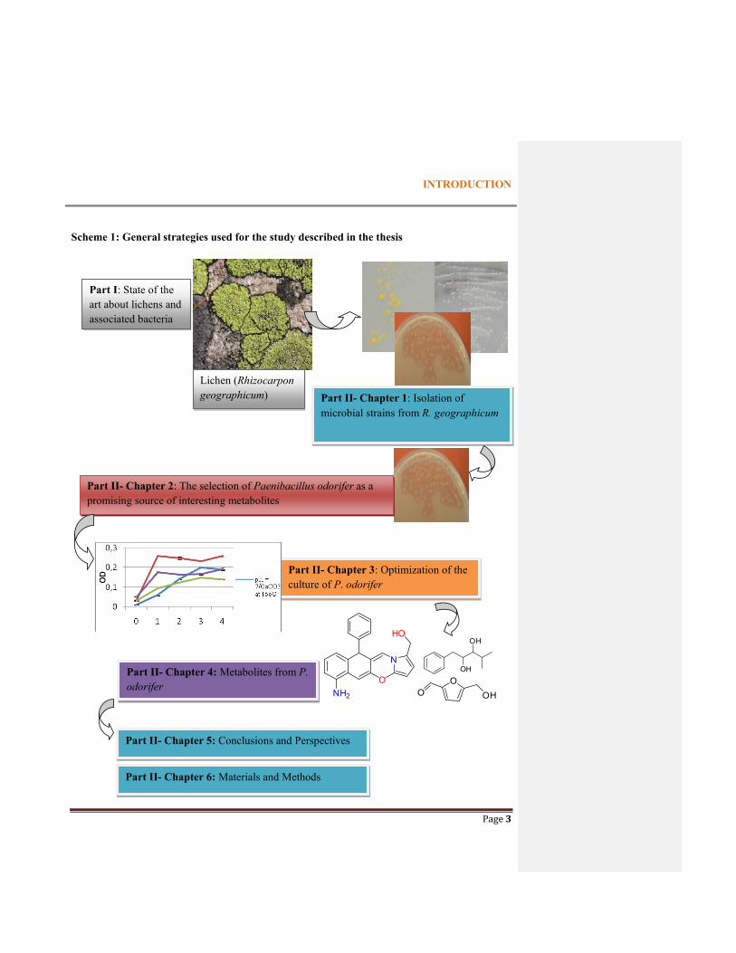
All strategies were summarized in Scheme 1.
INTRODUCTION
Page 3
Scheme 1: General strategies used for the study described in the thesis
Part I: State of the art about lichens and associated bacteria
Lichen (Rhizocarpon geographicum) Part II- Chapter 1: Isolation of
microbial strains from R. geographicum
Part II- Chapter 2: The selection of Paenibacillus odorifer as a promising source of interesting metabolites
Part II- Chapter 3: Optimization of the culture of P. odorifer
OO OH
OH
OHPart II- Chapter 4: Metabolites from P. odorifer
Part II- Chapter 5: Conclusions and Perspectives
Part II- Chapter 6: Materials and Methods
O
N
HO
NH2
INTRODUCTION
Page 4
References
Lammers, A., Wang, R., Cetnar, J., Prasad, V., 2017. Time from US Food and Drug Administration approval to
publication of data for cancer drugs: a comparison of first and subsequent approvals. Blood Cancer Journal 7,
637. https://doi.org/10.1038/s41408-017-0008-9
Suzuki, M.T., Parrot, D., Berg, G., Grube, M., Tomasi, S., 2015. Lichens as natural sources of biotechnologically
relevant bacteria. Appl Microbiol Biotechnol 100, 583–595. https://doi.org/10.1007/s00253-015-7114-z
Koehn, F.E., Carter, G.T., 2005. The evolving role of natural products in drug discovery. Nature Reviews Drug
Discovery 4, 206–220. https://doi.org/10.1038/nrd1657

Page 5
PART 1: STATE OF THE ART ON LICHEN-
ASSOCIATED BACTERIA
Part I : State of the art on lichen-associated bacteria
Page 6
PART 1: STATE OF THE ART ON LICHEN-ASSOCIATED BACTERIA
This general part will be divided into 5 items describing general information on:
- Lichens
- Bacteria associated with lichens and their role in the symbiotic association. This part will report
the recent studies focused on the bacterial communities found on lichens.
- Production of metabolites from lichen-associated bacteria.
- Natural cytotoxic compounds produced by bacteria.
- Reasons for the selection of Rhizocarpon geographicum as a subject of our research.
1.1. INTRODUCTION ON LICHENS
1.1.1. The lichens
1.1.1.1 Description
Lichen, one of the oldest life-forms, appears in several places on our planet (Grube and Berge,
2009). It can grow on most surfaces in the earth and even amazingly on some extreme
environments (Boustie, Tomasi and Grube, 2010). It is estimated that 8% of Earth’s land surface is
covered by lichens (Grube et al., 2013). Around 18500 distinct lichen species have been
characterized and they can adapt themselves to a drastic array of ecological conditions (Shukla et
al., 2010). Lichen is a self-support system, a mini-ecosystem with a perfect combination between
symbiotic parts including a fungal (mycobiont), green alga and/ or cyanobacterium (photobiont),
forming a unique structure called the thallus, but also non-photobiont bacteria (Hodkinson and
Lutzoni 2009; Cardinale et al., 2006, 2008; Grube and Berg, 2009). The majority of fungi
adopting this lifestyle correspond to ascomycetes and more rarely to basidiomycetes. More
recently Spribille and co-workers (2016) have discovered the presence of Cyphobasidiales yeasts
in Bryoria fremontii and B. tortuosa which conducted Pr Grube Martin to define lichens as a
―symbiotic network‖ (personal communication). The photobionts produce carbohydrate by
photosynthesis, bacteria provide to lichens nutrients by fixing nitrogen in the atmosphere (Grube et
al., 2015), while fungal counterparts supply water, mineral elements and protection for all system

Part I : State of the art on lichen-associated bacteria
Page 7
(Parrot et al. 2016a, Benedict 2009) (Figure 1.1). Fungal endophytes have been also described
from various thalli (Wang et al., 2016; Park et al., 2015). In this thesis we will not give details on
this group of microorganisms existing in lichen thalli.
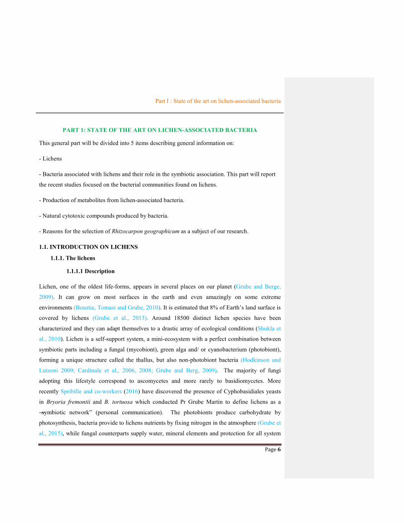
Figure 1.1 Exchange nutrients between lichen symbiotic partners (R. geographicum as the model) (adapted from Grube et al., 2015).
Lichen substances: depsidones, depsides… depsidesdepsides
-Water, minerals and vitamins supply -Protection of all systems
Production of carbohydrates, vitamins
Water, minerals, light, CO2, N2
-Nutrient supply -Pathogen defensis -Provision of Vitamin B12 for photosynthesis -Provision of hormones to support fungal, algae growth
Part I : State of the art on lichen-associated bacteria
Page 8
1.1.1.2 Morphology
The appearance of the thallus is predominantly determined by the mycobiont and could be
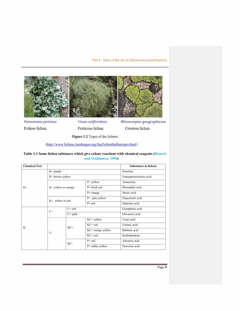
divided into three main morphological groups: crustose, foliose and fruticose types (Figure 1.2).
An additional type corresponds to the gelatinous thallus which possesses a particular aspect due to
the presence of cyanobacteria as photobiont (Büdel and Scheidegger 2008). The identification of
lichens is based on their morphological characteristics but also on thalline reactions which
correspond to the chemical reaction after addition of various chemical reagents (Huneck and
Yoshimura 1996).
There are several tests which are commonly used to identify lichen species; they include the
C, Pd, K and KC test. The C test is executed using sodium hypochlorite (NaClO), readily
accessible as it is contained in an appropriate dilution in most commercial sources of bleach. The
Pd test is performed using the chemical reagent para-phenylenediamine. This chemical is known
to be carcinogenic but generally accepted as a rather week carcinogen and relatively safe to use for
chemical tests on lichens. The K test is carried out with the chemical compound potassium
hydroxide. The final test, the KC test, is a combined test where the K test is performed followed by
the application of the C test to achieve a stronger reaction (see Table 1.1). These tests are designed
to create a chemical reaction when they come into contact with the lichen and are used as color
spot tests to identify the various types of lichen. The tests are not always helpful in the
identification of a specific lichen species. These thalline reactions correspond to a chemical
reaction between one or some lichen compound(s) found on the thallus and the reagent deposited
on it. Moreover, depending on the location where the drop of reagent was added (medulla, cortex,
apothecia…) the reaction will be different due to the host localization of some lichen compounds
(Parrot et al., 2014).
Part I : State of the art on lichen-associated bacteria
Page 9
Figure 1.2 Types of the lichens
(http://www.lichens.lastdragon.org/faq/lichenthallustypes.html)
Table 1.1 Some lichen subtances which give colour reactions with chemical reagents (Huneck and Yoshimura, 1996)
Chemical Test Substances in lichens
K+
K+ purple Parietine
K+ brown-yellow Fumarprotocetraric acid
K+ yellow or orange
P+ yellow Atranorine
P+ brick red Physodalic acid
P+ orange Stictic acid
K+ yellow to red P+ pale yellow Hypostictic acid
P+ red Salazinic acid
K-
C+ C+ red Gyrophoric acid
C+ pink Olivetoric acid
C-
KC+
KC+ yellow Usnic acid
KC+ red Lobaric acid
KC+ orange yellow Barbatic acid
KC+ red Norlobaridone
KC- P+ red Alectoric acid
P+ sulfur yellow Psoromic acid
Part I : State of the art on lichen-associated bacteria
Page 10
1.1.1.3 Metabolites produced by lichen thallus
Metabolites could be divided into two main classes. The first class concerns primary
metabolites e.g. proteins, amino acids, carotenoids, polyols, polysaccharides, vitamins which are
water-soluble and more often produced by the fungus than by the alga. The second class
corresponds to secondary or specialized metabolites most often found on the surface of the hyphae
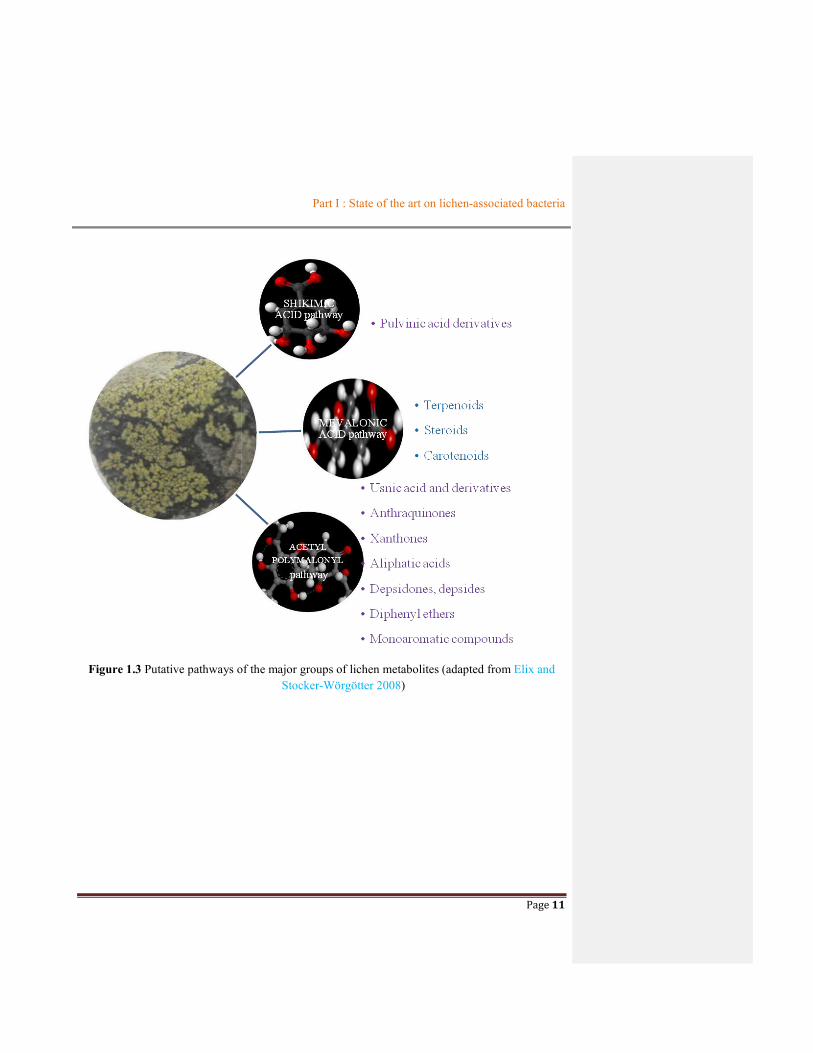
than intracellular. In the Figure 1.3 are reported the biosynthetic pathways of the major groups of
lichen metabolites such as moroaromatic phenolic compounds, depsides, depsidones, diphenyl
ethers, dibenzofurane derivatives (Elix. and Stocker-Wörgötter 2008); and in Figure 1.4, 1.5 and
1.6 were described the structures of some secondary metabolites from lichens corresponding with
the different pathways.
Part I : State of the art on lichen-associated bacteria
Page 11
Figure 1.3 Putative pathways of the major groups of lichen metabolites (adapted from Elix and Stocker-Wörgötter 2008)

Part I : State of the art on lichen-associated bacteria
Page 12
OO
OOH
(CH2)12CH3
(+)-Protolichesterinic Acid
OH
O
(H2C)11
OHORoccellic acid
H3C SR
Aliphatic acids
HO OH
OH
O
Orsellinic acid
HO OH
OH
O
Orsellinic acid
Phenolic compounds
O
HO O
OH
OHO
HO
Siphulin (Chromone)
O
OH O
H3CO OCH3
Lichexanthone (Xanthone)
OH OHO
HO OHO
Averythrin (Anthraquinone)
O
OH O
OOH
HOOCH2C
Haemaventosin (Naphthaquinone)
O
O
HO
O
HO OH
OHPara-Depside(Lecanoric acid)
O
O
OH
O
O OH
OH
O
Meta-Depside(Sekikaic acid)
OH
HO
O
O
OH
O
O
OH
O
OH
Tridepside(Gyrophoric acid)
O
O
HO
OHO
O
OUsnic acid
O
O
O
OH
COOHCHO
HO
Depsidone(Verensic acid)
OH
OO
HO
OH
COOH
Depside(4-O-dimethylbarbatic acid)
O
HOHO
O
O
O
O
Depsone(Picrolichen acid)
Figure 1.4 Structure of typical lichen products derived from the Acetyl-polymalonyl pathway

Part I : State of the art on lichen-associated bacteria
Page 13
OOH
OOH
O
Pulvinic acid and Derivatives
Pulvinic acid
O
O
O
OHO
Calycin
Terphenylquinones
OH
OHO
O
Polyporic acid
O
OHO
HO
O
O
OH
OH
Thelephoric acid
Amino acid derivatives
N
O N
OO
O
O
O
OCOCH3
4-Acetyl-4'-butyrylcabrosin
NN
O
OAcO
O
O
OCORS
S
Ambew elamide A1 (R = C3H7)Ambew elamide B2 (R = C5H11)
Figure 1.5 Structure of typical lichen products derived from the Shikimic acid pathway

Part I : State of the art on lichen-associated bacteria
Page 14
O
R
S
S
O
O
H
Terpenoids
(-)--Thujoine(Monoterpenoid) HO O
H
H
S
R
R
RR
(-)-Sandaracopimaric acid(Diterpenoid)
OHOH
OH
Hopan- -triol(Triterpenoid)
Fukinanolide A(Sesquiterpenoid)
Steroid
Ergosterol
Carotenoid
-Carotene
Figure 1.6 Structure of typical lichen products derived from the Mevalonic acid pathway
1.2. BACTERIA ASSOCIATED WITH LICHENS
1.2.1. State of art about bacterial communities on lichens
Although common knowledge dictated that the lichen thallus was formed by a fungus that
constructed a symbiotic relationship with an alga and/or a cyanobacterium (photobiont), the non-
photobiont bacteria were increasingly considered as an integral component of lichen thalli
(Hodkinson et al., 2009). Therefore, the classical view of this symbiotic relationship should be
expanded to include bacteria. This concept has taken its origin from researches in the beginning of
this century using either cultivation method or cultivation independent method to study these
communities. Cardinale and co-workers (2006) has used cultivation methods investigated on
eleven different lichen samples collected on different sites and highlighted interesting results about
the microorganism communities. More than 100 bacteria were identified belonging to Firmicutes,
Actinobacteria and Proteobacteria phylum. Although the dominant community among these

Part I : State of the art on lichen-associated bacteria
Page 15
bacterial strains was not presented in this report, Paenibacilllus and Burkholderia phylotypes were
commonly found in almost lichens of this study. A distinct research also reported in the same year
(Liba et al. 2006) used cultivation-dependent method with a nitrogen-free minimal medium to
screen five species of cyanobacteria-deprive lichens Canoparmelia caroliniana, crozalsiana,
texana; Parmotrema sancti-angeli and tinctorum harvested in San Paulo state (Brazil). The results
demonstrated that seventeen strains were isolated and all isolates were belonged to
Gamaproteobacteria (Proteobacteria) group.
Two years later, Cardinale and co-workers (2008) studied bacterial communities found on the
shrub-like reindeer lichen Cladonia arbuscula. This research was based on cultivation independent
method using general DNA staining and fluorescent in situ hydridization (FISH) coupled with
confocal laser scanning microscopy (CLSM). This work exhibited that about 6.107 bacteria g-1
hosted on C. arbuscula. The report also showed that the dominance on bacterial communities
corresponded to Alphaproteobacteria with more than 60% of all bacteria following by
Actinobacteria and Betaproteobacteria phylum (lower than 10% for each). Whereas Firmicutes
were rarely detected, no Gammaproteobacteria was found.
A non-cultivation method using FISH, CLSM and imaging analysis applied on three lichens
(Cladonia arbuscula, Lecanora polytropa and Umbilicaria cylindrica) highlighted that the
abundance of bacterial colonies was up to 108 cells per gram fresh weight (Grube and Berg 2009).
This experiment also indicated the predominance of the bacterial communities on these three
lichens being Alphaproteobacteria followed by Firmicutes with Paenibacillus cited as an example.
Moreover, a different subsequent report from non-culture method carried out on Cladonia
arbuscula lichens concluded that lichen-associated microbial communities consisted of diverse
taxonomic groups and the majority of bacteria belonged to Alphaproteobacteria (Grube et al
2009). Another study used a set of PCR-based methods applied on different lichens from several
sites in United States to examine the putative microorganism communities associated with lichen
thalli. This work has revealed the identity of several bacterial associates consisting of the
extremophilic Acidobacteria, Brucellaceae, and members of an undescribed lineage belonging to
the Rhizobiales (Hodkinson and Lutzoni 2009). Nevertheless, a distinct research using culture
dependent method from thirteen species of lichens collecting in different locations in the United

Part I : State of the art on lichen-associated bacteria
Page 16
States highlighted the presence of thirty pure strains from main bacterial lineages as
Actinobacteria, Firmicutes, Proteobacteria and Deinococcus-Thermus (Selbmann et al., 2009).
The next studies published in 2011 by research groups such as Bates et al., Schneider et al.
and Bjelland et al. using unculture-based methods led to the results that Proteobacteria
(Alphaproteobacteria or Betaproteobacteria) expressed again its dominance in bacterial
communities from the studied lichens. It is then noted that a novel Actinobacterium belonging to
the family Microbacteriaceae was first found in lichen Cladonia arbuscula via cultivation method
and identified by 16S rRNA sequencing technology (Cardinale et al., 2011). This study led to the
discovery of a novel member among the microorganism colonies associated with lichens. During
the following years, the revolution of discovery on lichen-associated prokaryotic colonies bloomed
throughout a series of surveys on many distinct lichens collected in the same site (Cardinale et al.,
2012b) or on only lichen species collected at different locations (Cardinal et al., 2012a; Printzen el
al., 2012). Most studies used non-culture based methods and they gave the same results about the
dominant fraction of Alphaproteobacteria in bacteria communities. A different result, however,
was provided by the team of Grube and his co-workers (2012b) where Acidobacteria was proposed
as predominant colonies for all bacterial symbiotic partner of Solorina crocea found in the Alpine
then followed by Planctomycetes and Proteobacteria.
Further study of Kim et al. (2012) performed on the Arctic lichen Stereocaulon sp. using
culture method exhibited the presence of three bacterial members (Pseudomonas graminis,
Mucilaginibacter rugui, Bosea vestrisii). The study also specified the antimicrobial properties of
these bacteria against six tested bacteria (such as Staphylococus aureus, Bacillus bacillus,
Micrococcus luteus, Escherichia coli, Pseudomonas aeruginosa and Enterobacter cloacae). A
different research during this year using cultivation-independent method instead of cultivation
method screened abundance of bacterial communities on lichens from Alpine soil crusts and led to
the result that Alphaproteobacteria and Acidobacteria were predominant in these lichen-associated
prokaryotic colonies (Muggia et al., 2013).
The most recent report using unculture method compared the lichen-associated bacterial
community compositions on both thallus and isidioid soredia of the lichen Lobaria pulmonaria.
The results showed that Alphaproteobacteria were the predominant phylum on both samples
followed by Sphingobacteria (Aschenbrenner et al., 2014). Another study based on culture-based

Part I : State of the art on lichen-associated bacteria
Page 17
method investigated on 4 lichen species found in northern Iceland (Lecanora helicopis, Verrucaria
ceuthocarpa, Hydropunctaria maura and Caloplaca verruculifera) indicated that strains which
were found belonged to 7 classes: Alphaproteobacteria, Bacilli, Actinobacteria, Flavobacteria,
Cytophagia, Sphingobacteria, and Gammaproteobacteria (Sigurbjornsdottir et al. 2014). A report
about 20 cultivable bacteria species isolated from the Antarctic lichen Psoroma sp. by Kim et al
(2014) provided new knowledge about antimicrobial and antioxidant properties as a potential
application of these symbiotic partners of lichens.
In summary
A series of researches have been undertaken to affiliate the third symbiotic partners of lichens
using well-definite approaches, either unculturable or culturable methods, depending on the
purpose of the studies. Each method displayed its advantages and disadvantages. While
uncultivable methods led to discover a greater bacterial diversity on lichens, culturable approaches
only supported information about cultivable bacterial strains and it can lead to wrong conclusions
about the diversity of these microorganisms. A strong point for these cultivable methods, however,
is to easily permit to study properties and production of metabolites from isolated bacteria which
cannot be performed in non-culture based methods.
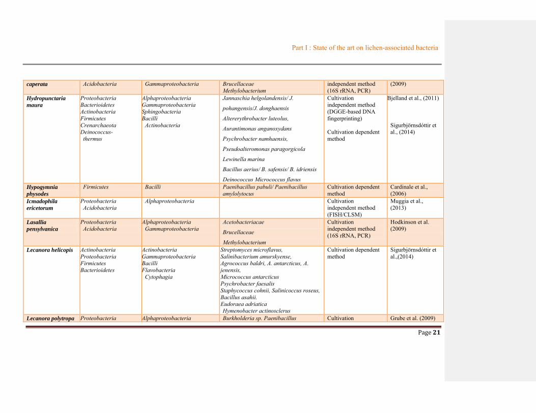
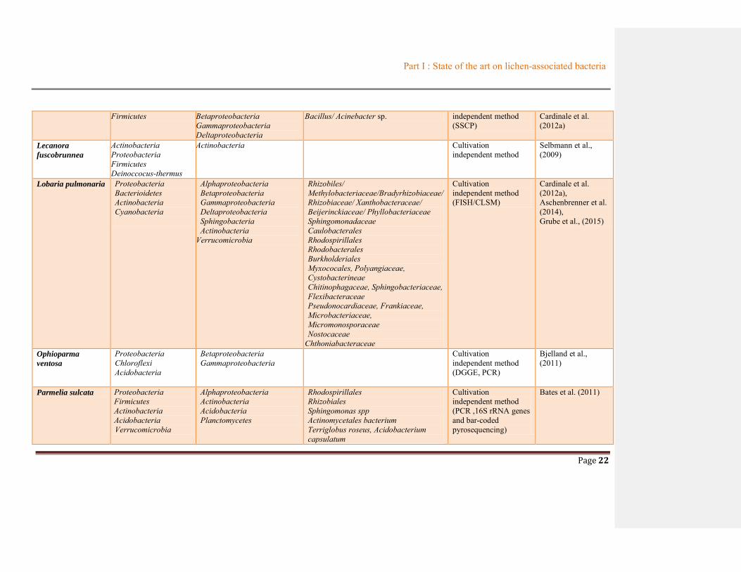
The detailed data indicated in Table 1.2 summarized information about bacterial communities
(up to genus level) from lichen species with their names noted in alphabetical order. One lichen
species can be studied by either a team or many teams. The methods used in these studies were
noted clearly as cultivation-dependent or the independent one. From 52 lichen species mentioned
in Table 1.2, Proteobacteria always exhibited as dominant phylum (with 39.5%) in a total of 119
genera reported. This value was over more twice than those of Acidobacteria (16.8%) and
Firmicutes (15.1%). These data permit to conclude that three phyla were commonly presented in
bacteria associated with lichens. In the dominant phylum Proteobacteria, the class present in the
most cases was Alphaproteobacteria, following by Gammaproteobacteria and Betaproteobacteria,
while Deltaproteobacteria rarely appeared from this phylum. The other phyla sometimes found in
lichens were Actinobacteria (with 10.7%) and Bacteroidetes (about 5.8%). These two bacterial
phyla were often present in foliose and crustose lichens. Indeed, Actinobacteria were found in the
total of 11 lichen species consisting of 8 foliose lichens, 3 crustose lichens, 3 placodioid lichens
and only one fructicose lichen; Bacteroidetes were found in 7 lichen species consisting of 3 foliose

Part I : State of the art on lichen-associated bacteria
Page 18
lichens, 3 crustose lichens and 1 placodioid lichen. In addition, diverse bacterial phyla as
Deinococcus-thermus, Verrucomicrobia … were presented in a little part from lichens. It is finally
to note that, an ancient phylum also reported as a partner with lichens was Chloroflexi found from
the following lichens Ophioparma ventosa, Pertusaria corallina and Rhizocarpon geographicum.

Part I : State of the art on lichen-associated bacteria
Page 19
Table 1.2 Summary of bacterial communities associated with lichens
Lichen Bacterial communities Method References Phylum Class Family or genus
Arthrorhaphis
citrinella
Proteobacteria Acidobacteria
Alphaproteobacteria Cultivation independent method (FISH/CLSM)
Muggia et al., (2013)
Caloplaca
verruculifera
Proteobacteria Bacteroidetes Actinobacteria
Alphaproteobacteria Gammaproteobacteria Flavobacteria Sphingobacteria Actinobacteria
Jannaschia pohagensis/
Sphingopysix marina
/S. desiccabilis/Aurantimonas coralicida/
Aurantimonas kwanggyangensis/
Sphingomonas suberifaciens
Psychrobacter frigidicola.
Micrococcus luteus/ Micrococcus flavus
Aquimarina intermedia
Lewinella antartica
Cultivation dependent method
Sigurbjörnsdóttir et al., (2014)
Canoparmelia
caroliniana
Proteobacteria
Gammaproteobacteria
Stenotrophomonas maltophilia Cultivation dependent method
Liba et al. (2006)
Canoparmelia
crozalsiana
Proteobacteria
Gammaproteobacteria
Stenotrophomonas maltophilia/ Serratia
marcescens
Cultivation dependent method
Canoparmelia
texana
Proteobacteria
Gammaproteobacteria
Serratia marcescens/ Pseudomonas
stutzeri
Cultivation dependent method
Cetraria islandica Proteobacteria Actinobacteria Firmicutes
Alphaproteobacteria Betaproteobacteria Gammaproteobacteria Actinobacteria
Cultivation independent method (FISH/CLSM)
Cardinale et al. (2012a) Grube et al. (2009)
Cetraria sp. Proteobacteria
Alphaproteobacteria
Sphingomonas faebi Cultivation dependent method
Kim et al. (2012)

Part I : State of the art on lichen-associated bacteria
Page 20
Cetraria aculeata Proteobacteria
Alphaproteobacteria
Cultivation independent (DNA/PCR)
Printzen et al. (2012)
Cladonia arbusculla
Proteobacteria Firmicutes
Alphaproteobacteria Betaproteobacteria Bacilli
Burkholderia sp./ Paenibacillus Bacillus
Cultivation independent (SSCP)
Grube et al.,(2009); Cardinale et al. (2012a)
Cladonia coccifera Proteobacteria Firmicutes
Alphaproteobacteria Betaproteobacteria Deltaproteobacteria
Burkolderia phenazinium Paenibacillus amylolyticus/ Paenibacillus agarexedens
Cultivation dependent ITS (Cultivation independent method)
Cardinale et al. (2006); Cardinale et al. (2012a)
Cladonia cristatella Proteobacteria Acidobacteria
Alphaproteobacteria Gammaproteobacteria
Acetobacteriacae Brucellaceae Methylobacterium
Cultivation independent method (16S rRNA, PCR )
Hodkinson et al., (2009)
Cladonia
cryptochlorophaea
Proteobacteria Acidobacteria
Alphaproteobacteria Gammaproteobacteria
Acetobacteriacae Brucellaceae Methylobacterium
Cladonia
peziziformis
Proteobacteria Acidobacteria
Alphaproteobacteria Gammaproteobacteria
Acetobacteriacae Brucellaceae Methylobacterium
Cladonia gracilis Actinobacteria Actinobacteria Streptomyces sp. Cultivation dependent method (16S rRNA)
Cheenpracha et al., (2010)
Cladonia sp. Proteobacteria Betaproteobacteria Burkholderia sordidicola Cultivation dependent method
Kim et al. (2012)
Cladonia pyxidata Proteobacteria Betaproteobacteria Burkholderia glathei/ Burkholderia sordidicola
Cultivation dependent method
Cardinale et al., (2006)
Cladonia
rangiferina
Proteobacteria Firmicutes
Betaproteobacteria Bacilli
Burkholderia glathei/ Burkholderia sordidicola Paenibacillus pabuli
Cultivation dependent method
Cladonia subtenuis Proteobacteria Acidobacteria
Alphaproteobacteria Gammaproteobacteria
Acetobacteriacae Brucellaceae Methylobacterium
Cultivation dependent method (16S rRNA, PCR)
Hodkinson et al., (2009)
Cladonia uncialis Actinobacteria Actinobacteria Streptomyces uncialis Cultivation dependent method
Davies et al., (2005), Williams et al. (2008)
Flavoparmelia Proteobacteria Alphaproteobacteria Acetobacteriacae Cultivation Hodkinson et al.
Part I : State of the art on lichen-associated bacteria
Page 21
caperata Acidobacteria Gammaproteobacteria Brucellaceae Methylobacterium
independent method (16S rRNA, PCR)
(2009)
Hydropunctaria
maura
Proteobacteria Bacterioidetes Actinobacteria Firmicutes Crenarchaeota Deinococcus- thermus
Alphaproteobacteria Gammaproteobacteria Sphingobacteria Bacilli
Actinobacteria
Jannaschia helgolandensis/ J.
pohangensis/J. donghaensis
Altererythrobacter luteolus,
Aurantimonas anganoxydans
Psychrobacter namhaensis,
Pseudoalteromonas paragorgicola
Lewinella marina
Bacillus aerius/ B. safensis/ B. idriensis
Deinococcus Micrococcus flavus
Cultivation independent method (DGGE-based DNA fingerprinting) Cultivation dependent method
Bjelland et al., (2011)
Sigurbjörnsdóttir et al., (2014)
Hypogymnia
physodes
Firmicutes Bacilli Paenibacillus pabuli/ Paenibacillus amylolytocus
Cultivation dependent method
Cardinale et al., (2006)
Icmadophila
ericetorum
Proteobacteria Acidobacteria
Alphaproteobacteria Cultivation independent method (FISH/CLSM)
Muggia et al., (2013)
Lasallia
pensylvanica
Proteobacteria Acidobacteria
Alphaproteobacteria Gammaproteobacteria
Acetobacteriacae
Brucellaceae
Methylobacterium
Cultivation independent method (16S rRNA, PCR)
Hodkinson et al. (2009)
Lecanora helicopis Actinobacteria Proteobacteria Firmicutes Bacterioidetes
Actinobacteria Gammaproteobacteria Bacilli Flavobacteria
Cytophagia
Streptomyces microflavus, Salinibacterium amurskyense, Agrococcus baldri, A. antarcticus, A. jenensis, Micrococcus antarcticus Psychrobacter faesalis Staphycoccus cohnii, Salinicoccus roseus, Bacillus asahii. Eudoraea adriatica Hymenobacter actinosclerus
Cultivation dependent method
Sigurbjörnsdóttir et al.,(2014)
Lecanora polytropa Proteobacteria Alphaproteobacteria Burkholderia sp. Paenibacillus Cultivation Grube et al. (2009)
Part I : State of the art on lichen-associated bacteria
Page 22
Firmicutes Betaproteobacteria Gammaproteobacteria Deltaproteobacteria
Bacillus/ Acinebacter sp. independent method (SSCP)
Cardinale et al. (2012a)
Lecanora
fuscobrunnea
Actinobacteria Proteobacteria Firmicutes Deinoccocus-thermus
Actinobacteria Cultivation independent method
Selbmann et al., (2009)
Lobaria pulmonaria Proteobacteria Bacterioidetes Actinobacteria Cyanobacteria
Alphaproteobacteria Betaproteobacteria Gammaproteobacteria Deltaproteobacteria Sphingobacteria Actinobacteria
Verrucomicrobia
Rhizobiles/ Methylobacteriaceae/Bradyrhizobiaceae/ Rhizobiaceae/ Xanthobacteraceae/ Beijerinckiaceae/ Phyllobacteriaceae Sphingomonadaceae Caulobacterales Rhodospirillales Rhodobacterales Burkholderiales Myxococales, Polyangiaceae, Cystobacterineae Chitinophagaceae, Sphingobacteriaceae, Flexibacteraceae Pseudonocardiaceae, Frankiaceae, Microbacteriaceae, Micromonosporaceae Nostocaceae
Chthoniabacteraceae
Cultivation independent method (FISH/CLSM)
Cardinale et al. (2012a), Aschenbrenner et al. (2014), Grube et al., (2015)
Ophioparma
ventosa
Proteobacteria Chloroflexi Acidobacteria
Betaproteobacteria Gammaproteobacteria
Cultivation independent method (DGGE, PCR)
Bjelland et al., (2011)
Parmelia sulcata Proteobacteria Firmicutes Actinobacteria Acidobacteria Verrucomicrobia
Alphaproteobacteria Actinobacteria Acidobacteria Planctomycetes
Rhodospirillales Rhizobiales Sphingomonas spp Actinomycetales bacterium Terriglobus roseus, Acidobacterium capsulatum
Cultivation independent method (PCR ,16S rRNA genes and bar-coded pyrosequencing)
Bates et al. (2011)

Part I : State of the art on lichen-associated bacteria
Page 23
Nostocoida limicola Rubritalea spp.
Parmotrema
perforatum
Proteobacteria Acidobacteria
Alphaproteobacteria Gammaproteobacteria
Acetobacteriacae Brucellaceae Methylobacterium
Cultivation independent method (16S rRNA, PCR)
Hodkinson et al. (2009)
Liba et al. (2006) Parmotrema sancti-
angeli
Proteobacteria Gammaproteobacteria Serratia marcesens/ Acinetobacter calcoaceticus
Parmotrema
tinctirum
Proteobacteria Gammaproteobacteria Serratia marcesens/ Pseodomonas sp. Stenotrophomonas maltophilia/ Pentoea sp.
Pertusaria corallina Proteobacteria Acidobacteria Chloroflexi
Alphaproteobacteria Betaproteobacteria
Cultivation independent method(DGGE, PCR)
Bjelland et al. (2011)
Peltigera
membranacea
Proteobacteria Actinobacteria Bacteriodetes
Alphaproteobacteria Betaproteobacteria
Cultivation independent (Pyrosequencing)
Grube et al. (2014)
Peltigera phyllidosia Proteobacteria Acidobacteria
Alphaproteobacteria Gammaproteobacteria
Acetobacteriacae Brucellaceae
Methylobacterium
Cultivation independent (16S rRNA, PCR)
Hodkinson et al. (2009)
Pseudevenia
furfuracea
Firmicutes Bacilli Paenibacillus mendilii/ Paenibacillus phyllosphaerae
Cultivation dependent Cardinale et al. (2006)
Psora decipiens Proteobacteria Acidobacteria
Alphaproteobacteria Cultivation independent method(FISH/CLSM)
Muggia et al. (2013)
Rhizocarpon
geographicum
Proteobacteria Acidobacteria Chloroflexi
Alphaproteobacteria Betaproteobacteria
Cultivation independent method (DGGE, PCR)
Bjelland et al (2011)
Rhizocarpon
chrysoleuca
Proteobacteria Acidobacteria Firmicutes Verrucomicrobia
Alphaproteobacteria Acidobacteria Firmicutes Verrucimicrobia Planctomycetes
Rhodospirillales/ Acetobacteria/Acidiphilum sp Rhizobiales Sphingomonas spp. Acidobacteriales/Terriglobus roseus
Cultivation independent method (Pyrosequencing)
Bates et al. (2011)

Part I : State of the art on lichen-associated bacteria
Page 24
Rubritalea spp. Nostocoida limicola Desulfotomaculum sp.
Solorina crocea Acidobacteria Planctomycetes Proteobacteria
Acidobacteria Planctomycetes Alphaproteobacteria
Acidobacterium sp./Edaphobacter Isosphaera sp./Gemmata Novosphingobium
Sphingomonas
Cultivation independent method
Grube et al. (2012a)
Stereocaulon sp. Proteobacteria Betaprotebacteria Burkholderia sordidicola Cultivation dependent method
Kim et al. (2012)
Trapeliopsis
granulosa
Proteobacteria Acidobacteria
Alphaproteobacteria Cultivation independent method (FISH/CLSM)
Muggia et al. (2013)
Umbilicaria
americana
Proteobacteria Acidobacteria Actinobacteria Firmicutes Bacteroidetes
Alphaproteobacteria Acidobacteria Actinobacteria Firmicutes Bacteroidetes
Rhodospirillales/ Acidiphilum sp. Phenylobacterium spp. Sphingomonas spp. Acidobacteriales/Terriglobus roseus Actinomycetsles bacterium Desulfotomaculum sp. Sphingobacteria/Pedobacter solani
Cultivation independent method (pyrosequencing)
Bates et al. (2011)
Umbilicaria
cylindrica
Proteobacteria Firmicutes
Alphaproteobacteria Betaproteobacteria Gammaproteobacteria Bacilli
Acetobacteraceae Burkholderia sp. Acinebacter
Bacillus sp.
Cultivation independent method (SSCP)
Grube et al. (2009), Cardinale et al.
(2012a)
Umbilicaria
decussata
Actinobacteria Proteobacteria Firmicutes Deinococcusthermus
Actinobacteria Actinomycetales/ Intrasporangiaceae Knoellia
Cultivation dependent method
Selbmann et al. (2009)
Umbilicarria phaea Proteobacteria Acidobacteria Firmicutes
Planctomycetes
Alphaproteobacteria Acidobacteria Firmicutes Planctomycetes
Rhodospirillales/Acidiphilum sp. Acidobacterium apsulatum Desulfotomaculum sp. Nostocoida limicola
Cultivation independent method (Pyrosequencing)
Bates et al., (2011)
Umbilicaria
mammulata
Acidobacteria Proteobacteria
Acidobacteria Alphaproteobacteria Gammaproteobacteria
Acetobacteraceae Brucellaceae
Methylobacterium
Cultivation independent method (16s rRNA, PCR)
Hodkinson et al. (2009)

Part I : State of the art on lichen-associated bacteria
Page 25
Umbilicaria sp. Proteobacteria Betaproteobacteria Burkholderia sordidicola Cultivation dependent method
Kim et al. (2012)
Usnea antarctica
Actinobacteria Proteobacteria Firmicutes Deinococcusthermus
Actinobacteria Gammaproteobacteria
Micrococcaceae/Arthrobacter Pseudomonadaceae/Pseudomonas
Cultivation dependent method
Selbmann et al. (2009)
Xanthoria elegans Actinobacteria Proteobacteria Firmicutes Deinococcusthermus
Gammaproteobacteria Actinobacteria Firmicutes
Pseudomonadaceae/Pseudomonas Mycobacterriun Paenibacillus Bacillus
Verrucaria
ceuthocarpa
Proteobacteria Actinobacteria Firmicutes Bacteroidetes
Alphaproteobacteria Gammaproteobacteria Actinobacteria Bacilli Bacteroidetes
Sphingopysix baekryungensis Loktanella salsilacus Altererythrobacter luteolus Psychrobacter maritimus Micrococcus antarcticus Micrococcus luteus Bacillus murimartini Bacillus safensis Rhodothermus marinus
Cultivation dependent method
Sigurbjörnsdóttir et al., (2014)

Part I : State of the art on lichen-associated bacteria
Page 26
1.2.2. The roles of bacteria
Bacterial communities were identified as stable, specific and structurally integrated partners of
the lichen symbiosis, but their role has remained largely elusive in comparison to the well-known
functions of the fungal and algal partners. One of the first studies to question about the roles of the
bacterial communities in lichens was those of Grube (2015) which found that more than 800
bacterial species have the ability to contribute multiple aspects to the symbiotic system, including
essential functions such as (i) nutrient supply, especially nitrogen, phosphorous and sulfur, (ii)
resistance against biotic stress factors (pathogen defenses as an example), (iii) resistance against
abiotic factors, (iv) support of photosynthesis by provision of vitamin B12, (v) fungal and algal
growth support by provision of hormones, (vi) detoxification of metabolites, and (vii) degradation
of older parts of the lichen thallus (Grube et al., 2015).
Figure 1.7 Roles of bacteria in lichens (adapted from Grube et al., 2015)

Part I : State of the art on lichen-associated bacteria
Page 27
1.3. METABOLITES FROM THE LICHEN-ASSOCIATED BACTERIA
As lichens are a promising source of bioactive metabolites (Boustie and Grube, 2005), bacteria
from lichens possess an unexploited potential of effective new metabolites (Grube et al., 2012b).
Recent investigations focused on a strain of Streptomyces isolated from the lichen Cladonia
uncialis. The extracts from the broth of the cultivation of this strain produced a series of novel
active compounds. Among them, uncialamycin, an enediyne compound (Davies et al., 2005)
exhibited a strong antibacterial activity against the human pathogens Burkholderia cepacia and
Staphylococcus aureus but also as potent cytotoxic against MCF-7 cells. This strain also produces
seven new bis-indole alkaloids, named cladoniamides, which possess an unprecedented skeleton in
natural products (Williams et al., 2008). In these alkaloids, cladoniamide G presented a potential
as the anticancer compound by its cytotoxicity against human breast cancer MCF-7 cells in vitro at
10 µg/mL. Further investigations on this strain in 2015 (Williams et al., 2015) yielded a new
compound, unciaphenol, resulted from the expected Bergman cycloaromatization of uncialamycin.
It is interesting that the unciaphenol presented in vitro anti-HIV activity against drug-resistant
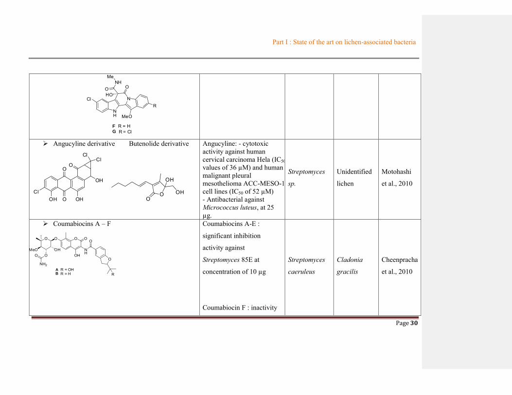
isolates of the virus. Another lichen-derived Streptomyces strain (Streptomyces sp.) from
unidentified lichen in Japan produced a new angucycline which also displayed inhibitory effects on
certain cell lines and on bacterial strains and a new butenolide (Motohashi et al., 2010). Another
research on Streptomyces caeruleus obtained from the lichen Cladonia gracilis afforded six
compounds, coumabiocins A-F, which exhibited significant inhibition activity against
Streptomyces-85 using agar diffusion assay (Cheenpracha et al., 2010).
Moreover, a culture of bacterium Streptomyces cyaneofuscatus isolated from a marine lichen
Lichina confinis yielded a new bioactive compound cyaneodimycin (Parrot et al., 2016b). This
compound showed activity against human keratinocyte HaCaT and murine melanoma B16 cell
lines [Inhibitory concentration (IC50) values of 47 ± 11 and 27 ± 4 µM, respectively]. A potent
cytotoxic compound N-methyldactinomycine derived from the known anticancer drug
actinomycine D was also isolated from this same species. Although diketopiperazines, also found
from this species, did not display noticeable cytotoxic properties, they seem becoming as chemical
signals for lichen-associated bacteria. They were not only found from the strain cited above (Parrot

Part I : State of the art on lichen-associated bacteria
Page 28
et al. 2016b), but they also are produced by another lichen-associated actinobacterium as Nocardia
sp associated to Collema auriforme (Noël el al., 2017).
IN SUMMARY
Up to now, the rare studies on bioactive metabolites from lichen-associated bacteria have
almost focused on Streptomyces strains isolated from Cladonia genus or Lichina sp. That is one of
the reasons that encouraged us to investigate another type of lichen to study its bacterial symbiotic
communities and to study the interesting metabolites produced by one of these strains. The reasons
for the selection of the bacterial species will be displayed in part 1.5. The bacteria belonging to
Streptomyces genus, isolated from different natural origins, are an important source to produce
bioactive metabolites such as antibiotic, antitumor and immunosuppressant drugs (Lucas et al.,
2013). Around two-thirds of all known natural antibiotics are produced from these kinds of
bacteria which belong to Actinobacteria phylum. Streptomyces genus from lichens confirmed
again their crucial roles via antibacterial properties, cytotoxicity, or even, anti-HIV virus isolates.
The Table 1.3 cited below showed the summary of metabolites isolated from lichen-associated
bacteria. The information including origin, structure and biological activities were displayed in
Table 1.3 organized following the year of publication.

Part I : State of the art on lichen-associated bacteria
Page 29
Table 1.3 Summary of metabolites from lichen-associated bacteria
Compounds Biological activities
Origin
References Bacterial
species
Lichen
species
Uncialamycin
O
O OH
HN OHO OH
Antibacterial activity against Burkholderia cepacia (MIC 0.001 µg/mL). Staphylococcus aureus ((MIC 0.0000064 µg/mL), Escherichia coli (MIC 0.002 µg/mL)
Streptomyces
uncialis
Cladonia
uncialis
Davies et al.,
2005
Cladoniamide A-G
NH
N
N
R1
MeO
MeOO
R2
HO
A R1 = Cl; R2 = HB R1 = R2 = ClC R1 = R2 = H
OH
NH
N
HN
Cl
MeO
O
R
D R = HE R = Cl
OOH
Me
Cytotoxicity against human
breast cancer MCF-7 cells
(for cladoniamide G) in
vitro at 10 μg/mL
Streptomyces
uncialis
Cladonia
uncialis
Williams et
al., 2008
Part I : State of the art on lichen-associated bacteria
Page 30
NH
NCl
MeO
R
F R = HG R = Cl
NHMe
O OHO
Angucyline derivative Butenolide derivative
ClOH O
O
OH
OH
O
ClCl
O
O
OH
OH
Angucyline: - cytotoxic activity against human cervical carcinoma Hela (IC50 values of 36 µM) and human malignant pleural mesothelioma ACC-MESO-1 cell lines (IC50 of 52 µM) - Antibacterial against Micrococcus luteus, at 25 µg.
Streptomyces
sp.
Unidentified
lichen
Motohashi
et al., 2010
Coumabiocins A – F
O O O
NH
O
O
R
MeOO
NH2
OOH
OH
O
A R = OHB R = H
Coumabiocins A-E :
significant inhibition
activity against
Streptomyces 85E at
concentration of 10 µg
Coumabiocin F : inactivity
Streptomyces
caeruleus
Cladonia
gracilis
Cheenpracha
et al., 2010

Part I : State of the art on lichen-associated bacteria
Page 31
O O O
NH
O
MeOO
NH2
OOH
OH
O
C R = HD R = OH
O
R
O O O
NH
O
MeOO
NH2
OOH
OH
O
EOHOH
HO O
NH
O
OH
O
OHOH
F
Novobiocin (G) and isonovobiocin (H)
O O O
NH
O
MeOOR2
OR1OH
O
OH
G R1 = H; R2 = CONH2H R1 = CONH2; R2 = H
Novobiocin : potent
inhibition activity
Isonovobiocin : weaker
activity
Unciaphenol
HNO
O OH
OH
HO
O OH
in vitro anti-HIV activity
against drug-resistant
isolates of the virus.
Streptomyces
uncialis
Cladonia
uncialis
Williams et
al., 2015
Cyaneodimycin Cyaneomycin Cyaneodimycin : cytotoxic Streptomyces Lichina Parrot et al.,

Part I : State of the art on lichen-associated bacteria
Page 32
O
O
O
OOH
OO
O
O
O
OOH
HO
Diketopiperazines : Cyclo-(L-Phe-L-Pro); Cyclo-
(L-Leu-L-Pro)
(3-hydroxyacetyl)indole
N-methyldactinomycin
Usnic acid
O
O
HO
OHO
O
O
against human keratinocyte
HaCaT (IC50 of 47±11 µM),
murine melanoma B16
(IC50 of 27±4 µM) and
leukemic (Jurkat) cell lines
(IC50 = 18.5±0.5 μM).
N-
methyldactinomycin
cytotoxic activities (IC50 ≈ 0.05 μM) against various normal or cancer cell lines after 48 h of incubation
cyaneofuscatus confinis 2016b
Diketopiperazines : cyclo (L-Pro-L-OMet), cyclo
(L-Pro-L-Tyr), cyclo (D-Pro-L-Tyr), cyclo (L-Pro-L-Val),
cyclo (L-Pro-L-Leu), cyclo (D-Pro-L-Br-Tyr), cyclo (L-
Pro-L-Br-Tyr)
Indole-carboxaldehyde
No cytotoxic activities
Nocardia
ignorata
Collema
auriforme
Noël et al.,
2017
Nanaomycin Compounds 1- 4: inactive Streptomyces Lepidostroma Liu et al.,

Part I : State of the art on lichen-associated bacteria
Page 33
antibacterial at
concentration of 100 µg/mL.
Compounds 5-6 :
antibacterial activities
against Staphylococcus
aureus, Candida albicans
and Bacillus subtilis with
MIC values ranging from
3.13 to 100 μg/mL.
hebeiensis
Lepidostroma
sp.,
sp., 2017

Part I : State of the art on lichen-associated bacteria
Page 34
1.4. FOCUS ON RHIZOCARPON GEOGRAPHICUM
1.4.1. Description of Rhizocarpon genus
Rhizocarpon is a large lichen genus containing approximately 200 species worldwide. They
dwelled mainly on siliceous rocks; a few are the parasite of other lichens. Rhizocarpon is green to
yellow-green, white, grey, brown or rust-red, rarely sorediate or blastidiate. Its prothallus usually
appears with black color or sometimes white to brown-grey (Benedict 1998). This cosmopolitan
kind of lichen is generally found in temperate, Arctic and Antarctic areas (Ihlen et al., 2004).
The Rhizocarpon genus is divided into two large sub-genera Phaeothallus and Rhizocarpon based
on the presence or absence of rhizocarpic acid (Innes, 1985; Benedict, 2009), but this classification
has recently been questioned based on genetic studies (Ihlen & Ekman, 2002). The subgenus
Rhizocarpon can be further subdivided into four sections: Alpicola, Rhizocarpon, Superficiale and
Viridiatrum, based on thallus morphology, spore characteristics, and chemistry of the secondary
metabolites found in the medulla (Innes, 1985) (see Table 1.4).
These sections can be broadly subdivided based on the number of cells in a spore (Benedict, 1988).
Superficiale and Alpicola have uniseptate spores (two cells) whereas Rhizocarpon and Viridiatrum
contain pluriseptate (usually muriform of submuriform) spores (several cells) (Innes, 1985).
Section Alpicola and section Superficiale can then be further subdivided by the size of the spores,
the hymenial dimensions and colour of the apihymenium. In the sections with pluriseptate spores,
Rhizocarpon and Viridiatrum apihymenium colour and the reaction of the medulla to iodine
(turning blue) are common distinguishing characteristics (Benedict, 1988). A cross-section of a
Rhizocarpon thallus can be found in Figure 1.8 and demonstrates the characteristic of a stratified
thallus concluding three parts: upper cortex, alga layer and medulla part.

Part I : State of the art on lichen-associated bacteria
Page 35
Figure 1.8: Scheme of the cross section of R. geographicum
http://www.imlichenit.com/imlichenit/about.html)
Table 1.4 Classification of Rhizocarpon subgenus (Innes, 1985)
Genus Section Species
Rhizocarpon
Superficiale
R. dispersum Runem. R. ejjiguratum (Anzi) Th.Fr. R. norvegicum Ras. R. parvum Runem. R. pusillum Runem. R. superjiciale (Schaer.) Vain
Alpicola R. alpicola (Hepp.) Rahb. R. eupetraeoides (Nyl.) 810mb. R. inarense (Vain.) Vain.
Viridiatrum
R. atrovirellum (Nyl.) Zahlbr. R. kakurgon Poelt R. lusitanicum (Nyl.) Arnold R. oportense (Vain.) Ras. R. subtile Runem. R. tetras porum Runem. R. viridiatrum (Wulf.) Koerb
Rhizocarpon
R. atroglavescens Lynge R. carpaticum Runem. R. ferax H. Magn. R.furax Poelt et V. R. geographicum (L.) DC. R. intermediellum Ras. R. lecanorinum Anders R. macros porum Ras. R. pulverulentum (Schaer.) Ras. R. rapax V. Wirth et Poelt R. riparium Ras. R. saanense Ras. R. sphaerosporum Ras. R. sublucidum
Part I : State of the art on lichen-associated bacteria
Page 36
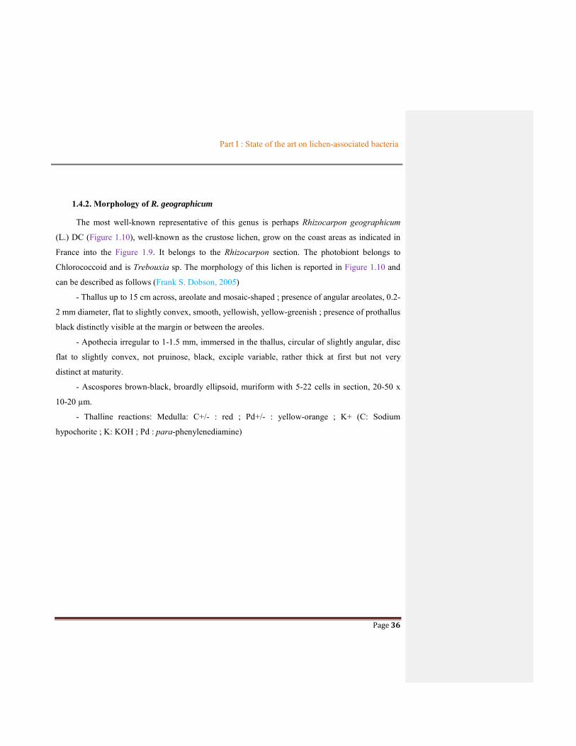
1.4.2. Morphology of R. geographicum
The most well-known representative of this genus is perhaps Rhizocarpon geographicum
(L.) DC (Figure 1.10), well-known as the crustose lichen, grow on the coast areas as indicated in
France into the Figure 1.9. It belongs to the Rhizocarpon section. The photobiont belongs to
Chlorococcoid and is Trebouxia sp. The morphology of this lichen is reported in Figure 1.10 and
can be described as follows (Frank S. Dobson, 2005)
- Thallus up to 15 cm across, areolate and mosaic-shaped ; presence of angular areolates, 0.2-
2 mm diameter, flat to slightly convex, smooth, yellowish, yellow-greenish ; presence of prothallus
black distinctly visible at the margin or between the areoles.
- Apothecia irregular to 1-1.5 mm, immersed in the thallus, circular of slightly angular, disc
flat to slightly convex, not pruinose, black, exciple variable, rather thick at first but not very
distinct at maturity.
- Ascospores brown-black, broardly ellipsoid, muriform with 5-22 cells in section, 20-50 x
10-20 µm.
- Thalline reactions: Medulla: C+/- : red ; Pd+/- : yellow-orange ; K+ (C: Sodium
hypochorite ; K: KOH ; Pd : para-phenylenediamine)

Part I : State of the art on lichen-associated bacteria
Page 37
Figure 1.9: Map of the sites of Rhizocarpon geographicum in France
(http://www.lichensmaritimes.org)
Figure 1.10 Morphology of Rhizocarpon geographicum
Areola
Prothallus

Part I : State of the art on lichen-associated bacteria
Page 38
(by Olivier Gonner at http://www.afl-
lichenologie.fr/Photos_AFL/Photos_AFL_R/Rhizocarpon_geographicum.htm)
1.4.3. Studies on R. geographicum
In the report of McCathay and Elix (2014), rhizocarpic acid and psoromic acid and barbatic
acid were considered major organic compounds from R. geographicum originated from Australia.
Besides, alectoronic acid, α-collatolic acid, bourgeanic acid, confluentic acid, 2-O’-
methylperlatolic acid, 2-O’-methylanziaic acid and gyrophoric acid were also found in this lichen.
The structure of these compounds were shown in Figure 1.11.
Rhizocarpic acid Psoromic acid Barbatic acid
Alectoronic acid α-collatolic acid Bourgeanic acid
2-O’-methylanziaic acid Gyrophoric acid
2-O’-methylperlatolic acid
Figure 1.11 Structure of some compounds from R. geographicum

Part I : State of the art on lichen-associated bacteria
Page 39
This yellow-green species grows very slowly. It is estimated that its annual radial growth rate is
from 0.02 to 2.0 mm per year (Armstrong et al., 1996). The exceptionally slow growth rate and
longevity of these species have made them especially valuable in lichenometry studies (Innes et
al., 1985) and were the focus of many geographical researches (Table 1.5). The lichenometry used
the lichen growth to determine the age of exposed rock based on a presumed specific rate of
increase in radial size time by time. The Radial Growth Rate (RGR) of R. geographicum depended
on many environmental factors as temperature, wind, humidity, sunny and geographical regions
(Armstrong et al., 2006, Bench et al., 2001, Bradwell et al., 2007, O’Neal et al., 2003). Studies
suggested that although a number of parameters affected RGR, the most important influence
appeared to be with temperature. The RGR of R. geographicum was increased by high temperature
and inhibited by low temperature. The maximum growth of R. geographicum was period of July-
September (Armstrong et al., 2006). The Table 1.5 summarized studies of lichenometry on R.
geographicum displayed by the location studied, period time, and growth size and the technique
used as well.

Part I : State of the art on lichen-associated bacteria
Page 40
Author Location Study period Size range
(mm)
Technique
Armstrong
(1983)
Ordovician slate rock field,
Gwynedd, North Wales
1.5 years (6 month
intervals)
2-65 Beck Kassal lens (fitted with a 1 cm
micrometer with x8 magnification
Proctor (1983) Valsorey moraine, Switzerland 4 years (2 year
intervals)
2-18 Photographs with a 0.05 mm scale
micrometer placed next to the lichen
Haworth et al.
(1986)
Brooks Range, Alaska 4-6 years 3-107 Tracing thallus area onto acetate and
converting to equivalent diameter
McCarthy
(2012)
Illecillewaet glacier, GNP,
British Columbia
Annual intervals over 4
years
5-50 Digital caliper (+/-0.02mm)
Armstrong
(2005)
Cascade mountains, Washington
State
6 years 3-102 Lens with x 8 magnification with a 1
cm micrometer scale
Bradwell and
Armstrong
(2007)
Gigjokuk glacier, Iceland 4.33 years <78 Field-ruler and 8xhand lens
(precision is probably 0.5mm)

Part I : State of the art on lichen-associated bacteria
Page 41
Table 1.5 Summary of studies on Rhizocarpon geographicum
In summary, most studies on R. geographicum were focus on its lichenometry and some metabolites were presented from it. This species
is promising as interesting objective to discover.
Bradwell (2010)
Lochiniver, Scotland Churchyard-5 years
Mill-7 years
2-28
2-43
Digital camera and Adobe Photoshop
8.0
McCarthy
(2012)
Selkirk Mountains of Canada’s
glacier, British Columbia
3-7 years <5 Adobe, photoshop CS3 extended
software

Part I : State of the art on lichen-associated bacteria
Page 42
1.4.4. Bacteria communities of R. geographicum
As mentioned above, almost all studies on this lichen species mainly focused on its
characteristic for lichenometry. However, a few rare investigations about bacterial communities on
R. geographicum were conducted and they only applied cultivation independent method to
discover these microorganisms.
Bacterial communities involved in this yellow-green colonization of rocks were mainly composed
of Acidobacteria, Proteobacteria with distinct variations among sites (Bjelland et al., 2011 and
Bates et al., 2011). The study of Esposito and co-workers (2013) also showed that bacterial
communities were mainly composed of Acidobacteria, Proteobacteria, and Cyanobacteria with
distinct variations among sites. In another study (reviewed by Bjelland et al., 2011) the bacterial
community of R. geographicum consisted mainly of Acidobacteria, Proteobacteria (Alpha- and
Betaproteobacteria) and Chloroflexi classes (Table 1.1). Therefore, regarding previous researches
about the third symbiotic on lichens, Proteobacteria (specify as Alphaproteobacteria) was not
found as dominant in bacterial communities of R. geographicum. Instead of, it seems that the
dominant community on this yellow-green lichen was Acidobacteria using cultivation-independent
method.
Because of no reports using culture-based method to isolate bacterial colonies associated with
R. geographicum, so far, no metabolites produced from isolates have been described. This is one of
the reasons of the focus of our study on this lichen and its bacterial communities.
IN SUMMARY
A number of studies of Rhizocarpon geographicum have almost focused on the lichenometry.
Moreover, the research about the bacterial communities as well as the secondary metabolites of
this lichen has been still limited. As a result, for a deeper understanding of Rhizocarpon
geographicum and particularly to learn more about its culturable bacterial communities and their
ability to produce metabolites of interest, we have focused on Rhizocarpon geographicum in our
work.

Part I : State of the art on lichen-associated bacteria
Page 43
1.5. NATURAL CYTOTOXIC COMPOUNDS FROM BACTERIAL CULTURE
Natural products are an important source in the development of drugs due to their significant
activities as antioxidant, antibiotic, cytotoxic … Among them, the natural cytotoxic compounds
become potential candidates for the development of novel anticancer agents. Over 50% of
anticancer drugs were derived from natural products (Kim and Park, 2002, Ruiz-Torres et al.,
2017). Most of these compounds were isolated from plants and some were produced from other
sources as fungi (Debbab et al., 2010) or marine sponges (Mioso et al., 2017)…. To date, bacteria
have become a potential source in the production of active compounds. Obviously, the bacterial
cytotoxic compounds have also attracted many chemists. Therefore, many new cytotoxic
compounds derived from bacteria were continuously reported. Some examples are reported in
Table 1.6 as lucentamycins isolated from Actinomycete (Cho et al., 2007), napyradiomycin
derivatives from a marine Actinomycete (Farnaes et al., 2014), thiasporine A from
Actinomycetospora chlora (Fu and MacMillan, 2015) e.g. and cytotoxic compounds from lichen-
associated bacteria mentioned in part 1.3 (Table 1.3). Therefore, the efforts to isolate cytotoxic
compounds from selected bacteria associated with R. geographicum are one of our aims in this
thesis.

Part I : State of the art on lichen-associated bacteria
Page 44
Table 1.6 Summary of some cytotoxic compounds produced by bacteria
Compounds Cytotoxicity, IC50 Bacterial origin References
Lucentamycin A Lucentamycin B
HO
ONH
ONO
HN
HN NH2
HN
O
HO
ONH
ONO
HN
HN NH2
HN
O
NH
in Vitro against HCT-116 human colon carcinoma cell line IC50 values of 0.20 µM and 11 µM (for Lucentamycin A and B, respectively)
Nocardiopsis lucentensis Cho et al., 2007
Napyradiomycin
O
OH
HO
O
O
R2
R3
R1
O
H
Napyradiomycin 1 (R1 = Cl, R2 = Cl, R3=Me)Napyradiomycin 3 (R1 = Br, R2 = Cl, R3=Me)
Against HCT-116 colon carcinoma at concentrations of 17, 6, 49 μM for Napyradiomycin 1, 2 and 4,
respectively
Actinomycete strain
CNQ525
Farnaes et al.,
2014
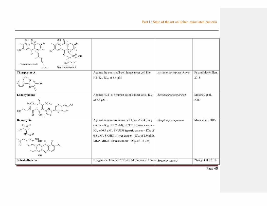
Part I : State of the art on lichen-associated bacteria
Page 45
O
OH
HO
O
O
ClBr
Napyradiomycin 2
O
OH
HO
O
O
H
OH
Br
H
BrNapyradiomycin 4
Thiasporine A
N
S
O
OH
NH2
Against the non-small-cell lung cancer cell line
H2122 , IC50 of 5.4 μM
Actinomycetospora chlora Fu and MacMillan,
2015
Lodopyridone
N
H3CSHN
HO
OOCH3
S
NN
O CH3
Cl
Against HCT-116 human colon cancer cells, IC50
of 3.6 µM.
Saccharomonospora sp Maloney et al.,
2009
Buanmycin
O
O
HN OHO
HO O
OHOH
OH
O OHO
Against human carcinoma cell lines: A594 (lung
cancer – IC50 of 1.7 µM), HCT116 (colon cancer –
IC50 of 0.9 µM), SNU638 (gastric cancer – IC50 of
0.8 µM), SKHEP1 (liver cancer – IC50 of 1.9 µM),
MDA-MB231 (breast cancer – IC50 of 1.2 µM)
Streptomyces cyaneus Moon et al., 2015
Spiroindimicins B: against cell lines: CCRF-CEM (human leukemia Streptomyces sp. Zhang et al., 2012

Part I : State of the art on lichen-associated bacteria
Page 46
N
HN
Cl Cl
NHR2
OCH3
OR1
Spiroindimicin B (R1 = CH3 , R2 = H)Spiroindimicin D (R1 = CH3 , R2 = COOCH3)
– IC50 of 4 µg/mL), B16 (mouse melanoma – IC50
of 5 µg/mL), H460 (human lung cancer – IC50 of
12 µg/mL).
D: against cell lines: B16 (mouse melanoma – IC50
of 20 µg/mL), H460 (human lung cancer – IC50 of
18 µg/mL), HepG2 (human hepatocellular liver
carcinoma – IC50 22 µg/mL).
Marthiapeptid
Against human carcinoma cell lines: SF-268
(glioblastoma cancer - IC50 of 0.38 ± 0.02 μM),
MCF-7 (breast cancer –IC50 of 0.43 ± 0.005 μM),
NCI-H460 ( lung cancer – IC50 of 0.47 ± 0.003
μM) and HepG2 (hepatocarcinoma cancer – IC50
of 0.52 ± 0.01 μM)
Marinactinospora
thermotolerans
Zhou et al., 2012
Indolocarbazole Compound 1, 2 and 3: against human prostate
PC-3 cancer cell line, IC50 of 0.8 ; 16.2 and 1.5
µM, respectively
Streptomyces sp. Qin et al., 2018

Part I : State of the art on lichen-associated bacteria
Page 47
NH
N
HN O
R
O
HOHN O
Indolocarbazole 1 (R = a)Indolocarbazole 2 (R = b)Indolocarbazole 3 (R = ac
(a)
O
HOHN O
(b)
O
HOHN
H
O(c)
Ieodoglucomide B
O
OHHOHO
O
O
O
HN
OH
O
H
O OHO
Against lung cancer and stomach cancer cell
lines, GI50 of 25.18 and 17.78 µg/mL,
respectively.
Bacillus licheniformis Tareq et al., 2012
Spiruchostatin C Burkholderia thailandensis Klausmeyer et al.,
2011
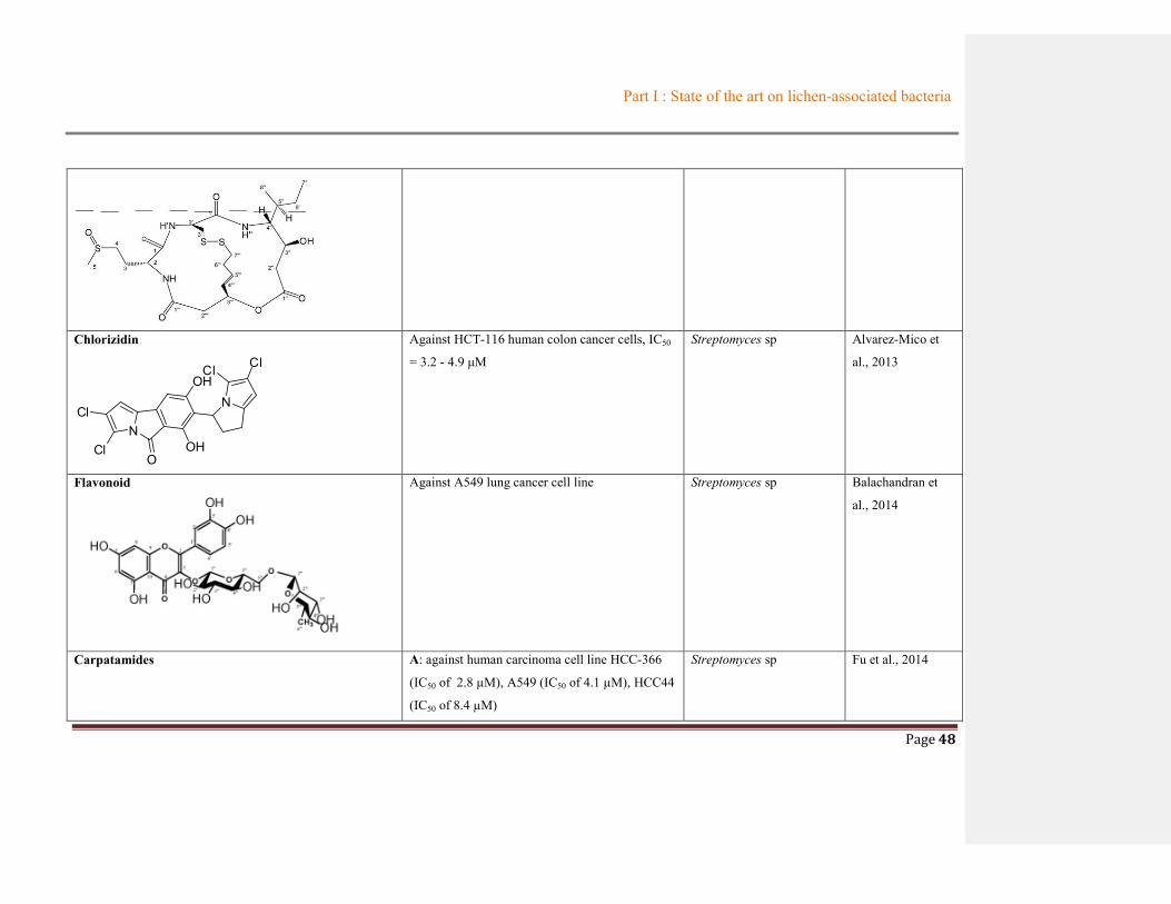
Part I : State of the art on lichen-associated bacteria
Page 48
Chlorizidin
NOH
OCl
Cl
OHN
Cl Cl
Against HCT-116 human colon cancer cells, IC50
= 3.2 - 4.9 μM
Streptomyces sp Alvarez-Mico et
al., 2013
Flavonoid
Against A549 lung cancer cell line Streptomyces sp Balachandran et
al., 2014
Carpatamides A: against human carcinoma cell line HCC-366
(IC50 of 2.8 µM), A549 (IC50 of 4.1 µM), HCC44
(IC50 of 8.4 µM)
Streptomyces sp Fu et al., 2014
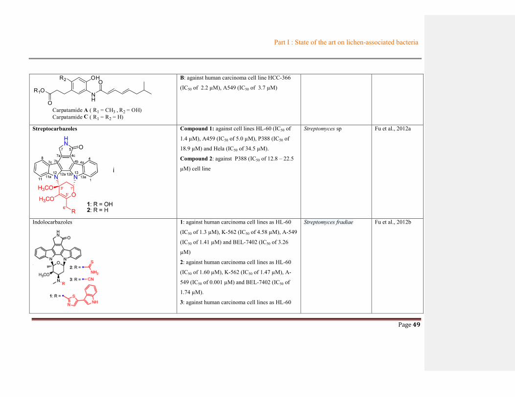
Part I : State of the art on lichen-associated bacteria
Page 49
R1O
O
R2
NH
OHO
Carpatamide A ( R1 = CH3 , R2 = OH)Carpatamide C ( R1 = R2 = H)
B: against human carcinoma cell line HCC-366
(IC50 of 2.2 µM), A549 (IC50 of 3.7 µM)
Streptocarbazoles
Compound 1: against cell lines HL-60 (IC50 of
1.4 µM), A459 (IC50 of 5.0 µM), P388 (IC50 of
18.9 µM) and Hela (IC50 of 34.5 µM).
Compound 2: against P388 (IC50 of 12.8 – 22.5
µM) cell line
Streptomyces sp Fu et al., 2012a
Indolocarbazoles
1: against human carcinoma cell lines as HL-60
(IC50 of 1.3 µM), K-562 (IC50 of 4.58 µM), A-549
(IC50 of 1.41 µM) and BEL-7402 (IC50 of 3.26
µM)
2: against human carcinoma cell lines as HL-60
(IC50 of 1.60 µM), K-562 (IC50 of 1.47 µM), A-
549 (IC50 of 0.001 µM) and BEL-7402 (IC50 of
1.74 µM).
3: against human carcinoma cell lines as HL-60
Streptomyces fradiae Fu et al., 2012b

Part I : State of the art on lichen-associated bacteria
Page 50
(IC50 of 0.13 µM), K-562 (IC50 of 0.43 µM), A-
549 (IC50 of 0.02 µM) and BEL-7402 (IC50 of
0.68 µM)
Mansouramycin B
N
O
ONH
Against lung cancer (LXFA 629 - IC50 1.63 µM)
and melanoma cells (MEXF 276 and MEXF 514,
IC50 0.33 and 0.05 µM)
Streptomyces sp Hawas et al., 2009
Summary
Throughout a screening of the data of metabolites isolated from bacterial cultures, bacteria showed a great potential source of cytotoxic
metabolites that might serve as useful leads in the development of new pharmaceutical agents. Moreover, the information from Table 1.6
highlighted that most of these cytotoxic compounds are alkaloids and they were frequently produced from Streptomyces genus, while
different bacterial genera are still unexploited. Thus, these data can give us helps in the selection of the genus producing cytotoxic
metabolites.
Part I : State of the art on lichen-associated bacteria
Page 51
REFERENCES Alvarez-Mico, X., Jensen, P.R., Fenical, W., Hughes, C.C., 2013. Chlorizidine, a Cytotoxic 5H-Pyrrolo[2,1-a]isoindol-
5-one-Containing Alkaloid from a Marine Streptomyces sp. Org. Lett. 15, 988–991.
https://doi.org/10.1021/ol303374e
Armstrong, R., 2006. Seasonal growth of the crustose lichen Rhizocarpon geographicum (L.) DC. in South Gwynedd,
Wales [WWW Document]. URL https://research.aston.ac.uk/portal/en/researchoutput/seasonal-growth-of-the-
crustose-lichen-rhizocarpon-geographicum-l-dc-in-south-gwynedd-wales%28e71ef783-51fa-4a86-bf43-
daca4c892686%29.html (accessed 12.23.15).
Armstrong, R.A., 2005. Radial Growth of Rhizocarpon Section Rhizocarpon Lichen Thalli over Six Years at
Snoqualmie Pass in the Cascade Range, Washington State. Arctic, Antarctic, and Alpine Research 37, 411–415.
Armstrong, R.A., 1983. Growth Curve of the Lichen Rhizocarpon Geographicum. New Phytologist 94, 619–622.
https://doi.org/10.1111/j.1469-8137.1983.tb04870.x
Armstrong, R.A., Smith, S.N., 1996. Experimental studies of hypothallus growth in the lichen Rhizocarpon
geographicum. New Phytologist 132, 123–126. https://doi.org/10.1111/j.1469-8137.1996.tb04517.x
Aschenbrenner, I.A., Cardinale, M., Berg, G., Grube, M., 2014. Microbial cargo: do bacteria on symbiotic propagules
reinforce the microbiome of lichens? Environ Microbiol 16, 3743–3752. https://doi.org/10.1111/1462-
2920.12658
Balachandran, C., Sangeetha, B., Duraipandiyan, V., Raj, M.K., Ignacimuthu, S., Al-Dhabi, N.A., Balakrishna, K.,
Parthasarathy, K., Arulmozhi, N.M., Arasu, M.V., 2014. A flavonoid isolated from Streptomyces sp.
(ERINLG-4) induces apoptosis in human lung cancer A549 cells through p53 and cytochrome c release caspase
dependant pathway. Chemico-Biological Interactions 224, 24–35. https://doi.org/10.1016/j.cbi.2014.09.019
Bates, S.T., Cropsey, G.W.G., Caporaso, J.G., Knight, R., Fierer, N., 2011. Bacterial Communities Associated with the
Lichen Symbiosis. Appl Environ Microbiol 77, 1309–1314. https://doi.org/10.1128/AEM.02257-10
Belnap, J., 2001. Factors Influencing Nitrogen Fixation and Nitrogen Release in Biological Soil Crusts, in: Belnap,
P.D.J., Lange, P.D.D. h c O.L. (Eds.), Biological Soil Crusts: Structure, Function, and Management, Ecological
Studies. Springer Berlin Heidelberg, pp. 241–261. https://doi.org/10.1007/978-3-642-56475-8_19

Part I : State of the art on lichen-associated bacteria
Page 52
Bench, G., Clark, B.M., Mangelson, N.F., St. Clair, L.L., Rees, L.B., Grant, P.G., Southon, J.R., 2001. Accurate
lifespan estimates cannot be obtained from14C profiles in the crustose lichen Rhizocarpon geographicum (L.)
DC. The Lichenologist 33, 539–542. https://doi.org/10.1006/lich.2001.0353
Benedict, J.B., 2009. A Review of Lichenometric Dating and Its Applications to Archaeology. American Antiquity 74,
143–172. https://doi.org/10.2307/25470542
Benedict, J.B., 1988. Techniques in Lichenometry: Identifying the Yellow Rhizocarpons. Arctic and Alpine Research
20, 285–291. https://doi.org/10.2307/1551260
Bjelland, T., Grube, M., Hoem, S., Jorgensen, S.L., Daae, F.L., Thorseth, I.H., Øvreås, L., 2011. Microbial
metacommunities in the lichen–rock habitat. Environmental Microbiology Reports 3, 434–442.
https://doi.org/10.1111/j.1758-2229.2010.00206.x
Boustie, J., Grube, M., 2005. Lichens—a promising source of bioactive secondary metabolites. Plant Genetic Resources 3, 273–287. https://doi.org/10.1079/PGR200572
Boustie, J., Tomasi, S., Grube, M., 2010. Bioactive lichen metabolites: alpine habitats as an untapped source.
Phytochem Rev 10, 287–307. https://doi.org/10.1007/s11101-010-9201-1
Bradwell, T., 2010. Studies on the Growth of Rhizocarpon Geographicum in Nw Scotland, and Some Implications for
Lichenometry. Geografiska Annaler: Series A, Physical Geography 92, 41–52. https://doi.org/10.1111/j.1468-
0459.2010.00376.x
Bradwell, T., Armstrong, R.A., 2007. Growth rates of Rhizocarpon geographicum lichens: a review with new data
from Iceland. J. Quaternary Sci. 22, 311–320. https://doi.org/10.1002/jqs.1058
Büdel B. and Scheidegger S. 2008, Thallus morphology and anatomy (Chapter 4). p 40. Lichen Biology, Edited by
Nash S. Thomas, Cambridge university Press, 486.
Cardinale, M., Castro, J.V. de, Müller, H., Berg, G., Grube, M., 2008. In situ analysis of the bacterial community
associated with the reindeer lichen Cladonia arbuscula reveals predominance of Alphaproteobacteria. FEMS
Microbiology Ecology 66, 63–71. https://doi.org/10.1111/j.1574-6941.2008.00546.x
Cardinale, M., Grube, M., Berg, G., 2011. Frondihabitans cladoniiphilus sp. nov., an actinobacterium of the family
Microbacteriaceae isolated from lichen, and emended description of the genus Frondihabitans. International
Journal of Systematic and Evolutionary Microbiology 61, 3033–3038. https://doi.org/10.1099/ijs.0.028324-0

Part I : State of the art on lichen-associated bacteria
Page 53
Cardinale, M., Grube, M., Castro, J.V., Müller, H., Berg, G., 2012a. Bacterial taxa associated with the lung lichen
Lobaria pulmonaria are differentially shaped by geography and habitat. FEMS Microbiol Lett 329, 111–115.
https://doi.org/10.1111/j.1574-6968.2012.02508.x
Cardinale, M., Puglia, A.M., Grube, M., 2006. Molecular analysis of lichen-associated bacterial communities. FEMS
Microbiol Ecol 57, 484–495. https://doi.org/10.1111/j.1574-6941.2006.00133.x
Cardinale Massimiliano, Steinová Jana, Rabensteiner Johannes, Berg Gabriele, Grube Martin, 2012b. Age, sun and
substrate: triggers of bacterial communities in lichens. Environmental Microbiology Reports 4, 23–28.
https://doi.org/10.1111/j.1758-2229.2011.00272.x
Cheenpracha, S., Vidor, N.B., Yoshida, W.Y., Davies, J., Chang, L.C., 2010. Coumabiocins A−F, Aminocoumarins
from an Organic Extract of Streptomyces sp. L-4-4. J. Nat. Prod. 73, 880–884.
https://doi.org/10.1021/np900843b
Cho, J.Y., Williams, P.G., Kwon, H.C., Jensen, P.R., Fenical, W., 2007. Lucentamycins A−D, Cytotoxic Peptides
from the Marine-Derived Actinomycete Nocardiopsis lucentensis. J. Nat. Prod. 70, 1321–1328.
https://doi.org/10.1021/np070101b
Davies, J., Wang, H., Taylor, T., Warabi, K., Huang, X.-H., Andersen, R.J., 2005. Uncialamycin, A New Enediyne
Antibiotic. Org. Lett. 7, 5233–5236. https://doi.org/10.1021/ol052081f
Debbab, A., Aly, A.H., Lin, W.H., Proksch, P., 2010. Bioactive Compounds from Marine Bacteria and Fungi. Microb
Biotechnol 3, 544–563. https://doi.org/10.1111/j.1751-7915.2010.00179.x
Dobson S. Frank, 2005. Lichen-A illustrated guide to the British and Irish species. p 386. The Richmond publishing
Co.Ltd. 480
Elix J.A. and Stocker-Wörgötte, 2008, Biochemistry and secondary metabolites (Chapter 7), Lichen Biology, Edited
by Nash S. Thomas, Cambridge university Press. 486
Esposito, A., Ciccazzo, S., Borruso, L., Zerbe, S., Daffonchio, D., Brusetti, L., 2013. A Three-Scale Analysis of
Bacterial Communities Involved in Rocks Colonization and Soil Formation in High Mountain Environments.
Curr Microbiol 67, 472–479. https://doi.org/10.1007/s00284-013-0391-9
Farnaes, L., Coufal, N.G., Kauffman, C.A., Rheingold, A.L., DiPasquale, A.G., Jensen, P.R., Fenical, W., 2014.
Napyradiomycin Derivatives, Produced by a Marine-Derived Actinomycete, Illustrate Cytotoxicity by
Induction of Apoptosis. J. Nat. Prod. 77, 15–21. https://doi.org/10.1021/np400466j

Part I : State of the art on lichen-associated bacteria
Page 54
Fu, P., Johnson, M., Chen, H., Posner, B.A., MacMillan, J.B., 2014. Carpatamides A–C, Cytotoxic Arylamine
Derivatives from a Marine-Derived Streptomyces sp. J Nat Prod 77, 1245–1248.
https://doi.org/10.1021/np500207p
Fu, P., MacMillan, J.B., 2015. Thiasporines A–C, Thiazine and Thiazole Derivatives from a Marine-Derived
Actinomycetospora chlora. J. Nat. Prod. 78, 548–551. https://doi.org/10.1021/np500929z
Fu, P., Yang, C., Wang, Y., Liu, P., Ma, Y., Xu, L., Su, M., Hong, K., Zhu, W., 2012a. Streptocarbazoles A and B,
Two Novel Indolocarbazoles from the Marine-Derived Actinomycete Strain Streptomyces sp. FMA. Org. Lett.
14, 2422–2425. https://doi.org/10.1021/ol3008638
Fu, P., Zhuang, Y., Wang, Y., Liu, P., Qi, X., Gu, K., Zhang, D., Zhu, W., 2012b. New Indolocarbazoles from a
Mutant Strain of the Marine-Derived Actinomycete Streptomyces fradiae 007M135. Org. Lett. 14, 6194–6197.
https://doi.org/10.1021/ol302940y
Grube, M., Cardinale, M., de Castro, J.V., Müller, H., Berg, G., 2009a. Species-specific structural and functional
diversity of bacterial communities in lichen symbioses. ISME J 3, 1105–1115.
https://doi.org/10.1038/ismej.2009.63
Grube, M., Berg, G., 2009b. Microbial consortia of bacteria and fungi with focus on the lichen symbiosis. Fungal
Biology Reviews 23, 72–85. https://doi.org/10.1016/j.fbr.2009.10.001
Grube, M., Berg, G., S. Andrésson, Ó., Vilhelmsson, O., Dyer, P.S., Miao, V.P.W., 2013. Lichen Genomics, in:
Francisrtin (Ed.), The Ecological Genomics of Fungi. John Wiley & Sons, Inc, pp. 191–212.
Grube, M., Cardinale, M., Berg, G., 2012a. 17 Bacteria and the Lichen Symbiosis, in: Hock, B. (Ed.), Fungal
Associations, The Mycota. Springer Berlin Heidelberg, pp. 363–372. https://doi.org/10.1007/978-3-642-30826-
0_17
Grube, M., Cernava, T., Soh, J., Fuchs, S., Aschenbrenner, I., Lassek, C., Wegner, U., Becher, D., Riedel, K., Sensen,
C.W., Berg, G., 2015. Exploring functional contexts of symbiotic sustain within lichen-associated bacteria by
comparative omics. ISME J 9, 412–424. https://doi.org/10.1038/ismej.2014.138
Grube, Martin, Köberl, M., Lackner, S., Berg, C., Berg, G., 2012b. Host–parasite interaction and microbiome
response: effects of fungal infections on the bacterial community of the Alpine lichen Solorina crocea. FEMS
Microbiology Ecology 82, 472–481. https://doi.org/10.1111/j.1574-6941.2012.01425.x

Part I : State of the art on lichen-associated bacteria
Page 55
Hawas, U.W., Shaaban, M., Shaaban, K.A., Speitling, M., Maier, A., Kelter, G., Fiebig, H.H., Meiners, M., Helmke,
E., Laatsch, H., 2009. Mansouramycins A−D, Cytotoxic Isoquinolinequinones from a Marine Streptomycete. J.
Nat. Prod. 72, 2120–2124. https://doi.org/10.1021/np900160g
Haworth, L.A., Calkin, P.E., Ellis, J.M., 1986. Direct Measurement of Lichen Growth in the Central Brooks Range,
Alaska, U.S.A., and Its Application to Lichenometric Dating. Arctic and Alpine Research 18, 289–296.
https://doi.org/10.2307/1550886
Hodkinson, B.P., Lutzoni, F., 2009. A microbiotic survey of lichen-associated bacteria reveals a new lineage from the
Rhizobiales. Symbiosis 49, 163–180. https://doi.org/10.1007/s13199-009-0049-3
Huneck S. and Yoshimura I., 1996. Identification of lichen substances, p. 13 Springer,493
Ihlen, P.G., 2004. Taxonomy of the non-yellow species of Rhizocarpon (Rhizocarpaceae, lichenized Ascomycota) in
the Nordic countries, with hyaline and muriform ascospores. Mycol. Res. 108, 533–570.
Ihlen, P.G., Ekman, S., 2002. Outline of phylogeny and character evolution in Rhizocarpon (Rhizocarpaceae,
lichenized Ascomycota) based on nuclear ITS and mitochondrial SSU ribosomal DNA sequences. Biological
Journal of the Linnean Society 77, 535–546. https://doi.org/10.1046/j.1095-8312.2002.00127.x
Innes, J.L., 1985. A standard Rhizocarpon nomenclature for lichenometry. Boreas 14, 83–85.
https://doi.org/10.1111/j.1502-3885.1985.tb00890.x
Kim, J., Park, E.J., 2002. Cytotoxic anticancer candidates from natural resources. Curr Med Chem Anticancer Agents
2, 485–537.
Kim, M.-K., Park, H., Oh, T.-J., 2014. Antibacterial and antioxidant capacity of polar microorganisms isolated from
Arctic lichen Ochrolechia sp. Pol. J. Microbiol. 63, 317–322.
Kim, M.K., Park, H., Oh, T.J., 2012. Antibacterial Properties Associated with Microorganisms Isolated from Arctic
Lichens. Korean Journal of Microbiology and Biotechnology.
Klausmeyer, P., Shipley, S., Zuck, K.M., McCloud, T.G., 2011. Histone Deacetylase Inhibitors from Burkholderia
Thailandensis. J Nat Prod 74, 2039–2044. https://doi.org/10.1021/np200532d
Liba, C. M., Ferrara, F. I. S., Manfio, G. P., Fantinatti-Garboggini, F., Albuquerque, R. C., Pavan, C., Ramos, P. L.,
Moreira-Filho, C. A., Barbosa, H. R., 2006. Nitrogen-fixing chemo-organotrophic bacteria isolated from
cyanobacteria-deprived lichens and their ability to solubilize phosphate and to release amino acids and

Part I : State of the art on lichen-associated bacteria
Page 56
phytohormones. Journal of Applied Microbiology 101, 1076–1086. https://doi.org/10.1111/j.1365-
2672.2006.03010.x
Liu, C., Jiang, Y., Lei, H., Chen, X., Ma, Q., Han, L., Huang, X., 2017. Four New Nanaomycins Produced by
Streptomyces hebeiensis Derived from Lichen. Chem. Biodivers. 14. https://doi.org/10.1002/cbdv.201700057
Lucas, X., Senger, C., Erxleben, A., Grüning, B.A., Döring, K., Mosch, J., Flemming, S., Günther, S., 2013.
StreptomeDB: a resource for natural compounds isolated from Streptomyces species. Nucl. Acids Res. 41,
D1130–D1136. https://doi.org/10.1093/nar/gks1253
Maloney, K.N., MacMillan, J.B., Kauffman, C.A., Jensen, P.R., DiPasquale, A.G., Rheingold, A.L., Fenical, W.,
2009. Lodopyridone, a Structurally Unprecedented Alkaloid from a Marine Actinomycete. Org. Lett. 11, 5422–
5424. https://doi.org/10.1021/ol901997k
McCarthy, D.P., Henry, N., 2012. Measurement of growth in the lichen Rhizocarpon geographicum using a new
photographic technique. The Lichenologist 44, 679–693. https://doi.org/10.1017/S0024282912000175
McCarthy, P., Elix, J., 2014. The lichen genus Rhizocarpon in mainland Australia. Telopea 16, 195–211.
https://doi.org/10.7751/telopea20148124
Mioso, R., Marante, F.J.T., Bezerra, R. de S., Borges, F.V.P., Santos, B.V. de O., Laguna, I.H.B. de, 2017. Cytotoxic
Compounds Derived from Marine Sponges. A Review (2010–2012). Molecules 22, 208.
https://doi.org/10.3390/molecules22020208
Moon, K., Chung, B., Shin, Y., Rheingold, A.L., Moore, C.E., Park, S.J., Park, S., Lee, S.K., Oh, K.-B., Shin, J., Oh,
D.-C., 2015. Pentacyclic Antibiotics from a Tidal Mud Flat-Derived Actinomycete. J. Nat. Prod. 78, 524–529.
https://doi.org/10.1021/np500736b
Motohashi, K., Takagi, M., Yamamura, H., Hayakawa, M., Shin-ya, K., 2010. A new angucycline and a new
butenolide isolated from lichen-derived Streptomyces spp. J Antibiot 63, 545–548.
https://doi.org/10.1038/ja.2010.94
Muggia, L., Klug, B., Berg, G., Grube, M., 2013. Localization of bacteria in lichens from Alpine soil crusts by
fluorescence in situ hybridization. Applied Soil Ecology 68, 20–25.
https://doi.org/10.1016/j.apsoil.2013.03.008
Noël, A., Ferron, S., Rouaud, I., Gouault, N., Hurvois, J.-P., Tomasi, S., 2017. Isolation and Structure Identification of
Novel Brominated Diketopiperazines from Nocardia ignorata—A Lichen-Associated Actinobacterium.
Molecules 22, 371. https://doi.org/10.3390/molecules22030371

Part I : State of the art on lichen-associated bacteria
Page 57
O’Neal, M.A., Schoenenberger, K.R., 2003. A Rhizocarpon geographicum growth curve for the Cascade Range of
Washington and northern Oregon, usa. Quaternary Research 60, 233–241. https://doi.org/10.1016/S0033-
5894(03)00104-2
Parrot, D., Legrave, N., Delmail, D., Grube, M., Suzuki, M.T., Tomasi, S., 2016a. Review – Lichen-associated
bacteria as a hot spot of chemodiversity: focus on uncialamycin, a promising compound for future medicinal
applications. Planta Medica 82, 1143–1152. https://doi.org/10.1055/s-0042-105571
Parrot, D., Legrave, N., Intertaglia, L., Rouaud, I., Legembre, P., Grube, M., Suzuki, M.T., Tomasi, S., 2016b.
Cyaneodimycin, a Bioactive Compound Isolated from the Culture of Streptomyces cyaneofuscatus Associated
with Lichina confinis. Eur. J. Org. Chem. 2016, 3977–3982. https://doi.org/10.1002/ejoc.201600252
Parrot, D., Peresse, T., Hitti, E., Carrie, D., Grube, M., Tomasi, S., 2015. Qualitative and Spatial Metabolite Profiling of Lichens by a LC–MS Approach Combined With Optimised Extraction. Phytochemical Analysis 26, 23–33. https://doi.org/10.1002/pca.2532
Printzen, C., Fernández-Mendoza, F., Muggia, L., Berg, G., Grube, M., 2012. Alphaproteobacterial communities in
geographically distant populations of the lichen Cetraria aculeata. FEMS Microbiology Ecology 82, 316–325.
https://doi.org/10.1111/j.1574-6941.2012.01358.x
Proctor, M.C.F., 1983. Sizes and Growth-Rates of Thalli of the Lichen Rhizocarpon Geographicum on the Moraines of
the Glacier De Valsorey, Valais, Switzerland. The Lichenologist 15, 249–261.
https://doi.org/10.1017/S0024282983000389
Qin, L.-L., Zhou, B., Ding, W., Ma, Z., 2018. Bioactive metabolites from marine-derived Streptomyces sp. A68 and its
Rifampicin resistant mutant strain R-M1. Phytochemistry Letters 23, 46–51.
https://doi.org/10.1016/j.phytol.2017.11.002
Rai, A. N., 2002. Cyanolichens: Nitrogen Metabolism, in: Rai, Amar N., Bergman, B., Rasmussen, U. (Eds.),
Cyanobacteria in Symbiosis. Springer Netherlands, pp. 97–115. https://doi.org/10.1007/0-306-48005-0_6
Ruiz-Torres, V., Encinar, J.A., Herranz-López, M., Pérez-Sánchez, A., Galiano, V., Barrajón-Catalán, E., Micol, V.,
2017. An Updated Review on Marine Anticancer Compounds: The Use of Virtual Screening for the Discovery
of Small-Molecule Cancer Drugs. Molecules 22, 1037. https://doi.org/10.3390/molecules22071037
Schneider, T., Schmid, E., de Castro, J.V., Cardinale, M., Eberl, L., Grube, M., Berg, G., Riedel, K., 2011. Structure
and function of the symbiosis partners of the lung lichen (Lobaria pulmonaria L. Hoffm.) analyzed by
metaproteomics. Proteomics 11, 2752–2756. https://doi.org/10.1002/pmic.201000679

Part I : State of the art on lichen-associated bacteria
Page 58
Selbmann, L., Zucconi, L., Ruisi, S., Grube, M., Cardinale, M., Onofri, S., 2009. Culturable bacteria associated with
Antarctic lichens: affiliation and psychrotolerance. Polar Biol 33, 71–83. https://doi.org/10.1007/s00300-009-
0686-2
Shukla, V., Joshi, G.P., Rawat, M.S.M., 2010. Lichens as a potential natural source of bioactive compounds: a review.
Phytochem Rev 9, 303–314. https://doi.org/10.1007/s11101-010-9189-6
Sigurbjörnsdóttir, M.A., Heiðmarsson, S., Jónsdóttir, A.R., Vilhelmsson, O., 2014. Novel bacteria associated with
Arctic seashore lichens have potential roles in nutrient scavenging. Can. J. Microbiol. 60, 307–317.
https://doi.org/10.1139/cjm-2013-0888
Spribille, T., Tuovinen, V., Resl, P., Vanderpool, D., Wolinski, H., Aime, M.C., Schneider, K., Stabentheiner, E.,
Toome-Heller, M., Thor, G., Mayrhofer, H., Johannesson, H., McCutcheon, J.P., 2016. Basidiomycete yeasts in
the cortex of ascomycete macrolichens. Science 353, 488–492. https://doi.org/10.1126/science.aaf8287
Tareq, F.S., Kim, J.H., Lee, M.A., Lee, H.-S., Lee, Y.-J., Lee, J.S., Shin, H.J., 2012. Ieodoglucomides A and B from a
Marine-Derived Bacterium Bacillus licheniformis. Org. Lett. 14, 1464–1467. https://doi.org/10.1021/ol300202z
Wang, Y., Zheng, Y., Wang, X., Wei, X., Wei, J., 2016. Lichen-Associated Fungal Community in Hypogymnia
hypotrypa (Parmeliaceae, Ascomycota) Affected by Geographic Distribution and Altitude. Front Microbiol 7.
https://doi.org/10.3389/fmicb.2016.01231
Williams, D.E., Bottriell, H., Davies, J., Tietjen, I., Brockman, M.A., Andersen, R.J., 2015. Unciaphenol, an
Oxygenated Analogue of the Bergman Cyclization Product of Uncialamycin Exhibits Anti-HIV Activity. Org.
Lett. 17, 5304–5307. https://doi.org/10.1021/acs.orglett.5b02664
Williams, D.E., Davies, J., Patrick, B.O., Bottriell, H., Tarling, T., Roberge, M., Andersen, R.J., 2008. Cladoniamides
A−G, Tryptophan-Derived Alkaloids Produced in Culture by Streptomyces uncialis. Org. Lett. 10, 3501–3504.
https://doi.org/10.1021/ol801274c
Zhang, W., Liu, Z., Li, S., Yang, T., Zhang, Q., Ma, L., Tian, X., Zhang, H., Huang, C., Zhang, S., Ju, J., Shen, Y.,
Zhang, C., 2012. Spiroindimicins A–D: New Bisindole Alkaloids from a Deep-Sea-Derived Actinomycete.
Org. Lett. 14, 3364–3367. https://doi.org/10.1021/ol301343n
Zhou, X., Huang, H., Chen, Y., Tan, J., Song, Y., Zou, J., Tian, X., Hua, Y., Ju, J., 2012. Marthiapeptide A, an Anti-
infective and Cytotoxic Polythiazole Cyclopeptide from a 60 L Scale Fermentation of the Deep Sea-Derived
Marinactinospora thermotolerans SCSIO 00652. J. Nat. Prod. 75, 2251–2255.
https://doi.org/10.1021/np300554f

Part I : State of the art on lichen-associated bacteria
Page 59
Webography
http://www.lichens.lastdragon.org/faq/lichenthallustypes.html
http://www.lichensmaritimes.org
http://www.afl-lichenologie.fr/Photos_AFL/Photos_AFL_R/Rhizocarpon_geographicum.ht
http://www.imlichenit.com/imlichenit/about.html

The objectives of the work
Page 60
1.6. THE OBJECTIVES OF THE WORK
According to the state of the art detailed in the previous part, we have decided to focus our
research work on the isolation of bioactive novel metabolites from interesting and original sources
such as lichen-associated bacteria which are poorly studied. The first step was the selection of the
lichen which could possess specific and novel bacterial partners. Due to the ubiquitary presence of
the lichen Rhizocarpon geographicum in coastal areas which could afford particular environments
to their hosted organisms (Delmail et al., 2013) and due to the lack of knowledge of culturable
bacterial communities associated with this organism, we decided to select this lichen to study its
associated bacteria via a culture-based approach. These data will be presented in chapter 1 of part
II of this manuscript.
After isolation of various pure strains from R. geographicum, a species Paenibacillus
odorifer with special characteristics known through several references would be preferred for
studying its ability to produce secondary metabolites of interest (reported in chapter 2). We have
focused our research on the discovery of potential cytotoxic compounds (tested on two HaCaT and
B16 cell lines) as we have seen previously the particular ability of some bacteria to produce novel
and potent cytotoxic metabolites possessing original skeletons. The optimization of the culture in
order to increase the production of bioactive compounds has then been applied for this selected
strain and described in chapter 3. Two optimization steps using various vessels (bioreactor,
Erlenmeyer flask) have been undertaken. The parameters selected for this process included pH of
medium, temperature, stirring, inoculum ratio, and the quantity and bioactivity of extracts from the
broth of the culture as well. The best cultured conditions were applied for a large volume culture as
explained in chapter 3.
Finally, the isolation, identification as well as the evaluation of bioactivities of a number of
pure bacterial compounds were performed and has detailed in chapter 4. The chapter 5 was
displayed conclusions and perspectives. The chapter 6 finally reported the materials and methods
used during this work.

The objectives of the work
Page 61
References Delmail, D., Grube, M., Parrot, D., Cook-Moreau, J., Boustie, J., Labrousse, P., Tomasi, S., 2013. Halotolerance in
Lichens: Symbiotic Coalition Against Salt Stress, in: Ecophysiology and Responses of Plants under Salt Stress.
Springer, New York, NY, pp. 115–148. https://doi.org/10.1007/978-1-4614-4747-4_4

Page 62
PART II: RESULTS

Page 63
PART II. RESULTS
This part displayed the results of this PhD work divided into distinct 5 sections which are
reported either as articles (in progress) or as classical parts.
In the first part, the isolation and identification of thirteen pure strains from Rhizocarpon
geographicum will be presented. This result would be proposed as an article in progress. (Chapter
1)
The second section detailed the selection of bacterial species with the objective to produce
metabolites. It is classically reported in this thesis as Chapter 2. After the strain selected, the
optimization to find the best conditions for the culture will be displayed in Chapter 3 in a classical
form.
The metabolites isolated will be introduced in Chapter 4 including a fraction of
polysaccharide, two tert-butylphenol compounds and a cytotoxic alkaloid that are presented as
articles (in progress), while other metabolites with no cytotoxicity are displayed as a part in the
thesis.
After conclusions and perspectives (Chapter 5), the final section will be the description of
materials and methods used in this work (Chapter 6).
Page 64
Chapter 1: ISOLATION OF BACTERIAL STRAINS
FROM R. GEOGRAPHICUM

Chapter 1 : Isolation of bacterial strains from R. geographicum
Page 65
CHAPTER 1: ISOLATION OF BACTERIAL STRAINS FROM R. GEOGRAPHICUM
The results of chapter 1 were reported as an article (in preparation).
Culturable bacterial communities from the lichen Rhizocarpon greographicum
--------------------------------------------------------------------------------------------------------------
An article in progress
--------------------------------------------------------------------------------------------------------------
Nguyen Thi Bach Le, Delmail David, Tomasi Sophie
Univ Rennes, CNRS, ISCR – UMR 6226, F-35000 Rennes, France
Abstract
Eleven microorganisms associated with the lichen Rhizocarpon geographicum were isolated using
a culture-based method. They were identified based on their 16s rRNA gene sequence analysis and
led to their affiliation as 11 microbial strains along with 8 ones belonged to the Bacteria kingdom.
The results first demonstrated that the cultured bacterial communities in Rhizocarpon
geographicum were dominated by Bacilli class (Firmicutes phylum) followed by Proteobacteria.
INTRODUCTION
Many studies in previous decades had not admitted bacterial communities as an important
part of lichens which were only considered as a result of the symbiotic association between a
mycobiont (known as fungus) and a photobiont (alga or cyanobacterium). These previous studies,
therefore, cannot address the question why lichens can grow on extremely nutrient-poor life
conditions (Brodo and Irwin 1973) such as rocky surface, frosted polar or barren desert where
fungi or algae cannot survive alone. Several recent studies have demonstrated that microorganisms
mostly supplied food for lichens by fixing nitrogen, carbon dioxide in the atmosphere to produce a
substantial source of crucial nutrients (González et al. 2005; Liba et al. 2006; Hodkinson et al.

Chapter 1 : Isolation of bacterial strains from R. geographicum
Page 66
2012; Grube et al. 2015). As a result, lichens, forming the symbiotic association between fungi,
algae/cyanobacteria and with bacterial cohabitation, are one of the most successful life-form on the
earth which can grow in any harshly environmental conditions (Boustie and Grube 2005; Boustie,
Tomasi, and Grube 2010). Moreover, bacterial communities played significant roles in the ecology
of lichens (Cardinale, Puglia, and Grube 2006; Grube and Berg, 2009; Liba et al. 2006 ,
Hodkinson and Lutzoni 2009) and recent years, these communities became an interesting subject
for researches in the field of Lichenology.
Several studies demonstrated that the majority of lichen-associated bacteria live on or near
the fungal surfaces of lichen thalli and they formed biofilm-like coatings on lichen surfaces
(Cardinale et al. 2008; Grube and Berg 2009). As a result, bacteria provide to lichens nutrients by
fixing nitrogen in the atmosphere whereas green algae cannot (Grube et al., 2015). Therefore, this
hypothesis could address the question why lichen can grow in any habitat and geographic area on
the Earth.
These microorganisms were often described by culture or unculture-based methods. While
the latter method highlighted a great diversity of bacteria in each lichen thallus (Cardinale, Puglia,
and Grube 2006), the culture-based methods cannot collect uncultured bacteria and cannot afford
the identification of the vast majority of microorganisms in the laboratory (Amann, Ludwig, and
Schleifer 1995). It may lead to false conclusions about the abundance and importance of certain
bacteria in nature. However, the significant advantages of culture-based methods are the abilities to
isolate bacterial strains as well as to produce metabolites from these populations and to check
bioactivities of the substances produced.
Rhizocarpon geographicum, one of the most popular species of the crustose lichen family
(Ihlen et al., 2004) grows mainly on nutrient-poor substrates as rock surface (especially rocky
promontory) or under harsh coastal conditions (Armstrong and Bradwell 2010, Armstrong and
Smith 2009). However, the information on bacterial communities from this lichen remains still
poor. One unusual study on microorganism communities of Rhizocarpon geographicum was
performed by Bjelland and co-workers (2011). The results provided by this literature using
uncultured-based method indicated that Alphaproteobacteria was predominant in bacterial
population associated with Rhizocarpon geographicum.

Chapter 1 : Isolation of bacterial strains from R. geographicum
Page 67
In this study, we first report the bacterial communities associated with R. geographicum by a
culture-based method and identify selected strains at the genus level by partial 16S rRNA
sequencing. Interestingly, the results from our study illustrated a dominance of Bacilli class instead
of Alphaproteobacteria class which is previously presented as dominant in previous studies
performed on microorganisms of crustose lichens (Bjelland et al., 2011; Bates et al., 2011).
MATERIALS AND METHODS
Sample collection
The lichen samples of Rhizocarpon geographicum were collected at a small rocky
promontory in Bretagne, France near Saint-Malo in February 2015 as reported in Figures 1 and 2.
Three points of the collection in the same site have been labeled using specific colors following the
distance to the sea (blue for the close site, pink one for the medium distance then green one for the
farthest site). At each location, fragments of lichen species were collected together with rock
surface below them using a sterile hammer and a chisel. The samples were transferred into sterile
Eppendorf® tubes before being brought to the laboratory.
Figure 1: The sites of R. geographicum harvest and their associated color
R. geographicum labeled Green
R. geographicum labeled Pink
R.geographicum labeled Blue
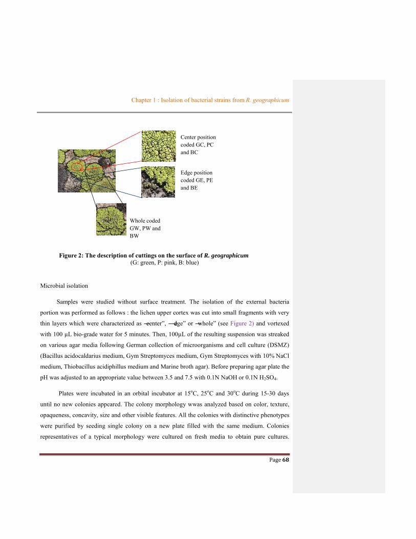
Chapter 1 : Isolation of bacterial strains from R. geographicum
Page 68
Figure 2: The description of cuttings on the surface of R. geographicum
(G: green, P: pink, B: blue)
Microbial isolation
Samples were studied without surface treatment. The isolation of the external bacteria
portion was performed as follows : the lichen upper cortex was cut into small fragments with very
thin layers which were characterized as ―center‖, ―edge‖ or ―whole‖ (see Figure 2) and vortexed
with 100 µL bio-grade water for 5 minutes. Then, 100µL of the resulting suspension was streaked
on various agar media following German collection of microorganisms and cell culture (DSMZ)
(Bacillus acidocaldarius medium, Gym Streptomyces medium, Gym Streptomyces with 10% NaCl
medium, Thiobacillus acidiphillus medium and Marine broth agar). Before preparing agar plate the
pH was adjusted to an appropriate value between 3.5 and 7.5 with 0.1N NaOH or 0.1N H2SO4.
Plates were incubated in an orbital incubator at 15oC, 25oC and 30oC during 15-30 days
until no new colonies appeared. The colony morphology wwas analyzed based on color, texture,
opaqueness, concavity, size and other visible features. All the colonies with distinctive phenotypes
were purified by seeding single colony on a new plate filled with the same medium. Colonies
representatives of a typical morphology were cultured on fresh media to obtain pure cultures.
Center position coded GC, PC and BC
Edge position coded GE, PE and BE
Whole coded GW, PW and BW

Chapter 1 : Isolation of bacterial strains from R. geographicum
Page 69
Finally, thirteen pure strains were collected and identified by 16S rRNA gene sequencing analysis.
The process of isolation was demonstrated in Scheme.1.
Scheme 1: Process of isolation of microbial strains from R. geographicum
The cryopreservation was prepared by suspending a loop full of colonies in 1.0 mL of the 47.5%
(v/v) glycerol and 5% (v/v) of dimethyl sulfoxide (DMSO) in 50% (v/v) of stetile water and stored
at -800C in the laboratory of the University of Rennes 1 (CORINT team).
The isolated strains were coded following the sites of the lichen harvest, the position of cutting on
the surface of R. geographicum, the growth medium and their morphological characteristics. For
instance, the strain coded GC-MA-OP means that it was isolated from lichen labeled Green, cut at
the center position on the surface of the lichen, cultured on Marine agar medium, and with
characteristics as orange-pink color (Table 1).
Phylogenetic analysis
Isolated strains were selected for identification based on colony morphology and phylogenetic
analysis. For DNA extraction from pure cultures, a Kit Promega® was used according to the
manufacturer’s instruction and performed by L. Intertaglia (Plateform, Banyuls/Mer, France).
Extracted DNA was PCR amplified in an ABI- Applied Biosystems thermocycler (Observatoire
Océanologique de Banyuls sur Mer, France) using the universal primer pair 27F mod (5’-
AGRGTTTGATCMTGGCTCAG-3’) and 1492R mod (5’-TACGGYTACCTTGTTAYGACTT-
3’) at a final concentration of 1.0µmol/L in total volume of 10µL of PCR mixture. The PCR
reaction was performed as follows: initial denaturation at 94oC for 5 min, followed by 35 cycles of
94oC for 30s, 51oC for 30s, 72oC for 1.30 min, and final extension was performed at 72oC for 10
R. geographicum Mixture of the small lichen thalli in biograde water
Culture at 15oC, 250C and 30oC
Pure strains Different media

Chapter 1 : Isolation of bacterial strains from R. geographicum
Page 70
min. PCR products were then separated by electrophoresis on 1% agarose gel. The separated bands
were visualized using a UV transilluminator. Partial sequencing of the purified PCR products was
performed with a Big Dye terminator kit run on DNA analyzer. Characterization by 16S rRNA
sequencing showed one strain to be related with percentage after comparison using the EZ Taxon
server.
RESULTS AND DISCUSSION
Bacterial isolation
Cultivable bacterial communities found on the surface of Rhizocarpon geographicum were isolated
at 15oC, 25oC and 30oC from distinguishable media named as Bacillus acido, Gym Streptomyces,
Gym Streptomyces with 10% NaCl, Thiobacillus and Marine agar. These media were selected from
the marine media (following references of DSMZ catalogue) in order to easily harvest
corresponding maritime bacteria. The results reported in Table 1 highlighted that thirteen pure
strains were isolated using our culture-based method from R. geographicum samples collected near
the coasts. The most number of strains were obtained at 15oC (eight strains) in comparison to any
other temperatures, such as at 25oC (four isolates) and at 30oC (only one strain). Moreover, the
results also demonstrated that the strains were more collected from Gym Streptomyces and Marine
agar media (with 38.5 % for each) than from the three other media (7.7% for each). On the other
hand, when we consider the position of the harvest on lichen thalli, the data showed that the most
of the strains were isolated from all the surface on lichen thalli (61 % of the total of the isolated
strains), the rest of the strains were harvested from center position of the lichen thalli, while no
strains were found at the edge location. Interestingly while the main groups of the isolates (69%)
were found on samples harvested on the site at the medium distance to the sea (labeled P) no
strains were isolated from samples located closer to the sea in the culture conditions used in this
study.

Chapter 1 : Isolation of bacterial strains from R. geographicum
Page 71
Table 1: Factors for microbial isolation from R. geographicum.
Bacterial strains
(code)
Part collected on
lichen samples Media
Temperature oC
15 25 30
GC-GYM-YT Center Gym Streptomyces + - -
PW-GYM-WS Whole Gym Streptomyces - + -
PC-GYM-TO Center Gym Streptomyces + + -
PW-GYM-LY Whole Gym Streptomyces + - -
PW-GYM-CY Whole Gym Streptomyces + - -
PW-GYM+10%NaCl- PY Whole
Gym Streptomyces plus
10% NaCl - - +
PW-MA-YF Whole Marine agar - + -
PW-MA-LG Whole Marine agar + - -
PW-MA-LB Whole Marine agar + - -
PW-MA-OF Whole Marine agar - + -
GC-MA-OP Center Marine agar + - -
GC-Thio-DG Center Thiobacillus acidphillus + - -
GC-Bac-LW Center Bacillus acidocaldarius + - -
A total of thirteen strains were isolated from Rhizocarpon geographicum based on their
morphology characteristics such as color, texture, opaqueness, concavity, size and other visible
features (Table 2 and Figure 3). Following all these features these isolates could be classified into
11 types (Figure 3). These strains were finally stored with a solution of 47,5% (v/v) glycerol and
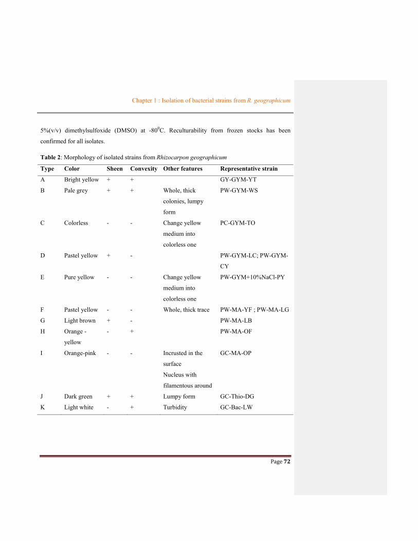
Chapter 1 : Isolation of bacterial strains from R. geographicum
Page 72
5%(v/v) dimethylsulfoxide (DMSO) at -800C. Reculturability from frozen stocks has been
confirmed for all isolates.
Table 2: Morphology of isolated strains from Rhizocarpon geographicum
Type Color Sheen Convexity Other features Representative strain
A Bright yellow + + GY-GYM-YT
B Pale grey + + Whole, thick
colonies, lumpy
form
PW-GYM-WS
C Colorless - - Change yellow
medium into
colorless one
PC-GYM-TO
D Pastel yellow + - PW-GYM-LC; PW-GYM-
CY
E Pure yellow - - Change yellow
medium into
colorless one
PW-GYM+10%NaCl-PY
F Pastel yellow - - Whole, thick trace PW-MA-YF ; PW-MA-LG
G Light brown + - PW-MA-LB
H Orange -
yellow
- + PW-MA-OF
I Orange-pink - - Incrusted in the
surface
Nucleus with
filamentous around
GC-MA-OP
J Dark green + + Lumpy form GC-Thio-DG
K Light white - + Turbidity GC-Bac-LW

Chapter 1 : Isolation of bacterial strains from R. geographicum
Page 73
Figure 3: The pure strains isolated from Rhizocarpon geographicum. (a:GC-GYM-YT; b: PW-GYM-WS; c: PC-GYM-TO; d: PW-GYM-LY and PW-GYM-CY; e: PY-GYM +10%NaCl-PY; f: PW-MA-YF and PW-MA-LG; g: PW-MA-LB; h: PW-MA-OF; i: GC-MA-OP; j: GC-Thio-DG; k: GC-Bac-LW) Table 3 : The most common colony types in samples after 14 days of incubation Samples Dominant
colonies Other colony types
GC A I,J,K PC C PW D,F B,E,G,H
GC: thin layer at the center position on the surface of R. geographicum collected at the farthest distance from the sea PC: thin layer at the center position on the surface of R. geographicum collected at the medium distance PW: whole of Rhizocarpon geographicum collected at the medium distance
A clear difference in the dominance of the colony morphology was observed in agar plates
obtained from Rhizocarpon geographicum samples collected at the different locations on the
thallus. Most colony types were scarce, with one or few representatives per plate, the samples
being strongly dominated by one or few colony types (See Table 3). The crustose lichen-associated
strains appear to be characterized by a strong presence of bacteria forming colony type D, F, A, C.
However, the individual samples appear to harbor populations distinct from another type with GC
a b d f c e
g h i j k

Chapter 1 : Isolation of bacterial strains from R. geographicum
Page 74
samples harboring populations strongly dominated by colony type A, whereas colony type C
dominated on PC samples and the PW samples are co-dominated by bacteria forming colony types
D and F.
Microbial identification
The isolates were identified by partial 16S rRNA gene sequence analysis (see Table 4). All
sequences matched with entries in GenBank with similarity ranging from 85% to 100%. The
collection was found to contain members of three classes such as Alphaproteobacteria,
Betaproteobacteria and Bacilli. The taxonomic diversity of selected strains is shown in Figure 4.

Chapter 1 : Isolation of bacterial strains from R. geographicum
Page 75
Table 4: Identify of cultured strains as revealed by partial 16S rRNA gene sequencing
Strain Colony type
Sequence length (pb)
GenBank accession number
Most similar strains
% Identity
Family Order Classe
GC-GYM-YT A 1445 AF131295
Sphingomonas aquatilis
98.39 Sphingomonadaceae Sphingomonadales Alphaproteobacteria
PW-GYM-WS
B 1489 AF512826
Burkholderia sordidicola
98.98 Burkholderiaceae Burkholderiales Betaproteobacteria
PC-GYM-TO C 1512 AJ223990 Paenibacillus odorifer
98.46 Paenibacillaceae Bacillales Bacilli
PW-GYM-LY D 1503
EU099594
Paenibacillus castaneae
98.34 Paenibacillaceae Bacillales Bacilli
PW-GYM-CY
D 1503 EU099594
Paenibacillus castaneae
98.34 Paenibacillaceae Bacillales Bacilli
PW-GYM+10%NaCl- PY
E 1508 AJ316316 Bacillus murimartini
99.93 Bacillaceae Bacillales Bacilli
PW-MA-YF F 1291 AJ277984 Psychrobacillus psychrodurans
99.84 Bacillaceae Bacillales Bacilli
PW-MA-LG F 1291 AJ277984 Psychrobacillus psychrodurans
99.84 Bacillaceae Bacillales Bacilli
PW-MA-LB G 1494 HM054474
Bacillus xiaoxiensis 99.11 Bacillaceae Bacillales Bacilli
PW-MA-OF H 1487 AB300598
Lysinibacillus parviboronicapiens
99.12 Planococcaceae Bacillales Bacilli
GC-MA-OP I 556 EF600972 Elsinoe verbenae 84.19 Elsinoaceae Myriangiales Dothideomycetes GC-Thio-DG J 1572 Coccomyxa sp. 100.00 Coccomyxaceae Trebouxiophyceae
ordo incertae sedis
trebouxiophyceae
GC-Bac-LW K 1493 AXCJ01000001
Candidatus “Xenolissoclinum pacificiencis”
68.78
Among the strains harvested from R. geographicum, ten isolates were bacteria; one was a
fungus (Sphaceloma sp.), one was a cyanobacterium and one seems to be not pure regarding its
identity (coded GC-Bac-LW) (see Table 4 and Figure 3).Two isolates with the codes PW-GYM-
LY and PW-GYM-CY were affiliated to the same species named Paenibacillus castaneae with a
percentage of identity as 98.34 %. This result was obvious because of their similarity of the growth
medium and their morphological characteristics as well. Additionally, two other isolates named
PW-MA-YF and PW-MA-LC belonged to Psychrobacillus psychrodurans with a percentage of
similarity up to 99.84%. Two species belonging to Proteobacteria phylum were identified as
Sphingomonas aqualitis and Burkholderia sordidicola. The yellow pigmented Sphingomonas
aqualitis species was firstly reported from the several mineral water sources in Korea by Lee and
co-workers (2001). Herein, this strain was the first example found from R. geographicum.
However, the Sphingomonas genus was already described commonly from lichens in some
previous studies. It was isolated from Caloplaca verruculifera (Sigurbjörnsdóttir et al., 2014)

Chapter 1 : Isolation of bacterial strains from R. geographicum
Page 76
Cetraria sp. (Kim et al., 2012), Lobaria pulmonaria (Cardinale et al., 2011 ; Aschenbrenner et al.,
2014; Grube et al., 2015), Solorina crocea (Grube et al., 2012), Umbilicaria americana (Bates et
al., 2011). Therefore, it is the first report of Sphingomonas aqualitis found from R. geographicum.
Another species of Proteobacteria, Burkholderia sordidicola which was firstly identified from
white-rot fungus Phanerochaete (Lim et al., 2003) was commonly found in the third symbiotic
communities of lichens such as from Cladonia pyxidata and Cladonia rangiferina (Cardinale et al.,
2006) and from Cladonia sp., Steroacaulon sp. and Umbilicaria sp. in the Arctic (Kim et al.,
2012). This study is thus considered as the first description of this species from R. geographicum
samples.
The other six bacterial species all belonged to Bacilli class, Firmicutes phylum consisting of
two Paenibacillus, two Bacillus, one Psychrobacillus and one Lysinibacillus. Among these, two
species Psychrobacillus psychrodurans and Lysinibacillus parviboronicapiens (or their genera)
were for the first time discovered from lichens. Although it is the first report of the other species
Paenibacillus odorifer, P. castaneae, Bacillus murimartini and B. xiaoxiensis from R.
geographicum, the genera from which they belong were often found in lichens. For instance,
Paenibacillus genus was recorded from Cladonia sp. lichens (Grube et al., 2009; Cardinale et al.,
2011; Cardinale et al., 2006; Hodkinson et al., 2009), Hypogymnia physodes (Cardinale et al.,
2006), Lecanora polytropa (Grube et al., 2009; Cardinale et al., 2011) ; Bacillus species were
found from Cladonia arbuscula (Grube et al., 2009; Cardinale et al., 2011), Hydropunctaria
maura (Sigurbjörnsdóttir et al., 2014), Lecanora species (Sigurbjörnsdóttir et al., 2014 ; Grube et
al., 2009; Cardinale et al., 2011), Umbilicaria cylindrica (Grube et al., 2009; Cardinale et al.,
2011) and Xanthoria elegans (Selbmann et al., 2009). As a result, most isolates from R.
geographicum were for the first time reported from lichens, excepted B. sordidicola species which
was already reported from Umbilicaria sp. (Kim et al., 2012) and Cladonia pyxidata (Cardinale et
al., 2006). Further, we need to investigate biological interests of these species and detailed
investigations about their activities are essential.
Besides, two non-bacterial isolates were Coccomyxa sp., a genus of green alga, and Sphaceloma
araliae, a genus of Ascomycete fungi. The Coccomyxa genus was already found from Rhizocarpon

Chapter 1 : Isolation of bacterial strains from R. geographicum
Page 77
lecanorinum (Clayden 1998). The presence of this alga can be explained by the fact that it came
from the process without surface treatment of the lichen before cutting it.
Figure 4: Taxonomic diversity of selected strains
CONCLUSION
Herein, our culture-based strategy led to the isolation of thirteen solates and among them 11 were
bacteria. These data highlighted that the method used was not enough selective to the bacteria
kingdom. Alphaprotebacteria was not the major part of cultivable lichen-associated microbial
communities on lichen R. geographicum. Bacilli belonging to Firmicutes phylum became a
dominant class for these communities. This result was similar to those reported from Cardinale’s
team (2006) where Firmicutes dominated all bacterial communities associated with lichens
studied.

Chapter 1 : Isolation of bacterial strains from R. geographicum
Page 78
References
Amann, R I, W Ludwig, and Schleifer K H. 1995. ―Phylogenetic Identification and in Situ Detection of Individual
Microbial Cells without Cultivation.‖ Microbiological Reviews 59 (1): 143–69.
Armstrong, R. A., and Smith S. N.. 2009. ―Carbohydrates in the Hypothallus and Areolae of the Crustose Lichen
Rhizocarpon Geographicum (L.) DC.‖ Symbiosis 49 (2): 95–100. https://doi.org/10.1007/s13199-009-0016-z.
Armstrong, R., and Bradwell T.. 2010. ―Growth of Crustose Lichens: A Review.‖ Geografiska Annaler: Series A,
Physical Geography 92 (1): 3–17. https://doi.org/10.1111/j.1468-0459.2010.00374.x.
Aschenbrenner, I. A.,. Cardinale M, Berg G., and Grube M.. 2014. ―Microbial Cargo: Do Bacteria on Symbiotic
Propagules Reinforce the Microbiome of Lichens?‖ Environmental Microbiology 16 (12): 3743–52.
https://doi.org/10.1111/1462-2920.12658.
Bates, S. T., Garrett W. G. C., J. Gregory Caporaso, Rob Knight, and Noah Fierer. 2011. ―Bacterial Communities
Associated with the Lichen Symbiosis.‖ Applied and Environmental Microbiology 77 (4): 1309–14.
https://doi.org/10.1128/AEM.02257-10.
Bjelland, T., Grube M., Hoem S.,. Jorgensen S. L, Daae F. L., Thorseth I. H., and Øvreås L. 2011. ―Microbial
Metacommunities in the Lichen–rock Habitat.‖ Environmental Microbiology Reports 3 (4): 434–42.
https://doi.org/10.1111/j.1758-2229.2010.00206.x.
Boustie, J.l, and Grube M. 2005. ―Lichens—a Promising Source of Bioactive Secondary Metabolites.‖ Plant Genetic
Resources 3 (02): 273–287. https://doi.org/10.1079/PGR200572.
Boustie, J., Tomasi S., and Grube M.. 2010. ―Bioactive Lichen Metabolites: Alpine Habitats as an Untapped Source.‖
Phytochemistry Reviews 10 (3): 287–307. https://doi.org/10.1007/s11101-010-9201-1.
Brodo, Irwin M. 1973. ―Chapter 12 - Substrate Ecology.‖ 401–41. In The Lichens, edited by Vernon Ahmadjianmason
E. Hale,. Academic Press. http://www.sciencedirect.com/science/article/pii/B9780120449507500179.
Cardinale, M., de Castro J. V., Müller H., Berg G., and Grube M.. 2008. ―In Situ Analysis of the Bacterial Community
Associated with the Reindeer Lichen Cladonia Arbuscula Reveals Predominance of Alphaproteobacteria.‖
FEMS Microbiology Ecology 66 (1): 63–71. https://doi.org/10.1111/j.1574-6941.2008.00546.x.
Cardinale, M., Puglia A. M., and Grube M.. 2006. ―Molecular Analysis of Lichen-Associated Bacterial Communities.‖
FEMS Microbiology Ecology 57 (3): 484–95. https://doi.org/10.1111/j.1574-6941.2006.00133.x.
Chapter 1 : Isolation of bacterial strains from R. geographicum
Page 79
Cardinale M., Steinová J., Rabensteiner J., Berg G., and Grube M.. 2011. ―Age, Sun and Substrate: Triggers of
Bacterial Communities in Lichens.‖ Environmental Microbiology Reports 4 (1): 23–28.
https://doi.org/10.1111/j.1758-2229.2011.00272.x.
Clayden R. S. 1998. "Thallus inititaion and developments in lichen Rhizocarpon lecanorium". New phytol 139, 685 - 695
González, I., Angel A.-S., Anderson A., and Genilloud O.. 2005. ―Actinomycetes Isolated from Lichens: Evaluation
of Their Diversity and Detection of Biosynthetic Gene Sequences.‖ FEMS Microbiology Ecology 54 (3): 401–
15. https://doi.org/10.1016/j.femsec.2005.05.004.
Grube, Martin, Köberl M., Lackner S., Berg C., and Berg G.. 2012. ―Host–parasite Interaction and Microbiome
Response: Effects of Fungal Infections on the Bacterial Community of the Alpine Lichen Solorina Crocea.‖
FEMS Microbiology Ecology 82 (2): 472–81. https://doi.org/10.1111/j.1574-6941.2012.01425.x.
Grube M., and Berg G.. 2009. ―Microbial Consortia of Bacteria and Fungi with Focus on the Lichen Symbiosis.‖
Fungal Biology Reviews 23 (3): 72–85. https://doi.org/10.1016/j.fbr.2009.10.001.
Grube M., Cernava T., Soh J., Fuchs S., Aschenbrenner I., Lassek C., Wegner U., 2015. ―Exploring Functional
Contexts of Symbiotic Sustain within Lichen-Associated Bacteria by Comparative Omics.‖ The ISME Journal
9 (2): 412–24. https://doi.org/10.1038/ismej.2014.138.
Hodkinson B. P., Gottel N. R., Schadt C. W., and Lutzoni F.. 2012. ―Photoautotrophic Symbiont and Geography Are
Major Factors Affecting Highly Structured and Diverse Bacterial Communities in the Lichen Microbiome.‖
Environmental Microbiology 14 (1): 147–61. https://doi.org/10.1111/j.1462-2920.2011.02560.x.
Hodkinson, B. P., and Lutzoni F.. 2009. ―A Microbiotic Survey of Lichen-Associated Bacteria Reveals a New Lineage
from the Rhizobiales.‖ Symbiosis 49 (3): 163–80. https://doi.org/10.1007/s13199-009-0049-3.
Ihlen, P. G. 2004. ―Taxonomy of the Non-Yellow Species of Rhizocarpon (Rhizocarpaceae, Lichenized Ascomycota)
in the Nordic Countries, with Hyaline and Muriform Ascospores.‖ Mycological Research 108 (Pt 5): 533–70.
Kim, S. H. , Shin Y. K., Sohn Y. C., and Kwon H. C.. 2012. ―Two New Cholic Acid Derivatives from the Marine
Ascidian-Associated Bacterium Hasllibacter Halocynthiae.‖ Molecules 17 (10): 12357–64.
https://doi.org/10.3390/molecules171012357.
Lee, J S, Shin Y. K., Yoon J. H., Takeuchi M., Pyun Y. R., and Park Y. H. 2001. ―Sphingomonas Aquatilis Sp. Nov.,
Sphingomonas Koreensis Sp. Nov., and Sphingomonas Taejonensis Sp. Nov., Yellow-Pigmented Bacteria
Isolated from Natural Mineral Water.‖ International Journal of Systematic and Evolutionary Microbiology 51
(4): 1491–98. https://doi.org/10.1099/00207713-51-4-1491.

Chapter 1 : Isolation of bacterial strains from R. geographicum
Page 80
Liba, C.M., Ferrara F. I. S., Manfio G. P., Fantinatti-Garboggini F., Albuquerque R. C., Pavan C., Ramos P. L.,
Moreira-Filho C. A., and. Barbosa H. R.. 2006. ―Nitrogen-Fixing Chemo-Organotrophic Bacteria Isolated from
Cyanobacteria-Deprived Lichens and Their Ability to Solubilize Phosphate and to Release Amino Acids and
Phytohormones.‖ Journal of Applied Microbiology 101 (5): 1076–86. https://doi.org/10.1111/j.1365-
2672.2006.03010.x.
Lim, Y. W., Baik K. S., Han S. K., Kim S. B., and Bae K. S.. 2003. ―Burkholderia Sordidicola Sp. Nov., Isolated from
the White-Rot Fungus Phanerochaete Sordida.‖ International Journal of Systematic and Evolutionary
Microbiology 53 (5): 1631–36. https://doi.org/10.1099/ijs.0.02456-0.
Selbmann, L., Zucconi L., Ruisi S., Grube M., Cardinale M., and Onofri S.. 2009. ―Culturable Bacteria Associated
with Antarctic Lichens: Affiliation and Psychrotolerance.‖ Polar Biology 33 (1): 71–83.
https://doi.org/10.1007/s00300-009-0686-2.
Sigurbjörnsdóttir, M. A., Heiðmarsson S., Jónsdóttir A. R. and Vilhelmsson O.. 2014. ―Novel Bacteria Associated
with Arctic Seashore Lichens Have Potential Roles in Nutrient Scavenging.‖ Canadian Journal of
Microbiology 60 (5): 307–17. https://doi.org/10.1139/cjm-2013-0888.

Page 81
CHAPTER 2: THE SELECTION OF PAENIBACILLUS ODORIFER AS A
PROMISING SOURCE OF INTERESTING METABOLITES

Chapter 2: The selection of P. odorifer as a promising source of interesting metabolites
Page 82
CHAPTER 2: THE SELECTION OF PAENIBACILLUS ODORIFER AS A PROMISING SOURCE OF INTERESTING METABOLITES
Because we have focused our work on the isolation of bioactive bacterial metabolites,
the bibliographic data concerning the production of interesting compounds from the isolated
strains from R. geographicum had to be studied. In this chapter, the chemical production of
these strains will be introduced and we will give details on studies from which we will be
based for our selection of the strain(s) for further researches.
2.1. State of art on the chemical production of the isolated strains
Firstly, it seems that no report had described metabolites from Sphingomonas aquatilis
belonging to Sphingomonas genus. However this genus produced some primary metabolites
like enzymes (Kmunícek et al., 2005) or glycosphingolipids (Kawahara et al., 1991; Kubota
et al., 2009) which demonstrated an activity on invariant natural killer T (iNKT) cells. The
Table 2.2.1 provides data on the metabolites already isolated from this genus.
Table 2.2.1 Summary about chemical studies on Sphingomonas genus
Producing strains Compounds Nature of bioactivities References
S. paucimobilis Haloalkane dehalogenase LiB Dehalogenation Kmunícek et al., 2005
Two novel glycosphingolipids Kawahara et al., 1991
S. yanoikyae Glycosphingolipid (GSL-7) Activity on invariant natural killer T (iNKT) cells
Kubota et al., 2009
S. terrae Glycosphingolipid (GSL-13)
S. adhaesiva Glycosphingolipid (GSL-4B) Kawahara et al., 1991
Studies on Burkholderia genus described many active compounds (primary or
secondary metabolites) isolated from this genus such as antifungal pyrrolnitrin (El-Banna and
Winkelmann, 1998; Hwang et al., 2002; Keum et al., 2009); diketopiperazines (Wang et al.,
2010) or antitumor spiruchostatin (Klausmeyer et al., 2011). The summary of some
compounds isolated from Burkholderia genus was shown in Table 2.2.2. We can conclude the
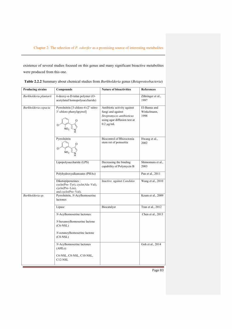
Chapter 2: The selection of P. odorifer as a promising source of interesting metabolites
Page 83
existence of several studies focused on this genus and many significant bioactive metabolites
were produced from this one.
Table 2.2.2 Summary about chemical studies from Burkholderia genus (Betaproteobacteria)
Producing strains Compounds Nature of bioactivities References
Burkholderia plantarii 6-deoxy-α-D-talan polymer (O-acetylated homopolysaccharide)
Zähringer et al., 1997
Burkholderia cepacia Pyrrolnitrin [3-chloro-4-(2’-nitro-3’-chloro phenyl)pyrrol]
NH
Cl
NO2
Cl
Antibiotic activity against fungi and against Streptomyces antibioticus using agar diffusion test at 0.2 µg/mL
El-Banna and Winkelmann, 1998
Pyrrolnitrin
NH
Cl
NO2
Cl
Biocontrol of Rhizoctonia stem rot of poinsettia
Hwang et al., 2002
Lipopolysaccharide (LPS) Decreasing the binding capability of Polymycin B
Shimomura et al., 2003
Polyhydroxyalkanoates (PHAs) Pan et al., 2011
Diketopiperazines : cyclo(Pro–Tyr), cyclo(Ala–Val), cyclo(Pro–Leu), and cyclo(Pro–Val),
Inactive against Candidas Wang et al., 2010
Burkholderia sp. Pyrrolnitrin, N-Acylhomoserine lactones
Keum et al., 2009
Lipase Biocatalyst Tran et al., 2012
N-Acylhomoserine lactones:
N-hexanoylhomoserine lactone (C6-NSL)
N-octanoylhomoserine lactone (C8-NSL)
Chen et al., 2013
N-Acylhomoserine lactones (AHLs):
C6-NSL, C8-NSL, C10-NSL, C12-NSL
Goh et al., 2014

Chapter 2: The selection of P. odorifer as a promising source of interesting metabolites
Page 84
Burkholderia vietnamiensis
Endotoxin Immunostimulatory activity on human myelomonocytic U937 cells.
Ieranò et al., 2009
Burkholderia thailandensis
Bactobolin
Bactobolins A, B: antibiotics against MRSA and V. parahemolyticus with MICs < 1 µg/mL
Seyedsayamdost et al., 2010
Spiruchostatins
Antitumor towards LOX IMVI melanoma cells using murine hollow fiber assay
Klausmeyer et al., 2011
B. dolosa Endotoxin Lorenzo et al., 2013
Burkholderia gladioli pv. agaricicola
Volatile organic compounds (major compound as 1-methyl-4-(1-methylethenyl)-cyclohexene)
Antifungal activity against Botrytis cinerea, Aspergillus flavus, Aspergillus niger, Penicillium digitatum, Penicillium expansum, Sclerotinia sclerotiorum and Phytophthora cactorum using agar diffusion test
Elshafie et al., 2012
Exopolysaccharide Andolfi et al., 2008
O-specific polysaccharide (containing : D-mannose, D-rhamnose, D-galactose)
Karapetyan et al., 2006
Burkholderia arboris Pyochelin
Phytotoxicity against pine callus at EC50 of 171.98 μg ml−1
Dang et al., 2011
The Paenibacillus genus possess the ability to produce some metabolites with industrial
applications such as xylanase in paper industry (Zheng et al., 2012; Yeasmin et al., 2010),

Chapter 2: The selection of P. odorifer as a promising source of interesting metabolites
Page 85
acetoin in cosmetics and foods (Zhang et al., 2012). Especially, the genus produced
fusaracidin - a compound having remarkable antibiotic activity against Micrococcus luteus
(Kim et al., 2014) or abling to control Phytophthora blight infection (a disease blighted
leaves) caused by Phytophthora capsic (Lee et al., 2012). This genus thus is promising as a
source of interesting compounds. The data of some species production belonging to this genus
was reported in Table 2.2.3.
Table 2.2.3 Summary about chemical studies on Paenibacillus genus (Bacilli)
Producing strains Compounds Nature of bioactivities
References
P. campinasensis Xylanase Industrial application: pulp bleaching pretreatment.
Zheng et al., 2012
P. polymyxa Acetoin
OH
O
Cosmetic, food Zhang et al., 2012
Xylanase enzyme Paper industry Yeasmin et al., 2010
Lipopeptide Anti-phytopathogen Xanthomonas campestris using agar diffusion test
Mageshwaran et al., 2011
Fusaracidin
Antibiotic : control Phytophthora blight infection caused by Phytophthora capsic at concentration 0.1 ppm
Lee et al., 2012
Antimicrobial activity against Micrococcus luteus using agar diffusion test
Kim et al., 2014
-1,4-mannanase Production of mannooligosaccharide
Hori et al., 2014
Paenibacillus sp. Fibrinolytic enzyme Vijayaraghavan et

Chapter 2: The selection of P. odorifer as a promising source of interesting metabolites
Page 86
al, 2014
Mutanase (α-1,3-glucan) An oral hygiene product
Shimotsuura et al., 2008
Chitinase Meena et al., 2013
KB 425796-A (nikkomycin)
KB 425796-B
Antifungal activity against Aspergillus fumigatus (MIC of 0.5 and 2.5 µg.mL-1, respectively.)
Kai et al., 2013b
-1,3-glucanase PgIA Fungal disease biocontrol
Cheng et al., 2013
KB 425796-C macrocyclic lipopeptidolactone
Anifungal activity activities against T. asahii (MEC of 1.56 mg.ml-1, MIC of 3.13 mg.ml-1) and Aspergillus fumigatus (MEC of 3.13 mg.ml-1, MIC > 50 mg.ml-1).
Kai et al., 2013a
P. terrae Xylanase
(endo-β-1,4-xylanase KRICT PX-3)
Song et al., 2014
P. woosongensis Alkaline keratinolytic protease In the laundry industry Paul et al., 2014
P.alvei Cyclic lipopeptide Antimicrobial activity against E. coli, Salmonella, and Staphylococcus aureus using agar diffusion test
Knolhoff et al., 2015
P. elgii Lipopeptide Antibiotic Ding et al., 2011
To date, to our knowledge, the genus Lysinibacillus was not already found as symbiotic
partner of lichens. Herein, it was firstly reported from R. geographicum. The studies
describing the production of its metabolites were also limited. Some researches (summarized
in Table 2.2.4) highlighted that some bioactive compounds were isolated from this genus as
spirobacillenes, soraphinol A and kurasoin B (Park et al., 2012), biosulfactants which possess

Chapter 2: The selection of P. odorifer as a promising source of interesting metabolites
Page 87
potent activity as anticancer agent with cytotoxic effect on human embryonic kidney
cancerous cell (HEK-293) with LC50 75 μg.ml−1 (Pradhan et al., 2014).
Table 2.2.4 Summary about chemical studies on Lysinibacillus genus (Bacilli)
Producing strains Compounds Nature of bioactivities References
Lysinibacillus fusiformis Spirobacillenes A and B:
Soraphinol A
HNOH
O
HO Kurasoin B
HN
O
HO
Activity against the production of nitric oxide (NO) and reactive oxygen species
Park et al., 2012
Protease and esterase Prabha et al., 2014
Biosurfactants Cytotoxicity effect on human embryonic kidney cancerous cells (HEK-293) with LC50 75 μg ml−1
Pradhan et al., 2014
To date, the Bacillus genus has already attracted many interests for its metabolites. It
produced many significant bioactive compounds (see Table 2.2.5). Diverse metabolites
extracted from its culture were isolated e.g. enzymes as β-mannanase (Zang et al., 2015),
lipoamide (Berrue et al., 2009), a series of bioactive compounds as surfactin, macrolactin
(Nastro et al., 2013; Mondol et al., 2011a, 2011b) and diketopiperazines (Kumar et al., 2013).
Table 2.2.5 Chemical studies on Bacillus genus (Bacilli)
Producing strains
Compounds Nature of bioactivities References

Chapter 2: The selection of P. odorifer as a promising source of interesting metabolites
Page 88
Bacillus pumilus β-mannanase Monno-oligosaccharides production
Zang et al., 2015
Lipoamide A
Lipopeptides 1-4
Amicoumacins A, B
O
NH
O
H2N
O
OOHOH
OH
H2N
No active
Antibacterial activity against S. aureus, P. vulgaris, E. faecalis (MIC from 6.5-25 µg/mL)
Antibacterial activity against S. aureus, P. vulgaris, E. faecalis, S. aureus (MIC from 6.5-50 µg/mL)
Berrue et al., 2009
Bacillus amyloliquefaciens
Surfactin
Fengycin
Iturin A
Macrolactin
Difficidin
Bacillaene
Nastro et al., 2013
Difficidin
Oxidifficidin
Bacillaene
Chen et al., 2006
Macrolactin S
Macrolactin V
Macrolactin S : antibacterial activity against E.coli and S.aureus (MIC of 0.3 and 0.1µg.mL-
1, respectively)
Macrolactin V : antibacterial activity against Bacillus subtilis, E.coli, S.aureus (MIC of 0.1µg.mL-1)
Gao et al., 2010

Chapter 2: The selection of P. odorifer as a promising source of interesting metabolites
Page 89
Bacillopeptin B1 and B Antifungal activity Ma et al., 2014
Bacillus lichenformis
Glycolipids : ieodoglucomide C and ieodoglycolipid
Antibiotic properties against Staphylococcus aureus, Bacillus subtilis, Bacillus cereus, Salmonella typhi, Escherichia coli and Pseudomonas aeruginosa with MICs ranging from 0.01 to 0.05 μM
Tareq et al., 2015
Ieodoglucomides A and B
O
OHHOHO
O
O
O
HN
OH
O
H8
O OHO
A : R = CH3B : R = H
Antimicrobial activity
B: Anti-lung cancer (GI50 25.18 µg.mL1), stomach cancer (GI50 17.78 µg.mL1)
Tareq et al., 2012
Bacteriocin BL8 Antimicrobial activity Smitha and Bhat, 2013
Bacillus subtilis Tetraprenyl-β-curcumene
Tetraprenyl-α-curcumene
C35-terpenol
Takigawa et al., 2010
Chitinase emzyme Antifungal activity
Anti-insecticid pests
Senol et al., 2014

Chapter 2: The selection of P. odorifer as a promising source of interesting metabolites
Page 90
Bacilosarcins A
Bacilosarcins B
Bacilosarcin A showed 82% inhibition at 50 mM against growth of barnyard millet
Azumi et al., 2008
7-O-malonyl macrolactins A
R = CO-CH2-COOH
Antibacterial activity Romero-Tabarez et al., 2006
1-deoxynojirimycin
HN
HO OH
OHHO
Increasing of GLUT4 and glucose uptake into adipocytes at 0.5 μM
Lee et al., 2013
Sporulene
H
H
H
Terpenoid
Kontnik et al., 2008
Bacillus claucii β-geranylfarnesene
β-hexaprene
Sato et al., 2013
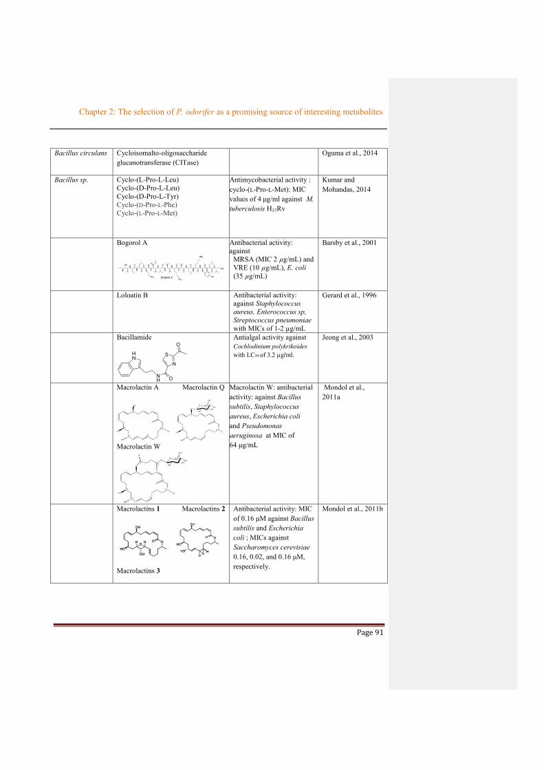
Chapter 2: The selection of P. odorifer as a promising source of interesting metabolites
Page 91
Bacillus circulans Cycloisomalto-oligosaccharide glucanotransferase (CITase)
Oguma et al., 2014
Bacillus sp.
Cyclo-(L-Pro-L-Leu) Cyclo-(D-Pro-L-Leu) Cyclo-(D-Pro-L-Tyr) Cyclo-(D-Pro-L-Phe) Cyclo-(L-Pro-L-Met)
Antimycobacterial activity : cyclo-(L-Pro-L-Met): MIC values of 4 μg/ml against M. tuberculosis H37Rv
Kumar and Mohandas, 2014
Bogorol A
Antibacterial activity: against
MRSA (MIC 2 µg/mL) and VRE (10 µg/mL), E. coli (35 µg/mL)
Barsby et al., 2001
Loloatin B Antibacterial activity: against Staphylococcus aureus, Enterococcus sp, Streptococcus pneumoniae with MICs of 1-2 µg/mL
Gerard et al., 1996
Bacillamide
S
N
ONH
HN
O
Antialgal activity against Cochlodinium polykrikoides with LC50 of 3.2 µg/ml.
Jeong et al., 2003
Macrolactin A Macrolactin Q
Macrolactin W
Macrolactin W: antibacterial activity: against Bacillus subtilis, Staphylococcus aureus, Escherichia coli and Pseudomonas aeruginosa at MIC of 64 μg/mL
Mondol et al., 2011a
Macrolactins 1 Macrolactins 2
Macrolactins 3
Antibacterial activity: MIC of 0.16 μM against Bacillus subtilis and Escherichia coli ; MICs against Saccharomyces cerevisiae 0.16, 0.02, and 0.16 μM, respectively.
Mondol et al., 2011b

Chapter 2: The selection of P. odorifer as a promising source of interesting metabolites
Page 92
Lichenase Maktouf et al., 2015
Semiquinone glucoside Antioxidant Mishra et al., 2014
3,5-dihydroxy-4-isopropyl stilbene
HO
HO
Antioxidant at concentration 100 µg/ml
Anticancer against cervical cancer cell line (HeLa), growth inhibition at IC50 of 25 µg/ml
Kumar et al., 2013
3,4’,5-trihydroxystilbene
OHHO
OH
3,5-dihydroxy-4-isopropyl stilbene
Antiphytopathogen: P. expansum (MIC of 4 µg/mL) Fusarium oxysporum (MIC of 2 µg/mL)
Antifungal : P. expansum (MIC of 8 µg/mL), Rhizotocnia solani (MIC of 8 µg/mL)
Kumar et al., 2012
Macrolactin F (1) and 7-O-succinyl macrolactin F (2), and A (3)
Antibacterial activity against B. subtilis and S. aureus (inhibition zones of 8-28 mm at 50-100 µg/disk
Jaruchoktaweechai et al., 2000
Bacillus marinus Marihysin A
Antifungal activity against Alternaria solani, Fusarium oxysporum, Verticillium alboatrum, F.graminearum, Sclerotium sp., Penicillium sp., Rhizoctonia solani, and Colletotrichum sp. with MIC values of 100 – 200 mg/ml
Liu et al., 2010
Macrolactins T and U Macrolactin B: antifungal activity against Alternaria solani with MIC values of
Xue et al., 2008
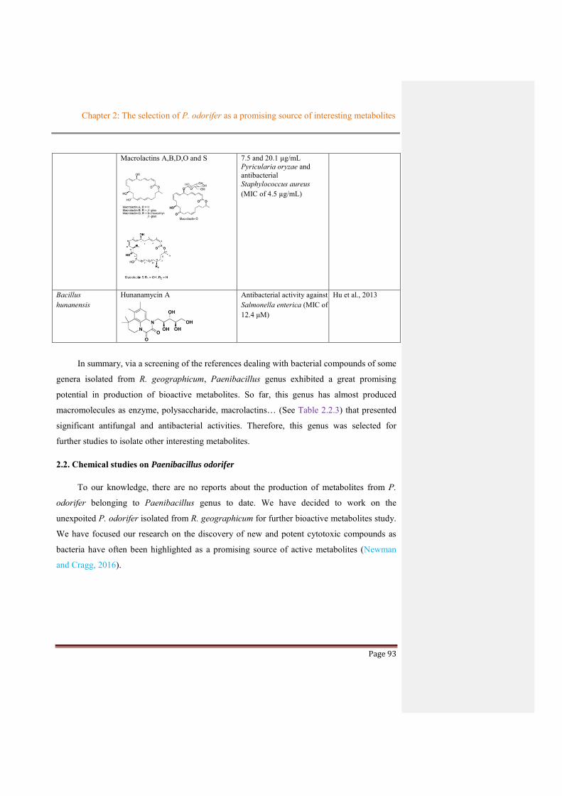
Chapter 2: The selection of P. odorifer as a promising source of interesting metabolites
Page 93
Macrolactins A,B,D,O and S
7.5 and 20.1 µg/mL Pyricularia oryzae and antibacterial Staphylococcus aureus (MIC of 4.5 µg/mL)
Bacillus hunanensis
Hunanamycin A
Antibacterial activity against Salmonella enterica (MIC of 12.4 μM)
Hu et al., 2013
In summary, via a screening of the references dealing with bacterial compounds of some
genera isolated from R. geographicum, Paenibacillus genus exhibited a great promising
potential in production of bioactive metabolites. So far, this genus has almost produced
macromolecules as enzyme, polysaccharide, macrolactins… (See Table 2.2.3) that presented
significant antifungal and antibacterial activities. Therefore, this genus was selected for
further studies to isolate other interesting metabolites.
2.2. Chemical studies on Paenibacillus odorifer
To our knowledge, there are no reports about the production of metabolites from P.
odorifer belonging to Paenibacillus genus to date. We have decided to work on the
unexpoited P. odorifer isolated from R. geographicum for further bioactive metabolites study.
We have focused our research on the discovery of new and potent cytotoxic compounds as
bacteria have often been highlighted as a promising source of active metabolites (Newman
and Cragg, 2016).

Chapter 2: The selection of P. odorifer as a promising source of interesting metabolites
Page 94

Chapter 2: The selection of P. odorifer as a promising source of interesting metabolites
Page 95
References
Azumi, M., Ogawa, K., Fujita, T., Takeshita, M., Yoshida, R., Furumai, T., Igarashi, Y., 2008. Bacilosarcins A and B, novel bioactive isocoumarins with unusual heterocyclic cores from the marine-derived bacterium Bacillus subtilis. Tetrahedron 64, 6420–6425. https://doi.org/10.1016/j.tet.2008.04.076
Barsby, T., Kelly, M.T., Gagné, S.M., Andersen, R.J., 2001. Bogorol A Produced in Culture by a Marine Bacillus sp. Reveals a Novel Template for Cationic Peptide Antibiotics. Org. Lett. 3, 437–440. https://doi.org/10.1021/ol006942q
Berrue, F., Ibrahim, A., Boland, P., Kerr, R.G., 2009. Newly isolated marine Bacillus pumilus (SP21): A source of novel lipoamides and other antimicrobial agents. Pure and applied chemistry 81, 1027–1031.
Chen, J.W., Koh, C.-L., Sam, C.-K., Yin, W.-F., Chan, K.-G., 2013. Short Chain N-acyl Homoserine Lactone Production by Soil Isolate Burkholderia sp. Strain A9. Sensors 13, 13217–13227. https://doi.org/10.3390/s131013217
Chen, X.-H., Vater, J., Piel, J., Franke, P., Scholz, R., Schneider, K., Koumoutsi, A., Hitzeroth, G., Grammel, N., Strittmatter, A.W., Gottschalk, G., Süssmuth, R.D., Borriss, R., 2006. Structural and Functional Characterization of Three Polyketide Synthase Gene Clusters in Bacillus amyloliquefaciens FZB 42. J Bacteriol 188, 4024–4036. https://doi.org/10.1128/JB.00052-06
Cheng, R., Chen, J., Yu, X., Wang, Y., Wang, S., Zhang, J., 2013. Recombinant production and characterization of full-length and truncated β-1,3-glucanase PglA from Paenibacillus sp. S09. BMC Biotechnology 13, 105. https://doi.org/10.1186/1472-6750-13-105
Dang, Q.L., Son, S.W., Cheon, H.-M., Choi, G.J., Choi, Y.H., Jang, K.S., Lim, C.H., Kim, J.-C., 2011. Pyochelin isolated from Burkholderia arboris KRICT1 carried by pine wood nematodes exhibits phytotoxicity in pine callus. Nematology 13, 521–528. https://doi.org/10.1163/138855410X528271
Ding, R., Wu, X.-C., Qian, C.-D., Teng, Y., Li, O., Zhan, Z.-J., Zhao, Y.-H., 2011. Isolation and identification of lipopeptide antibiotics from Paenibacillus elgii B69 with inhibitory activity against methicillin-resistant Staphylococcus aureus. J Microbiol. 49, 942–949. https://doi.org/10.1007/s12275-011-1153-7
El-Banna, Winkelmann, 1998. Pyrrolnitrin from Burkholderia cepacia: antibiotic activity against fungi and novel activities against streptomycetes. Journal of Applied Microbiology 85, 69–78. https://doi.org/10.1046/j.1365-2672.1998.00473.x
Elshafie, H.S., Camele, I., Racioppi, R., Scrano, L., Iacobellis, N.S., Bufo, S.A., 2012. In Vitro Antifungal Activity of Burkholderia gladioli pv. agaricicola against Some Phytopathogenic Fungi. Int J Mol Sci 13, 16291–16302. https://doi.org/10.3390/ijms131216291
Gao, C.-H., Tian, X.-P., Qi, S.-H., Luo, X.-M., Wang, P., Zhang, S., 2010. Antibacterial and antilarval compounds from marine gorgonian-associated bacterium Bacillus amyloliquefaciens SCSIO 00856. The Journal of Antibiotics 63, 191–193. https://doi.org/10.1038/ja.2010.7
Gerard, J., Haden, P., Kelly, M.T., Andersen, R.J., 1996. Loloatin B, A cyclic decapeptide antibiotic produced in culture by a tropical marine bacterium. Tetrahedron Letters 37, 7201–7204. https://doi.org/10.1016/0040-4039(96)01624-3
Comment [ST1]: Write in ref Zahringer D-talan

Chapter 2: The selection of P. odorifer as a promising source of interesting metabolites
Page 96
Goh, S.Y., Tan, W.-S., Khan, S.A., Chew, H.P., Kasim, N.H.A., Yin, W.-F., Chan, K.-G., 2014. Unusual Multiple Production of N-Acylhomoserine Lactones a by Burkholderia sp. Strain C10B Isolated from Dentine Caries. Sensors (Basel) 14, 8940–8949. https://doi.org/10.3390/s140508940
Hori, K., Kawabata, Y., Nakazawa, Y., Nishizawa, M., Toeda, K., 2014. A Novel β-1,4-mannanase Isolated from Paenibacillus polymyxa KT551. Food Science and Technology Research 20, 1261–1265. https://doi.org/10.3136/fstr.20.1261
Hu, Y., Wang, K., MacMillan, J.B., 2013. Hunanamycin A, an Antibiotic from a Marine-Derived Bacillus hunanensis. Org. Lett. 15, 390–393. https://doi.org/10.1021/ol303376c
Hwang, J., Chilton, W.S., Benson, D.M., 2002. Pyrrolnitrin production by Burkholderia cepacia and biocontrol of Rhizoctonia stem rot of poinsettia. Biological Control 25, 56–63. https://doi.org/10.1016/S1049-9644(02)00044-0
Ieranò, T., Silipo, A., Sturiale, L., Garozzo, D., Bryant, C., Lanzetta, R., Parrilli, M., Aldridge, C., Gould, F.K., Corris, P.A., Khan, C.M.A., Soyza, A.D., Molinaro, A., 2009. First structural characterization of Burkholderia vietnamiensis lipooligosaccharide from cystic fibrosis-associated lung transplantation strains. Glycobiology 19, 1214–1223. https://doi.org/10.1093/glycob/cwp112
Jaruchoktaweechai, C., Suwanborirux, K., Tanasupawatt, S., Kittakoop, P., Menasveta, P., 2000. New Macrolactins from a Marine Bacillus sp. Sc026. J. Nat. Prod. 63, 984–986. https://doi.org/10.1021/np990605c
Jeong, S.-Y., Ishida, K., Ito, Y., Okada, S., Murakami, M., 2003. Bacillamide, a novel algicide from the marine bacterium, Bacillus sp. SY-1, against the harmful dinoflagellate, Cochlodinium polykrikoides. Tetrahedron Letters 44, 8005–8007. https://doi.org/10.1016/j.tetlet.2003.08.115
Kai, H., Yamashita, M., Nakamura, I., Yoshikawa, K., Nitta, K., Watanabe, M., Inamura, N., Fujie, A., 2013a. Synergistic antifungal activity of KB425796-C in combination with micafungin against Aspergillus fumigatus and its efficacy in murine infection models. J Antibiot 66, 479–484. https://doi.org/10.1038/ja.2013.57
Kai, H., Yamashita, M., Takase, S., Hashimoto, M., Muramatsu, H., Nakamura, I., Yoshikawa, K., Kanasaki, R., Ezaki, M., Nitta, K., Watanabe, M., Inamura, N., Fujie, A., 2013b. Identification of ten KB425796-A congeners from Paenibacillus sp. 530603 using an antifungal assay against Aspergillus fumigatus in combination with micafungin. J Antibiot 66, 473–478. https://doi.org/10.1038/ja.2013.64
Karapetyan, G., Kaczynski, Z., Iacobellis, N.S., Evidente, A., Holst, O., 2006. The structure of the O-specific polysaccharide of the lipopolysaccharide from Burkholderia gladioli pv. agaricicola. Carbohydrate Research 341, 930–934. https://doi.org/10.1016/j.carres.2006.02.010
Kawahara, K., Seydel, U., Matsuura, M., Danbara, H., Rietschel, E.T., Za¨hringer, U., 1991. Chemical structure of glycosphingolipids isolated fromSphingomonas paucimobilis. FEBS Letters 292, 107–110. https://doi.org/10.1016/0014-5793(91)80845-T
Keum, Y.S., Lee, Y.J., Lee, Y.H., Kim, J.H., 2009. Effects of nutrients on quorum signals and secondary metabolite productions of Burkholderia sp. O33. J. Microbiol. Biotechnol. 19, 1142–1149.
Kim, H.-R., Park, S.-Y., Kim, S.-B., Jeong, H., Choi, S.-K., Park, S.-H., 2014. Inactivation of the phosphoglucomutase gene pgm in Paenibacillus polymyxa leads to overproduction of fusaricidin. J Ind Microbiol Biotechnol 41, 1405–1414. https://doi.org/10.1007/s10295-014-1470-z

Chapter 2: The selection of P. odorifer as a promising source of interesting metabolites
Page 97
Klausmeyer, P., Shipley, S., Zuck, K.M., McCloud, T.G., 2011. Histone Deacetylase Inhibitors from Burkholderia Thailandensis. J Nat Prod 74, 2039–2044. https://doi.org/10.1021/np200532d
Kmunícek, J., Hynková, K., Jedlicka, T., Nagata, Y., Negri, A., Gago, F., Wade, R.C., Damborský, J., 2005. Quantitative Analysis of Substrate Specificity of Haloalkane Dehalogenase LinB from Sphingomonas paucimobilis UT26†. Biochemistry 44, 3390–3401. https://doi.org/10.1021/bi047912o
Knolhoff, A.M., Zheng, J., McFarland, M.A., Luo, Y., Callahan, J.H., Brown, E.W., Croley, T.R., 2015. Identification and Structural Characterization of Naturally-Occurring Broad-Spectrum Cyclic Antibiotics Isolated from Paenibacillus. J. Am. Soc. Mass Spectrom. 26, 1768–1779. https://doi.org/10.1007/s13361-015-1190-2
Kontnik, R., Bosak, T., Butcher, R.A., Brocks, J.J., Losick, R., Clardy, J., Pearson, A., 2008. Sporulenes, Heptaprenyl Metabolites from Bacillus subtilis Spores. Org Lett 10, 3551–3554. https://doi.org/10.1021/ol801314k
Kubota, M., Takimoto, H., Kaneko, M., Inoue, J., Kumazawa, Y., 2009. Potentiation of murine innate immunity by α-galacturonosyl-type glycosphingolipids isolated from Sphingomonas yanoikuyae and S. terrae. Immunopharmacology and Immunotoxicology 31, 363–369. https://doi.org/10.1080/08923970802438409
Kumar, S. N., Siji, J. V., Rajasekharan, K. N., Nambisan, B., Mohandas, C., 2012. Bioactive stilbenes from a Bacillus sp. N strain associated with a novel rhabditid entomopathogenic nematode. Letters in Applied Microbiology 54, 410–417. https://doi.org/10.1111/j.1472-765X.2012.03223.x
Kumar, S.N., Mohandas, C., 2014. Antimycobacterial activity of cyclic dipeptides isolated from Bacillus sp. N strain associated with entomopathogenic nematode. Pharmaceutical Biology 52, 91–96. https://doi.org/10.3109/13880209.2013.815635
Kumar, S.N., Nambisan, B., Kumar, B.S.D., Vasudevan, N.G., Mohandas, C., Cheriyan, V.T., Anto, R.J., 2013. Antioxidant and anticancer activity of 3,5-dihydroxy-4-isopropylstilbene produced by Bacillus sp. N strain isolated from entomopathogenic nematode. Arch. Pharm. Res. 1–11. https://doi.org/10.1007/s12272-013-0207-2
Lee, S.H., Cho, Y.E., Park, S.-H., Balaraju, K., Park, J.W., Lee, S.W., Park, K., 2012. An antibiotic fusaricidin: a cyclic depsipeptide from Paenibacillus polymyxa E681 induces systemic resistance against Phytophthora blight of red-pepper. Phytoparasitica 41, 49–58. https://doi.org/10.1007/s12600-012-0263-z
Lee, S.-M., Do, H.J., Shin, M.-J., Seong, S.-I., Hwang, K.Y., Lee, J.Y., Kwon, O., Jin, T., Chung, J.H., 2013. 1-Deoxynojirimycin isolated from a Bacillus subtilis stimulates adiponectin and GLUT4 expressions in 3T3-L1 adipocytes. J. Microbiol. Biotechnol. 23, 637–643.
Liu, R.-F., Zhang, D.-J., Li, Y.-G., Tao, L.-M., Tian, L., 2010. A New Antifungal Cyclic Lipopeptide from Bacillus marinus B-9987. HCA 93, 2419–2425. https://doi.org/10.1002/hlca.201000094
Lorenzo, F.D., Sturiale, L., Palmigiano, A., Fazio, L.L.-, Paciello, I., Coutinho, C.P., Sá-Correia, I., Bernardini, M., Lanzetta, R., Garozzo, D., Silipo, A., Molinaro, A., 2013. Chemistry and Biology of the Potent Endotoxin from a Burkholderia dolosa Clinical Isolate from a Cystic Fibrosis Patient. ChemBioChem 14, 1105–1115. https://doi.org/10.1002/cbic.201300062
Ma, Z., Hu, J., Wang, X., Wang, S., 2014. NMR spectroscopic and MS/MS spectrometric characterization of a new lipopeptide antibiotic bacillopeptin B1 produced by a marine sediment-derived Bacillus amyloliquefaciens SH-B74. J Antibiot 67, 175–178. https://doi.org/10.1038/ja.2013.89

Chapter 2: The selection of P. odorifer as a promising source of interesting metabolites
Page 98
Mageshwaran, V., Walia, S., Annapurna, K., 2011. Isolation and partial characterization of antibacterial lipopeptide produced by Paenibacillus polymyxa HKA-15 against phytopathogen Xanthomonas campestris pv. phaseoli M-5. World J Microbiol Biotechnol 28, 909–917. https://doi.org/10.1007/s11274-011-0888-y
Maktouf, S., Moulis, C., Miled, N., Ellouz Chaabouni, S., Remaud-Simeon, M., 2015. A highly thermostable lichenase from Bacillus sp. UEB-S: Biochemical and molecular characterization. Journal of Molecular Catalysis B: Enzymatic 115, 8–12. https://doi.org/10.1016/j.molcatb.2015.01.016
Meena, S., Gothwal, R.K., Saxena, J., Mohan, M.K., Ghosh, P., 2013. Chitinase production by a newly isolated thermotolerant Paenibacillus sp. BISR-047. Ann Microbiol 64, 787–797. https://doi.org/10.1007/s13213-013-0715-9
Mishra, S., Reddy, D.S.K., Jamwal, V.S., Bansal, D.D., Patel, D.D., Malhotra, P., Gupta, A.K., Singh, P.K., Jawed, S., Kumar, R., 2013. Semiquinone derivative isolated from Bacillus sp. INM-1 protects cellular antioxidant enzymes from γ-radiation-induced renal toxicity. Mol Cell Biochem 379, 19–27. https://doi.org/10.1007/s11010-013-1622-x
Mondol, M.A., Kim, J.H., Lee, H.-S., Lee, Y.-J., Shin, H.J., 2011a. Macrolactin W, a new antibacterial macrolide from a marine Bacillus sp. Bioorganic & Medicinal Chemistry Letters 21, 3832–3835. https://doi.org/10.1016/j.bmcl.2010.12.050
Mondol, M.A.M., Tareq, F.S., Kim, J.H., Lee, M. ah, Lee, H.-S., Lee, Y.-J., Lee, J.S., Shin, H.J., 2011b. Cyclic Ether-Containing Macrolactins, Antimicrobial 24-Membered Isomeric Macrolactones from a Marine Bacillus sp. J. Nat. Prod. 74, 2582–2587. https://doi.org/10.1021/np200487k
Nastro, R.A., Arguelles-Arias, A., Ongena, M., Costanzo, A.D., Trifuoggi, M., Guida, M., Fickers, P., 2013. Antimicrobial Activity of Bacillus amyloliquefaciens ANT1 Toward Pathogenic Bacteria and Mold: Effects on Biofilm Formation. Probiotics & Antimicro. Prot. 5, 252–258. https://doi.org/10.1007/s12602-013-9143-1
Newman, D.J., Cragg, G.M., 2016. Natural Products as Sources of New Drugs from 1981 to 2014. J. Nat. Prod. 79, 629–661. https://doi.org/10.1021/acs.jnatprod.5b01055
Oguma, T., Kitao, S., Kobayashi, M., 2014. Purification and Characterization of Cycloisomaltooligosaccharide Glucanotransferase and Cloning of cit from Bacillus circulans U-155. Journal of Applied Glycoscience advpub. https://doi.org/10.5458/jag.jag.JAG-2013_017
Pan, W., Perrotta, J.A., Stipanovic, A.J., Nomura, C.T., Nakas, J.P., 2011. Production of polyhydroxyalkanoates by Burkholderia cepacia ATCC 17759 using a detoxified sugar maple hemicellulosic hydrolysate. J Ind Microbiol Biotechnol 39, 459–469. https://doi.org/10.1007/s10295-011-1040-6
Park, H.B., Kim, Y.-J., Lee, J.K., Lee, K.R., Kwon, H.C., 2012. Spirobacillenes A and B, Unusual Spiro-cyclopentenones from Lysinibacillus fusiformis KMC003. Org. Lett. 14, 5002–5005. https://doi.org/10.1021/ol302115z.
Park, C.H., Kim, K.M., Elvebakk, A., Kim, O.-S., Jeong, G., Hong, S.G., 2015. Algal and Fungal Diversity in
Antarctic Lichens. Journal of Eukaryotic Microbiology 62, 196–205. https://doi.org/10.1111/jeu.12159
Paul, T., Das, A., Mandal, A., Halder, S.K., Jana, A., Maity, C., DasMohapatra, P.K., Pati, B.R., Mondal, K.C., 2014. An efficient cloth cleaning properties of a crude keratinase combined with detergent: towards

Chapter 2: The selection of P. odorifer as a promising source of interesting metabolites
Page 99
industrial viewpoint. Journal of Cleaner Production 66, 672–684. https://doi.org/10.1016/j.jclepro.2013.10.054
Prabha, M.S., Divakar, K., Priya, J.D.A., Selvam, G.P., Balasubramanian, N., Gautam, P., 2014. Statistical analysis of production of protease and esterase by a newly isolated Lysinibacillus fusiformis AU01: purification and application of protease in sub-culturing cell lines. Ann Microbiol 65, 33–46. https://doi.org/10.1007/s13213-014-0833-z
Pradhan, A.K., Pradhan, N., Mohapatra, P., Kundu, C.N., Panda, P.K., Mishra, B.K., 2014. Cytotoxic Effect of Microbial Biosurfactants Against Human Embryonic Kidney Cancerous Cell: HEK-293 and Their Possible Role in Apoptosis. Appl Biochem Biotechnol 174, 1850–1858. https://doi.org/10.1007/s12010-014-1168-8.
Romero-Tabarez, M., Jansen, R., Sylla, M., Lünsdorf, H., Häußler, S., Santosa, D.A., Timmis, K.N., Molinari,
G., 2006. 7-O-Malonyl Macrolactin A, a New Macrolactin Antibiotic from Bacillus subtilis Active
against Methicillin-Resistant Staphylococcus aureus, Vancomycin-Resistant Enterococci, and a Small-
Colony Variant of Burkholderia cepacia. Antimicrob Agents Chemother 50, 1701–1709.
https://doi.org/10.1128/AAC.50.5.1701-1709.2006
Sato, T., Yamaga, H., Kashima, S., Murata, Y., Shinada, T., Nakano, C., Hoshino, T., 2013. Identification of Novel Sesterterpene/Triterpene Synthase from Bacillus clausii. ChemBioChem 14, 822–825. https://doi.org/10.1002/cbic.201300035
Senol, M., Nadaroglu, H., Dikbas, N., Kotan, R., 2014. Purification of Chitinase enzymes from Bacillus subtilis bacteria TV-125, investigation of kinetic properties and antifungal activity against Fusarium culmorum. Ann Clin Microbiol Antimicrob 13, 35. https://doi.org/10.1186/s12941-014-0035-3
Seyedsayamdost, M.R., Chandler, J.R., Blodgett, J.A.V., Lima, P.S., Duerkop, B.A., Oinuma, K.-I., Greenberg, E.P., Clardy, J., 2010. Quorum-Sensing-Regulated Bactobolin Production by Burkholderia thailandensis E264. Org Lett 12, 716–719. https://doi.org/10.1021/ol902751x
Shimomura, H., Matsuura, M., Saito, S., Hirai, Y., Isshiki, Y., Kawahara, K., 2003. Unusual Interaction of a Lipopolysaccharide Isolated from Burkholderia cepacia with Polymyxin B. Infect Immun 71, 5225–5230. https://doi.org/10.1128/IAI.71.9.5225-5230.2003
Shimotsuura, I., Kigawa, H., Ohdera, M., Kuramitsu, H.K., Nakashima, S., 2008. Biochemical and Molecular Characterization of a Novel Type of Mutanase from Paenibacillus sp. Strain RM1: Identification of Its Mutan-Binding Domain, Essential for Degradation of Streptococcus mutans Biofilms. Appl Environ Microbiol 74, 2759–2765. https://doi.org/10.1128/AEM.02332-07
Smitha, S., Bhat, S. g., 2013. Thermostable Bacteriocin BL8 from Bacillus licheniformis isolated from marine sediment. J Appl Microbiol 114, 688–694. https://doi.org/10.1111/jam.12097
Song, H.Y., Lim, H.K., Kim, D.R., Lee, K.I., Hwang, I.T., 2014. A new bi-modular endo-β-1,4-xylanase KRICT PX-3 from whole genome sequence of Paenibacillus terrae HPL-003. Enzyme and Microbial Technology 54, 1–7. https://doi.org/10.1016/j.enzmictec.2013.09.002
Takigawa, H., Sugiyama, M., Shibuya, Y., 2010. C35-Terpenes from Bacillus subtilis KSM 6-10. J. Nat. Prod. 73, 204–207. https://doi.org/10.1021/np900705q

Chapter 2: The selection of P. odorifer as a promising source of interesting metabolites
Page 100
Tareq, F.S., Kim, J.H., Lee, M.A., Lee, H.-S., Lee, Y.-J., Lee, J.S., Shin, H.J., 2012. Ieodoglucomides A and B from a Marine-Derived Bacterium Bacillus licheniformis. Org. Lett. 14, 1464–1467. https://doi.org/10.1021/ol300202z
Tareq, F.S., Lee, H.-S., Lee, Y.-J., Lee, J.S., Shin, H.J., 2015. Ieodoglucomide C and Ieodoglycolipid, New Glycolipids from a Marine-Derived Bacterium Bacillus licheniformis 09IDYM23. Lipids 50, 513–519. https://doi.org/10.1007/s11745-015-4014-z
Tran, D.-T., Chen, C.-L., Chang, J.-S., 2012. Immobilization of Burkholderia sp. lipase on a ferric silica nanocomposite for biodiesel production. Journal of Biotechnology 158, 112–119. https://doi.org/10.1016/j.jbiotec.2012.01.018
Vijayaraghavan, P., Prakash Vincent, S.G., Vijayaraghavan, P., Prakash Vincent, S.G., 2014. Medium Optimization for the Production of Fibrinolytic Enzyme by Paenibacillus sp. IND8 Using Response Surface Methodology, Medium Optimization for the Production of Fibrinolytic Enzyme by Paenibacillus sp. IND8 Using Response Surface Methodology. The Scientific World Journal, The Scientific World Journal 2014, 2014, e276942. https://doi.org/10.1155/2014/276942, 10.1155/2014/276942
Wang, J.-H., Quan, C.-S., Qi, X.-H., Li, X., Fan, S.-D., 2010. Determination of diketopiperazines of Burkholderia cepacia CF-66 by gas chromatography–mass spectrometry. Anal Bioanal Chem 396, 1773–1779. https://doi.org/10.1007/s00216-009-3379-3
Xue, C., Tian, L., Xu, M., Deng, Z., Lin, W., 2008. A New 24-membered Lactone and a New Polyene δ-Lactone from the Marine Bacterium Bacillus marinus. The Journal of Antibiotics 61, 668–674. https://doi.org/10.1038/ja.2008.94
Yeasmin, S., Kim, C.H., Park, H.J., Sheikh, M.I., Lee, J.Y., Kim, J.W., Back, K.K., Kim, S.H., 2010. Cell Surface Display of Cellulase Activity–Free Xylanase Enzyme on Saccharomyces Cerevisiae EBY100. Appl Biochem Biotechnol 164, 294–304. https://doi.org/10.1007/s12010-010-9135-5
Zähringer, U., Rettenmaier, H., Moll, H., Senchenkova, S.N., Knirel, Y.A., 1997. Structure of a new 6-deoxy-α-D-talan from Burkholderia (Pseudomonas) plantarii strain DSM 6535, which is different from the O-chain of the lipopolysaccharide. Carbohydrate Research 300, 143–151. https://doi.org/10.1016/S0008-6215(96)00304-7
Zang, H., Xie, S., Wu, H., Wang, W., Shao, X., Wu, L., Rajer, F.U., Gao, X., 2015. A novel thermostable GH5_7 β-mannanase from Bacillus pumilus GBSW19 and its application in manno-oligosaccharides (MOS) production. Enzyme and Microbial Technology 78, 1–9. https://doi.org/10.1016/j.enzmictec.2015.06.007
Zhang, L., Chen, S., Xie, H., Tian, Y., Hu, K., 2012. Efficient acetoin production by optimization of medium components and oxygen supply control using a newly isolated Paenibacillus polymyxa CS107. J. Chem. Technol. Biotechnol. 87, 1551–1557. https://doi.org/10.1002/jctb.3791
Zheng, H., Liu, Y., Liu, X., Wang, J., Han, Y., Lu, F., 2012. Isolation, purification, and characterization of a thermostable xylanase from a novel strain, Paenibacillus campinasensis G1-1. J. Microbiol. Biotechnol. 22, 930–938.

Page 101
CHAPTER 3: OPTIMIZATION OF THE CULTURE OF
PAENIBACILLUS ODORIFER

Chapter 3: Optimization of the culture of P. odorifer
Page 102
CHAPTER 3: OPTIMIZATION OF THE CULTURE OF PAENIBACILLUS
ODORIFER
When P. odorifer is cultivated in liquid media, the selection of culture parameters in this
process is crucial because they could dramatically affect the production of bioactive
compounds. We have decided to perform the optimization of the culture in order to obtain
sufficient amount of active extracts for further purification steps.
In this chapter, we will describe two optimal processes used for the culture of this strain.
The first optimization was based on the selection of culture parameters as pH, temperature,
and CaCO3 supplementation in medium to obtain the best bacterial growth. The second
process was then applied using the results of the first one and was set up using new
parameters corresponding to stirring rate, inoculum ratio, quantity and biological activities of
the crude extracts obtained.
3.1. THE FIRST OPTIMIZATION OF PROCESS
3.1.1. The selected parameters
P. odorifer was isolated from Gym Streptomyces agar medium at 15o and 25oC. The
DSMZ (German collection of microorganisms and cell culture) suggested that CaCO3 can be
removed if Gym Streptomyces medium was liquid. The supplementation with or without
CaCO3 in liquid medium was selected as factors for optimal process. Beside, the pH values of
medium were also chosen to measure its eventual impact on bacterial growth. Indeed, the
parameters selected for the first optimization were pH of medium (4, 7, 8, 9 and 10),
temperature (15°C and 25°C) and medium supplementation with or without CaCO3.
The experimental plans were carried out at small scale (25 mL of medium). The cultures
were performed in Gym Streptomyces medium with parameters shown in Table 2.3.1.
However, the stirring rate was fixed at 150 rpm.
Table 2.3.1 Parameters for the first optimization
pH 4 4 7 7 8 8 9 9 10 10
Supplementation
in medium CaCO3 CaCO3 CaCO3 CaCO3 CaCO3
Temperature
(oC) 15 25 15 25 15 25 15 25 15 25 15 25 15 25 15 25 15 25 15 25

Chapter 3: Optimization of the culture of P. odorifer
Page 103
The growth of bacterium was checked each day by measurement of optical density
(OD) at 620 nm. The curves of bacterial growth provide information to select the best
conditions for culture. The process is reported in Scheme 2.3.1.

Chapter 3: Optimization of the culture of P. odorifer
Page 104
Scheme 2.3.1: Diagram of the first optimal process

Chapter 3: Optimization of the culture of P. odorifer
Page 105
3.1.2. Results
Figure 2.3.1 The curves of bacterial growth at 15oC and at 25°C with different culture conditions
The results in Figure 2.3.1 indicated that P. odorifer did not grow at pH 4 at any temperature
in medium with or without CaCO3. This strain grew well at other pH values. Based on OD
measurement, the growth was higher at 25oC than at 15oC. The comparison between OD values led
to a conclusion that the best conditions for P. odorifer growth was in Gym Streptomyces medium
supplemented with CaCO3 at pH 7 and 25oC.

Chapter 3: Optimization of the culture of P. odorifer
Page 106
3.1.3. The application to bioreactor fermentation
The first optimization provided the best parameters of pH and kind of medium for the culture
of P.odorifer in liquid medium with small scale. These conditions were applied to the fermentation
step using a bioreactor (Figure 2.3.2).
Figure 2.3.2 Bioreactor (BioFlo® 115)
The process was set up in 4.5 litres of Gym Streptomyces medium supplemented with
CaCO3, at pH 7, 25oC, with a 1/4.5 ratio of VVM during 4 days. The bacterial growth in bioreactor
was also checked by measurement of OD at 620 nm (Figure 2.3.3).
Figure 2.3.3 The curves of bacterial growth cultured using a bioreactor

Chapter 3: Optimization of the culture of P. odorifer
Page 107
After a 4-day culture, the crude extracts from the fermentation were collected following the
Scheme 2.4.1 (Chapter 4, part 4.1). LC-UV and LC-MS analyses were undertaken on the obtained
extracts (Chapter 6, part 6.2.2) to study the production of metabolites (reported in Chapter 4, part
4.4).
The metabolites isolated from the fermentation using bioreactor corresponded to a fraction
of polysaccharide, to two diol derivatives together with some different metabolites. The
polysaccharide structure will be detailed in an article (Chapter 4, part 4.2, 4.2.2), the diol
derivatives along with other metabolites will be presented in Chapter 4, part 4.5 as a report of
thesis. Unfortunately other compounds, due to their presence in a too low amount (< 0.8 mg),
could not be submitted to structural elucidation and biological evaluation steps. Therefore, to
increase the amount of secondary metabolites produced, a second optimization process was
established using Erlenmeyer flasks.
3.2. THE SECOND OPTIMIZATION PROCESS
3.2.1. The selected parameters
In the first optimization step, the factors consisting of stirring, inoculum ratio injected in the
culture, the quantities and the activities of crude extracts from the broth were not chosen.
Therefore, these factors became the parameters of the second optimization. Moreover, the best
culture conditions determined from the first optimization were also applied in this stage.
Firstly, experimental plans were performed with parameters as inoculum ratio and stirring
(See Table 2.3.2). Each experiment was carried out at 25oC using 4 liters of each medium
supplemented with CaCO3 at pH=7 in Erlenmeyer flasks (300 mL volume). The culture process
was checked by OD measurement and it was stopped until decreasing of the OD values which
corresponded to the reach of the stationary phase. It is well known that the production of secondary
metabolites is made at this step of growth. Besides, the colony forming unit (CFU) was counted at
the same time with OD measurement. The method to determine CFU was shown in Chapter 6, part
6.2.
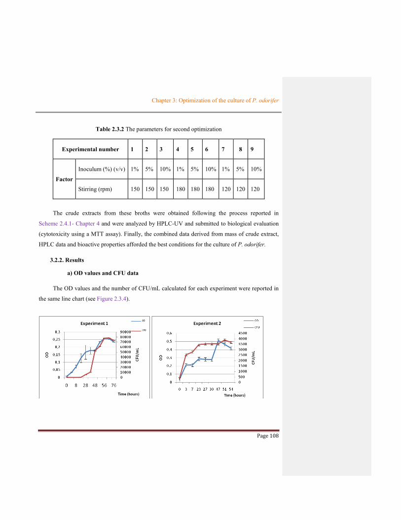
Chapter 3: Optimization of the culture of P. odorifer
Page 108
Table 2.3.2 The parameters for second optimization
Experimental number 1 2 3 4 5 6 7 8 9
Factor
Inoculum (%) (v/v) 1% 5% 10% 1% 5% 10% 1% 5% 10%
Stirring (rpm) 150 150 150 180 180 180 120 120 120
The crude extracts from these broths were obtained following the process reported in
Scheme 2.4.1- Chapter 4 and were analyzed by HPLC-UV and submitted to biological evaluation
(cytotoxicity using a MTT assay). Finally, the combined data derived from mass of crude extract,
HPLC data and bioactive properties afforded the best conditions for the culture of P. odorifer.
3.2.2. Results
a) OD values and CFU data
The OD values and the number of CFU/mL calculated for each experiment were reported in
the same line chart (see Figure 2.3.4).

Chapter 3: Optimization of the culture of P. odorifer
Page 109

Chapter 3: Optimization of the culture of P. odorifer
Page 110
Figure 2.3.4 The OD and number of CFU/mL for each experiment at different culture conditions
As seen in Figure 2.3.4, the OD and CFU/mL data has evolved for each experiment in the
same manner indicating that there is a linear relationship between these two parameters.
b) The mass of the crude extracts
The mass of crude extracts were shown in Table 2.3.3. Following the process described in
Scheme 2.4.1 in Chapter 4 – part 4.1 two different crudes were obtained for each experiment
labeled resin or supernatant extracts. In most experiments, the mass of resin extracts was higher
than those of the supernatant extracts expected for experiments 5 and 9.
Table 2.3.3 The mass of crude extracts (mg) from experiments obtained during the second optimization step
Experiment No 1 2 3 4 5 6 7 8 9
mass of crude extracts (mg)
Resin extract
176.2 164.2 200.4 229.8 237.6 176 439.5 307.9 136.0
Supernatant extract
59.0 39.6 85.0 103.9 264.8 128.0 61.9 111.6 196.3
c) The analysis of chemical profile of each extract by HPLC
The analysis by HPLC-DAD was performed using the same concentration of 1 mg/mL for
each sample, with a volume injection of 20 µL, using a reverse phase (Prevail C18 column), and a
gradient of CH3CN and H2O as mobile phase (See Chapter 6, part 6.2; 6.2.2.4).

Chapter 3: Optimization of the culture of P. odorifer
Page 111
Figure 2.3.5 Chemical profiles obtained by HPLC-DAD of crude extracts from resin (R1: extract from resin of experiment number 1, similar for R2, R3, R4, R5, R7, R8, and R9)
Figure 2.3.6 Chemical profiles obtained by HPLC--DAD of crude extracts from supernatant (S1: extract from supernatant of experiment number 1)
The data reported in Figures 2.3.5 and 2.3.6 highlighted a similar chemical profiling of
extracts of all the experiments depending on the nature of these extracts (resin or supernatant).
Moreover, the resin extracts seem richer in term of metabolites than the supernatants which
contained more polar metabolites (compounds with Tr between around 13 and 24 min).
R1
R3 R4 R5
R7 R8 R9
R2
R6
S1 S2 S3 S4 S5 S6 S7 S8 S9

Chapter 3: Optimization of the culture of P. odorifer
Page 112
d) The evaluation of biological activities
The cytotoxic effect on all the crude extracts were evaluated using a MTT assay against
HaCaT human keratinocyte and B16 murine melanoma cell lines with doxorubicine as positive
control (See Chapter 6 - part 6.2 - 6.2.7). The results showed that the extracts possessing the
highest cytotoxicity (IC50 of 26 ± 3 and 17 ± 7) against the two cell lines were the resin extract 7,
which was harvested from culture at 25oC, 120 rpm of stirring with 1% inoculum in medium
supplemented with CaCO3 at pH 7, followed by resin extract 5. However the supernatant extract
of experiment 7 (in the same conditions of culture) exhibited low values of cytotoxicity (IC50 of
93±11 and 150±10 for HaCaT and B16, respectively). The difference can be explained that the
vast majority of cytotoxic metabolites in the broth number 7 were present on extract resin in
comparison to the supernatant extract.
Table 2.3.4 The results of the second optimization (in Gym Streptomyces medium supplemented with CaCO3 at 25oC, pH = 7)
Experimental conditions Results
Crude extract from resin Crude extract from supernatant
Experiment Stirring (rpm)
Inoculum (%)
Amount (mg) (in 4.0 L medium) (mg/L yield)
IC50 (µg/mL) Amount (mg) (in 4.0 L medium) mg/mL yield)
IC50 (µg/mL)
HaCaT B16 HaCaT B16
1 150 1 176.2, (43.25) 193±23 190±40 59.0, (14.75) >200 >200
2 150 5 164.2, (41.05) >200 >200 29.6, (7.40) 158±31 190±42
3 150 10 200.4, (50.1) >200 >200 85.0, (21.25) 123±17 133±15
4 180 1 229.8, (57.45) 152±24 190±34 103.9, (25.96) 118±13 110±21
5 180 5 237.6, (59.4) 63±5 50±18 246.8, (61.7) 47±11 100±16
6 180 10 176.0, (44.0) 130±10 46±20 128.0, (32.0) 56±5 96±8
7 120 1 439.5, (108.86) 26±3 17±7 61.9, (15.48) 93±11 150±10
8 120 5 307.9, (76.96) 105±15 45±18 111.6, (27.9) 39±6 115±6
9 120 10 136.0, (34.0) 167±17 18±8 196.0, (49.0) 187±34 >200

Chapter 3: Optimization of the culture of P. odorifer
Page 113
e) Conclusions
The combination of the data from the secondary optimization (see Table 2.3.4) indicated that
the best conditions of culture to produce bioactive compounds were those of experiment noted 5
and 7. Although the OD values of experiment 7 was lower than that of experiment 5, its yield
(mg/L) and cytotoxic properties exhibited values better than those of experiment 5. Therefore, the
parameters of experiment 7, corresponding to culture in Gym Streptomyces medium supplemented
with CaCO3, pH 7 at 25oC with stirring at 120 rpm, inoculation of 1% (v/v) during 7 days, were
selected as the best conditions for culture of P. odorifer.
Besides that, the summary of results in Table 2.3.4 emphasized that the parameters including
stirring and inoculum ratio were important factors dramatically affecting the production of
cytotoxic metabolites. For instance, using the same inoculum ratio, the stirring of experiment 1
was stronger than that of experiment 7, but the mass and biological effects of extracts from
experiment 1 were less important than those of experiment 7. Similarly, the cultures with the same
stirring rate gave noticeable differences on the mass and cytotoxicity of crude extracts as displayed
for experiments 6, 7.
3.2.3. The application of these results for cytotoxic compounds discovery
The crude extracts from experiments number 7 (R7 and S7) which exhibited higher activities
were directly used to isolate metabolites. The extract R7 (439.5 mg) was submitted to various
chromatography approaches as flash chromatography, analytical and semi-preparative HPLC (See
Chapter 6, part 6.2) to afford 9 compounds as two tert-butyl compounds, furfural derivatives …
Among them, one of the two tert-butyl compounds possessed a significant cytotoxic effect against
HaCaT human keratinocyte and B16 murine melanoma cell lines. Moreover, these compounds are
considered to be rare in nature. The isolation, structural elucidation and evaluation of bioactive
properties of these compounds were detailed as an article (Chapter 4 - part 4.3). The other
compounds were classically reported in this manuscript in Chapter 4 - part 4.5.
Further, the culture with a large volume (40 L in Erlenmeyer) using the best conditions
selected following the results of the second optimization was carried out to find bioactive

Chapter 3: Optimization of the culture of P. odorifer
Page 114
metabolites with higher amounts. Bioassay-guided fractionation of the resin extract led to the
identification of a new alkaloid formed by a dihydronaphthalen part fused to a pyrrolooxazine unit.
The details of the isolation, structural elucidation and cytotoxic activity of this alkaloid were
reported as an article (see Chapter 4 - part 4.4).

Page 115
CHAPTER 4: ISOLATION OF METABOLITES FROM
PAENIBACILLUS ODORIFER
Molecules 2018, 23, x; doi: www.mdpi.com/journal/molecules
CHAPTER 4: ISOLATION OF METABOLITES FROM PAENIBACILLUS
ODORIFER
Once the parameters of culture of P. odorifer have been selected we have carried
out the cultures either by Erlenmeyer Flasks or by Bioreactor following the general process
described below (chapter 4.1). We have focused our work on the resin or supernatant
extracts and a bio-guided approach has been attempted in order to highlight the metabolites
of interest. This chapter will be divided into 5 parts including part 4.1 being introduction of
the process for the production of crude extracts from many fermentations, part 4.2 dealing
with the presentation of an oligosaccharide obtained in high amount during a culture in
bioreactor, the isolation of tert-butyl compounds will be discussed into part 4.3, the
selection of an active novel alkaloid will be described into part 4.4 and finally a report of
the other metabolites produced by this species following various kind of cultures will be
done on part 4.5.
4.1 THE PROCESS FOR THE PRODUCTION OF CRUDE EXTRACTS FROM FERMENTATION
After separation of supernatant and bacterial cells by centrifugation, a XAD-7HP
resin was added to the supernatant to absorb organic compounds with stirring for 4 hours.
After filtration, the resulting supernatant was extracted by EtOAc to afford supernatant
extract. While the resin was desorbed using MeOH : acetone 50:50 (v/v) three times, the
resulting phase was concentrated then extracted by EtOAc to afford a crude extract labeled
resin extract. For each culture, two different crude extracts were each time obtained and
called supernatant extract (SE) and resin extract (RE). The general process was reported in
Scheme 2.4.1.

Molecules 2018, 23, x 2 of 294
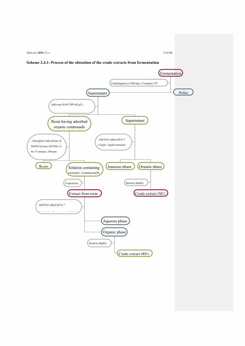
Molecules 2018, 23, x 3 of 294
Scheme 2.4.1: Process of the obtention of the crude extracts from fermentation
Fermentation
Supernatant
Pellet
Resin having adsorbed organic compounds
Supernatant
Add resin XAD-7HP (40 g/L)
-Stirring at 240 rpm during 4 hours
Resin
Aqueous phase
Solution containing organic compounds
containing organic compounds
Extract from resin
Aqueous phase
Organic phase
Crude extract (SE)
Centrifugation at 3500 rpm, 15 minutes, 40C
- Desorption with mixture of
MeOH/Acetone (50/50)(v/v)
for 15 minutes, 180 rpm -
Repeated 3 times
Evaporation
-Add H2O, adjust pH to 7
-Liquid –liquid extraction with EtOAc
Organic phase
Crude extract (RE)
(RE)
Dried by MgSO4
- Vacuum
evaporation
-Add H2O, adjust pH to 7
-Liquid –liquid extraction
with Ethyl acetate
Dried by MgSO4

Molecules 2018, 23, x 4 of 294
4.2. ISOLATION OF A POLYSACCHARIDE UNIT
During our work, a polysaccharide fraction was isolated and its antioxidant and
cytotoxic activities were exhibited. This part will be described in chapter 4 – part 4.2.2 as
an article. We will first describe a general presentation on bacterial polysaccharides and the
polysaccharide fraction isolated from P. odorifer culture from bioreactor will be then
reported.
4.2.1. General presentation of bacterial polysaccharides: structure and properties
Polysaccharides are polymers of high molecular weights formed by chains of sugar
units connected by many ether glycosidic linkages. They are ubiquitary present in all living
organisms and are important for the life due to their properties such as responsible of
membrane rigidity or gel formation, energy storage, protection against dehydration etc.
Bacterial polysaccharides have recently attracted much attention from chemists due to their
easier biotechnological production and their significant properties (see in Ruas-Madiedo et
al., 2002). Thus, many active bacterial polysaccharides were reported continuously such as
an exopolysaccharide isolated from Micrococcus luteus possessing antioxidant property
using DPPH assay (EC50 of 180 µg/mL) (Asker et al., 2014); an extracellular
polysaccharide from Streptomyces sp. exhibiting antioxidant DPPH activity and
cytotoxicity using MTT assay against human breast cancer (MDA-MB-231) and mouse
breast cancer (4T1) cell lines (Elnahas et al., 2017); an exopolysaccharide from Bacillus
coagulans with an effect of lipid peroxidation inhibition (TBA assay) (Kodali et al., 2011);
an extracellular polysaccharide from Paenibacillus polymyxa having antioxidant activity
using NBT assay (Raza et al., 2011), an exopolysaccharide from Bacillus amyloliquefaciens
sp. owning antitumor ability (MTT assay) against gastric carcinoma cell lines MC-4 (IC50
of 19.7 µg/µL) and SGC-7901 (IC50 of 26.8 µg/µL) (Chen et al., 2013); a sulfate
polysaccharide from Pseudomonas sp. displaying its cytotoxicity against many human
cancer cell lines (Matsuda et al., 2011).

Molecules 2018, 23, x 5 of 294
Bacterial polysaccharides represent a diverse range of macromolecules that include
peptidoglycan (Manna et al., 2017) consisting of sugars and aminoacids;
lipopolysaccharides (Kokoulin et al., 2016; Ravenscroft et al., 2015) containing lipid and
polysaccharide, capsular polysaccharides (Kadirvelraj et al., 2006; Petersen et al., 2014)
and exopolysaccharides (Asker et al., 2014; Hung et al., 2005) containing polysaccharide
and protein. These compounds demonstrate functions like structural cell-wall components
(e.g. peptidoglycan (Schleifer et al., 1972)), immunostimulating effect (Manna et al., 2017),
as important virulence factors (e.g. Poly-N-acetylglucosamine in Staphylococcus aureus
(Kropec et al., 2005)) or permitting the bacterium to survive in harsh environments (e.g.
Pseudomonas aeruginosa in the human lung (Sadikot et al., 2005)).
4.2.2. Production of one polysaccharide fraction from P. odorifer
The polysaccharide fraction was extracted from the broth of the culture carried out on
bioreactor (4.5 L) at 25oC, with Gym Streptomyces medium supplemented with CaCO3 at
pH =7 (Chapter 3 - 3.1). The crude extract (353.7 mg) collected from the culture (following
Scheme 2.4.1) was separated by flash chromatography using a reverse phase Reveleris
(Grace) C18 column with parameters described in 5.2.2 and Figure 2.5.2 (Chapter 5) to
afford 21 fractions. The twentieth fraction was purified several times by methanol to give a
polysaccharide fraction (116.4 mg) (Table 2.5.1) yielding a 0.0073 g/L.
The polysaccharide fraction was first identified by IR spectrum; the elucidation of its
structure performed via various steps and approaches was reported in an article (in
progress).

Molecules 2018, 23, x 6 of 294
References
Asker, M.M.S., EL Sayed, O.H., Mahmoud, M.G., Ramadan, M.F., 2014. Chemical structure and antioxidant
activity of a new exopolysaccharide produced from Micrococcus luteus. Journal of Genetic
Engineering and Biotechnology 12, 121–126. https://doi.org/10.1016/j.jgeb.2014.08.002
Chen Y.-T., Yuan, Q., Shan, L.-T., Lin, M.-A., Cheng, D.-Q., Ly, C.-Y., 2013. Antitumor activity of bacterial
exopolysaccharides from the endophyte Bacillus amyloliquefaciens sp. isolated from Ophiopogon
japonicus. Oncol Lett 5, 1787–1792. https://doi.org/10.3892/ol.2013.1284
Elnahas, M.O., Amin, M.A., Hussein, M.M.D., Shanbhag, V.C., Ali, A.E., Wall, J.D., 2017. Isolation,
Characterization and Bioactivities of an Extracellular Polysaccharide Produced from Streptomyces sp.
MOE6. Molecules 22, 1396. https://doi.org/10.3390/molecules22091396
Hung, C.-C., Santschi, P.H., Gillow, J.B., 2005. Isolation and characterization of extracellular polysaccharides
produced by Pseudomonas fluorescens Biovar II. Carbohydrate Polymers 61, 141–147.
https://doi.org/10.1016/j.carbpol.2005.04.008
Kadirvelraj, R., Gonzalez-Outeiriño, J., Foley, B.L., Beckham, M.L., Jennings, H.J., Foote, S., Ford, M.G.,
Woods, R.J., 2006. Understanding the bacterial polysaccharide antigenicity of Streptococcus agalactiae
versus Streptococcus pneumoniae. Proc Natl Acad Sci U S A 103, 8149–8154.
https://doi.org/10.1073/pnas.0602815103
Kodali, V.P., Perali, R.S., Sen, R., 2011. Purification and Partial Elucidation of the Structure of an
Antioxidant Carbohydrate Biopolymer from the Probiotic Bacterium Bacillus coagulans RK-02. J. Nat.
Prod. 74, 1692–1697. https://doi.org/10.1021/np1008448
Kokoulin, M.S., Kuzmich, A.S., Kalinovsky, A.I., Tomshich, S.V., Romanenko, L.A., Mikhailov, V.V.,
Komandrova, N.A., 2016. Structure and anticancer activity of sulfated O-polysaccharide from marine
bacterium Cobetia litoralis KMM 3880T. Carbohydrate Polymers 154, 55–61.
https://doi.org/10.1016/j.carbpol.2016.08.036
Kropec, A., Maira-Litran, T., Jefferson, K.K., Grout, M., Cramton, S.E., Götz, F., Goldmann, D.A., Pier,
G.B., 2005. Poly-N-Acetylglucosamine Production in Staphylococcus aureus Is Essential for Virulence
in Murine Models of Systemic Infection. Infect Immun 73, 6868–6876.
https://doi.org/10.1128/IAI.73.10.6868-6876.2005
Manna, D.K., Maity, P., Nandi, A.K., Pattanayak, M., Panda, B.C., Mandal, A.K., Tripathy, S., Acharya, K.,
Sahoo, A.K., Gupta, N., Roy, S., Islam, S.S., 2017. Structural elucidation and immunostimulating
property of a novel polysaccharide extracted from an edible mushroom Lentinus fusipes. Carbohydrate
Polymers 157, 1657–1665. https://doi.org/10.1016/j.carbpol.2016.11.048

Molecules 2018, 23, x 7 of 294
Matsuda, M., Yamori, T., Naitoh, M., Okutani, K., 2003. Structural Revision of Sulfated Polysaccharide B-1
Isolated from a Marine Pseudomonas Species and Its Cytotoxic Activity Against Human Cancer Cell
Lines. Mar. Biotechnol. 5, 13–19. https://doi.org/10.1007/s10126-002-0046-5
Petersen, B.O., Hindsgaul, O., Paulsen, B.S., Redondo, A.R., Skovsted, I.C., 2014. Structural elucidation of
the capsular polysaccharide from Streptococcus pneumoniae serotype 47A by NMR spectroscopy.
Carbohydrate Research 386, 62–67. https://doi.org/10.1016/j.carres.2013.11.013
Ravenscroft, N., Cescutti, P., Gavini, M., Stefanetti, G., MacLennan, C.A., Martin, L.B., Micoli, F., 2015.
Structural analysis of the O-acetylated O-polysaccharide isolated from Salmonella paratyphi A and
used for vaccine preparation. Carbohydrate Research 404, 108–116.
https://doi.org/10.1016/j.carres.2014.12.002
Raza, W., Makeen, K., Wang, Y., Xu, Y., Qirong, S., 2011. Optimization, purification, characterization and
antioxidant activity of an extracellular polysaccharide produced by Paenibacillus polymyxa SQR-21.
Bioresource Technology 102, 6095–6103. https://doi.org/10.1016/j.biortech.2011.02.033
Ruas-Madiedo, P., Hugenholtz, J., Zoon, P., 2002. An overview of the functionality of exopolysaccharides
produced by lactic acid bacteria. International Dairy Journal, NIZO Dairy Conference on Food
Microbes 2001 12, 163–171. https://doi.org/10.1016/S0958-6946(01)00160-1
Sadikot, R.T., Blackwell, T.S., Christman, J.W., Prince, A.S., 2005. Pathogen–Host Interactions in
Pseudomonas aeruginosa Pneumonia. Am J Respir Crit Care Med 171, 1209–1223.
https://doi.org/10.1164/rccm.200408-1044SO
Schleifer, K.H., Kandler, O., 1972. Peptidoglycan types of bacterial cell walls and their taxonomic
implications. Bacteriol Rev 36, 407–477.

Molecules 2018, 23, x 8 of 294
Article
Isolation and structural characterization of an
antioxidant and cytotoxic fraction of polysaccharide
purified from culture of Paenibacillus odorifer – a
bacterium associated with the lichen Rhizocarpon
geographicum
----------------------------------------------------------------------------------------------------------------------------- -------
-------
An article in progress
---------------------------------------------------------------------------------------------------------------------------------------------------
-------
Le Thi Bach Nguyen1,2, Isabelle Rouard1, Aurélie Sauvager1, and Sophie Tomasi 1,*
1 Univ Rennes, CNRS, ISCR – UMR 6226, F-35000 Rennes, France
2 Faculty of Chemical and Food technology, HCMC University of Technology and Education, Vietnam
* Correspondence: [email protected] ; Tel.: +33(0)2-23-23-48-17
Academic Editor: name
Received: date; Accepted: date; Published: date
Abstract: Extracellular polysaccharide (EPS), coded as PO-QL1, was isolated from Paenibacillus
odorifer – a bacterium associated with the lichen Rhizocarpon geographicum. The EPS was obtained
from methanol extraction by successive purification via flash chromatography using C18 reserve
phase column, with elution gradient as H2O and acetonitrile mixture. Its structural feature analyzed
by IR, HPLC, NMR, LC-MS indicated that it was mainly composed of β-D-glucuronic acid, β-D-
fructose and β-L-fucose as monomeric constituent units with an approximate molar ratio as 4:2:1.
Further, the process sequencing comprising methylation, hydrolysis, reduction and acetylation was
applied to determine the structure of PO-QL1. The results based on GC-MS data indicated that the
carbohydrate contained a fraction of linked monosaccharides as →2)- β-D-GlcAp-(1→2)-β-D-GlcAp-
(1→2)-β-D-GlcAp-(1→2)-β-D-GlcAp-(1→4)-β-D-Fruf-(2→4)-β-D-Fruf-(2→4)-β-L-Fucp-(1→. This
extracellular polymeric substance also displayed a moderate antioxidant activity using NBT assay
(IC50 of 22.5 ± 1.5 µg/mL), while it possessed a significant cytotoxicity measured by MTT assay with
IC50 value of 19 µg/mL and 27 µg/mL on HaCaT human keratinocyte and B16 murine melanoma
cell lines, respectively. This may provide a potential source of antitumor agents as microbial
polysaccharide fraction from P. odorifer.
Keywords: Extracellular polysaccharide fraction; Paenibacillus odorifer; Rhizocarpon geographicum.
1. Introduction
Natural polysaccharides, including those secreted by bacteria, have drawn the attention of
many researchers during the last decades due to their significant biological activities. Indeed, the
microbial polysaccharides, or extracellular polysaccharides (EPSs), extracted from the broth of
culture process from bacteria were recently postulated as a potential source of antioxidants [1] – for
Molecules 2018, 23, x 9 of 294
important and useful applications in pharmaceutical and cosmetic industries [2] and for antitumor
agents discovery [3,4]. Recently, EPSs derived from Paenibacillus genus have attracted much
attention because of their biotechnological potential in many distinct fields such as the production
of antioxidant active EPSs [5];; of EPS for application in cosmetic field [6]; of polysaccharides useful
in wastewater treatment [7]; of bioflocculant polymeric substance [8]; of curdlan with potential in
pharmaceutical industry [9]; of removal heavy metal EPS [10], of a multi-functional EPS [11]; or of
-glucan with enhancing immunity ability for animals [12]. However, no EPSs has been reported
from Paenibacillus odorifer species. Herein, we first report structural characterization of a EPS
fraction isolated from P. odorifer that exhibited remarkable antioxidant and cytotoxic activities.
2. Results and Discussion
2.1. Characterization of a polysaccharide fraction PO-QL1 isolated from Paenibacillus odorifer
2.1.1. Analysis by Fourier transform Infrared (FT-IR) spectroscopy
The main structural groups of polysaccharide PO-QL1 were first identified by characteristic
absorbance band maxima highlighted by FT-IR spectrum analysis (Figure 1). It revealed a typical
major broad stretching vibration at 3342 cm-1 for hydroxyl groups that were assigned to functional
groups of polysaccharides. The sharp bands at 2917 and 2849 cm-1 introduced the C-H bond that
was always present in carbohydrates. The strong absorption at 1656 cm-1 is characteristic to
stretching band of carbonyl groups (C=O). Moreover, the prominent band at 1062 cm-1 was
attributed to stretching vibration of C-O-C bond of glycosidic linkages. Interestingly, the band at
the frequency of 950 cm-1 suggested the presence of β-anomer linkage in PO-QL1 [13].
Figure 1. Fourier transform infrared (FT-IR) spectrum of polysaccharide QL1 in the range of 400 – 4000
cm-1
2.1.2. Analysis of 1H-NMR data of PO-QL1
The structure of the polysaccharide fraction PO-QL1 was further identified by one-dimensional
(1D) 1H-NMR spectrum (Figure 2). The 1H-NMR data showed signals at δH 2.8-2.9 ppm
corresponding to acetyl groups [14], and it also presented an overlap signal region at δH 3.0-4.0 ppm
assigned to protons belonging to ring monosaccharide of many sugar units – a typical structure of
polysaccharide. One interesting point was highlighted in the 1H-NMR with the presence of a signal
at δH 4.5-5.5 ppm characteristic of β-anomer linkage between monosaccharide units. These results
are similar to those shown by FTIR spectrum of PO-QL1 (Figure 1).
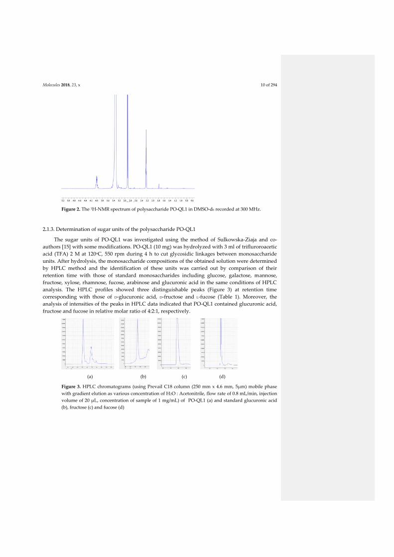
Molecules 2018, 23, x 10 of 294
Figure 2. The 1H-NMR spectrum of polysaccharide PO-QL1 in DMSO-d6 recorded at 300 MHz.
2.1.3. Determination of sugar units of the polysaccharide PO-QL1
The sugar units of PO-QL1 was investigated using the method of Sulkowska-Ziaja and co-
authors [15] with some modifications. PO-QL1 (10 mg) was hydrolyzed with 3 ml of trifluroroacetic
acid (TFA) 2 M at 120oC, 550 rpm during 4 h to cut glycosidic linkages between monosaccharide
units. After hydrolysis, the monosaccharide compositions of the obtained solution were determined
by HPLC method and the identification of these units was carried out by comparison of their
retention time with those of standard monosaccharides including glucose, galactose, mannose,
fructose, xylose, rhamnose, fucose, arabinose and glucuronic acid in the same conditions of HPLC
analysis. The HPLC profiles showed three distinguishable peaks (Figure 3) at retention time
corresponding with those of D-glucuronic acid, D-fructose and L-fucose (Table 1). Moreover, the
analysis of intensities of the peaks in HPLC data indicated that PO-QL1 contained glucuronic acid,
fructose and fucose in relative molar ratio of 4:2:1, respectively.
(a) (b) (c) (d)
Figure 3. HPLC chromatograms (using Prevail C18 column (250 mm x 4.6 mm, 5µm) mobile phase
with gradient elution as various concentration of H2O : Acetonitrile, flow rate of 0.8 mL/min, injection
volume of 20 µL, concentration of sample of 1 mg/mL) of PO-QL1 (a) and standard glucuronic acid
(b), fructose (c) and fucose (d)

Molecules 2018, 23, x 11 of 294
Table 1. Comparison of retention time between polysaccharide after treatment by TFA with standard
monosaccharides in HPLC analysis using reserve-phase Prevail column and elution with a gradient of
mixture H2O/Acetonitrile.
Polysaccharide
PO-QL1 Retention time
(min)
Standard
monosaccharide
Retention time
(min)
Peak 1 3.12 Glucuronic acid 3.16
Peak 2 3.92 Fructose 3.92
Peak 3 4.4 Fucose 4.4
2.1.4. Determination of the order of linked sugar units by LC-MS
NMR spectroscopy and HPLC analysis were valuable tools for the structural elucidation of
polysaccharides, affording saccharide compositions, ring glycosidic linkage informations...
Nevertheless these techniques were limited by the requirement of relatively large amounts of
polysaccharides. Mass spectrometry (MS) is another powerful technique for structural elucidation
of carbohydrates, including those based on electrospray ionization (ESI-MS) and matrix-assisted
laser desorption/ionization time–of-flight (MALDI-TOF-MS). Both ESI-MS and MALDI-TOF-MS
possess particularly advantages in the analysis of macromolecules [16]. In MALDI-TOF-MS, the
parent ion can be easily formed, but this ion cannot accurately give data on major ion in order to
determine molecule weight of polysaccharide because of the easily damaged structure occurring
during the analysis. While ESI-MS, combined with on-line liquid phase chromatography (LC),
provided very gentle ionization that lead to more informations about mono-, di-, tri-<
oligosaccharides from fragments collected in the analysis process [17,18,19] without having ability
to give true molecular weight of polysaccharides. In this report, a LC-MS method was applied to
analysis structural characterization of polysaccharide PO-QL1 (Figure 4). The results from the most
abundant ion series were characteristic of the small saccharides formed in ionization source. The
major components of PO-QL1 were mainly glucuronic acid, fructose, fucose according to HPLC
analysis. Indeed the analysis of LC-MS data focused on these three kinds of monosaccharides and
the results were reported in Table 2. After analysis of all these data a small structure of
polysaccharide PO-QL1 corresponding to an oligosaccharide fraction was described in Figure 5.
Figure 4. TIC (Total ion current) chromatogram and full mass rang scan ESI-MS spectrum in
positive mode of polysaccharide PO-QL1
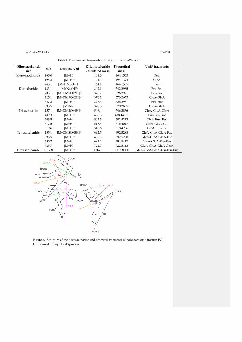
Molecules 2018, 23, x 12 of 294
Table 2. The observed fragments of PO-QL1 from LC-MS data.
Oligosaccharide
size m/z Ion observed
Oligosaccharide
calculated mass
Theoretical
mass
Unit/ fragments
Monosaccharide 165.0 [M+H]+ 164.0 164.1565 Fuc
195.3 [M+H]+ 194.3 194.1394 GlcA
243.1 [M+DMSO+H]+ 164.1 164.1565 Fuc
Disaccharide 183.1 [M+Na+H]2+ 342.1 342.2965 Fru-Fru
203.1 [M+DMSO+2H]2+ 326.2 326.2971 Fru-Fuc
225.1 [M+DMSO+2H]2+ 370.2 370.2635 GlcA-GlcA
327.3 [M+H]+ 326.3 326.2971 Fru-Fuc
393.5 [M+Na]+ 370.5 370.2635 GlcA-GlcA
Trisaccharide 157.1 [M+DMSO+4H]4+ 546.4 546.3876 GlcA-GlcA-GlcA
489.3 [M+H]+ 488.3 488.44352 Fru-Fru-Fuc
503.5 [M+H]+ 502.5 502.4212 GlcA-Fru- Fuc
517.5 [M+H]+ 516.5 516.4047 GlcA-GlcA-Fuc
519.6 [M+H]+ 518.6 518.4206 GlcA-Fru-Fru
Tetrasaccharide 155.1 [M+DMSO+5H]5+ 692.5 692.5288 GlcA-GlcA-GlcA-Fuc
693.5 [M+H]+ 692.5 692.5288 GlcA-GlcA-GlcA-Fuc
695.2 [M+H]+ 694.2 694.5447 GlcA-GlcA-Fru-Fru
723.7 [M+H]+ 722.7 722.5118 GlcA-GlcA-GlcA-GlcA
Hexasaccharide 1017.8 [M+H]+ 1016.8 1016.8100 GlcA-GlcA-GlcA-Fru-Fru-Fuc
O
O
COOH
O
HO
HO
O
OHO
HO
HOOC
O
OHOOC
HOHO O
HOOC
O
OH
OH
O
CH2OH
HOH2CO O
CH2OH
HOH2C
HOHO
O
O
HO
OH
O
195.3
165.0327.3
723.7
393.5
489.3
519.6
695.2
Figure 5. Structure of the oligosaccharide and observed fragments of polysaccharide fraction PO-
QL1 formed during LC-MS process.

Molecules 2018, 23, x 13 of 294
2.1.5. Determination of linkage position of sugar units in polysaccharide PO-QL1 by GC-MS
The alditol acetate and partially methylated alditol acetate method applied on carbohydrates
and analyzed by gas chromatography mass spectrometry (GC-MS) is mostly employed [20]. This
procedure involves the conversion of all the free hydroxyl groups on polysaccharide into methoxyl
group followed by cleavage of glycosidic linkage, reduction reaction to open monosaccharide ring
and acetylated process. The substitution patterns of O-methyl groups in monomers revealed the
carbon atoms in polysaccharide that are not involved in glycosidic linkage. After a chain reaction,
these monosaccharides were analyzed by GC-MS providing information of position linkage
between sugar units in polysaccharides. In GC-MS, the collected fragments derived from the
cleavage between two adjacent carbon atoms containing simultaneously two methoxyl groups or
one bearing methoxyl group and one linked to acetyl group. The cleavage of two adjacent carbon
atoms bearing two acetyl groups rarely happened [20]. Therefore, the linked positions of
monosaccharide ring can be easily confirmed via the mechanism of the cleavage. The results of GC-
MS analysis of polysaccharide PO-QL1 after treatment was shown in Figure 6 and exhibited that
glycosidic linkage between units of glucuronic acid, fructose and fucose were at carbon atoms with
position 1,2 ; 2,4 and 1,4 respectively (Table 3).
(a) (b)
(c)
Figure 6. The observed fragments of glucuronic acid (a), fructose (b) and fucose (c) from GC-MS.

Molecules 2018, 23, x 14 of 294
Table 3. GC-MS data for alditol acetate derivatives from polysaccharide PO-QL1
Methylated sugar Retention time
(min)
Mass fragments
(m/z)
Type of
linkage
3,4,6-tri-O-Me-glcA1 41.65
55, 69, 71, 83, 85, 97,
99, 111, 113, 129, 131,
145, 175, 189, 219, 233
→2)-glcA-(1→
1,3-di-O-Me-fru 38.09
57, 69, 71, 85, 99,
113, 117, 129, 159,
161, 189, 233
→4)-fru-(2→
2,3-di-O-Me-fuc 40.98
41, 55, 57, 69, 83,
97, 111, 117, 159,
161, 203
→4)-fuc-(1→
1 1,2,5-tri-O-acetyl-3,4,6-tri-O-methyl glucuronic acid.
2.1.6. Conclusion of structural characterization of the polysaccharide fraction PO-QL1
No single method can be completely determined the structure of polysaccharide due to its
complex structure. Therefore, the simultaneous combination of many evident data from IR, NMR,
HPLC, LC-MS, GC-MS with distinguished chemical modifications led to the structural
characterization of a fraction from PO- QL1 as a polysaccharide containing a chain of
oligosaccharide as follows → 2)- β-D-GlcAp-(1→2)-β-D-GlcAp-(1→2)-β-D-GlcAp-(1→2)-β-D-GlcAp-
(2→2)-β-D-Fruf-(2→4)-β-D-Fruf-(2→4)- β-L-Fucp-(1→ (Figure 5).
2.2. Biological activities of PO-QL1
2.2.1. Antioxidant activity
The polysaccharides were recently reported as effective free radical scavengers and
antioxidants, playing a critical role in protection against oxidation damages in living organisms
[21,22,23]. Although they lacked phenolic-type structures which were essential for scavenging free
radicals, the natural polysaccharides emerged as an important kind of antioxidants. Their
antioxidant potentials were not determined by a single factor but a combination of several related
factors [24] including molecular weight [25], monosaccharide components such as galacturonic
acid, glucuronic acid...[26,27], the presence of electrophilic groups like keto, aldehyde<which
facilitated the liberation of hydrogen from O-H bond [21,28].
2.2.1.1. 2,2-Diphenyl-1-picrylhydrazyl (DPPH) Radical Scavenging
The DPPH assay is popular in natural product antioxidant studies. This assay is based on the
theory that a hydrogen donor is an antioxidant. The antioxidant effect can be evaluated by
following the decrease of UV absorption at 540 nm corresponding to the transformation of DPPH
radical (purple) into DPPH (yellow). Antioxidant activity of polysaccharide PO-QL1 was
investigated using DPPH assay and gallic acid, quecertine were used as positive controls. PO-QL1
did not display antioxidant activity by DPPH method whereas it was described in references for
other natural polysaccharides.
2.2.1.2. Nitroblue tetrazolium (NBT)
The superoxide radical scavenging activity assay was performed using the method of nitroblue
tetrazolium (NBT) reduction with Vitamin C as a control sample. Polysaccharide was tested at
various concentrations from 0.25 - 50 mg/mL. The results exhibited antioxidant activity of PO-QL1
with IC50 value 22.5 ± 1.5 µg/mL, in comparison with a 6.1 ± 0.2 µg/mL IC50 value of Vitamin C.

Molecules 2018, 23, x 15 of 294
2.2.2. MTT Assay
Cytotoxic activity of PO-QL1 was investigated by MTT assay on two cell lines as human
keratinocyte cells (HaCaT) and murine melanoma cell (B16) with doxorubicine as positive control
[29]. PO-QL1 demonstrated a significant cytotoxic activity with an IC50 value of 19 µg/mL and 27
µg/mL on HaCaT and B16, respectively.
3. Materials and Methods
3.1. Materials and Regents
2,2-Diphenyl-1-picrylhydazyl (DPPH), nitroblue tetrazolium (NBT), 3-(4,5-dimethylthiazol -2-
yl)-2,5-diphenyltetrazolium bromide (NBT), trifluoroacetic acid (TFA), sodium borohydride
(NaBH4), glucose, malt extract, yeast extract, agar, XAD-7HP-resin were purchased from Sigma-
Aldrich, Germany. RMPI 1640 medium was purchased from Eurobio-Abcys, France.
3.2. PO-QL1 Isolation and Purification
PO-QL1 was isolated from the Gym Streptomyces medium (containing 4 g glucose, 4 g yeast
extract, 10 g malt extract, 2 g CaCO3 in 1 L) for the culture broth of Paenibacillus odorifer – a
bacterium identified from the lichen Rhizocarpon geographicum. After 7 days culture, the
fermentation (4.2 L) was separated by centrifugation (Allegra Série X-12- Thermo Ficher Scientific,
USA) at 3500 x g, 4oC for 15 min to eliminate the pellet. The XAD-7HP resin was then added into
the broth (40 g/L, 180 rpm during 4 hours) and it was filtered and de-adsorbed by solvent mixture
of Methanol : Acetone (1:1) (shaken at 180 rpm for 15 min). The mixture of solvent was removed
under reduced pressure; the resulting aqueous layer was extracted with ethyl acetate (3 x 300 mL).
The crude extract (439.5 mg) was collected after drying of the EtOAc – solute extract under vacuum.
This crude extract was subjected to flash chromatography (Puriflash INTERCHIM) with a
reverse phase C18 Reveleris (Grace) column, using a sequential mixture of solvent of ACN and H2O
to furnish 21 fractions. The fraction 21 was washed several times by MeOH to give PO-QL1 (116.4
mg) yielding a 0.0073 g/L.
3.3. PO-QL1 Spectra Analysis
The infrared spectrum of PO-QL1 was recorded on Fourier transform infrared (FTIR)
spectroscopy (Thermo Fisher, Germany) to identify structural groups of polysaccharide. A
polysaccharide sample of two milligrams was pressed and scanned in the frequency range of 4000 –
400 cm-1.
The NMR analysis was performed in DMSO-d6 solution on a Bruker DMX 300 spectrometry
[300 MHz for 1H and 75 MHz for 13C).
The LC-MS experiment was first carried out on HPLC system (Shimadzu- DGU-20AsA,
Shimadzu Corporation, Kyoto, Japan) using Prevail C18 column (250 mm x 4.6 mm, 5 µm); mobile
phase with gradient elution as various ratio of H2O : Acetonitrile (beginning at 0% of ACN in 5 min,
then increasing up to 100% of ACN at 35 min, and maintaining this concentration in next 10 min,
finally decreasing to 0% at 55 min) ; flow rate of 0.8 mL/min; injection volume of 20 µL;
concentration sample 1 mg/mL, UV wavelength of 220 nm. Advion Expression CMS plus ion-trap
mass spectrometer was used for HPLC-MS analysis (Advion, Inc, NY, USA). The MS spectrum was
recorded in scanning range of m/z 100 – 1200.
3.4. Compositional Analysis of PO-QL1
3.4.1. PO-QL1 acid hydrolysis.
The sugar units of polysaccharide QL1 was investigated using the method of Sulkowska-Ziaja
[15] with some modifications. QL1 (10 mg) was hydrolyzed with 3 ml of 2 M triflouroacetic (TFA) in

Molecules 2018, 23, x 16 of 294
the heat vial at 120oC, shaking of 550 rpm during 4 h to cut glycosidic linkages between
monosaccharide units. After reaction, the excess acid was removed by evaporation on the water
bath with a temperature of 40oC and under vacuum with water (3 x 5 mL). The product obtained
after hydrolysis was investigated by HPLC with chromatographic conditions described as above.
3.4.2. Partially methylated alditol acetates
Methylation reaction. Polysaccharide was submitted to methylation according to the method of
Osborn [30]. A sample of PO-QL1 (20 mg) and dimethylsulfoxide (DMSO) (2 mL) were placed into
a 20 ml of glass vial and was left on overnight at room temperature with shaking of 610 rpm. 15 g
powder NaOH was added the next day and the mixture was shaken for 10 min. Methyl iodide (2.5
mL) and more than 5 mg of powder NaOH were then added. The reaction was set at 20oC, shaking
of 610 rpm during 4 hours. The mixture was then put for 5 min at ambient temperature and the
milky white liquid was pulled off into a centrifuge tube. Dichloromethane (DCM) (2 mL) was
added to remaining sample left in the tube and 2 mL of distilled water was then supplemented to
centrifugation tube. The centrifugation was performed at 4000 rpm for 15 min and the water layer
was removed. The process was repeated three times to wash the sample of any impurities. After the
last centrifugation, the DCM layer was removed into the flask and was evaporated under reduced
pressure.
Hydrolysis of product from methylation. The product from methylation was dissolved in mixture
consisting of 10 drops of methanol and 1 mL of chloroform, and hydrolyzed by 2 mL of TFA (4M) at
120oC, 550 rpm during 4 hours. The mixture after reaction was treated as above.
Sodium Borohydride Reduction. The sample after hydrolysis was dissolved in 5 mL of deionized
water and added by a 3 milligram of anhydrous sodium borohydride (NaBH4). The vial was sealed
with the Teflon cap and left for 22 hours at room temperature with periodic vortexing. Glacial acetic
was then added to the reaction vial the next day until no effervescence was observed. The solution
in glass vial was evaporated to dryness under reduced pressure.
Acetylation by Acetic anhydride. After reduction, the dried sample was dissolved in an 5mL of
acidic methanol (95% methanol and 5% acetic acid) and evaporation to dryness to convert boric
acid formed during the reduction stage to volatile methyl borate. This was repeated three times to
completely remove boric acid which can interfere with the acetylation process. The product was
dried and the formation of a white powder was observed. This powder was then extracted by DCM
(3 x 3 mL) and dried by sodium sulfate.
GC-MS Analysis of Alditol Acetates. The final product obtained after derivatization was
dissolved in 1 mL of DCM. This solution was subjected in GC-MS-system (Agilent Technologies
GC-MS 7820A-GC, Agilent, Waldbronn, Germany) with Nitrogen as carrier gas, using DB-5 column
(30 M x 0.25 mm x 0.25 µm) and injection volume of 1 µL. The initial 37oC oven temperature was set
for 6s following injection, raised to 140oC for 30 min at a rate of 200C/min, then to 180oC for 40 min
at a rate of 40oC/min, and finally maintained at 230oC for 30 min. The detector temperature was held
at 270oC.
3.5. Biological evaluation
3.5.1. DDPH assay
The free radical scavenging activity of PO-QL1 was measured by DPPH method with
Quercetin and Galic acid as positive controls. 100 µL of DPPH methanol solution (freshly prepared
at a concentration of 1 mg/mL) were mixed with 10 µL of PO-QL1 at different concentrations (4.00;
11.1; 33.3; 100 µg/mL) in DMSO. After 15 min culture in the dark, the absorbance was measured at
540 nm using a UV-visible spectrophotometer.
The DPPH scavenging percentage activity was calculated as follows [14]:
Scavenging ability % = [1- (As – Ab)/Ac] x 100
Comment [ST2]: Is it ok this ref? to
check

Molecules 2018, 23, x 17 of 294
Where As is the absorbance of polysaccharide sample with DPPH, Ab is the absorbance of
polysaccharide sample without DPPH (blank), Ac is the absorbance of negative control.
3.5.2. NBT assay
Phenazine methosulphate in the presence of NADH and under aerobic conditions (+ O2)
produces superoxide anions whose formation is appreciated by a dye nitro blue tetrazolium (NBT).
In the solution containing 40 µL of tris hydrochloride buffer (16 mM, pH = 8), 50 µL nicotinamid
adenine dinucleotide (NADH) (78 µM), 50 µL NBT (50 µM) and 50 µL phenazin methosulphate 910
µM) were added to test with different concentrations of polysaccharide (0.25; 1.85; 2.5; 5.5; 16.0;
25.0; 50 mg/mL in DMSO). The color reaction of superoxide radical and NBT was monitored by
measurement of the absorbance at 560 nm. Vitamin C was used as positive control. The experiment
was repeated three times. The inhibition percentage was calculated [31] as follows :
Scavenging effect (%) = (1 – ASample at 560 nm/AControl at 560 nm) x 100
3.5.3. MTT assay
PO-QL1 (40 mg/mL) was submitted to MTT assay against HaCaT human keratinocyte and B16
murine melanoma cell lines on 96-well plate as described in literature [29]. Firstly, the HaCaT (2000
cells/well) and B16 (1800 cells/well) were cultivated in RMPI 1640 medium (Thermo Fisher
Scientific, USA) supplemented with 5% of foetal calf serum (FCS) and antibiotics (penicillin,
streptomycin) in atmosphere of 5% CO2 at 37oC. After 24 hours culture, the samples were added at
different concentrations of PO-QL1 (1, 10, 50, 100 and 200 µg/mL) and each 96-well plate was
continuously incubated at the same temperature and atmosphere as above. The cell growth after
48h culture were then measured at 540 nm by Microplate photometer (Thermo Scientific™
Multiskan™ FC, USA) using a MTT (3-[4,5-dimethylthiazole-2-yl]-2,5-diphenyltetrazolium
bromide) assay. The doxorubicine was used as a positive control. Each experiment was repeated
three times.
4. Conclusion
A polysaccharide fraction was isolated from the culture of Paenibacillus odorifer – a bacterium
associated with the lichen Rhizocarpon geographicum. Its chemical components were analyzed by IR,
NMR, HPLC and LC-MS and indicated that it contains an oligosaccharide fraction including mainly
D-glucuronic acid, D- fructose, L-fucose in a molar ratio of 4:2:1, respectively. The fraction structure
of this saccharide determined via GC-MS spectrometry was →2)- β-D-GlcAp-(1→ 2)-β-D-GlcAp-
(1→2)-β-D-GlcAp-(1→2)-β-D-GlcAp-(2→2)-β-D-Fruf-(2→4)-β-D-Fruf-(2→4)- β-L-Fucp-(1→. The
polysaccharide possessed an antioxidant activity using NBT assay (IC50 of 22.5 ± 1.6 µg/mL), and it
also presented significant cytotoxic activity measured by MTT with an IC50 value of 19 µg/mL and
27 µg/mL on HaCaT and B16 cell lines, respectively.
Acknowledgments: We sincerely thank M. Blot for assistant technology in GC-MS, L. Intertaglia from
Banyuls/mer Platform for the identification of the strain. Vietnamese government is gratefully acknowledged
for the PhD grant for Nguyen Thi Bach Le.
Author Contributions: N.T.B.L designed and performed most of the experiments and wrote manuscript; I.R.
evaluated biological tests; A.S. and S.F. were technical assistance in HPLC, LC-MS and NMR. S.T. designed,
supervised the experiments and edited the manuscript.
Conflicts of Interest: The authors declare no conflict of interest.
References

Molecules 2018, 23, x 18 of 294
1. Nwodo, U.U.; Green, E.; Okoh, A.I. Bacterial Exopolysaccharides: Functionality and Prospects. Int. J.
Mol. Sci. 2012, 13 (11), 14002–14015.
2. Freitas, F.; Alves, V.D.; Reis, M.A.M. Advances in Bacterial Exopolysaccharides: From Production to
Biotechnological Applications. Trends Biotechnol. 2011, 29 (8), 388–398.
3. Zheng, X.; Liu, Z.; Li, S.; Wang, L.; Lv, J.; Li, J.; Ma, X.; Fan, L.; Qian, F. Identification and
Characterization of a Cytotoxic Polysaccharide from the Flower of Abelmoschus Manihot. Int. J. Biol. Macromol.
2016, 82, 284–290.
4. Goodarzi, N.; Varshochian, R.; Kamalinia, G.; Atyabi, F.; Dinarvand, R. A Review of Polysaccharide
Cytotoxic Drug Conjugates for Cancer Therapy. Carbohydr. Polym. 2013, 92 (2), 1280–1293.
5. Wang, C.-L.; Huang, T.-H.; Liang, T.-W.; Fang, C.-Y.; Wang, S.-L. Production and Characterization of
Exopolysaccharides and Antioxidant from Paenibacillus Sp. TKU023. New Biotechnol. 2011, 28 (6), 559–565.
6. Liang, T.-W.; Wang, S.-L. Recent Advances in Exopolysaccharides from Paenibacillus Spp.:
Production, Isolation, Structure, and Bioactivities. Mar. Drugs 2015, 13 (4), 1847–1863.
7. Aguilera, M.; Quesada, M.T.; del Águila, V.G.; Morillo, J.A.; Rivadeneyra, M.A.; Ramos-Cormenzana,
A.; Monteoliva-Sánchez, M. Characterisation of Paenibacillus Jamilae Strains That Produce Exopolysaccharide
during Growth on and Detoxification of Olive Mill Wastewaters. Bioresour. Technol. 2008, 99 (13), 5640–5644.
8. Li, O.; Lu, C.; Liu, A.; Zhu, L.; Wang, P.-M.; Qian, C.-D.; Jiang, X.-H.; Wu, X.-C. Optimization and
Characterization of Polysaccharide-Based Bioflocculant Produced by Paenibacillus Elgii B69 and Its
Application in Wastewater Treatment. Bioresour. Technol. 2013, 134 (Supplement C), 87–93.
9. Rafigh, S.M.; Yazdi, A.V.; Vossoughi, M.; Safekordi, A.A.; Ardjmand, M. Optimization of Culture
Medium and Modeling of Curdlan Production from Paenibacillus Polymyxa by RSM and ANN. Int. J. Biol.
Macromol. 2014, 70 (Supplement C), 463–473.
10. Mokaddem, H.; Sadaoui, Z.; Boukhelata, N.; Azouaou, N.; Kaci, Y. Removal of Cadmium from
Aqueous Solution by Polysaccharide Produced from Paenibacillus Polymyxa. J. Hazard. Mater. 2009, 172 (2),
1150–1155.
11. Raza, W.; Makeen, K.; Wang, Y.; Xu, Y.; Qirong, S. Optimization, Purification, Characterization and
Antioxidant Activity of an Extracellular Polysaccharide Produced by Paenibacillus Polymyxa SQR-21.
Bioresour. Technol. 2011, 102 (10), 6095–6103.
12. Jung, H.-K.; Hong, J.-H.; Park, S.-C.; Park, B.-K.; Nam, D.-H.; Kim, S.-D. Production and
Physicochemical Characterization of β-Glucan Produced byPaenibacillus Polymyxa JB115. Biotechnol. Bioprocess
Eng. 2007, 12 (6), 713–719.

Molecules 2018, 23, x 19 of 294
13. Kac̆uráková, M.; Capek, P.; Sasinková, V.; Wellner, N.; Ebringerová, A. FT-IR Study of Plant Cell Wall
Model Compounds: Pectic Polysaccharides and Hemicelluloses. Carbohydr. Polym. 2000, 43 (2), 195–203.
14. Elnahas, M.O.; Amin, M.A.; Hussein, M.M.D.; Shanbhag, V.C.; Ali, A.E.; Wall, J.D. Isolation,
Characterization and Bioactivities of an Extracellular Polysaccharide Produced from Streptomyces Sp. MOE6.
Molecules 2017, 22 (9), 1396.
15. Sułkowska-Ziaja, K.; Muszyńska, B.; Ekiert, H. Chemical Composition and Cytotoxic Activity of the
Polysaccharide Fractions in Sarcodon Imbricatus (Basidiomycota). Acta Mycol. 2013, 47 (1), 49–56.
16. Thanawiroon, C.; Rice, K.G.; Toida, T.; Linhardt, R.J. Liquid Chromatography/Mass Spectrometry
Sequencing Approach for Highly Sulfated Heparin-Derived Oligosaccharides. J. Biol. Chem. 2004, 279 (4), 2608–
2615.
17. Hu, P.; Fang, L.; Jones, C.M.; Chess, E.K. Collective Sampling of Intact Anionic Polysaccharide
Components and Application in Quantitative Determination by LC–MS. Carbohydr. Res. 2011, 346 (14), 2268–
2273.
18. Cai, X.; Lei, Q.P.; Lamb, D.H.; Shannon, A.; Jacoby, J.; Kruk, J.; Kensinger, R.D.; Ryall, R.; Zablackis,
E.; Cash, P. LC/MS Characterization of Meningococcal Depolymerized Polysaccharide Group C Reducing
Endgroup and Internal Repeating Unit. Anal. Chem. 2004, 76 (24), 7387–7390.
19. Wang, B.-B.; Wang, J.-L.; Yuan, J.; Quan, Q.-H.; Ji, R.-F.; Tan, P.; Han, J.; Liu, Y.-G. Sugar Composition
Analysis of Fuzi Polysaccharides by HPLC-MSn and Their Protective Effects on Schwann Cells Exposed to
High Glucose. Molecules 2016, 21 (11), 1496.
20. Björndal, H.; Lindberg, B.; Svensson, S. Mass Spectrometry of Partially Methylated Alditol Acetates.
Carbohydr. Res. 1967, 5 (4), 433–440.
21. Al-Sheraji, S.H.; Ismail, A.; Manap, M.Y.; Mustafa, S.; Yusof, R.M.; Hassan, F.A. Purification,
Characterization and Antioxidant Activity of Polysaccharides Extracted from the Fibrous Pulp of Mangifera
Pajang Fruits. LWT - Food Sci. Technol. 2012, 48 (2), 291–296.
22 Asker, M.M.S.; EL Sayed, O.H.; Mahmoud, M.G.; Ramadan, M.F. Chemical Structure and Antioxidant
Activity of a New Exopolysaccharide Produced from Micrococcus Luteus. J. Genet. Eng. Biotechnol. 2014, 12 (2),
121–126.
23. Asker, M.M.S.; Shawky, B.T. Structural Characterization and Antioxidant Activity of an Extracellular
Polysaccharide Isolated from Brevibacterium Otitidis BTS 44. Food Chem. 2010, 123 (2), 315–320.
24. Wang, J.; Hu, S.; Nie, S.; Yu, Q.; Xie, M. Reviews on Mechanisms of In Vitro Antioxidant Activity of
Polysaccharides. Oxid. Med. Cell. Longev. 2015, 2016, e5692852.

Molecules 2018, 23, x 20 of 294
25. Xing, R.; Liu, S.; Guo, Z.; Yu, H.; Wang, P.; Li, C.; Li, Z.; Li, P. Relevance of Molecular Weight of
Chitosan and Its Derivatives and Their Antioxidant Activities in Vitro. Bioorg. Med. Chem. 2005, 13 (5), 1573–
1577.
26. Tsiapali, E.; Whaley, S.; Kalbfleisch, J.; Ensley, H.E.; Browder, I.W.; Williams, D.L. Glucans Exhibit
Weak Antioxidant Activity, but Stimulate Macrophage Free Radical Activity. Free Radic. Biol. Med. 2001, 30 (4),
393–402.
27. Rao, R.S.P.; Muralikrishna, G. Water Soluble Feruloyl Arabinoxylans from Rice and Ragi: Changes
upon Malting and Their Consequence on Antioxidant Activity. Phytochemistry 2006, 67 (1), 91–99.
28. Wu, H.; Min, T.; Li, X.; Li, L.; Lai, F.; Tang, Y.; Yang, X. Physicochemical Properties and Antioxidant
Activities of Acidic Polysaccharides from Wampee Seeds. Int. J. Biol. Macromol. 2013, 59, 90–95.
29. Millot, M.; Tomasi, S.; Studzinska, E.; Rouaud, I.; Boustie, J. Cytotoxic Constituents of the Lichen
Diploicia Canescens. J. Nat. Prod. 2009, 72 (12), 2177–2180.
30. Osborn, H.M.I.; Lochey, F.; Mosley, L.; Read, D. Analysis of Polysaccharides and Monosaccharides in
the Root Mucilage of Maize (Zea Mays L.) by Gas Chromatography. J. Chromatogr. A 1999, 831 (2), 267–276.
31. Liu, D.; Liao, N.; Ye, X.; Hu, Y.; Wu, D.; Guo, X.; Zhong, J.; Wu, J.; Chen, S. Isolation and Structural
Characterization of a Novel Antioxidant Mannoglucan from a Marine Bubble Snail, Bullacta Exarata (Philippi).
Mar. Drugs 2013, 11 (11), 4464–4477.
4.3. ISOLATION OF TERT-BUTYL PHENOLIC COMPOUNDS
4.3.1. State of art about tert-butyl compounds
The tert-butylphenol compounds have become novel compounds to focus for
chemists due to their interesting properties. Almost of tert-butylphenol compounds were
volatile organic compounds and they were frequently discovered from plants (see in review
of Dembitsky et al., 2006) and some characteristic structures were shown in Figure 2.4.1.

Molecules 2018, 23, x 21 of 294
MeO
OHOH
OHC
OH
OH
OH
NMe2
CHOOH
HO
R
OHR = MeR = OH
HO
OH
OMe
OMe
OH
OH
OH
OH
OHCHO
R
OH
R = COOMeR= H
OH
O
O(CH2)17Me
OH OH
2-ter t-butyl-4-methoxyphenol Topanol A
fruits of Zizyphus jujube f lowers of Aloe vera
green alga Enteromorpha compressa
red alga Hypnea musciformis
3,5-di-tert-butyl-4-hydroxybenzaldehyde
odors of apple tree
Butylphenol
fresh flowers of Lamium album, L.
galeobdolon, L. garganicum, L.
maculatum, and L. purpureum
3,5-di-tert-butyl-2-hydroxybenzaldehyde
f lowers of Fissistigmashangtzeense
3-tert-butylphenol
from Malus domestica
2-tert-butyl-6-methylphenol
Leaves of Lippia graveolens
2-tert-butyl-4-methoxyphenol
Leaves of Aloe arborescens,
Chrysosplenium nudicaule
3-tert-butyl-4-methoxyphenol
whole of Tubermelanosporum
roots of Japanese ginseng,Korean ginseng
2-tert-butyl-4-(2,4,4-trimethylpentan-2-yl)phenol
whole of Ar temisia scopar ia
2-tert-butylbenzene-1,4-diol
Leaves of Chrysanthellum indicum
octadecyl 3-(3,5-di-tert-butyl-4-hydroxyphenyl)propanoate
sawdust of Oak wood (Quercus)
3-tert-butyl-6-hydroxy-5-isopropyl-2-methylbenzaldehyde
bark of Scorodophloeus zenkere,
Hua gabonii, Af rostyrax lepidophylleus
roots of X inkaihe ginseng
2-tert-butylphenol
leaves of Filipendula palmata
2-tert-butyl-3-methylphenol
roots of Siphonostegia chinensis,
Oster icum grosseserratum
Figure 2.4.1 Tert-butylphenols isolated from nature (from Dembitsky et al., 2006)
Beside, some tert-butylphenol compounds produced from bacteria were recently
reported. They included (Figure 2.4.2) 2,4-di-tert-butylphenol from Streptomyces mutabilis
(Belghit et al., 2016), from Pseudomonas monteilii (Dharni et al., 2014) and from
Lactococcus sp. (Varsha et al., 2015); 2,5-di-tert-butylphenol from Streptomyces sp. (Jannu
et al., 2015)…etc. These tert-butylphenols exhibited an interesting antioxidant activity
reported in many studies (Choi et al., 2013; Chuah et al., 2015; Varsha et al., 2015; Yoon et

Molecules 2018, 23, x 22 of 294
al., 2006), antifungal properties (Varsha et al., 2015; Dharni et al., 2014) and cytotoxicity (
Varsha et al., 2015) as well.
OH
2,4-di-ter t-butylphenol
OH
2,5-di-ter t-butylphenol
Figure 2.4.2 Tert-Butylphenols produced by various bacteria
In our study, two tert-butylphenol derivatives were isolated. One of the compounds
possessed a strong cytotoxic effect against B16 murine melanoma and HaCaT human
keratinocyte cell lines. This compound possess a symmetrical structure composed of two
tert-butylphenol linked together by a sulfur-bond. The second compound was well-known
but its NMR spectroscopic data was for the first time reported in this work.
4.3.2. Production of the tert-butyl phenol compounds
The tert-butyl phenol compounds were isolated from the supernatant extract of the
culture broth of P. odorifer obtained during the second optimization (Experiment 7 –
Chapter 3 – 3.2 – 3.2.3). The supernatant extract (439.5 mg) was injected into flash
chromatography using a silica gel Chromabond column with parameters described in
Chapter 6 – 6.2.2.3 and Figure 2.6.1 to furnish 14 fractions.
The detailed process of isolation and the structural elucidation of these two tert-butyl
phenol derivatives and their evaluation of biological properties were described as an article
(Published by Molecules).
The NMR data for these compounds were introduced in ANNEXE 1.

Molecules 2018, 23, x 23 of 294
References
Belghit, S., Driche, E.H., Bijani, C., Zitouni, A., Sabaou, N., Badji, B., Mathieu, F., 2016. Activity of 2,4-Di-
tert-butylphenol produced by a strain of Streptomyces mutabilis isolated from a Saharan soil against
Candida albicans and other pathogenic fungi. Journal de Mycologie Médicale / Journal of Medical
Mycology 26, 160–169. https://doi.org/10.1016/j.mycmed.2016.03.001
Choi, S.J., Kim, J.K., Kim, H.K., Harris, K., Kim, C.-J., Park, G.G., Park, C.-S., Shin, D.-H., 2013. 2,4-Di-
tert-butylphenol from Sweet Potato Protects Against Oxidative Stress in PC12 Cells and in Mice. J
Med Food 16, 977–983. https://doi.org/10.1089/jmf.2012.2739
Chuah, T.S., Norhafizah, M.Z., Ismail, B.S., 2015. Evaluation of the biochemical and physiological activity of
the natural compound, 2,4-ditert-butylphenol on weeds. Crop Pasture Sci. 66, 214–223.
https://doi.org/10.1071/CP13386
Dembitsky, V.M., 2006. Natural neo acids and neo alkanes: their analogs and derivatives. Lipids 41, 309–340.
Dharni, S., Sanchita, Maurya, A., Samad, A., Srivastava, S.K., Sharma, A., Patra, D.D., 2014. Purification,
Characterization, and in Vitro Activity of 2,4-Di-tert-butylphenol from Pseudomonas monteilii PsF84:
Conformational and Molecular Docking Studies. J. Agric. Food Chem. 62, 6138–6146.
https://doi.org/10.1021/jf5001138
Jannu, V.G., Sanjenbam, P., Kannabiran, K., 2015. Preclinical evaluation and molecular docking of 2,5-di-
tert-butyl-1,4-benzoquinone (DTBBQ) from Streptomyces sp. VITVSK1 as a potent antibacterial
agent. International Journal of Bioinformatics Research and Applications 11, 142.
https://doi.org/10.1504/IJBRA.2015.068089
Varsha, K.K., Devendra, L., Shilpa, G., Priya, S., Pandey, A., Nampoothiri, K.M., 2015. 2,4-Di-tert-butyl
phenol as the antifungal, antioxidant bioactive purified from a newly isolated Lactococcus sp.
International Journal of Food Microbiology 211, 44–50.
https://doi.org/10.1016/j.ijfoodmicro.2015.06.025
Yoon, M.-A., Jeong, T.-S., Park, D.-S., Xu, M.-Z., Oh, H.-W., Song, K.-B., Lee, W.S., Park, H.-Y., 2006.
Antioxidant Effects of Quinoline Alkaloids and 2,4-Di-tert-butylphenol Isolated from Scolopendra
subspinipes. Biological and Pharmaceutical Bulletin 29, 735–739. https://doi.org/10.1248/bpb.29.735

Molecules 2018, 23, x 24 of 294
Molecules 2018, 23, x 25 of 294
Article
tert-Butylphenolic Derivatives from
Paenibacillus odorifer—A Case of Bioconversion
Thi-Bach-Le Nguyen 1, Olivier Delalande 2, Isabelle Rouaud 1, Solenn Ferron 1, Laura Chaillot 3,
Rémy Pedeux 3 and Sophie Tomasi 1,*
1 University of Rennes 1, CNRS, ISCR—UMR 6226, F-35000 Rennes, France;
[email protected] (T.-B.-L.N.); [email protected] (I.R.); solenn.ferron@univ-
rennes1.fr (S.F.) 2 University of Rennes 1, CNRS, IGDR—UMR 6290, F-35000 Rennes, France; olivier.delalande@univ-
rennes1.fr 3 Chemistry, Oncogenesis, Stress, Signaling, Centre Eugène Marquis, Université de Rennes 1,
INSERM U1242, 35000 Rennes, France; [email protected] (L.C.);
[email protected] (R.P.)
* Correspondence: [email protected]; Tel.: +33-223-234-817
Received: 14 June 2018; Accepted: 28 July 2018; Published: 5 August 2018
Abstract: Two compounds (1) and (2) containing tert-butylphenol groups were, for the first time,
produced during the culture of Paenibacillus odorifer, a bacterial strain associated with the crustose
lichen, Rhizocarpon geographicum. Their entire structures were identified by one-dimensional (1D)
and two-dimensional (2D) NMR and high-resolution electrospray ionisation mass spectrometry
(HRESIMS) spectroscopic analyses. Among them, Compound 1 exhibited significant cytotoxicity
against B16 murine melanoma and HaCaT human keratinocyte cell lines with micromolar half
maximal inhibitory concentration (IC50) values. Furthermore, after supplementation studies, a
putative biosynthesis pathway was proposed for Compound 1 throughout a bioconversion by this
bacterial strain of butylated hydroxyanisole (BHA), an antioxidant polymer additive.
Keywords: tert-butylphenols; Paenibacillus odorifer; Rhizocarpon geographicum; bioconversion
1. Introduction
Organic products with branched tert-butyl groups represent a relatively important number of
active compounds [1,2]. In the past, this group was already exploited in many organic syntheses
due to its chemical reactivity. There are more than 200 compounds containing tert-butyl groups,
described as natural products with interesting bioactivities [3]. Indeed, the tert-butyl moiety can be
found in a variety of compounds produced by various sources, such as plants, fungi, algae,
cyanobacteria [4–9], and especially from bacteria which were admitted as a source of novel and
interesting bioactive products [10]. The tert-butylated compounds from natural sources reported in
these papers often belong to classes of terpenes or polyketides. Although metabolites containing
tert-butyl functionality on the aromatic ring are considered to be rare, several tert-butylphenyl
derivatives were already isolated from organisms and they also exhibited remarkable antitumor,
antibacterial, and antioxidant activities [11–13]. Chemical studies on Paenibacillus odorifer, a
bacterium associated with the crustose lichen, Rhizocarpon geographicum, led to the identification of
two tert-butylphenol derivatives (1, 2) described in Figure 1. Interestingly, aromatic compounds
simultaneously bearing a tert-butyl group and a sulfur atom are really uncommon in nature. The
aims of this work were to characterize these compounds, to give some hypothesis about their
origin, and to evaluate their cytotoxic activities. Herein, to our knowledge, is the first report of the
bacterial production of Compound 1 carrying a tert-butyl phenol moiety and a sulfur atom.

Molecules 2018, 23, x 26 of 294
S
OH OH1
O
O17
2OH
Figure 1. The structure of tert-butylphenolic derivatives found in the culture of Paenibacillus odorifer.
2. Results
2.1. Process of Purification
Paenibacillus odorifer (P. odorifer) was cultivated via a scale-up (4.0 L) shaken fermentation at 25
°C, pH 7, 120 rpm with 1% (v/v) inoculum in GYM Streptomyces medium supplemented with
CaCO3. From the broth, the excreted metabolites were collected using Amberlite® XAD-7-HP resin,
and the resulting extract was separated via a combination of liquid/liquid extraction and normal-
phase flash chromatography to give several fractions. Final purification combining silica gel
chromatography, semi-preparative HPLC, and preparative silica thin-layer chromatography (TLC)
afforded 5,5′-thiobis(2-tert-butyl-4-methylphenol) (1; 5.0 mg) and octadecyl 3-(3,5-di-tert-butyl-4-
hydroxyphenyl)propanoate (2; 5.9 mg).
2.2. Structural Elucidation
Compound 1 was obtained as a white amorphous powder. Its molecular formula was assigned
as C22H30O2S from the [M − H]− ion at m/z 357.1983 according to high-resolution electrospray
ionisation mass spectrometry (HRESIMS) data, and required eight degrees of unsaturation. Its 13C-
NMR data highlighted nine carbon signals, which were classified by Jmod and HSQC analyses as
two aromatic methine carbons, four aromatic quaternary carbons, one aliphatic quaternary carbon,
and four methyl carbons (Table 1). The lack of coupling between two aromatic protons at δH 6.56
ppm (1H, s, H-6) and 7.00 (1H, s, H-3) indicated a para-relationship between them, and the existence
of a 1,2,4,5-tetrasubstituted phenyl nucleus. The presence of three methyl groups at δH 1.28 (9H, s,
H-9/H-10/H-11) along with their HMBC correlations to a quaternary carbon at δC 34.4 (C-8) revealed
the presence of a tert-butyl group which was one of the substituted groups on the aromatic ring. On
the other hand, the 13C shift of C-2 at δC 134.6 together with its HMBC correlations with H-9/H-
10/H-11 confirmed the position of the tert-butyl group at C-2. In addition, analysis of the HMBC
spectrum exhibiting the correlations from H-3 to C-8 (δC 34.4), C-4 (δC 125.5), C-5 (δC 137.8), and C-1
(δC 153.4) provided the presence of a methyl group at C-4, a hydroxyl group at C-1, and a sulfur-
bond at C-5. The latter could also be indicated through the HMBC correlations from the aromatic
methyl protons at δH 2.28 (3H, s, H-7) to C-4, C-5, and C-6 (δC 118.8). Moreover, HMBC correlations
from H-6 to C-1, C-2, C-4, and C-7 were also observed. Interestingly, NOE correlations between two
kinds of protons at δH 7.00 (H-3) and δH 1.28 (H-9/H-10/H-11) indicated that the tert-butyl group
possessed a position close to C-3. The remaining structural assignment of Compound 1 (Figure 2)
required C11H15O, which corresponds to the already assigned part of Compound 1. On the basis of
molecular formula and HMBC correlations, Compound 1 was suggested to possess a symmetrical
structure containing two 1,2,4,5-tetrasubstituted phenyl nuclei which were linked via a sulfur bond
This structure is close to that of a known synthetic compound Santonox (CAS registry number 96-
69-5) 78 (Figure 3). This is the first natural isolation of Compound 1.

Molecules 2018, 23, x 27 of 294
S
OH OH1
3 5
7
8
9
1'
5 '
7 '
8 '9 '
HMBC2
4
610
11
Figure 2. Key correlations for the structural assignment of 1.
S
OHHO
S
OH OH1 1'
22'
3 3'
4 4'5 5'
6 6'
7 7'
8 8'
9 9'
10 10'
11 11'
1 1'
2 2'3 3'
4 4'5 5'
6 6'
7 7'
88'
9 9'10 10'
11 11'
1 Santonox 5,5'-thiobis(2-ter t-butyl-4-methylphenol) 4,4'-thiobis(2-ter t-butyl-5-methylphenol)
Figure 3. Structures of Compound 1 and Santonox.
From the comparison of the NMR spectroscopic data, minor differences in the chemical shifts
of protons and carbons were highlighted for Compound 1 and standard Santonox. Thus, the
identification of the exact structures could not be based on these data, with the exception of NOE
correlations. For Santonox, the NOE correlations were clearly displayed between two series of
protons at δH 7.00 (aromatic proton, H-3) and δH 1.28 (tert-butyl protons), and at δH 6.54 (aromatic
proton, H-6) and δH 2.27 (methyl protons). For Compound 1, the NOE experiments only highlighted
the correlations between δH 7.00 (aromatic proton, H-3) and δH 1.28 (tert-butyl protons). The absence
of the NOE correlation between H-6 (δH 6.56) and protons of the tert-butyl group (δH 1.28) indicated
that H-6 in Compound 1 was not close to the tert-butyl group. NOE predictions obtained through
molecular dynamics simulations confirmed the NOE experimental data (see Supplementary
Materials, Figure S20 and Table S1). As a result, the data from NOE correlations finally highlighted
that Compound 1 was an isomer of Santonox. Moreover, to our best knowledge, it is the first report
of the isolation of Compound 1 from a bacterial culture.
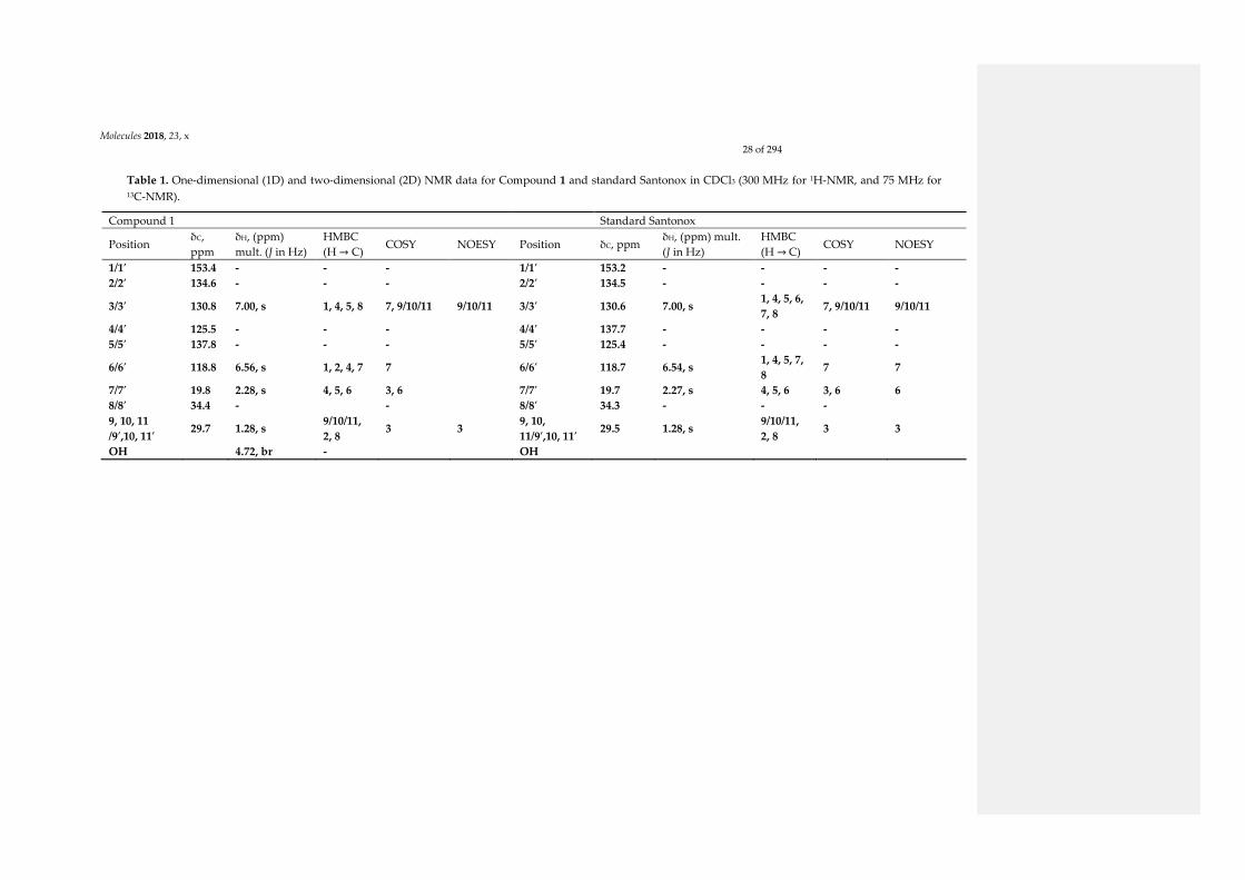
Molecules 2018, 23, x
28 of 294
Table 1. One-dimensional (1D) and two-dimensional (2D) NMR data for Compound 1 and standard Santonox in CDCl3 (300 MHz for 1H-NMR, and 75 MHz for 13C-NMR).
Compound 1 Standard Santonox
Position δC,
ppm
δH, (ppm)
mult. (J in Hz)
HMBC
(H → C) COSY NOESY Position δC, ppm
δH, (ppm) mult.
(J in Hz)
HMBC
(H → C) COSY NOESY
1/1′ 153.4 - - - 1/1′ 153.2 - - - -
2/2′ 134.6 - - - 2/2′ 134.5 - - - -
3/3′ 130.8 7.00, s 1, 4, 5, 8 7, 9/10/11 9/10/11 3/3′ 130.6 7.00, s 1, 4, 5, 6,
7, 8 7, 9/10/11 9/10/11
4/4′ 125.5 - - - 4/4′ 137.7 - - - -
5/5′ 137.8 - - - 5/5′ 125.4 - - - -
6/6′ 118.8 6.56, s 1, 2, 4, 7 7 6/6′ 118.7 6.54, s 1, 4, 5, 7,
8 7 7
7/7′ 19.8 2.28, s 4, 5, 6 3, 6 7/7′ 19.7 2.27, s 4, 5, 6 3, 6 6
8/8′ 34.4 - - 8/8′ 34.3 - - -
9, 10, 11
/9′,10, 11′ 29.7 1.28, s
9/10/11,
2, 8 3 3
9, 10,
11/9′,10, 11′ 29.5 1.28, s
9/10/11,
2, 8 3 3
OH 4.72, br - OH

Chapter 4: Isolation of metabolites from P. odorifer
Page 29
Compound 2 was isolated as a white solid and had a C35H62O3 molecular formula determined by the [M +
Na]+ peak at m/z 553.4592 from its (+)HRESIMS data. The analysis of 1H-NMR and Jmod, along with HSQC data
(Table 2), indicated the presence of two aromatic protons, 19 methylene groups (one of them oxygenated), seven
methyl groups (six of them as singlets), four aromatic quaternary carbons, and one carbonyl carbon. The
presence of two aromatic protons at δH 6.99 (2H, s, H-3′/H-5′) pointed to the existence of a 1,2,4,6-
tetrasubstituted phenyl moiety (Figure 4). Four spin systems could be revealed via analysis of COSY
correlations, corresponding to the C-1 to C-2, C-1′’ to C-2′’, C-3′’ to C-17′’, and C-17′’ to C-18′’ fragments. The
HMBC correlations from an exchangeable proton (δH 5.07, 1H, bs) to C-1′ demonstrated that C-1′ might be
substituted by a hydroxyl group, and it was confirmed by the 13C shift of C-1′ at δC 152.1. Compound 2 also
presented two tert-butyl groups containing six methyl groups at δC/H 30.3/1.43 (18H, s, H-8′, H-9′, H-10′, H-12′,
H-13′, H-14′) connected with two quaternary carbons at δC 34.3 (C-7′/C-11′) as two of the substituted groups on
the aromatic ring. In addition, the HMBC data provided correlations from the protons of tert-butyl groups to C-
7′/C-11′, C-2′/C-6′ (δC 135.8), C-3′/C-5′ (δC 124.8), and C-4′ (δC 131.1); and from H-3′/H-5′ to C-1′ (δC 152.1), C-3′/C-
5′ (δC 124.8), and C-1 (δC 31.0). Moreover, the presence of two coupled methylene groups at δH 2.85 (2H, dd, J =
9.1, 6.9 Hz, H-1) and δH 2.60 (2H, dd, J = 9.1, 6.9 Hz, H-2) was also observed. The HMBC correlations from H-1 to
C-4′, C-3′/C-5′, C-2 (δC 36.5), and C-3 (δC 173.4); and from H-2 to C-4′, C-1, and C-3 provided more evidence that
this group made a linkage between a phenyl nucleus and a carbonyl carbon (Figure 4). Furthermore, the
oxygenated methylene at δH 4.07 (2H, t, J = 6.8Hz, H-1′’), δC 64.6 (C-1′’) was linked to carbonyl carbon C-3 and to
other several methylene groups as indicated by the HMBC data. Thus, the structure of Compound 2 was
established as octadecyl 1-(2′,6′-di-tert-butyl-1′-hydroxyphenyl)propanoate, introduced in Figure 4. This
structure was already reported in the literature [3]; however, its full NMR data are not yet published, and this is
the first report of its isolation from a culture of P. odorifer.
O
O14
12 3
7'8 ' 11' 12'
1 ''2 '' 3 ''
OH1'
2 '
3 '5 '
HMBC COSY
Figure 4. Key correlations for the structural assignment of 2.
Table 2. 1D and 2D NMR data for Compound 2 in CDCl3 (300 MHz for 1H-NMR, and 75 MHz for 13C-NMR).
Compound 2
Position δC Type δH, mult. (J in Hz) COSY HMBC (H → C)
1′ 152.1 C - -
2′ 135.8 C - -
3′ 124.8 CH 6.99, s 1′, 5′, 12′
4′ 131.1 C -
5′ 124.8 CH 6.99, s 1′, 3′, 1, 8′
6′ 135.8 C -
1 31.0 CH2 2.85, dd (9.1, 6.9) 2 3′/5′, 4′, 2, 3
2 36.5 CH2 2.60, dd (9.1, 6.9) 1 5′, 1, 3
3 173.4 C -
1′’ 64.6 CH2 4.07, t (6.8) 2″ 3, 2″, 3″
2′’ 29.7 CH2 1.56–1.61, m 1″, 3″ 1″, 3″
3′’ 29.7 CH2 1.56–1.61,m 2″ 2′’
4″–17″ 22.7–32.0 CH2 1.24, m 18″ 5″–17″, 2″, 3″, 18″
18′’ 14.1 CH3 0.88, t (6.7) 17″ 17′’
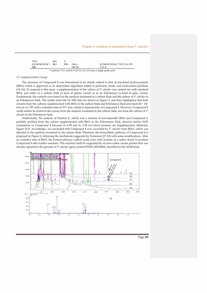
Chapter 4: Isolation of metabolites from P. odorifer
Page 30
7′/11′c 34.3 C -
8′,9‘,10′/12′,13‘,14′ a 30.3 CH3 1.43, s 8′,9‘,10′/12′,13‘,14′, 7′/11′, 2′, 6′, 3′/5′
OH 5.07, bs 1′, 2′, 6′
a Carbons 7′/11′ and 8′,9’,10′/12′,13’,14′ form a single peak each.
2.3. Supplementation Assays
The structure of Compound 1 was determined to be closely related to that of butylated hydroxyanisole
(BHA) which is approved as an antioxidant ingredient added to polymers, foods, and food-related products
[14–16]. To respond to this issue, a supplementation of the culture of P. odorifer was carried out with standard
BHA, put either in a culture flask (a kind of plastic vessel) or in an Erlenmeyer (a kind of glass vessel).
Furthermore, the controls were based on the medium incubated in a culture flask and the culture of P. odorifer in
an Erlenmeyer flask. The results from the LC–MS data are shown in Figure 5, and they highlighted that both
extracts from the cultures supplemented with BHA in the culture flask and Erlenmeyer flask provided [M − H]−
ions at m/z 357 with a retention time of 35.7 min, which is characteristic of Compound 1. However, Compound 1
could neither be found in the extract from the medium incubated in the culture flask, nor from the culture of P.
odorifer in the Erlenmeyer flask.
Additionally, the analysis of Fraction 1′, which was a mixture of non-separable BHA and Compound 1,
partially purified from the culture supplemented with BHA in the Erlenmeyer flask, showed similar NOE
correlations to Compound 1 between δH 6.99 and δH 1.28 (tert-butyl protons; see Supplementary Materials,
Figure S13). Accordingly, we concluded that Compound 1 was converted by P. odorifer from BHA, which was
detected in the medium incubated in the culture flask. Therefore, the biosynthetic pathway of Compound 1 is
proposed in Figure 6, following the mechanism suggested by Fontecave [17,18] with some modifications. After
an oxidative step of BHA, the formed phenoxy radical could react with cysteine as a sulfur donor to produce
Compound 1 after further reactions. This reaction could be supported by an iron–sulfur cluster protein that was
already reported in the genome of P. odorifer (gene symbol PODO_RS22860), described in the NCBI bank.
(f)
(b)
Compound 1
(a)
(c)
(d)
(e)

Chapter 4: Isolation of metabolites from P. odorifer
Page 31
Figure 5. HPLC chromatograms of Compound 1 (a), of the extracts from P. odorifer culture supplemented with
butylated hydroxyanisole (BHA) in the culture flask (b), or in the glass Erlenmeyer flask (c), of medium in the
culture flask (d), and of the Paenibacillus odorifer (P. odorifer) culture in the Erlenmeyer flask (e). Electrospray
ionisation (ESI)-MS (−) spectra of extracts from the P odorifer culture supplemented with BHA in the culture flask
(f), or in the glass Erlenmeyer flask (g).
OH
OMeBHA
O
OMe
O
OMe
SS
Fe3+
CysO
OMe
SFe3+
O
OMe
Cys-S
OH
OMe
SO
OMeOH
S
OH
H
OH
OMe
SFe3+
OH
OMe
SOH
OMe1
Figure 6. Putative biosynthetic pathway for Compound 1 from BHA supported by an iron–sulfur cluster protein
with cysteine as a sulfur donor.
Compound 2, as with Compound 1, was isolated from the culture process using a culture flask in the pre-
culture stage. In order to discover the origin of Compound 2, butylated hydroxytoluene (BHT), with its close
structure to that of Compound 2, was used as a supplemented material during the culture of P. odorifer, put
either in a culture flask or in an Erlenmeyer flask. The blank controls were the medium (without bacteria) in the
culture flask and the culture of P. odorifer in the Erlenmeyer flask. The HPLC-MS data introduced in Figure 7
exhibited that Compound 2, with a retention time at 38 min, was associated with an ion at m/z 296, which
occurred in extracts from media supplemented with standard BHT in both the culture flask and the Erlenmeyer
flask. This ion was detected in the MS spectrum of Compound 2 due to the hydrolysis of its ester group in LC–
MS process. However, Compound 2 was not found in the medium put in the culture flask, but was found in the
culture broth of P. odorifer in the Erlenmeyer flask. On the other hand, this compound was already reported in
the literature [3] from Oakwood. Therefore, we propose that Compound 2 came from the bioconversion of BHT,
or as a natural metabolite from the culture of P. odorifer. Furthermore, our P. odorifer strain could be considered
as a new example of tert-butylphenol-utilizing bacterium.
(g)
O
O
OH
14(a)

Chapter 4: Isolation of metabolites from P. odorifer
Page 32
Figure 7. ESI-MS (+) spectra of Compound 2 (a), of the extracts from the culture supplemented with BHT in either
the culture flask (b) or the Erlenmeyer flask (c), and of the culture of P. odorifer in the Erlenmeyer flask (d).
2.4. Cytotoxic Activity
The biological activities of Compounds 1 and 2 were tested using a 3-(4,5-dimethylthiazol-2-yl)-2,5-
diphenyltetrazolium bromide (MTT) assay on HaCaT (human keratinocytes) and B16 (murine melanoma) cell
lines (Table 3) [19]. Although neither compound showed activity significantly greater than the positive control
(doxorubicin) against the two cell lines, Compound 1 exhibited a significant half maximal inhibitory
concentration (IC50) on B16 (4.75 µM) and HaCaT (8.38 µM), while Compound 2 was less active. Additionally,
DNA damage assays, using γH2AX as a biomarker, were performed with Compound 1 on U2OS cells (Table 4).
These cells are frequently used since they are sensitive to DNA damage. Although the compound was highly
cytotoxic at 1 µM (cell death > 90%), no significant induction in γH2AX foci was observed at 1 µM or 0.1 µM
within the nuclei, suggesting that no significant DNA damage was triggered compared to untreated cells. These
results suggest that the cytotoxicity of Compound 1 was not driven by DNA damage.
Table 3. Cytotoxic assay of Compounds 1 and 2.
Compound IC50 (µM)
HaCaT B16
1 8.38 4.75
2 >377.4 169.8 ± 1
Doxorubicin 0.096 ± 0.009 0.034 ± 0.001
(b)
(c)
(d)

Chapter 4: Isolation of metabolites from P. odorifer
Page 33
Table 4. DNA damage assay of Compound 1.
Concentration (µM)
γH2AX Foci/Nuclei
0 12.9 ± 0.4
0.1 12.8 ± 0.2
1 3.6 ± 0.3
3. Materials and Methods
3.1. General Experimental Procedures
One-dimensional (1D) and two-dimensional (2D) NMR spectroscopic data were recorded in MeOH-d4 and
CDCl3 on a Bruker DMX 300 spectrometer (300 MHz (1H) and 75 MHz (13C), Bruker BioSpin, Billerica, MA,
USA). NMR spectroscopic data were processed using the MestRenoVa version 10.0 software (Mestrelab
Research, S.L., Santiago de Compostela, Spain). HRMS measurements for exact mass determination were
performed with a Q-Extractive Focus at the Centre Regional de Mesure Physique de l’Ouest (CRMPO), Rennes,
France. Analytical HPLC and semi-preparative HPLC were performed on a 5-µm Prevail C18 column (250 mm ×
4.6 mm for the former, and 250 mm × 10 mm for the later), GRACE, Columbia, MD, USA.
3.2. Collection and Phylogenetic Analysis of PC-GYM-TO Strain
The PC-GYM-TO strain was isolated from the crustose lichen, Rhizocarpon geographicum, collected in
Brittany, France in February 2015. The strain was identified at Banyuls/mer Platform (L. Intertaglia) as
Paenibacillus odorifer based on 16S ribosomal RNA (rRNA) gene sequence analysis (GenBank accession number
AJ223990). A comparative BLAST similarities search of the 16S rRNA gene sequence gave a 98.46% similarity to
that of P. odorifer (Gene bank entry PODO_RS03805). After culture in GYM Streptomyces medium (containing 4 g
of glucose (Sigma-Aldrich, St Louis, MO, USA), 4 g of yeast extract (Sigma-Aldrich, St Louis, MO, USA), 10 g of
malt extract (Sigma-Aldrich, St Louis, MO, USA), 2 g of CaCO3 (Merck KGaA, St Frankfurter, Darmstadt,
Germany), and 12 g of agar (Sigma-Aldrich, St Louis, MO, USA) in 1 L), the bacterium was stored in a mixture
of 47.5% (v/v) glycerol, 47.5% (v/v) H2O, and 5% (v/v) DMSO at −80 °C with a reference of PC-GYM-TO
(CORINT collection).
3.3. Cultivation and Extraction
P. odorifer (strain PC-GYM-TO) was cultured on GYM Streptomyces medium agar (2 g of glucose, 4 g of
yeast extract, 4 g of malt extract, 2 g of CaCO3, and 12 g of agar in 1 L at pH 7). The inoculum was prepared by
transferring one loop full of culture (PC-GYM-TO) from agar medium to a 250-mL culture flask, containing 50
mL of liquid GYM Streptomyces medium (2 g of glucose, 4 g of yeast extract, 4 g of malt extract, and 2 g of
CaCO3 in 1 L at pH 7). The bacterium culture was grown at 25 °C on a rotary shaker incubator at 120 rpm for
seven days. After seven days for pre-culture, 42 mL of bacterium culture was transferred into 14 Erlenmeyer
flasks (500 mL), each containing 300 mL of liquid GYM Streptomyces medium. The fermentation culture was
then incubated at 25 °C with 120-rpm shaking for seven days. After seven days of culture, the fermentation
broth was collected and centrifuged at 3500 rpm, at 4 °C for 15 min. After removal of the pellet, sterilized XAD-
7-HP resin (40 g/L) was added to the supernatant to absorb the organic products from the culture, and the resin
was then shaken at 220 rpm for 4 h. The resin was filtered and de-adsorbed by a mixture of solvent
acetone/MeOH (50/50, v/v). This mixture of solvent was removed under reduced pressure; the resulting
aqueous layer was extracted with ethyl acetate (EtOAc; 3 × 300 mL). The EtOAc/solute extract was dried under
vacuum to yield 439.5 mg of organic extract from 4.0 L of the culture.
Chapter 4: Isolation of metabolites from P. odorifer
Page 34
Supplementation assays: BHA was supplemented with a quantity of 0.1 mg per 25 mL of liquid GYM
Streptomyces medium at day zero of the culture to check the origin of isolated compounds.
3.4. Purification and Isolation
The organic extract (439.5 mg) from strain PC-GYM-TO, after biological assays, was subjected to flash
chromatography with a 50-g SiOH Chromabond® Flash column, using a sequential mixture of solvent with
increasing polarity from cyclohexane to dichloromethane, EtOAc, and MeOH for 4 h to furnish 14 fractions.
Guided by HPLC analysis, the first fraction containing Compound 2 (FA, 39.6 mg) and the second fraction
containing Compound 1 (FB, 46.6 mg) were purified with semi-preparative HPLC (using a Prevail® C18 column
with a gradient of 0% to 100% CH3OH in H2O for 60 min, and a flow rate of 2.5 mL/min) and preparative TLC to
afford Compound 1 (5.0 mg) and Compound 2 (5.9 mg), with yields of 1.25 mg/L and 1.5 mg/L, respectively.
LC–MS was applied using a Prevail® C18 column, with a gradient of 0% to 100% CH3CN in H2O for 60 min,
a flow rate of 0.8 mL/min, a sample concentration of 1 mg/mL, and an MS full range from 100–1200.
5,5′-Thiobis(2-tert-butyl-4-methylphenol) (Compound 1): white amorphous powder, retention time = 35.7
min; Rf = 0.45 (chloroform 100%). 1H NMR (300 MHz, CDCl3) and 13C NMR (75 MHz, CDCl3) are described in
Table 1. HRESIMS m/z 357.1983 [M − H]− (calculated for C22H29O2S, Δ = 0 ppm).
Octadecyl 3-(3,5-di-tert-butyl-4-hydroxyphenyl)propanoate (Compound 2): while solid, retention time = 26.7
min. 1H NMR (300 MHz, CDCl3) and 13C NMR (75 MHz, CDCl3) are described in Table 2. HRESIMS m/z
553.4592 [M + Na]+ (calculated for C35H32O3Na).
3.5. Molecular Models and Dynamic Simulations
The structures of Compound 1 and Santonox were built using the Yasara program and were parameterized
for the Yamber3 force field following the automated AutoSMILE procedure [20]. Both geometries were
optimized through the standardized minimization protocol of Yasara. Finally, to enhance the conformational
space exploration available to the structures, molecular models were used as an initial point for molecular
dynamics (MD) simulations. Each isomer was placed in an explicit chloroform solvent box and simulated under
periodic boundary conditions at a constant temperature of 300 K. Structures were relaxed during a 2-ns MD
simulation and trajectories were collected at 1-ps intervals. Analyses of the MD trajectories (root-mean-square
deviation (RMSD) and clustering) was performed using Gromacs tools [21].
3.6. Cytotoxicity Assays
The cytotoxic assays were performed on pure compounds (with a concentration for each sample as 40
mg/mL) against HaCaT human keratinocytes and B16 murine melanoma cell lines as described in the literature
[19]. HaCaT (2000 cells/well) and B16 (1800 cells/well) were cultivated in Roswell Park Memorial Institute RPMI
1640 medium supplemented with 5% fetal calf serum (FCS) and antibiotics in an atmosphere of 5% CO2 at 37 °C.
After a 24-h culture, the samples were added at different concentrations (1, 10, 50, 100, and 200 µg/mL) and each
96-well plate was continuously incubated at the same temperature and atmosphere as above. After a 48-h
culture, cell growth and viability were then measured at 540 nm using a 3-(4,5-dimethylthiazol-2-yl)-2,5-
diphenyltetrazolium bromide (MTT) assay. Doxorubicin was used as a positive control. Each experiment was
repeated three times.
3.7. DNA Damage Assays
U2OS cells were cultivated in Dulbecco's Modified Eagle's medium DMEM supplemented with 10% fetal
calf serum and antibiotics in an atmosphere of 5% CO2 at 37 °C. The γH2AX staining was performed as
previously described [22]. Images were acquired on an ArrayScan VTI high-content screening reader with a 320
lens (Thermo Scientific, Villebon sur Yvette, France). The images were analyzed using the Cell Profiler software
(http://www.cellprofiler.org, Broad Institute). For all analyses, raw data files were obtained with the total

Chapter 4: Isolation of metabolites from P. odorifer
Page 35
amount of Hoechst fluorescence and the total amount of γH2AX fluorescence. The number of γH2AX foci per
nucleus is indicated for each condition in Table 4 with more than 3000 cells counted except for the 1 µM
concentration because of the high cytotoxicity.
4. Conclusions
In summary, two tert-butylphenol compounds were firstly isolated from the culture of a bacterium, P.
odorifer, associated with the lichen, Rhizocarpon geographicum. Compound 1 displayed a symmetric structure
including two units of BHA linked by a sulfur bond. This point can be explained by the fact that Compound 1
was putatively formed via the bioaccumulation of BHA from the culture flask used in the culture process,
followed by the biotransformation of BHA into Compound 1. Therefore, a putative biosynthesis pathway was
proposed for this compound, and involved an iron–sulfur cluster protein with cysteine as a sulfur donor.
Compound 1 exhibited a moderate cytotoxicity, making it promising for further investigation to determine its
mechanism. The results also highlighted P. odorifer as a new case of tert-butylphenol-utilizing bacterium.
Supplementary Materials: Supplementary Materials are available online.
Author Contributions: S.T. and T.-B.-L.N. conceived and designed the experiments; T.-B.-L.N. performed the experiments;
T.-B.-L.N. analyzed the data; I.R. realized the biological assays on HaCaT and B16; R.P. and L.C. designed and realized the
assays on DNA damage; O.D. designed and realized the NOE calculations; S.F. ran NMR; T.-B.-L.N and S.T. wrote the
paper.
Funding: This research received no external funding.
Acknowledgments: We sincerely thank D. Delmail for his help in the isolation of the strain P. odorifer from R. geographicum;
P. Jehan, F. Lambert, and T. Delhaye for the mass spectrometer measurements; L. Intertaglia from Banyuls/mer Platform
Bio2Mar for the identification of the strain; E. Poupon (Biocis, Paris Saclay) and J-P. Hurvois for a critical discussion about
biosynthesis; P. Uriac for a discussion about NMR data; and J. Boustie for his recommendations in the writing. The
Vietnamese government is gratefully acknowledged for the PhD grant of Thi-Bach-Le Nguyen.
Conflicts of Interest: The authors declare no conflict of interest.
References
1. Wang, Y.; Schnell, B.; Baumann, S.; Müller, R.; Begley, T.P. Biosynthesis of Branched Alkoxy Groups: Iterative Methyl
Group Alkylation by a Cobalamin-Dependent Radical SAM Enzyme. J. Am. Chem. Soc. 2017, 139, 1742–1745.
2. Bisel, P.; Al-Momani, L.; Müller, M. The Tert-Butyl Group in Chemistry and Biology. Org. Biomol. Chem. 2008, 6, 2655–
2665.
3. Dembitsky, V.M. Natural Neo Acids and Neo Alkanes: Their Analogs and Derivatives. Lipids 2006, 41, 309–340.
4. Luesch, H.; Yoshida, W.Y.; Moore, R.E.; Paul, V.J.; Corbett, T.H. Total Structure Determination of Apratoxin A, a Potent
Novel Cytotoxin from the Marine Cyanobacterium Lyngbya majuscula. J. Am. Chem. Soc. 2001, 123, 5418–5423.
5. Lopanik, N.; Gustafson, K.R.; Lindquist, N. Structure of Bryostatin 20: A Symbiont-Produced Chemical Defense for
Larvae of the Host Bryozoan, Bugula Neritina. J. Nat. Prod. 2004, 67, 1412–1414.
6. Pereira, A.R.; Cao, Z.; Engene, N.; Soria-Mercado, I.E.; Murray, T.F.; Gerwick, W.H. Palmyrolide A, an Unusually
Stabilized Neuroactive Macrolide from Palmyra Atoll Cyanobacteria. Org. Lett. 2010, 12, 4490–4493.
7. Matthew, S.; Salvador, L.A.; Schupp, P.J.; Paul, V.J.; Luesch, H. Cytotoxic Halogenated Macrolides and Modified
Peptides from the Apratoxin-Producing Marine Cyanobacterium Lyngbya bouillonii from Guam. J. Nat. Prod. 2010, 73,
1544–1552.
8. Tidgewell, K.; Engene, N.; Byrum, T.; Media, J.; Doi, T.; Valeriote, F.A.; Gerwick, W.H. Evolved Diversification of a
Modular Natural Product Pathway: Apratoxins F and G, Two Cytotoxic Cyclic Depsipeptides from a Palmyra
Collection of Lyngbya bouillonii. ChemBioChem 2010, 11, 1458–1466.
9. Toyama, T.; Momotani, N.; Ogata, Y.; Miyamori, Y.; Inoue, D.; Sei, K.; Mori, K.; Kikuchi, S.; Ike, M. Isolation and
Characterization of 4-Tert-Butylphenol-Utilizing Sphingobium fuliginis Strains from Phragmites australis Rhizosphere
Sediment. Appl. Environ. Microbiol. 2010, 76, 6733–6740.

Chapter 4: Isolation of metabolites from P. odorifer
Page 36
10. Suzuki, M.T.; Parrot, D.; Berg, G.; Grube, M.; Tomasi, S. Lichens as Natural Sources of Biotechnologically Relevant
Bacteria. Appl. Microbiol. Biotechnol. 2015, 100, 583–595.
11. Varsha, K.K.; Devendra, L.; Shilpa, G.; Priya, S.; Pandey, A.; Nampoothiri, K.M. 2,4-Di-Tert-Butyl Phenol as the
Antifungal, Antioxidant Bioactive Purified from a Newly Isolated Lactococcus Sp. Int. J. Food Microbiol. 2015, 211, 44–50.
12. Jaivel, N.; Uvarani, C.; Rajesh, R.; Velmurugan, D.; Marimuthu, P. Natural Occurrence of Organofluorine and Other
Constituents from Streptomyces Sp. TC1. J. Nat. Prod. 2014, 77, 2–8.
13. Belghit, S.; Driche, E.H.; Bijani, C.; Zitouni, A.; Sabaou, N.; Badji, B.; Mathieu, F. Activity of 2,4-Di-Tert-Butylphenol
Produced by a Strain of Streptomyces mutabilis Isolated from a Saharan Soil against Candida albicans and Other
Pathogenic Fungi. J. Med. Mycol. 2016, 26, 160–169.
14. Yang, C.Z.; Yaniger, S.I.; Jordan, V.C.; Klein, D.J.; Bittner, G.D. Most Plastic Products Release Estrogenic Chemicals: A
Potential Health Problem That Can Be Solved. Environ. Health Perspect. 2011, 119, 989–996.
15. Li, B.; Wang, Z.-W.; Lin, Q.-B.; Hu, C.-Y.; Su, Q.-Z.; Wu, Y.-M. Determination of Polymer Additives-Antioxidants,
Ultraviolet Stabilizers, Plasticizers and Photoinitiators in Plastic Food Package by Accelerated Solvent Extraction
Coupled with High-Performance Liquid Chromatography. J. Chromatogr. Sci. 2015, 53, 1026–1035.
16. Babich, H. Butylated Hydroxytoluene (BHT): A Review. Environ. Res. 1982, 29, 1–29.
17. Fontecave, M.; Ollagnier-de-Choudens, S.; Mulliez, E. Biological Radical Sulfur Insertion Reactions. Chem. Rev. 2003,
103, 2149–2166.
18. Fontecave, M.; Py, B.; Ollagnier de Choudens, S.; Barras, F. From Iron and Cysteine to Iron-Sulfur Clusters: The
Biogenesis Protein Machineries. EcoSal Plus 2008, 3, 1–24.
19. Millot, M.; Tomasi, S.; Studzinska, E.; Rouaud, I.; Boustie, J. Cytotoxic Constituents of the Lichen Diploicia canescens. J.
Nat. Prod. 2009, 72, 2177–2180.
20. Krieger, E.; Darden, T.; Nabuurs, S.B.; Finkelstein, A.; Vriend, G. Making optimal use of empirical energy functions:
Force-field parameterization in crystal space. Proteins 2004, 57, 678–683.
21. Van-der-Spoel, D.; Lindahl, E.; Hess, B.; Groenhof, G.; Mark, A.E.; Berendsen, H.J. GROMACS: Fast, flexible, and free.
J. Comput. Chem. 2005, 26, 1701–1718.
22. Bigot, N.; Mouche, A.; Preti, M.; Loisel, S.; Renoud, M.L.; Le Guével, R.; Sensebé, L.; Tarte, K.; Pedeux, R. Hypoxia
Differentially Modulates the Genomic Stability of Clinical‐Grade ADSCs and BM‐MSCs in Long‐Term Culture. Stem
Cells 2015, 33, 3608–3620.
Sample Availability: Samples of the Compounds 1, 2 are available from the authors.
© 2018 by the authors. Licensee MDPI, Basel, Switzerland. This article is an open access article
distributed under the terms and conditions of the Creative Commons Attribution (CC BY) license
(http://creativecommons.org/licenses/by/4.0/).

Chapter 4: Isolation of metabolites from P. odorifer
Page 37
4.4. THE ISOLATION PROCESS OF A NOVEL ALKALOID
4.4.1. General presentation of alkaloids
Alkaloids are a large and structurally diverse group of natural products which could be isolated
from distinct sources as plants, fungi and bacteria or even animals. Many alkaloids have been
developed like therapeutic agents to treat various diseases including malaria, diabetis, cancer, cardiac
dysfunctions etc (see in Ain et al., 2016). To date, more than 18000 alkaloids have been found
(Dembitsky el al., 2005) and this number will continuously increase in the future. Alkaloids bearing
nitrogen atoms were identified as compounds containing either indole, indolizidine, quinoline,
isoquinoline, piperazine, indole-quinoline... etc (Figure 2.4.3) moities.
Indole
HN
Indolizidine
N
Quinoline
N
Isoquinoline
N
PiperazineNH
HN
PiperidineNH
PyrimidineN
N
Pyridine
N
Pyridone
NHO
Pyrrole
NH
Pyrrolidine
NH
QuinoloneNH
O
Figure 2.4.3 Some structural features found in alkaloids
A vast majority of alkaloids were frequently found in plant kingdom. These compounds exhibited
pharmacological properties including analgesic (e.g. codeine) (Honig et al., 1984), antihypertensive
(e.g. reserpine) (see in Yu et al., 2013), antipyretic (e.g. quinine) (Santos et al., 1998), anticholinergic
(e.g. atropine) (Braun et al., 1993), antitumor (e.g. vinblastine) (Fodstad et al., 1996), antimalarial (e.g.
quinine) (see in Achan et a., 2011) The structures of some compounds were shown in Figure 2.4.4.
Moreover some new alkaloids with significant biological activities are continuously reported in many
studies..

Chapter 4: Isolation of metabolites from P. odorifer
Page 38
However, when bacteria became a new source of exploitation of active natural products,
alkaloids were also produced by these organisms. These compounds showed interesting biological
properties. Some of them recently isolated are reported in Figure 2.4.5 including an antibacterial
dihydroquinoline from Pseudomonas aeruginosa (Uzair et al., 2006), a cytotoxic ammosamide D from
Streptomyces variabilis (Pan et al., 2012), an antibiotic hunanamycin D from Bacillus hunanensis (Hu
et al., 2013), a potent inhibitor of the developement of human neuroblastoma streptonigrin from
Micromonospora sp. (Wang et al., 2002) and also active alkaloids isolated from lichen-associated
bacteria such as an antibiotic and cytotoxic uncialamycin (Davies et al., 2005), an anti-HIV derivative
unciaphenol (Williams et al., 2015) and cytotoxic cladoniamides from Streptomyces uncialis (Williams
et al., 2008).
Vinblastine - anticancer Isolated from Madagascar periwinkle
Reserpine- antihypertensive Isolated from Rauwolfia canescens Figure 2.4.4 Structures of some drugs as natural alkaloids

Chapter 4: Isolation of metabolites from P. odorifer
Page 39
N
1-methyl-1,4-dihydroquinoline(Uzair et al., 2006)
N
OO
ClNH2
H2N
O
NHO
Ammosamide D(Pan et al., 2012)
NN
OO
OHOHOH
OH
Hunanamycin D(Hu et al., 2013)
NN
O
O
H3CO
H2N
H2N
CO2H
OH
OCH3OCH3
Streptonigrin(Wang et al., 2001)
O
O OH
HN OHO OH
Uncialamycin(Davies et al., 2005)
NH
N
N
R1
MeO
MeOO
R2
HO
A R1 = Cl; R2 = HB R1 = R2 = ClC R1 = R2 = H
NH
N
HN
Cl
MeO
O
R
D R = HE R = Cl
OOH
Me
NH
NCl
MeO
R
F R = HG R = Cl
NHMe
O OHO
Cladoniamides (Williams et al., 2008)
HNO
O OH
OH
HO
O OH
Unciaphenol(Williams et al., 2015)
OH
Figure 2.4.5 Some cytotoxic alkaloids recently isolated from bacteria
4.4.2 Isolation of alkaloid
In our study, a new cytotoxic alkaloid was also isolated. Its structure was a combination of
dihydronaphthalene moiety fused to a pyrrolooxazine unit. Moreover, it exhibited a cytotoxicity
against B16 murine melanoma and HaCaT human keratinocyte cell lines with micromolar IC50 values.
This property promised it as a potent agent for further pharmaceutical researches. The isolation,
structural elucidation and bioactive evaluation of this alkaloid were displayed as an article. The
supporting information of the publication will be reported in ANNEXE 2.

Chapter 4: Isolation of metabolites from P. odorifer
Page 40
References
Achan, J., Talisuna, A.O., Erhart, A., Yeka, A., Tibenderana, J.K., Baliraine, F.N., Rosenthal, P.J., D’Alessandro, U., 2011. Quinine, an old anti-malarial drug in a modern world: role in the treatment of malaria. Malar J 10, 144. https://doi.org/10.1186/1475-2875-10-144
Ain, Q.-U.-, Khan, H., Mubarak, M.S., Pervaiz, A., 2016. Plant Alkaloids as Antiplatelet Agent: Drugs of the Future in the Light of Recent Developments. Front Pharmacol 7. https://doi.org/10.3389/fphar.2016.00292
Asif, M., 2014. Chemical Characteristics, Synthetic Methods, and Biological Potential of Quinazoline and Quinazolinone Derivatives [WWW Document]. International Journal of Medicinal Chemistry. https://doi.org/10.1155/2014/395637
Braun, G.G., Schywalsky, M., Wölfel, L., Müller, H., Danner, U., Albert, R., 1993. [A comparison of the two anticholinergic agents atropine and glycopyrrolate during antagonism of a muscle relaxation with pyridostigmine]. Anaesthesiol Reanim 18, 120–122, 125–127.
Davies, J., Wang, H., Taylor, T., Warabi, K., Huang, X.-H., Andersen, R.J., 2005. Uncialamycin, A New Enediyne Antibiotic. Org. Lett. 7, 5233–5236. https://doi.org/10.1021/ol052081f
Dembitsky Valery M., 2005. Astonishing diversity of natural surfactants: 6. Biologically active marine and terrestrial alkaloid glycosides. Lipids 40, 1081. https://doi.org/10.1007/s11745-005-1473-2
Fodstad, O., Breistøl, K., Pettit, G.R., Shoemaker, R.H., Boyd, M.R., 1996. Comparative antitumor activities of halichondrins and vinblastine against human tumor xenografts. J. Exp. Ther. Oncol. 1, 119–125.
Honig, S., Murray, K.A., 1984. An appraisal of codeine as an analgesic: single-dose analysis. J Clin Pharmacol 24, 96–102.
Hu, Y., Wang, K., MacMillan, J.B., 2013. Hunanamycin A, an Antibiotic from a Marine-Derived Bacillus hunanensis. Org. Lett. 15, 390–393. https://doi.org/10.1021/ol303376c
Pan, E., Jamison, M., Yousufuddin, M., MacMillan, J.B., 2012. Ammosamide D, an Oxidatively Ring Opened Ammosamide Analog from a Marine-Derived Streptomyces variabilis. Org. Lett. 14, 2390–2393. https://doi.org/10.1021/ol300806e
Santos F. A., Rao V. S. N., 2011. A Study of the Anti‐ pyretic Effect of Quinine, an Alkaloid Effective Against Cerebral Malaria, on Fever Induced by Bacterial Endotoxin and Yeast in Rats. Journal of Pharmacy and Pharmacology 50, 225–229. https://doi.org/10.1111/j.2042-7158.1998.tb06180.x
Uzair, B., Ahmed, N., Ahmad, V.U., Kousar, F., 2006. A new antibacterial compound produced by an indigenous marine bacteria--fermentation, isolation, and biological activity. Nat. Prod. Res. 20, 1326–1331. https://doi.org/10.1080/14786410601102017
Wang, H., Yeo, S.L., Xu, J., Xu, X., He, H., Ronca, F., Ting, A.E., Wang, Y., Yu, V.C., Sim, M.M., 2002. Isolation of Streptonigrin and Its Novel Derivative from Micromonospora as Inducing Agents of p53-Dependent Cell Apoptosis. J. Nat. Prod. 65, 721–724. https://doi.org/10.1021/np0104572
Williams, D.E., Bottriell, H., Davies, J., Tietjen, I., Brockman, M.A., Andersen, R.J., 2015. Unciaphenol, an Oxygenated Analogue of the Bergman Cyclization Product of Uncialamycin Exhibits Anti-HIV Activity. Org. Lett. 17, 5304–5307. https://doi.org/10.1021/acs.orglett.5b02664
Chapter 4: Isolation of metabolites from P. odorifer
Page 41
Williams, D.E., Davies, J., Patrick, B.O., Bottriell, H., Tarling, T., Roberge, M., Andersen, R.J., 2008. Cladoniamides A−G, Tryptophan-Derived Alkaloids Produced in Culture by Streptomyces uncialis. Org. Lett. 10, 3501–3504. https://doi.org/10.1021/ol801274c
Yu, J., Ma, Y., Drisko, J., Chen, Q., 2013. Antitumor Activities of Rauwolfia vomitoria Extract and Potentiation of Carboplatin Effects Against Ovarian Cancer. Curr Ther Res Clin Exp 75, 8–14. https://doi.org/10.1016/j.curtheres.2013.04.001
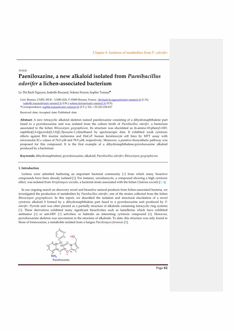
Chapter 4: Isolation of metabolites from P. odorifer
Page 42
Article
Paeniloxazine, a new alkaloid isolated from Paenibacillus
odorifer a lichen-associated bacterium
Le Thi Bach Nguyen, Isabelle Rouaud, Solenn Ferron, Sophie Tomasi*
Univ Rennes, CNRS, ISCR – UMR 6226, F-35000 Rennes, France ; [email protected] (L.N);
[email protected] (I.R.); [email protected] (S.F);
*Correspondence: [email protected] (S.T.); Tel.: +33-223-234-817
Received: date; Accepted: date; Published: date
Abstract: A new tetracyclic alkaloid skeleton named paeniloxazine consisting of a dihydronaphthalene part
fused to a pyrrolooxazine unit was isolated from the culture broth of Paenibacillus odorifer, a bacterium
associated to the lichen Rhizocarpon geographicum. Its structure was elucidated as (6-amino-10-phenyl-10H-
naphtho[2,3-e]pyrrolo[2,1-b][1,3]oxazin-1-yl)methanol by spectroscopic data. It exhibited weak cytotoxic
effects against B16 murine melanoma and HaCaT human keratinocyte cell lines by MTT assay with
micromolar IC50 values of 76.0 µM and 78.9 µM, respectively. Moreover, a putative biosynthetic pathway was
proposed for this compound. It is the first example of a dihydronaphthalene-pyrrolooxazine alkaloid
produced by a bacterium
Keywords: dihydronaphtalene; pyrrolooxazine; alkaloid; Paenibacillus odorifer; Rhizocarpon geographicum.
1. Introduction
Lichens were admitted harboring an important bacterial community [1] from which many bioactive
compounds have been already isolated [2]. For instance, uncialamycin, a compound showing a high cytotoxic
effect, was isolated from Streptomyces uncialis, a bacterial strain associated with the lichen Cladonia uncialis [3, 4].
In our ongoing search on discovery novel and bioactive natural products from lichen-associated bacteria, we
investigated the production of metabolites by Paenibacillus odorifer, one of the strains collected from the lichen
Rhizocarpon geographicum. In this report, we described the isolation and structural elucidation of a novel
cytotoxic alkaloid 1 formed by a dihydronaphthalene part fused to a pyrrolooxazine unit produced by P.
odorifer. Pyrrole unit was often present as a partially structure of alkaloids containing tetracyclic ring systems
[5]. These derivatives exhibited many significant bioactivities such as lamellerins which have exhibited
antitumor [6] or anti-HIV [7] activities; or halitulin an interesting cytotoxic compound [8]. However,
pyrrolooxazine skeleton was uncommon in the structure of alkaloids. To date, this structure was only found in
those of formoxazine, a metabolite isolated from a fungus Paecilomyces formosus [9].
O
N
HO
NH2
1
2
34
4a
5
5a
67
8
99a 10
10a
11
3a
1'2'
3'
4'
Paeniloxazine
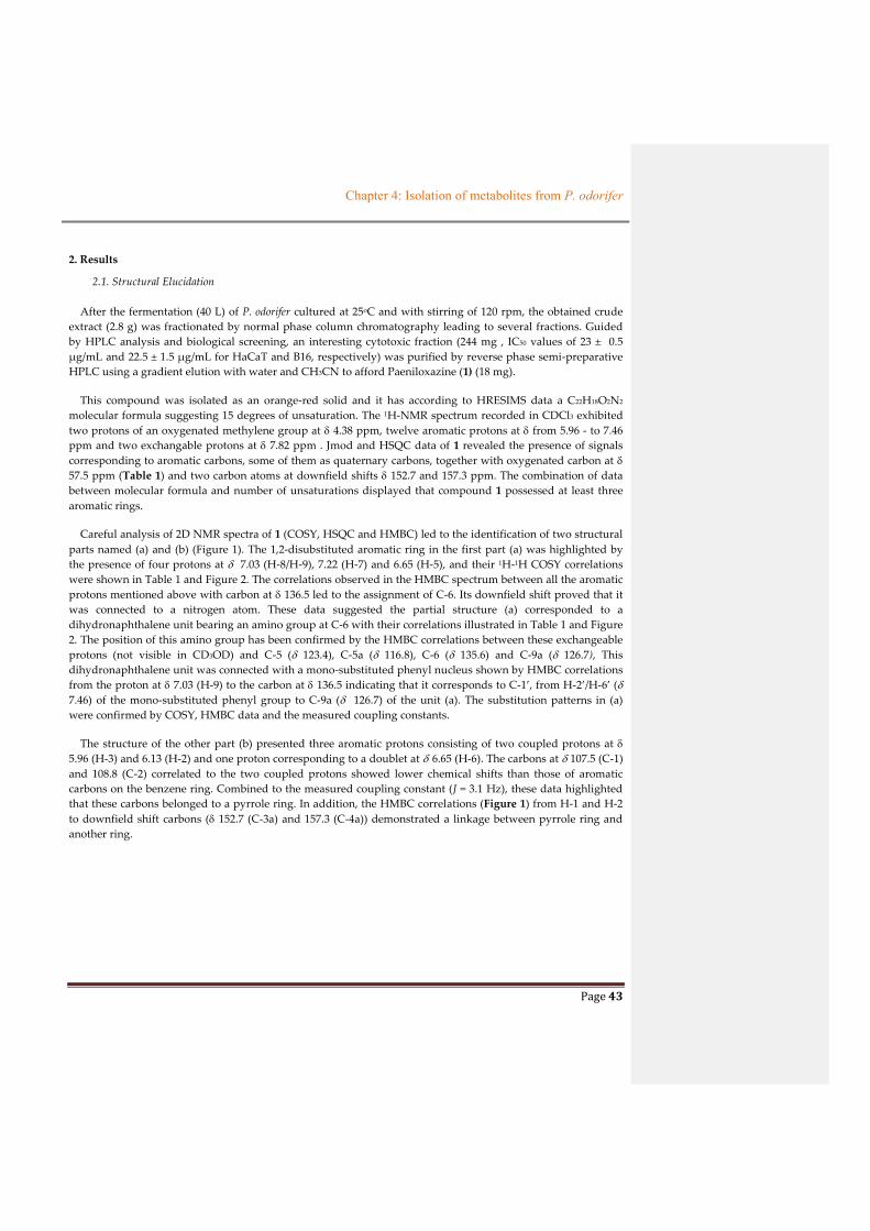
Chapter 4: Isolation of metabolites from P. odorifer
Page 43
2. Results
2.1. Structural Elucidation
After the fermentation (40 L) of P. odorifer cultured at 25oC and with stirring of 120 rpm, the obtained crude
extract (2.8 g) was fractionated by normal phase column chromatography leading to several fractions. Guided
by HPLC analysis and biological screening, an interesting cytotoxic fraction (244 mg , IC50 values of 23 ± 0.5
µg/mL and 22.5 ± 1.5 µg/mL for HaCaT and B16, respectively) was purified by reverse phase semi-preparative
HPLC using a gradient elution with water and CH3CN to afford Paeniloxazine (1) (18 mg).
This compound was isolated as an orange-red solid and it has according to HRESIMS data a C22H18O2N2
molecular formula suggesting 15 degrees of unsaturation. The 1H-NMR spectrum recorded in CDCl3 exhibited
two protons of an oxygenated methylene group at 4.38 ppm, twelve aromatic protons at from 5.96 - to 7.46
ppm and two exchangable protons at 7.82 ppm . Jmod and HSQC data of 1 revealed the presence of signals
corresponding to aromatic carbons, some of them as quaternary carbons, together with oxygenated carbon at
57.5 ppm (Table 1) and two carbon atoms at downfield shifts 152.7 and 157.3 ppm. The combination of data
between molecular formula and number of unsaturations displayed that compound 1 possessed at least three
aromatic rings.
Careful analysis of 2D NMR spectra of 1 (COSY, HSQC and HMBC) led to the identification of two structural
parts named (a) and (b) (Figure 1). The 1,2-disubstituted aromatic ring in the first part (a) was highlighted by
the presence of four protons at 7.03 (H-8/H-9), 7.22 (H-7) and 6.65 (H-5), and their 1H-1H COSY correlations
were shown in Table 1 and Figure 2. The correlations observed in the HMBC spectrum between all the aromatic
protons mentioned above with carbon at 136.5 led to the assignment of C-6. Its downfield shift proved that it
was connected to a nitrogen atom. These data suggested the partial structure (a) corresponded to a
dihydronaphthalene unit bearing an amino group at C-6 with their correlations illustrated in Table 1 and Figure
2. The position of this amino group has been confirmed by the HMBC correlations between these exchangeable
protons (not visible in CD3OD) and C-5 ( 123.4), C-5a ( 116.8), C-6 ( 135.6) and C-9a ( 126.7), This
dihydronaphthalene unit was connected with a mono-substituted phenyl nucleus shown by HMBC correlations
from the proton at 7.03 (H-9) to the carbon at 136.5 indicating that it corresponds to C-1’, from H-2’/H-6’ (
7.46) of the mono-substituted phenyl group to C-9a ( 126.7) of the unit (a). The substitution patterns in (a)
were confirmed by COSY, HMBC data and the measured coupling constants.
The structure of the other part (b) presented three aromatic protons consisting of two coupled protons at
5.96 (H-3) and 6.13 (H-2) and one proton corresponding to a doublet at 6.65 (H-6). The carbons at 107.5 (C-1)
and 108.8 (C-2) correlated to the two coupled protons showed lower chemical shifts than those of aromatic
carbons on the benzene ring. Combined to the measured coupling constant (J = 3.1 Hz), these data highlighted
that these carbons belonged to a pyrrole ring. In addition, the HMBC correlations (Figure 1) from H-1 and H-2
to downfield shift carbons ( 152.7 (C-3a) and 157.3 (C-4a)) demonstrated a linkage between pyrrole ring and
another ring.
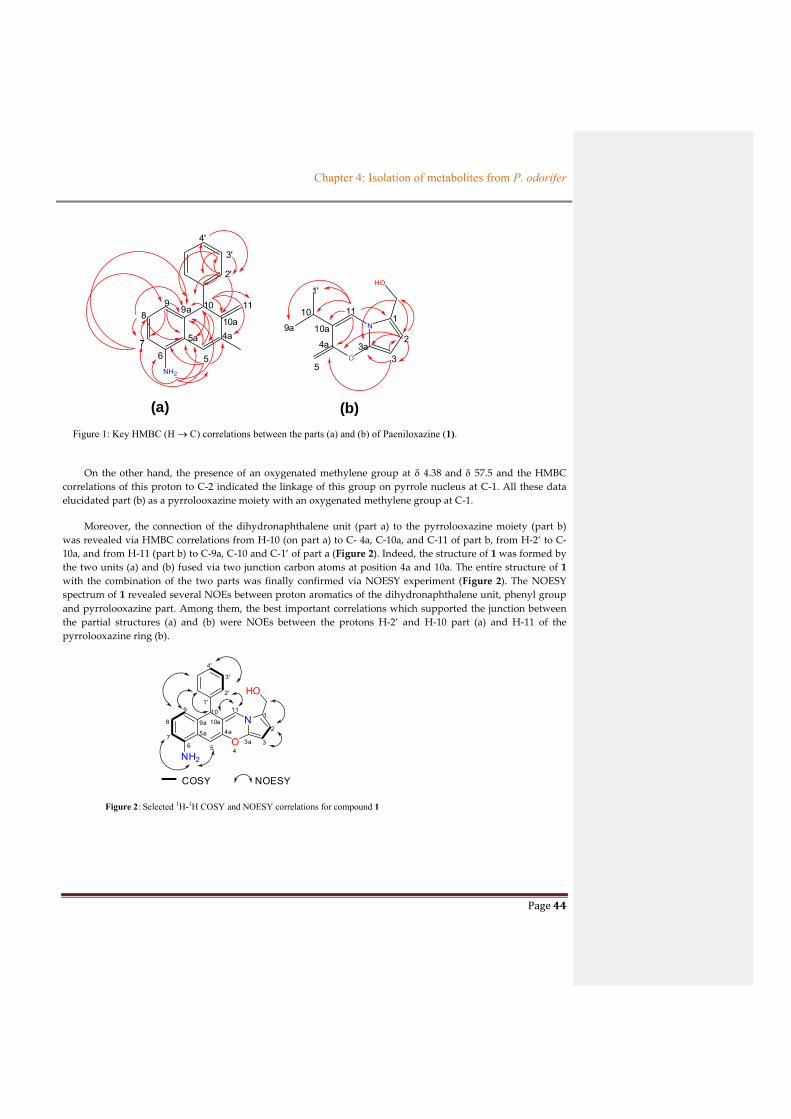
Chapter 4: Isolation of metabolites from P. odorifer
Page 44
NH2
4a
5
5a
67
89
9a 10
10a
11
2'
3'
4'
O
N
HO
1
2
3
1'
4a
5
9a
10
10a
11
3a
(a) (b)
Figure 1: Key HMBC (H C) correlations between the parts (a) and (b) of Paeniloxazine (1).
On the other hand, the presence of an oxygenated methylene group at 4.38 and 57.5 and the HMBC
correlations of this proton to C-2 indicated the linkage of this group on pyrrole nucleus at C-1. All these data
elucidated part (b) as a pyrrolooxazine moiety with an oxygenated methylene group at C-1.
Moreover, the connection of the dihydronaphthalene unit (part a) to the pyrrolooxazine moiety (part b)
was revealed via HMBC correlations from H-10 (on part a) to C- 4a, C-10a, and C-11 of part b, from H-2’ to C-
10a, and from H-11 (part b) to C-9a, C-10 and C-1’ of part a (Figure 2). Indeed, the structure of 1 was formed by
the two units (a) and (b) fused via two junction carbon atoms at position 4a and 10a. The entire structure of 1
with the combination of the two parts was finally confirmed via NOESY experiment (Figure 2). The NOESY
spectrum of 1 revealed several NOEs between proton aromatics of the dihydronaphthalene unit, phenyl group
and pyrrolooxazine part. Among them, the best important correlations which supported the junction between
the partial structures (a) and (b) were NOEs between the protons H-2’ and H-10 part (a) and H-11 of the
pyrrolooxazine ring (b).
O
N
HO
NH2
1
2
34
4a
5
5a
67
8
9
9a
1010a
11
3a
1'2'
3'
4'
COSY NOESY
Figure 2: Selected 1H-1H COSY and NOESY correlations for compound 1

Chapter 4: Isolation of metabolites from P. odorifer
Page 45
Table 1. 1D and 2D-NMR data of 1 (300 and 75 MHz, CDCl3, ppm)
Position C Type H mult (J in
Hz)
HMBC (H C) COSY NOESY
1 126.7 qC
2 108.8 CH 6.13, d (3.1) 3, 3a, 4a, OCH2 3, OCH2 3, OCH2
3 107.5 CH 5.96, d (3.1) 2, 3a, 4a 2 2
3a 152.7 qC
4a 157.3 qC
5 123.4 CH 6.65, d, (1.8) 4a, 5a, 6, 7, 9a, 10 10, NH2 10, NH2
5a 116.8 qC
6 136.5 qC
7 111.4 CH 7.22, d, (8.0) 8, 9, 9a 8, 9 NH2
8 119.4 CH 7.03, m 5a, 7, 9a 7 2’/6’
9 119.4 CH 7.03, m 7, 5a, 9a, 1’ 7, 2’/6’ 2’/6’
9a 126.7 qC
10 34.2 CH 5.86, s 4a, 5a/10a, 5/11, 9a 5/11 11, 2’/6’
10a 116.8 qC
11 123.4 CH 6.65, d, (1.8) 1/9a, 4a, 10, 10a, 1’ 10 10, 2’/6’
1’ 136.5 qC
2’/ 6’ 119.6 CH 7.46, d (8.0) 1’, 3’/5’, 4’, 9a, 10a 3’/5’, 9 9, 10, 11
3’/ 5’ 122.0 CH 7.14, m 1’, 2’/6’, 9a 2’/6’, 4’ 9
4’ 111.4 CH 7.22, d (8.0) 2’/6’ 3’/5’, 2’/6’ 2’/6’
OCH2 57.5 CH2 4.38, s 2, 3a 2 2
NH2 7.82, s 5, 5a, 6, 9a 5, 7
Therefore, all data confirmed that compound 1, produced by this bacterium P. odorifer, could be a
tetracyclic alkaloid skeleton consisting of a dihydronaphthalene unit fused to a pyrrolooxazine moiety and was

Chapter 4: Isolation of metabolites from P. odorifer
Page 46
named Paeniloxozine. To our knowledge, it is the first report of this skeleton from natural sources. The new
alkaloid 1 is probably biosynthesized, according to Dewick [10], via an amino acid pathway (Scheme 1)
combined to a polyketide pathway. After the formation of a dihydronaphtalene part coming from the
condensation between 4 unit of malonyl CoA and one phenylmalonylCoA derivative, the obtained bicyclic
compound was condensed to a gamma-butyric acid unit to form the pyrroloxazine part. The last steps could
involve various reactions from hydroxylation, dehydration, C-methylation to lead the substitution of the
pyrrole group by an oxygenated methylene.
Scheme 1: Putative biosynthetic pathway for compound 1
CoASPh
O O
+ 4x CoAS OH
O O
O O
O
reduction Ph
O
OH O
Ph
OHOH
OH
O
Ph
H2NO
HO
-aminobutyric acid
Ph
N
OHO
HO
Ph
NH
OO
HO
Ph
N
OHO
H
-H2O
Ph
O
N
OHOHOHOH
Ph
O
N
NH2
Ph
O
N
NH2
Ph
O
N
NH2
Ph
O
N
NH2
hydroxylation -H2O
Ph
O
N
NH2
SAM
SEnz
O
O
OH
reduction decarboxylation
oxidation
OH
OH
O
Ph
-H2Oreduction
NH3
OHHO
Paeniloxazine
hydroxylation
2.2. Cytotoxic Activity
Paeniloxazine was also investigated in vitro for its cytotoxic effects against B16 murine melanoma and
HaCaT human keratinocyte cell lines by MTT assay [11] with doxorubicin as a positive control. The compound 1
exhibited a weak activity with micromolar IC50 values (76.0 1 and 78.9 µM for B16 and HaCaT cell lines,
respectively) which could explain the activity of the fraction.
3. Materials and Methods
3.1. General Experimental Procedures.
1D and 2D NMR spectroscopic data were recorded in MeOH-d4 solution containing Me4Si as internal
standard on Bruker DMX 300 spectrometry [300 MHz (1H) and 75 MHz (13C)]. NMR spectroscopic data were
processed using the MestReNoVa version 10.0 software. HRMS measurements for exact mass determination
were performed with a Q-Extractive Focus at CRMPO (Centre Regional de Measure Physique de l’Ouest),
Rennes, France. Analytical HPLC (High Resolution Analysis Performed) and semi preparative HPLC were
performed on Prevail C18 column 5µm (250 mm x 4.6 mm for the former and 250 mm x 10 mm for the later),
GRACE, Columbia, MD, USA. The chiral HPLC was set up using a chiral AD-H column. Optical rotations were
determined at 589 nm (sodium D line) using PerkinElmer-343 polarimeter. Infrared spectra were recorded on
Perkin Elmer apparatus. Wavelengths of maximum absorbance (νmax) are quoted in wave numbers (cm−1).UV
spectrum was recorded on SPECORD 205-222A190 in MeOH.
Chapter 4: Isolation of metabolites from P. odorifer
Page 47
3.2. Collection and Analysis of PC-GYM-TO Strain
PC-GYM-TO Strain was isolated from the crustose lichen Rhizocarpon geographicum-collected in Brittany,
France in February 2015. The strain has been identified as Paenibacillus odorifer based on its 16S rRNA gene
sequence analysis (GenBank accession number AJ223990) (L. Intertaglia, Banyuls/mer Platform). Comparative
Blast similarities search of the 16S rRNA gene sequence gave a 98.46 % similarity to that of P. odorifer (Gene
bank entry PODO_RS03805). After culture in Gym streptomyces medium (containing 4 g Glucose (Sigma-
Aldrich, St Louis, MO, USA), 4 g yeast extract (Sigma-Aldrich, St Louis, MO, USA), 10 g malt extract (Sigma-
Aldrich, St Louis, MO, USA), 2 g CaCO3 (Merck, Germany), 12 g agar (Sigma-Aldrich, St Louis, MO, USA) in
1L), the bacterium was stored in mixture of 47.5% (v/v) glycerol, 47.5% (v/v) H2O and 5%(v/v) DMSO at -80oC
with reference as PC-GYM-TO (CORINT collection).
3.3. Cultivation and Extraction.
P. odorifer (strain PC-GYM-TO) was cultured on Gym streptomyces medium agar (2 g glucose, 4 g yeast
extract, 4 g malt extract, 2 g CaCO3 and 12 g agar in 1 L at pH 7). The inoculum was prepared by transferring
one loop full of culture (PC-GYM-TO) from medium agar to culture flask (250 mL) consisted of 50 mL of liquid
Gym streptomyces medium (2 g glucose, 4 g yeast extract, 4 g malt extract and 2 g CaCO3 in 1 L at pH 7). The
bacterium culture was grown at 25oC on a rotary shaker incubator at 120 rpm for 7 days. After 7 days for pre-
culture, 400 mL of bacterium culture was put into 140 Erlenmeyer flasks (500 mL), each containing 300 mL of
liquid Gym streptomyces medium (1% inoculum). The fermentation culture was then performed at 25oC with 120
rpm of stirring for 7 days. After 7 days of culture, the broth was collected and centrifuged at 3500 rpm, 4oC for
15 min. After removal of the pellet, sterilized XAD-7-HP resin (40 g/L) was added into supernatant to absorb the
organic products from the culture. The mixture then was shaken at 220 rpm for 4h. The resin was filtered and
de-adsorbed by mixture of solvent acetone/MeOH (50/50, v/v). This mixture of solvent was removed under
reduce pressure; the resulting aqueous layer was extracted with ethyl acetate (EtOAc) (3 x 300 mL). The EtOAc-
solute extract was dried under vacuum to yield 3.0 g of organic extract from 40.0 L of the culture.
3.4. Extraction and Isolation
The organic extract (2.9 g) from strain PC-GYM-TO, used after biological assay, was subjected to normal
phase chromatography column using silica gel 0.06-0.2 mm, 60 Ao (Acros Organics, Germany) and a sequential
mixture of solvent elution with increasing polarity from cyclohexane, dichloromethane (DCM), ethyl acetate
(EtOAc) and methanol (MeOH) to furnish 18 fractions. Guided by HPLC analysis and bioactivities for each
fraction, the bioactive fraction containing Paeniloxazine (F8, 244.0 mg, IC50 23 ± 0.5 µg/mL and 22.5 ± 1.5µg/mL
using MTT assay against HaCaT and B16, respectively) was purified by semi-preparative HPLC (using Prevail®
C18 column with gradient of 0% to 100% CH3OH in H2O for 60 min, a flow rate of 2.0 mL/min) and preparative
TLC to afford Paeniloxazine (18.0 mg).
Paeniloxazine: (6-amino-10-phenyl-10H-naphtho[2,3-e]pyrrolo[2,1-b][1,3]oxazin-1-yl)methanol: orange-red
solid, Rf = 0.45 (in CHCl3/EtOAc = 2/1)); HR-MS-ESI at m/z 365.1261 of [M+Na]+ ion, calcul. C22H18N2O2 molecular
formula; FT-IR max (cm-1): 3400, 2921, 1705, 1610, 1415; UV-vis (MeOH,) max (log ): 224 (3.27), 281(2.47); 1H-
NMR (CDCl3) and 13C-NMR (CDCl3) see Table 1. [α]D20 was not measurable at the operating wavelength of the
polarimeter (589 nm) at c, 0.01 in EtOH.

Chapter 4: Isolation of metabolites from P. odorifer
Page 48
3.5. Cytotoxic evaluation
The cytotoxic effects were evaluated on crude extract and pure compounds (with mother concentration for
each sample as 40 mg/mL) against HaCaT human keratinocytes and B16 murine melanoma cell line described in
literature [11]. HaCaT (2000 cells/well) and B16 (1800 cells/well) were cultivated in RMPI 1640 medium
supplemented with 5% of fetal calf serum (FCS) and antibiotic in atmosphere of 5% CO2 at 37oC. After 24h
culture, the samples were added at different concentrations (1, 10, 50, 100 and 200 µg/mL) and each 96-well
plate was continuously incubated at the same temperature and atmosphere as above. After 48 culture, cell
growth and viability were then measured at 540 nm using a MTT (3-[4,5-dimethylthiazol-2-yl]-2,5-
diphenyltetrazolium bromide) assay. Doxorubicin was used as a positive control. Each experiment was
repeated three times.
4. Conclusions
In summary, a new tetracyclic alkaloid skeleton was firstly isolated from P. odorifer associated with the
lichen Rhizocarpon geographicum. Its structure consists of dihydronaphthalene unit that is a well-known group of
natural products, fused to a rare pyrrolooxazine moiety. This compound exhibited a weak cytotoxicity against
HaCaT and B16 cells. Further experiments will be done to determine its activity on a larger panel of cancer cell
lines and its eventual mechanism of action.
Supplementary Materials: Supplementary Materials are available online
Author Contributions: S.T. and L.N. conceived of and designed the experiments; L.N. performed the
experiments; L.N. analyzed the data; I.R. realized the biological assays; S.F. helps for UV measurement; L.N.
and S.T. wrote the paper.
Acknowledgments: We sincerely thank Dr. D. Delmail for his help for the isolation of the strain P. odorifer from
R. geographicum, to A. Sauvager for technical assistance in chiral HPLC analysis, to Dr O. Delalande for
discussion about NOEs calculation, to Dr P. Jehan from CRMPO for the measurement of HRMS, to Pr. P. Uriac
and Dr. A. Bondon for critical discussion in NMR spectra, and to Vietnamese Government for PhD grant of
Nguyen T. B. Le.
Conflicts of Interest: The authors declare no conflict of interest.
References
1. Grube, M.; Cernava, T.; Soh, J.; Fuchs, S.; Aschenbrenner, I.; Lassek, C.; Wegner, U.; Becher, D.; Riedel, K.; Sensen, C.W.; Berg, G. Exploring Functional Contexts of Symbiotic Sustain within Lichen-Associated Bacteria by Comparative Omics. ISME J 2015, 9 (2), 412–424.
2. Suzuki, M.T.; Parrot, D.; Berg, G.; Grube, M.; Tomasi, S. Lichens as Natural Sources of Biotechnologically Relevant Bacteria. Appl Microbiol Biotechnol 2015, 100 (2), 583–595.
3. Davies, J.; Wang, H.; Taylor, T.; Warabi, K.; Huang, X.-H.; Andersen, R.J. Uncialamycin, A New Enediyne Antibiotic. Org. Lett. 2005, 7 (23), 5233–5236.
4. Nicolaou, K.C.; Chen, J.S.; Zhang, H.; Montero, A. Asymmetric Synthesis and Biological Properties of Uncialamycin and 26-Epi-Uncialamycin. Angewandte Chemie International Edition 47 (1), 185–189.
5. Estévez, V.; Villacampa, M.; Menéndez, J.C. Recent Advances in the Synthesis of Pyrroles by Multicomponent Reactions. Chem Soc Rev 2014, 43 (13), 4633–4657.

Chapter 4: Isolation of metabolites from P. odorifer
Page 49
6. Bailly, C. Anticancer Properties of Lamellarins. Mar Drugs 2015, 13 (3), 1105–1123.
7. Reddy, M.V.R.; Rao, M.R.; Rhodes, D.; Hansen, M.S.T.; Rubins, K.; Bushman, F.D.; Venkateswarlu, Y.; Faulkner, D.J. Lamellarin α 20-Sulfate, an Inhibitor of HIV-1 Integrase Active against HIV-1 Virus in Cell Culture. J. Med. Chem. 1999, 42 (11), 1901–1907.
8. Kashman, Y.; Koren-Goldshlager, G.; Gravalos, M.D.G.; Schleyer, M. Halitulin, a New Cytotoxic Alkaloid from the Marine Sponge Haliclona Tulearensis. Tetrahedron Letters 1999, 40 (5), 997–1000.
9. Yun, K.; Leutou, A.S.; Rho, J.-R.; Son, B.W. Formoxazine, a New Pyrrolooxazine, and Two Amines from the Marine–Mudflat-Derived Fungus Paecilomyces Formosus. Bulletin of the Korean Chemical Society 37 (1), 103–104.
10. Dewick, P.M. The Acetate pathway. Medicinal Natural Products; John Wiley & Sons, Ltd, 2009; pp 507, .
11. Millot, M.; Tomasi, S.; Studzinska, E.; Rouaud, I.; Boustie, J. Cytotoxic Constituents of the Lichen Diploicia Canescens. J. Nat. Prod. 2009, 72 (12), 2177–2180.
Sample Availability: Samples of the compounds 1 is available from the authors.
© 2018 by the authors. Submitted for possible open access publication under the terms and conditions
of the Creative Commons Attribution (CC BY) license (http://creativecommons.org/licenses/by/4.0/).

Chapter 4: Isolation of metabolites from P. odorifer
Page 50
4.5. DESCRIPTION OF THE OTHER ISOLATED METABOLITES
Along with cytotoxic compounds reported above, other known and no cytotoxic metabolites
were also isolated from the different cultures carried out during this work including two diol
compounds (1, 2) (found from sawdust of Oak wood reported in Rukachaisirikul et al., 2012), an
aliphatic ester (3), two furfural derivatives (4, 5) a butyrolactone (6), an acrylate derivative
cyaneodimycin (7) (isolated from Streptomyces cyaneofuscatus - Parrot et al., 2016), a cyclopentene
(8), a salicylate ester (9) (Green et al., 2008), a diphenol (10) and an indole compound (11) (found
from Nocardia ignorata Noël et al 2017) (Figure 2.4.6). The physical and chemical properties of these
compounds will be introduced in Chapter 6 – 6.3 and their NMR spectra will be provided in ANNEXE
3. Among them, the two diols and (1, 2) and cyaneodimycin (7) were found in both of the resin
extracts from the culture performed using bioreactor or Erlenmeyer flasks. The other compounds were
isolated from extracts obtained by culture in either bioreactor or Erlenmeyer with too small yield. The
Schemes 2.4.1 to 2.4.3 described the process of isolation of these compounds. The isolation process is
currently in progress from the Erlenmeyer 40L culture.
Scheme 2.4.1: Description of the isolated process on resin extract from culture in bioreactor with
the yield of compounds in mg/L.
P. odorifer culture in bioreactor (4.5 L)
EtOAc extract from resin (353.7 mg)
Fraction 14
21.8 mg
Fraction 20
116.4 mg
Flash chromatography using C18 column
Compound 1 2.0 mg
(0.45 mg/L)
Compound 2 1.9 mg
(0.42 mg/L)
Compound 7 0.6 mg
(0.13 mg/L)
Polysaccharide (PO-QL1) Semi-preparative HPLC using Prevail C18 column

Chapter 4: Isolation of metabolites from P. odorifer
Page 51
P. odorifer culture in Erlenmeyer (4.0 L)
Resin extract (439.5 mg)
Fraction 1 39.6 mg
Fraction 2 46.6 mg
Fraction 3 24.7 mg
Flash chromatography using silica gel column
Fraction 4 39.8 mg
Fraction 5 23.3 mg
Fraction 7 13.7 mg
Subfraction 1 18.6 mg
Column chromatography using silica gel 60A, elution
of cyclohexan/DCM and DCM/EtOAc
Subfraction 2 12.6 mg
Compound 3 3.6 mg
(0.9 mg/L)
Semi-preparative
HPLC
R7-FA-2*
5.9 mg (1.5 mg/L)
Semi-preparative
HPLC
R7-FB-1* 5 mg
1.25 mg/L)
Semi-preparative
HPLC
Compound 4 2.93 mg
(0.73 mg/L)
Semi-preparative
HPLC
Compound 5 2.3 mg
(0.57 mg/L)
Semi-preparative
HPLC
Subfraction 18.6 mg
Compound 6 1.5 mg
(0.38 mg/L)
Semi-preparative
TLCa
Fraction12 8.9 mg
Compound 7 0.9 mg
(0.26 mg/L)
Semi-preparative
HPLC
Compound 8 1.9 mg
(0.48 mg/L)
Semi-preparative
TLCa
Compound 9 0.9 mg
(0.23 mg/L)
Semi-preparative
HPLC
Scheme 2.4.2: Description of the isolation process on resin extract from culture in Erlenmeyer (4.0 L) with the yield of compounds. * showed tert-butyl compound reported in Chapter 4 – part 4.3. a preparative TLC with elution solvent as n-hexane/chloroform/ethyl acetate/methanol 2/2/2/0.5 (v/v/v/v).
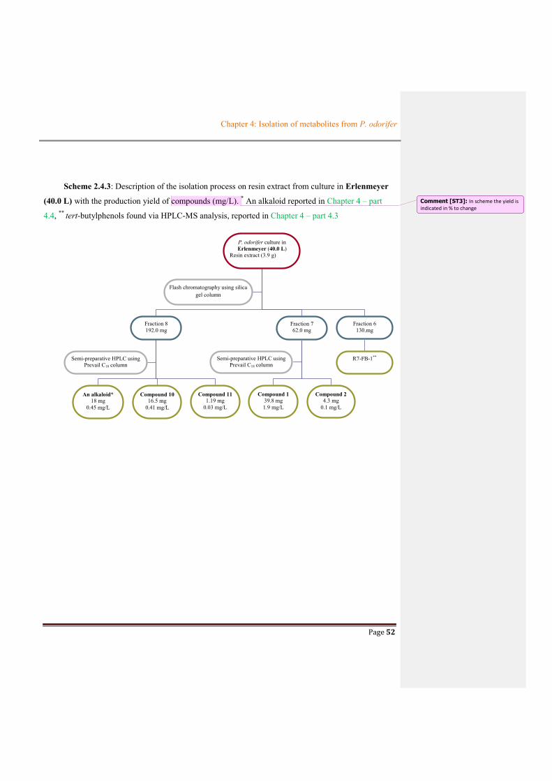
Chapter 4: Isolation of metabolites from P. odorifer
Page 52
Scheme 2.4.3: Description of the isolation process on resin extract from culture in Erlenmeyer
(40.0 L) with the production yield of compounds (mg/L). * An alkaloid reported in Chapter 4 – part
4.4, ** tert-butylphenols found via HPLC-MS analysis, reported in Chapter 4 – part 4.3
P. odorifer culture in Erlenmeyer (40.0 L)
Resin extract (3.9 g)
Fraction 8 192.0 mg
Fraction 7 62.0 mg
Flash chromatography using silica gel column
An alkaloid* 18 mg
0.45 mg/L
Semi-preparative HPLC using Prevail C18 column
Compound 11 1.19 mg
0.03 mg/L
Compound 10 16.5 mg
0.41 mg/L
Compound 1 39.8 mg 1.9 mg/L
Semi-preparative HPLC using Prevail C18 column
Compound 2 4.3 mg
0.1 mg/L
Fraction 6 130.mg
R7-FB-1**
Comment [ST3]: In scheme the yield is indicated in % to change
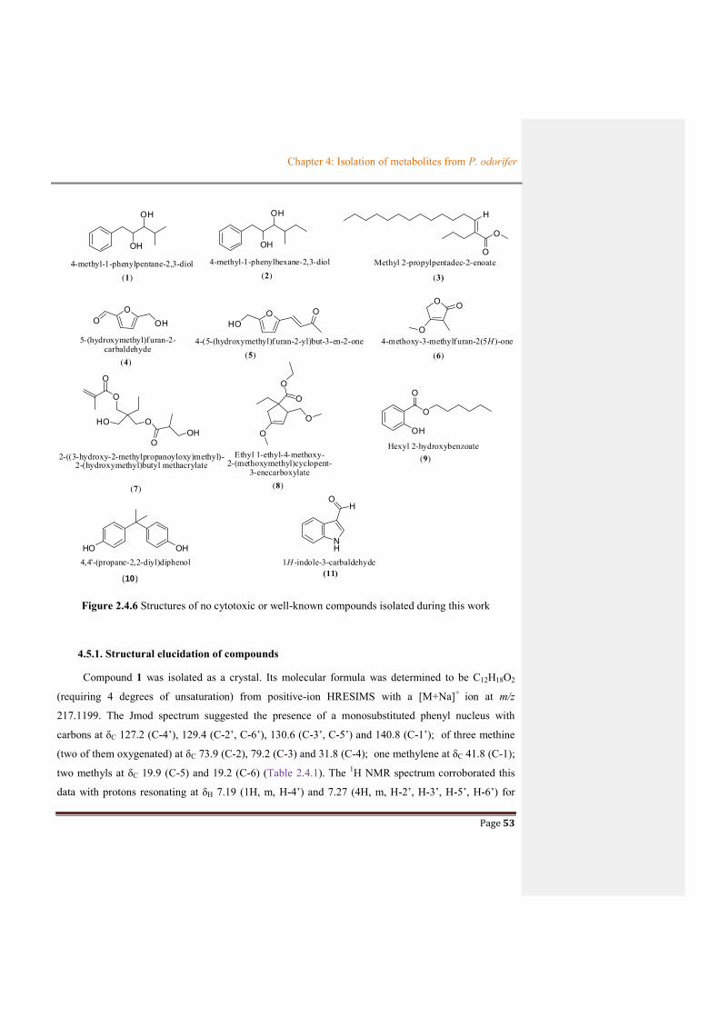
Chapter 4: Isolation of metabolites from P. odorifer
Page 53
OH
OH
(1)
OH
OH
(2)
H
O
O
(3)
OO OH
(4)
OHO
(5)
OO O
(6)
O
O
O
HO O
OOH
(7)
O
O
O
O
(8)
4-methyl-1-phenylpentane-2,3-diol 4-methyl-1-phenylhexane-2,3-diol Methyl 2-propylpentadec-2-enoate
4-(5-(hydroxymethyl)f uran-2-yl)but-3-en-2-one 4-methoxy-3-methylf uran-2(5H )-one
2-((3-hydroxy-2-methylpropanoyloxy)methyl)-2-(hydroxymethyl)butyl methacrylate
O
5-(hydroxymethyl)f uran-2-carbaldehyde
OH
O
Hexyl 2-hydroxybenzoateEthyl 1-ethyl-4-methoxy-
2-(methoxymethyl)cyclopent-3-enecarboxylate
(9)
HO OH4,4'-(propane-2,2-diyl)diphenol
(10)
NH
OH
1H -indole-3-carbaldehyde(11)
Figure 2.4.6 Structures of no cytotoxic or well-known compounds isolated during this work
4.5.1. Structural elucidation of compounds
Compound 1 was isolated as a crystal. Its molecular formula was determined to be C12H18O2
(requiring 4 degrees of unsaturation) from positive-ion HRESIMS with a [M+Na]+ ion at m/z
217.1199. The Jmod spectrum suggested the presence of a monosubstituted phenyl nucleus with
carbons at δC 127.2 (C-4’), 129.4 (C-2’, C-6’), 130.6 (C-3’, C-5’) and 140.8 (C-1’); of three methine
(two of them oxygenated) at δC 73.9 (C-2), 79.2 (C-3) and 31.8 (C-4); one methylene at δC 41.8 (C-1);
two methyls at δC 19.9 (C-5) and 19.2 (C-6) (Table 2.4.1). The 1H NMR spectrum corroborated this
data with protons resonating at δH 7.19 (1H, m, H-4’) and 7.27 (4H, m, H-2’, H-3’, H-5’, H-6’) for

Chapter 4: Isolation of metabolites from P. odorifer
Page 54
monosubstituted phenyl ring, two oxygenated methine protons at δH 3.85 (1H, ddd, J = 2.8, 6.1, 7.6
Hz, H-2), 2.99 (1H, dd, J = 2.8, 7.6, H-3) and one aliphatic methine at at δH 1.87 (1H, m, H-4) and two
deshielded methyl group at δH 0.88 (3H, d, J = 6.7 Hz, H-5) and 0.97 (3h, d, J = 6.7 Hz, H-6). Analysis
of 2D NMR spectra of 1 (COSY, HSQC and HMBC) led to the identification of two partial structure
(a-b) (Figure 2.4.7). The first moiety (a) was a monosubstituted phenyl nucleus that was connected
with another part (b) via HMBC correlations from H-1 to C-2’/C-6’, and from H-2’/H-6’ to C-2 to give
the full structure of 1.
aOH
OH
b
OH
OH OH
OH1 3 5
6
1'
2'4'
HMBC COSY
Figure 2.4.7 Key correlations for the structural assignment of 1
The stereochemistry of H-2 and H-3 in compound 1 can be assigned by the analysis of coupling
constants of H-2, H-3. According to literature (Silverstein and Webster, 1996), if J2,3 is smaller than
7.0 Hz, the two protons H-2 and H-3 are in a trans-configuration, otherwise, if J2,3 is more than 7.0 Hz,
they are in cis configuration. In the case of 1, the trans configuration between H-2 and H-3 was
attributed by their small coupling content (J2,3 = 2.8 Hz). Moreover, the absolute configuration of 1
could be determined by Mosher’s method. The determination of the absolute configuration of 1 will be
further realized. Compound 2 was obtained as a crystal. Its molecular formula was assigned to be
C13H20O2 based on the HRESIMS data which corresponded to a supplementar 14 mass unit (one CH2
group) in comparison with 1. Analysis of 1H, 13C NMR spectra revealed that the structure of 2 was
similar to 1. The major difference was the presence of one methylene group at δC/H 26.1(C-5)/1.15
(2H, m, H-5) (Table 2.4.1). Furthermore, the COSY and HMBC correlations also confirmed this
structure (Figure 2.4.8). The stereochemistry at C-2 and C-3 of 2 was also determined as described
earlier for compound 1. It was suggested as trans configuration due to a small coupling constant of
J2,3 as 2.3 Hz. Likewise, the absolute configuration of 2 could be determined in further work.
Comment [ST4]: Pas ds la liste

Chapter 4: Isolation of metabolites from P. odorifer
Page 55
OH
OH
12
3 45
61'
2'
4' 7
HMBC
Figure 2.4.8 Key correlations for the structural assignment of 2
Table 2.4.1 Comparison of 1H NMR (500MHz, CD3OD) and 13C NMR (75MHz, CD3OD) spectroscopic data of compounds 1 and 2
H, mult. (J in Hz) C Compound 1 Compound 2 Compound 1 Compound 2
1a 2.80, dd (7.6, 13.5) 2.85, dd (6.6, 5.5) 41.8 41.8 1b 2.87, dd (6.1, 13.5) 2 3.85, ddd (7.6, 6.1, 2.8) 3.87, ddd (7.4, 6.6, 2.3) 73.9 73.5 3 2.99, dd (2.8, 7.7) 3.07, dd (8.2, 2.3) 79.2 77.3 4 1.87, m 1.67, m 31.8 38.2 5 0.88, d (6.7) 1.15, m 19.9 26.1 6 0.97, d (6.7) 0.88, t (7.5) 19.2 11.4 7 - 0.85, d (6.8) 15.6 1’ - - 140.8 140.6
2’/6’ 7.27, m 7.27, m 129.4 129.3 3’/5’ 130.6 130.5
4’ 7.19, m 7.19, m 127.2 127.1
The compounds 1 and 2 were already found in Mangrove Rhizophora (Rukachaisirikul et al.,
2012) but their spectroscopic data were not shown.
Compound 3 was obtained as colorless oil. Its molecular formula was determined to be C19H36O2
from the [M+Na] + ion at m/z 319.26130 (calcd for C19H36O2Na 319.26075) observed in positive ion
HR-ESI-MS spectrum.
The 1H-NMR spectrum of 3 exhibited a methoxy group at δ 3.62 (H-19), two methylene groups
linked to an alkene at δ 2.26 and 1.97 (H-16, H-4), a chain of aliphatic protons at δ 1.24-1.54 (H-5-14,
H-17) and two methyl groups at δ 0.86 (H-15, H-18). The presence of only one olefinic proton at δ
5.38 (H-3) highlighted the presence of a monosubstituted alkene. The 13C-NMR data of 3, which were

Chapter 4: Isolation of metabolites from P. odorifer
Page 56
assigned by Jmod, HSQC and HMBC analysis, displayed signals for a carbonyl at δ 173.5 (C-1), two
olefinic carbons at δ 135.4 (C-2) and 129.8 (C-3), a oxygenated carbon at δ 50.6 (C-19) and a chain of
aliphatic carbons at δ 28.8 to 32.0. The positions of these groups were confirmed by HMBC
correlations from H-4 to C-3, from H-3 and H-5 to C-4, H-18 to C-17, C-16, H-17 to C-16, C-2 and H-
16 to C-1 (shown in Chapter 5 - 5.3.2).
Moreover, the 1H-1H COSY experiments highlighted correlations between protons H-4 with H-3
and H-5, H16-and H17 as well as H14 and H15 (Figure 2.4.9).
Additionally, the HMBC data exhibited the correlation from protons of methoxy to carbonyl
carbon that formed an ester group. On the other hand, protons of methylene group attached to alkene
(H-16, δ 2.26) also presented HMBC correlations with carbon of the carbonyl group. This observation
suggested that the ester group was one of the substituted groups in the double bond. Therefore, with
above data, 3 presented an entire structure as methyl 2-propylpentadec-2-enoate. To our knowlegede,
this compound was an aliphatic ester found for the first time from bacteria. However, it exhibited no
cytotoxicity on HaCaT and B16 cell lines
Compound 4 and 5 were assigned as furfural derivatives. Compound 4 was collected as colorless
oil while compound 5 was found as yellow oil. Their molecular formula was determined to be C6H6O3
for 4 and C9H10O3 for 5
The 1H NMR spectrum of 4 exhibited a symmetrical structure with three proton signals including
a methylene group at δ 4.70 (H-6) joined to an oxygen, two aromatic protons at δ 7.22 (H-3) and
6.52 (H-4) and one proton of carbonyl group at δ 9.59 (H-7). The presence of a small coupling
constant (3.5 Hz) between the two aromatic protons supported an ortho relationship of them on furan
ring. The Jmod and HSQC spectra of 4 showed carbon signals including carboxyl groups at δ 178.1,
H
O
O
12
3
4
5
6
7
8
9
10
11
12
13
14
15
16
1718 19
HMBCFigure 2.4.9 Key HMBC correlations in 3

Chapter 4: Isolation of metabolites from P. odorifer
Page 57
aromatic carbons at δ 123.0 and 110.6 and methylene group at δ 58.7. The assignements of these
groups were confirmed by HMBC correlations (Figure 2.4.10).
The 1H and 13C NMR data of compound 5 revealed four signals at downfield shifts as methine
groups at δC 131.8 (δH 7.41, H-3), δC 124.7 (δH 6.58, H-4), δC 118.6 (δH 6.80, H-3’) and δC 111.3 (δH
6.46, H-4’). It simultaneously presented a carbonyl group at δC 200.9; a methyl group at δC 27.2 (δH
2.34, H-2), an oxygenated methylene moiety at δC 57.5 (δH 4.56, H-6’) and two quaternary carbons at
δC 151.9 (C-2’) and δC 159.6 (C-5’). The COSY correlations established two spin systems from these
four methine groups as H-3 and H-4, H3’and H4’ whereas, the HMBC correlations from protons of
methyl group from H-1 to C-2, C-3, C-4 ; from H-3 to C-2, C-4, C-2’ and C-3’; from H-4 to C-2, C-
2’; from H-3’ to C-3, C-2’, C-4’, C-5’; from H-3’ to C-1’, C-2’, C-4’; and from H-6’ to C-4’ C-5’
demonstrated the structure of 5 (Figure 2.4.10).
Although two compounds 4 and 5 were well- known synthetic compounds (Rigal and Gaset et
al.,1983), it was the first example in nature. Both compound 4 and 5 showed no cytotoxicity on HaCaT
and B16 cell lines.
Compound 7 was isolated as a white amorphous powder, it has C10H18O4 molecular formula
from the [M+Na]+ ion at m/z 225.1100 according to HRESIMS data. Its 1D and 2D NMR were
analyzed in CDCl3. The NMR data analysis of 7 was assigned to cyaneodimycin which has been
already reported in the literature from S. cyaneofuscatus associated to Lichina pygmeae (Delphine
Parrot et al., 2016).The NNR data are characteristic and exhibited two germinal protons at δH 6.12,
OO OH
1
2
3 4
5
67O
O
HO 12
34
1'
2'
3'4'
5'
6'
COSY HMBC
4 5
Figure 2.4.10 Key HMBC correlations in 4 and 5

Chapter 4: Isolation of metabolites from P. odorifer
Page 58
5.63 corresponded to a methylene group with δC 126.5, one carbonyl carbon atom at δC 166.5 and one
methyl group at δC 18.0, δH 1.94. All of them displayed a methacrylate moiety - a rare group in natural
products (See in Table 2.4.2). Moreover, 7 also possessed one carbonyl carbon (at δC 175.2), four
oxygenated methylene group (at δC 64.7, 66.2, 68.1, 60.5 corresponding with δH 4.28, 3.68, 3.65, 3.72,
respectively), two methyl groups (at δC 7.6, 14.4 and δH 0.88, 1.30, respectively) and a quaternary
carbon at δC 43.2. Interestingly, HRESIMS data of 7 supported a [M+Na]+ ion at m/z 225.1100 related
to molecular formula as C10H18O4Na (instead of C14H24O6) which corresponds to a loss of a C4H6O2
unit. This observation was fully explained by Delphine Parrot and it confirmed again that this kind of
compound was easily hydrolyzed during the analysis process.
O
O
OH
O
O
OH
12
3 4
56 7
8
9
10
11
12
13
14
Figure 2.4.11 Key COSY (black line) and HMBC (arrows H to C) correlations for compound 7
Table 2.4.2 Comparison of NMR data between compound 7 and literature
Compound 7 (in CDCl3) Reference (Parrot et al., 2016) in CDCl3
Position δH , mul, (J[Hz]) δC δH , mul, (J[Hz]) δC
1 - 131.0 - 136.7 2 - 166.5 167.3 3 4.28, s 64.7 4.06,s 64.9 4 - 43.2 - 42.2 5 3.65 68.1 3.60-4.10 71.5 6 - 175.2 - 176.1 7 2.03 31.1 2.69-2.73, m 39.9 8 3.73 60.5 3.46-3.56 73.4 9 1.23, m 14.4 1.12, d, (10) 13.6 10 3.68 66.2 4.06, s 64.9 11 1.94 18.5 1.90, br. s 18.4 12a 6.12, s 126.5 6.04, br. s 125.6 12b 5.63, m 5.54, br. s 13 1.30, q, (7.6) 22.9 1.48, q, (7.5) 23.6 14 0.88, t, 7.6 7.6 0.87, t, (7.5) 7.6

Chapter 4: Isolation of metabolites from P. odorifer
Page 59
Compound 8 was obtained as colorless oil. Its molecular formula was determined to be C13H22O4
from the [M-H] - peak at m/z 241.1448 (calcd. for C13H21O4 241.14453) in negative ion HR-ESI-MS
spectrum. The 1H-NMR spectrum of 8 showed signals of methine proton at δH 6.17 (s, H-3), two
oxygenated methylene protons at δH 4.47 (d, J = 5.9 Hz, H-9) and 4.05 (q, J = 7.1 Hz, H-7).
Additionally, it displayed two methyl groups at δH 1.19 (t, J = 7.1 Hz, H-8) and 0.87 (t, J = 6.7 Hz, H-
12), two methylene groups at δH 1.27 (br, H-5 and H-11), two methoxyl groups at δH 3.31 (s, H-13)
and 3.30 (s, H-10). The 13C-NMR data highlighted also the presence of two carbons sp2 at δC 107.3 (C-
3) and 155.0 (C-4), two oxygenated methylene carbons at δC 59.7 (C-7) and 56.4 (C-9), two methoxy
carbons at δC 48.9 (C-13 and C-10), two methylene carbons at δC 31.4 (C-5) and 22.4 (C-11) and two
methyl carbon atoms at δC 13.8 (C-8) and 13.6 (C-12). Moreover, the presence of a carbonyl resonance
at δC 163.9 (C-6) and one quaternary carbon at δC 78.3 (C-1) together with HMBC correlations (Table
in Chapter 6- 6.3.8) supported a fully structure for 8.reported in Figure 2.4.12. To date, in our
knowledge, no report exists about this compound 8, but this novel compound exhibited no cytotoxic
effect on HaCaT and B16 cell lines
O
O
O
O
12
4 3
5
6
7
8
910
11
12
13
Figure 2.4.12 Key COSY (black lines) and HMBC (arrows H to C) correlations for compound 8
Compound 9 was isolated as white powder. Its molecular formula was determined to be C-
13H18O3 from the [M+Na] + peak at m/z 245.1152 (calcd. for C13H18O3Na 245.11481) in positive ion
HR-ESI-MS spectrum. The 1H-NMR spectrum of 9 presented signals of four aromatic protons at δH
7.87 (dd, J = 7.9 and 1.7 Hz, H-6), 6.89 (m, H-5), 7.48 (m, H-4) and 7.01 (dd, J = 8.4 and 0.9 Hz, H-
3); five methylene groups (one oxygenated at δH 4.37 (t, J = 6.7 Hz, H-8), 1.79 (m, H-9), 1.65 (m, H-
10) and 1.30 (m, H-11, H-12) and one methyl group at δH 0.91 (m, H-13). Additionally, these
functional groups were confirmed in 13C-NMR spectrum that showed six aromatic carbons (two of

Chapter 4: Isolation of metabolites from P. odorifer
Page 60
them as a quaternary carbon and one is an oxygenated aromatic carbon) at δC 115.3 (C-1), 161.9 (C-
2), 117.9 (C-3), 135.9 (C-4), 119.4 (C-5) and 130.2 (C-6); one carbonyl carbon at δC 170.5 (C-7); five
methylene carbons at δC 65.8 (C-8) (oxygenated carbon), 28.5 (C-9), 25.0 (C-10), 23.0-33.8 (C-11, C-
12) and one methyl group at δC 14.4. Furthermore, the combination of the all correlations highlighted
in 2D-NMR spectra (HSQC, HMBC, COSY) (Figure 2.4.13) supported the assignment of the full
structure of 9 as 1-hexylsalicylate.
O
OH
O
1
24
67
8
9 2
13
Figure 2.4.13 Key HMBC correlations for compound 9
Compound 9 was reported as a synthetic compound (Green et al., 2008) but its NMR data was
not shown. It was, thus, the first report in nature. Compound 10 has a commercial name as bisphenol A
that was synthetic compound and was ingredient of plastic. However, its derivative (bisphenol A
propyl ether analogue) was found from Streptomyces sp. (Qin et al., 2018) and it was confirmed to not
be an artifact product. Compound 10 exhibited a moderate cytotoxic effect oh HaCaT and B16 cell
lines with IC50 values of 34 ± 2 and 74 ± 4 µg/mL, respectively.
Compound 11 was isolated as a yellow crystal with a C9H7ON molecular formula. The 1H NMR
spectrum presented five aromatic protons and a carbonyl proton. This data and its molecular formula
provided the assignment of this compound to be indole carbadehyde which was already reported from
Nocardia sp isolated from Collema auriforme (Noël et al 2017), and from plant (Shankar et al., 2009),
from marine sponge (McKay et al., 2005) and from bacteria belonging to Actinomyces (Wang et al.,
2014). The HMBC correlations of compound 11 were shown in Figure 2.4.13.
HN
O
1
23
4
5
6
77a
3a
Figure 2.4.14 Key HMBC correlations for compound 11

Chapter 4: Isolation of metabolites from P. odorifer
Page 61
Table 2.4.3 Comparison of NMR data between compound 11 and reference
Compound 11
Reference (Noël et al., 2017)
Position δH , mul, (J[Hz]) δC HMBC COSY δH , mul, (J[Hz]) δC
1-NH - - - - 2-CH 8.12, s 138.3 C-3, C-4, C-7a, C-8 - 8.10, s 139.7
3 - 118.6 - - - 120.1 3a - 126.5 - - - 125.6
4-CH 7.28, m 123.6 C-6 H-5 7.25, dt (7.6; 1.2) 123.6 5-CH 8.17, m 121.0 C-4 H-4/H-6 8.16, dt (7.6; 0.9) 122.3 6-CH 7.28, m 122.2 C-7 H-5, H-7 7.28, td (7.6; 7.8; 1.2) 125.0 7-CH 7.50, m 111.7 C-4 H-6 7.48, dt (8.0; 0.8) 113.1
7a - 137.8 - - - 139.1 8-CHO 9.90, s 190.0 C-3, C-4 - 9.89, s 187.2
Conclusions
The compounds isolated from the culture of P. odorifer showed a variety of skeleton from
primary metabolite (polysaccharide) to secondary metabolites (alkaloid, tert-butylphenol compounds,
diol compounds, furfural derivatives, ester salicylate, indole carbaldehyde...). Although
polysaccharides were reported in many cultures of bacteria, it was firstly found in P. odorifer species.
Excepted the alkaloid, most compounds isolated from P. odorifer were either rare as natural products
(e.g. tert-butylphenol compounds) or were synthetic compounds (furfural derivative, ester salicylate).
A novel compound but no cytotoxic has been also described. Besides, indole carbaldehyde, a well-
known compound that was found from many bacteria, was isolated from this bacterial species.
However, no diketopiperazines which act as chemical signals (Brelles-Mariño and Bedmar, 2001)
were found from P. odorifer.

Chapter 4: Isolation of metabolites from P. odorifer
Page 62
References
McKay, M.J., Carroll, A.R., Quinn, R.J., Hooper, J.N.A., 2002. 1,2-Bis(1H-indol-3-yl)ethane-1,2-dione, an Indole Alkaloid from the Marine Sponge Smenospongia sp. J. Nat. Prod. 65, 595–597. https://doi.org/10.1021/np010347v
Noël, A., Ferron, S., Rouaud, I., Gouault, N., Hurvois, J.-P., Tomasi, S., 2017. Isolation and Structure Identification of Novel Brominated Diketopiperazines from Nocardia ignorata—A Lichen-Associated Actinobacterium. Molecules 22, 371. https://doi.org/10.3390/molecules22030371
Parrot, D., Legrave, N., Intertaglia, L., Rouaud, I., Legembre, P., Grube, M., Suzuki, M.T., Tomasi, S., 2016. Cyaneodimycin, a Bioactive Compound Isolated from the Culture of Streptomyces cyaneofuscatus Associated with Lichina confinis. Eur. J. Org. Chem. 2016, 3977–3982. https://doi.org/10.1002/ejoc.201600252
Rigal, L., Gaset, A., 1983. Direct preparation of 5-hydroxymethyl-2-furancarboxaldehyde from polyholosides: a chemical valorisation of the Jerusalem artichoke (Helianthus tuberosus L.). Biomass 3, 151–163. https://doi.org/10.1016/0144-4565(83)90003-3
Rukachaisirikul, V., Rodglin, A., Sukpondma, Y., Phongpaichit, S., Buatong, J., Sakayaroj, J., 2012. Phthalide and Isocoumarin Derivatives Produced by an Acremonium sp. Isolated from a Mangrove Rhizophora apiculata. J. Nat. Prod. 75, 853–858. https://doi.org/10.1021/np200885e
Silverstein M. R., Webster X. F., Kiemle J. D., 2005, spectrometric identification of organic compounds, 550, John Wiley & Son, INC, USA, p. 172.
Shankar, T., Muthusubramanian, S., Gandhidasan, R., 2009. A chromanone alkaloid from Derris ovalifolia stem. Nat. Prod. Res. 23, 1652–1656. https://doi.org/10.1080/14786410903306126
Wang, P., Kong, F., Wei, J., Wang, Y., Wang, W., Hong, K., Zhu, W., 2014. Alkaloids from the Mangrove-Derived Actinomycete Jishengella endophytica 161111. Mar Drugs 12, 477–490. https://doi.org/10.3390/md12010477

Page 63
CHAPTER 5: CONCLUSIONS AND PERSPECTIVES
Chapter 5: Conclusions and Perspectives
Page 64
CHAPTER 5: CONCLUSIONS AND PERSPECTIVES
Lichens are complex organisms which can grow on many different supports. They can be
found on trees, soil, rock surfaces and even inside rocks as well as on glass ceramics, metal
objects... Lichens are also perfect self-supply mini-ecosystems growing very slowly and formed
by the association of three partners such as fungi (mycobiont), algae or/and cyanobacteria
(photobiont) and bacterial communities. Each partner plays distinct roles and participates together
to permit to this organism to survive under drastic conditions of light, temperature and water
supply, from the poles to the tropics, from the intertidal zones to peaks of the mountains (Brodo et
al., 2001).
Among partners of the lichen, the third partner corresponding to the bacterial communities
became a new interesting source for chemists to study due to their ability to produce many active
compounds. In our efforts, using a culture-based strategy, in discovery and study these
communities found on Rhizocarpon geographicum, one of the crustose lichens living on the rock
surface, 13 pure strains including 10 of bacteria, one cyanobacterium and one fungus (Chapter 1)
have been collected. The ten bacteria strains belonged to the two phyla of Firmicutes and
Proteobacteria. Indeed, unlike to the other lichens, Proteobacteria was not the predominant class of
bacteria among all the symbiotic microorganisms of R. geographicum. Instead, Firmicutes phylum
took place in this dominance among these bacterial communities. Moreover, in this phylum, the
genus which was present with the highest percentage (33%) was Paenibacillus genus. Throughout
the studies of literature already published on the strains of this genus, Paenibacillus odorifer was
selected to isolate its metabolites due to its potential production of active compounds (Chapter 2).
The next step of this work was to determine the optimal parameters of culture of P. odorifer
in liquid medium. The first optimization (Chapter 3 – 3.1) carrying out with small scale (25 mL)
resulted that P. odorifer grew the best in Gym Streptomyces medium supplemented with CaCO3 at
pH =7 and 25oC. These parameters chosen from the first optimization step were applied for the
culture by bioreactor and yielded polysaccharide (Chapter 4 – 4.2) and two diol compounds
(Chapter 4 – 4.5).
The polysaccharide, firstly identified by IR and NMR data, was formed by three
sugar units of glucuronic acid, fructose and fucose with an average molecular ratio of 4/2/1,
respectively. The results were determined from the comparison of retention time observed in

Chapter 5: Conclusions and Perspectives
Page 65
HPLC chromatograms between sugar units derived from the hydrolysis of polysaccharide and
those of standard monosaccharides. Then, a series of reactions involving methylation, hydrolysis,
sodium borohydride reduction, acetylation by acetic anhydride and final GC-MS analysis of
alditol acetates led to a hypothesis of the structure of this polysaccharide fraction as →2)- β-D-
GlcAp-(1→ 2)-β-D-GlcAp-(1→2)-β-D-GlcAp-(1→2)-β-D-GlcAp-(2→2)-β-D-Fruf-(2→4)-β-D-
Fruf-(2→4)- β-L-Fucp-(1→. This polysaccharide also possessed a significant cytotoxicity
measured by MTT assay with IC50 value of 19µg/mL and 27 µg/mL on HaCaT human
keratinocyte and B16 murine melanoma cell lines respectively. This may provide a potential
source of antitumor agents as microbial polysaccharide fraction produced from P. odorifer.
The two diol compounds (named 4-methyl-1-phenylpentane-2,3-diol (1) and 4-
methyl-1-phenylhexane-2,3-diol (2)) harvested from culture in bioreactor, did not show any
cytotoxic activity. Although these two compounds were identified by Rukachaisirikul and co-
workers (2011), their spectroscopic data was not shown. To our knowledge it is the first report of
these compounds from P. odorifer culture.
The disadvantage of the culture performed in the bioreactor (4.5 L) is the presence of other
metabolites in a too scale amount. As a result, to increase the amount of secondary metabolites
produced, a secondary optimization process was established using Erlenmeyer flasks (Chapter 3 –
3.2). The difference of production following the vessels used could be explained by the different
mode of aeration and the stirring system inducing different stress conditions for the strain. In fact,
the stirring in the bioreactor is realized by agitation blade submerged in the culture broth which
implicates hyphal breaks, while in shaken flasks the stirring is performed with an orbital shaker
allowing the hyphal formation. By the same way while in shaken flasks there is no specific system
for aeration those in bioreactor consisted in a tube submerged in the culture broth which conducts
oxygen into the medium causing the formation of bubbles.
Based on the results of the first optimization, the second one was carried out using
Erlenmeyer flasks (4.0 L) leading to the selection of the best conditions for the production of
metabolites from P. odorifer as medium Gym Streptomyces supplemented with CaCO3 at pH 7,
25oC, and 120 rpm stirring as well as 1% inoculum. The resin extract obtained using the best
conditions of the culture was subjected to chromatography approaches to give some compounds as
described here. Among them, two tert-butylphenols were isolated and one of them exhibited

Chapter 5: Conclusions and Perspectives
Page 66
significant cytotoxic activity against HaCaT and B16 cell lines (Chapter 4 – 4.3). These two
compounds were rare in nature. Their presence in the broth of the culture could be explained by
the bioaccumulation and the biotransformation from tert-butyl derivatives such as BHA
(benzohydroxyanisole). Besides, P. odorifer has also produced some other compounds either no
cytotoxic or well-known (Chapter 4 – 4.5) such as methyl 2-propylpentadec-2-enoate (3); 5-
(hydroxymethyl)furan-2-carbaldehyde (4); 4-(5-(hydroxymethyl)furan-2-yl)but-3-en-2-one (5); 4-
methoxy-3-methylfuran-2(5H)-one (6); 2-((3-hydroxy-2-methylpropanoyloxy)methyl)-2-
(hydroxymethyl)butyl methacrylate (7); Ethyl 1-ethyl-4-methoxy-2-(methoxymethyl)cyclopent-3-
enecarboxylate (8); hexyl 2-hydroxybenzoate (9).
Further, the culture with a large volume (40 L in Erlenmeyer) using the best conditions
selected following the results of the second optimization was carried out to find bioactive
metabolites with higher amounts. A new alkaloid was found from a cytotoxic fraction (fraction 8).
Its structure consists of a dihydronaphthalene moiety, which is a well-known structural group in
natural products, fused to a rare pyrrolooxazine unit (Chapter 4 – 4.4). This new compound was
the first example of this skeleton produced by any living organisms (and especially P. odorifer).
This compound also presented week cytotoxicity with IC50 values of 76.0 µM and 78.9 µM on
B16 and HaCaT cell lines, respectively. Moreover, two well- known compounds were also found
from the fraction 8 such as 4,4'-(propane-2,2-diyl)diphenol (10) and 1H-indole-3-carbaldehyde
(11) which were no active.
By comparing the report of all compounds isolated from each culture of P. odorifer using
different means as bioreator and Erlenmeyer, it seems that the polysaccharide was only found
from the culture using bioreactor. This can be explained by the fact that for culture in bioreator,
the inoculum ratio was not controlled and the stirring was fixed at 150 rpm during the culture.
These factors are different to the optimal parameters selected from optimization stages whereas
these factors seem to play an important role for the production of active metabolites (Chapter 3 –
3.2.2). Moreover, the difference in the aeration method used could be also one of the reasons for
this difference in production. Therefore, the realization of a culture in the bioreactor following the
best conditions found during the second optimization could afford an accurate conclusion for the
production of this polysaccharide fraction by P. odorifer. Endeavors will be done to control in a
better way all the parameters of the culture in bioreactor (aeration rate, stirring, inoculum ratio
Chapter 5: Conclusions and Perspectives
Page 67
etc…).
In the same way, for the other metabolites, the two tert-butylphenols were found from the
culture in Erlenmeyer but were absent in culture using bioreactor. The origin of this kind of
compounds must be studied and especially their putative biosynthetic pathway. We envisaged
realizing the entire genome sequencing in order to emphasize some interesting metabolic
pathways.
Unlike, the diol compounds 1 and 2 were reported from the culture in both using bioreactor
and Erlenmeyer flask. Thus, we could conclude that they are one of the major compounds
produced by P. odorifer. Interestingly, when the volume of culture increased the mass of these
compounds also increased (see in Scheme 2.3.1 and 2.4.2). In the same manner, to increase the
mass of cytotoxic compounds, it is better to cultivate this strain in a large volume of medium.
Besides, two furfural derivatives (4, 5) were also found in the cultures using Erlenmeyer
flask. The medium used for the culture of P. odorifer contained malt extract which is a mixture of
cereal seeds. It is important to note that furfural was reported to be formed from the treatment of
cereals at high temperature (Mesias et al., 2017). Correspondingly, we could suggest that the
furfural derivatives (4, 5) were formed by the bioaccumulation of furfural which was present in
medium after sterilization step and following by biotransformation by P. odorifer. Therefore, the
bioaccumulation and biotransformation seem to be a characteristic behavior of P. odorifer. As
discussed above this particular behavior must be studied in further experiments. We also suggest
submitting this interesting strain in the biotransformation studies of lichen compounds.
Finally, P. odorifer is an interesting producer as this strain could produce original
compounds such as a new alkaloid, some diol compounds in a large amount, some furfural
derivatives, and some tert-butylphenols which could be derived from the biotransformation of
particular precursors. Interestingly, while a significant amount of diketopiperazines have already
been isolated from the culture of various lichen-associated bacteria, none of these compounds,
classically found from cultures of microorganisms, were found herein. These observations
highlight that this bacterium could be an interesting strain for biotransformation or producer of
novel active compounds.

Chapter 5: Conclusions and Perspectives
Page 68
The isolation of other potential active metabolites is in progress from other active fractions
obtained from our culture in large volume. Other evaluation of activities such as antibiotic or
antifungal properties will be undertaken to valorize these compounds particularly the diol
compounds obtained in large amount. We also envisage studying the other extracts as supernatant
of from pellets which could contain potentially active intracellular compounds.
Moreover, the co-culture between P. odorifer and the fungus which has already been
isolated from R. geographicum should be done to give some hypothesis about the ability of this
bacterium to produce defensive weapons in the presence of this fungus. These further experiments
will give us some argues to highlight the competition between various microorganisms which can
be appeared inside this complex micro-ecosystem which could be the lichen.
References
Brodo I.M., Sharnoff S.D., Sharnoff S. (2001). Lichens of North Ameica. Yale University Press, New Haven CT. 828pp.
Mesías, M., Morales, F.J., 2017. Effect of Different Flours on the Formation of Hydroxymethylfurfural, Furfural, and Dicarbonyl Compounds in Heated Glucose/Flour Systems. Foods 6. https://doi.org/10.3390/foods6020014
Rukachaisirikul, V., Rodglin, A., Sukpondma, Y., Phongpaichit, S., Buatong, J., Sakayaroj, J., 2012. Phthalide and Isocoumarin Derivatives Produced by an Acremonium sp. Isolated from a Mangrove Rhizophora apiculata. J. Nat. Prod. 75, 853–858. https://doi.org/10.1021/np200885e.

Page 69
CHAPTER 6: MATERIALS AND METHODS

Chapter 6: Materials and Methods
Page 70
CHAPTER 6: MATERIALS AND METHODS
6.1. MATERIALS
All solvents, reagents and materials used in all experiments were purchased from Sigma-
Aldrich (Lyon-France), Carlo Erba Reactif (Val de Reuil, France), VWR (Fontenay-sous-bois,
France), Acros Organics (Halluin, France). The solvent used for analysis or separation in HPLC
were the quality of HPLC. The water was used from the EasyPure system (BarnsteadTM,
ThermoFisher Waltham, MA, USA).
The Table 2.6.1 describes the ingredients composing of the media used to isolate the strains
from R. geographicum.
Table 2.6.1 Ingredient of media used to isolate bacterial strains from R. geographicum
Elements (g/liter sterile water) Bacillus acido
Gym Gym + 10%NaCl
Thiobacillus Marine agar
Yeast extract (Sigma-Aldrich, France)
1.0 4.0 4.0
Malt extract (Sigma-Aldrich, France)
10.0 10.0
Glucose (Sigma-Aldrich, Germany)
1.0 4.0 4.0 10.0
Marine broth (DifcoTM) 37.4
Agar 20.0 15.0 15.0 15 15.0
(NH4)2SO4 0.2 3.0
MgSO4.7H2O 0.5 1.0
CaCl2.2H2O 0.25
KH2PO4 0.6 0.5
CaCO3 2.0 2.0
KCl 0.1
Ca(NO3)2.4H2O 1.8.10-2
FeSO4.7H2O 10-5
NaCl 100.0
pH 3.5 7.2 7.2 4.5

Chapter 6: Materials and Methods
Page 71
6.2. METHODS
6.2.1. The process for the production of crude extracts from fermentation
After the culture at selected parameters, the supernatant and the bacterial cells (pellets)
were separated by the centrifugation at 3500 rpm at 4°C during 15 minutes. A XAD-7HP resin was
added to the supernatant (40 g of resin per 1 L of supernatant) to adsorb organic compounds
during 4 hours at 240 rpm of orbital rotation. After 4 hours, resin and supernatant were separated
by filtration. The desorption from the resin was practiced 3 times with 800 mL of
methanol:acetone mixture (50:50, v:v) for 15 minutes at 180 rpm of stirring. After the filtration
step, the solution containing organic compounds was adjusted pH up to 7, concentrated under
vacuum and extracted three times with ethyl acetate solvent. The resulting organic phase was dried
by magnesium sulfate before evaporation under vacuum to afford a crude extract named resin
extract (Scheme 2.4.1 chapter 4, part 4.1).
Similarly, the supernatant was also extracted 3 times by ethyl acetate solvent and the
resulting organic phase was dried by MgSO4 before evaporation under vacuum to give the
supernatant extract.
6.2.2. Analytical methods used for isolation steps
6.2.2.1. Thin layer chromatography (TLC)
The TLC was performed on Aluminum sheets (20 x 20 cm) (silica gel 60 F254, Merck,
Germany) for normal phase and using the solvent of elution as several mixtures of CHCl3, EtOAc,
MeOH with different ratios. The substances on silica gel sheets were observed under UV light at
254 nm, 312 nm or 365 nm and often visualized by anisaldehyde (ANS) reagent.
The ANS reagent was prepared by 3mL of ANS dissolved in 40 mL of glacial acetic acid
and 90 mL of ethanol. 2% H2SO4 was then added to the resulting solution before the use.
6.2.2.2. Classical column chromatography
The separation using classical column chromatography was performed by a column with
silica gel 60A (0.06 – 0.2 mm) (ACROS-ORGANICS). The elution solvents were solvents with
increasing polarity from cyclohexane, dichloromethane (DCM), EtOAc to MeOH. The process
was checked by TLC on normal phase.

Chapter 6: Materials and Methods
Page 72
6.2.2.3. Flash chromatography
The fractionation of crude extracts from fermentation was carried out by Flash
chromatography (PuriFlash, Interchim, Montlucon, France) using a packaged column either as
normal phase (40g SiOH Chromabond®Flash column) or reverse phase (40g, C18, Reveleris,
Grace), flow rate of 15 mL/min, collection of 10 mL, UV detector at 272 nm. The elution solvent
for normal phase was a sequential mixture of solvents with increasing polarity from cyclohexane,
dichloromethane, EtOAc to MeOH (see Figure 2.6.1) during 4 hours. The gradient of elution for
reverse phase was a mixture of H2O and acetonitrile (ACN) during 75 min, beginning from ACN
at 2% to 10% in 6 min, increasing to 15% at 15 min, 30% at 30 min, 40% at 45 min, 65% at 60
min and 100% at 75 min (See Figure 2.6.2).
Figure 2.6.1 Gradient of elution for the separation of extracts by flash chromatography using a 40g SiOH Chromabond column
Figure 2.6.2 Gradient of elution in flash chromatography using a reverse phase C18 Reveleris (Grace) column

Chapter 6: Materials and Methods
Page 73
6.2.2.4. HPLC-UV/MS analysis
The chemical profiles of extracts or of some fractions were analyzed by HPLC using Prevail
C18 column (5 µm, 250 x 4.6 mm, Grace, Columbia, MD, USA), with a flow rate of 0.8 mL/min,
concentration of sample as 1 mg/mL, volume injection of 20 µL, with elution solvents as gradient
of a mixture of H2O and Acetonitrile during 60 min, beginning at 0% of ACN in 5 min, then
increasing up to 100% of ACN at 35 min and maintaining this concentration in next 10 min, then
decreasing to 0% at 50 min (Figure 2.6.3). The mass spectrometry detector was connected on-line
with HPLC. The analysis on MS was performed by either positive or negative mode ESI
(Electrospray) on CMS of Advion (Ithaca, NY, CA). The data were analyzed by the software
Labsolution for HPLC and by Advion for MS.
Figure 2.6.3 Gradient of solvent in HPLC analysis using Prevail C18 column

Chapter 6: Materials and Methods
Page 74
6.2.2.5. Semi-preparative HPLC
The purification was performed by semi-preparative HPLC (Shimadzu, Marne-la-Vallée,
France) with a diode detector, using Prevail C18 column (5 µm, 250 x 10 mm, Grace, Columbia,
MD, USA). The gradients of elution were various mixtures of H2O and ACN as shown in Figure
2.6.4
Figure 2.6.4 Gradients of elution used in semi-preparative HPLC (using Prevail C18 column)
6.2.2.6. GC-MS (Gas Chromatography- Mass Spectrometry)
The GC-MS system spectrometry (Agilent Technologies GC-MS 7820A-GC, Agilent,
Waldbronn, Germany) was set up with nitrogen as carrier gas, using a DB-5 column (30 M x 0.25
mm x 0.25 µm), the injection volume of 1 µL at injection temperature 260oC and detector
temperature 270oC during 105.05 min. The initial 37oC oven temperature was set for 6s following
injection, raised to 140oC for 30 min at a rate of 200C/min, then to 180oC for 40 min at a rate of
40oC/min, and finally maintained at 230oC for 30 min (Table 2.6.2).
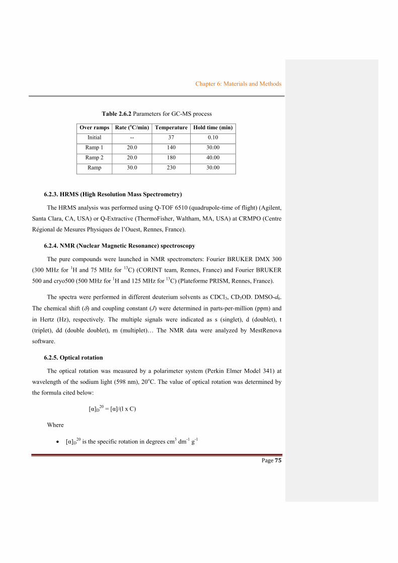
Chapter 6: Materials and Methods
Page 75
Table 2.6.2 Parameters for GC-MS process
Over ramps Rate (oC/min) Temperature Hold time (min)
Initial -- 37 0.10
Ramp 1 20.0 140 30.00
Ramp 2 20.0 180 40.00
Ramp 30.0 230 30.00
6.2.3. HRMS (High Resolution Mass Spectrometry)
The HRMS analysis was performed using Q-TOF 6510 (quadrupole-time of flight) (Agilent,
Santa Clara, CA, USA) or Q-Extractive (ThermoFisher, Waltham, MA, USA) at CRMPO (Centre
Régional de Mesures Physiques de l’Ouest, Rennes, France).
6.2.4. NMR (Nuclear Magnetic Resonance) spectroscopy
The pure compounds were launched in NMR spectrometers: Fourier BRUKER DMX 300
(300 MHz for 1H and 75 MHz for 13C) (CORINT team, Rennes, France) and Fourier BRUKER
500 and cryo500 (500 MHz for 1H and 125 MHz for 13C) (Plateforme PRISM, Rennes, France).
The spectra were performed in different deuterium solvents as CDCl3, CD3OD. DMSO-d6.
The chemical shift () and coupling constant (J) were determined in parts-per-million (ppm) and
in Hertz (Hz), respectively. The multiple signals were indicated as s (singlet), d (doublet), t
(triplet), dd (double doublet), m (multiplet)… The NMR data were analyzed by MestRenova
software.
6.2.5. Optical rotation
The optical rotation was measured by a polarimeter system (Perkin Elmer Model 341) at
wavelength of the sodium light (598 nm), 20oC. The value of optical rotation was determined by
the formula cited below:
[α]D20 = [α]/(l x C)
Where
[α]D20 is the specific rotation in degrees cm3 dm-1 g-1

Chapter 6: Materials and Methods
Page 76
[α] is the measured angle of rotation of a substance.
l is the path length in decimeter (dm)
C is the concentration in g/mL

Chapter 6: Materials and Methods
Page 77
6.2.6. Fourier transform infra-red (FT-IR) spectroscopy
The Infra-red spectrum was recorded on Fourier transform infrared (FTIR) spectrometer
(Thermo Fisher, Germany) using Attenuated Total Reflectance (ATR). The sample of one
milligram was pressed and scanned in the frequency range of 4000 – 400 cm-1. The vibration
bands were indicated at in cm-1.
6.2.7. Biological assays
6.2.7.1. Cytotoxicity evaluation using MTT assay
The cytotoxic effects were evaluated on extracts and pure compounds against HaCaT human
keratinocytes and B16 murine melanoma cell lines. HaCaT (10 000 cells/well) and B16 (6000
cells/well) were cultivated in RMPI 1640 medium supplemented with 5% of foetal calf serum
(FCS) and antibiotics (penicillin/streptomycin 1%) in atmosphere of 5% CO2 at 37oC. After 24h
culture, the samples were added at different concentrations (1, 10, 50, 100 and 200 µg/mL) and
each 96-well plate was continuously incubated at the same temperature and atmosphere as above.
After 48 culture, cell growth and viability were added 200 µL of DMSO and were then measured
at 540 nm using a MTT (3-[4,5-dimethylthiazol-2-yl]-2,5-diphenyltetrazolium bromide) assay.
Doxorubicin was used as a positive control. Each experiment was repeated three times.
6.2.7.2. Antioxidant evaluation
a) DPPH assay
The free radical scavenging activity was measured by DPPH (2,2-diphenyl-1-picrylhydrazyl
radical) method with quercetin and gallic acid as positive controls. 100 µL of DPPH methanol
solution (freshly prepared at a concentration of 1 mg/mL) were mixed with 10 µL of substance at
different concentrations (4.00; 11.1; 33.3; 100 µg/mL) in DMSO. After 15 min culture in the dark,
the absorbance was measured at 540 nm using a UV-visible spectrophotometer.
The DPPH scavenging percentage activity was calculated as follow:
Scavenging abilty % = [1- (As – Ab)/Ac] x 100
Where As is the absorbance of sample with DPPH, Ab is the absorbance of the sample without
DPPH (blank), Ac is the absorbance of negative control.

Chapter 6: Materials and Methods
Page 78
b) NBT assay
Phenazine methosulphate in the presence of NADH and under aerobic conditions (+ O2)
produces superoxide anions whose formation is determined by a dye nitro blue tetrazolium (NBT).
In the solution containing 40 µL of tris hydrochloride buffer (16 mM, pH = 8), 50 µL
nicotinamide adenine dinucleotide (NADH) (78 µM), 50 µL NBT (50 µM) and 50 µL phenazine
methosulfate (910 µM) were added to test different concentrations of the substance (0.25; 1.85;
2.5; 5.5; 16.0; 25.0; 50 mg/mL in DMSO). The color reaction between superoxide radical and
NBT was monitored by measurement of the absorbance at 560 nm. Vitamin C was used as
positive control. The experiment was repeated three times. The inhibition percentage was
calculated as follows:
Scavenging effect (%) = (1 – ASample at 560 nm/AControl at 560 nm) x 100
6.2.8. CFU (Colony-forming unit)
At the same time of the measurement of the optical density (OD) of the bacterial growth,
100 µL of the broth was diluted several times following demonstration of Figure 2.6.5 before
cultured on the agar plates. The counting of the colonies appearing on the agar was carried out the
next days.
Figure 2.6.5 The process of viable plate counts

Chapter 6: Materials and Methods
Page 79
The colony forming unit (CFU) per milliliter was calculated by equal as following:
CFU/ml = number of colony x (1/ total of dilution)

Chapter 6: Materials and Methods
Page 80
6.3. DESCRIPTIONS OF ISOLATED COMPOUNDS
The metabolites isolated from all the culture processes in either bioreactor or Erlenmeyer
will be presented in this part and their spectroscopic data will be reported in Annexe 3.
6.3.1. 4-methyl-1-phenylpentane-2,3-diol (1)
OH
OH2 3 4 5
1'
2'3'
4'
HaHb
1
The process of isolation for compound 1
Aspect: white crystal
Molecular formula: C12H18O2 (MW: 194 g.mol-1)
[α]20D = - 3.4 (c, 10-3, MeOH) (dm-1g-1.cm3).
HRESIMS: m/z [M+Na]+ found : 217.1202 , calculated: 217.1199
NMR spectra recorded in CD3OD at 300 MHz or 75 MHz
Position δC δH, mult. (J in Hz) COSY HMBC 1a-CH 41.8 2.80, dd (7.6, 13.5) 2 1’, 2’, 6’, 2, 3 1b-CH 2.87, dd (6.1, 13.5) 2 2-CH 73.9 3.85, ddd (2.8, 6.1, 7.6) 1, 3 3-CH 79.2 2.99, dd (2.8, 7.6) 2, 4 4, 5, 6 4-CH 31.8 1.87, m 3, 5, 6 3, 5, 6 5-CH3 19.9 0.88, d (6.7) 4 3, 4, 6 6-CH3 19.2 0.97, d (6.7) 4 3, 4, 5
1’ 140.8 - 2’/6’-CH 129.4 7.20 - 7.30, m 4’ 4’ 3’/5’-CH 130.6
4’-CH 127.2 7.19, m 3’/ 5’
Resin extract from culture in bioreactor
(4.5 L) (353.7 mg)
Reverse phase Flash chromatography (ACN/H2O)
Fraction 14 (21.8 mg) Semi-preparative
HPLC, Prevail C18 column, gradient 1
Compound 1 (2.0 mg)
Resin extract from culture in
Erlenmeyer (40.0 L) (3.9 g)
Normal phase Flash chromatography (Cyclohexane/DCM, DCM/EtOAc, ETOAc/MeOH
Fraction 7 (62.0 mg) Semi-preparative
HPLC, Prevail C18 column, gradient 5
Compound 1 (39.8 mg)

Chapter 6: Materials and Methods
Page 81
6.3.2. 4-methyl-1-phenylhexane-2,3-diol (2)
OH
OH2 3 4
5
1'
2'3'
4'
1 6
The process of isolation for compound 2
Aspect: white crystal
Molecular formula: C13H20O2 (MW: 208 g.mol-1)
[α]20D = - 8 (c, 10-3, MeOH) (dm-1g-1.cm3).
HRESIMS: m/z [M+ Na]+ found : 231.1356 , calculated: 231.13555
NMR spectra recorded in CD3OD at 300 MHz or 75 MHz
Position δC δH, mult.(J in Hz) COSY HMBC 1-CH2 41.8 2.85, dd (6.8, 5.5) 2 1’, 2’, 6’, 2, 3 2-CH 73.5 3.87, ddd (7.4, 6.6, 2.3) 1, 3 3-CH 77.3 3.07, dd (8.2, 2.3) 2, 4 4-CH 38.2 1.67, m 3, 5, 7 5-CH2 26.1 1.15, m 4, 6 6-CH3 11.4 0.88, t (7.5) 5 4, 5 7-CH3 15.6 0.85, d (6.8) 4 3, 4, 6
1’ 140.6 - 2’/6’-CH 129.3 7.20 – 7.30, m 4’ 4’, 1’ 3’/5’-CH 130.5
4’-CH 127.1 7.19, m 3’ 5’
Resin extract from culture in bioreactor
(4.5 L) (353.7 mg)
Fraction 14 (21.8 mg) Semi-preparative
HPLC, Prevail C18 column, gradient 1
Compound 2 (1.9 mg)
Resin extract from culture in
Erlenmeyer (40.0L) (3.9 g)
Fraction 7 (62.0 mg) Semi-preparative
HPLC, Prevail C18 column, gradient 5
Compound 2 (4.3 mg)
Reverse phase Flash chromatography (ACN/H2O)
Normal phase Flash chromatography (Cyclohexane/DCM, DCM/EtOAc, ETOAc/MeOH

Chapter 6: Materials and Methods
Page 82
6.3.3. Methyl 2-propylpentadec-2-enoate (3)
1
2
34
5
6
7
8
9
10
11
12
13
1415
1617
H
O
O18 19
The process of isolation for compound 3
Aspect: white powder
Molecular formula: C19H36O2 (MW: 296 g.mol-1)
Rf = 0. 565 (n-Hexane/Chloroform = 1/1 (v/v))
HRESIMS: m/z [M+ Na]+ found : 319.26075 , calculated: 319.2613
NMR spectra recorded in CD2Cl2, 300 MHz or 75 MHz
Position δH , mul, (J[Hz]) δC (Jmod, HSQC) HMBC COSY 1 - 173.5 - - 2 - 135.4 - -
3-CH 5.38, m 129.8 H-4 4-CH2 1.97, m 31.4 C-3 H-3, H-5
5-14-CH2 1.24, m 28.5-32.0 C-4, C-15 H-4, H-15 15/18-CH3 0.86, m 13.3 C-14/C16, C-17 H-4
16-CH2 2.26, t, 7.5 33.4 C-17, C-1 H-17 17-CH2 1.54, m 24.3 C-16, C-2 H-16
19-OCH3 3.62, s 50.6 C-1 -
Resin extract from culture in
Erlenmeyer (4.0L) (439.5 mg)
Fraction 1 (39.6 mg) Semi-preparative
HPLC, Prevail C18 column, gradient 4
Compound 3 (3.6 mg)
Subfraction 2 (18.6 mg) Reverse phase
column chromatography
Normal phase Flash chromatography (Cyclohexane/DCM, DCM/EtOAc, ETOAc/MeOH

Chapter 6: Materials and Methods
Page 83
6.3.4. 5-(hydroxymethyl) furan-2-carbaldehyde (4)
OO OH
1
2
3 4
5
67
The process of isolation for compound 4
Aspect: yellow oil
Molecular formula: C6H6O3 (MW: 126 g.mol-1)
Rf = 0.41 (Chloroform/Methanol (9/1) (v/v))
HRESIMS: m/z [M+ Na]+ found : 149.0208 , calculated: 149.02091
NMR spectra recorded in CDCl3, 300 MHz or 75 MHz
Position δH , mul, (J[Hz]) δC HMBCa COSY
2 - 153.3 - - 3-CH 7.22, d (3.5) 123.0 C2,C4,C5,C7 H-4 4-CH 6.52, d (3.5) 110.6 C2, C3, C5 H-3
5 - 161.0 - - 6-OCH2 4.70, s 58.7 C2, C3,C4, C5 - 7-CHO 9.59, s 178.1 - -
Resin extract from culture in
Erlenmeyer (4.0 L) (439.5 mg)
Fraction 3 (24.7 mg)
Compound 4 (1.93 mg)
Semi-preparative HPLC, Prevail C18 column, gradient 4
Resin extract from culture in
Erlenmeyer (40.0L) (3.9 mg)
Fraction 10 (79.4 mg) Semi-preparative
HPLC, Prevail C18 column, gradient 5
Compound 4 (2.57 mg)
Normal phase Flash chromatography (Cyclohexane/DCM, DCM/EtOAc, ETOAc/MeOH
Normal phase Flash chromatography (Cyclohexane/DCM, DCM/EtOAc, ETOAc/MeOH
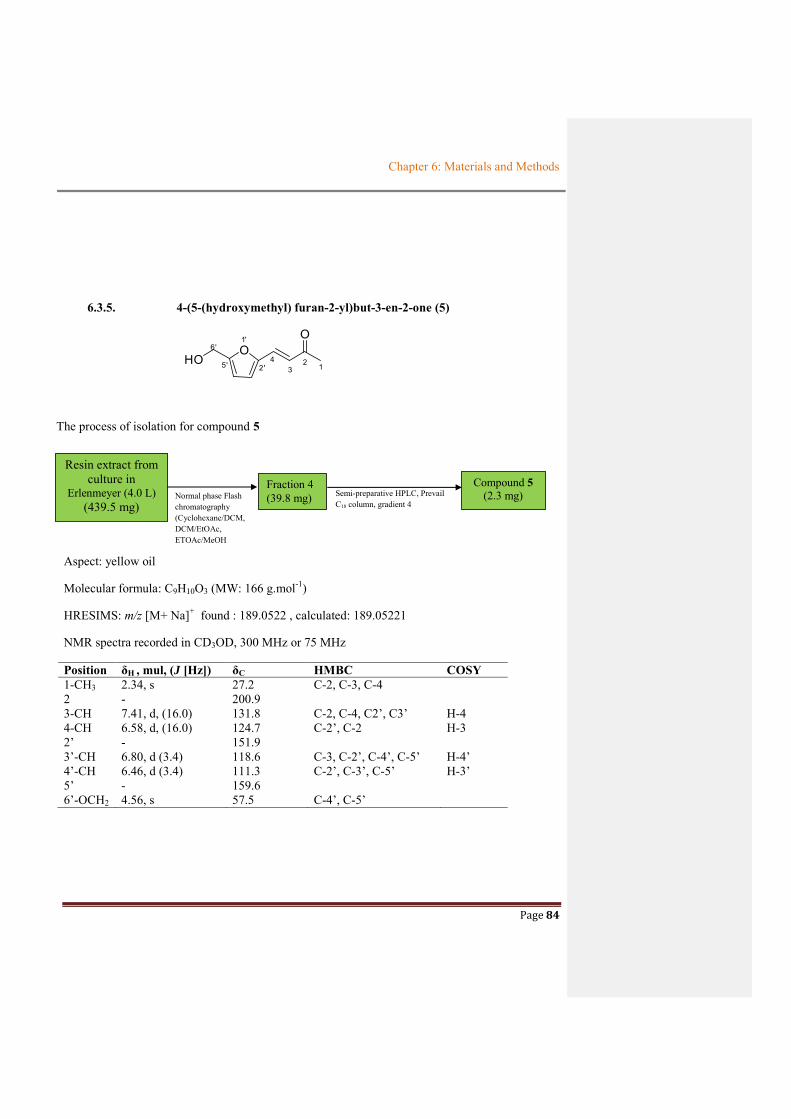
Chapter 6: Materials and Methods
Page 84
6.3.5. 4-(5-(hydroxymethyl) furan-2-yl)but-3-en-2-one (5)
OO
HO12
34
1'
2'5'
6'
The process of isolation for compound 5
Aspect: yellow oil
Molecular formula: C9H10O3 (MW: 166 g.mol-1)
HRESIMS: m/z [M+ Na]+ found : 189.0522 , calculated: 189.05221
NMR spectra recorded in CD3OD, 300 MHz or 75 MHz
Position δH , mul, (J [Hz]) δC HMBC COSY 1-CH3 2.34, s 27.2 C-2, C-3, C-4 2 - 200.9 3-CH 7.41, d, (16.0) 131.8 C-2, C-4, C2’, C3’ H-4 4-CH 6.58, d, (16.0) 124.7 C-2’, C-2 H-3 2’ - 151.9 3’-CH 6.80, d (3.4) 118.6 C-3, C-2’, C-4’, C-5’ H-4’ 4’-CH 6.46, d (3.4) 111.3 C-2’, C-3’, C-5’ H-3’ 5’ - 159.6 6’-OCH2 4.56, s 57.5 C-4’, C-5’
Resin extract from culture in
Erlenmeyer (4.0 L) (439.5 mg)
Fraction 4 (39.8 mg)
Compound 5 (2.3 mg)
Semi-preparative HPLC, Prevail C18 column, gradient 4
Normal phase Flash chromatography (Cyclohexane/DCM, DCM/EtOAc, ETOAc/MeOH

Chapter 6: Materials and Methods
Page 85
6.3.6. 4-methoxy-3-methylfuran-2(5H)-one (6)
O O
O
1
2
34
5
6
7
The process of isolation for compound 6
Aspect: colorless crystal
Molecular formula: C6H8O3 (MW: 128 g.mol-1)
HRESIMS: m/z [M+ Na]+ found : 151.0366 , calculated: 151.03656
NMR spectra recorded in CDCl3, 300 MHz or 75 MHz
Position δH , mul, (J [Hz]) δC HMBC 1 - - 2 - 170.2 3 - 128.7 4 - 129.4 5-CH2 4.14, q (7.0) 60.4 C-2 6-OCH3 3.51, s 50.9 7-CH3 2.07, s 21.1 C-2
Resin extract from culture in
Erlenmeyer (4.0 L) (439.5 mg)
Fraction 4 (39.8 mg)
Compound 6 (1.5 mg)
Subfraction 2 (18.6 mg) Semi-preparative
HPLC, Prevail C18 column, gradient 4
Preparative TLC (n-Hexane/CHCl3/ EtOAc/MeOH 2/2/2/0.5)
Rf = 0.57
Normal phase Flash chromatography (Cyclohexane/DCM, DCM/EtOAc, ETOAc/MeOH

Chapter 6: Materials and Methods
Page 86
6.3.7. 2-((3-hydroxy-2-methylpropanoyloxy)methyl)-2-(hydroxymethyl)butyl methacrylate (7)
O
O
OH
O
O
OH
12
3 4
56 7
8
9
10
11
12
13
14
The process of isolation for compound 7
Aspect: white, amorphous powder
Molecular formula: C14H24O6 (MW: 256 g.mol-1)
Rf = 0.13 (n-Hexane/Chloroform/Ethyl acetate/ Methanol (2/2/2/0.5), (v/v))
HRESIMS: m/z [M+ Na]+ (C10H18O4Na) found : 225.11 , calculated: 225.10973 (lost of a C4H6O2 unit due to hydrolysis)
NMR spectra recorded in CDCl3, 500 MHz or 125 MHz
Compound 7 (in CDCl3) Reference (Parrot et al., 2016) in
CDCl3 Position δH , mul, (J[Hz]) δC
HMBCb COSY δH , mul, (J[Hz]) δC 1 - 131.0 - - - 136.7 2 - 166.5 - - 167.3 3 4.28, s 64.7 C2,C4,C10 H-13 4.06,s 64.9 4 - 43.2 - 42.2 5 3.65 68.1 C3, C4, C13 H13 3.6-4.1 71.5 6 - 175.2 - 176.1 7 2.03 31.1 C6 2.69-2.73, m 39.9 8 3.73 60.5 C6 H-9 3.46-3.56 73.4 9 1.23, m 14.4 C8 H-8 1.12, d, (10) 13.6 10 3.68 66.2 C3, C4, C13 4.06, s 64.9 11 1.94 18.5 C1, C2, C12 H-12 1.90, br. s 18.4 12a 6.12, s 126.5 C1, C2, C11 H-11 6.04, br. s 125.6 12b 5.63, m 5.54, br. s 13 1.30, q, (7.6) 22.9 C3, C4, C14 H-14 1.48, q, (7.5) 23.6 14 0.88, t, (7.6) 7.6 C4, C13 H-13 0.87, t, (7.5) 7.6
Resin extract from culture in bioreactor
(4.5 L) (353.7 mg)
Fraction 14 (21.8 mg) Semi-preparative
HPLC, Prevail C18 column, gradient 1
Compound 7 (0.6 mg)
Fraction 5 (23.3 mg) Semi-preparative
HPLC, Prevail C18 column, gradient 2
Compound 7 (0.9 mg)
Resin extract from culture in
Erlenmeyer (4.0 L) (439.5 mg)
Normal phase Flash chromatography (Cyclohexane/DCM, DCM/EtOAc, ETOAc/MeOH
Reverse phase Flash chromatography (ACN/H2O)

Chapter 6: Materials and Methods
Page 87
6.3.8 Ethyl 1-ethyl-4-methoxy-2-(methoxymethyl)cyclopent-3-enecarboxylate (8)
OO
O
O
12
4 3
5
6
7
8
9 10
11
12
13
The process of isolation for compound 8
Aspect: colorless oil
Molecular formula: C13H22O4 (MW: 242 g.mol-1)
HRESIMS: m/z [M+ H]- found : 241.1448 , calculated: 241.14453
NMR spectra: recorded in CD3COCD3, 300 MHz or 75 MHz
Position δH , mul, (J[Hz]) δC HMBC COSY
1 - 78.3 - - 2-CH 1.96, s 19.9 C6 H-9 3-CH 6.17, s 107.3 C4 -
4 - 155.0 5-CH2 1.27, br 31.4 C11
6 - 163.9 7-OCH2 4.05, q (7.1) 59.7 C6, C8 H-8 8-CH3 1.19, t (7.1) 13.5 C6 H-7
9-OCH2 4.47, d (5.9) 56.4 C3, C4, C6 H-2 10-OCH3 3.30, s 48.9 11-CH2 1.27, br 22.4 C5, C9 H-12 12-CH3 0.87, t (6.7) 13.6 C5, C11 H-11
13-OCH3 3.31, s 48.9
Fraction 7 (13.7 mg)
Compound 8 (1.9 mg)
Resin extract from culture in
Erlenmeyer (4.0 L) (439.5 mg)
Preparative TLC (n-Hexane/CHCl3/EtOAc /MeOH 2/2/2/0.5)
Rf = 0.27
Normal phase Flash chromatography (Cyclohexane/DCM, DCM/EtOAc, ETOAc/MeOH

Chapter 6: Materials and Methods
Page 88
6.3.9. Hexyl 2-hydroxybenzoate (9)
O
OH
O
1
24
67
8
9
10
11
12
13
The process of isolation for compound 9
Aspect: white powder
Molecular formula: C13H18O3 (MW: 222 g.mol-1)
HRESIMS: m/z [M+ Na]+ found : 245.1152 , calculated: 245.11481
NMR spectra: recorded in CDCl3, 500 MHz or 125 MHz
Position δH , mul, (J [Hz]) δC HMBC COSY
1 - 115.3 - - 2 - 161.9 - - 3-CH 7.01, dd (8.4 and 0.9) 117.9 C-1, C-2, C-5 H-4 4-CH 7.48, m 135.9 C-2, C-6 H-3, H-5 5-CH 6.89, m 119.4 C-1 H-6, H-6 6-CH 7.87, dd (7.9 and 1.7) 130.2 C-2, C-4, C-7 H-5 7 - 170.5 8-CH2 4.37, t (6.7) 65.8 C-7, C-9, C-10 H-9 9-CH2 1.79, m 28.5 C-8, C-10 H-8, H-10 10-CH2 1.65, m 25.0 C-9, C-11/12 H-9, H-11/12 11, 12-CH2 1.30, m 23.0-33.8 C-10, C-13 H-10, H-13 13-CH3 0.91, m 14.4 C-11/12 H-11/12
Fraction 12 (8.9 mg) Semi-preparative
HPLC, Prevail C18 column, gradient 4
Compound 9 (0.9 mg)
Resin extract from culture in
Erlenmeyer (4.0 L) (439.5 mg)
Normal phase Flash chromatography (Cyclohexane/DCM, DCM/EtOAc, ETOAc/MeOH
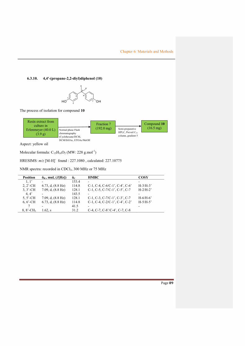
Chapter 6: Materials and Methods
Page 89
6.3.10. 4,4'-(propane-2,2-diyl)diphenol (10)
HO OH1
2
4
1'
4'
7
88'
The process of isolation for compound 10
Aspect: yellow oil
Molecular formula: C15H16O2 (MW: 228 g.mol-1)
HRESIMS: m/z [M-H]- found : 227.1080 , calculated: 227.10775
NMR spectra: recorded in CDCl3, 300 MHz or 75 MHz
Position δH , mul, (J[Hz]) δC HMBC COSY
1, 1’ - 153.4 - - 2, 2’-CH 6.73, d, (8.8 Hz) 114.8 C-1, C-4, C-6/C-1’, C-4’, C-6’ H-3/H-3’ 3, 3’-CH 7.09, d, (8.8 Hz) 128.1 C-1, C-5, C-7/C-1’, C-5’, C-7 H-2/H-2’
4, 4’ - 143.5 - - 5, 5’-CH 7.09, d, (8.8 Hz) 128.1 C-1, C-3, C-7/C-1’, C-3’, C-7 H-6/H-6’ 6, 6’-CH 6.73, d, (8.8 Hz) 114.8 C-1, C-4, C-2/C-1’, C-4’, C-2’ H-5/H-5’
7 - 41.5 - - 8, 8’-CH3 1.62, s 31.2 C-4, C-7, C-8’/C-4’, C-7, C-8
Fraction 7 (192.0 mg) Semi-preparative
HPLC, Prevail C18 column, gradient 5
Compound 10 (16.5 mg)
Resin extract from culture in
Erlenmeyer (40.0 L) (3.9 g)
Normal phase Flash chromatography (Cyclohexane/DCM, DCM/EtOAc, ETOAc/MeOH

Chapter 6: Materials and Methods
Page 90
6.3.11. 1H-indole-3-carbaldehyde (11)
1 2
34
5
6
77a
3a
1'
NH
OH
The process of isolation for compound 11
Aspect: yellow crystal
Molecular formula: C9H7ON (MW: 145 g.mol-1)
HRESIMS: m/z [M + Na]+ found : 168.0420 , calculated: 168.04198
NMR spectra: recorded in CD3OD, 300 MHz or 75 MHz
Position δH , mul, (J[Hz]) δC HMBC COSY
1-NH - - 2-CH 8.12, s 138.3 C-3, C-4, C-7a, C-8 -
3 - 118.6 - - 3a - 126.5 - -
4-CH 7.28, m 123.6 C-6 H-5 5-CH 8.17, m 121.0 C-4 H-4/H-6 6-CH 7.28, m 122.2 C-7 H-5, H-7 7-CH 7.50, m 111.7 C-4 H-6
7a - 137.8 - - 8-CHO 9.90, s 190.0 C-3, C-4 -
Fraction 8 (192.0 mg) Semi-preparative
HPLC, Prevail C18 column, gradient 5
Compound 11 (1.91 mg)
Crude extract from resin of culture in Erlenmeyer (40 L)
(3.9 g) Normal phase Flash chromatography (Cyclohexane/DCM, DCM/EtOAc, ETOAc/MeOH

Page 91
REFERENCES

References
Page 92
REFERENCES
Achan, J., Talisuna, A.O., Erhart, A., Yeka, A., Tibenderana, J.K., Baliraine, F.N., Rosenthal, P.J.,
D’Alessandro, U., 2011. Quinine, an old anti-malarial drug in a modern world: role in the
treatment of malaria. Malar J 10, 144. https://doi.org/10.1186/1475-2875-10-144
Aguilera, M., Quesada, M.T., del Águila, V.G., Morillo, J.A., Rivadeneyra, M.A., Ramos-
Cormenzana, A., Monteoliva-Sánchez, M., 2008. Characterisation of Paenibacillus jamilae
strains that produce exopolysaccharide during growth on and detoxification of olive mill
wastewaters. Bioresource Technology 99, 5640–5644.
https://doi.org/10.1016/j.biortech.2007.10.032
Ain, Q.-U.-, Khan, H., Mubarak, M.S., Pervaiz, A., 2016. Plant Alkaloids as Antiplatelet Agent:
Drugs of the Future in the Light of Recent Developments. Front Pharmacol 7.
https://doi.org/10.3389/fphar.2016.00292
Al-Sheraji, S.H., Ismail, A., Manap, M.Y., Mustafa, S., Yusof, R.M., Hassan, F.A., 2012.
Purification, characterization and antioxidant activity of polysaccharides extracted from the
fibrous pulp of Mangifera pajang fruits. LWT - Food Science and Technology 48, 291–296.
https://doi.org/10.1016/j.lwt.2012.04.002
Alvarez-Mico, X., Jensen, P.R., Fenical, W., Hughes, C.C., 2013. Chlorizidine, a Cytotoxic 5H-
Pyrrolo[2,1-a]isoindol-5-one-Containing Alkaloid from a Marine Streptomyces sp. Org.
Lett. 15, 988–991. https://doi.org/10.1021/ol303374e
Amann, R.I., Ludwig, W., Schleifer, K.H., 1995. Phylogenetic identification and in situ detection
of individual microbial cells without cultivation. Microbiol Rev 59, 143–169.
Armstrong, R., 2006. Seasonal growth of the crustose lichen Rhizocarpon geographicum (L.) DC.
in South Gwynedd, Wales [WWW Document]. URL
https://research.aston.ac.uk/portal/en/researchoutput/seasonal-growth-of-the-crustose-lichen-
rhizocarpon-geographicum-l-dc-in-south-gwynedd-wales%28e71ef783-51fa-4a86-bf43-
daca4c892686%29.html (accessed 12.23.15).
Armstrong, R., Bradwell, T., 2010. Growth of Crustose Lichens: A Review. Geografiska Annaler:
Series A, Physical Geography 92, 3–17. https://doi.org/10.1111/j.1468-0459.2010.00374.x

References
Page 93
Armstrong, R.A., 2005. Radial Growth of Rhizocarpon Section Rhizocarpon Lichen Thalli over
Six Years at Snoqualmie Pass in the Cascade Range, Washington State. Arctic, Antarctic,
and Alpine Research 37, 411–415.
Armstrong, R.A., 1983. Growth Curve of the Lichen Rhizocarpon Geographicum. New
Phytologist 94, 619–622. https://doi.org/10.1111/j.1469-8137.1983.tb04870.x
Armstrong, R.A., Smith, S.N., 2009. Carbohydrates in the hypothallus and areolae of the crustose
lichen Rhizocarpon geographicum (L.) DC. Symbiosis 49, 95–100.
https://doi.org/10.1007/s13199-009-0016-z
Armstrong, R.A., Smith, S.N., 1996. Experimental studies of hypothallus growth in the lichen
Rhizocarpon geographicum. New Phytologist 132, 123–126. https://doi.org/10.1111/j.1469-
8137.1996.tb04517.x
Aschenbrenner, I.A., Cardinale, M., Berg, G., Grube, M., 2014. Microbial cargo: do bacteria on
symbiotic propagules reinforce the microbiome of lichens? Environ Microbiol 16, 3743–
3752. https://doi.org/10.1111/1462-2920.12658
Asif, M., 2014. Chemical Characteristics, Synthetic Methods, and Biological Potential of
Quinazoline and Quinazolinone Derivatives [WWW Document]. International Journal of
Medicinal Chemistry. https://doi.org/10.1155/2014/395637
Asker, M.M.S., EL Sayed, O.H., Mahmoud, M.G., Ramadan, M.F., 2014. Chemical structure and
antioxidant activity of a new exopolysaccharide produced from Micrococcus luteus. Journal
of Genetic Engineering and Biotechnology 12, 121–126.
https://doi.org/10.1016/j.jgeb.2014.08.002
Asker, M.M.S., Shawky, B.T., 2010. Structural characterization and antioxidant activity of an
extracellular polysaccharide isolated from Brevibacterium otitidis BTS 44. Food Chemistry
123, 315–320. https://doi.org/10.1016/j.foodchem.2010.04.037
Azumi, M., Ogawa, K., Fujita, T., Takeshita, M., Yoshida, R., Furumai, T., Igarashi, Y., 2008.
Bacilosarcins A and B, novel bioactive isocoumarins with unusual heterocyclic cores from
the marine-derived bacterium Bacillus subtilis. Tetrahedron 64, 6420–6425.
https://doi.org/10.1016/j.tet.2008.04.076

References
Page 94
Balachandran, C., Sangeetha, B., Duraipandiyan, V., Raj, M.K., Ignacimuthu, S., Al-Dhabi, N.A.,
Balakrishna, K., Parthasarathy, K., Arulmozhi, N.M., Arasu, M.V., 2014. A flavonoid
isolated from Streptomyces sp. (ERINLG-4) induces apoptosis in human lung cancer A549
cells through p53 and cytochrome c release caspase dependant pathway. Chemico-
Biological Interactions 224, 24–35. https://doi.org/10.1016/j.cbi.2014.09.019
Barsby, T., Kelly, M.T., Gagné, S.M., Andersen, R.J., 2001. Bogorol A Produced in Culture by a
Marine Bacillus sp. Reveals a Novel Template for Cationic Peptide Antibiotics. Org. Lett. 3,
437–440. https://doi.org/10.1021/ol006942q
Bates, S.T., Cropsey, G.W.G., Caporaso, J.G., Knight, R., Fierer, N., 2011. Bacterial Communities
Associated with the Lichen Symbiosis. Appl Environ Microbiol 77, 1309–1314.
https://doi.org/10.1128/AEM.02257-10
Belghit, S., Driche, E.H., Bijani, C., Zitouni, A., Sabaou, N., Badji, B., Mathieu, F., 2016. Activity
of 2,4-Di-tert-butylphenol produced by a strain of Streptomyces mutabilis isolated from a
Saharan soil against Candida albicans and other pathogenic fungi. Journal de Mycologie
Médicale / Journal of Medical Mycology 26, 160–169.
https://doi.org/10.1016/j.mycmed.2016.03.001
Belnap, J., 2001. Factors Influencing Nitrogen Fixation and Nitrogen Release in Biological Soil
Crusts, in: Belnap, P.D.J., Lange, P.D.D. h c O.L. (Eds.), Biological Soil Crusts: Structure,
Function, and Management, Ecological Studies. Springer Berlin Heidelberg, pp. 241–261.
https://doi.org/10.1007/978-3-642-56475-8_19
Bench, G., Clark, B.M., Mangelson, N.F., St. Clair, L.L., Rees, L.B., Grant, P.G., Southon, J.R.,
2001. Accurate lifespan estimates cannot be obtained from14C profiles in the crustose lichen
Rhizocarpon geographicum (L.) DC. The Lichenologist 33, 539–542.
https://doi.org/10.1006/lich.2001.0353
Benedict, J.B., 2009. A Review of Lichenometric Dating and Its Applications to Archaeology.
American Antiquity 74, 143–172. https://doi.org/10.2307/25470542
Benedict, J.B., 1988. Techniques in Lichenometry: Identifying the Yellow Rhizocarpons. Arctic
and Alpine Research 20, 285–291. https://doi.org/10.2307/1551260

References
Page 95
Berrue, F., Ibrahim, A., Boland, P., Kerr, R.G., 2009. Newly isolated marine Bacillus pumilus
(SP21): A source of novel lipoamides and other antimicrobial agents. Pure and applied
chemistry 81, 1027–1031.
Bisel, P., Al-Momani, L., Müller, M., 2008. The tert-butyl group in chemistry and biology. Org.
Biomol. Chem. 6, 2655–2665. https://doi.org/10.1039/b800083b
Bjelland, T., Grube, M., Hoem, S., Jorgensen, S.L., Daae, F.L., Thorseth, I.H., Øvreås, L., 2011.
Microbial metacommunities in the lichen–rock habitat. Environmental Microbiology
Reports 3, 434–442. https://doi.org/10.1111/j.1758-2229.2010.00206.x
Björndal, H., Lindberg, B., Svensson, S., 1967. Mass spectrometry of partially methylated alditol
acetates. Carbohydrate Research 5, 433–440. https://doi.org/10.1016/S0008-6215(00)81115-
5
Boustie, J., Grube, M., 2005. Lichens—a promising source of bioactive secondary metabolites.
Plant Genetic Resources 3, 273–287. https://doi.org/10.1079/PGR200572
Boustie, J., Tomasi, S., Grube, M., 2010. Bioactive lichen metabolites: alpine habitats as an
untapped source. Phytochem Rev 10, 287–307. https://doi.org/10.1007/s11101-010-9201-1
Bradwell, T., 2010. Studies on the Growth of Rhizocarpon Geographicum in Nw Scotland, and
Some Implications for Lichenometry. Geografiska Annaler: Series A, Physical Geography
92, 41–52. https://doi.org/10.1111/j.1468-0459.2010.00376.x
Bradwell, T., Armstrong, R.A., 2007. Growth rates of Rhizocarpon geographicum lichens: a
review with new data from Iceland. J. Quaternary Sci. 22, 311–320.
https://doi.org/10.1002/jqs.1058
Braun, G.G., Schywalsky, M., Wölfel, L., Müller, H., Danner, U., Albert, R., 1993. [A comparison
of the two anticholinergic agents atropine and glycopyrrolate during antagonism of a muscle
relaxation with pyridostigmine]. Anaesthesiol Reanim 18, 120–122, 125–127.
Brelles-Mariño, G., Bedmar, E.J., 2001. Detection, purification and characterisation of quorum-
sensing signal molecules in plant-associated bacteria. Journal of Biotechnology, Co-
References
Page 96
operation between Europe and Developing Countries on Research about Biological Nitrogen
Fixation-Needa and Means 91, 197–209. https://doi.org/10.1016/S0168-1656(01)00330-3
Brodo, I.M., 1973. Chapter 12 - Substrate Ecology, in: HaLe, V.A.E. (Ed.), The Lichens.
Academic Press, pp. 401–441.
Büdel B. and Scheidegger S. 2008, Thallus morphology and anatomy (Chapter 4). p 40. Lichen
Biology, Edited by Nash S. Thomas, Cambridge university Press, 486.
Cai, X., Lei, Q.P., Lamb, D.H., Shannon, A., Jacoby, J., Kruk, J., Kensinger, R.D., Ryall, R.,
Zablackis, E., Cash, P., 2004. LC/MS Characterization of Meningococcal Depolymerized
Polysaccharide Group C Reducing Endgroup and Internal Repeating Unit. Anal. Chem. 76,
7387–7390. https://doi.org/10.1021/ac0491760
Cardinale, M., Castro, J.V. de, Müller, H., Berg, G., Grube, M., 2008. In situ analysis of the
bacterial community associated with the reindeer lichen Cladonia arbuscula reveals
predominance of Alphaproteobacteria. FEMS Microbiology Ecology 66, 63–71.
https://doi.org/10.1111/j.1574-6941.2008.00546.x
Cardinale, M., Grube, M., Berg, G., 2011. Frondihabitans cladoniiphilus sp. nov., an
actinobacterium of the family Microbacteriaceae isolated from lichen, and emended
description of the genus Frondihabitans. International Journal od Systematic and
Microbiology 61, 3033–3038. https://doi.org/10.1099/ijs.0.028324-0
Cardinale, M., Grube, M., Castro, J.V., Müller, H., Berg, G., 2012a. Bacterial taxa associated with
the lung lichen Lobaria pulmonaria are differentially shaped by geography and habitat.
FEMS Microbiol Lett 329, 111–115. https://doi.org/10.1111/j.1574-6968.2012.02508.x
Cardinale, M., Puglia, A.M., Grube, M., 2006. Molecular analysis of lichen-associated bacterial
communities. FEMS Microbiol Ecol 57, 484–495. https://doi.org/10.1111/j.1574-
6941.2006.00133.x
Cardinale Massimiliano, Steinová Jana, Rabensteiner Johannes, Berg Gabriele, Grube Martin,
2012b. Age, sun and substrate: triggers of bacterial communities in lichens. Environmental
Microbiology Reports 4, 23–28. https://doi.org/10.1111/j.1758-2229.2011.00272.x

References
Page 97
Cheenpracha, S., Vidor, N.B., Yoshida, W.Y., Davies, J., Chang, L.C., 2010. Coumabiocins A−F,
Aminocoumarins from an Organic Extract of Streptomyces sp. L-4-4. J. Nat. Prod. 73, 880–
884. https://doi.org/10.1021/np900843b
Chen, J.W., Koh, C.-L., Sam, C.-K., Yin, W.-F., Chan, K.-G., 2013. Short Chain N-acyl
Homoserine Lactone Production by Soil Isolate Burkholderia sp. Strain A9. Sensors 13,
13217–13227. https://doi.org/10.3390/s131013217
Chen, X.-H., Vater, J., Piel, J., Franke, P., Scholz, R., Schneider, K., Koumoutsi, A., Hitzeroth, G.,
Grammel, N., Strittmatter, A.W., Gottschalk, G., Süssmuth, R.D., Borriss, R., 2006.
Structural and Functional Characterization of Three Polyketide Synthase Gene Clusters in
Bacillus amyloliquefaciens FZB 42. J Bacteriol 188, 4024–4036.
https://doi.org/10.1128/JB.00052-06
Chen, Y.-T., Yuan, Q., Shan, L.-T., Lin, M.-A., Cheng, D.-Q., Li, C.-Y., 2013. Antitumor activity
of bacterial exopolysaccharides from the endophyte Bacillus amyloliquefaciens sp. isolated
from Ophiopogon japonicus. Oncol Lett 5, 1787–1792. https://doi.org/10.3892/ol.2013.1284
Cheng, R., Chen, J., Yu, X., Wang, Y., Wang, S., Zhang, J., 2013. Recombinant production and
characterization of full-length and truncated β-1,3-glucanase PglA from Paenibacillus sp.
S09. BMC Biotechnology 13, 105. https://doi.org/10.1186/1472-6750-13-105
Cho, J.Y., Williams, P.G., Kwon, H.C., Jensen, P.R., Fenical, W., 2007. Lucentamycins A−D,
Cytotoxic Peptides from the Marine-Derived Actinomycete Nocardiopsis lucentensis. J. Nat.
Prod. 70, 1321–1328. https://doi.org/10.1021/np070101b
Choi, S.J., Kim, J.K., Kim, H.K., Harris, K., Kim, C.-J., Park, G.G., Park, C.-S., Shin, D.-H.,
2013. 2,4-Di-tert-butylphenol from Sweet Potato Protects Against Oxidative Stress in PC12
Cells and in Mice. J Med Food 16, 977–983. https://doi.org/10.1089/jmf.2012.2739
Chuah, T.S., Norhafizah, M.Z., Ismail, B.S., 2015. Evaluation of the biochemical and
physiological activity of the natural compound, 2,4-ditert-butylphenol on weeds. Crop
Pasture Sci. 66, 214–223. https://doi.org/10.1071/CP13386.
Clayden R. S. 1998. Thallus inititaion and developments in lichen Rhizocarpon lecanorium. New phytol 139, 685 - 695

References
Page 98
Dang, Q.L., Son, S.W., Cheon, H.-M., Choi, G.J., Choi, Y.H., Jang, K.S., Lim, C.H., Kim, J.-C.,
2011. Pyochelin isolated from Burkholderia arboris KRICT1 carried by pine wood
nematodes exhibits phytotoxicity in pine callus. Nematology 13, 521–528.
https://doi.org/10.1163/138855410X528271
Davies, J., Wang, H., Taylor, T., Warabi, K., Huang, X.-H., Andersen, R.J., 2005. Uncialamycin,
A New Enediyne Antibiotic. Org. Lett. 7, 5233–5236. https://doi.org/10.1021/ol052081f
Debbab, A., Aly, A.H., Lin, W.H., Proksch, P., 2010. Bioactive Compounds from Marine Bacteria
and Fungi. Microb Biotechnol 3, 544–563. https://doi.org/10.1111/j.1751-
7915.2010.00179.x
Delmail, D., Grube, M., Parrot, D., Cook-Moreau, J., Boustie, J., Labrousse, P., Tomasi, S., 2013.
Halotolerance in Lichens: Symbiotic Coalition Against Salt Stress, in: Ecophysiology and
Responses of Plants under Salt Stress. Springer, New York, NY, pp. 115–148.
https://doi.org/10.1007/978-1-4614-4747-4_4
Dembitsky Valery M., 2005. Astonishing diversity of natural surfactants: 6. Biologically active
marine and terrestrial alkaloid glycosides. Lipids 40, 1081. https://doi.org/10.1007/s11745-
005-1473-2
Dembitsky, V.M., 2006. Natural neo acids and neo alkanes: their analogs and derivatives. Lipids
41, 309–340.
Dewick, P.M., 2009. Alkaloids, in: Medicinal Natural Products. John Wiley & Sons, Ltd, pp. 311–
420. https://doi.org/10.1002/9780470742761.ch6
Dharni, S., Sanchita, Maurya, A., Samad, A., Srivastava, S.K., Sharma, A., Patra, D.D., 2014.
Purification, Characterization, and in Vitro Activity of 2,4-Di-tert-butylphenol from
Pseudomonas monteilii PsF84: Conformational and Molecular Docking Studies. J. Agric.
Food Chem. 62, 6138–6146. https://doi.org/10.1021/jf5001138
Ding, R., Wu, X.-C., Qian, C.-D., Teng, Y., Li, O., Zhan, Z.-J., Zhao, Y.-H., 2011. Isolation and
identification of lipopeptide antibiotics from Paenibacillus elgii B69 with inhibitory activity
against methicillin-resistant Staphylococcus aureus. J Microbiol. 49, 942–949.
https://doi.org/10.1007/s12275-011-1153-7

References
Page 99
Elix J.A. and Stocker-Wörgötte, 2008, Biochemistry and secondary metabolites (Chapter 7),
Lichen Biology, Edited by Nash S. Thomas, Cambridge university Press. 486
El-Banna, Winkelmann, 1998. Pyrrolnitrin from Burkholderia cepacia: antibiotic activity against
fungi and novel activities against streptomycetes. Journal of Applied Microbiology 85, 69–
78. https://doi.org/10.1046/j.1365-2672.1998.00473.x
Elnahas, M.O., Amin, M.A., Hussein, M.M.D., Shanbhag, V.C., Ali, A.E., Wall, J.D., 2017.
Isolation, Characterization and Bioactivities of an Extracellular Polysaccharide Produced
from Streptomyces sp. MOE6. Molecules 22, 1396.
https://doi.org/10.3390/molecules22091396
Elshafie, H.S., Camele, I., Racioppi, R., Scrano, L., Iacobellis, N.S., Bufo, S.A., 2012. In Vitro
Antifungal Activity of Burkholderia gladioli pv. agaricicola against Some Phytopathogenic
Fungi. Int J Mol Sci 13, 16291–16302. https://doi.org/10.3390/ijms131216291
Esposito, A., Ciccazzo, S., Borruso, L., Zerbe, S., Daffonchio, D., Brusetti, L., 2013. A Three-
Scale Analysis of Bacterial Communities Involved in Rocks Colonization and Soil
Formation in High Mountain Environments. Curr Microbiol 67, 472–479.
https://doi.org/10.1007/s00284-013-0391-9
Farnaes, L., Coufal, N.G., Kauffman, C.A., Rheingold, A.L., DiPasquale, A.G., Jensen, P.R.,
Fenical, W., 2014. Napyradiomycin Derivatives, Produced by a Marine-Derived
Actinomycete, Illustrate Cytotoxicity by Induction of Apoptosis. J. Nat. Prod. 77, 15–21.
https://doi.org/10.1021/np400466j
Fodstad, O., Breistøl, K., Pettit, G.R., Shoemaker, R.H., Boyd, M.R., 1996. Comparative
antitumor activities of halichondrins and vinblastine against human tumor xenografts. J.
Exp. Ther. Oncol. 1, 119–125.
Fontecave, M., Ollagnier-de-Choudens, S., Mulliez, E., 2003. Biological Radical Sulfur Insertion
Reactions. Chem. Rev. 103, 2149–2166. https://doi.org/10.1021/cr020427j
Fontecave, M., Py, B., Ollagnier de Choudens, S., Barras, F., 2008. From Iron and Cysteine to
Iron-Sulfur Clusters: the Biogenesis Protein Machineries. EcoSal Plus 3.
https://doi.org/10.1128/ecosalplus.3.6.3.14

References
Page 100
Freitas, F., Alves, V.D., Reis, M.A.M., 2011. Advances in bacterial exopolysaccharides: from
production to biotechnological applications. Trends in Biotechnology 29, 388–398.
https://doi.org/10.1016/j.tibtech.2011.03.008
Fu, P., Johnson, M., Chen, H., Posner, B.A., MacMillan, J.B., 2014. Carpatamides A–C, Cytotoxic
Arylamine Derivatives from a Marine-Derived Streptomyces sp. J Nat Prod 77, 1245–1248.
https://doi.org/10.1021/np500207p
Fu, P., MacMillan, J.B., 2015. Thiasporines A–C, Thiazine and Thiazole Derivatives from a
Marine-Derived Actinomycetospora chlora. J. Nat. Prod. 78, 548–551.
https://doi.org/10.1021/np500929z
Fu, P., Yang, C., Wang, Y., Liu, P., Ma, Y., Xu, L., Su, M., Hong, K., Zhu, W., 2012a.
Streptocarbazoles A and B, Two Novel Indolocarbazoles from the Marine-Derived
Actinomycete Strain Streptomyces sp. FMA. Org. Lett. 14, 2422–2425.
https://doi.org/10.1021/ol3008638
Fu, P., Zhuang, Y., Wang, Y., Liu, P., Qi, X., Gu, K., Zhang, D., Zhu, W., 2012b. New
Indolocarbazoles from a Mutant Strain of the Marine-Derived Actinomycete Streptomyces
fradiae 007M135. Org. Lett. 14, 6194–6197. https://doi.org/10.1021/ol302940y
Gao, C.-H., Tian, X.-P., Qi, S.-H., Luo, X.-M., Wang, P., Zhang, S., 2010. Antibacterial and
antilarval compounds from marine gorgonian-associated bacterium Bacillus
amyloliquefaciens SCSIO 00856. The Journal of Antibiotics 63, 191–193.
https://doi.org/10.1038/ja.2010.7
Gerard, J., Haden, P., Kelly, M.T., Andersen, R.J., 1996. Loloatin B, A cyclic decapeptide
antibiotic produced in culture by a tropical marine bacterium. Tetrahedron Letters 37, 7201–
7204. https://doi.org/10.1016/0040-4039(96)01624-3
Goh, S.Y., Tan, W.-S., Khan, S.A., Chew, H.P., Kasim, N.H.A., Yin, W.-F., Chan, K.-G., 2014.
Unusual Multiple Production of N-Acylhomoserine Lactones a by Burkholderia sp. Strain
C10B Isolated from Dentine Caries. Sensors (Basel) 14, 8940–8949.
https://doi.org/10.3390/s140508940
References
Page 101
González, I., Ayuso-Sacido, A., Anderson, A., Genilloud, O., 2005. Actinomycetes isolated from
lichens: Evaluation of their diversity and detection of biosynthetic gene sequences. FEMS
Microbiology Ecology 54, 401–415. https://doi.org/10.1016/j.femsec.2005.05.004
Goodarzi, N., Varshochian, R., Kamalinia, G., Atyabi, F., Dinarvand, R., 2013. A review of
polysaccharide cytotoxic drug conjugates for cancer therapy. Carbohydrate Polymers 92,
1280–1293. https://doi.org/10.1016/j.carbpol.2012.10.036
Green, I.R., Tocoli, F.E., Lee, S.H., Nihei, K., Kubo, I., 2008. Design and evaluation of anacardic
acid derivatives as anticavity agents. European Journal of Medicinal Chemistry 43, 1315–
1320. https://doi.org/10.1016/j.ejmech.2007.08.012
Grube, M., Berg, G., 2009. Microbial consortia of bacteria and fungi with focus on the lichen
symbiosis. Fungal Biology Reviews 23, 72–85. https://doi.org/10.1016/j.fbr.2009.10.001
Grube, M., Berg, G., S. Andrésson, Ó., Vilhelmsson, O., Dyer, P.S., Miao, V.P.W., 2013. Lichen
Genomics, in: Francisrtin (Ed.), The Ecological Genomics of Fungi. John Wiley & Sons,
Inc, pp. 191–212.
Grube, M., Cardinale, M., Berg, G., 2012a. 17 Bacteria and the Lichen Symbiosis, in: Hock, B.
(Ed.), Fungal Associations, The Mycota. Springer Berlin Heidelberg, pp. 363–372.
https://doi.org/10.1007/978-3-642-30826-0_17
Grube, M., Cardinale, M., de Castro, J.V., Müller, H., Berg, G., 2009. Species-specific structural
and functional diversity of bacterial communities in lichen symbioses. ISME J 3, 1105–
1115. https://doi.org/10.1038/ismej.2009.63
Grube, M., Cernava, T., Soh, J., Fuchs, S., Aschenbrenner, I., Lassek, C., Wegner, U., Becher, D.,
Riedel, K., Sensen, C.W., Berg, G., 2015. Exploring functional contexts of symbiotic sustain
within lichen-associated bacteria by comparative omics. ISME J 9, 412–424.
https://doi.org/10.1038/ismej.2014.138
Grube, Martin, Köberl, M., Lackner, S., Berg, C., Berg, G., 2012b. Host–parasite interaction and
microbiome response: effects of fungal infections on the bacterial community of the Alpine
lichen Solorina crocea. FEMS Microbiology Ecology 82, 472–481.
https://doi.org/10.1111/j.1574-6941.2012.01425.x

References
Page 102
Gutierrez-Lugo, M.-T., Woldemichael, G.M., Singh, M.P., Suarez, P.A., Maiese, W.M.,
Montenegro, G., Timmermann, B.N., 2005. Isolation of three new naturally occurring
compounds from the culture of Micromonospora sp. P1068. Nat. Prod. Res. 19, 645–652.
https://doi.org/10.1080/14786410412331272040
Hawas, U.W., Shaaban, M., Shaaban, K.A., Speitling, M., Maier, A., Kelter, G., Fiebig, H.H.,
Meiners, M., Helmke, E., Laatsch, H., 2009. Mansouramycins A−D, Cytotoxic
Isoquinolinequinones from a Marine Streptomycete. J. Nat. Prod. 72, 2120–2124.
https://doi.org/10.1021/np900160g
Haworth, L.A., Calkin, P.E., Ellis, J.M., 1986. Direct Measurement of Lichen Growth in the
Central Brooks Range, Alaska, U.S.A., and Its Application to Lichenometric Dating. Arctic
and Alpine Research 18, 289–296. https://doi.org/10.2307/1550886
Hodkinson, B.P., Gottel, N.R., Schadt, C.W., Lutzoni, F., 2012. Photoautotrophic symbiont and
geography are major factors affecting highly structured and diverse bacterial communities in
the lichen microbiome. Environmental Microbiology 14, 147–161.
https://doi.org/10.1111/j.1462-2920.2011.02560.x
Hodkinson, B.P., Lutzoni, F., 2009. A microbiotic survey of lichen-associated bacteria reveals a
new lineage from the Rhizobiales. Symbiosis 49, 163–180. https://doi.org/10.1007/s13199-
009-0049-3
Honig, S., Murray, K.A., 1984. An appraisal of codeine as an analgesic: single-dose analysis. J
Clin Pharmacol 24, 96–102.
Hori, K., Kawabata, Y., Nakazawa, Y., Nishizawa, M., Toeda, K., 2014. A Novel β-1,4-
mannanase Isolated from Paenibacillus polymyxa KT551. Food Science and Technology
Research 20, 1261–1265. https://doi.org/10.3136/fstr.20.1261
Hu, P., Fang, L., Jones, C.M., Chess, E.K., 2011. Collective sampling of intact anionic
polysaccharide components and application in quantitative determination by LC–MS.
Carbohydrate Research 346, 2268–2273. https://doi.org/10.1016/j.carres.2011.07.017
Hu, Y., Wang, K., MacMillan, J.B., 2013. Hunanamycin A, an Antibiotic from a Marine-Derived
Bacillus hunanensis. Org. Lett. 15, 390–393. https://doi.org/10.1021/ol303376c
References
Page 103
Hung, C.-C., Santschi, P.H., Gillow, J.B., 2005. Isolation and characterization of extracellular
polysaccharides produced by Pseudomonas fluorescens Biovar II. Carbohydrate Polymers
61, 141–147. https://doi.org/10.1016/j.carbpol.2005.04.008
Hwang, J., Chilton, W.S., Benson, D.M., 2002. Pyrrolnitrin production by Burkholderia cepacia
and biocontrol of Rhizoctonia stem rot of poinsettia. Biological Control 25, 56–63.
https://doi.org/10.1016/S1049-9644(02)00044-0
Ieranò, T., Silipo, A., Sturiale, L., Garozzo, D., Bryant, C., Lanzetta, R., Parrilli, M., Aldridge, C.,
Gould, F.K., Corris, P.A., Khan, C.M.A., Soyza, A.D., Molinaro, A., 2009. First structural
characterization of Burkholderia vietnamiensis lipooligosaccharide from cystic fibrosis-
associated lung transplantation strains. Glycobiology 19, 1214–1223.
https://doi.org/10.1093/glycob/cwp112
Ihlen, P.G., 2004. Taxonomy of the non-yellow species of Rhizocarpon (Rhizocarpaceae,
lichenized Ascomycota) in the Nordic countries, with hyaline and muriform ascospores.
Mycol. Res. 108, 533–570.
Ihlen, P.G., Ekman, S., 2002. Outline of phylogeny and character evolution in Rhizocarpon
(Rhizocarpaceae, lichenized Ascomycota) based on nuclear ITS and mitochondrial SSU
ribosomal DNA sequences. Biological Journal of the Linnean Society 77, 535–546.
https://doi.org/10.1046/j.1095-8312.2002.00127.x
Innes, J.L., 1985. A standard Rhizocarpon nomenclature for lichenometry. Boreas 14, 83–85.
https://doi.org/10.1111/j.1502-3885.1985.tb00890.x
Jaivel, N., Uvarani, C., Rajesh, R., Velmurugan, D., Marimuthu, P., 2014. Natural Occurrence of
Organofluorine and Other Constituents from Streptomyces sp. TC1. J. Nat. Prod. 77, 2–8.
https://doi.org/10.1021/np400360h
Jannu, V.G., Sanjenbam, P., Kannabiran, K., 2015. Preclinical evaluation and molecular docking
of 2,5-di-tert-butyl-1,4-benzoquinone (DTBBQ) from Streptomyces sp. VITVSK1 as a
potent antibacterial agent. International Journal of Bioinformatics Research and Applications
11, 142. https://doi.org/10.1504/IJBRA.2015.068089

References
Page 104
Jaruchoktaweechai, C., Suwanborirux, K., Tanasupawatt, S., Kittakoop, P., Menasveta, P., 2000.
New Macrolactins from a Marine Bacillus sp. Sc026. J. Nat. Prod. 63, 984–986.
https://doi.org/10.1021/np990605c
Jeong, S.-Y., Ishida, K., Ito, Y., Okada, S., Murakami, M., 2003. Bacillamide, a novel algicide
from the marine bacterium, Bacillus sp. SY-1, against the harmful dinoflagellate,
Cochlodinium polykrikoides. Tetrahedron Letters 44, 8005–8007.
https://doi.org/10.1016/j.tetlet.2003.08.115
Jung, H.-K., Hong, J.-H., Park, S.-C., Park, B.-K., Nam, D.-H., Kim, S.-D., 2007. Production and
physicochemical characterization of β-glucan produced byPaenibacillus polymyxa JB115.
Biotechnol. Bioprocess Eng. 12, 713–719. https://doi.org/10.1007/BF02931090
Kac̆uráková, M., Capek, P., Sasinková, V., Wellner, N., Ebringerová, A., 2000. FT-IR study of
plant cell wall model compounds: pectic polysaccharides and hemicelluloses. Carbohydrate
Polymers 43, 195–203. https://doi.org/10.1016/S0144-8617(00)00151-X
Kadirvelraj, R., Gonzalez-Outeiriño, J., Foley, B.L., Beckham, M.L., Jennings, H.J., Foote, S.,
Ford, M.G., Woods, R.J., 2006. Understanding the bacterial polysaccharide antigenicity of
Streptococcus agalactiae versus Streptococcus pneumoniae. Proc Natl Acad Sci U S A 103,
8149–8154. https://doi.org/10.1073/pnas.0602815103
Kai, H., Yamashita, M., Nakamura, I., Yoshikawa, K., Nitta, K., Watanabe, M., Inamura, N.,
Fujie, A., 2013a. Synergistic antifungal activity of KB425796-C in combination with
micafungin against Aspergillus fumigatus and its efficacy in murine infection models. J
Antibiot 66, 479–484. https://doi.org/10.1038/ja.2013.57
Kai, H., Yamashita, M., Takase, S., Hashimoto, M., Muramatsu, H., Nakamura, I., Yoshikawa, K.,
Kanasaki, R., Ezaki, M., Nitta, K., Watanabe, M., Inamura, N., Fujie, A., 2013b.
Identification of ten KB425796-A congeners from Paenibacillus sp. 530603 using an
antifungal assay against Aspergillus fumigatus in combination with micafungin. J Antibiot
66, 473–478. https://doi.org/10.1038/ja.2013.64
Karapetyan, G., Kaczynski, Z., Iacobellis, N.S., Evidente, A., Holst, O., 2006. The structure of the
O-specific polysaccharide of the lipopolysaccharide from Burkholderia gladioli pv.

References
Page 105
agaricicola. Carbohydrate Research 341, 930–934.
https://doi.org/10.1016/j.carres.2006.02.010
Kawahara, K., Seydel, U., Matsuura, M., Danbara, H., Rietschel, E.T., Za¨hringer, U., 1991.
Chemical structure of glycosphingolipids isolated fromSphingomonas paucimobilis. FEBS
Letters 292, 107–110. https://doi.org/10.1016/0014-5793(91)80845-T
Keum, Y.S., Lee, Y.J., Lee, Y.H., Kim, J.H., 2009. Effects of nutrients on quorum signals and
secondary metabolite productions of Burkholderia sp. O33. J. Microbiol. Biotechnol. 19,
1142–1149.
Kim, H.-R., Park, S.-Y., Kim, S.-B., Jeong, H., Choi, S.-K., Park, S.-H., 2014. Inactivation of the
phosphoglucomutase gene pgm in Paenibacillus polymyxa leads to overproduction of
fusaricidin. J Ind Microbiol Biotechnol 41, 1405–1414. https://doi.org/10.1007/s10295-014-
1470-z
Kim, J., Park, E.J., 2002. Cytotoxic anticancer candidates from natural resources. Curr Med Chem
Anticancer Agents 2, 485–537.
Kim, M.-K., Park, H., Oh, T.-J., 2014. Antibacterial and antioxidant capacity of polar
microorganisms isolated from Arctic lichen Ochrolechia sp. Pol. J. Microbiol. 63, 317–322.
Kim, M.K., Park, H., Oh, T.J., 2012. Antibacterial Properties Associated with Microorganisms
Isolated from Arctic Lichens. Korean Journal of Microbiology and Biotechnology.
Kim, S.H., Shin, Y.K., Sohn, Y.C., Kwon, H.C., 2012. Two New Cholic Acid Derivatives from
the Marine Ascidian-Associated Bacterium Hasllibacter halocynthiae. Molecules 17, 12357–
12364. https://doi.org/10.3390/molecules171012357
Klausmeyer, P., Shipley, S., Zuck, K.M., McCloud, T.G., 2011. Histone Deacetylase Inhibitors
from Burkholderia Thailandensis. J Nat Prod 74, 2039–2044.
https://doi.org/10.1021/np200532d
Kmunícek, J., Hynková, K., Jedlicka, T., Nagata, Y., Negri, A., Gago, F., Wade, R.C.,
Damborský, J., 2005. Quantitative Analysis of Substrate Specificity of Haloalkane

References
Page 106
Dehalogenase LinB from Sphingomonas paucimobilis UT26†. Biochemistry 44, 3390–3401.
https://doi.org/10.1021/bi047912o
Knolhoff, A.M., Zheng, J., McFarland, M.A., Luo, Y., Callahan, J.H., Brown, E.W., Croley, T.R.,
2015. Identification and Structural Characterization of Naturally-Occurring Broad-Spectrum
Cyclic Antibiotics Isolated from Paenibacillus. J. Am. Soc. Mass Spectrom. 26, 1768–1779.
https://doi.org/10.1007/s13361-015-1190-2
Kodali, V.P., Perali, R.S., Sen, R., 2011. Purification and Partial Elucidation of the Structure of an
Antioxidant Carbohydrate Biopolymer from the Probiotic Bacterium Bacillus coagulans
RK-02. J. Nat. Prod. 74, 1692–1697. https://doi.org/10.1021/np1008448.
Koehn, F.E., Carter, G.T., 2005. The evolving role of natural products in drug discovery. Nature
Reviews Drug Discovery 4, 206–220. https://doi.org/10.1038/nrd1657
Kokoulin, M.S., Kuzmich, A.S., Kalinovsky, A.I., Tomshich, S.V., Romanenko, L.A., Mikhailov,
V.V., Komandrova, N.A., 2016. Structure and anticancer activity of sulfated O-
polysaccharide from marine bacterium Cobetia litoralis KMM 3880T. Carbohydrate
Polymers 154, 55–61. https://doi.org/10.1016/j.carbpol.2016.08.036
Kontnik, R., Bosak, T., Butcher, R.A., Brocks, J.J., Losick, R., Clardy, J., Pearson, A., 2008.
Sporulenes, Heptaprenyl Metabolites from Bacillus subtilis Spores. Org Lett 10, 3551–3554.
https://doi.org/10.1021/ol801314k
Kropec, A., Maira-Litran, T., Jefferson, K.K., Grout, M., Cramton, S.E., Götz, F., Goldmann,
D.A., Pier, G.B., 2005. Poly-N-Acetylglucosamine Production in Staphylococcus aureus Is
Essential for Virulence in Murine Models of Systemic Infection. Infect Immun 73, 6868–
6876. https://doi.org/10.1128/IAI.73.10.6868-6876.2005
Kubota, M., Takimoto, H., Kaneko, M., Inoue, J., Kumazawa, Y., 2009. Potentiation of murine
innate immunity by α-galacturonosyl-type glycosphingolipids isolated from Sphingomonas
yanoikuyae and S. terrae. Immunopharmacology and Immunotoxicology 31, 363–369.
https://doi.org/10.1080/08923970802438409
Kumar, S. n., Siji, J. v., Rajasekharan, K. n., Nambisan, B., Mohandas, C., 2012. Bioactive
stilbenes from a Bacillus sp. N strain associated with a novel rhabditid entomopathogenic
References
Page 107
nematode. Letters in Applied Microbiology 54, 410–417. https://doi.org/10.1111/j.1472-
765X.2012.03223.x
Kumar, S.N., Mohandas, C., 2014. Antimycobacterial activity of cyclic dipeptides isolated from
Bacillus sp. N strain associated with entomopathogenic nematode. Pharmaceutical Biology
52, 91–96. https://doi.org/10.3109/13880209.2013.815635
Kumar, S.N., Nambisan, B., Kumar, B.S.D., Vasudevan, N.G., Mohandas, C., Cheriyan, V.T.,
Anto, R.J., 2013. Antioxidant and anticancer activity of 3,5-dihydroxy-4-isopropylstilbene
produced by Bacillus sp. N strain isolated from entomopathogenic nematode. Arch. Pharm.
Res. 1–11. https://doi.org/10.1007/s12272-013-0207-2
Lammers, A., Wang, R., Cetnar, J., Prasad, V., 2017. Time from US Food and Drug
Administration approval to publication of data for cancer drugs: a comparison of first and
subsequent approvals. Blood Cancer Journal 7, 637. https://doi.org/10.1038/s41408-017-
0008-9
Lee, J.S., Shin, Y.K., Yoon, J.H., Takeuchi, M., Pyun, Y.R., Park, Y.H., 2001. Sphingomonas
aquatilis sp. nov., Sphingomonas koreensis sp. nov., and Sphingomonas taejonensis sp. nov.,
yellow-pigmented bacteria isolated from natural mineral water. International Journal of
Systematic and Evolutionary Microbiology 51, 1491–1498.
https://doi.org/10.1099/00207713-51-4-1491
Lee, S.H., Cho, Y.E., Park, S.-H., Balaraju, K., Park, J.W., Lee, S.W., Park, K., 2012. An
antibiotic fusaricidin: a cyclic depsipeptide from Paenibacillus polymyxa E681 induces
systemic resistance against Phytophthora blight of red-pepper. Phytoparasitica 41, 49–58.
https://doi.org/10.1007/s12600-012-0263-z
Lee, S.-M., Do, H.J., Shin, M.-J., Seong, S.-I., Hwang, K.Y., Lee, J.Y., Kwon, O., Jin, T., Chung,
J.H., 2013. 1-Deoxynojirimycin isolated from a Bacillus subtilis stimulates adiponectin and
GLUT4 expressions in 3T3-L1 adipocytes. J. Microbiol. Biotechnol. 23, 637–643.
Li, O., Lu, C., Liu, A., Zhu, L., Wang, P.-M., Qian, C.-D., Jiang, X.-H., Wu, X.-C., 2013.
Optimization and characterization of polysaccharide-based bioflocculant produced by

References
Page 108
Paenibacillus elgii B69 and its application in wastewater treatment. Bioresource Technology
134, 87–93. https://doi.org/10.1016/j.biortech.2013.02.013
Liang, T.-W., Wang, S.-L., 2015. Recent Advances in Exopolysaccharides from Paenibacillus
spp.: Production, Isolation, Structure, and Bioactivities. Mar Drugs 13, 1847–1863.
https://doi.org/10.3390/md13041847
Liba, C. M., Ferrara, F. I. S., Manfio, G. P., Fantinatti-Garboggini, F., Albuquerque, R. C., Pavan,
C., Ramos, P. L., Moreira-Filho, C. A., Barbosa, H. R., 2006. Nitrogen-fixing chemo-
organotrophic bacteria isolated from cyanobacteria-deprived lichens and their ability to
solubilize phosphate and to release amino acids and phytohormones. Journal of Applied
Microbiology 101, 1076–1086. https://doi.org/10.1111/j.1365-2672.2006.03010.x
Lim, Y.W., Baik, K.S., Han, S.K., Kim, S.B., Bae, K.S., 2003. Burkholderia sordidicola sp. nov.,
isolated from the white-rot fungus Phanerochaete sordida. International Journal of
Systematic and Evolutionary Microbiology 53, 1631–1636.
https://doi.org/10.1099/ijs.0.02456-0
Liu, C., Liu, Q., Sun, J., Jiang, B., Yan, J., 2014. Extraction of water-soluble polysaccharide and
the antioxidant activity from Semen cassiae. Journal of Food and Drug Analysis 22, 492–
499. https://doi.org/10.1016/j.jfda.2014.01.027
Liu, D., Liao, N., Ye, X., Hu, Y., Wu, D., Guo, X., Zhong, J., Wu, J., Chen, S., 2013a. Isolation
and Structural Characterization of a Novel Antioxidant Mannoglucan from a Marine Bubble
Snail, Bullacta exarata (Philippi). Mar Drugs 11, 4464–4477.
https://doi.org/10.3390/md11114464
Liu, D., Liao, N., Ye, X., Hu, Y., Wu, D., Guo, X., Zhong, J., Wu, J., Chen, S., 2013b. Isolation
and Structural Characterization of a Novel Antioxidant Mannoglucan from a Marine Bubble
Snail, Bullacta exarata (Philippi). Marine Drugs 11, 4464–4477.
https://doi.org/10.3390/md11114464
Liu, R.-F., Zhang, D.-J., Li, Y.-G., Tao, L.-M., Tian, L., 2010. A New Antifungal Cyclic
Lipopeptide from Bacillus marinus B-9987. HCA 93, 2419–2425.
https://doi.org/10.1002/hlca.201000094

References
Page 109
Liu, W., Wang, H., Pang, X., Yao, W., Gao, X., 2010. Characterization and antioxidant activity of
two low-molecular-weight polysaccharides purified from the fruiting bodies of Ganoderma
lucidum. International Journal of Biological Macromolecules 46, 451–457.
https://doi.org/10.1016/j.ijbiomac.2010.02.006
Liu, C., Jiang, Y., Lei, H., Chen, X., Ma, Q., Han, L., Huang, X., 2017. Four New Nanaomycins
Produced by Streptomyces hebeiensis Derived from Lichen. Chem. Biodivers. 14.
https://doi.org/10.1002/cbdv.201700057
Lopanik, N., Gustafson, K.R., Lindquist, N., 2004. Structure of Bryostatin 20: A Symbiont-
Produced Chemical Defense for Larvae of the Host Bryozoan, Bugula neritina. J. Nat. Prod.
67, 1412–1414. https://doi.org/10.1021/np040007k
Lorenzo, F.D., Sturiale, L., Palmigiano, A., Fazio, L.L.-, Paciello, I., Coutinho, C.P., Sá-Correia,
I., Bernardini, M., Lanzetta, R., Garozzo, D., Silipo, A., Molinaro, A., 2013. Chemistry and
Biology of the Potent Endotoxin from a Burkholderia dolosa Clinical Isolate from a Cystic
Fibrosis Patient. ChemBioChem 14, 1105–1115. https://doi.org/10.1002/cbic.201300062
Lucas, X., Senger, C., Erxleben, A., Grüning, B.A., Döring, K., Mosch, J., Flemming, S., Günther,
S., 2013. StreptomeDB: a resource for natural compounds isolated from Streptomyces
species. Nucl. Acids Res. 41, D1130–D1136. https://doi.org/10.1093/nar/gks1253
Luesch, H., Yoshida, W.Y., Moore, R.E., Paul, V.J., Corbett, T.H., 2001. Total Structure
Determination of Apratoxin A, a Potent Novel Cytotoxin from the Marine Cyanobacterium
Lyngbya majuscula. J. Am. Chem. Soc. 123, 5418–5423. https://doi.org/10.1021/ja010453j
Ma, Z., Hu, J., Wang, X., Wang, S., 2014. NMR spectroscopic and MS/MS spectrometric
characterization of a new lipopeptide antibiotic bacillopeptin B1 produced by a marine
sediment-derived Bacillus amyloliquefaciens SH-B74. J Antibiot 67, 175–178.
https://doi.org/10.1038/ja.2013.89
Mageshwaran, V., Walia, S., Annapurna, K., 2011. Isolation and partial characterization of
antibacterial lipopeptide produced by Paenibacillus polymyxa HKA-15 against
phytopathogen Xanthomonas campestris pv. phaseoli M-5. World J Microbiol Biotechnol
28, 909–917. https://doi.org/10.1007/s11274-011-0888-y
References
Page 110
Maktouf, S., Moulis, C., Miled, N., Ellouz Chaabouni, S., Remaud-Simeon, M., 2015. A highly
thermostable lichenase from Bacillus sp. UEB-S: Biochemical and molecular
characterization. Journal of Molecular Catalysis B: Enzymatic 115, 8–12.
https://doi.org/10.1016/j.molcatb.2015.01.016
Maloney, K.N., MacMillan, J.B., Kauffman, C.A., Jensen, P.R., DiPasquale, A.G., Rheingold,
A.L., Fenical, W., 2009. Lodopyridone, a Structurally Unprecedented Alkaloid from a
Marine Actinomycete. Org. Lett. 11, 5422–5424. https://doi.org/10.1021/ol901997k
Manna, D.K., Maity, P., Nandi, A.K., Pattanayak, M., Panda, B.C., Mandal, A.K., Tripathy, S.,
Acharya, K., Sahoo, A.K., Gupta, N., Roy, S., Islam, S.S., 2017. Structural elucidation and
immunostimulating property of a novel polysaccharide extracted from an edible mushroom
Lentinus fusipes. Carbohydrate Polymers 157, 1657–1665.
https://doi.org/10.1016/j.carbpol.2016.11.048
Matsuda, M., Yamori, T., Naitoh, M., Okutani, K., 2003. Structural Revision of Sulfated
Polysaccharide B-1 Isolated from a Marine Pseudomonas Species and Its Cytotoxic Activity
Against Human Cancer Cell Lines. Mar. Biotechnol. 5, 13–19.
https://doi.org/10.1007/s10126-002-0046-5
Matthew, S., Salvador, L.A., Schupp, P.J., Paul, V.J., Luesch, H., 2010. Cytotoxic Halogenated
Macrolides and Modified Peptides from the Apratoxin-Producing Marine Cyanobacterium
Lyngbya bouillonii from Guam. J. Nat. Prod. 73, 1544–1552.
https://doi.org/10.1021/np1004032
McCarthy, D.P., Henry, N., 2012. Measurement of growth in the lichen Rhizocarpon
geographicum using a new photographic technique. The Lichenologist 44, 679–693.
https://doi.org/10.1017/S0024282912000175
McCarthy, P., Elix, J., 2014. The lichen genus Rhizocarpon in mainland Australia. Telopea 16,
195–211. https://doi.org/10.7751/telopea20148124
McKay, M.J., Carroll, A.R., Quinn, R.J., Hooper, J.N.A., 2002. 1,2-Bis(1H-indol-3-yl)ethane-1,2-
dione, an Indole Alkaloid from the Marine Sponge Smenospongia sp. J. Nat. Prod. 65, 595–
597. https://doi.org/10.1021/np010347v
References
Page 111
Meena, S., Gothwal, R.K., Saxena, J., Mohan, M.K., Ghosh, P., 2013. Chitinase production by a
newly isolated thermotolerant Paenibacillus sp. BISR-047. Ann Microbiol 64, 787–797.
https://doi.org/10.1007/s13213-013-0715-9
Mesías, M., Morales, F.J., 2017. Effect of Different Flours on the Formation of
Hydroxymethylfurfural, Furfural, and Dicarbonyl Compounds in Heated Glucose/Flour
Systems. Foods 6. https://doi.org/10.3390/foods6020014
Millot, M., Tomasi, S., Studzinska, E., Rouaud, I., Boustie, J., 2009. Cytotoxic Constituents of the
Lichen Diploicia canescens. J. Nat. Prod. 72, 2177–2180. https://doi.org/10.1021/np9003728
Mioso, R., Marante, F.J.T., Bezerra, R. de S., Borges, F.V.P., Santos, B.V. de O., Laguna, I.H.B.
de, 2017. Cytotoxic Compounds Derived from Marine Sponges. A Review (2010–2012).
Molecules 22, 208. https://doi.org/10.3390/molecules22020208
Mishra, S., Reddy, D.S.K., Jamwal, V.S., Bansal, D.D., Patel, D.D., Malhotra, P., Gupta, A.K.,
Singh, P.K., Jawed, S., Kumar, R., 2013. Semiquinone derivative isolated from Bacillus sp.
INM-1 protects cellular antioxidant enzymes from γ-radiation-induced renal toxicity. Mol
Cell Biochem 379, 19–27. https://doi.org/10.1007/s11010-013-1622-x
Mojid Mondol, M.A., Kim, J.H., Lee, H.-S., Lee, Y.-J., Shin, H.J., 2011. Macrolactin W, a new
antibacterial macrolide from a marine Bacillus sp. Bioorganic & Medicinal Chemistry
Letters 21, 3832–3835. https://doi.org/10.1016/j.bmcl.2010.12.050
Mokaddem, H., Sadaoui, Z., Boukhelata, N., Azouaou, N., Kaci, Y., 2009. Removal of Cadmium
from aqueous solution by polysaccharide produced from Paenibacillus polymyxa. Journal of
Hazardous Materials 172, 1150–1155. https://doi.org/10.1016/j.jhazmat.2009.07.116
Mondol, M.A.M., Tareq, F.S., Kim, J.H., Lee, M. ah, Lee, H.-S., Lee, Y.-J., Lee, J.S., Shin, H.J.,
2011. Cyclic Ether-Containing Macrolactins, Antimicrobial 24-Membered Isomeric
Macrolactones from a Marine Bacillus sp. J. Nat. Prod. 74, 2582–2587.
https://doi.org/10.1021/np200487k
Moon, K., Chung, B., Shin, Y., Rheingold, A.L., Moore, C.E., Park, S.J., Park, S., Lee, S.K., Oh,
K.-B., Shin, J., Oh, D.-C., 2015. Pentacyclic Antibiotics from a Tidal Mud Flat-Derived
Actinomycete. J. Nat. Prod. 78, 524–529. https://doi.org/10.1021/np500736b

References
Page 112
Motohashi, K., Takagi, M., Yamamura, H., Hayakawa, M., Shin-ya, K., 2010. A new angucycline
and a new butenolide isolated from lichen-derived Streptomyces spp. J Antibiot 63, 545–
548. https://doi.org/10.1038/ja.2010.94
Muggia, L., Klug, B., Berg, G., Grube, M., 2013. Localization of bacteria in lichens from Alpine
soil crusts by fluorescence in situ hybridization. Applied Soil Ecology 68, 20–25.
https://doi.org/10.1016/j.apsoil.2013.03.008
Nastro, R.A., Arguelles-Arias, A., Ongena, M., Costanzo, A.D., Trifuoggi, M., Guida, M.,
Fickers, P., 2013. Antimicrobial Activity of Bacillus amyloliquefaciens ANT1 Toward
Pathogenic Bacteria and Mold: Effects on Biofilm Formation. Probiotics & Antimicro. Prot.
5, 252–258. https://doi.org/10.1007/s12602-013-9143-1
Newman, D.J., Cragg, G.M., 2016. Natural Products as Sources of New Drugs from 1981 to 2014.
J. Nat. Prod. 79, 629–661. https://doi.org/10.1021/acs.jnatprod.5b01055
Noël, A., Ferron, S., Rouaud, I., Gouault, N., Hurvois, J.-P., Tomasi, S., 2017. Isolation and
Structure Identification of Novel Brominated Diketopiperazines from Nocardia ignorata—A
Lichen-Associated Actinobacterium. Molecules 22, 371.
https://doi.org/10.3390/molecules22030371
Nwodo, U.U., Green, E., Okoh, A.I., 2012. Bacterial Exopolysaccharides: Functionality and
Prospects. Int J Mol Sci 13, 14002–14015. https://doi.org/10.3390/ijms131114002
Oguma, T., Kitao, S., Kobayashi, M., 2014. Purification and Characterization of
Cycloisomaltooligosaccharide Glucanotransferase and Cloning of cit from Bacillus
circulans U-155. Journal of Applied Glycoscience advpub.
https://doi.org/10.5458/jag.jag.JAG-2013_017
O’Neal, M.A., Schoenenberger, K.R., 2003. A Rhizocarpon geographicum growth curve for the
Cascade Range of Washington and northern Oregon, usa. Quaternary Research 60, 233–241.
https://doi.org/10.1016/S0033-5894(03)00104-2
Osborn, H.M.I., Lochey, F., Mosley, L., Read, D., 1999. Analysis of polysaccharides and
monosaccharides in the root mucilage of maize (Zea mays L.) by gas chromatography.

References
Page 113
Journal of Chromatography A 831, 267–276. https://doi.org/10.1016/S0021-9673(98)00935-
2
Pan, E., Jamison, M., Yousufuddin, M., MacMillan, J.B., 2012. Ammosamide D, an Oxidatively
Ring Opened Ammosamide Analog from a Marine-Derived Streptomyces variabilis. Org.
Lett. 14, 2390–2393. https://doi.org/10.1021/ol300806e
Pan, W., Perrotta, J.A., Stipanovic, A.J., Nomura, C.T., Nakas, J.P., 2011. Production of
polyhydroxyalkanoates by Burkholderia cepacia ATCC 17759 using a detoxified sugar
maple hemicellulosic hydrolysate. J Ind Microbiol Biotechnol 39, 459–469.
https://doi.org/10.1007/s10295-011-1040-6
Park, H.B., Kim, Y.-J., Lee, J.K., Lee, K.R., Kwon, H.C., 2012. Spirobacillenes A and B, Unusual
Spiro-cyclopentenones from Lysinibacillus fusiformis KMC003. Org. Lett. 14, 5002–5005.
https://doi.org/10.1021/ol302115z.
Park, C.H., Kim, K.M., Elvebakk, A., Kim, O.-S., Jeong, G., Hong, S.G., 2015. Algal and Fungal
Diversity in Antarctic Lichens. Journal of Eukaryotic Microbiology 62, 196–205.
https://doi.org/10.1111/jeu.12159
Parrot, D., Legrave, N., Delmail, D., Grube, M., Suzuki, M.T., Tomasi, S., 2016a. Review –
Lichen-associated bacteria as a hot spot of chemodiversity: focus on uncialamycin, a
promising compound for future medicinal applications. Planta Medica 82, 1143–1152.
https://doi.org/10.1055/s-0042-105571
Parrot, D., Legrave, N., Intertaglia, L., Rouaud, I., Legembre, P., Grube, M., Suzuki, M.T.,
Tomasi, S., 2016b. Cyaneodimycin, a Bioactive Compound Isolated from the Culture of
Streptomyces cyaneofuscatus Associated with Lichina confinis. Eur. J. Org. Chem. 2016,
3977–3982. https://doi.org/10.1002/ejoc.201600252
Parrot, D., Peresse, T., Hitti, E., Carrie, D., Grube, M., Tomasi, S., 2015. Qualitative and Spatial
Metabolite Profiling of Lichens by a LC–MS Approach Combined With Optimised
Extraction. Phytochemical Analysis 26, 23–33. https://doi.org/10.1002/pca.2532
Paul, T., Das, A., Mandal, A., Halder, S.K., Jana, A., Maity, C., DasMohapatra, P.K., Pati, B.R.,
Mondal, K.C., 2014. An efficient cloth cleaning properties of a crude keratinase combined

References
Page 114
with detergent: towards industrial viewpoint. Journal of Cleaner Production 66, 672–684.
https://doi.org/10.1016/j.jclepro.2013.10.054
Pereira, A.R., Cao, Z., Engene, N., Soria-Mercado, I.E., Murray, T.F., Gerwick, W.H., 2010.
Palmyrolide A, an Unusually Stabilized Neuroactive Macrolide from Palmyra Atoll
Cyanobacteria. Org. Lett. 12, 4490–4493. https://doi.org/10.1021/ol101752n
Petersen, B.O., Hindsgaul, O., Paulsen, B.S., Redondo, A.R., Skovsted, I.C., 2014. Structural
elucidation of the capsular polysaccharide from Streptococcus pneumoniae serotype 47A by
NMR spectroscopy. Carbohydrate Research 386, 62–67.
https://doi.org/10.1016/j.carres.2013.11.013
Prabha, M.S., Divakar, K., Priya, J.D.A., Selvam, G.P., Balasubramanian, N., Gautam, P., 2014.
Statistical analysis of production of protease and esterase by a newly isolated Lysinibacillus
fusiformis AU01: purification and application of protease in sub-culturing cell lines. Ann
Microbiol 65, 33–46. https://doi.org/10.1007/s13213-014-0833-z
Pradhan, A.K., Pradhan, N., Mohapatra, P., Kundu, C.N., Panda, P.K., Mishra, B.K., 2014.
Cytotoxic Effect of Microbial Biosurfactants Against Human Embryonic Kidney Cancerous
Cell: HEK-293 and Their Possible Role in Apoptosis. Appl Biochem Biotechnol 174, 1850–
1858. https://doi.org/10.1007/s12010-014-1168-8
Printzen, C., Fernández-Mendoza, F., Muggia, L., Berg, G., Grube, M., 2012.
Alphaproteobacterial communities in geographically distant populations of the lichen
Cetraria aculeata. FEMS Microbiology Ecology 82, 316–325.
https://doi.org/10.1111/j.1574-6941.2012.01358.x
Proctor, M.C.F., 1983. Sizes and Growth-Rates of Thalli of the Lichen Rhizocarpon
Geographicum on the Moraines of the Glacier De Valsorey, Valais, Switzerland. The
Lichenologist 15, 249–261. https://doi.org/10.1017/S0024282983000389
Qin, L.-L., Zhou, B., Ding, W., Ma, Z., 2018. Bioactive metabolites from marine-derived
Streptomyces sp. A68 and its Rifampicin resistant mutant strain R-M1. Phytochemistry
Letters 23, 46–51. https://doi.org/10.1016/j.phytol.2017.11.002

References
Page 115
Rafigh, S.M., Yazdi, A.V., Vossoughi, M., Safekordi, A.A., Ardjmand, M., 2014. Optimization of
culture medium and modeling of curdlan production from Paenibacillus polymyxa by RSM
and ANN. International Journal of Biological Macromolecules 70, 463–473.
https://doi.org/10.1016/j.ijbiomac.2014.07.034
Rai, A.N., 2002. Cyanolichens: Nitrogen Metabolism, in: Rai, Amar N., Bergman, B., Rasmussen,
U. (Eds.), Cyanobacteria in Symbiosis. Springer Netherlands, pp. 97–115.
https://doi.org/10.1007/0-306-48005-0_6
Rao, R.S.P., Muralikrishna, G., 2006. Water soluble feruloyl arabinoxylans from rice and ragi:
Changes upon malting and their consequence on antioxidant activity. Phytochemistry 67,
91–99. https://doi.org/10.1016/j.phytochem.2005.09.036
Ravenscroft, N., Cescutti, P., Gavini, M., Stefanetti, G., MacLennan, C.A., Martin, L.B., Micoli,
F., 2015. Structural analysis of the O-acetylated O-polysaccharide isolated from Salmonella
paratyphi A and used for vaccine preparation. Carbohydrate Research 404, 108–116.
https://doi.org/10.1016/j.carres.2014.12.002
Raza, W., Makeen, K., Wang, Y., Xu, Y., Qirong, S., 2011. Optimization, purification,
characterization and antioxidant activity of an extracellular polysaccharide produced by
Paenibacillus polymyxa SQR-21. Bioresource Technology 102, 6095–6103.
https://doi.org/10.1016/j.biortech.2011.02.033
Rigal, L., Gaset, A., 1983. Direct preparation of 5-hydroxymethyl-2-furancarboxaldehyde from
polyholosides: a chemical valorisation of the Jerusalem artichoke (Helianthus tuberosus L.).
Biomass 3, 151–163. https://doi.org/10.1016/0144-4565(83)90003-3
Romero-Tabarez, M., Jansen, R., Sylla, M., Lünsdorf, H., Häußler, S., Santosa, D.A., Timmis,
K.N., Molinari, G., 2006. 7-O-Malonyl Macrolactin A, a New Macrolactin Antibiotic from
Bacillus subtilis Active against Methicillin-Resistant Staphylococcus aureus, Vancomycin-
Resistant Enterococci, and a Small-Colony Variant of Burkholderia cepacia. Antimicrob
Agents Chemother 50, 1701–1709. https://doi.org/10.1128/AAC.50.5.1701-1709.2006
Ruas-Madiedo, P., Hugenholtz, J., Zoon, P., 2002. An overview of the functionality of
exopolysaccharides produced by lactic acid bacteria. International Dairy Journal, NIZO
References
Page 116
Dairy Conference on Food Microbes 2001 12, 163–171. https://doi.org/10.1016/S0958-
6946(01)00160-1
Ruiz-Torres, V., Encinar, J.A., Herranz-López, M., Pérez-Sánchez, A., Galiano, V., Barrajón-
Catalán, E., Micol, V., 2017. An Updated Review on Marine Anticancer Compounds: The
Use of Virtual Screening for the Discovery of Small-Molecule Cancer Drugs. Molecules 22,
1037. https://doi.org/10.3390/molecules22071037
Rukachaisirikul, V., Rodglin, A., Sukpondma, Y., Phongpaichit, S., Buatong, J., Sakayaroj, J.,
2012a. Phthalide and Isocoumarin Derivatives Produced by an Acremonium sp. Isolated
from a Mangrove Rhizophora apiculata. J. Nat. Prod. 75, 853–858.
https://doi.org/10.1021/np200885e
Rukachaisirikul, V., Rodglin, A., Sukpondma, Y., Phongpaichit, S., Buatong, J., Sakayaroj, J.,
2012b. Phthalide and Isocoumarin Derivatives Produced by an Acremonium sp. Isolated
from a Mangrove Rhizophora apiculata. J. Nat. Prod. 75, 853–858.
https://doi.org/10.1021/np200885e
Sadikot, R.T., Blackwell, T.S., Christman, J.W., Prince, A.S., 2005. Pathogen–Host Interactions in
Pseudomonas aeruginosa Pneumonia. Am J Respir Crit Care Med 171, 1209–1223.
https://doi.org/10.1164/rccm.200408-1044SO
Santos F. A., Rao V. S. N., 2011. A Study of the Anti‐pyretic Effect of Quinine, an Alkaloid
Effective Against Cerebral Malaria, on Fever Induced by Bacterial Endotoxin and Yeast in
Rats. Journal of Pharmacy and Pharmacology 50, 225–229. https://doi.org/10.1111/j.2042-
7158.1998.tb06180.x
Sato, T., Yamaga, H., Kashima, S., Murata, Y., Shinada, T., Nakano, C., Hoshino, T., 2013.
Identification of Novel Sesterterpene/Triterpene Synthase from Bacillus clausii.
ChemBioChem 14, 822–825. https://doi.org/10.1002/cbic.201300035
Schleifer, K.H., Kandler, O., 1972. Peptidoglycan types of bacterial cell walls and their taxonomic
implications. Bacteriol Rev 36, 407–477.
Schneider, T., Schmid, E., de Castro, J.V., Cardinale, M., Eberl, L., Grube, M., Berg, G., Riedel,
K., 2011. Structure and function of the symbiosis partners of the lung lichen (Lobaria

References
Page 117
pulmonaria L. Hoffm.) analyzed by metaproteomics. Proteomics 11, 2752–2756.
https://doi.org/10.1002/pmic.201000679
Selbmann, L., Zucconi, L., Ruisi, S., Grube, M., Cardinale, M., Onofri, S., 2009. Culturable
bacteria associated with Antarctic lichens: affiliation and psychrotolerance. Polar Biol 33,
71–83. https://doi.org/10.1007/s00300-009-0686-2
Senol, M., Nadaroglu, H., Dikbas, N., Kotan, R., 2014. Purification of Chitinase enzymes from
Bacillus subtilis bacteria TV-125, investigation of kinetic properties and antifungal activity
against Fusarium culmorum. Ann Clin Microbiol Antimicrob 13, 35.
https://doi.org/10.1186/s12941-014-0035-3
Seyedsayamdost, M.R., Chandler, J.R., Blodgett, J.A.V., Lima, P.S., Duerkop, B.A., Oinuma, K.-
I., Greenberg, E.P., Clardy, J., 2010. Quorum-Sensing-Regulated Bactobolin Production by
Burkholderia thailandensis E264. Org Lett 12, 716–719. https://doi.org/10.1021/ol902751x.
Silverstein M. R., Webster X. F., Kiemle J. D., 2005, spectrometric identification of organic
compounds, 550, John Wiley & Son, INC, USA, p. 172.
Shankar, T., Muthusubramanian, S., Gandhidasan, R., 2009. A chromanone alkaloid from Derris
ovalifolia stem. Nat. Prod. Res. 23, 1652–1656.
https://doi.org/10.1080/14786410903306126
Shimomura, H., Matsuura, M., Saito, S., Hirai, Y., Isshiki, Y., Kawahara, K., 2003. Unusual
Interaction of a Lipopolysaccharide Isolated from Burkholderia cepacia with Polymyxin B.
Infect Immun 71, 5225–5230. https://doi.org/10.1128/IAI.71.9.5225-5230.2003
Shimotsuura, I., Kigawa, H., Ohdera, M., Kuramitsu, H.K., Nakashima, S., 2008. Biochemical and
Molecular Characterization of a Novel Type of Mutanase from Paenibacillus sp. Strain
RM1: Identification of Its Mutan-Binding Domain, Essential for Degradation of
Streptococcus mutans Biofilms. Appl Environ Microbiol 74, 2759–2765.
https://doi.org/10.1128/AEM.02332-07
Shukla, V., Joshi, G.P., Rawat, M.S.M., 2010. Lichens as a potential natural source of bioactive
compounds: a review. Phytochem Rev 9, 303–314. https://doi.org/10.1007/s11101-010-
9189-6
References
Page 118
Sigurbjörnsdóttir, M.A., Heiðmarsson, S., Jónsdóttir, A.R., Vilhelmsson, O., 2014. Novel bacteria
associated with Arctic seashore lichens have potential roles in nutrient scavenging. Can. J.
Microbiol. 60, 307–317. https://doi.org/10.1139/cjm-2013-0888
Smitha, S., Bhat, S. g., 2013. Thermostable Bacteriocin BL8 from Bacillus licheniformis isolated
from marine sediment. J Appl Microbiol 114, 688–694. https://doi.org/10.1111/jam.12097
Song, H.Y., Lim, H.K., Kim, D.R., Lee, K.I., Hwang, I.T., 2014. A new bi-modular endo-β-1,4-
xylanase KRICT PX-3 from whole genome sequence of Paenibacillus terrae HPL-003.
Enzyme and Microbial Technology 54, 1–7.
https://doi.org/10.1016/j.enzmictec.2013.09.002
Spribille, T., Tuovinen, V., Resl, P., Vanderpool, D., Wolinski, H., Aime, M.C., Schneider, K.,
Stabentheiner, E., Toome-Heller, M., Thor, G., Mayrhofer, H., Johannesson, H.,
McCutcheon, J.P., 2016. Basidiomycete yeasts in the cortex of ascomycete macrolichens.
Science 353, 488–492. https://doi.org/10.1126/science.aaf8287
Sułkowska-Ziaja, K., Muszyńska, B., Ekiert, H., 2013. Chemical composition and cytotoxic
activity of the polysaccharide fractions in Sarcodon imbricatus (Basidiomycota). Acta
Mycologica 47, 49–56. https://doi.org/10.5586/am.2012.006
Suzuki, M.T., Parrot, D., Berg, G., Grube, M., Tomasi, S., 2015. Lichens as natural sources of
biotechnologically relevant bacteria. Appl Microbiol Biotechnol 100, 583–595.
https://doi.org/10.1007/s00253-015-7114-z
Takigawa, H., Sugiyama, M., Shibuya, Y., 2010. C35-Terpenes from Bacillus subtilis KSM 6-10.
J. Nat. Prod. 73, 204–207. https://doi.org/10.1021/np900705q
Tareq, F.S., Kim, J.H., Lee, M.A., Lee, H.-S., Lee, Y.-J., Lee, J.S., Shin, H.J., 2012a.
Ieodoglucomides A and B from a Marine-Derived Bacterium Bacillus licheniformis. Org.
Lett. 14, 1464–1467. https://doi.org/10.1021/ol300202z
Tareq, F.S., Kim, J.H., Lee, M.A., Lee, H.-S., Lee, Y.-J., Lee, J.S., Shin, H.J., 2012b.
Ieodoglucomides A and B from a Marine-Derived Bacterium Bacillus licheniformis. Org.
Lett. 14, 1464–1467. https://doi.org/10.1021/ol300202z
References
Page 119
Tareq, F.S., Lee, H.-S., Lee, Y.-J., Lee, J.S., Shin, H.J., 2015. Ieodoglucomide C and
Ieodoglycolipid, New Glycolipids from a Marine-Derived Bacterium Bacillus licheniformis
09IDYM23. Lipids 50, 513–519. https://doi.org/10.1007/s11745-015-4014-z
Thanawiroon, C., Rice, K.G., Toida, T., Linhardt, R.J., 2004. Liquid Chromatography/Mass
Spectrometry Sequencing Approach for Highly Sulfated Heparin-derived Oligosaccharides.
J. Biol. Chem. 279, 2608–2615. https://doi.org/10.1074/jbc.M304772200
Tidgewell, K., Engene, N., Byrum, T., Media, J., Doi, T., Valeriote, F.A., Gerwick, W.H., 2010.
Evolved Diversification of a Modular Natural Product Pathway: Apratoxins F and G, Two
Cytotoxic Cyclic Depsipeptides from a Palmyra Collection of Lyngbya bouillonii.
ChemBioChem 11, 1458–1466. https://doi.org/10.1002/cbic.201000070
Toyama, T., Momotani, N., Ogata, Y., Miyamori, Y., Inoue, D., Sei, K., Mori, K., Kikuchi, S.,
Ike, M., 2010. Isolation and Characterization of 4-tert-Butylphenol-Utilizing Sphingobium
fuliginis Strains from Phragmites australis Rhizosphere Sediment. Appl Environ Microbiol
76, 6733–6740. https://doi.org/10.1128/AEM.00258-10
Tran, D.-T., Chen, C.-L., Chang, J.-S., 2012. Immobilization of Burkholderia sp. lipase on a ferric
silica nanocomposite for biodiesel production. Journal of Biotechnology 158, 112–119.
https://doi.org/10.1016/j.jbiotec.2012.01.018
Tsiapali, E., Whaley, S., Kalbfleisch, J., Ensley, H.E., Browder, I.W., Williams, D.L., 2001.
Glucans exhibit weak antioxidant activity, but stimulate macrophage free radical activity.
Free Radical Biology and Medicine 30, 393–402. https://doi.org/10.1016/S0891-
5849(00)00485-8
Uzair, B., Ahmed, N., Ahmad, V.U., Kousar, F., 2006. A new antibacterial compound produced
by an indigenous marine bacteria--fermentation, isolation, and biological activity. Nat. Prod.
Res. 20, 1326–1331. https://doi.org/10.1080/14786410601102017
Varsha, K.K., Devendra, L., Shilpa, G., Priya, S., Pandey, A., Nampoothiri, K.M., 2015. 2,4-Di-
tert-butyl phenol as the antifungal, antioxidant bioactive purified from a newly isolated
Lactococcus sp. International Journal of Food Microbiology 211, 44–50.
https://doi.org/10.1016/j.ijfoodmicro.2015.06.025

References
Page 120
Vijayaraghavan, P., Prakash Vincent, S.G., Vijayaraghavan, P., Prakash Vincent, S.G., 2014.
Medium Optimization for the Production of Fibrinolytic Enzyme by Paenibacillus sp. IND8
Using Response Surface Methodology, Medium Optimization for the Production of
Fibrinolytic Enzyme by Paenibacillus sp. IND8 Using Response Surface Methodology. The
Scientific World Journal, The Scientific World Journal 2014, 2014, e276942.
https://doi.org/10.1155/2014/276942, 10.1155/2014/276942
Wang, B.-B., Wang, J.-L., Yuan, J., Quan, Q.-H., Ji, R.-F., Tan, P., Han, J., Liu, Y.-G., 2016.
Sugar Composition Analysis of Fuzi Polysaccharides by HPLC-MSn and Their Protective
Effects on Schwann Cells Exposed to High Glucose. Molecules 21, 1496.
https://doi.org/10.3390/molecules21111496
Wang, C.-L., Huang, T.-H., Liang, T.-W., Fang, C.-Y., Wang, S.-L., 2011. Production and
characterization of exopolysaccharides and antioxidant from Paenibacillus sp. TKU023.
New Biotechnology, Special issues on Biocatalysis and Agricultural Biotechnology: number
7-9 28, 559–565. https://doi.org/10.1016/j.nbt.2011.03.003
Wang, H., Yeo, S.L., Xu, J., Xu, X., He, H., Ronca, F., Ting, A.E., Wang, Y., Yu, V.C., Sim,
M.M., 2002. Isolation of Streptonigrin and Its Novel Derivative from Micromonospora as
Inducing Agents of p53-Dependent Cell Apoptosis. J. Nat. Prod. 65, 721–724.
https://doi.org/10.1021/np0104572
Wang, J., Hu, S., Nie, S., Yu, Q., Xie, M., 2015. Reviews on Mechanisms of In Vitro Antioxidant
Activity of Polysaccharides. Oxidative Medicine and Cellular Longevity 2016, e5692852.
https://doi.org/10.1155/2016/5692852
Wang, J.-H., Quan, C.-S., Qi, X.-H., Li, X., Fan, S.-D., 2010. Determination of diketopiperazines
of Burkholderia cepacia CF-66 by gas chromatography–mass spectrometry. Anal Bioanal
Chem 396, 1773–1779. https://doi.org/10.1007/s00216-009-3379-3
Wang, P., Kong, F., Wei, J., Wang, Y., Wang, W., Hong, K., Zhu, W., 2014. Alkaloids from the
Mangrove-Derived Actinomycete Jishengella endophytica 161111. Mar Drugs 12, 477–490.
https://doi.org/10.3390/md12010477

References
Page 121
Wang, Y., Schnell, B., Baumann, S., Müller, R., Begley, T.P., 2017. Biosynthesis of Branched
Alkoxy Groups: Iterative Methyl Group Alkylation by a Cobalamin-Dependent Radical
SAM Enzyme. J. Am. Chem. Soc. 139, 1742–1745. https://doi.org/10.1021/jacs.6b10901.
Wang, Y., Zheng, Y., Wang, X., Wei, X., Wei, J., 2016. Lichen-Associated Fungal Community in
Hypogymnia hypotrypa (Parmeliaceae, Ascomycota) Affected by Geographic Distribution
and Altitude. Front Microbiol 7. https://doi.org/10.3389/fmicb.2016.01231
Williams, D.E., Bottriell, H., Davies, J., Tietjen, I., Brockman, M.A., Andersen, R.J., 2015.
Unciaphenol, an Oxygenated Analogue of the Bergman Cyclization Product of
Uncialamycin Exhibits Anti-HIV Activity. Org. Lett. 17, 5304–5307.
https://doi.org/10.1021/acs.orglett.5b02664
Williams, D.E., Davies, J., Patrick, B.O., Bottriell, H., Tarling, T., Roberge, M., Andersen, R.J.,
2008a. Cladoniamides A−G, Tryptophan-Derived Alkaloids Produced in Culture by
Streptomyces uncialis. Org. Lett. 10, 3501–3504. https://doi.org/10.1021/ol801274c
Williams, D.E., Davies, J., Patrick, B.O., Bottriell, H., Tarling, T., Roberge, M., Andersen, R.J.,
2008b. Cladoniamides A−G, Tryptophan-Derived Alkaloids Produced in Culture by
Streptomyces uncialis. Org. Lett. 10, 3501–3504. https://doi.org/10.1021/ol801274c
Wu, H., Min, T., Li, X., Li, L., Lai, F., Tang, Y., Yang, X., 2013. Physicochemical properties and
antioxidant activities of acidic polysaccharides from wampee seeds. International Journal of
Biological Macromolecules 59, 90–95. https://doi.org/10.1016/j.ijbiomac.2013.04.020
Wu, S.J., Fotso, S., Li, F., Qin, S., Laatsch, H., 2007. Amorphane Sesquiterpenes from a Marine
Streptomyces sp.,1. J. Nat. Prod. 70, 304–306. https://doi.org/10.1021/np050358e
Xing, R., Liu, S., Guo, Z., Yu, H., Wang, P., Li, C., Li, Z., Li, P., 2005. Relevance of molecular
weight of chitosan and its derivatives and their antioxidant activities in vitro. Bioorganic &
Medicinal Chemistry 13, 1573–1577. https://doi.org/10.1016/j.bmc.2004.12.022
Xue, C., Tian, L., Xu, M., Deng, Z., Lin, W., 2008. A New 24-membered Lactone and a New
Polyene δ-Lactone from the Marine Bacterium Bacillus marinus. The Journal of Antibiotics
61, 668–674. https://doi.org/10.1038/ja.2008.94

References
Page 122
Yang, Y., Hu, C., Zhong, H., Chen, X., Chen, R., Yam, K.L., 2016. Effects of Ultraviolet (UV) on
Degradation of Irgafos 168 and Migration of Its Degradation Products from Polypropylene
Films. J. Agric. Food Chem. 64, 7866–7873. https://doi.org/10.1021/acs.jafc.6b03018
Yeasmin, S., Kim, C.H., Park, H.J., Sheikh, M.I., Lee, J.Y., Kim, J.W., Back, K.K., Kim, S.H.,
2010. Cell Surface Display of Cellulase Activity–Free Xylanase Enzyme on Saccharomyces
Cerevisiae EBY100. Appl Biochem Biotechnol 164, 294–304.
https://doi.org/10.1007/s12010-010-9135-5
Yoon, M.-A., Jeong, T.-S., Park, D.-S., Xu, M.-Z., Oh, H.-W., Song, K.-B., Lee, W.S., Park, H.-
Y., 2006. Antioxidant Effects of Quinoline Alkaloids and 2,4-Di-tert-butylphenol Isolated
from Scolopendra subspinipes. Biological and Pharmaceutical Bulletin 29, 735–739.
https://doi.org/10.1248/bpb.29.735
Yu, J., Ma, Y., Drisko, J., Chen, Q., 2013. Antitumor Activities of Rauwolfia vomitoria Extract
and Potentiation of Carboplatin Effects Against Ovarian Cancer. Curr Ther Res Clin Exp 75,
8–14. https://doi.org/10.1016/j.curtheres.2013.04.001
Zähringer, U., Rettenmaier, H., Moll, H., Senchenkova, S.N., Knirel, Y.A., 1997. Structure of a
new 6-deoxy-α-d-talan from Burkholderia (Pseudomonas) plantarii strain DSM 6535, which
is different from the O-chain of the lipopolysaccharide. Carbohydrate Research 300, 143–
151. https://doi.org/10.1016/S0008-6215(96)00304-7
Zang, H., Xie, S., Wu, H., Wang, W., Shao, X., Wu, L., Rajer, F.U., Gao, X., 2015. A novel
thermostable GH5_7 β-mannanase from Bacillus pumilus GBSW19 and its application in
manno-oligosaccharides (MOS) production. Enzyme and Microbial Technology 78, 1–9.
https://doi.org/10.1016/j.enzmictec.2015.06.007
Zhang, L., Chen, S., Xie, H., Tian, Y., Hu, K., 2012. Efficient acetoin production by optimization
of medium components and oxygen supply control using a newly isolated Paenibacillus
polymyxa CS107. J. Chem. Technol. Biotechnol. 87, 1551–1557.
https://doi.org/10.1002/jctb.3791
Zhang, W., Liu, Z., Li, S., Yang, T., Zhang, Q., Ma, L., Tian, X., Zhang, H., Huang, C., Zhang, S.,
Ju, J., Shen, Y., Zhang, C., 2012. Spiroindimicins A–D: New Bisindole Alkaloids from a
References
Page 123
Deep-Sea-Derived Actinomycete. Org. Lett. 14, 3364–3367.
https://doi.org/10.1021/ol301343n
Zheng, H., Liu, Y., Liu, X., Wang, J., Han, Y., Lu, F., 2012. Isolation, purification, and
characterization of a thermostable xylanase from a novel strain, Paenibacillus campinasensis
G1-1. J. Microbiol. Biotechnol. 22, 930–938.
Zheng, X., Liu, Z., Li, S., Wang, L., Lv, J., Li, J., Ma, X., Fan, L., Qian, F., 2016. Identification
and characterization of a cytotoxic polysaccharide from the flower of Abelmoschus manihot.
International Journal of Biological Macromolecules 82, 284–290.
https://doi.org/10.1016/j.ijbiomac.2015.10.004
Zhou, X., Huang, H., Chen, Y., Tan, J., Song, Y., Zou, J., Tian, X., Hua, Y., Ju, J., 2012.
Marthiapeptide A, an Anti-infective and Cytotoxic Polythiazole Cyclopeptide from a 60 L
Scale Fermentation of the Deep Sea-Derived Marinactinospora thermotolerans SCSIO
00652. J. Nat. Prod. 75, 2251–2255. https://doi.org/10.1021/np300554f

Page 124
ANNEXES
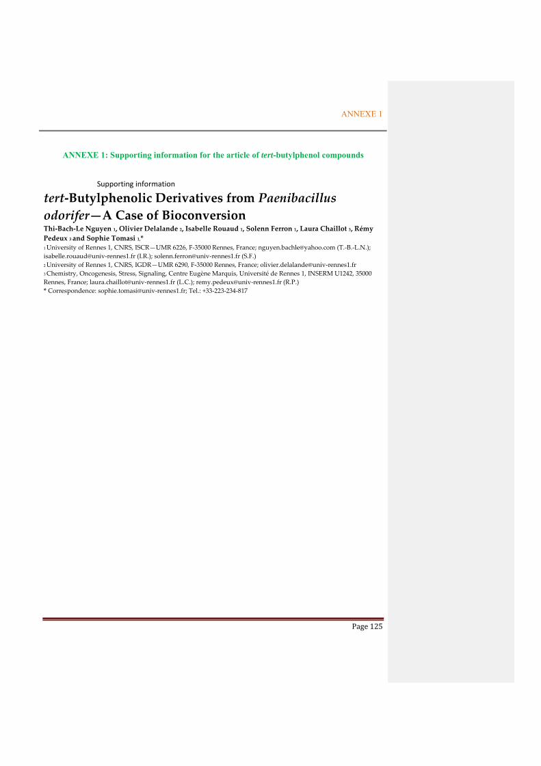
ANNEXE 1
Page 125
ANNEXE 1: Supporting information for the article of tert-butylphenol compounds
Supporting information
tert-Butylphenolic Derivatives from Paenibacillus
odorifer—A Case of Bioconversion Thi-Bach-Le Nguyen 1, Olivier Delalande 2, Isabelle Rouaud 1, Solenn Ferron 1, Laura Chaillot 3, Rémy
Pedeux 3 and Sophie Tomasi 1,* 1 University of Rennes 1, CNRS, ISCR—UMR 6226, F-35000 Rennes, France; [email protected] (T.-B.-L.N.);
[email protected] (I.R.); [email protected] (S.F.)
2 University of Rennes 1, CNRS, IGDR—UMR 6290, F-35000 Rennes, France; [email protected]
3 Chemistry, Oncogenesis, Stress, Signaling, Centre Eugène Marquis, Université de Rennes 1, INSERM U1242, 35000
Rennes, France; [email protected] (L.C.); [email protected] (R.P.)
* Correspondence: [email protected]; Tel.: +33-223-234-817

ANNEXE 1
Page 126
S1. 1H-NMR spectrum of compound 1 in CDCl3 (300 MHz)
S
OH OH
12
345
6
7
8
9
10
1'
11
1

ANNEXE 1
Page 127
S2. Jmod spectrum of compound 1 in CDCl3 (75 MHz)
S3. 2D-NMR HSQCedit spectrum of compound 1 in CDCl3 (300 MHz)
S
OH OH
12
345
6
7
8
9
10
1'
11
1

ANNEXE 1
Page 128
S4. 2D-NMR HMBC spectrum of compound 1 in CDCl3 (300 MHz)
S5. 2D-NMR COSY spectrum of compound 1 in CDCl3 (300 MHz)
S6. 2D-NMR NOESY spectrum of compound 1 in CDCl3 (300 MHz)
S
OH OH
12
345
6
7
8
9
10
1'
11
1
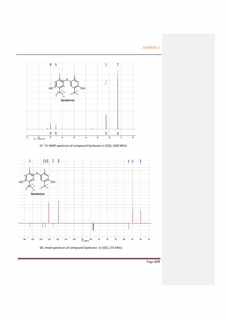
ANNEXE 1
Page 129
S7. 1H- NMR spectrum of compound Santonox in CDCl3 (300 MHz)
S8. Jmod-spectrum of compound Santonox in CDCl3 (75 MHz)
S
HO OH
12
35
6
7
8
9 10
1'
Santonox
11
S
HO OH
12
35
6
7
8
9 10
1'
Santonox
11

ANNEXE 1
Page 130
S9. 2D-NMR HSQC spectrum of Santonox in CDCl3 (300 MHz)
S10. 2D-NMR HMBC spectrum of Santonox in CDCl3 (300 MHz)
S
HO OH
12
35
6
7
8
9 10
1'
Santonox
11

ANNEXE 1
Page 131
S11. 2D-NMR COSY spectrum of Santonox in CDCl3 (300 MHz)
S12. 2D-NMR NOESY spectrum of Santonox in CDCl3 (300 MHz)
S
HO OH
12
35
6
7
8
9 10
1'
Santonox
11

ANNEXE 1
Page 132
S13: NOESY spectrum of compound 1’ (mixture of BHA and compound 1) from the extract of the culture
supplemented with BHA in Erlenmeyer flask
S14: 1H – NMR spectra of compound 1’ (mixture of compound 1 and BHA) (a); compound 1 (b) and BHA (c)
H-6
Compound 1
Compound 1
H-3
Compound 1
H-9/10/11
(a)
(b)
(c)
OH
OMe
OH
OMe
BHA
S
OH OH
12
345
6
7
8
9
10
1'
11
1

ANNEXE 1
Page 133
S15. 1H-NMR spectrum of compound 2 in CDCl3 (300 MHz)
S16. Jmod-NMR spectra of compound 2 in CDCl3 (75 MHz)
O
O
OH
14
2

ANNEXE 1
Page 134
S17. 2D-NMR HSQCedit spectrum of compound 2 in CDCl3 (300 MHz)
S18. 2D-NMR HMBC spectrum of compound 2 in CDCl3 (300 MHz)
O
O
OH
14
2

ANNEXE 1
Page 135
S19. 2D-NMR COSY of compound 2 in CDCl3 (300 MHz)
O
O
OH
14
2

ANNEXE 1
Page 136
Table 1: Predicted inter-protons distances on the base of molecular models corresponding to the major conformers of compound 1 and santonox extracted from the molecular dynamics simulation (see S20 ).
Santonox Santonox Isoform
H3-tBut 3,58 5,66
H6-tBut 5,83 3,87
S20: Major conformers for the compound 1 (0.34) and santonox (0.46) extracted from the molecular dynamics simulations performed in chloroform.

ANNEXE 2
Page 137
ANNEXE 2: Supporting information for the article of alkaloid compound
Paeniloxazine, a new alkaloid isolated from Paenibacillus odorifer a lichen-associated
bacterium.
Le Thi Bach Nguyen, Isabelle Rouaud, Solenn Ferron, Sophie Tomasi*,
Univ Rennes, CNRS, ISCR – UMR 6226, F-35000 Rennes, France
*Correspondence: [email protected]
† Univ Rennes, CNRS, ISCR – UMR 6226, F-35000 Rennes, France
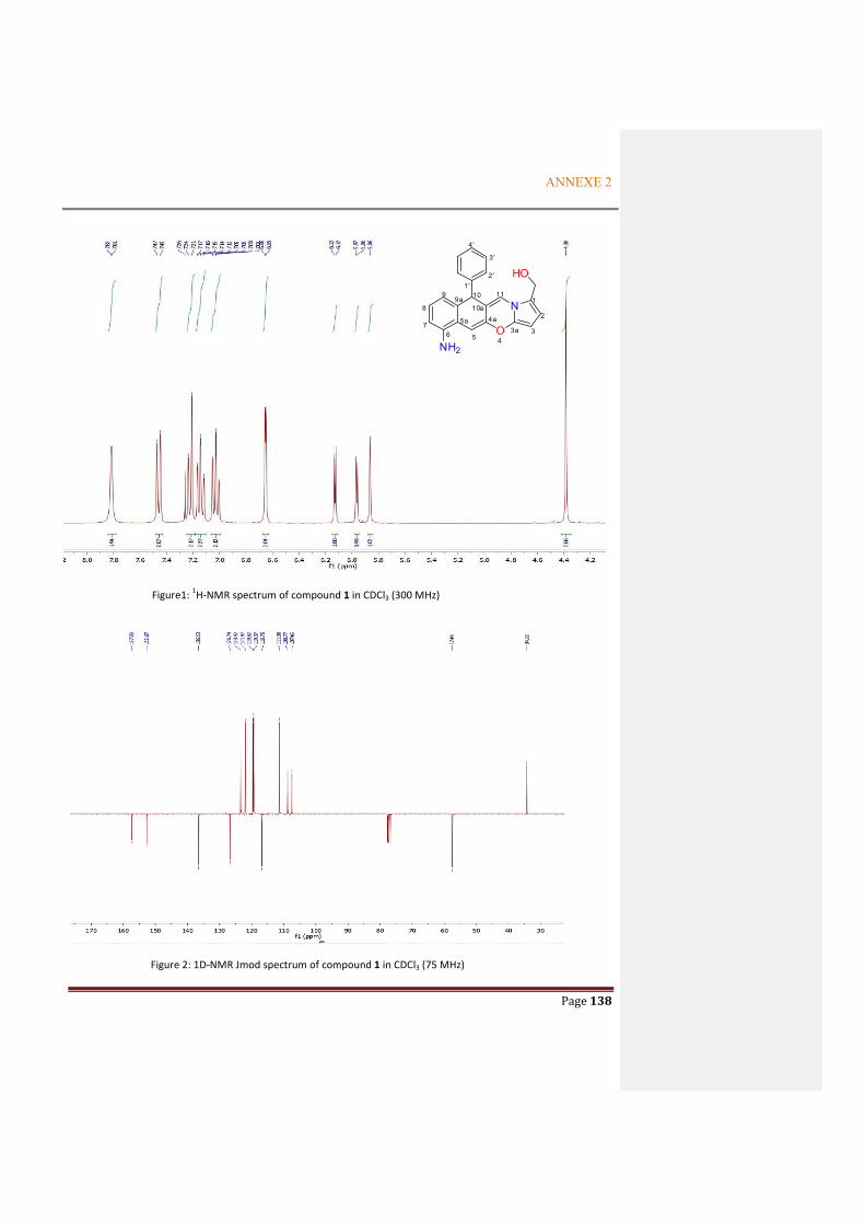
ANNEXE 2
Page 138
Figure1: 1H-NMR spectrum of compound 1 in CDCl3 (300 MHz)
Figure 2: 1D-NMR Jmod spectrum of compound 1 in CDCl3 (75 MHz)
O
N
HO
NH2
1
2
34
4a
5
5a
67
8
99a 10
10a
11
3a
1'2'
3'
4'
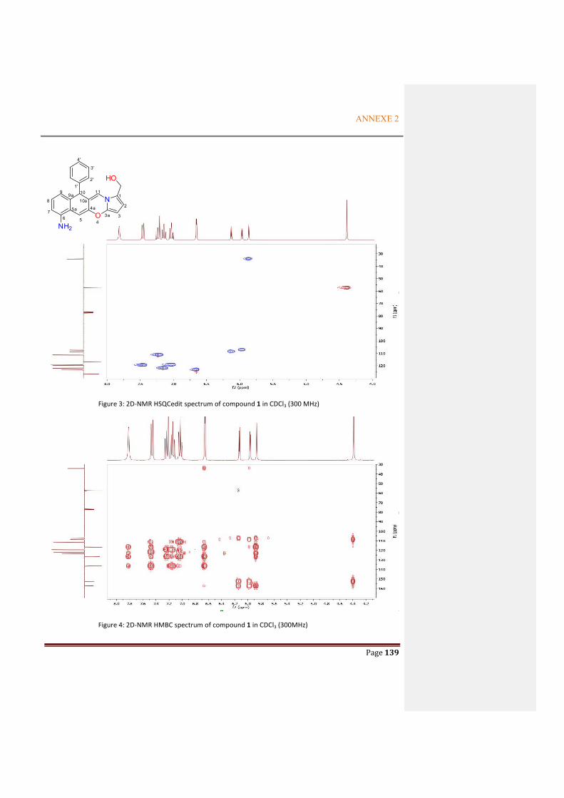
ANNEXE 2
Page 139
Figure 3: 2D-NMR HSQCedit spectrum of compound 1 in CDCl3 (300 MHz)
Figure 4: 2D-NMR HMBC spectrum of compound 1 in CDCl3 (300MHz)
O
N
HO
NH2
1
2
34
4a
5
5a
67
8
99a 10
10a
11
3a
1'2'
3'
4'

ANNEXE 2
Page 140
Figure 5: 2D-NMR COSY spectrum of compound 1 in CDCl3 (300MHz)
Figure 6: 2D-NMR NOESY spectrum of compound 1 in CDCl3 (300MHz)
O
N
HO
NH2
1
2
34
4a
5
5a
67
8
99a 10
10a
11
3a
1'2'
3'
4'
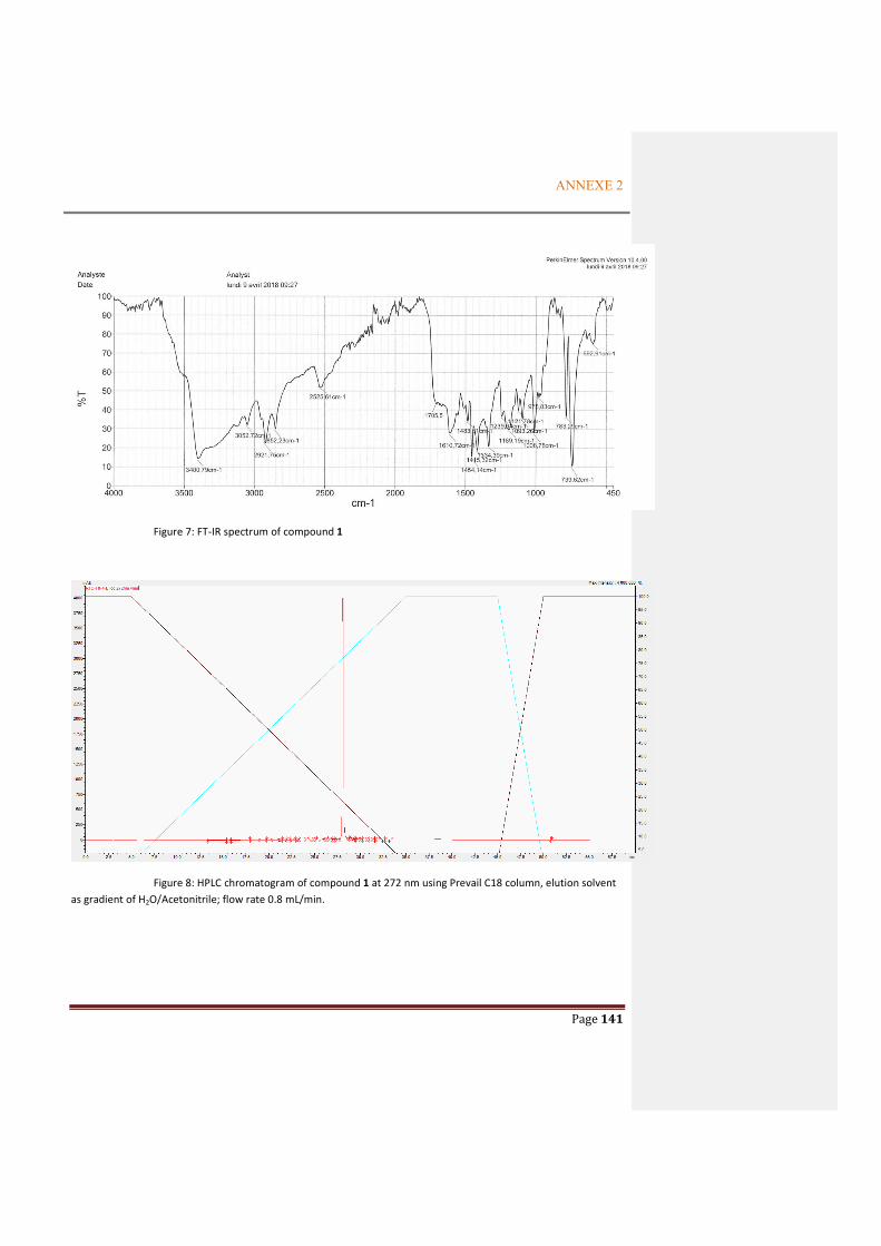
ANNEXE 2
Page 141
Figure 7: FT-IR spectrum of compound 1
Figure 8: HPLC chromatogram of compound 1 at 272 nm using Prevail C18 column, elution solvent
as gradient of H2O/Acetonitrile; flow rate 0.8 mL/min.

ANNEXE 2
Page 142
Figure 9: UV spectrum of compound 1
Figure 10: Thin layer chromatography (TLC) of compound 1, visualization by anisaldehyde (ANS), Rf = 0.45; 0.72 and
0.05 in elution solvents of CHCl3/EtOAc (2/1); EtOAc (100%) and CHCl3 (100%), respectively.
EtOAc = 100% CHCl3 = 100%
Compound 1 Compound 1 Compound 1
CHCl3/EtOAc = 2/1

ANNEXE 3
Page 143
ANNEXE 3: NMR spectra of the metabolites from P. odorifer
NMR spectra (500 MHz) of compound 1 (4-methyl-1-phenylpentane-2,3-diol) in CD3OD
1H-NMR spectrum of compound 1
13C-NMR spectrum of compound 1
OH
OH2 3 4 5
1'
2'3'
4'
HaHb
1

ANNEXE 3
Page 144
HSQCedit spectrum of compound 1
HMBC spectrum of compound 1
OH
OH2 3 4 5
1'
2'3'
4'
HaHb
1

ANNEXE 3
Page 145
NMR spectra (500 MHz) of compound 2 (4-methyl-1-phenylhexane-2,3-diol) in CD3OD
1H-NMR spectrum of compound 2
Jmod spectrum of compound 2
OH
OH2 3 4
5
1'
2'3'
4'
1 6
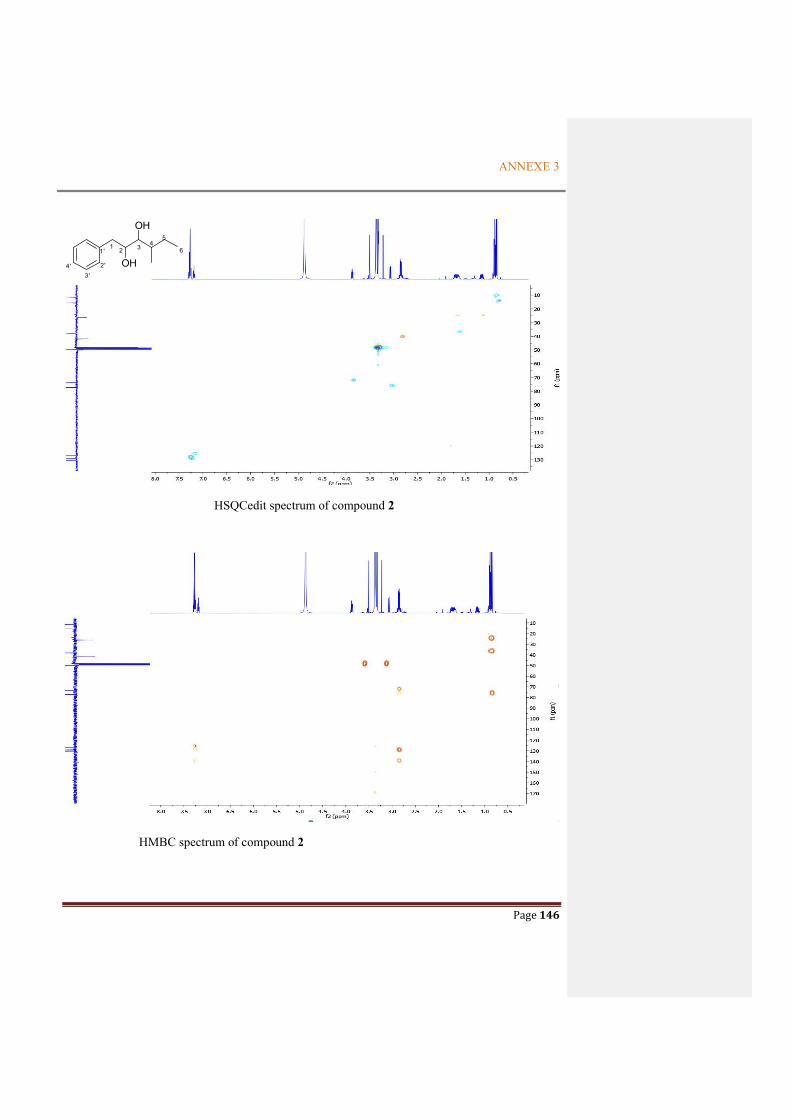
ANNEXE 3
Page 146
HSQCedit spectrum of compound 2
HMBC spectrum of compound 2
OH
OH2 3 4
5
1'
2'3'
4'
1 6
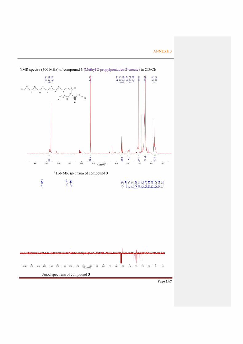
ANNEXE 3
Page 147
NMR spectra (300 MHz) of compound 3 (Methyl 2-propylpentadec-2-enoate) in CD2Cl2
1 H-NMR spectrum of compound 3
Jmod spectrum of compound 3
1
2
34
5
6
7
8
9
10
11
12
13
1415
1617
H
O
O18 19

ANNEXE 3
Page 148
HSQCedit spectrum of compound 3
HMBC spectrum of compound 3
1
2
34
5
6
7
8
9
10
11
12
13
1415
1617
H
O
O18 19

ANNEXE 3
Page 149
NMR spectra (300 MHz) of compound 4 (5-(hydroxymethyl)furan-2-carbaldehyde) in CDCl3
1H-NMR spectrum of compound 4
13C –NMR spectrum of compound 4
OO OH
1
2
3 4
5
67

ANNEXE 3
Page 150
HSQCedit spectrum of compound 4
HMBC spectrum of compound 4
OO OH
1
2
3 4
5
67

ANNEXE 3
Page 151
NMR spectra (300 MHz) of compound 5 (4-(5-(hydroxymethyl)furan-2-yl)but-3-en-2-one) in CD3OD
1H-NMR spectrum of compound 5
Jmod spectrum of compound 5
OO
HO12
34
1'
2'5'
6'
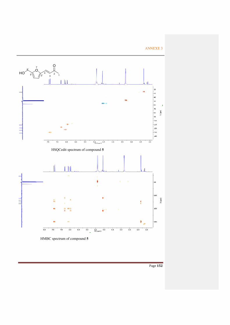
ANNEXE 3
Page 152
HSQCedit spectrum of compound 5
HMBC spectrum of compound 5
OO
HO12
34
1'
2'5'
6'

ANNEXE 3
Page 153
NMR spectra (300 MHz) of compound 6 (4-methoxy-3-methylfuran-2(5H)-one) in CDCl3
1H-NMR spectrum compound 6
Jmod spectrum compound 6
O O
O
1
2
34
5
6
7

ANNEXE 3
Page 154
HSQCedit spectrum of compound 6
HMBC spectrum of compound 6
O O
O
1
2
34
5
6
7
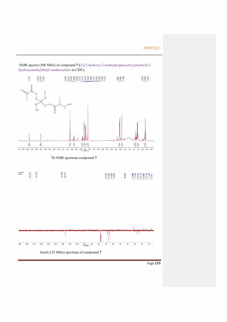
ANNEXE 3
Page 155
NMR spectra (500 MHz) of compound 7 (2-((3-hydroxy-2-methylpropanoyloxy)methyl)-2-(hydroxymethyl)butyl methacrylate) in CDCl3
1H-NMR spectrum compound 7
Jmod (125 MHz) spectrum of compound 7
O
O
OH
O
O
OH
12
3 4
56 7
8
9
10
11
12
13
14

ANNEXE 3
Page 156
NMR spectra (300 MHz) of compound 8 (Ethyl 1-ethyl-4-methoxy-2-(methoxymethyl)cyclopent-3-enecarboxylate) in CD3COCD3
1H-NMR spectrum of compound 8
13C-NMR spectrum of compound 8
OO
O
O
12
4 3
5
6
7
8
9 10
11
12
13

ANNEXE 3
Page 157
HSQCedit spectrum of compound 8
HMBC spectrum of compound 8
OO
O
O
12
4 3
5
6
7
8
9 10
11
12
13
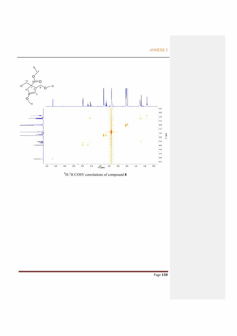
ANNEXE 3
Page 158
1H-1H COSY correlations of compound 8
OO
O
O
12
4 3
5
6
7
8
9 10
11
12
13

ANNEXE 3
Page 159
NMR spectra (500 MHz) of compound 9 (Hexyl 2-hydroxybenzoate) in CDCl3
1H-NMR spectrum of compound 9
Jmod spectrum of compound 9
O
OH
O
1
24
67
8
9
10
11
12
13

ANNEXE 3
Page 160
HSQCedit spectrum of compound 9
HMBC spectrum of compound 9
O
OH
O
1
24
67
8
9
10
11
12
13

ANNEXE 3
Page 161
1H-1H- COSY spectrum of compound 9
O
OH
O
1
24
67
8
9
10
11
12
13

ANNEXE 3
Page 162
NMR spectra (300 MHz) of compound 10 (4,4'-(propane-2,2-diyl)diphenol) in CDCl3
1H-NMR spectrum of compound 10
Jmod spectrum of compound 10
HO OH1
2
4
1'
4'
7
88'

ANNEXE 3
Page 163
HSQCedit spectrum of compound 10
HMBC spectrum of compound 10
HO OH1
2
4
1'
4'
7
88'

ANNEXE 3
Page 164
NMR spectra (300 MHz) of compound 11 (1H-indole-3-carbaldehyde) in CD3OD
1H-NMR spectrum of compound 11
Jmod spectrum of compound 11
HN
O
12
34
5
6
77a
3a
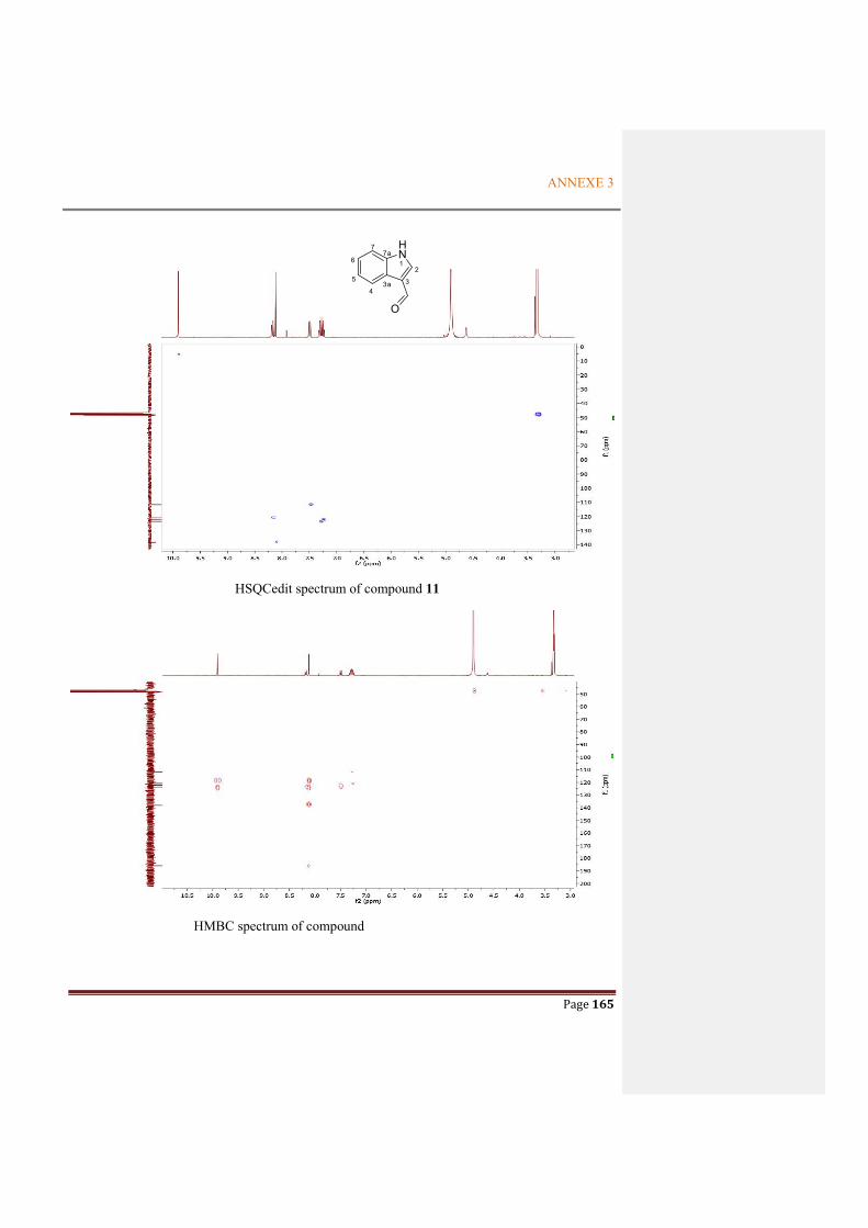
ANNEXE 3
Page 165
HSQCedit spectrum of compound 11
HMBC spectrum of compound
HN
O
12
34
5
6
77a
3a


Titre : Découverte des métabolites secondaires actifs de Paenibacillus odorifer, une bactérie associée à
un lichen
Mots clés : Paenibacillus odorifer, Rhizocarpon geographicum, bacterial compounds, alkaloid, tert-butylphenols
Résumé : Les bactéries qui sont des sources prolifiques d'antibiotiques et des fournisseurs importants d’agents pharmaceutiques peuvent produire une grande variété de métabolites. Ainsi, la découverte de métabolites issus bactéries est un nouveau challenge pour les chimistes. Parmi ces sources, les lichens sont admis comme niches intéressantes de nouvelles bactéries et de nouveaux composés bactériens. Par conséquent, les communautés de micro-organismes associées aux lichens sont devenues des sources prometteuses pour la production de composés naturels actifs.
Dans cette thèse, nous avons concentré notre travail sur l'isolement des bactéries de Rhizocarpon geogaphicum, l'un des lichens crustacés les plus populaires vivant sur la roche. Parmi les souches isolées, Paenibacillus odorifer a été sélectionnée pour poursuivre les travaux visant à produire des composés actifs.
Après des étapes d’optimisation de culture, l’étude des extraits issus des cultures de P. odorifer soit par le bioréacteur soit en Erlenmeyer a permis l’isolement des métabolites : un polysaccharide antioxydant, deux dérivés tert-butylphénoliques cytotoxiques issus de la bioaccumulation et de la biotransformation de précurseurs, d'un nouvel alcaloïde cytotoxique, de deux diols, de deux dérivés de type furfural et quelques autres composés connus. Des hypothèses de biosynthèse ont pu être proposés pour certains composés.
La diversité des métabolites isolés de P. odorifer indique que cette espèce possède un grand potentiel de production des composés actifs et est une bactérie utilisatrice de substrats tert-butyl phénoliques.
Title: : Discovery of active secondary metabolites from Paenibacillus odorifer, a lichen-associated
bacterium
Keywords : Paenibacillus odorifer, Rhizocarpon geographicum, bacterial compounds, alkaloid, tert-butylphenols
Abstract: Bacteria which are prolific sources of antibiotics and important suppliers to the pharmaceutical agents can produce a wide variety of metabolites. Thus finding metabolites from the bacterial lineages represented new interests for chemists. Among that, lichens are admitted as a rich source of new bacterial lineages and novel bacterial compounds. Therefore, microorganism communities associated with lichens became significant subjects as great potential for the production of active natural compounds.
In this thesis, we focus our work on the isolation of bacterial lineages from the lichen Rhizocarpon
geogaphicum, one of the most popular crustose lichens dwelling on the rock. Among the strains isolated, Paenibacillus odorifer was selected for further work to produce active compounds.
After the culture optimization steps, the study of extracts from the P. odorifer cultures either in the bioreactor or in Erlenmeyer flask led to the production of metabolites: an antioxidant polysaccharide, two cytotoxic tert-butylphenol derivatives which came from the bioaccumulation and biotransformation of precursors, a novel and cytotoxic alkaloid compound, two diol compounds, two furfural derivatives and some other known compounds. Putative biosynthetic pathways have been proposed for some compounds.
The diversity of metabolites isolated from P. odorifer highlighted that this species possessed a great potential of the production active compounds and were a new case of tert-butyl phenol utilizing bacterium.
1
LE RÉSUMÉ DE LA THÈSE par Thi Bach Le NGUYEN
Discovery of active secondary metabolites from Paenibacillus odorifer, a lichen-
associated bacterium
Les lichens sont des organismes complexes qui peuvent se développer sur de nombreux
supports différents. On les trouve sur les arbres, les sols, les surfaces rocheuses et même à
l'intérieur des roches, ainsi que sur la vitrocéramique, les objets métalliques… Les lichens
sont également des mini-écosystèmes auto-approvisionnements parfaits qui se développent
très lentement et sont formés par l'association de trois partenaires tels que champignons
(mycobionte), algues et / ou cyanobactéries (photobionte) et communautés bactériennes.
Chaque partenaire joue un rôle distinct et participe ensemble pour permettre à cet organisme
de survivre dans des conditions extrêmes de lumière, de température et d’eau, des pôles aux
tropiques, des zones intertidales aux sommets des montagnes. (Brodo et al., 2001).
Parmi les partenaires du lichen, le troisième partenaire correspondant aux communautés
bactériennes est devenu une nouvelle source intéressante à étudier pour les chimistes en raison
de leur capacité à produire de nombreux composés actifs. Dans nos efforts, en utilisant une
stratégie basée sur la culture, pour découvrir et étudier ces communautés trouvées sur
Rhizocarpon Geographicum, l'un des lichens crustacés vivant à la surface de la roche, 13
souches pures comprenant 10 bactéries, une cyanobactérie et un champignon ont été collectés.
Les dix souches de bactéries appartenaient aux deux phylums de Firmicutes et
Proteobacteria. En effet, contrairement aux autres lichens, les protéobactéries ne constituaient
pas la classe prédominante de bactéries parmi tous les microorganismes symbiotiques de R.
geographycum. Au lieu de cela, le phylum Firmicutes a eu lieu dans cette domination parmi
ces communautés bactériennes. De plus, dans ce phylum, le genre Paenibacillus présentait le
pourcentage le plus élevé (33%). Tout au long des études de la littérature déjà publiées sur les
souches de ce genre, Paenibacillus odorifer a été sélectionné pour isoler ses métabolites en
raison de la production potentielle de composés actifs.
La prochaine étape de ce travail consistait à déterminer les paramètres optimaux de la
culture de P. odorifer en milieu liquide. La première optimisation effectuée à petite échelle
(25 ml) a permis à P. odorifer de bénéficier de la meilleure croissance dans le milieu Gym
Streptomyces supplémenté en CaCO3 à pH 7 et à 25°C. Ces paramètres choisis dans la

2
première étape d’optimisation ont été appliqués à la culture par bioréacteur et ont donné le
polysaccharide et deux composés diols.
Le polysaccharide, identifié premièrement par les données IR et RMN, était
formé de trois unités de sucre d'acide glucuronique, de fructose et de fucose avec un rapport
moléculaire moyen de 4/2/1, respectivement. Les résultats ont été déterminés à partir de la
comparaison du temps de rétention observé dans les chromatogrammes HPLC entre les unités
de sucre dérivées de l'hydrolyse de polysaccharide et celles de monosaccharides classiques.
Ensuite, une série de réactions impliquant la méthylation, l'hydrolyse, la réduction du
borohydrure de sodium, l'acétylation par l'anhydride acétique et l'analyse finale par GC-MS
des acétates d'alditol ont conduit à une hypothèse de la structure de cette fraction de
polysaccharide telle que → 2) - β-D-GlcAp- (1 → 2) -β-D-GlcAp- (1 → 2) -β-D-GlcAp- (1
→ 2) -β-D-GlcAp- (2 → 2) -β-D-Fruf- (2 → 4) -β-D-Fruf- (2 → 4) - β-L-Fucp- (1 →. Ce
polysaccharide possédait également une cytotoxicité significative mesurée par titrage au MTT
avec une valeur de CI50 de 19 µg / mL et de 27 µg / mL HaCaT et des lignées cellulaires de
mélanomes murins B16, respectivement, ce qui pourrait constituer une source potentielle
d’agents antitumoraux sous forme de fraction polysaccharidique microbienne produite à partir
de P. odorifer.
Les deux composés de diol (nommés 4-méthyl-1-phénylpentane-2,3-diol (1) et
4-méthyl-1-phénylhexane-2,3-diol (2)) récoltés de la culture dans un bioréacteur ne montrent
pas toute activité cytotoxique. Bien que ces deux composés aient été identifiés par
Rukachaisirikul et ses collaborateurs (2011), leurs données spectroscopiques n'ont pas été
présentées. A notre connaissance, il s'agit du premier signalement de ces composés issus de la
culture de P. odorifer.
L'inconvénient de la culture réalisée dans le bioréacteur (4,5 L) est la présence d'autres
métabolites en quantité trop importante. En conséquence, afin d’augmenter la quantité de
métabolites secondaires produits, un processus d’optimisation secondaire a été établi à l’aide
des flacons Erlenmeyer. La différence de production suivant les récipients utilisés pourrait
s'expliquer par le mode d'aération différent et par le système d'agitation induisant des
conditions de contrainte différentes pour la souche. En fait, l'agitation dans le bioréacteur est
réalisée par une lame d'agitation immergée dans le bouillon de culture qui implique des
pauses hypha, tandis que dans les flacons agités, l'agitation est réalisée avec un agitateur
orbital permettant la formation de l'hyphale. De même, dans les flacons agités, il n’existe pas

3
de système spécifique d’aération; ceux du bioréacteur consistent en un tube immergé dans le
bouillon de culture qui conduit l’oxygène dans le milieu, ce qui provoque la formation de
bulles.
A la base des résultats de la première optimisation, la seconde a été réalisée à l'aide de
fioles Erlenmeyer (4,0 L) permettant de sélectionner les meilleures conditions de production
de métabolites à partir de P. odorifer sous forme de Gym Streptomyces moyen additionné de
CaCO3 à pH 7, Sous agitation à 25°C et à 120 tr/min, ainsi qu’à 1% d’inoculum. L'extrait de
résine obtenu en utilisant les meilleures conditions de la culture a été soumis à des approches
de chromatographie pour donner certains composés qui seront décrits suivant. Parmi eux,
deux tert-butylphénols ont été isolés et l'un d'eux a montré une activité cytotoxique
significative contre les lignées de cellules HaCaT et B16. Ces deux composés étaient rares
dans la nature. Leur présence dans le bouillon de culture pourrait s'expliquer par la
bioaccumulation et la biotransformation à partir de dérivés de tert-butyle tels que le BHA
(benzohydroxyanisole). Par ailleurs, P. odorifer a également produit d'autres composés non
cytotoxiques ou bien connus, tels que le 2-propylpentadec-2-énoate de méthyle (3); Le 5-
(hydroxyméthyl) furanne-2-carbaldéhyde (4); 4- (5- (hydroxyméthyl) furanne-2-yl) but-3-én-
2-one (5); 4-méthoxy-3-méthylfurane-2 (5H) -one (6); Méthacrylate de 2 - ((3-hydroxy-2-
méthylpropanoyloxy) méthyl) -2- (hydroxyméthyl) butyle (7); 1-éthyl-4-méthoxy-2-
(méthoxyméthyl) cyclopent-3-énécarboxylate d'éthyle (8); le 2-hydroxybenzoate d'hexyle (9).
De plus, la culture avec un volume important (40 L dans l’Erlenmeyer flask) en utilisant
les meilleures conditions choisies à la suite des résultats de la seconde optimisation a été
réalisée pour trouver des métabolites bioactifs en plus grande quantité. Un nouvel alcaloïde a
été trouvé dans une fraction cytotoxique (fraction 8). Sa structure consiste à une fraction
dihydronaphtalène, qui est un groupe structurel bien connu dans les produits naturels,
fusionnée à une unité rare de pyrrolooxazine. Ce nouveau composé était le premier exemple
de ce squelette produit par des organismes vivants (notamment P. odorifer). Ce composé
présentait également une cytotoxicité hebdomadaire avec des valeurs de CI50 de 76,0 µM et
78,9 µM sur les lignées cellulaires B16 et HaCaT, respectivement. De plus, deux composés
bien connus ont également été trouvés dans la fraction 8, tels que le 4,4 '- (propane-2,2-diyl)
diphénol (10) et le 1H-indole-3-carbaldéhyde (11) qui n'étaient pas actifs.

4
Par comparer le compte-rendu de tous les composés isolés de chaque culture de P.
odorifer en utilisant différents moyens comme bioréacteur et Erlenmeyer flasks, il semble que
le polysaccharide n'ait été trouvé que dans la culture utilisant le bioréacteur. Ceci s'explique
que, pour la culture en bioréacteur, le rapport d'inoculum n'était pas contrôlé et l'agitation était
fixée à 150 tr / min pendant la culture. Ces facteurs diffèrent des paramètres optimaux choisis
parmi les étapes d'optimisation, alors qu'ils semblent jouer un rôle important dans la
production de métabolites actifs. De plus, la différence de méthode d'aération utilisée pourrait
également être l'une des raisons de cette différence de production. Par conséquent, la
réalisation d'une culture dans le bioréacteur en respectant les meilleures conditions trouvées
lors de la deuxième optimisation pourrait permettre de conclure avec précision pour la
production de cette fraction de polysaccharide par P. odorifer. Ces efforts seront faits pour
mieux contrôler tous les paramètres de la culture dans le bioréacteur (taux d'aération,
brassage, rapport d'inoculum, etc.).
De la même manière, pour les autres métabolites, les deux tert-butylphénols ont été
trouvés dans la culture à Erlenmeyer flasks mais étaient absents dans la culture au bioréacteur.
L’origine de ce type de composés doit être étudiée particulièrement par leur voie de
biosynthèse supposée. Nous avons envisagé de réaliser le séquençage complet du génome afin
de mettre en évidence certaines voies métaboliques intéressantes.
Contrairement aux composés 1 et 2 du diol, ils ont été rapportés de la culture à la fois
par utiliser un bioréacteur et une fiole Erlenmeyer flask. Nous pourrions donc conclure qu'ils
constituent l'un des principaux composés produits par P. odorifer. Fait intéressant, lorsque le
volume de culture a augmenté, la masse de ces composés a également augmenté. De la même
manière, pour augmenter la masse de composés cytotoxiques, il est préférable de cultiver cette
souche dans un grand volume de milieu.
En outre, deux dérivés du furfural (4, 5) ont également été trouvés dans les cultures en
utilisant un erlenmeyer. Le milieu utilisé pour la culture de P. odorifer contenait de l'extrait de
malt, qui est un mélange de graines de céréales. Il est important de noter que le furfural aurait
été formé par le traitement des céréales à haute température (Mesias et al., 2017). De manière
correspondante, nous pourrions suggérer que les dérivés de furfural (4, 5) ont été formés par
la bioaccumulation de furfural présent dans le milieu après l'étape de stérilisation et ensuite
par biotransformation par P. odorifer. Par conséquent, la bioaccumulation et la

5
biotransformation semblent être un comportement caractéristique de P. odorifer. Comme
discuté ci-dessus, ce comportement particulier doit être étudié dans des expériences
ultérieures. Nous suggérons également de soumettre cette souche intéressante dans les études
de biotransformation de composés du lichen.
Enfin, P. odorifer est un producteur intéressant car cette souche pourrait produire des
composés originaux tels qu'un nouvel alcaloïde, certains composés de diol en grande quantité,
certains dérivés du furfural et certains tert-butylphénols qui pourraient être dérivés de la
biotransformation de précurseurs particuliers. Fait intéressant, bien qu'une quantité importante
de dicétopipérazines ait déjà été isolée de la culture de diverses bactéries associées au lichen,
aucun de ces composés, classiquement trouvé dans des cultures de microorganismes, n'a été
trouvé ici. Ces observations montrent que cette bactérie pourrait constituer une souche
intéressante pour la biotransformation ou produire de nouveaux composés actifs.
L'isolement d'autres métabolites actifs potentiels est en cour traiter sur les autres
fractions actives obtenues de notre grand volume de culture. D'autres évaluations d'activités
telles que les propriétés antibiotiques ou antifongiques seront entreprises pour valoriser ces
composés, particulièrement une grande quantité de ces composées diols a été obtenue. Nous
envisageons également d’étudier les autres extraits en tant que surnageant de pellets pouvant
contenir des composés intracellulaires potentiellement actifs.
De plus, la co-culture entre P. odorifer et le champignon qui a déjà été isolé de R.
geographicum devrait être réalisée afin de donner une hypothèse sur la capacité de cette
bactérie à produire des armes défensives sur la présence de ce champignon. Ces expériences
ultérieures nous donneront quelques arguments pour mettre en évidence la compétition parmi
les divers microorganismes pouvant apparaître dans ce micro-écosystème complexe qui
pourrait être le lichen.
References
Brodo I.M., Sharnoff S.D., Sharnoff S. (2001). Lichens of North Ameica. Yale University Press, New Haven CT. 828pp.
Mesías, M., Morales, F.J., 2017. Effect of Different Flours on the Formation of Hydroxymethylfurfural, Furfural, and Dicarbonyl Compounds in Heated Glucose/Flour Systems. Foods 6. https://doi.org/10.3390/foods6020014
Rukachaisirikul, V., Rodglin, A., Sukpondma, Y., Phongpaichit, S., Buatong, J., Sakayaroj, J., 2012. Phthalide and Isocoumarin Derivatives Produced by an Acremonium sp. Isolated from a Mangrove Rhizophora apiculata. J. Nat. Prod. 75, 853–858. https://doi.org/10.1021/np200885e.