Discovery of a Novel Orally Active Small-Molecule gp130...
14
Chemical Therapeutics Discovery of a Novel Orally Active Small-Molecule gp130 Inhibitor for the Treatment of Ovarian Cancer Shili Xu 1 , Fedora Grande 1,2 , Antonio Garofalo 2 , and Nouri Neamati 1 Abstract Interleukin (IL)-6 and Stat3 play key roles in ovarian cancer progression. However, the role of glycoprotein 130 (gp130), the signal transducer of this signaling axis, is not well-established. Currently, there are no small- molecule inhibitors of gp130 under clinical development. In this study, we show that gp130 is an attractive drug target in ovarian cancer due to its role in promoting cancer progression via the activation of its downstream Stat3 signaling. We also present preclinical studies of SC144, the first-in-class orally active small-molecule gp130 inhibitor. SC144 shows greater potency in human ovarian cancer cell lines than in normal epithelial cells. SC144 binds gp130, induces gp130 phosphorylation (S782) and deglycosylation, abrogates Stat3 phosphor- ylation and nuclear translocation, and further inhibits the expression of downstream target genes. In addition, SC144 shows potent inhibition of gp130 ligand–triggered signaling. Oral administration of SC144 delays tumor growth in a mouse xenograft model of human ovarian cancer without significant toxicity to normal tissues. Mol Cancer Ther; 12(6); 937–49. Ó2013 AACR. Introduction Ovarian cancer is the fourth-leading cause of death in women with gynecologic diseases in the United States. About 70% of the ovarian cancer cases are diagnosed at the late and distant stage and therefore are poorly treatable (1). Prolonged use of paclitaxel and carboplatin in the standard treatment often induces drug resistance, causes ovarian cancer relapse, and eventually the death of patients (2). In this context, there is an urgent need for breakthrough drugs with effective therapeutic impact on ovarian cancer. Gp130 is part of the receptor signaling complexes for at least 8 cytokines [interleukin (IL)-6, IL-11, IL-27, LIF, CNTF, OSM, CT-1, and CLC; ref. (3)]. Ligand binding induces the association of gp130 with a cytokine-specific receptor-a chain, followed by the activation of down- stream signaling cascades including JAK/STAT, RAS/ RAF/MAPK, and PI3K/AKT pathways (4). It has been shown that phosphorylation of gp130 at Ser782 down- regulates cell surface expression of gp130 (5, 6). As a ubiquitously expressed receptor, gp130 is involved in a wide range of important biologic processes including inflammation, autoimmunity, cancer (7), stem cell main- tenance, and embryonic development (8). In recent years, increasing attention has been paid to the rela- tionship between gp130 and cancer progression. How- ever, the role of gp130 in ovarian cancer progression remains elusive, and there is a dearth of small-molecule inhibitors selectively targeting gp130. Previously, we discovered novel quinoxalinhydrazides to have desirable physicochemical and drug-like proper- ties and a broad-spectrum anticancer activity (9–11). Here, we show that the lead compound, SC144, is a first-in-class small-molecule gp130 inhibitor with oral activity in ovar- ian cancer. Materials and Methods Reagents SC144, a quinoxalinhydrazide derivative, was synthe- sized as described (9). Stattic, a Stat3 inhibitor (12), was purchased from Sigma-Aldrich. Stock solutions of 10 mmol/L SC144 and Stattic were prepared in dimethyl sulfoxide (DMSO) and stored at 20 C. Further dilutions were made fresh in cell culture medium. IL-6, IFN-g , and platelet-derived growth factor (PDGF) were purchased from PeproTech. LIF was purchased from Santa Cruz Biotechnology. SDF-1a plasmid was a generous gift from Dr. Ghalib A. Alkhatib (Indiana University, Indianapolis, IN) and the protein was expressed and purified as described (13). Anti-phospho-Stat3 (Y705, 9145), anti-Stat3 (9132), anti-gp130 (3732), anti-phospho-Stat1 (Y701, 9171), anti-Stat1 (9172), anti-Cyclin D1 (2978), anti-Bcl-X L (2764), anti-Bcl2 (2870), anti-phospho-Akt (T308, 2965), anti- phospho-Akt (S473, 4060), anti-Akt (9272), and anti-Ape1 (4128) were purchased from Cell Signaling Technology. Authors' Affiliations: 1 Department of Pharmacology and Pharmaceutical Sciences, School of Pharmacy, University of Southern California, Los Angeles, California; and 2 Dipartimento di Farmacia e Scienze della Salute e della Nutrizione, Universit a della Calabria, Arcavacata di Rende, Italy Note: Supplementary data for this article are available at Molecular Cancer Therapeutics Online (http://mct.aacrjournals.org/). Corresponding Author: Nouri Neamati, Department of Pharmacology and Pharmaceutical Sciences, School of Pharmacy, University of Southern California, Los Angeles, CA 90033. Phone: 323-442-2341; Fax: 323-442- 1390; E-mail: [email protected] doi: 10.1158/1535-7163.MCT-12-1082 Ó2013 American Association for Cancer Research. Molecular Cancer Therapeutics www.aacrjournals.org 937 on February 18, 2019. © 2013 American Association for Cancer Research. mct.aacrjournals.org Downloaded from Published OnlineFirst March 27, 2013; DOI: 10.1158/1535-7163.MCT-12-1082
Transcript of Discovery of a Novel Orally Active Small-Molecule gp130...

Chemical Therapeutics
Discovery of a Novel Orally Active Small-Molecule gp130Inhibitor for the Treatment of Ovarian Cancer
Shili Xu1, Fedora Grande1,2, Antonio Garofalo2, and Nouri Neamati1
AbstractInterleukin (IL)-6 and Stat3 play key roles in ovarian cancer progression. However, the role of glycoprotein
130 (gp130), the signal transducer of this signaling axis, is not well-established. Currently, there are no small-
molecule inhibitors of gp130under clinical development. In this study,we show that gp130 is an attractive drug
target in ovarian cancer due to its role in promoting cancer progression via the activation of its downstream
Stat3 signaling. We also present preclinical studies of SC144, the first-in-class orally active small-molecule
gp130 inhibitor. SC144 shows greater potency in human ovarian cancer cell lines than in normal epithelial cells.
SC144 binds gp130, induces gp130 phosphorylation (S782) and deglycosylation, abrogates Stat3 phosphor-
ylation and nuclear translocation, and further inhibits the expression of downstream target genes. In addition,
SC144 showspotent inhibition of gp130 ligand–triggered signaling.Oral administration of SC144 delays tumor
growth in amouse xenograftmodel of human ovarian cancerwithout significant toxicity to normal tissues.Mol
Cancer Ther; 12(6); 937–49. �2013 AACR.
IntroductionOvarian cancer is the fourth-leading cause of death in
women with gynecologic diseases in the United States.About 70%of theovarian cancer cases arediagnosed at thelate and distant stage and therefore are poorly treatable(1). Prolonged use of paclitaxel and carboplatin in thestandard treatment often induces drug resistance, causesovarian cancer relapse, and eventually the death ofpatients (2). In this context, there is an urgent need forbreakthrough drugs with effective therapeutic impact onovarian cancer.Gp130 is part of the receptor signaling complexes for
at least 8 cytokines [interleukin (IL)-6, IL-11, IL-27, LIF,CNTF, OSM, CT-1, and CLC; ref. (3)]. Ligand bindinginduces the association of gp130 with a cytokine-specificreceptor-a chain, followed by the activation of down-stream signaling cascades including JAK/STAT, RAS/RAF/MAPK, and PI3K/AKT pathways (4). It has beenshown that phosphorylation of gp130 at Ser782 down-regulates cell surface expression of gp130 (5, 6). As aubiquitously expressed receptor, gp130 is involved in a
wide range of important biologic processes includinginflammation, autoimmunity, cancer (7), stem cell main-tenance, and embryonic development (8). In recentyears, increasing attention has been paid to the rela-tionship between gp130 and cancer progression. How-ever, the role of gp130 in ovarian cancer progressionremains elusive, and there is a dearth of small-moleculeinhibitors selectively targeting gp130.
Previously, we discovered novel quinoxalinhydrazidesto have desirable physicochemical and drug-like proper-ties and abroad-spectrumanticancer activity (9–11).Here,we show that the lead compound, SC144, is a first-in-classsmall-molecule gp130 inhibitor with oral activity in ovar-ian cancer.
Materials and MethodsReagents
SC144, a quinoxalinhydrazide derivative, was synthe-sized as described (9). Stattic, a Stat3 inhibitor (12), waspurchased from Sigma-Aldrich. Stock solutions of 10mmol/L SC144 and Stattic were prepared in dimethylsulfoxide (DMSO) and stored at�20�C. Further dilutionswere made fresh in cell culture medium. IL-6, IFN-g , andplatelet-derived growth factor (PDGF) were purchasedfrom PeproTech. LIF was purchased from Santa CruzBiotechnology. SDF-1a plasmid was a generous gift fromDr. Ghalib A. Alkhatib (Indiana University, Indianapolis,IN) and the protein was expressed and purified asdescribed (13). Anti-phospho-Stat3 (Y705, 9145), anti-Stat3(9132), anti-gp130 (3732), anti-phospho-Stat1 (Y701, 9171),anti-Stat1 (9172), anti-Cyclin D1 (2978), anti-Bcl-XL (2764),anti-Bcl2 (2870), anti-phospho-Akt (T308, 2965), anti-phospho-Akt (S473, 4060), anti-Akt (9272), and anti-Ape1(4128) were purchased from Cell Signaling Technology.
Authors' Affiliations: 1Department of Pharmacology and PharmaceuticalSciences, School of Pharmacy, University of Southern California, LosAngeles, California; and 2Dipartimento di Farmacia e Scienze della Salutee della Nutrizione, Universit�a della Calabria, Arcavacata di Rende, Italy
Note: Supplementary data for this article are available at Molecular CancerTherapeutics Online (http://mct.aacrjournals.org/).
Corresponding Author:Nouri Neamati, Department of Pharmacology andPharmaceutical Sciences, School of Pharmacy, University of SouthernCalifornia, Los Angeles, CA 90033. Phone: 323-442-2341; Fax: 323-442-1390; E-mail: [email protected]
doi: 10.1158/1535-7163.MCT-12-1082
�2013 American Association for Cancer Research.
MolecularCancer
Therapeutics
www.aacrjournals.org 937
on February 18, 2019. © 2013 American Association for Cancer Research. mct.aacrjournals.org Downloaded from
Published OnlineFirst March 27, 2013; DOI: 10.1158/1535-7163.MCT-12-1082

Anti-phospho-gp130 (Ser782, 12978-R), anti-actin (1616-R),normal rabbit IgG (3888), and goat anti-rabbit IgG-HRP(2030) were purchased from Santa Cruz Biotechnology.Anti-gp130 (B-R3, 852-060-000) was purchased from CellSciences. Mouse IgG2a (010-0141) and goat anti-mouseIgG-HRP (610-1302) were purchased from Rockland.Goat anti-mouse Cy5-linked IgG and goat anti-rabbitCy5-linked were purchased from GE Life Sciences.gp130 siRNA, Stat3 siRNA, control siRNA, siRNAtransfection reagent, and siRNA transfection mediumwere purchased from Santa Cruz Biotechnology. Try-pan Blue 0.4% solution was purchased from Lonza Inc.
Cell cultureOVCAR-8, OVCAR-5, and OVCAR-3 cells (National
Cancer Institute, Developmental Therapeutics Pro-gram, Bethesda, MD) were maintained in RPMI-1640supplemented with 10% heat-inactivated FBS (Gemini-Bioproducts). NCI/ADR-RES cells (National CancerInstitute) were maintained in RPMI-1640 supplemen-ted with 10% heat-inactivated FBS and 5 mmol/LL-glutamine. HEY and Caov-3 cell lines were kindlyprovided as gifts by Dr. Louis Dubeau (University ofSouthern California, Keck School of Medicine, LosAngeles, CA). HEY cells were maintained in Dulbecco’sModified Eagle Media (DMEM) supplemented with10% heat-inactivated FBS and 5 mmol/L L-glutamine.Caov-3 were maintained in MEM supplemented with10% heat-inactivated FBS. Human normal kidney epi-thelial and human normal endometrial epithelial cellswere kindly provided by Dr. Alan Epstein (Universityof Southern California, Keck School of Medicine) andmaintained in RPMI-1640 supplemented with 10%heat-inactivated FBS. Cells were grown as monolayersat 37�C in a humidified atmosphere of 5% CO2. Toremove adherent cells from the flask for subculture andcounting, cells were washed with PBS without calciumor magnesium, incubated with a small volume of 0.25%trypsin–EDTA solution (Mediatech, Inc.) for 5 to 10minutes, resuspended with culture medium, and cen-trifuged. All experiments were carried out using cellsin the exponential growth phase. Cells were routinelychecked for mycoplasma contamination by using Plas-moTest (InvivoGen).
Colony formation assayColony formation assays were conducted as described
(14). Briefly, OVCAR-8 cells (600 cells/well) and Caov-3 cells (800 cells/well) were seeded in 24-well platesand allowed to attach. After 48 hours, serial dilutionsof the corresponding compounds or antibodies wereadded to the culture medium and incubated for 48hours. Cells were cultured until colonies were formed(10 days for OVCAR-8 and 15 days for Caov-3), thensubsequently washed, stained with crystal violet solu-tion (2%) for 1 hour, and thoroughly washed withwater.
Western blottingCells (only attached cells were collected) or xenograft
tumor tissues were washed with ice-cold PBS and lysedin cold lysis buffer (20 mmol/L Tris-HCl, 150 mmol/LNaCl, 1 mmol/L EDTA, 1% Triton X-100, pH 7.5) with1� protease and phosphatase inhibitors. Protein con-centration was determined by BCA protein assay(Thermo Scientific). Proteins were resolved on 8% or10% SDS-PAGE and electrotransferred to Immun-BlotPVDF membrane (Bio-Rad). After blocking with 5%milk in Tris-buffered Saline with Tween-80 (TBST),membranes were probed with the indicated primaryantibodies, subsequently with horseradish peroxidase–conjugated secondary antibody, and developed usingDura Extended Duration Substrate (Thermo Scientific).Immunoreactive proteins were visualized with theChemi-Doc System (Bio-Rad).
Growth inhibition assayGrowth inhibition was assessed using an MTT assay as
described earlier (15). Cells were seeded in 96-well micro-titer plates, allowed to attach 24 hours before siRNAtransfections or the addition of corresponding com-pounds to the culture medium. After 72 hours, cells wereincubated with 0.3 mg/mL MTT (Amresco) for an addi-tional 3 hours at 37�C. After removal of the supernatant,DMSO was added to the wells and the absorbance wasread at 570 nm. All assays were conducted in triplicate.Percentageof cell growth inhibitionwas expressedas: (1�A/C) � 100% (A and C were the absorbance values fromexperimental and control cells, respectively). Inhibitoryconcentration 50% (IC50) valueswere determined for eachdrug from a plot of log (drug concentration) versus per-centage of cell growth inhibition. SDs were calculated onthe basis of the IC50 values obtained from at least 3independent experiments.
DARTS assayThe Drug Affinity Responsive Target Stability
(DARTS) assay was optimized and used to assess thebinding of SC144 to gp130 via the protease protectionfrom pronase (Roche Applied Science, Inc.) as previ-ously described (16, 17). OVCAR-8 cells were lysedusing M-PER (Thermo Scientific, Inc.) supplementedwith protease and phosphatase inhibitors. The super-natant of cell lysate containing 4 to 6 mg/mL totalproteins was incubated with SC144 at indicated con-centrations at room temperature for 1 hour, followed byproteolysis with 1 mg pronase to every 9,600 mg of lysatefor 30 minutes at room temperature. Final concentrationof DMSO was 1% in all samples. To stop proteolysis,5� SDS sample loading buffer [Tris-HCl 0.25 mol/L, pH6.8, SDS 10%, glycerol 50%, bromophenol blue 0.5%,dithiothreitol (DTT) 100 mmol/L] was added to eachsample at a 1:4 ratio, mixed well, and boiled at 100�Cfor 5 minutes. Samples were analyzed by Westernblotting.
Xu et al.
Mol Cancer Ther; 12(6) June 2013 Molecular Cancer Therapeutics938
on February 18, 2019. © 2013 American Association for Cancer Research. mct.aacrjournals.org Downloaded from
Published OnlineFirst March 27, 2013; DOI: 10.1158/1535-7163.MCT-12-1082
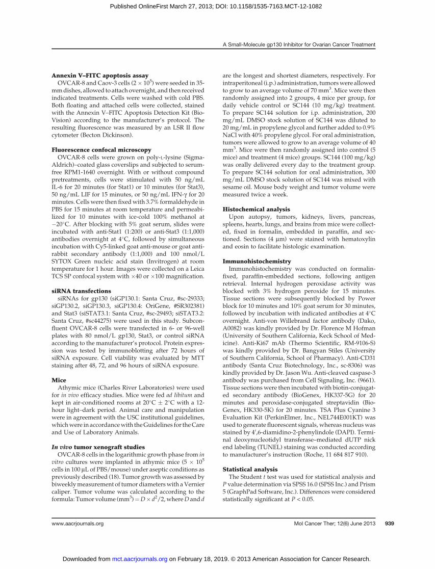
Annexin V–FITC apoptosis assayOVCAR-8 and Caov-3 cells (2� 105) were seeded in 35-
mmdishes, allowed to attach overnight, and then receivedindicated treatments. Cells were washed with cold PBS.Both floating and attached cells were collected, stainedwith the Annexin V–FITC Apoptosis Detection Kit (Bio-Vision) according to the manufacturer’s protocol. Theresulting fluorescence was measured by an LSR II flowcytometer (Becton Dickinson).
Fluorescence confocal microscopyOVCAR-8 cells were grown on poly-L-lysine (Sigma-
Aldrich)–coated glass coverslips and subjected to serum-free RPM1-1640 overnight. With or without compoundpretreatments, cells were stimulated with 50 ng/mLIL-6 for 20 minutes (for Stat1) or 10 minutes (for Stat3),50 ng/mL LIF for 15 minutes, or 50 ng/mL IFN-g for 20minutes. Cellswere then fixedwith 3.7% formaldehyde inPBS for 15 minutes at room temperature and permeabi-lized for 10 minutes with ice-cold 100% methanol at�20�C. After blocking with 5% goat serum, slides wereincubated with anti-Stat1 (1:200) or anti-Stat3 (1:1,000)antibodies overnight at 4�C, followed by simultaneousincubation with Cy5-linked goat anti-mouse or goat anti-rabbit secondary antibody (1:1,000) and 100 nmol/LSYTOX Green nucleic acid stain (Invitrogen) at roomtemperature for 1 hour. Images were collected on a LeicaTCS SP confocal system with �40 or �100 magnification.
siRNA transfectionssiRNAs for gp130 (siGP130.1: Santa Cruz, #sc-29333;
siGP130.2, siGP130.3, siGP130.4: OriGene, #SR302381)and Stat3 (siSTAT3.1: Santa Cruz, #sc-29493; siSTAT3.2:Santa Cruz, #sc44275) were used in this study. Subcon-fluent OVCAR-8 cells were transfected in 6- or 96-wellplates with 80 nmol/L gp130, Stat3, or control siRNAaccording to the manufacturer’s protocol. Protein expres-sion was tested by immunoblotting after 72 hours ofsiRNA exposure. Cell viability was evaluated by MTTstaining after 48, 72, and 96 hours of siRNA exposure.
MiceAthymic mice (Charles River Laboratories) were used
for in vivo efficacy studies. Mice were fed ad libitum andkept in air-conditioned rooms at 20�C � 2�C with a 12-hour light–dark period. Animal care and manipulationwere in agreement with the USC institutional guidelines,whichwere in accordancewith theGuidelines for theCareand Use of Laboratory Animals.
In vivo tumor xenograft studiesOVCAR-8 cells in the logarithmic growth phase from in
vitro cultures were implanted in athymic mice (5 � 105
cells in 100 mL of PBS/mouse) under aseptic conditions aspreviously described (18). Tumor growthwas assessed bybiweeklymeasurement of tumor diameterswith aVerniercaliper. Tumor volume was calculated according to theformula: Tumor volume (mm3)¼D� d2/2,whereD and d
are the longest and shortest diameters, respectively. Forintraperitoneal (i.p.) administration, tumorswere allowedto grow to an average volume of 70 mm3. Mice were thenrandomly assigned into 2 groups, 4 mice per group, fordaily vehicle control or SC144 (10 mg/kg) treatment.To prepare SC144 solution for i.p. administration, 200mg/mL DMSO stock solution of SC144 was diluted to20 mg/mL in propylene glycol and further added to 0.9%NaCl with 40% propylene glycol. For oral administration,tumors were allowed to grow to an average volume of 40mm3. Mice were then randomly assigned into control (5mice) and treatment (4 mice) groups. SC144 (100 mg/kg)was orally delivered every day to the treatment group.To prepare SC144 solution for oral administration, 300mg/mL DMSO stock solution of SC144 was mixed withsesame oil. Mouse body weight and tumor volume weremeasured twice a week.
Histochemical analysisUpon autopsy, tumors, kidneys, livers, pancreas,
spleens, hearts, lungs, and brains from mice were collect-ed, fixed in formalin, embedded in paraffin, and sec-tioned. Sections (4 mm) were stained with hematoxylinand eosin to facilitate histologic examination.
ImmunohistochemistryImmunohistochemistry was conducted on formalin-
fixed, paraffin-embedded sections, following antigenretrieval. Internal hydrogen peroxidase activity wasblocked with 3% hydrogen peroxide for 15 minutes.Tissue sections were subsequently blocked by Powerblock for 10 minutes and 10% goat serum for 30 minutes,followed by incubation with indicated antibodies at 4�Covernight. Anti-von Willebrand factor antibody (Dako,A0082) was kindly provided by Dr. Florence M Hofman(University of Southern California, Keck School of Med-icine). Anti-Ki67 mAb (Thermo Scientific, RM-9106-S)was kindly provided by Dr. Bangyan Stiles (Universityof Southern California, School of Pharmacy). Anti-CD31antibody (Santa Cruz Biotechnology, Inc., sc-8306) waskindly provided by Dr. JasonWu. Anti-cleaved caspase-3antibody was purchased from Cell Signaling, Inc. (9661).Tissue sectionswere then incubatedwith biotin-conjugat-ed secondary antibody (BioGenex, HK337-5G) for 20minutes and peroxidase-conjugated streptavidin (Bio-Genex, HK330-5K) for 20 minutes. TSA Plus Cyanine 3Evaluation Kit (PerkinElmer, Inc., NEL744E001KT) wasused to generate fluorescent signals, whereas nucleuswasstained by 40,6-diamidino-2-phenylindole (DAPI). Termi-nal deoxynucleotidyl transferase–mediated dUTP nickend labeling (TUNEL) staining was conducted accordingto manufacturer’s instruction (Roche, 11 684 817 910).
Statistical analysisThe Student t test was used for statistical analysis and
P value determination via SPSS 16.0 (SPSS Inc.) and Prism5 (GraphPad Software, Inc.). Differences were consideredstatistically significant at P < 0.05.
A Small-Molecule gp130 Inhibitor for Ovarian Cancer Treatment
www.aacrjournals.org Mol Cancer Ther; 12(6) June 2013 939
on February 18, 2019. © 2013 American Association for Cancer Research. mct.aacrjournals.org Downloaded from
Published OnlineFirst March 27, 2013; DOI: 10.1158/1535-7163.MCT-12-1082

ResultsSC144 shows cytotoxicity in a panel of humanovarian cancer cell lines
SC144 inhibits cell growth in a panel of human ovariancancer cell lineswith IC50 values in a submicromolar range(Table 1, Fig. 1A). The potency of SC144 toward NCI/ADR-RES (paclitaxel- and doxorubicin-resistant, IC50 ¼0.43 mmol/L; refs. 19–21) and HEY (cisplatin-resistant,IC50¼ 0.88 mmol/L) suggests an ability to overcome drug
resistance in ovarian cancer. SC144 also inhibited colonyformation in Caov-3 and OVCAR-8 cells (SupplementaryFig. S1A). We compared the abilities of SC144 to induceapoptosis and cell death in human ovarian cancer cells(OVCAR-8 and Caov-3) with human normal epithelialcells (kidney epithelial cells and endometrial epithelialcells). The kidney epithelial cells and the endometrialepithelial cells do not express cancer specific markersurvivin (Supplementary Fig. S1B; ref. 22). SC144 induced
C
0
20
40
60
80
DM
SO
SC
144
Pa
clit
axe
l
DM
SO
SC
144
Pa
clit
axe
l
DM
SO
SC
144
Pa
clit
axe
l
DM
SO
SC
144
Pa
clit
axe
l
KidneyCaov-3OVCAR-8
epithelial
cells
Endometrial
epithelial
cells
Ce
ll k
ill (%
)
***
******
***
BOVCAR-8
DM
SO
SC
144
Pacli
taxel
Kidney
epithelial
cells
Endometrial
epithelial
cells
An
exin
V-F
ITC
PI
Ap
op
tos
is (
%)
0
20
40
60
80
DM
SO
SC
144
Pa
clit
axe
l
DM
SO
SC
144
Pa
clit
axe
l
DM
SO
SC
144
Pa
clit
axe
l
DM
SO
SC
144
Pa
clit
axe
l
Late apoptosis
Early apoptosis
Caov-3OVCAR-8Kidney
epithelial
cells
Endometrial
epithelial
cells
Caov-3
A
N
NHN
NH
O
N
NF
SC144
Figure1. SC144 is cytotoxic to humanovarian cancer cell lines. A, chemical structure ofSC144. B, SC144causes significantlymore apoptosis inOVCAR-8 andCaov-3 than normal kidney epithelial and normal endometrial cells. After treatment with SC144 (2 mmol/L), paclitaxel (2 mmol/L), or equal amount ofDMSO for 72 hours, cells were stained with Annexin V–FITC and propidium iodide (PI) and then analyzed by flow cytometry. Top, cells in the bottom leftquadrant of each panel (Annexin V–negative, PI-negative) are viable, whereas cells in the top left quadrant (Annexin V–positive, PI-negative) are in the earlyapoptotic stage, and cells in the top right quadrant (Annexin V–positive, PI-positive) are in the late apoptotic/necrotic stage. Bottom, the percentage ofapoptotic cells is shown in a histogram. C, analysis of SC144 induced cell death in OVCAR-8, Caov-3, normal kidney epithelial, and normal endometrial cells.After treatment with SC144 (2 mmol/L), paclitaxel (2 mmol/L), or equal amount of DMSO for 72 hours, cells were stained with trypan blue. Histogram shows thepercentage of trypan blue–stained cells after indicated treatment (���, P < 0.001; paired t test. Bar, SEM).
Table 1. Cytotoxicity of SC144 in a panel of human ovarian cancer cell lines
IC50,a mmol/L
OVCAR-8 OVCAR-5 OVCAR-3 Caov-3 SKOV-3 HEY NCI/ADR-RES
SC144 0.72 � 0.32b 0.49 � 0.44 0.95 � 0.35 0.44 � 0.14 0.53 � 0.04 0.88 � 0.46 0.43 � 0.12
aIC50 is defined as the drug concentration causing a 50% decrease in the cell population.bValues (mean � SD) are calculated from 3 independent experiments.
Xu et al.
Mol Cancer Ther; 12(6) June 2013 Molecular Cancer Therapeutics940
on February 18, 2019. © 2013 American Association for Cancer Research. mct.aacrjournals.org Downloaded from
Published OnlineFirst March 27, 2013; DOI: 10.1158/1535-7163.MCT-12-1082

substantial apoptosis in OVCAR-8 and Caov-3 cells, butno considerable apoptotic effect was induced in the kid-ney epithelial cells or the endometrial epithelial cells(Fig. 1B). Consistently, trypan blue staining showed thatSC144 significantly caused cell death in OVCAR-8 andCaov-3 cells, whereas no significant cell death wasobserved with SC144 treatment in the kidney epithelialcells or the endometrial epithelial cells (Fig. 1C). Theseresults suggest that SC144 is preferentially cytotoxic tohuman ovarian cancer cells.
SC144 induces gp130 (Ser782) phosphorylation anddownregulates gp130 glycosylationSC144 treatment substantially increased the phosphor-
ylation of gp130 (S782) in bothOVCAR-8 and Caov-3 cellsin a time- (Fig. 2A) and dose-dependent manner (Fig. 2B).In both cell lines, total gp130 protein was detected as 2individual bands in immunoblotting (Fig. 2A). Silencingof gp130 using siRNAs decreased the abundance of bothbands (Supplementary Fig. S2A), confirming that bothbands are gp130. Treatment of OVCAR-8 andCaov-3 cellswith brefeldin A (BFA), an inhibitor of protein glycosyl-ation produced a time-dependent shift of gp130 into thelower band (Supplementary Fig. S2B), indicating that theupper band represents glycosylated gp130 and the lowerband represents unglycosylated or minimal core glyco-sylated gp130. After 6-hour continuous SC144 treatmentin OVCAR-8 and Caov-3 cells, the upper bands of totalgp130 shifted to the lower band (Fig. 2A), indicating thatSC144 treatment induced gp130 deglycosylation follow-ing the stimulation of gp130phosphorylation.Also, SC144
is mechanistically different from BFA, which did notinduce gp130 phosphorylation (Supplementary Fig.S2C). In addition, we found that gp130 ligands IL-6 andLIF substantially enhanced Stat3 (Y705) phosphorylation,whereas the phosphorylation of gp130 (Ser782) was onlyincreased by LIF treatment (Supplementary Fig. S2D).These results suggest that phosphorylation at Ser782 iscytokine-specific and is not required for gp130 activation.Serine phosphorylation of surface receptors is often asso-ciatedwith receptor internalization. Therefore, we furtherexamined the effect of SC144 treatment on cell surfacegp130 abundance. SC144 significantly induced gp130internalization at 10 mmol/L with 6- and 24-hour treat-ment, whereas at 0.5 and 2 mmol/L, no significant changeswere detected at cell surface gp130 (SupplementaryFig. S2E).
Induction of gp130 phosphorylation and deglycosy-lation suggest that SC144 targets gp130. Binding of asmall-molecule compound to its target protein canresult in conformational changes and proteolytic stabi-lization of the protein by decreasing its sensitivity toproteases (17, 23). Similar in concept to DNA footprint-ing assay, DARTS assay (16, 17) was used to test thebinding of SC144 to gp130. Incubation of SC144 withOVCAR-8 cell lysate protected gp130 from digestion ina dose-dependent manner as shown by the increasedabundance of the gp130 band (Fig. 2C). Especially, 100and 1,000 mmol/L of SC144 significantly increased gp130abundance by 63% (P ¼ 0.035) and 102% (P ¼ 0.020)compared with vehicle control. These results show thedirect binding of SC144 to gp130 and suggest that SC144
Pronase
100 1,00010100SC144 (µmol/L):
gp130
C
130 kDa
95 kDa0
0.2
0.4
0.6
0.8
1
0 0 1
10
100
1,0
00
− +++++
gp
130
SC144 (µmol/L):
Pronase:
*
*
Actin 43 kDa
min h
SC144 (2 μmol/L):
Caov-3OVCAR-8 BA
SC144:
Caov-3OVCAR-8
(μmol/L)
p-gp130 (S782)
gp130
Actin
p-gp130 (S782)
gp130
Actin
50 15 621300.5 20 20 0.5
0 5 15 62130
hmin
130 kDa
130 kDa
43 kDa
130 kDa
130 kDa
43 kDa
Figure 2. SC144 induces gp130 phosphorylation and deglycosylation. Immunoblot detection of phosphorylation of gp130 (Ser782) in OVCAR-8 andCaov-3 cells treated by SC144 in a time- (A) and dose-dependent (B) manner as indicated. C, SC144 directly binds gp130. OVCAR-8 cell lysate was used forthe DARTS assay, conducted as described in Materials and Methods. Left, 1 of 3 representative experiments is shown. Right, statistical analyses ofdata from the 3 independent experiments. Error bars, SEM. �, P < 0.05; paired t test.
A Small-Molecule gp130 Inhibitor for Ovarian Cancer Treatment
www.aacrjournals.org Mol Cancer Ther; 12(6) June 2013 941
on February 18, 2019. © 2013 American Association for Cancer Research. mct.aacrjournals.org Downloaded from
Published OnlineFirst March 27, 2013; DOI: 10.1158/1535-7163.MCT-12-1082
may induce conformational changes in gp130 and affectits activity.
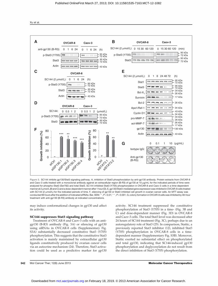
SC144 suppresses Stat3 signaling pathwayTreatment of OVCAR-8 and Caov-3 cells with an anti-
gp130 (B-R3) antibody (Fig. 3A) or silencing of gp130using siRNAs in OVCAR-8 cells (Supplementary Fig.S3A) substantially decreased constitutive Stat3 (Y705)phosphorylation. This suggests that the constitutive Stat3activation is mainly maintained by extracellular gp130ligands constitutively produced by ovarian cancer cellsvia an autocrine mechanism (24). Therefore, Stat3 activa-tion could be used as a predictive marker for gp130
activity. SC144 treatment suppressed the constitutivephosphorylation of Stat3 (Y705) in a time- (Fig. 3B andC) and dose-dependent manner (Fig. 3D) in OVCAR-8and Caov-3 cells. The total Stat3 level was decreased after24 hours of SC144 treatment (Fig. 3C), perhaps due to anautoregulatory role of Stat3 (25). In comparison, Stattic, apreviously reported Stat3 inhibitor (12), inhibited Stat3(Y705) phosphorylation in OVCAR-8 cells in a time-dependent manner (Supplementary Fig. S3B). Moreover,Stattic exerted no substantial effect on phosphorylatedand total gp130, indicating that SC144-induced gp130phosphorylation and deglycosylation do not result fromthe direct inhibition of Stat3 (Y705) phosphorylation.
72 kDa
95 kDa
72 kDa95 kDa
72 kDa
72 kDa95 kDa
95 kDa
43 kDa
B
C
p-Stat3 (Y705)
Stat3
Actin
(h)24610
OVCAR-8
SC144 (2 μmol/L):
SC144: (μmol/L)
D
p-Stat3 (Y705)
Stat3
Caov-3OVCAR-8
210.50 210.50
SC144 (2 μmol/L):
E
Stat1
Stat3
Bcl-XL
Survivin
Bcl-2
Ape1/Ref-1
Cyclin D1
pro-MMP-7
act-MMP-7
gp130
Actin
(h)24 48 72610
Caov-3OVCAR-8
(h)anti-gp130 (B-R3):
A
p-Stat3 (Y705)
Stat3
Actin
24610 24610
72 kDa
72 kDa95 kDa
95 kDa
43 kDa
72 kDa95 kDa
72 kDa95 kDa
26 kDa
34 kDa
17 kDa
26 kDa
34 kDa
34 kDa
43 kDa
34 kDa
26 kDa
26 kDa17 kDa
130 kDa
43 kDa
F
G
Isotype IgG:
Anti-gp130 (B-R3):
(μg/mL)1010.10
0
20
40
60
80
Gro
wth
in
hib
itio
n (
%)
**
**
0
20
40
60
80
Gro
wth
in
hib
itio
n (
%)
*** ***** ***
72 kDa95 kDa
72 kDa
95 kDa
Caov-3OVCAR-8
SC144 (2 μmol/L): (min)
p-Stat3 (Y705)
Stat3
60 12015 300 0 15 30 60 120
Figure 3. SC144 inhibits gp130/Stat3 signaling pathway. A, inhibition of Stat3 phosphorylation by anti-gp130 antibody. Protein extracts from OVCAR-8and Caov-3 cells treated with a monoclonal antibody against an extracellular region (B-R3) of gp130 at 10 mg/mL for the indicated periods of time wereanalyzed for phospho-Stat3 (Ser782) and total Stat3. SC144 inhibited Stat3 (Y705) phosphorylation in OVCAR-8 and Caov-3 cells in a time-dependentmanner at 2mmol/L (BandC) andadose-dependentmanner after 1hour (D). E, gp130/Stat3-mediatedgene expressionwas inhibited inOVCAR-8cells treatedwith SC144 (2 mmol/L) for the indicated periods of time. F, silencing of gp130 or Stat3 inhibited cell growth in ovarian cancer cells. An MTT assay wasconducted96hours after theaddition of indicated siRNAs.Bar, SEM. ��,P<0.01; ���,P<0.001.G, colony formation inOVCAR-8cellswas inhibitedby48-hourtreatment with anti-gp130 (B-R3) antibody at indicated concentrations.
Xu et al.
Mol Cancer Ther; 12(6) June 2013 Molecular Cancer Therapeutics942
on February 18, 2019. © 2013 American Association for Cancer Research. mct.aacrjournals.org Downloaded from
Published OnlineFirst March 27, 2013; DOI: 10.1158/1535-7163.MCT-12-1082

The activation of gp130/Stat3 promotes the expressionof multiple genes (26), including Bcl-2 (27), Bcl-XL (28),CyclinD1 (29), survivin (29),Ape1/Ref-1 (30), andMMP-7(31),which are involved in cell growth, survival, cell-cycleprogression, proliferation, and metastasis. The role ofgp130/Stat3 in promoting the expression of these genesin OVCAR-8 cells was confirmed by silencing of gp130(Supplementary Fig. S4A) and Stat3 (Supplementary Fig.S4B). Importantly, silencing of Stat3 also reduced gp130expression, which is consistent with a previous studyunveiling a STAT-binding element located in the gp130promoter region (29). To assess the effects of SC144 on theexpression of gp130/Stat3-regulated genes, we treatedOVCAR-8 cells with SC144 at various times. Survivin andMMP-7 were the most sensitive to SC144 treatment; theirabundance was significantly decreased during the first 24hours (Fig. 3E), whereas cyclin D1, Bcl-XL, Bcl-2, and
Ape1/Ref-1 expression started to decrease after 24 hours.Consistent with the Stat3 knockdown data, gp130 levelwas also significantly reduced within the first 24 hours ofSC144 treatment. It is also important to point out thatSC144 is mechanistically different from BFA due to thefact that total gp130 level was decreased after SC144treatment but was increased after BFA treatment (Sup-plementary Fig. S2B). In addition, Stat1 was not affectedby SC144 treatment. The inhibition of the gp130/Stat3downstream effectors (Bcl-2, BclXL, survivin, cyclin D1,Ape1/Rel-1, and MMP-7) further validates the mecha-nism of SC144 cytotoxicity in ovarian cancer cells. Inaddition, the cytotoxicity of SC144 (Supplementary Fig.S5A) as well as its inhibitory effects on gp130/Stat3pathway (Supplementary Fig. S5B) were also observedin 2 pancreatic cancer cell lines, Panc-1 and Mia-Paca-2,suggesting its potential use for other cancers.
72 kDa
55 kDa
95 kDa
55 kDa
55 kDa
43 kDa
72 kDa
95 kDa
72 kDa
72 kDa
72 kDa
72 kDa
55 kDa
55 kDa
55 kDa
43 kDa
72 kDa
72 kDa
72 kDa
95 kDa
72 kDa
72 kDa
72 kDa
72 kDa
55 kDa
55 kDa
55 kDa
43 kDa
72 kDa
95 kDa
95 kDa
72 kDa
72 kDa
95 kDa
95 kDa
72 kDa
72 kDa
95 kDa
72 kDa
72 kDa
95 kDa
95 kDa
55 kDa
55 kDa
72 kDa
43 kDa
72 kDa
95 kDa
72 kDa
72 kDa
95 kDa
72 kDa
72 kDa
95 kDa
55 kDa
55 kDa
72 kDa
55 kDa
43 kDa
72 kDa
72 kDa
95 kDa
95 kDa
A
p-Stat3 (Y705)
Stat3
p-Stat1 (Y701)
Stat1
p-Akt (T308)
p-Akt (S473)
Akt
Actin
p-Stat3 (Y705)
Stat3
p-Stat1 (Y701)
Stat1
p-Akt (T308)
p-Akt (S473)
Akt
Actin
CB
ED
p-Stat3 (Y705)
Stat3
p-Stat1 (Y701)
Stat1
p-Akt (T308)
Akt
Actin
p-Stat3 (Y705)
Stat3
p-Akt (T308)
p-Akt (S473)
Akt
Actin
p-Stat3(Y705)
p-Akt (T308)
p-Akt (S473)
Akt
Actin
LIF:
SC144: (μmol/L)
− ++++
5 10 20
IL-6:
SC144: (μmol/L)
− ++++
5 10 20
IFN-γ :
SC144: (μmol/L)
− ++++
5 10 20
SDF1-α:
SC144: (μmol/L)
− + + + +
5 10 20
PDGF:
SC144: (μmol/L)
− + + + +
5 10 20
Figure 4. SC144 inhibits downstream signaling induced by gp130 cytokines. SC144 inhibits the downstream signaling elicited by LIF (A) or IL-6 (B) ina dose-dependent manner without affecting the downstream signaling induced by IFN-g (C), SDF-1a (D), or PDGF (E). OVCAR-8 cells were serum-starvedovernight before 4-hour pretreatment with SC144 at the indicated concentrations, followed by stimulation with LIF (50 ng/mL) for 15minutes, IL-6 (50 ng/mL)for 10 minutes, IFN-g (50 ng/mL) for 20 minutes, SDF-1a (80 ng/mL) for 10 minutes, or PDGF (20 ng/mL) for 15 minutes. Whole-cell lysates weresubjected to immunoblotting using specific antibodies for the indicated proteins.
A Small-Molecule gp130 Inhibitor for Ovarian Cancer Treatment
www.aacrjournals.org Mol Cancer Ther; 12(6) June 2013 943
on February 18, 2019. © 2013 American Association for Cancer Research. mct.aacrjournals.org Downloaded from
Published OnlineFirst March 27, 2013; DOI: 10.1158/1535-7163.MCT-12-1082

Taken together, these results confirm that SC144 inhi-bits the activation of the Stat3 signaling pathway down-stream of gp130.
We further assessed whether the inhibition of gp130/Stat3 signaling contributes to the cytotoxicity of SC144 inovarian cancer cells. By using gp130 siRNAs or Stat3siRNAs, we found that silencing of gp130 or Stat3 signif-icantly inhibited OVCAR-8 cell growth (Fig. 3F). Whileanti-gp130 (B-R3) antibody substantially reduced the abil-ity ofOVCAR-8 cells to form colonies in adose-dependentmanner (Fig. 3G), it only induced limited apoptosis (Sup-plementary Fig. S6A) compared with that triggered bySC144 (Fig. 1B), suggesting there may be additionalmechanisms contributing to SC144-induced apoptosis.Furthermore, pharmacologic suppression of Stat3 activityby Stattic inhibited cell growth in OVCAR-8, Caov-3, andNCI/ADR-RES cells (Supplementary Fig. S6B). Takentogether, these results show that the gp130/Stat3 inhibi-tion results in cytotoxicity in human ovarian cancer cells.
SC144 potently blocks cytokine-triggered gp130signaling
We compared the effects of SC144 on the activation ofdownstream signaling stimulated by gp130 cytokines,including LIF and IL-6, and non-gp130 cytokines, includ-ing an IFN familymember IFN-g , a GPCR (CXCR4) ligandSDF-1a, and a growth factor PDGF. SC144 inhibited LIF-induced phosphorylation of Stat3 (Y705) and Stat1 (Y701)in a dose- and time-dependent manner (Fig. 4A andSupplementary Fig. S7A). SC144 also inhibited LIF-inducedAkt activation. Similar inhibitory effects of SC144were observed on IL-6–stimulated phosphorylation ofStat3, Stat1 and Akt (Fig. 4B). We further examined theinhibitory effect of SC144 against different concentrationsof IL-6. IL-6 stimulates Stat3 phosphorylation in a dose-dependent manner (Supplementary Fig. S7B). When 50ng/mL IL-6 was used to induce Stat3 phosphorylation,SC144 exhibited the inhibitory effect at over 10 mmol/L(Supplementary Fig. S7C). When 3 ng/mL IL-6, which isclose to the IL-6 concentration in thehumanbody (24),wasused to induce Stat3 phosphorylation, SC144 showedinhibitory effect at 2 mmol/L and a complete inhibitionat 5 mmol/L. These results suggest that SC144 inhibits thedownstream signaling activation elicited by gp130ligands. In contrast, SC144 did not affect the activationof phosphorylation of Stat3, Stat1, and Akt stimulated byIFN-g (Fig. 4C). Akt phosphorylation induced by SDF-1a(Fig. 4D) or PDGF (Fig. 4E) was not affected by SC144.Neither SDF-1a nor PDGF showed significant effects onStat3 phosphorylation in OVCAR-8 (Fig. 4D and E). Ourresults show that the inhibitory effect of SC144 on thedownstream signaling is gp130-specific and gp130-dependent.
The selectivity of SC144 in inhibiting the activation ofdownstream signaling stimulated by gp130 cytokineswasalso confirmed by examining the nuclear translocation ofStat3 and Stat1 induced by LIF, IL-6, or IFN-g , whichoccurs following tyrosine phosphorylation. Immunoflu-
orescencemicroscopy showed that SC144 treatment effec-tively inhibited LIF- and IL-6-induced nuclear transloca-tion of Stat3 and Stat1 but was not able to affect STATsnuclear translocation induced by IFN-g in OVCAR-8 cells(Fig. 5 and Supplementary Fig. S7D).
SC144 suppresses tumor growth in human ovariancancer xenografts
To determine in vivo efficacy of SC144, we tested itseffect on established tumors after subcutaneous inocula-tion of human ovarian cancer OVCAR-8 cells in the flankof nude mice. Compared with vehicle control treatment,daily i.p. administration of SC144 (10 mg/kg) for 58 dayssignificantly inhibited tumor growth by about 73% (from835.2� 228.1mm3 to 228.1� 92.5mm3;P¼ 0.032; Fig. 6A).To assess the oral bioavailability of SC144, we testedSC144 through daily oral administration. After we sacri-ficed the mice in the control group on day 69 due to theirlarge tumors, we continued SC144 treatment for another35 days. The average tumor volume in mice receivingdaily oral administration of SC144 (100mg/kg)was about
Control SC144
IL-6 IL-6 + SC144
LIF LIF + SC144
IFN-γ
Stat3 Nucleus Merge Stat3 Nucleus Merge
IFN-γ + SC144
Figure 5. SC144 inhibits Stat3 nuclear translocation induced by gp130cytokines. OVCAR-8 cells were serum-starved overnight before 4-hourpretreatment with or without SC144 (20 mmol/L), followed by stimulationwith indicated cytokines. Images are representative of 3 independentexperiments. Scale bar, 50 mm.
Xu et al.
Mol Cancer Ther; 12(6) June 2013 Molecular Cancer Therapeutics944
on February 18, 2019. © 2013 American Association for Cancer Research. mct.aacrjournals.org Downloaded from
Published OnlineFirst March 27, 2013; DOI: 10.1158/1535-7163.MCT-12-1082
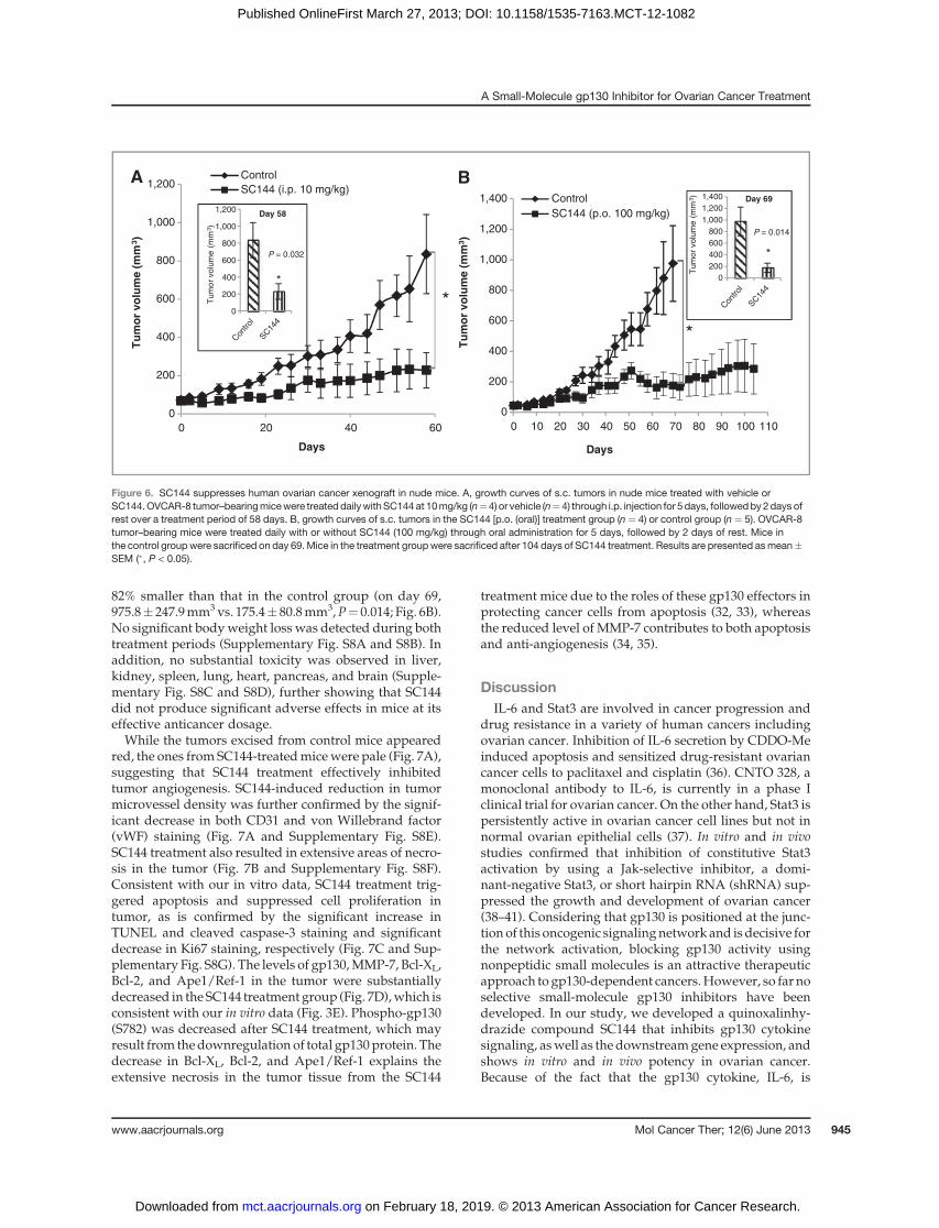
82% smaller than that in the control group (on day 69,975.8� 247.9mm3vs. 175.4� 80.8mm3,P¼ 0.014; Fig. 6B).No significant bodyweight loss was detected during bothtreatment periods (Supplementary Fig. S8A and S8B). Inaddition, no substantial toxicity was observed in liver,kidney, spleen, lung, heart, pancreas, and brain (Supple-mentary Fig. S8C and S8D), further showing that SC144did not produce significant adverse effects in mice at itseffective anticancer dosage.While the tumors excised from control mice appeared
red, the ones from SC144-treatedmicewere pale (Fig. 7A),suggesting that SC144 treatment effectively inhibitedtumor angiogenesis. SC144-induced reduction in tumormicrovessel density was further confirmed by the signif-icant decrease in both CD31 and von Willebrand factor(vWF) staining (Fig. 7A and Supplementary Fig. S8E).SC144 treatment also resulted in extensive areas of necro-sis in the tumor (Fig. 7B and Supplementary Fig. S8F).Consistent with our in vitro data, SC144 treatment trig-gered apoptosis and suppressed cell proliferation intumor, as is confirmed by the significant increase inTUNEL and cleaved caspase-3 staining and significantdecrease in Ki67 staining, respectively (Fig. 7C and Sup-plementary Fig. S8G). The levels of gp130,MMP-7, Bcl-XL,Bcl-2, and Ape1/Ref-1 in the tumor were substantiallydecreased in the SC144 treatment group (Fig. 7D),which isconsistent with our in vitro data (Fig. 3E). Phospho-gp130(S782) was decreased after SC144 treatment, which mayresult from thedownregulation of total gp130protein. Thedecrease in Bcl-XL, Bcl-2, and Ape1/Ref-1 explains theextensive necrosis in the tumor tissue from the SC144
treatment mice due to the roles of these gp130 effectors inprotecting cancer cells from apoptosis (32, 33), whereasthe reduced level of MMP-7 contributes to both apoptosisand anti-angiogenesis (34, 35).
DiscussionIL-6 and Stat3 are involved in cancer progression and
drug resistance in a variety of human cancers includingovarian cancer. Inhibition of IL-6 secretion by CDDO-Meinduced apoptosis and sensitized drug-resistant ovariancancer cells to paclitaxel and cisplatin (36). CNTO 328, amonoclonal antibody to IL-6, is currently in a phase Iclinical trial for ovarian cancer. On the other hand, Stat3 ispersistently active in ovarian cancer cell lines but not innormal ovarian epithelial cells (37). In vitro and in vivostudies confirmed that inhibition of constitutive Stat3activation by using a Jak-selective inhibitor, a domi-nant-negative Stat3, or short hairpin RNA (shRNA) sup-pressed the growth and development of ovarian cancer(38–41). Considering that gp130 is positioned at the junc-tion of this oncogenic signalingnetwork and isdecisive forthe network activation, blocking gp130 activity usingnonpeptidic small molecules is an attractive therapeuticapproach to gp130-dependent cancers.However, so far noselective small-molecule gp130 inhibitors have beendeveloped. In our study, we developed a quinoxalinhy-drazide compound SC144 that inhibits gp130 cytokinesignaling, aswell as the downstreamgene expression, andshows in vitro and in vivo potency in ovarian cancer.Because of the fact that the gp130 cytokine, IL-6, is
A
0
200
400
600
800
1,000
1,200
6040200
Control
SC144 (i.p. 10 mg/kg)
Con
trol
SC14
4
Con
trol
SC14
4
Tu
mo
r v
olu
me
(m
m3)
Days
*0
200
400
600
800
1,000
1,200T
um
or
vo
lum
e (
mm
3)
P = 0.032
*
Day 58
B
0
200
400
600
800
1,000
1,200
1,400
1101009080706050403020100
Days
Control
SC144 (p.o. 100 mg/kg)
Tu
mo
r v
olu
me
(m
m3)
*
Tu
mo
r vo
lum
e (
mm
3)
0
200
400
600
800
1,000
1,200
1,400
P = 0.014
Day 69
*
Figure 6. SC144 suppresses human ovarian cancer xenograft in nude mice. A, growth curves of s.c. tumors in nude mice treated with vehicle orSC144.OVCAR-8 tumor–bearingmicewere treated dailywithSC144 at 10mg/kg (n¼4) or vehicle (n¼4) through i.p. injection for 5days, followedby2days ofrest over a treatment period of 58 days. B, growth curves of s.c. tumors in the SC144 [p.o. (oral)] treatment group (n ¼ 4) or control group (n ¼ 5). OVCAR-8tumor–bearing mice were treated daily with or without SC144 (100 mg/kg) through oral administration for 5 days, followed by 2 days of rest. Mice inthe control group were sacrificed on day 69.Mice in the treatment groupwere sacrificed after 104 days of SC144 treatment. Results are presented asmean�SEM (�, P < 0.05).
A Small-Molecule gp130 Inhibitor for Ovarian Cancer Treatment
www.aacrjournals.org Mol Cancer Ther; 12(6) June 2013 945
on February 18, 2019. © 2013 American Association for Cancer Research. mct.aacrjournals.org Downloaded from
Published OnlineFirst March 27, 2013; DOI: 10.1158/1535-7163.MCT-12-1082

constitutively produced and the key effectors (Stat3, Akt)in the gp130 downstream signaling pathways are persis-tently active in ovarian cancer (24, 42–44), ovarian cancercells might be more sensitive to gp130 inhibitors thannormal cells. This concept is in accordwith our results thatSC144 is more cytotoxic in human ovarian cancer cellsthan human normal epithelial cells and inhibits humanovarian tumor growth in a mouse xenograft model with-out obvious toxicity to normal tissues. To our knowledge,SC144 is the first-in-class small-molecule gp130 inhibitorwith oral activity in ovarian cancer. In addition, ourresults showing that gp130 persistently activates Stat3 inovarian cancer cells, promoting cell growth and prolifer-ation, are consistent with a similar discovery in colitis-associated tumorigenesis (45). Although the identificationof amino residues on gp130 that SC144 interacts and theelucidation of off-targets are beyond the scope of thispresent study, the following evidence cumulatively showthat gp130 is directly inhibitedbySC144: (i) SC144binds togp130 (Fig. 2C) and induces gp130 (S782) phosphorylationand deglycosylation (Fig. 2A and B); (ii) anti-gp130 anti-body is able to completely suppress constitutive Stat3activation (Fig. 3A), indicating that Stat3 activation as an
indicator for the effect of SC144 on gp130; (iii) SC144inhibits Stat3 activation and the expression of down-stream genes (Fig. 3B–E); and (iv) SC144 potently inhibitsthe activation of the downstream signaling pathwaysstimulated by gp130 ligands but shows no effect on theactivation of the downstream signaling pathways stimu-lated by non-gp130 ligands (Figs. 4 and Fig. 5 and Sup-plementary Fig. S7).
The role of phospho-S782 in gp130 was first documen-ted in 2000 to downregulate gp130 cell surface expression(5). Recently, it has been reported that phospho-S782-associated downregulation of gp130 cell surface expres-sion is mediated by immediate internalization (46).Indeed, phosphorylation of many transmembrane recep-tors at Ser/Thr residues is commonly associated with thenegative modulation of receptor activity (47). The induc-tion of gp130 deglycosylation and the decrease in totalgp130 protein level may also contribute to Stat3 inhibitionat a later time. However, it required 10 mM of SC144 toinduce gp130 internalization (Supplementary Fig. S2E), aconcentration higher than for triggering gp130 deglyco-sylation. This may suggest that the amount of deglycosy-lation inducedbySC144 at 2mmol/Lmaynot be enough to
SC
144
130 kDa
130 kDa
26 kDa
26 kDa
17 kDa
34 kDa
26 kDa
34 kDa
26 kDa
34 kDa
43 kDa
BAControl SC144 (i.p.)
0
20
40
60
80
100
1.2
1
0.8
0.6
0.4
0.2
0
80
60
40
20
0
30
20
10
0
SC144CTRL(i.p.)
SC144CTRL
Control
(i.p.)
SC144CTRL(i.p.)
SC144CTRL(i.p.)
Necro
sis
(%
)
CD
31
-po
sit
ive
are
a
TU
NE
L-p
os
itiv
e c
ell
s (
%)
Ki6
7-p
os
itiv
e c
ell
s (
%)
***
**
**
***
P < 0.0001
P = 0.0034
P = 0.0026
P = 0.0003
200 µm 200 µm
C D
p-gp130 (S782)
gp130
pro-MMP-7
act-MMP-7
Bcl-XL
Bcl-2
Ape1/Ref-1
Actin
Control
SC144 (i.p.)
DAPICD31
CT
RL
SC
144
CT
RL
SC
144
CT
RL
SC
144
Merge
DAPITUNEL
Ki67
Merge
DAPI Merge
50 µm50 µm50 µm
50 µm50 µm
200 µm 200 µm 200 µm
200 µm 200 µm 200 µm
100 µm 100 µm 100 µm
100 µm 100 µm 100 µm
50 µm
Figure 7. Effects of SC144 on in vivoovarian tumor. A, SC144 treatmentreduced tumor blood vessels inOVCAR-8 xenograft mice. Top left,tumors excised from i.p. vehicle-treated and SC144-treatedmice are shown. Bottom,immunohistochemical staining forCD31. Percentage of positivelystained areas was determined bythe number of areas of stainedmicrovessels divided by number ofnuclei. Bar, SEM. ��, P < 0.01.B, SC144 (i.p.) treatment inducedextensive areas of necrosis inOVCAR-8 xenograft mice. Threedifferent fields were used forquantification of areas of necrosis.Bar, SEM. ���, P < 0.001. C,immunohistochemical staining ofTUNEL and Ki67. Percentage wasdetermined by the number ofdouble-stained cells divided by thenumber of nuclei. Bar, SEM.��, P < 0.01; ���, P < 0.001. D,SC144 (i.p.) treatmentdownregulated gp130/Stat3-mediated gene expression inOVCAR-8 tumors from xenograftmice.
Xu et al.
Mol Cancer Ther; 12(6) June 2013 Molecular Cancer Therapeutics946
on February 18, 2019. © 2013 American Association for Cancer Research. mct.aacrjournals.org Downloaded from
Published OnlineFirst March 27, 2013; DOI: 10.1158/1535-7163.MCT-12-1082

immediately initiate receptor internalization. However,the in vivo concentration of SC144 exceeds 10 mmol/Lunder our experimental conditions. Moreover, inhibitionof gp130/Stat3 amplifies the cytotoxicity of SC144 bydownregulating the expression of several downstreamgenes, such as Bcl-2, Bcl-XL, cyclin D1, MMP-7, Ape1/Ref-1, and survivin, in ovarian cancer cells that results inthe submicromolar IC50 values after 72-hour SC144 treat-ment (Table 1).On the basis of results presented in this study, we
propose a plausible model for the anticancer mecha-nism of SC144 in ovarian cancer (Fig. 8). SC144 bindinginduces conformational changes and activity loss in thecell surface–bound gp130 receptor, exposing the Ser782residue to protein kinases such as CaMKII, CaMKIV,and MK2 (6, 46). The decrease in gp130 activity leads tothe inactivation of downstream signaling pathwaysinvolving Stat3 and Akt. The suppression of gp130/Stat3-mediated gene expression amplifies the potency ofSC144 due to the inhibition of cell-cycle progression andangiogenesis and the promotion of apoptosis and celldeath. In addition, the downregulation of the PI3K/Aktpathway also contributes to apoptosis and cell death.Our working model may serve to identify a unifyinganticancer mechanism likely to be shared by futuregp130 inhibitors.Clinically, in patients with advanced ovarian cancer,
high plasma levels of IL-6 correlate with poor prognosis(48, 49), and increased levels of IL-6 are also present inmalignant ascites (50). Therefore, biopsy detection of IL-6
levels appears to be one strategy to identify subjects/patients whomay benefit most from IL-6/gp130-blockingtherapies. In addition, other components of the gp130receptor complexes (e.g., OSMR-b, LIFR-b) as well asother gp130 cytokines (e.g., OSM) have been reported tobe highly expressed in human ovarian carcinoma tissuesand have the ability to activate downstream signalingpathways (e.g., Stat3 pathway; ref. 51). On the basis ofsuch observations, these molecules can also be used asmarkers for selecting target subjects/patients. In fact, thisevidence also supports that gp130 antagonists may bemore effective for ovarian cancer treatment compared toIL-6 inhibitors (e.g., anti-IL-6 mAb), as other gp130 cyto-kines may compensate the effects of IL-6.
Disclosure of Potential Conflicts of InterestNo potential conflicts of interest were disclosed.
Authors' ContributionsConception and design: S. Xu, N. NeamatiDevelopment of methodology: S. Xu, F. Grande, A. Garofalo, N. NeamatiAnalysis and interpretation of data (e.g., statistical analysis, biostatis-tics, computational analysis): S. Xu, F. Grande, A. Garofalo, N. NeamatiWriting, review, and/or revision of the manuscript: S. Xu, N. NeamatiAdministrative, technical, or material support (i.e., reporting or orga-nizing data, constructing databases): N. NeamatiStudy supervision: N. Neamati
AcknowledgmentsThe authors thank Drs. Roger Duncan, Ebrahim Zandi, and Joe Miller
for their comments and suggestions and also thank Dr. Bangyan Stiles forher kind assistance for immunohistochemical staining.
Figure 8. A working model for theanticancer mechanism of SC144 inovarian cancer. We surmise thatSC144 is cytotoxic to ovarian cancercells via a mechanism involving theinhibition of gp130 activity, leading tothe inactivation of Akt and Stat3 aswell as the suppression of Stat3-regulated gene expression. As aresult, SC144 treatment eventuallycauses cell-cycle arrest, anti-angiogenesis, and apoptosis.
Internalization
& Degradation
Proliferation
Survival
Angiogenesis
& Metastasis
Survival
Deglycosylation
A Small-Molecule gp130 Inhibitor for Ovarian Cancer Treatment
www.aacrjournals.org Mol Cancer Ther; 12(6) June 2013 947
on February 18, 2019. © 2013 American Association for Cancer Research. mct.aacrjournals.org Downloaded from
Published OnlineFirst March 27, 2013; DOI: 10.1158/1535-7163.MCT-12-1082
Grant SupportThis studywas supported by funds through theDepartment ofDefense
Ovarian Cancer Program (OCRP) Idea Award (W81XWH-07-1-0414) to N.Neamati.
The costs of publication of this article were defrayed in part by thepayment of page charges. This article must therefore be hereby marked
advertisement in accordance with 18 U.S.C. Section 1734 solely to indicatethis fact.
ReceivedNovember 12, 2012; revised February 25, 2013; acceptedMarch20, 2013; published OnlineFirst March 27, 2013.
References1. Siegel R, Naishadham D, Jemal A. Cancer statistics, 2012. CA Cancer
J Clin 2012;62:10–29.2. Petty R, Evans A, Duncan I, Kurbacher C, Cree I. Drug resistance in
ovarian cancer - the role of p53. Pathol Oncol Res 1998;4:97–102.3. JonesSA,Scheller J,Rose-JohnS. Therapeutic strategies for theclinical
blockade of IL-6/gp130 signaling. J Clin Invest 2011;121:3375–83.4. Wang X, Lupardus P, Laporte SL, Garcia KC. Structural biology of
shared cytokine receptors. Annu Rev Immunol 2009;27:29–60.5. Gibson RM, Schiemann WP, Prichard LB, Reno JM, Ericsson LH,
Nathanson NM. Phosphorylation of human gp130 at Ser-782 adjacentto the Di-leucine internalization motif. Effects on expression andsignaling. J Biol Chem 2000;275:22574–82.
6. Gibson RM, Laszlo GS, Nathanson NM. Calmodulin-dependent pro-tein kinases phosphorylate gp130 at the serine-based dileucine inter-nalization motif. Biochim Biophys Acta 2005;1714:56–62.
7. Silver JS, Hunter CA. gp130 at the nexus of inflammation, autoimmu-nity, and cancer. J Leukoc Biol 2010;88:1145–56.
8. Rose-John S. GP130 stimulation and the maintenance of stem cells.Trends Biotechnol 2002;20:417–9.
9. Grande F, Aiello F, Grazia OD, Brizzi A, Garofalo A, Neamati N.Synthesis and antitumor activities of a series of novel quinoxalinhy-drazides. Bioorg Med Chem 2007;15:288–94.
10. Plasencia C, Grande F, Oshima T, Cao X, Yamada R, Sanchez T, et al.Discovery of a novel quinoxalinhydrazide with a broad-spectrumanticancer activity. Cancer Biol Ther 2009;8:458–65.
11. Oshima T, Cao X, Grande F, Yamada R, Garofalo A, Louie S, et al.Combination effects of SC144 and cytotoxic anticancer agents. Anti-cancer Drugs 2009;20:312–20.
12. Schust J, Sperl B, Hollis A, Mayer TU, Berg T. Stattic: a small-moleculeinhibitor of STAT3 activation and dimerization. Chem Biol 2006;13:1235–42.
13. Altenburg JD, Broxmeyer HE, Jin Q, Cooper S, Basu S, Alkhatib G. Anaturally occurring splice variant of CXCL12/stromal cell-derivedfactor 1 is a potent human immunodeficiency virus type 1 inhibitorwith weak chemotaxis and cell survival activities. J Virol 2007;81:8140–8.
14. Munshi A, Hobbs M, Meyn RE. Clonogenic cell survival assay. Meth-ods Mol Med 2005;110:21–8.
15. Yamada R, Kostova MB, Anchoori RK, Xu S, Neamati N, Khan SR.Biological evaluation of paclitaxel-peptide conjugates as a model forMMP2-targeted drug delivery. Cancer Biol Ther 2010;9:192–203.
16. Lomenick B, Jung G, Wohlschlegel JA, Huang J. Target identificationusing drug affinity responsive target stability (DARTS). Curr ProtocChem Biol 2011;3:163–80.
17. Lomenick B, Hao R, Jonai N, Chin RM, Aghajan M, Warburton S, et al.Target identification using drug affinity responsive target stability(DARTS). Proc Natl Acad Sci U S A 2009;106:21984–9.
18. Xu S, Butkevich AN, Yamada R, Zhou Y, Debnath B, Duncan R, et al.Discovery of an orally active small-molecule irreversible inhibitor ofprotein disulfide isomerase for ovarian cancer treatment. Proc NatlAcad Sci U S A 2012;109:16348–53.
19. Roschke AV, Tonon G, Gehlhaus KS, McTyre N, Bussey KJ, LababidiS, et al. Karyotypic complexity of the NCI-60 drug-screening panel.Cancer Res 2003;63:8634–47.
20. Alvarez M, Paull K, Monks A, Hose C, Lee JS, Weinstein J, et al.Generation of a drug resistance profile by quantitation of mdr-1/P-glycoprotein in the cell lines of the National Cancer InstituteAnticancer Drug Screen. J Clin Invest 1995;95:2205–14.
21. Lee JS, Paull K, Alvarez M, Hose C, Monks A, Grever M, et al.Rhodamine efflux patterns predict P-glycoprotein substrates in the
National Cancer Institute drug screen. Mol Pharmacol 1994;46:627–38.
22. Altieri DC. Validating survivin as a cancer therapeutic target. Nat RevCancer 2003;3:46–54.
23. Park C, Marqusee S. Pulse proteolysis: a simple method for quanti-tative determination of protein stability and ligand binding. Nat Meth-ods 2005;2:207–12.
24. Watson JM, Sensintaffar JL, Berek JS, Martinez-Maza O. Constitutiveproduction of interleukin 6 by ovarian cancer cell lines and by primaryovarian tumor cultures. Cancer Res 1990;50:6959–65.
25. IchibaM, Nakajima K, Yamanaka Y, Kiuchi N, Hirano T. Autoregulationof the Stat3 gene through cooperation with a cAMP-responsive ele-ment-binding protein. J Biol Chem 1998;273:6132–8.
26. Debnath B, Xu S, Neamati N. Small molecule inhibitors of signaltransducer and activator of transcription 3 (Stat3) protein. JMedChem2012;55:6645–68.
27. Selvendiran K, Bratasz A, Tong L, Ignarro LJ, Kuppusamy P.NCX-4016, a nitro-derivative of aspirin, inhibits EGFR and STAT3signaling and modulates Bcl-2 proteins in cisplatin-resistanthuman ovarian cancer cells and xenografts. Cell Cycle 2008;7:81–8.
28. Duan Z, Bradner JE, Greenberg E, Levine R, Foster R,Mahoney J, et al.SD-1029 inhibits signal transducer and activator of transcription 3nuclear translocation. Clin Cancer Res 2006;12:6844–52.
29. Cai L, Zhang G, Tong X, You Q, An Y, Wang Y, et al. Growthinhibition of human ovarian cancer cells by blocking STAT3 acti-vation with small interfering RNA. Eur J Obstet Gynecol Reprod Biol2010;148:73–80.
30. HagaS, Terui K, ZhangHQ, EnosawaS, OgawaW, InoueH, et al. Stat3protects against Fas-induced liver injury by redox-dependent and-independent mechanisms. J Clin Invest 2003;112:989–98.
31. Fukuda A, Wang SC, Morris JPt, Folias AE, Liou A, KimGE, et al. Stat3and MMP7 contribute to pancreatic ductal adenocarcinoma initiationand progression. Cancer Cell 2011;19:441–55.
32. Patel MP, Masood A, Patel PS, Chanan-Khan AA. Targeting the Bcl-2.Curr Opin Oncol 2009;21:516–23.
33. Fishel ML, KelleyMR. The DNA base excision repair protein Ape1/Ref-1 as a therapeutic and chemopreventive target. Mol Aspects Med2007;28:375–95.
34. Ii M, Yamamoto H, Adachi Y, Maruyama Y, Shinomura Y. Role ofmatrix metalloproteinase-7 (matrilysin) in human cancer invasion,apoptosis, growth, and angiogenesis. Exp Biol Med (Maywood)2006;231:20–7.
35. Sang QX. Complex role of matrix metalloproteinases in angiogenesis.Cell Res 1998;8:171–7.
36. DuanZ, AmesRY, RyanM,Hornicek FJ,Mankin H, SeidenMV.CDDO-Me, a synthetic triterpenoid, inhibits expression of IL-6 and Stat3phosphorylation in multi-drug resistant ovarian cancer cells. CancerChemother Pharmacol 2009;63:681–9.
37. HuangM, Page C, Reynolds RK, Lin J. Constitutive activation of stat 3oncogene product in human ovarian carcinoma cells. Gynecol Oncol2000;79:67–73.
38. Huang F, Tong X, Fu L, Zhang R. Knockdown of STAT3 by shRNAinhibits the growth ofCAOV3 ovarian cancer cell line in vitro and in vivo.Acta Biochim Biophys Sin (Shanghai) 2008;40:519–25.
39. BurkeWM, Jin X, Lin HJ, HuangM, Liu R, Reynolds RK, et al. Inhibitionof constitutively active Stat3 suppresses growth of human ovarian andbreast cancer cells. Oncogene 2001;20:7925–34.
40. NiuG,Heller R, Catlett-FalconeR, Coppola D, JaroszeskiM, DaltonW,et al. Gene therapy with dominant-negative Stat3 suppresses growth
Xu et al.
Mol Cancer Ther; 12(6) June 2013 Molecular Cancer Therapeutics948
on February 18, 2019. © 2013 American Association for Cancer Research. mct.aacrjournals.org Downloaded from
Published OnlineFirst March 27, 2013; DOI: 10.1158/1535-7163.MCT-12-1082
of the murine melanoma B16 tumor in vivo. Cancer Res 1999;59:5059–63.
41. Catlett-Falcone R, Landowski TH, Oshiro MM, Turkson J, Levitzki A,Savino R, et al. Constitutive activation of Stat3 signaling confersresistance to apoptosis in human U266 myeloma cells. Immunity1999;10:105–15.
42. Nicosia SV, Bai W, Cheng JQ, Coppola D, Kruk PA. Oncogenic path-ways implicated in ovarian epithelial cancer. Hematol Oncol Clin NorthAm 2003;17:927–43.
43. Liu X, Shi Y, Han EK, Chen Z, Rosenberg SH, Giranda VL, et al.Downregulation of Akt1 inhibits anchorage-independent cell growthand induces apoptosis in cancer cells. Neoplasia 2001;3:278–86.
44. Cheng JQ, Jiang X, Fraser M, Li M, Dan HC, Sun M, et al. Role of X-linked inhibitor of apoptosis protein in chemoresistance in ovariancancer: possible involvement of the phosphoinositide-3 kinase/Aktpathway. Drug Resist Updat 2002;5:131–46.
45. Bollrath J, Phesse TJ, von Burstin VA, Putoczki T, Bennecke M,Bateman T, et al. gp130-mediated Stat3 activation in enterocytesregulates cell survival and cell-cycle progression during colitis-asso-ciated tumorigenesis. Cancer Cell 2009;15:91–102.
46. Radtke S,Wuller S, Yang XP, Lippok BE,Mutze B,Mais C, et al. Cross-regulation of cytokine signalling: pro-inflammatory cytokines restrictIL-6 signalling through receptor internalisation and degradation. J CellSci 2010;123:947–59.
47. Sibley DR, Benovic JL, Caron MG, Lefkowitz RJ. Regulation of trans-membrane signaling by receptor phosphorylation. Cell 1987;48:913–22.
48. Lutgendorf SK,WeinribAZ,PenedoF,Russell D, DeGeest K,CostanzoES, et al. Interleukin-6, cortisol, and depressive symptoms in ovariancancer patients. J Clin Oncol 2008;26:4820–7.
49. Scambia G, Testa U, Benedetti Panici P, Foti E, Martucci R, GadducciA, et al. Prognostic significance of interleukin 6 serum levels in patientswith ovarian cancer. Br J Cancer 1995;71:354–6.
50. Plante M, Rubin SC, Wong GY, Federici MG, Finstad CL, Gastl GA.Interleukin-6 level in serum and ascites as a prognostic factor inpatients with epithelial ovarian cancer. Cancer 1994;73:1882–8.
51. Savarese TM, Campbell CL, McQuain C, Mitchell K, Guardiani R,Quesenberry PJ, et al. Coexpression of oncostatin M and its receptorsand evidence for STAT3 activation in human ovarian carcinomas.Cytokine 2002;17:324–34.
A Small-Molecule gp130 Inhibitor for Ovarian Cancer Treatment
www.aacrjournals.org Mol Cancer Ther; 12(6) June 2013 949
on February 18, 2019. © 2013 American Association for Cancer Research. mct.aacrjournals.org Downloaded from
Published OnlineFirst March 27, 2013; DOI: 10.1158/1535-7163.MCT-12-1082

2013;12:937-949. Published OnlineFirst March 27, 2013.Mol Cancer Ther Shili Xu, Fedora Grande, Antonio Garofalo, et al. for the Treatment of Ovarian CancerDiscovery of a Novel Orally Active Small-Molecule gp130 Inhibitor
Updated version
10.1158/1535-7163.MCT-12-1082doi:
Access the most recent version of this article at:
Material
Supplementary
http://mct.aacrjournals.org/content/suppl/2013/03/26/1535-7163.MCT-12-1082.DC1
Access the most recent supplemental material at:
Cited articles
http://mct.aacrjournals.org/content/12/6/937.full#ref-list-1
This article cites 51 articles, 12 of which you can access for free at:
Citing articles
http://mct.aacrjournals.org/content/12/6/937.full#related-urls
This article has been cited by 11 HighWire-hosted articles. Access the articles at:
E-mail alerts related to this article or journal.Sign up to receive free email-alerts
Subscriptions
Reprints and
To order reprints of this article or to subscribe to the journal, contact the AACR Publications Department at
Permissions
Rightslink site. Click on "Request Permissions" which will take you to the Copyright Clearance Center's (CCC)
.http://mct.aacrjournals.org/content/12/6/937To request permission to re-use all or part of this article, use this link
on February 18, 2019. © 2013 American Association for Cancer Research. mct.aacrjournals.org Downloaded from
Published OnlineFirst March 27, 2013; DOI: 10.1158/1535-7163.MCT-12-1082