
DISCOVERY LS - Activexray.com more accurate diagnosis, staging and treatment. Introducing Discovery...
12
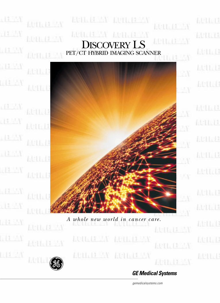
gemedicalsystems.com GE Medical Systems D ISCOVERY LS PET/CT HYBRID IMAGING SCANNER A whole new world in cancer care.
Transcript of DISCOVERY LS - Activexray.com more accurate diagnosis, staging and treatment. Introducing Discovery...
gemedicalsystems.com
GE Medical Systems
DISCOVERY LSPET/CT HYBRID IMAGING SCANNER
A whole new wor ld in cancer care .
Cancer. The escalating numbers speak volumes about the overwhelming challengewe face today.
In the United States, statistics predict oneout of two men and one out of three womenwill develop cancer. Its devastating reach nowextends to four out of five families. And cancerhas advanced to become the second leadingcause of death following heart disease.
At GE Medical Systems, we understand the pressure you face in the fight against cancer. So we’re working in tandem with you to find a better way to diagnose and treatcancer. To help physicians detect it early. To help physicians treat it more effectively. To monitor response to therapy accurately.And to help doctors bring cures faster.
This motivation has led us to create a remarkable new cancer-fighting tool, which will enable physicians to personalizecancer treatment.
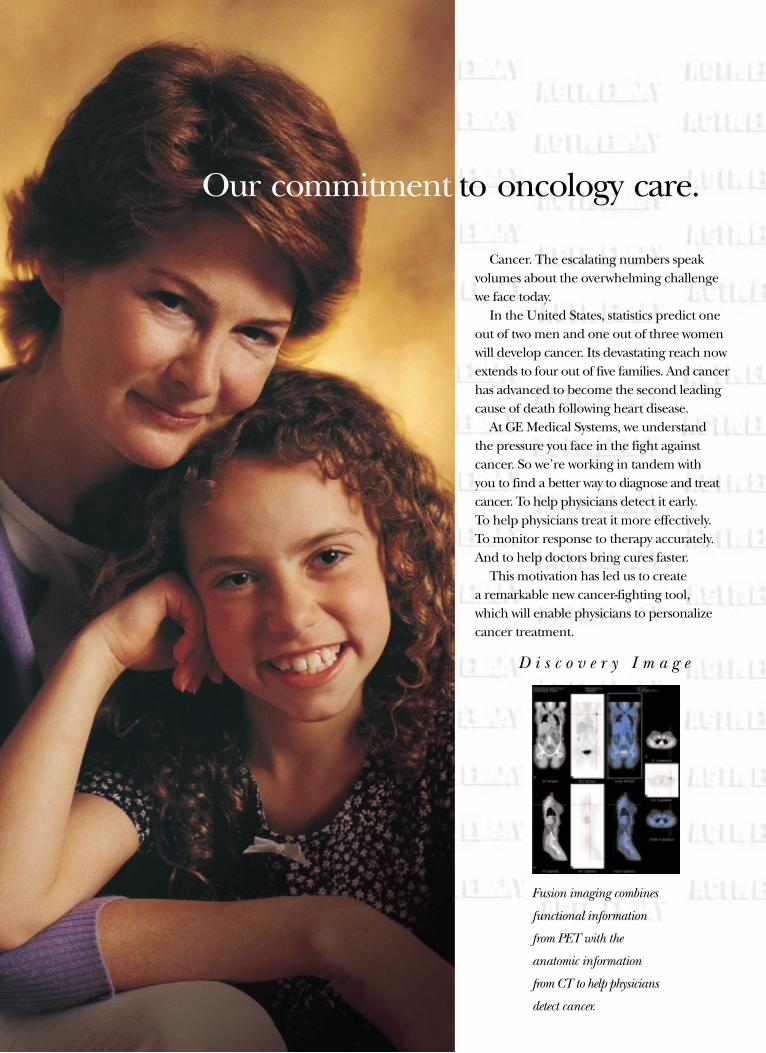
Our commitment to oncology care.
Fusion imaging combines
functional information
from PET with the
anatomic information
from CT to help physicians
detect cancer.
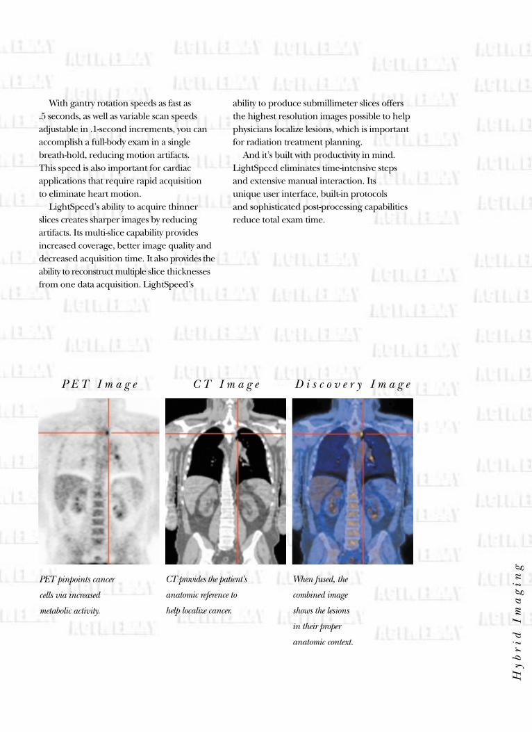
D i s c o v e r y I m a g e
Providing more accurate diagnosis, staging and treatment.
Introducing Discovery LS. Discovery LScombines the functional information ofPositron Emission Tomography (PET) withthe anatomic information of ComputedTomography (CT) in one, clinically proven,revolutionary scanner. By providing metabolicdata in an anatomical context, Discovery LShelps physicians provide more accurate andintuitive diagnosis, staging and treatment ofcancer. And that can result in improvedpatient outcomes, lower medical costs andincreased productivity.
Imagine, in one rapid exam, you’ll get thecritical diagnostic information you need togive your patients the best possible care.
Discovery LS can help you determine if a patient has cancer. Where it is. Whetherthere are multiple tumors or metastases.Whether it’s spreading. What stage it’s in. The optimal therapy. Whether the therapy is working. And if there’s recurrence.
It can streamline healthcare delivery andcosts by avoiding multiple and unnecessarytests and surgeries. And because Discovery LScan help reveal cancer before it can be seenwith other imaging modalities, your patientscan get a head start on treatment.
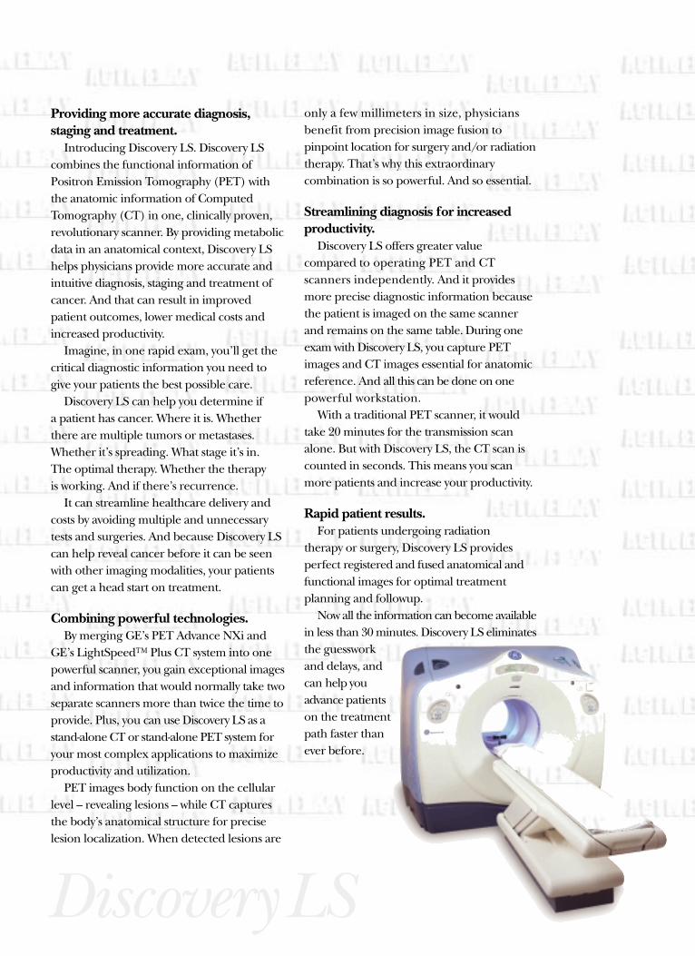
Combining powerful technologies.By merging GE’s PET Advance NXi and
GE’s LightSpeed™ Plus CT system into one powerful scanner, you gain exceptional imagesand information that would normally take twoseparate scanners more than twice the time toprovide. Plus, you can use Discovery LS as astand-alone CT or stand-alone PET system foryour most complex applications to maximizeproductivity and utilization.
PET images body function on the cellularlevel – revealing lesions – while CT capturesthe body’s anatomical structure for preciselesion localization. When detected lesions are
only a few millimeters in size, physiciansbenefit from precision image fusion to pinpoint location for surgery and/or radiationtherapy. That’s why this extraordinary combination is so powerful. And so essential.
Streamlining diagnosis for increased productivity.
Discovery LS offers greater value compared to operating PET and CT scanners independently. And it providesmore precise diagnostic information becausethe patient is imaged on the same scannerand remains on the same table. During oneexam with Discovery LS, you capture PETimages and CT images essential for anatomicreference. And all this can be done on one powerful workstation.
With a traditional PET scanner, it wouldtake 20 minutes for the transmission scanalone. But with Discovery LS, the CT scan iscounted in seconds. This means you scanmore patients and increase your productivity.
Rapid patient results.For patients undergoing radiation
therapy or surgery, Discovery LS provides perfect registered and fused anatomical andfunctional images for optimal treatment planning and followup.
Now all the information can become availablein less than 30 minutes. Discovery LS eliminatesthe guesswork and delays, andcan help youadvance patientson the treatmentpath faster thanever before.
DiscoveryLS
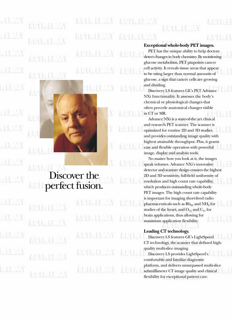
Discover the perfect fusion.
Exceptional whole-body PET images.PET has the unique ability to help doctors
detect changes in body chemistry. By monitoringglucose metabolism, PET pinpoints cancercell activity. It reveals tissue areas that appearto be using larger than normal amounts ofglucose, a sign that cancer cells are growingand dividing.
Discovery LS features GE’s PET AdvanceNXi functionality. It assesses the body’s chemical or physiological changes that often precede anatomical changes visible in CT or MR.
Advance NXi is a state-of-the art clinicaland research PET scanner. The scanner is optimized for routine 2D and 3D studies and provides outstanding image quality withhighest attainable throughput. Plus, it grantseasy and flexible operation with powerfulimage, display and analysis tools.
No matter how you look at it, the imagesspeak volumes. Advance NXi’s innovativedetector and scanner design ensures the highest2D and 3D sensitivity, full-field uniformity ofresolution and high count rate capability,which produces outstanding whole-body PET images. The high count rate capability is important for imaging short-lived radiopharmaceuticals such as Rb82 and NH3 forstudies of the heart, and O15 and C11 for brain applications, thus allowing for maximum application flexibility.
Leading CT technology.Discovery LS features GE’s LightSpeed
CT technology, the scanner that defined high-quality multi-slice imaging.
Discovery LS provides LightSpeed’s comfortable and familiar diagnostic platform, and delivers unsurpassed multi-slicesubmillimeter CT image quality and clinicalflexibility for exceptional patient care.
With gantry rotation speeds as fast as .5 seconds, as well as variable scan speedsadjustable in .1-second increments, you canaccomplish a full-body exam in a singlebreath-hold, reducing motion artifacts. This speed is also important for cardiac applications that require rapid acquisition to eliminate heart motion.
LightSpeed’s ability to acquire thinnerslices creates sharper images by reducing artifacts. Its multi-slice capability providesincreased coverage, better image quality anddecreased acquisition time. It also provides theability to reconstruct multiple slice thicknessesfrom one data acquisition. LightSpeed’s
ability to produce submillimeter slices offersthe highest resolution images possible to helpphysicians localize lesions, which is importantfor radiation treatment planning.
And it’s built with productivity in mind.LightSpeed eliminates time-intensive stepsand extensive manual interaction. Its unique user interface, built-in protocols and sophisticated post-processing capabilitiesreduce total exam time.
PET pinpoints cancer
cells via increased
metabolic activity.
CT provides the patient’s
anatomic reference to
help localize cancer.
When fused, the
combined image
shows the lesions
in their proper
anatomic context.
Hy
br
id I
ma
gin
g
P E T I m a g e C T I m a g e D i s c o v e r y I m a g e
By gaining functional and anatomical dataat the same time, you’ll get critical informationearly in the game – right when you need it.
Early detection can change patient outcomes significantly. Knowing the extentand location of cancer can help to determinethe best course of treatment.
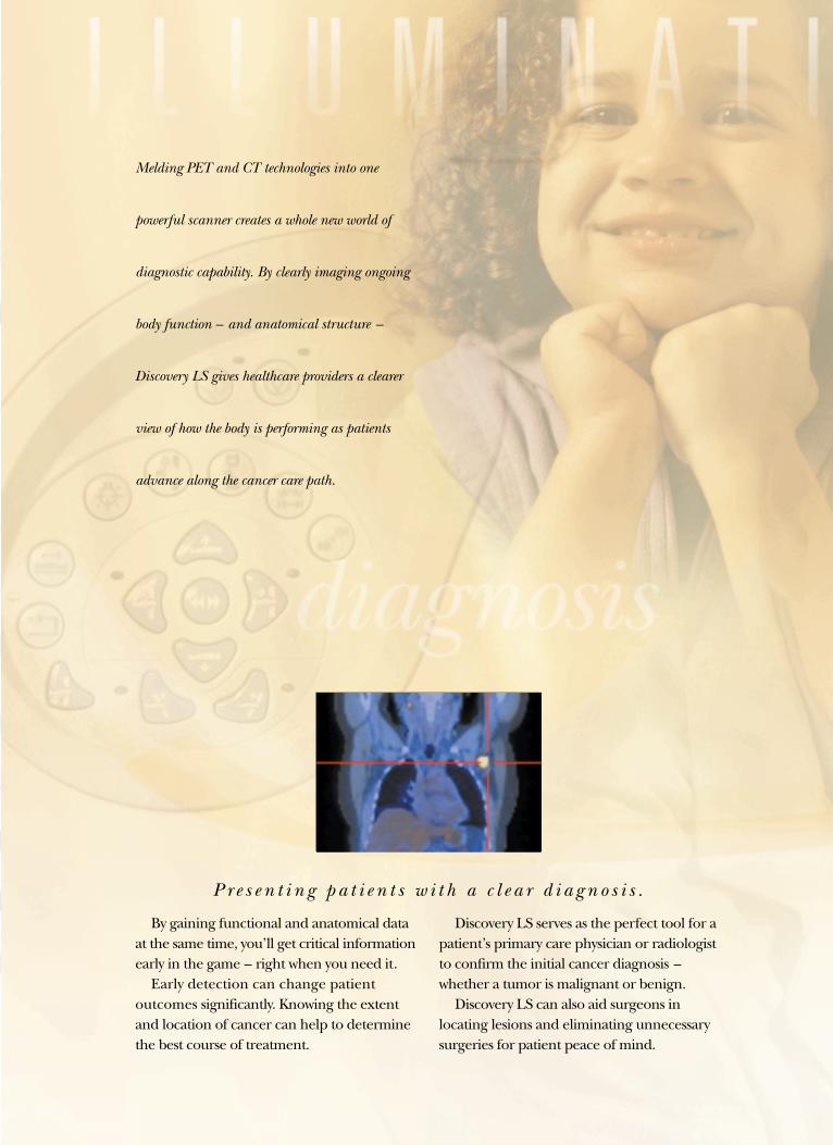
Melding PET and CT technologies into one
powerful scanner creates a whole new world of
diagnostic capability. By clearly imaging ongoing
body function – and anatomical structure –
Discovery LS gives healthcare providers a clearer
view of how the body is performing as patients
advance along the cancer care path.
Discovery LS serves as the perfect tool for apatient’s primary care physician or radiologistto confirm the initial cancer diagnosis –whether a tumor is malignant or benign.
Discovery LS can also aid surgeons in locating lesions and eliminating unnecessarysurgeries for patient peace of mind.
P r e s e n t i n g p a t i e n t s w i t h a c l e a r d i a g n o s i s .
I M A G I N E A W H
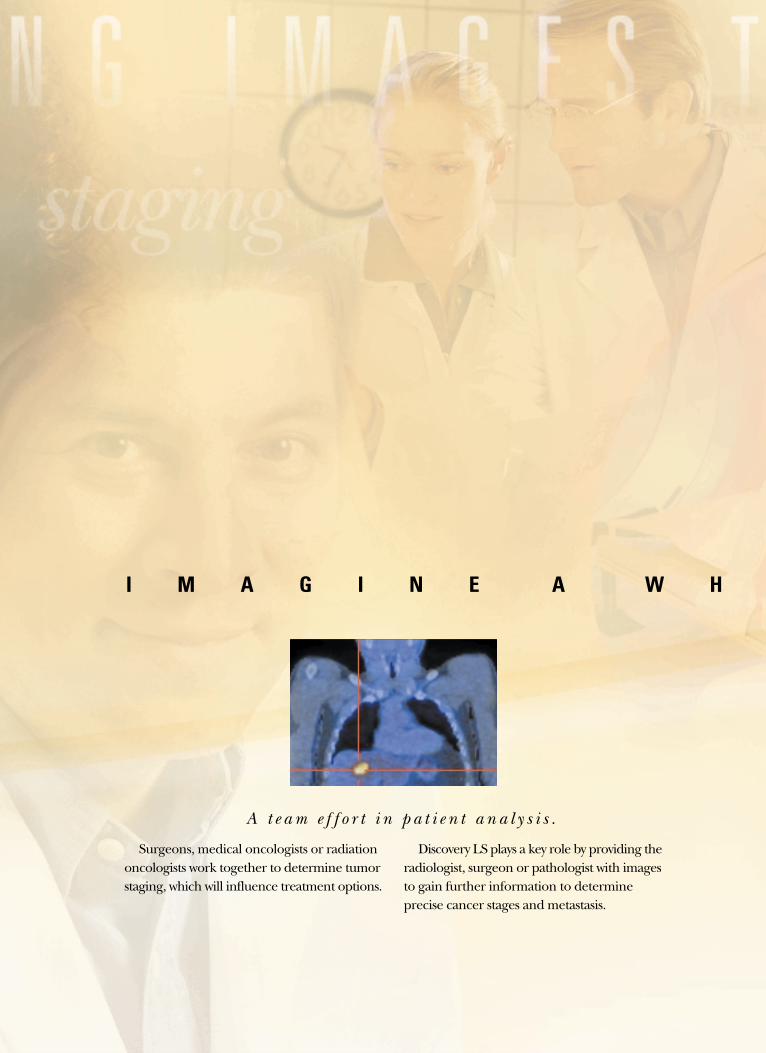
Surgeons, medical oncologists or radiationoncologists work together to determine tumorstaging, which will influence treatment options.
Discovery LS plays a key role by providing theradiologist, surgeon or pathologist with imagesto gain further information to determine precise cancer stages and metastasis.
A t e a m e f f o r t i n p a t i e n t a n a l y s i s .
L E N E W W O R L D
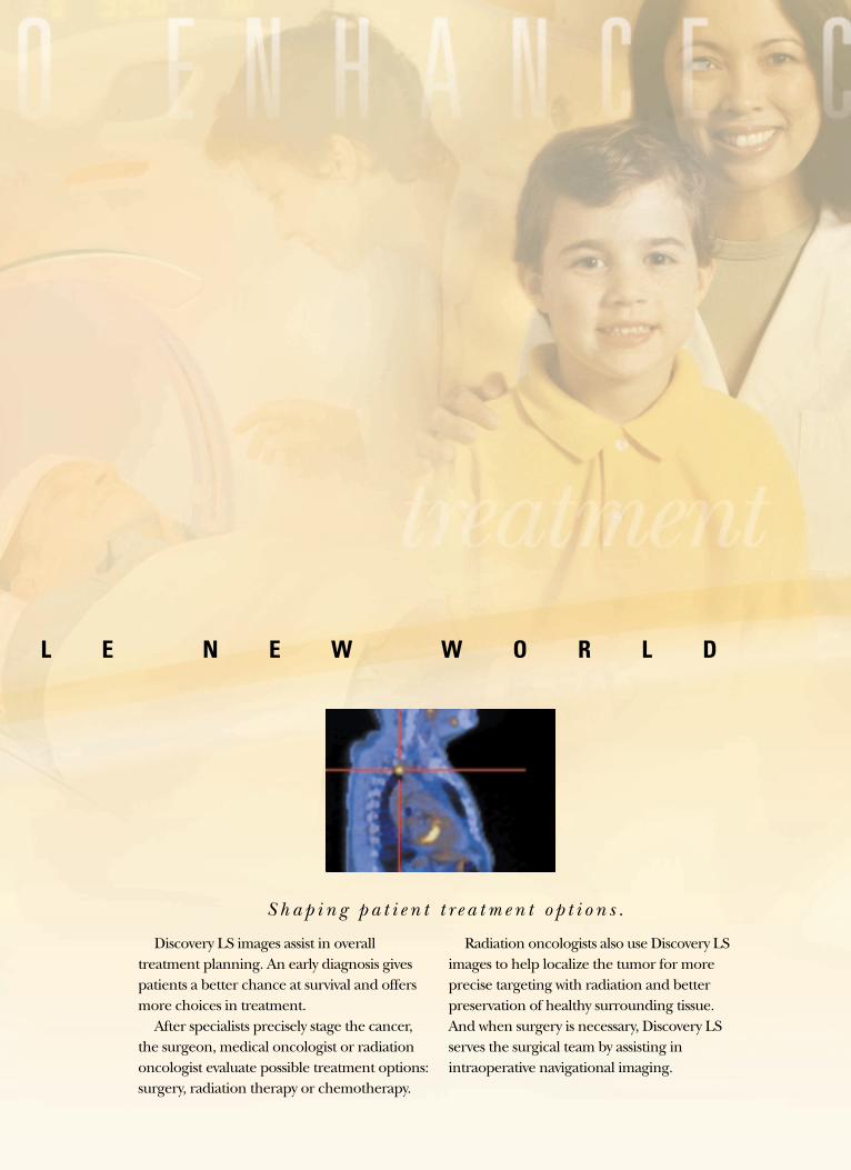
Discovery LS images assist in overall treatment planning. An early diagnosis givespatients a better chance at survival and offersmore choices in treatment.
After specialists precisely stage the cancer,the surgeon, medical oncologist or radiationoncologist evaluate possible treatment options:surgery, radiation therapy or chemotherapy.
Radiation oncologists also use Discovery LSimages to help localize the tumor for moreprecise targeting with radiation and betterpreservation of healthy surrounding tissue.And when surgery is necessary, Discovery LSserves the surgical team by assisting in intraoperative navigational imaging.
S h a p i n g p a t i e n t t r e a t m e n t o p t i o n s .
Discovery LS revolutionizes cancer care every step
of the way by providing detailed information for
diagnosis, treatment options and patient followup.
It delivers vital data to locate the primary tumor
site, grade the degree of malignancy, determine
tumor staging, develop precise radiation treatment
plans and monitor treatment success.
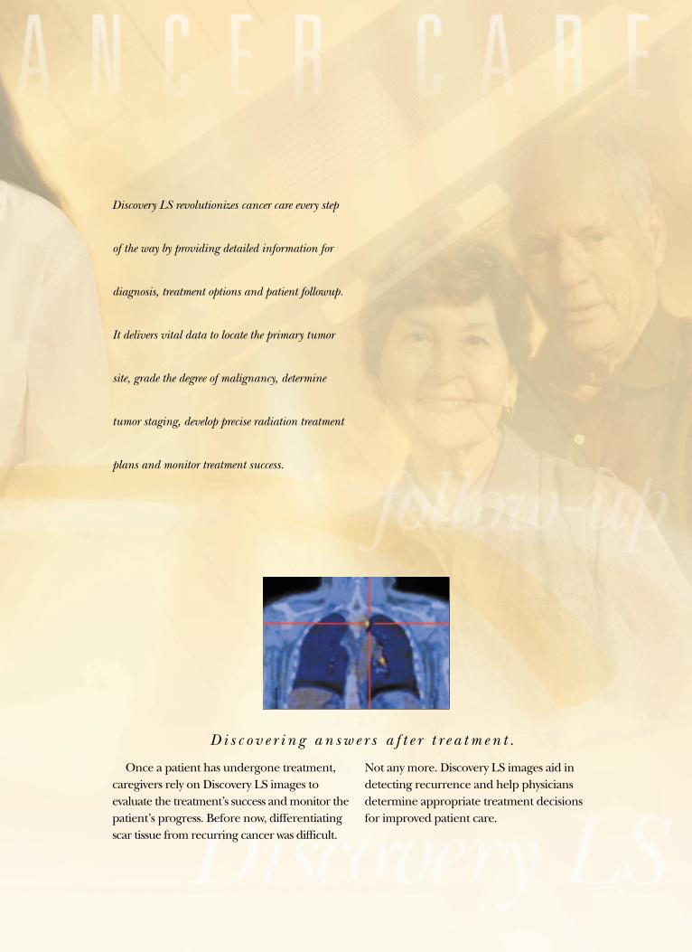
Once a patient has undergone treatment,caregivers rely on Discovery LS images to evaluate the treatment’s success and monitor thepatient’s progress. Before now, differentiatingscar tissue from recurring cancer was difficult.
Not any more. Discovery LS images aid indetecting recurrence and help physiciansdetermine appropriate treatment decisionsfor improved patient care.
D i s c o v e r i n g a n s w e r s a f t e r t r e a t m e n t .
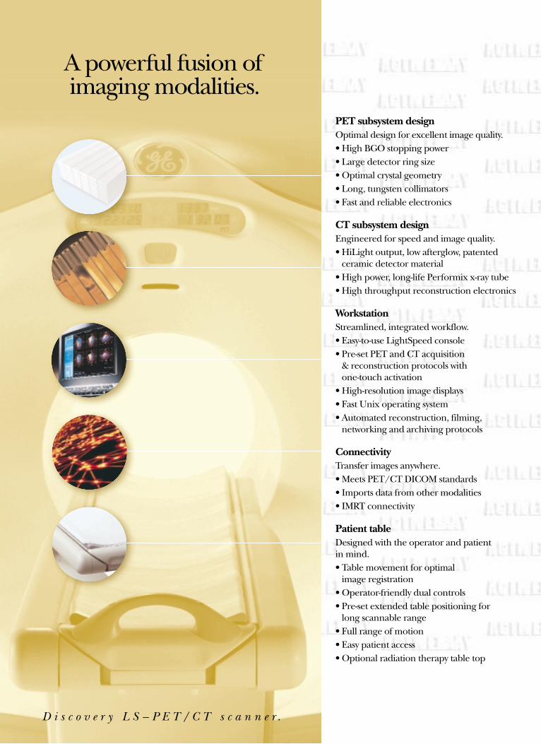
PET subsystem designOptimal design for excellent image quality.• High BGO stopping power • Large detector ring size• Optimal crystal geometry• Long, tungsten collimators • Fast and reliable electronics
CT subsystem designEngineered for speed and image quality.• HiLight output, low afterglow, patented
ceramic detector material • High power, long-life Performix x-ray tube• High throughput reconstruction electronics
WorkstationStreamlined, integrated workflow. • Easy-to-use LightSpeed console• Pre-set PET and CT acquisition
& reconstruction protocols with one-touch activation
• High-resolution image displays• Fast Unix operating system• Automated reconstruction, filming,
networking and archiving protocols
ConnectivityTransfer images anywhere.• Meets PET/CT DICOM standards• Imports data from other modalities• IMRT connectivity
Patient tableDesigned with the operator and patient in mind.• Table movement for optimal
image registration• Operator-friendly dual controls• Pre-set extended table positioning for
long scannable range• Full range of motion• Easy patient access• Optional radiation therapy table top
A powerful fusion ofimaging modalities.
D i s c o v e r y L S – P E T / C T s c a n n e r .
Through Six Sigma, a corporate-wide commit-ment to quality, GE designs customer-defined products, processes and services.
When you purchase a GE Discovery LS scanner, it comes with all the extras you’ve come to expectfrom the global leader in diagnostic imaging.
• Market analysis
• Customized financing solutions
• Site planning
• InSiteTM, diagnostic network
• World-class education and training
• Enterprise-wide networking solutions
• Productivity-enhancing process improvements
• Help in understanding complex reimbursement issues
• PET/Advantages – a comprehensive local marketing program
To make your purchasing decision even easier, Discovery LS offers premium technology, reliable system performance and customized financing solutions.
You can feel secure in knowing the GE Continuumof product development will keep you on the fore-front of emerging technologies and future applications,so you can help lead the way in the fight against cancer and other diseases, today and tomorrow.
GE is also forging new partnerships. GE MedicalSystems and Varian Medical Systems have cometogether in a unique alliance to bring you See &Treat™ Cancer Care – a visionary new approach tocancer care. This alliance of expertise combinesGE’s anatomical and functional imaging withVarian’s high-resolution Intensity ModulatedRadiation Therapy (IMRT) technology to help physicians localize and treat cancers more precisely.
The GE continuum keeps innovation flowing.
GE Medical SystemsWe bring good things to life.
Internet – gemedicalsystems.comGE Medical Systems – Americas: Fax 262-544-3384P.O. Box 414, Milwaukee, Wisconsin 53201 U.S.A.
GE Medical Systems – Europe: Fax 33-1-30-70-94-35Paris, France
GE Medical Systems – Asia:Tokyo, Japan – Fax: +81-425-85-5490Hong Kong – Fax: +852-2559-3588
General Electric Company reserves the right to make changes in specifications and featuresshown herein, or discontinue the product described at any time without notice or obligation.Contact your GE Representative for the most current information.
©2001 General Electric Company
01-7001 6/01 Printed in USA
For more than 50 years, healthcare providers worldwide
have relied on GE Medical Systems for medical technology,
services and productivity solutions.
So no matter what challenges your healthcare
system faces– you can always count on GE
to help you deliver the highest quality healthcare.
For details, please contact your GE representative today.


![¾L¹w ,IÀ - iranpotk.com 8 (mm) Taper punch with knurled shank nHk]A ¾²ILºj IM ½k¹¹¨ ZnIi ¾L¹w Code No. L(mm) (gr) LS 1030 LS 1230 LS 1430 LS 1630 LS 1830 LS 2030 LS 2230](https://static.fdocuments.in/doc/165x107/5b190a547f8b9a46258c4235/lw-ia-8-mm-taper-punch-with-knurled-shank-nhka-iloj-im-k.jpg)