DISCLOSURE OF RELEVANT RELATIONSHIPS WITH INDUSTRY … Leishmania.pdf · DISCLOSURE OF RELEVANT...
78
DISCLOSURE OF RELEVANT RELATIONSHIPS WITH INDUSTRY Ibrahim Khalifeh, MD I do not have any relevant relationships with industry. Cutaneous Leishmania
Transcript of DISCLOSURE OF RELEVANT RELATIONSHIPS WITH INDUSTRY … Leishmania.pdf · DISCLOSURE OF RELEVANT...

DISCLOSURE OF RELEVANT
RELATIONSHIPS WITH INDUSTRY
Ibrahim Khalifeh, MD
I do not have any relevant relationships with
industry.
Cutaneous Leishmania

Cutaneous Leishmania: Is it a Legend?
Ibrahim Khalifeh, M.D.
Assistant Professor
Department of Pathology
American University of Beirut Medical Center
XXIV International Academy of Pathology-Arab Division

History
• Lieutenant-General Sir William Boog Leishman (6 November 1865 – 2 June 1926) was a Scottish pathologist and British Army medical officer.
• He served in India, where he studied enteric fever and kala azar.
• In 1901, while examining pathologic specimens of a spleen from a patient who had died of kala azar he observed oval bodies and published his account of them in 1903.
• Leishman's name was engraved into the history of parasitology by Sir Ronald Ross, who was impressed by Leishman's work and classified the etiologic agent of kala azar into separate genus Leishmania.

Introduction
• Frequent disease: New and Old World
• Global Prevalence: 10 million cases
• Annual incidence: 1.5-2 million
• Annual mortality: 70,000
• Disease burden disability-adjusted life years (DALY): 2.4 million
• Endemic tropical disease
• World health organization (WHO) priority disease

Parasitology

Parasitology
• Leishmaniasis is caused by the vector-borne transmission of the kinetoplastid “leishmania”
• In the vector: Promastigote form.

Parasitology
• In the mammal host: “amastigote form”
- Exclusively in the macrophages
- Small, round immobile

Species implicated in human infection

Cutaneous Leishmania

Parasitology
• CL is transmitted by the bite of female sandflies:
- Phlebotomus in the old world
- Lutzomyia in the New World
• Leishmaniasis has both zoonotic and anthroponotic forms

Anthroponotic Cycle

Zoonotic Cycle

Parasitology
• The life cycle of the parasite depends on:
- resist the digestive enzymes of the sandfly
- adhere to the epithelium of the digestive tube
• Vector independent transmission is very rare: congenitally, transfusion, transplant, lab accidents, etc.

Parasitology
• New world (L. panamensis, guyanesis, amazonensis & mexicana): reservoir is wild animals.
• L. major and infantum: close to human habitats (demostic animals).
• L. tropica and donovani: Human
• Human infection is considered “epidemiologically dead end”
Domestic dog: reservoir for L . infantum


Transepidermal elimination in cutaneous Leishmaniasis: a multiregional study
• Skin biopsies (n=212) with Cutaneous Leishmaniasis
• L. tropica (88.2%) and L. major (11.8%)
• Transepidermal elimination: 28.3% of cases with a significant prevalence of L.major in this group (35% versus 2%, p<0.001).

Transepidermal elimination in cutaneous Leishmaniasis: a multiregional study
• Multivariate analysis: 1. L. major [OR (95% confidence interval) = 80 (9-712); p<0.001]
2. Parasitic index [OR= 3.4 (2.1-5.3); p<0.001],
3. Interface [OR= 6.24 (2.2-17.8); p<0.001]
4. Absence of necrosis [OR= 0.2 (0.1-0.8); p=0.026].


CD1a
P63
CK5/6

Clinical Presentation

Clinical Presentation
1. Parasitic factors:
- L. donovani & amazonensis: visceral form
- L. braziliensis: mucosal form.
2. Host factors:
- Disease extension
- Disease evolution

Clinical Presentation
1. Localized Cutaneous Leishmaniasis
2. Multilesional forms:
Post kala-azar dermal leishmaniasis
Diffuse cutaneous leishmaniasis
Disseminated cutaneous leishmaniasis
3. Rare cutaneous forms (still evolving)

1. Localized Cutaneous Leishmaniasis

• Well circumscribed Papule (1w)
• Nodule/plaque (3m)
• Ulcerated/verrucous (5m)
scarring (8m)

Clinical Classification • Ulcero-crusted type
• Abortive type
• Echtymatous type
• Sporotrichoid type
• Impetigoid type
• Verrucous type
• Tumor-like type
• Hypodermic type
• Eczematous type
• Rhinophyma-like type
• Keloid-like type
• Chalazion-like type
• Hemorragic type
• Pigmented type
• Chanciform type
• Vegetant type
• Recidivan type
• Lupoid type
• Psoriasifrom type
• Erysipeloid type


1: Mycobacterium marinum infection
2: Cutaneous leishmaniasis 3: Sporotrichosis

2. Multilesional form Post kala-azar dermal leishmaniasis
Diffuse cutaneous leishmaniasis
Disseminated cutaneous leishmaniasis

Post kala-azar dermal leishmaniasis Abundant hypochromatic papules and nodules on the trunk. Curtsey of Dr. Ali Khamesipour
Post kala-azar
• Occurs after Donovani visceral L. In India subcontinent
• Frequency: 5-60%
• DD: Lepromatous Leprosy
• LST: negative 33-100%
• Skin biopsy: negative in 20%
• PCR: positive 83-94%
• Papules: Contains parasite (reservoir)

Diffuse cutaneous leishmaniasis
• Follows localized form
• L. aethiopica & amazonensis
• Clinically resembles Lepromatous leprosy
• LST: negative
• Parasite count: High
• T-cell proliferation in vitro following Leishmania antigen: very weak
• Resistant to treatment

Disseminated cutaneous leishmaniasis
• Multilesional: >10 lesions in > 2 sites
• Brazil and Africa
• HIV patients
• L. amazonensis & major

Tunisia ZLC (2- 8 m)
Nodule/Sept Ulcer/Oct Scar/jan Papule/June
Curtsey of Dr. Mourad MOKNI

Types of CL (Iran) Anthroponotic CL (ACL)
– Urban type
– Dry lesions
– Chronic
– Longer incubation period (2-8
months)
– Longer duration of self healing
(up to 2 years)
– A few lesions usually
– Caused by L. tropica
Zoonotic CL (ZCL)
– Rural type
– Wet lesions
– Acute
– Shorter incubation period (2 weeks
to 2 months)
– Shorter duration of self healing
(up to 1 year)
– Could be numerous lesions
– Caused by L. major, L. aethiopica
& rarely by L. infantum
Curtsey of Dr. Ali Khamesipour

Chronic cutaneous leishmaniasis, a great mimicker with various clinical presentations: 12 years experience from Aleppo
J Eur Acad Dermatol Venereol. 2012 Oct;26(10):1224-9
Papulonodular form (a-d)
Plaque Form(e-i)
Gyrate From (j)

Tumoral form(e-a)
Verrucous form (b-c)
Ulcerative form (d–f )
Erysipeloid form (g-h)

Histopathology

II. Pan-necrosis
I. Normal skin/Collagen deg.
III. Mixed inflammatory inf.
V. Granulomas
IV. Scattered giant cells
Ridely’s classification
Trans R Soc Trop Med Hyg. 1980;74(4):508-14.


45.6% not classical 38/57:
67% PI≤1

57 cases
Atypical histology
6 cases (10.5 %)
Histologically mimicking SS
5 cases (83.3%)
Clinically suspected CL
4 cases (7.0 %)
Histologically mimicking TB
3 cases (75.0%)
Clinically suspected CL
5 cases (8.7 %)
Histologically mimicking PN
3 cases (60.0%)
Clinically suspected CL
3 cases (5.2 %)
Histologically mimicking MF
2 cases Clinically suspected AM
1 case Clinically suspected LY
8 cases
Histologically mimicking:
SA(2), PLEVA(2), IL(1), SASD(1),
LP(1), ALTCL(1)
20 cases (35.0 %)
Histologically mimicking SCC
6 cases (30.0%)
Clinically suspected SCC
12 cases (60.0%)
Clinically suspected CL
11 cases (19.2 %)
Histologically mimicking DFI
7 cases (63.6%)
Clinically suspected CL
ALTCL: anaplastic large T cell lymphoma; DFI: deep fungal infection; IL: indeterminate leprosy; LP: lichen planus; MF: mycosis fungoides; PLEVA: pityriasis lichenoides; PN: panniculitis; SCC: squamous cell carcinoma; SS: secondary syphilis; TB: tuberculosis.

Sarcoidosis SCC DFI

Panniculitis Lichen Planus

TB Secondary Syphilis

MF ACTL

Geography

Geographic distribution of the primary cutaneous leishmaniasis species Old World
Magill, A.J.; Dermatol Clin. 1995 Jul;13(3):505-23

Curtsey of Dr. Ali Khamesipour

Curtsey of Dr. Ali Khamesipour


Outbreak ZLC L. major
Sidi Saad Dam in KAIROUAN
District in 1982
Curtsey of Dr. Mourad MOKNI

Acta Trop. 2012 Dec;124(3):221-8. doi: 10.1016/j.actatropica.2012.08.012.
Geographical distribution updating of Tunisian leishmaniasis foci: About the isoenzymatic analysis of 694 strains

Diagnosis

Steps in parasitological confirmation in cutaneous leishmaniasis

Case Western Reserve University Pathology 2011 Fulbright S&T Fellow

PI Explanation
1+ 1 or more amastigotes per standard section
2+ 10 or more amastigotes per standard section
3+ 100 or more amastigotes per standard section
4+ 1 000 or more amastigotes per standard section
5+ 10 000 or more amastigotes per standard section
6+ 100 000 or more amastigotes per standard section
Table 1: Modified Ridley’s Parasitic Index
Modified Ridley’s Parasitic Index
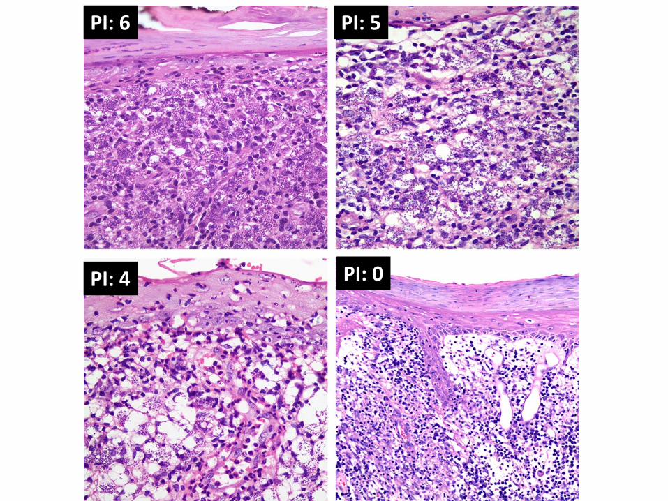
PI: 6
PI: 0 PI: 4
PI: 5


100bpL
300 bp
100bpL
• ITS1 Polymerase Chain Reaction (ITS1-PCR) Agarose gel electrophoresis of the amplicons of ITS1-PCR for representative samples. The gel reveals 4 specimen that are positive and show a DNA band between 300 and 350 basepairs. The gel also identifies 4 specimens that failed to show the leishmania specific band.
• All ITS1-PCR negative samples were subjected to nested ITS1-PCR (100 bpL represents a 100 basepair ladder used as a molecular marker).

20 bpL
200 bpL
60 bpL
200bp
60bp
20bpL
• Restriction Fragment Length Polymorphism (RFLP). RFLP pattern typical for Leishmania tropica showing two restriction fragments of 200 and 60 bp.
• RFLP patterns obtained after digestion of representative ITS1-PCR and nested ITS1-PCR amplicons with the restriction enzyme HaeIII. All 125 specimen show an RFLP pattern typical for Leishmania tropica showing two restriction fragments of 200 bp and 60 bp, respectively (20 bpL represents a 20 basepair ladder used as a molecular marker).

Treatment

Major Issues
1. Dermatologic impact
2. Oozing
3. Atrophic scaring/disfiguring
4. Concomitant visceral involvement (10% of New World L.)
5. Disease transition (L. Tropica)
6. L. Major: spontaneous resolution in 50-75% of cases in 6 months

CL Treatment
• CL is often self-limiting, but treatment is given to accelerate healing, reduce scarring, and prevent the risk of progression and complications
1. Physical treatment including surgery
2. Topical therapy
3. Systemic therapy

Pentavalent antimonate Glucantime
Meglumine antimonate
85 mg/mL (8.5%)
Pentostam
Sodium Stibogluconate
100 mg/mL
Both are on the WHO essential drug list
Glucantime is more readily available and less expensive than Pentostam.

Cutaneous Leishmaniasis
• Local and physical can be used for limited CL without the risk of dissemination.
• Systemic treatment is required in cases of Leishmania species with the potential to disseminate, immunocompromised host, mucocutaneous or diffuse disease, and severe cutaneous disease.
• Parenteral antinomials are usually first-line agents but are associated with considerable toxicity.

Lesion(s) Requires Treatment
1. Ulcer(s) which are located close to a vital organ or is cosmetically important, such as on the face.
2. Lesion(s) with no evidence of healing for several months after the onset.
3. Special forms of CL, such as sporotrichoid or lymphangietic with satellite lesions, which indicates spreading.
4. CL due to L. tropica, which could subsequent systemic involvement or be the reservoir of infection.
5. Requested by the patient

Lesion(s) caused by L. major
Single or a few lesion
Not close to a vital organ
Not in the face
Lesion size small
Requested by the patient
Lesion(s) Recommended not to Treat

Treatment Strategies
Single or a few lesions
Intralesional Glucantime
Freeze might be added
Systemic therapy
Multiple lesions
Lesion size larger than 3 cm in diameter
Lesion close to a vital organ
Lesion(s) on face

Collaboration

2. 2
1
OR
2
1. Pathology report
2. H&E and molecular sample
OR
2. Paraffin Block
Ibrahim Khalifeh AUBMC

Collaboration

Kashan, Iran

Kashan, Iran

Spring at American University of Beirut, Lebanon

Acknowledgment
AUBMC Team
Ghazi Zaatari, M.D.
Robert Habib, PhD
Lamis Yehia, M.S.
Jad Saab, M.D.
Faysal Fedda, M.D.
Ruba Khattab, M.D.
Sarah Karram, M.D.
Collaborators
Asif Loya, M.D. (Pakistan)
Mourad Mokni, M.D. (Tunisia)
Suad Taraif, M.D. (KSA)
Wasim Raslan, M.D. (KSA)
Mohammad Satti, M.D. (KSA)
Mohammad Houreih, M.D. (Syria)
Ali Khamesipour, M.D. (Iran)
Ali Firooz, M.D. (Iran)
Hadi Hammam, M.D. (Lebanon)