Disassociated relation between plasma tumor necrosis ...
9
Disassociated relation between plasma tumor necrosis factor-alpha, interleukin-6 and increased body weight in Amerindian women: A long-term prospective study of natural body weight variation and impaired glucose tolerance Lindgärde, Folke; Gottsäter, Anders; Ahrén, Bo Published in: Diabetology and Metabolic Syndrome DOI: 10.1186/1758-5996-2-38 2010 Link to publication Citation for published version (APA): Lindgärde, F., Gottsäter, A., & Ahrén, B. (2010). Disassociated relation between plasma tumor necrosis factor- alpha, interleukin-6 and increased body weight in Amerindian women: A long-term prospective study of natural body weight variation and impaired glucose tolerance. Diabetology and Metabolic Syndrome, 2. https://doi.org/10.1186/1758-5996-2-38 Total number of authors: 3 General rights Unless other specific re-use rights are stated the following general rights apply: Copyright and moral rights for the publications made accessible in the public portal are retained by the authors and/or other copyright owners and it is a condition of accessing publications that users recognise and abide by the legal requirements associated with these rights. • Users may download and print one copy of any publication from the public portal for the purpose of private study or research. • You may not further distribute the material or use it for any profit-making activity or commercial gain • You may freely distribute the URL identifying the publication in the public portal Read more about Creative commons licenses: https://creativecommons.org/licenses/ Take down policy If you believe that this document breaches copyright please contact us providing details, and we will remove access to the work immediately and investigate your claim. Download date: 27. Feb. 2022
Transcript of Disassociated relation between plasma tumor necrosis ...
LUND UNIVERSITY
PO Box 117221 00 Lund+46 46-222 00 00
Disassociated relation between plasma tumor necrosis factor-alpha, interleukin-6 andincreased body weight in Amerindian women: A long-term prospective study of naturalbody weight variation and impaired glucose tolerance
Lindgärde, Folke; Gottsäter, Anders; Ahrén, Bo
Published in:Diabetology and Metabolic Syndrome
DOI:10.1186/1758-5996-2-38
2010
Link to publication
Citation for published version (APA):Lindgärde, F., Gottsäter, A., & Ahrén, B. (2010). Disassociated relation between plasma tumor necrosis factor-alpha, interleukin-6 and increased body weight in Amerindian women: A long-term prospective study of naturalbody weight variation and impaired glucose tolerance. Diabetology and Metabolic Syndrome, 2.https://doi.org/10.1186/1758-5996-2-38
Total number of authors:3
General rightsUnless other specific re-use rights are stated the following general rights apply:Copyright and moral rights for the publications made accessible in the public portal are retained by the authorsand/or other copyright owners and it is a condition of accessing publications that users recognise and abide by thelegal requirements associated with these rights. • Users may download and print one copy of any publication from the public portal for the purpose of private studyor research. • You may not further distribute the material or use it for any profit-making activity or commercial gain • You may freely distribute the URL identifying the publication in the public portal
Read more about Creative commons licenses: https://creativecommons.org/licenses/Take down policyIf you believe that this document breaches copyright please contact us providing details, and we will removeaccess to the work immediately and investigate your claim.
Download date: 27. Feb. 2022
Lindgärde et al. Diabetology & Metabolic Syndrome 2010, 2:38http://www.dmsjournal.com/content/2/1/38
Open AccessR E S E A R C H
ResearchDisassociated relation between plasma tumor necrosis factor-α, interleukin-6 and increased body weight in Amerindian women: A long-term prospective study of natural body weight variation and impaired glucose toleranceFolke Lindgärde1, Anders Gottsäter*1 and Bo Ahrén2
AbstractBackground: Inflammatory cytokines are linked to obesity-related insulin resistance and may predict type 2 diabetes independently of obesity. We previously reported that a majority of a cohort of 73 non-diabetic women with normal plasma (p-)glucose with Amerindian heritage in Lima, Peru, during a 5-year period increased both body weight and p-glucose levels, yet p-insulin was unaltered. A high proportion of palmitoleic acid (16:1n-7) in serum (s) and systolic blood pressure (SBP) were independent predictors of high p-glucose. Whether cytokines also contributed is, however, not known.
Methods: During 5 years we prospectively investigated the relation between changed concentrations of p-tumor necrosis factor (TNF)-α, p-interleukin (IL)-6 and circulating insulin and glucose in relation to the natural variation of body weight. Study variables included anthropometric measurements, p-insulin, TNF-α, IL-6, SBP and the proportion of 16:1n-7 in s-fatty acid composition.
Results: Weight and waist differences correlated negatively to the difference in p-TNF-α but positively to differences in p-IL-6 and p-insulin, whereas the increase of p-glucose from baseline to follow-up did not correlate with changes in levels of the two cytokines. In multiple regression analysis changes of TNF-α and insulin contributed independently to the variance in weight. P-insulin at baseline and weight change were determinants of fasting p-insulin at follow-up. Multiple regression analysis revealed that weight change (t-value = - 2.42; P = 0.018) and waist change (t-value = 2.41; P = 0.019) together with S-16:1n-7 (p < 0.0001) and SBP (p = 0.0005) at baseline were significant predictors of p-glucose at follow-up.
Conclusion: Our prospective study of Amerindian women revealed disassociations between changes in p-TNF-α and p-IL-6 in relation to variation in body weight. A high proportion of s-16:1n-7, SBP at baseline together with weight and waist changes were independent predictors of p-glucose at follow-up. The exact role of the opposite effects and clinical impact of p-TNF-α and p-IL-6 on loss and gain of body weight and indirectly on the development of glucose intolerance is not known.
BackgroundSedentary socioeconomically marginalized women withan Amerindian heritage living in a northern suburb of
Lima, the capital of Peru, were in a previous study charac-terized by normal fasting plasma (p-)glucose levels, buthigh insulin levels [1,2]. A follow-up after five yearsrevealed that the majority of the women along withincreased body weight and fat mass had developed higherfasting p-glucose values possibly concomitant with insuf-ficient insulin secretion. The proportion of palmitoleic
* Correspondence: [email protected] Vascular Center, Lund University, Skåne University Hospital, S-205 02 Malmö, SwedenFull list of author information is available at the end of the article
© 2010 Lindgärde et al; licensee BioMed Central Ltd. This is an Open Access article distributed under the terms of the Creative CommonsAttribution License (http://creativecommons.org/licenses/by/2.0), which permits unrestricted use, distribution, and reproduction inany medium, provided the original work is properly cited.
Lindgärde et al. Diabetology & Metabolic Syndrome 2010, 2:38http://www.dmsjournal.com/content/2/1/38
Page 2 of 8
acid (16:1n-7) in serum (s) and systolic blood pressurewere strong independent determinants of p-glucose con-centration 5 years later [3].
In this study, we examined whether cytokines may con-tribute to this metabolic state as there is a growing bodyof evidence indicating that obesity may be associated tochronic activation of the innate immune system [4],resulting in progressive impairment of glucose toleranceand type 2 diabetes [5]. However, elevated levels ofinflammatory markers may also predict type 2 diabetesindependently of obesity [6], and future weight-gain [7].These results support the view that elevated levels ofinflammatory markers occur early in the process, leadingto glucose intolerance with or without concomitantweight gain. A possibility is therefore that cytokines, suchas interleukin (IL)-6 and tumor necrosis factor (TNF)-α,contribute to the development of insulin resistance andimpaired islet function. This is, however, controversialbecause in a cross-sectional study, neither p-TNF-α norp-IL-6 levels were independently associated with hepaticor peripheral insulin action [8]. Nevertheless, in peoplewith type 2 diabetes, the circulating p- IL-6 concentrationis correlated with adipose tissue mass, rather than withwhole-body insulin sensitivity [9], suggesting that IL-6may be a marker of obesity without any direct contribu-tion to the development of insulin resistance.
Cytokines are, in many ways, involved in the regula-tions of metabolism and food intake. TNF-α influencesenergy homeostasis, has an anorexigenic effect on thehypothalamus [10], and a role in the development of neo-plastic anorexia [11]. High concentrations of TNF-α andIL-6 are associated with lower muscle strength andendurance [12]. Furthermore, higher levels of TNF-α areassociated with greater 5-year decline in thigh musclearea and grip strength [13]. It is obvious that very smallalterations in metabolism or food intake can cause amoderate weight decrease.
The present study sought to explore whether a changeof circulating TNF-α or IL-6 during follow-up of 5 yearsin our material of Amerindian women [1-3] was associ-ated with gain or loss of body weight, and whether it pre-dicted fasting insulin and glucose concentrations.
MethodsStudy participantsThe original study [1] consisted of 182 Peruvian Amerin-dian women living in a northern urban area of Lima. Amigration antecedent from indigenous Andean commu-nities in Peru was identified in all participants. The agerange was 20-59 with a mean of 41 years. Fatty acid com-position in serum was measured in 141 subjects. All ofthese subjects had normal fasting p-glucose concentra-tions. This subgroup was invited to participate in a new
survey [3] five years later. Seventy-nine subjects attendedthe second investigation. P-glucose concentrations wereavailable from 73 subjects in both investigations. These73 women did not differ significantly from the remaining68 in baseline characteristics of p-concentrations of insu-lin and glucose, s-palmitoleic acid (16:1n-7) or bodyweight. None of the participants was taking cholesterollowering medication, was consuming a special diet, hadongoing cardiovascular disease, or was pregnant.
Approval for the study had been given in 1998 and 1999by the Ethics Committees of Lund University and SanMartin University Hospital in Lima. Furthermore, the fol-low-up examination was separately approved by the Eth-ics Committe of San Martin University Hospital. Thestudy was undertaken in accordance with the HelsinkiDeclaration of 1975, as revised in 1983. Informed consentwas obtained from all subjects.
Study methodsThe examinations took place in the morning after anovernight fast at Alternativa, a center for social researchand popular education in the district of San Martin,Lima.
Anthropometric measurementsTrained nurses measured weight, height and waist cir-cumferences. Changes in the respective values were cal-culated as measurement at follow-up subtracted from thebaseline value. Body mass index (BMI; in kg/m2) was cal-culated according to the standard formula. Body fat massand the percentage fat mass in relation to body mass weredetermined by measuring the resistance of the body to alow-level electrical current (Biodynamic Model 310e;Biodynamic Research Inc, Seattle, WA). Measurementswere performed with subjects lying on a couch for 5 min,and the electrodes were placed on the dorsal surfaces ofthe right hand and foot.
Measurement of p-insulin, glucose, TNF- α and IL-6Serum or plasma was separated from venous blood andstored within 1 hour at -20°C and then brought to Swe-den for analysis. P-insulin was measured with double-antibody radioimmunoassay techniques with the use ofguinea pig anti-human insulin antibodies and humaninsulin as the standard (Linco Research, St Charles, MO,USA). P-glucose was measured by using the glucose oxi-dase procedure. For the indirect determination of insulinsensitivity, the homeostasis model assessment of insulinresistance (HOMA-IR) was calculated as follows [14]:HOMA-IR = [fasting insulin (in pmol/L) x fasting glucose(in mmol/L)/22,5]. P-TNF- α and p-IL-6 were measuredby ELISA using commercially available test kits(Pharmingen, San Diego, CA, USA). Detection limitswere 0.12 pg/ml and 0.70 pg/ml [15], intraassay coeffi-
Lindgärde et al. Diabetology & Metabolic Syndrome 2010, 2:38http://www.dmsjournal.com/content/2/1/38
Page 3 of 8
cients of variation (CV) were 8.8% and 4.4%, and interas-say CV were 16.7% and 6.4%, respectively.
Lipid extraction and serum fatty acid measurementsThe fatty acid composition of s-cholesteryl esters wasmeasured as previously described [16]. Serum wasextracted with a hexane-isopropanol solution, and cho-lesteryl esters (only lipid esters that were measured atbaseline) were separated from the extract by thin-layerchromatography before interesterification with acidicmethanol was performed. Free cholesterol that had beenliberated in the reaction was removed by aluminum oxideto avoid contamination of the column. The compositionof methylated fatty acids was determined by gas chroma-tography (25-m NB-351 silica capillary column) with aflame ionization detector and helium as carrier gas. Every25th sample was a serum control pool. The CV betweensuccessive gas chromatography runs was 0.2-5%. The rel-ative amount of fatty acid was expressed as a percentageof the total amount of fatty acids reported.
Statistical analysesData are expressed as mean ± SD. The correlation coeffi-cients between 2 variables were determined by Spearmanrank analysis. Differences between measurements atbaseline and at follow-up levels were analysed by use ofWilcoxon signed rank tests (Table 1). Because p-concen-trations of glucose, TNF-α, IL-6, and insulin values fol-lowed a log-normal distribution, logarithmictransformation was used for these variables. Multipleregression analyses were undertaken to examine whichvariables predicted alterations of weight and waist duringthe observation period and p-glucose and insulin valuesat follow-up. The selected variables were forced into themodel based on analysis (Table 2) and prior findings suchas 16:1-n7 and systolic blood pressure [3]. All statisticalanalyses were conducted by using the statistical packageSTATVIEW (version 5.0.1, for Macintosh; SAS InstituteInc, Cary, NC).
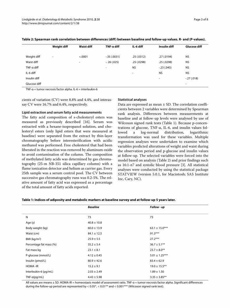
Table 1: Indices of adiposity and metabolic markers at baseline survey and at follow-up 5 years later.
Baseline Follow - up
N 73 73
Age (y) 40.8 ± 10.8
Body weight (kg) 60.0 ± 13.9 63.1 ± 15.0***
Waist (cm) 84.1 ± 12.5 91.3***
BMI (kg/m2) 25.9 ± 5.5 27.2***
Percentage fat mass (%) 35.2 ± 5.4 36.7 ± 5.1**
Fat mass kg 23.1 ± 8.1 23.7 ± 8.0**
P-glucose (mmol/L) 4.12 ± 0.45 5.01 ± 1.25***
Insulin (pmol/L) 80.9 ± 42.6 83.4 ± 62.9
HOMA -IR 15.2 ± 9.1 19.0 ± 15.5**
Interleukin-6 (pg/mL) 2.03 ± 2.49 1.89 ± 1.50
TNF-α(pg/mL) 4.43 ± 5.98 3.35 ± 3.85**
All values are means ± SD. HOMA-IR = homeostasis model of assessment ratio. TNF-α = tumor necrosis factor alpha. Significant differences during the follow-up period are represented by < 0.05*, < 0.01** and < 0.001*** (Wilcoxon signed rank test).
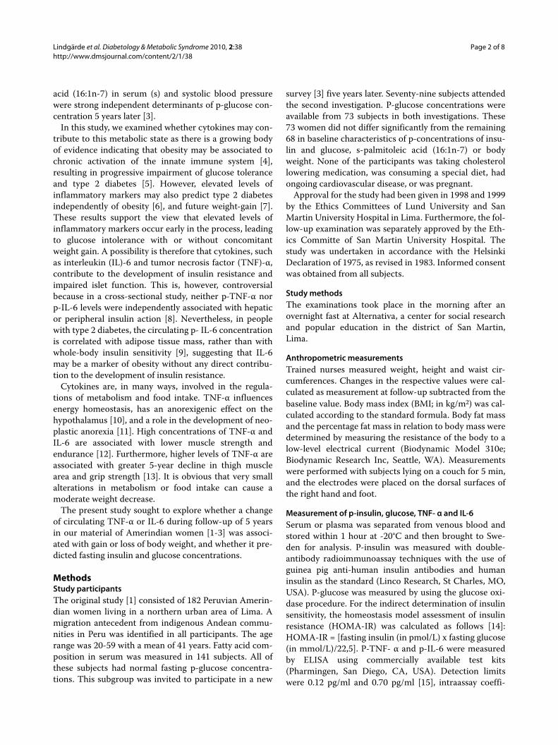
Table 2: Spearman rank correlation between differences (diff) between baseline and follow-up values. R- and (P-values).
Weight diff Waist diff TNF-α diff IL-6 diff Insulin diff Glucose diff
Weight diff - <.0001 -.35 (.0031) .25 (.0312) .27 (.0194) NS
Waist diff - -. 26 (.025) .25 (.0298) .25 (.0298) NS
TNF-α diff - NS -.23 (.045) NS
IL-6 diff - NS NS
Insulin diff - -.27 (.018)
Glucose diff -
TNF-α = tumor necrosis factor alpha. IL-6 = interleukin-6
Lindgärde et al. Diabetology & Metabolic Syndrome 2010, 2:38http://www.dmsjournal.com/content/2/1/38
Page 4 of 8
ResultsBody compositionAs can be seen in Table 1 body weight, BMI, waist cir-cumference, fat mass and fat as a percentage of bodyweight increased significantly. Twenty-one of the womenlost body weight (-2.0 ± 1.8 kg) while 52 gained bodyweight (+5.6 ± 4.0 kg).
Glucose and insulinMean p-glucose increased from 4.12 ± 0.45 to 5.01 ± 1.25mmol/l (p < 0.001). In contrast p-insulin was unchangedduring the 5 years. Consequently, when calculating insu-lin resistance according the homeostasis model of assess-ment ratio, HOMA-IR values increased significantly.
TNF-α and IL-6The decrease in p-IL-6 from 2.03 ± 2.49 to 1.89 ± 1.50 pg/ml did not reach significance while the decrease in TNF-α from 4.43 ± 1.75 to 3.35 ± 3.85 was significant (p =0.003). In subjects who lost body weight p-TNF-α and p-IL-6 concentrations were unchanged during the follow-up period. In women who gained body weight. p-TNF-αconcentration decreased from 4.9 ± 6.7 pg/ml to 3.3 ± 3.9(p < 0.0001) whereas p-IL-6 increased from 1.75 ± 2.27 to2.03 ± 1.63 (p = 0.002).
Weight change in relation to differences in waist circumference, p-cytokine, insulin and glucose concentrations between baseline and follow-up examinationsDifferences were calculated as the value at follow-up sub-tracted from the value at baseline. Spearman rank corre-lation was used to analyse the associations (Table 2).Weight and waist differences correlated negatively to thedifference in p-TNF-α but positively to differences in p-IL-6 and p-insulin, whereas the increase of p-glucosefrom baseline to follow-up did not correlate with changesin levels of the two cytokines. The decrease of p-TNF-αduring the observation period correlated negatively tochanges of p-insulin.
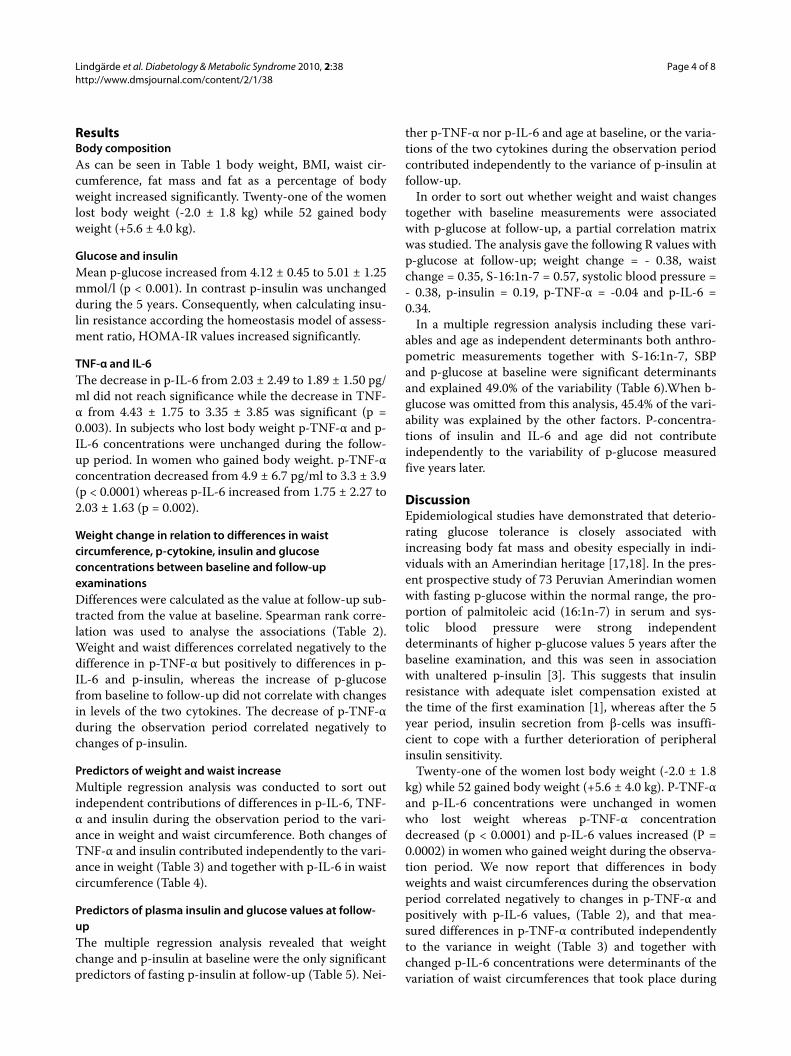
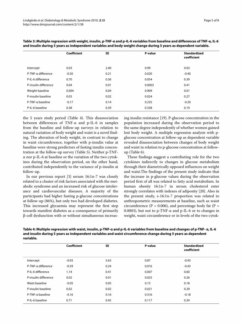
Predictors of weight and waist increaseMultiple regression analysis was conducted to sort outindependent contributions of differences in p-IL-6, TNF-α and insulin during the observation period to the vari-ance in weight and waist circumference. Both changes ofTNF-α and insulin contributed independently to the vari-ance in weight (Table 3) and together with p-IL-6 in waistcircumference (Table 4).
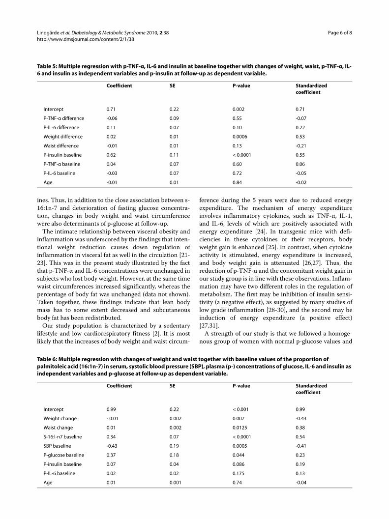
Predictors of plasma insulin and glucose values at follow-upThe multiple regression analysis revealed that weightchange and p-insulin at baseline were the only significantpredictors of fasting p-insulin at follow-up (Table 5). Nei-
ther p-TNF-α nor p-IL-6 and age at baseline, or the varia-tions of the two cytokines during the observation periodcontributed independently to the variance of p-insulin atfollow-up.
In order to sort out whether weight and waist changestogether with baseline measurements were associatedwith p-glucose at follow-up, a partial correlation matrixwas studied. The analysis gave the following R values withp-glucose at follow-up; weight change = - 0.38, waistchange = 0.35, S-16:1n-7 = 0.57, systolic blood pressure =- 0.38, p-insulin = 0.19, p-TNF-α = -0.04 and p-IL-6 =0.34.
In a multiple regression analysis including these vari-ables and age as independent determinants both anthro-pometric measurements together with S-16:1n-7, SBPand p-glucose at baseline were significant determinantsand explained 49.0% of the variability (Table 6).When b-glucose was omitted from this analysis, 45.4% of the vari-ability was explained by the other factors. P-concentra-tions of insulin and IL-6 and age did not contributeindependently to the variability of p-glucose measuredfive years later.
DiscussionEpidemiological studies have demonstrated that deterio-rating glucose tolerance is closely associated withincreasing body fat mass and obesity especially in indi-viduals with an Amerindian heritage [17,18]. In the pres-ent prospective study of 73 Peruvian Amerindian womenwith fasting p-glucose within the normal range, the pro-portion of palmitoleic acid (16:1n-7) in serum and sys-tolic blood pressure were strong independentdeterminants of higher p-glucose values 5 years after thebaseline examination, and this was seen in associationwith unaltered p-insulin [3]. This suggests that insulinresistance with adequate islet compensation existed atthe time of the first examination [1], whereas after the 5year period, insulin secretion from β-cells was insuffi-cient to cope with a further deterioration of peripheralinsulin sensitivity.
Twenty-one of the women lost body weight (-2.0 ± 1.8kg) while 52 gained body weight (+5.6 ± 4.0 kg). P-TNF-αand p-IL-6 concentrations were unchanged in womenwho lost weight whereas p-TNF-α concentrationdecreased (p < 0.0001) and p-IL-6 values increased (P =0.0002) in women who gained weight during the observa-tion period. We now report that differences in bodyweights and waist circumferences during the observationperiod correlated negatively to changes in p-TNF-α andpositively with p-IL-6 values, (Table 2), and that mea-sured differences in p-TNF-α contributed independentlyto the variance in weight (Table 3) and together withchanged p-IL-6 concentrations were determinants of thevariation of waist circumferences that took place during
Lindgärde et al. Diabetology & Metabolic Syndrome 2010, 2:38http://www.dmsjournal.com/content/2/1/38
Page 5 of 8
the 5 years study period (Table 4). This disassociationbetween differences of TNF-α and p-IL-6 in samplesfrom the baseline and follow-up surveys in relation tonatural variation of body weight and waist is a novel find-ing. The alteration of body weight, in contrast to changein waist circumference, together with p-insulin value atbaseline were strong predictors of fasting insulin concen-tration at the follow-up survey (Table 5). Neither p-TNF-α nor p-IL-6 at baseline or the variation of the two cytok-ines during the observation period, on the other hand,contributed independently to the variance of p-insulin atfollow-up.
In our previous report [3] serum 16:1n-7 was closelyrelated to a cluster of risk factors associated with the met-abolic syndrome and an increased risk of glucose intoler-ance and cardiovascular diseases. A majority of theparticipants had higher fasting p-glucose concentrationsat follow-up (86%), but only two had developed diabetes.This increased glycaemia may represent the first steptowards manifest diabetes as a consequence of primarilyβ-cell dysfunction with or without simultaneous increas-
ing insulin resistance [19]. P-glucose concentration in thepopulation increased during the observation period tothe same degree independently of whether women gainedlost body weight. A multiple regression analysis with p-glucose concentration at follow-up as dependent variablerevealed disassociation between changes of body weightand waist in relation to p-glucose concentration at follow-up (Table 6).
These findings suggest a contributing role for the twocytokines indirectly to changes in glucose metabolismthrough their diametrically opposed influences on weightand waist.The findings of the present study indicate thatthe increase in p-glucose values during the observationperiod first of all was related to fatty acid metabolism. Inhuman obesity 16:1n-7 in serum cholesterol esterstrongly correlates with indexes of adiposity [20]. Also inthe present study, s-16:1n-7 proportion was related toanthropometric measurements at baseline, such as waistcircumference (P = 0.006), and percentage body fat (P =0.0003), but not to p-TNF-α and p-IL-6 or to changes inweight, waist circumference or in levels of the two cytok-
Table 3: Multiple regression with weight, insulin, p-TNF-α and p-IL-6 variables from baseline and differences of TNF-α, IL-6 and insulin during 5 years as independent variables and body weight change during 5 years as dependent variable.
Coefficient SE P-value Standardized coefficient
Intercept 0.03 2.40 0.99 0.03
P-TNF-α difference -0.50 0.21 0.020 -0.40
P-IL-6 difference 0.70 0.36 0.054 0.39
P-insulin difference 0.04 0.01 0.0003 0.41
Weight baseline 0.004 0.04 0.909 0.01
P-insulin baseline 0.03 0.02 0.024 0.27
P-TNF-α baseline -0.17 0.14 0.235 -0.20
P-IL-6 baseline 0.38 0.39 0.338 0.19
Table 4: Multiple regression with waist, insulin, p-TNF-α and p-IL-6 variables from baseline and changes of p-TNF- α, IL-6 and insulin during 5 years as independent variables and waist circumference change during 5 years as dependent variable.
Coefficient SE P-value Standardized coefficient
Intercept -0.93 5.63 0.87 -0.93
P-TNF-α difference -0.59 0.24 0.016 -0.43
P-IL-6 difference 1.14 0.41 0.007 0.60
P-insulin difference 0.02 0.01 0.025 0.26
Waist baseline -0.05 0.05 0.15 0.18
P-insulin baseline 0.02 0.02 0.021 0.29
P-TNF-α baseline -0.16 0.16 0.316 -0.18
P-IL-6 baseline 0.71 0.45 0.117 0.34
Lindgärde et al. Diabetology & Metabolic Syndrome 2010, 2:38http://www.dmsjournal.com/content/2/1/38
Page 6 of 8
ines. Thus, in addition to the close association between s-16:1n-7 and deterioration of fasting glucose concentra-tion, changes in body weight and waist circumferencewere also determinants of p-glucose at follow-up.
The intimate relationship between visceral obesity andinflammation was underscored by the findings that inten-tional weight reduction causes down regulation ofinflammation in visceral fat as well in the circulation [21-23]. This was in the present study illustrated by the factthat p-TNF-α and IL-6 concentrations were unchanged insubjects who lost body weight. However, at the same timewaist circumferences increased significantly, whereas thepercentage of body fat was unchanged (data not shown).Taken together, these findings indicate that lean bodymass has to some extent decreased and subcutaneousbody fat has been redistributed.
Our study population is characterized by a sedentarylifestyle and low cardiorespiratory fitness [2]. It is mostlikely that the increases of body weight and waist circum-
ference during the 5 years were due to reduced energyexpenditure. The mechanism of energy expenditureinvolves inflammatory cytokines, such as TNF-α, IL-1,and IL-6, levels of which are positively associated withenergy expenditure [24]. In transgenic mice with defi-ciencies in these cytokines or their receptors, bodyweight gain is enhanced [25]. In contrast, when cytokineactivity is stimulated, energy expenditure is increased,and body weight gain is attenuated [26,27]. Thus, thereduction of p-TNF-α and the concomitant weight gain inour study group is in line with these observations. Inflam-mation may have two different roles in the regulation ofmetabolism. The first may be inhibition of insulin sensi-tivity (a negative effect), as suggested by many studies oflow grade inflammation [28-30], and the second may beinduction of energy expenditure (a positive effect)[27,31].
A strength of our study is that we followed a homoge-nous group of women with normal p-glucose values and
Table 5: Multiple regression with p-TNF-α, IL-6 and insulin at baseline together with changes of weight, waist, p-TNF-α, IL-6 and insulin as independent variables and p-insulin at follow-up as dependent variable.
Coefficient SE P-value Standardized coefficient
Intercept 0.71 0.22 0.002 0.71
P-TNF-α difference -0.06 0.09 0.55 -0.07
P-IL-6 difference 0.11 0.07 0.10 0.22
Weight difference 0.02 0.01 0.0006 0.53
Waist difference -0.01 0.01 0.13 -0.21
P-insulin baseline 0.62 0.11 < 0.0001 0.55
P-TNF-α baseline 0.04 0.07 0.60 0.06
P-IL-6 baseline -0.03 0.07 0.72 -0.05
Age -0.01 0.01 0.84 -0.02
Table 6: Multiple regression with changes of weight and waist together with baseline values of the proportion of palmitoleic acid (16:1n-7) in serum, systolic blood pressure (SBP), plasma (p-) concentrations of glucose, IL-6 and insulin as independent variables and p-glucose at follow-up as dependent variable.
Coefficient SE P-value Standardized coefficient
Intercept 0.99 0.22 < 0.001 0.99
Weight change - 0.01 0.002 0.007 -0.43
Waist change 0.01 0.002 0.0125 0.38
S-16:l-n7 baseline 0.34 0.07 < 0.0001 0.54
SBP baseline -0.43 0.19 0.0005 -0.41
P-glucose baseline 0.37 0.18 0.044 0.23
P-insulin baseline 0.07 0.04 0.086 0.19
P-IL-6 baseline 0.02 0.02 0.175 0.13
Age 0.01 0.001 0.74 -0.04

Lindgärde et al. Diabetology & Metabolic Syndrome 2010, 2:38http://www.dmsjournal.com/content/2/1/38
Page 7 of 8
hyperinsulinaemia [1] for a relatively long period of time,5 years [3]. Another strength of the study is that no activeintervention (diet or exercise programmes) took placesince all women had normal fasting p-glucose concentra-tion at the baseline survey. Accordingly it was possible toexamine relationships between circulating TNF-α, IL-6,insulin and glucose taking the natural variation of bodyweight into account.
It is well known that advancing age plays an importantrole in the progressive β-cell failure that characterisestype 2 diabetes. However, age did not correlate with anymeasured variable at baseline and was not a determinantof p-insulin (table 5) or p-glucose (table 6) at follow-up.These observations indicate that age in the present studymay not explain the increase of insulin resistance (table 1)during the 5-year observation period.
The report by Chan et al [32] is in agreement with ourobservations They found that obesity, dyslipidemia, IL-6,and TNF-α were the principal explanatory variables forthe various components of the metabolic syndrome inCaucasian non-diabetic subjects, with IL-6 and TNF-αhaving different explanatory variables and effects [32].Our findings of disassociations between p-TNF-α and p-IL-6 in relation to changes of body weight and waist cir-cumference are thus partly consistent with previous workand would suggest an importance for these cytokines alsoin this population of Amerindian women.
ConclusionsThe present prospective study revealed disassociationbetween the changes in p-TNF-α and p-IL-6 concentra-tions in samples from the baseline survey and the follow-up measurements in relation to body weight at follow-upfive years later when natural variation of body weight wastaken into account. The exact role of the opposite effectsand clinical impact of p-TNF-α and p-IL-6 on develop-ment of insulin resistance and perhaps type 2 diabetes insubjects with unintentional wasting and women whogained body weight now needs to be examined further.
Competing interestsThe authors declare that they have no competing interests.
Authors' contributionsFL and BA were responsible for the study design. FL was responsible for datacollection, BA and AG were responsible for data analysis, and all authors partic-ipated in writing the manuscript.
AcknowledgementsMiyaray Benavente Ercilla, and Laura Retamozo, at the Alternativa Center for Social Research and Popular Education in Lima, Peru, participated in the design and data collection and provided information to participants. We also thank Barbro Palmquist and Lilian Bengtsson for technical assistance. The study was supported by grants from Stiftelsen för forskning inom diabetes och kärlsjukd-om, The Ernhold Lundström Foundation, The Hulda Almroth Foundation, Swedish Research Council (no 6834), Region Skåne, and the Faculty of Medi-cine, Lund University.
Author Details1Vascular Center, Lund University, Skåne University Hospital, S-205 02 Malmö, Sweden and 2Department of Clinical Sciences, Division of Medicine, Lund University, B11 BMC, SE-221 84 Lund, Sweden
References1. Lindgärde F, Söderberg S, Olsson T, Ercilla MB, Correa LR, Ahrén B:
Overweight is associated with lower serum leptin in Peruvian Indian than in Caucasian women: a dissociation contributing to low blood pressure. Metabolism 2001, 50:325-329.
2. Lindgärde F, Ahrén B: Improved metabolic risk markers following two 6-month physical activity programs among socioeconomic marginalized women of native American ancestry in Lima, Peru. Diabetes Care 2007, 30:2230-2233.
3. Lindgärde F, Vessby B, Ahrén B: Serum cholesteryl fatty acid composition and plasma glucose concentrations in Amerindian women. Am J Clin Nutr 2006, 84:1009-1013.
4. Duncan BB, Schmidt MI, Chamb less LE, Folsom AR, Carpenter M, Heiss G: Fibrinogen, other putative markers of inflammation, and weight gain in middle-aged adults: the ARIC study: Atherosclerosis Risk in Communities. Obes Res 2000, 8:279-286.
5. Hajer GR, van Haeften TW, Visseren FL: Adipose tissue dysfunction in obesity, diabetes, and vascular diseases. Eur Heart J 2008, 29:2959-2971.
6. Engström G, Hedblad B, Eriksson KF, Janzon L, Lindgärde F: Complement C3 is a risk factor for the development of diabetes. A population-based cohort study. Diabetes 2005, 54:570-575.
7. Engström G, Hedblad B, Stavenow L, Lind P, Janzon L, Lindgärde F: Inflammation-sensitive plasma proteins are associated with future weight-gain. Diabetes 2003, 52:2097-2101.
8. Grunnet L, Poulsen P, Pedersen Klarlund B, Mandrup-Poulsen T, Vaag A: Plasma cytokine levels in young and elderly twins: genes versus environment and relation to in vivo insulin action. Diabetologia 2006, 49:343-350.
9. Carey AL, Bruce CR, Sacchetti M, et al.: Interleukin-6 and tumor necrosis factor-alpha are not increased in patients with Type 2 diabetes: evidence that plasma interleukin-6 is related to fat mass and not insulin responsiveness. Diabetologia 2004, 47:1029-1037.
10. Endo M, Masaki T, Seike M, Yoshimatsu M: Involvement of stomac gherlin and hypothalamic neuropeptides in tumor necrosis factor-alpha-induced hypophagia in mice. Regul Pept 2007, 140:94-100.
11. Meguid MM, Fetissov SO, Vendrell J, et al.: Hypothalamic dopamine and serotonin in the regulation of food intake. Nutrition 2000, 16:843-857.
12. Hsu F-C, Kritchevsky S, Liu Y, et al.: Association between inflammatory components and physical function in the health, aging, and body composition study: a principal component analysis approach. J Gerontol A Biol Sci Med Sc 2009, 64:581-589.
13. Schaap LA, Pluijm SM, Deeg DJ, et al.: Higher inflammatory Marker Levels in Older Persons: Associations With 5-Year Change in Muscle Mass and Muscle Strength. J Gerontol Biol Sci Med Sci 2009, 64:1183-1189.
14. Matthews DR, Hosker JP, Rudenski AS, Naylor BA, Treacher DF, Turnerer RC: Homeostasis model assessment: insulin resistance and beta-cell function from fasting plasma glucose and insulin concentrations in man. Diabetologia 1985, 28:412-419.
15. Barani J, Nilsson JÅ, Mattiasson I, Lindblad B, Gottsäter A: Inflammatory mediators are associated with 1-year mortality in critical limb ischemia. J Vasc Surg 2005, 42:75-80.
16. Boberg M, Croon LB, Gustafsson IB, Vessby B: Platelet fatty acid composition in relation to fatty acid composition in plasma and to s-lipoprotein lipids in healthy subjects with special reference to the linoleic acid pathway. Clin Sci 1985, 68:581-587.
17. Knowler WC, Pettitt DJ, Saad MF, Bennett PH: Diabetes mellitus in the Pima Indians: incidence, risk factors and pathogenesis. Diabetes Metab Rev 1990, 6:1-27.
18. Jacoby E, Goldstein J, Lopez A, Nunez E, Lopez T: Social class, family, and life-style factors associated with overweight and obesity among adults in Peruvian cities. Prev Med 2003, 37:396-405.
19. Rasmussen SS, Glümer C, Sandbaek A, Lauritzen T, Borch-Johnsen K: Determinants of progression from impaired fasting glucose and
Received: 4 February 2010 Accepted: 8 June 2010 Published: 8 June 2010This article is available from: http://www.dmsjournal.com/content/2/1/38© 2010 Lindgärde et al; licensee BioMed Central Ltd. This is an Open Access article distributed under the terms of the Creative Commons Attribution License (http://creativecommons.org/licenses/by/2.0), which permits unrestricted use, distribution, and reproduction in any medium, provided the original work is properly cited.Diabetology & Metabolic Syndrome 2010, 2:38
http://www.ncbi.nlm.nih.gov/entrez/query.fcgi?cmd=Retrieve&db=PubMed&dopt=Abstract&list_uids=3899825
http://www.ncbi.nlm.nih.gov/entrez/query.fcgi?cmd=Retrieve&db=PubMed&dopt=Abstract&list_uids=3919990
Lindgärde et al. Diabetology & Metabolic Syndrome 2010, 2:38http://www.dmsjournal.com/content/2/1/38
Page 8 of 8
impaired glucose tolerance to diabetes in a high-risk screened population: 3 year follow-up in the ADDITION study, Denmark. Diabetologia 2008, 51:249-257.
20. Kusenova M, Hainer V, Tvrzicka E, et al.: Assessment of dietary and genetic factors influencing serum and adipose fatty acid composition in obese female identical twins. Lipids 2002, 37:27-32.
21. Clement K, Viguerie N, Poitou C, et al.: Weight loss regulates inflammation-related genes in white adipose tissue of obese subjects. FASEB J 2004, 18:1657-1669.
22. Puglisi MJ, Fernandez ML: Modulation of C-reactive protein, tumor necrosis factor-alpha, and adiponectin by diet, exercise, and weight loss. J Nutr 2008, 138:2293-2296.
23. Samuelsson L, Gottsäter A, Lindgärde F: Decreasing levels of tumour necrosis factor alpha and interleukin 6 during lowering of body mass index with orlistat or placebo in obese subjects with cardiovascular risk factors. Diabetes Obes Metab 2003, 3:195-201.
24. Tisdale MJ: Biology of cachexia. Jrl Nat Cancer Inst 1997, 89:1763-1773.25. Pamir N, McMillen TS, Kaiyala KJ, Schwartz MW, LeBoeuf RC: Receptors for
tumor necrosis factor-α play a protective role against obesity and alter adipose tissue macrophage status. Endocrinology 2009, 150:4124-4134.
26. Xu H, Hirosumi J, Uysal KT, Guler AD, Hotamisligil GS: Exclusive action of transmembrane TNF -α in adipose tissue leads to reduced adipose mass and local but not systemic insulin resistance. Endocrinology 2002, 143:1502-1511.
27. Sadagurski M, Norquay L, Farhang J, D'Aquino K, Copps K, White MF: Human IL6 enhances leptin action in mice. Diabetologia 2010, 53:525-535.
28. Aguirre V, Uchida T, Yenush L, Davis R, White MF: The c-Jun NH(2)-terminal kinase promotes insulin resistance during association with insulin receptor substrate-1 and phosphorylation of Ser(307). J Biol Chem 2000, 24:9047-9054.
29. Hotamisligil GS, Peraldi P, Budavari A, Ellis R, White MF, Spiegelman BM: IRS-1-mediated inhibition of insulin receptor tyrosine kinase activity in TNF-alpha- and obesity-induced insulin resistance. Science 1996, 271:665-668.
30. Ruan H, Hachoen N, Golub TR, Van Parijs L, Lodish HF: Tumor necrosis factor alpha suppresses adipocyte-specific genes and activates expression of preadipocyte genes in 3T3-L1 adipocytes; nuclear factor-kappaB activation by TNF-alpha is obligatory. Diabetes 2002, 51:1319-1336.
31. Tang T, Zhang J, Yin J, et al.: Uncoupling of inflammation and insulin resistance by NF-kappaB in transgenic mice through elevated energy expenditure. J Biol Chem 2010, 285:4637-4644.
32. Chan JCN, Cheung JCK, Stehouwer CDA, et al.: The central roles of obesity-associated dyslipidaemia, endothelial activation and cytokines in the metabolic syndrome-an analysis by structural equation modeling. Int J Obes 2002, 26:994-1008.
doi: 10.1186/1758-5996-2-38Cite this article as: Lindgärde et al., Disassociated relation between plasma tumor necrosis factor-?, interleukin-6 and increased body weight in Amerin-dian women: A long-term prospective study of natural body weight variation and impaired glucose tolerance Diabetology & Metabolic Syndrome 2010, 2:38