disables neurons Neurons, Synapses, and Neurons … © 2008 Pearson Education Inc., publishing as...
14
1 Copyright © 2008 Pearson Education, Inc., publishing as Pearson Benjamin Cummings Lectures by Chris Romero, updated by Erin Barley with contributions from Joan Sharp and Janette Lewis PowerPoint Lectures for Biology, Eighth Edition Neurons, Synapses, and Signaling Chapter 48 Chapter 48 Copyright © 2008 Pearson Education Inc., publishing as Pearson Benjamin Cummings Overview: Lines of Communication • The cone snail kills prey with venom that disables neurons • Neurons are nerve cells that transfer information within the body • Neurons use two types of signals to communicate: electrical signals (long- distance) and chemical signals (short- distance) Copyright © 2008 Pearson Education Inc., publishing as Pearson Benjamin Cummings What makes this snail such a deadly predator? Copyright © 2008 Pearson Education Inc., publishing as Pearson Benjamin Cummings • Nervous system organization – The transmission of information depends on the path of neurons along which a signal travels – Processing of information takes place in simple clusters of neurons called ganglia or a more complex organization of neurons called a brain Copyright © 2008 Pearson Education Inc., publishing as Pearson Benjamin Cummings • Many animals have a complex nervous system which consists of: – A central nervous system (CNS) where integration takes place; this includes the brain and a nerve cord – A peripheral nervous system (PNS), which brings information into and out of the CNS Nerves with giant axons Ganglia Mantle Eye Brain Arm Nerve
Transcript of disables neurons Neurons, Synapses, and Neurons … © 2008 Pearson Education Inc., publishing as...
1
Copyright © 2008 Pearson Education, Inc., publishing as Pearson Benjamin Cummings
Lectures by Chris Romero, updated by Erin Barley with contributions from Joan Sharp and Janette Lewis
PowerPoint Lectures for Biology, Eighth Edition
Neurons, Synapses, and Signaling
Chapter 48Chapter 48
Copyright © 2008 Pearson Education Inc., publishing as Pearson Benjamin Cummings
Overview: Lines of Communication
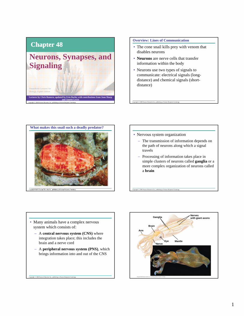
• The cone snail kills prey with venom that disables neurons
• Neurons are nerve cells that transfer information within the body
• Neurons use two types of signals to communicate: electrical signals (long-distance) and chemical signals (short-distance)
Copyright © 2008 Pearson Education Inc., publishing as Pearson Benjamin Cummings
What makes this snail such a deadly predator?
Copyright © 2008 Pearson Education Inc., publishing as Pearson Benjamin Cummings
• Nervous system organization– The transmission of information depends on
the path of neurons along which a signal travels
– Processing of information takes place in simple clusters of neurons called ganglia or a more complex organization of neurons called a brain
Copyright © 2008 Pearson Education Inc., publishing as Pearson Benjamin Cummings
• Many animals have a complex nervous system which consists of:– A central nervous system (CNS) where
integration takes place; this includes the brain and a nerve cord
– A peripheral nervous system (PNS), which brings information into and out of the CNS
Nerveswith giant axonsGanglia
MantleEye
Brain
Arm
Nerve
2
Copyright © 2008 Pearson Education Inc., publishing as Pearson Benjamin Cummings
Concept 48.1: Neuron organization and structure reflect function in information transfer
• Neuron organization and structure reflect function in information transfer – Neurons are the functional unit of the
nervous system– Nervous systems process information in
three stages: 1. sensory input2. integration3. motor output
Copyright © 2008 Pearson Education Inc., publishing as Pearson Benjamin Cummings
Neurons Transmit Information
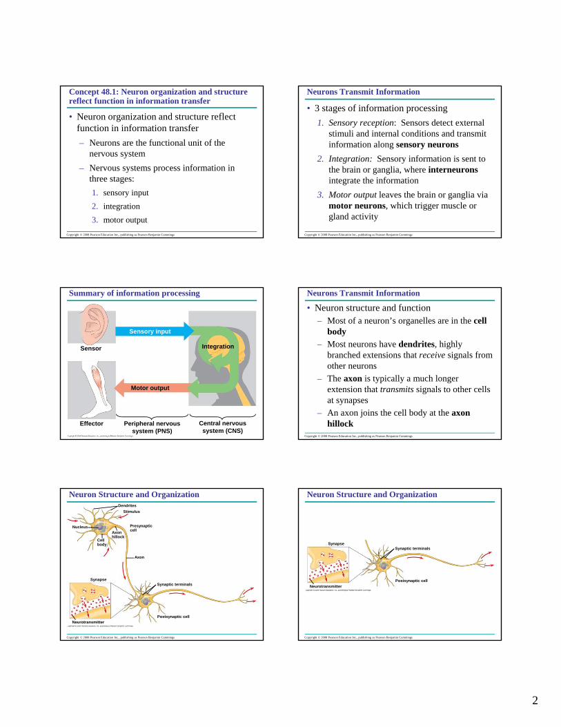
• 3 stages of information processing1. Sensory reception: Sensors detect external
stimuli and internal conditions and transmit information along sensory neurons
2. Integration: Sensory information is sent to the brain or ganglia, where interneurons integrate the information
3. Motor output leaves the brain or ganglia via motor neurons, which trigger muscle or gland activity
Copyright © 2008 Pearson Education Inc., publishing as Pearson Benjamin Cummings
Sensor
Sensory input
Integration
Effector
Motor output
Peripheral nervoussystem (PNS)
Central nervoussystem (CNS)
Summary of information processing
Copyright © 2008 Pearson Education Inc., publishing as Pearson Benjamin Cummings
Neurons Transmit Information
• Neuron structure and function– Most of a neuron’s organelles are in the cell
body– Most neurons have dendrites, highly
branched extensions that receive signals from other neurons
– The axon is typically a much longer extension that transmits signals to other cells at synapses
– An axon joins the cell body at the axon hillock
Copyright © 2008 Pearson Education Inc., publishing as Pearson Benjamin Cummings
DendritesStimulus
Nucleus
Cellbody
Axonhillock
Presynapticcell
Axon
Synaptic terminalsSynapse
Postsynaptic cellNeurotransmitter
Neuron Structure and Organization
Copyright © 2008 Pearson Education Inc., publishing as Pearson Benjamin Cummings
SynapseSynaptic terminals
Postsynaptic cellNeurotransmitter
Neuron Structure and Organization
3
Copyright © 2008 Pearson Education Inc., publishing as Pearson Benjamin Cummings
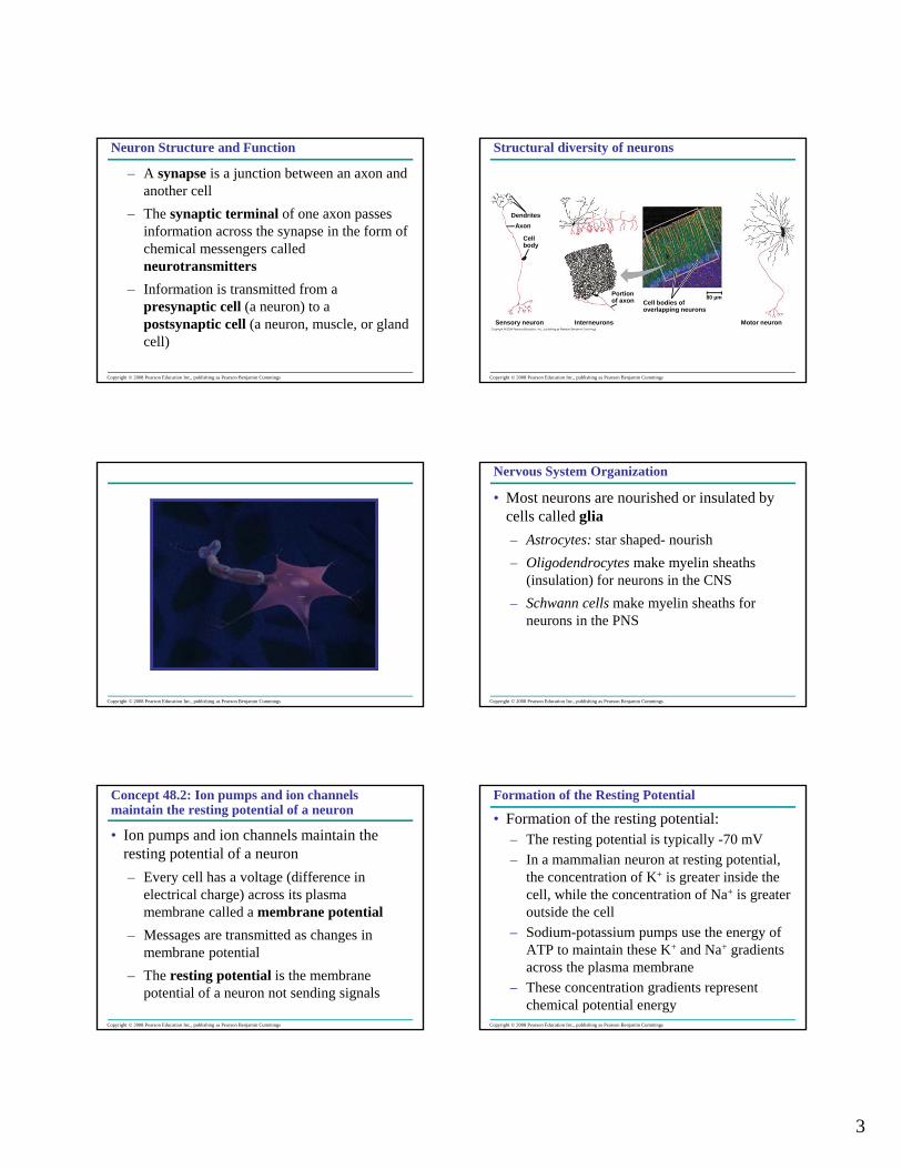
Neuron Structure and Function
– A synapse is a junction between an axon and another cell
– The synaptic terminal of one axon passes information across the synapse in the form of chemical messengers called neurotransmitters
– Information is transmitted from a presynaptic cell (a neuron) to a postsynaptic cell (a neuron, muscle, or gland cell)
Copyright © 2008 Pearson Education Inc., publishing as Pearson Benjamin Cummings
DendritesAxon
Cellbody
Sensory neuron Interneurons
Portion of axon Cell bodies of
overlapping neurons
80 µm
Motor neuron
Structural diversity of neurons
Copyright © 2008 Pearson Education Inc., publishing as Pearson Benjamin Cummings Copyright © 2008 Pearson Education Inc., publishing as Pearson Benjamin Cummings
Nervous System Organization
• Most neurons are nourished or insulated by cells called glia– Astrocytes: star shaped- nourish– Oligodendrocytes make myelin sheaths
(insulation) for neurons in the CNS– Schwann cells make myelin sheaths for
neurons in the PNS
Copyright © 2008 Pearson Education Inc., publishing as Pearson Benjamin Cummings
Concept 48.2: Ion pumps and ion channels maintain the resting potential of a neuron
• Ion pumps and ion channels maintain the resting potential of a neuron– Every cell has a voltage (difference in
electrical charge) across its plasma membrane called a membrane potential
– Messages are transmitted as changes in membrane potential
– The resting potential is the membrane potential of a neuron not sending signals
Copyright © 2008 Pearson Education Inc., publishing as Pearson Benjamin Cummings
Formation of the Resting Potential
• Formation of the resting potential: – The resting potential is typically -70 mV– In a mammalian neuron at resting potential,
the concentration of K+ is greater inside the cell, while the concentration of Na+ is greater outside the cell
– Sodium-potassium pumps use the energy of ATP to maintain these K+ and Na+ gradients across the plasma membrane
– These concentration gradients represent chemical potential energy
4
Copyright © 2008 Pearson Education Inc., publishing as Pearson Benjamin Cummings
Formation of the Resting Potential
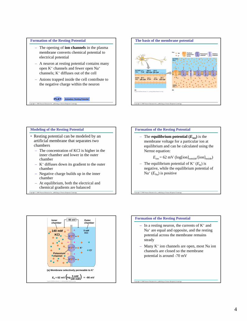
– The opening of ion channels in the plasma membrane converts chemical potential to electrical potential
– A neuron at resting potential contains many open K+ channels and fewer open Na+
channels; K+ diffuses out of the cell– Anions trapped inside the cell contribute to
the negative charge within the neuron
Animation: Resting Potential
Copyright © 2008 Pearson Education Inc., publishing as Pearson Benjamin Cummings
OUTSIDECELL
[K+]5 mM
[Na+]150 mM
[Cl–]120 mM
INSIDECELL
[K+]140 mM
[Na+]15 mM
[Cl–]10 mM
[A–]100 mM
(a) (b)
OUTSIDECELL
Na+Key
K+
Sodium-potassiumpump
Potassiumchannel
Sodiumchannel
INSIDECELL
The basis of the membrane potential
Copyright © 2008 Pearson Education Inc., publishing as Pearson Benjamin Cummings
Modeling of the Resting Potential
• Resting potential can be modeled by an artificial membrane that separates two chambers– The concentration of KCl is higher in the
inner chamber and lower in the outer chamber
– K+ diffuses down its gradient to the outer chamber
– Negative charge builds up in the inner chamber
– At equilibrium, both the electrical and chemical gradients are balanced
Copyright © 2008 Pearson Education Inc., publishing as Pearson Benjamin Cummings
Formation of the Resting Potential
– The equilibrium potential (Eion) is the membrane voltage for a particular ion at equilibrium and can be calculated using the Nernst equation:
Eion = 62 mV (log[ion]outside/[ion]inside)– The equilibrium potential of K+ (EK) is
negative, while the equilibrium potential of Na+ (ENa) is positive
Innerchamber
Outerchamber
–90 mV
140 mM 5 mM
KCI KCI
K+
Cl–Potassiumchannel
(a) Membrane selectively permeable to K+
EK = 62 mV(log 5 mM140 mM) = –90 mV
Copyright © 2008 Pearson Education Inc., publishing as Pearson Benjamin Cummings
Formation of the Resting Potential
– In a resting neuron, the currents of K+ and Na+ are equal and opposite, and the resting potential across the membrane remains steady
– Many K+ ion channels are open, most Na ion channels are closed so the membrane potential is around -70 mV

5
(b) Membrane selectively permeable to Na+
+62 mV
15 mMNaCI
Cl–
150 mMNaCI
Na+
Sodiumchannel
ENa = 62 mV (log 150 mM15 mM = +62 mV)
Innerchamber
Outerchamber
–90 mV
140 mM 5 mM
KCI KCI
K+
Cl–Potassiumchannel
(a) Membrane selectively permeable to K+ (b) Membrane selectively permeable to Na+
+62 mV
15 mMNaCI
Cl–
150 mMNaCI
Na+
Sodiumchannel
EK = 62 mV(log 5 mM140 mM ) = –90 mV ENa = 62 mV (log 150 mM
15 mM = +62 mV)
Copyright © 2008 Pearson Education Inc., publishing as Pearson Benjamin Cummings
Concept 48.3: Action potentials are the signals conducted by axons
• Neurons contain gated ion channels that open or close in response to stimuli
TECHNIQUE
Microelectrode
Voltagerecorder
Referenceelectrode
Copyright © 2008 Pearson Education Inc., publishing as Pearson Benjamin Cummings
Graded Potentials
• The membrane potential changes in response to opening or closing of these channels– Hyperpolarization
• When gated K+ channels open, K+ diffuses out, making the inside of the cell more negative
• This is hyperpolarization, an increase in (negative) magnitude of the membrane potential
• Remember that resting membrane potential is around -70 mV
Stimuli
+50
Mem
bran
e po
tent
ial (
mV)
–50 Threshold
Restingpotential
Hyperpolarizations–100
0 2 3 4Time (msec)
(a) Graded hyperpolarizations
0
1 5
Copyright © 2008 Pearson Education Inc., publishing as Pearson Benjamin Cummings
Graded Potentials
– Depolarizations• Other stimuli trigger a depolarization, a
reduction in the magnitude of the membrane potential
• For example, depolarization occurs if gated Na+ channels open and Na+ diffuses into the cell
• Graded potentials are changes in polarization where the magnitude of the change varies with the strength of the stimulus
6
Stimuli
+50
Mem
bran
e po
tent
ial (
mV)
–50 Threshold
Restingpotential
Depolarizations–100
0 2 3 4Time (msec)
(b) Graded depolarizations
1 5
0
Copyright © 2008 Pearson Education Inc., publishing as Pearson Benjamin Cummings
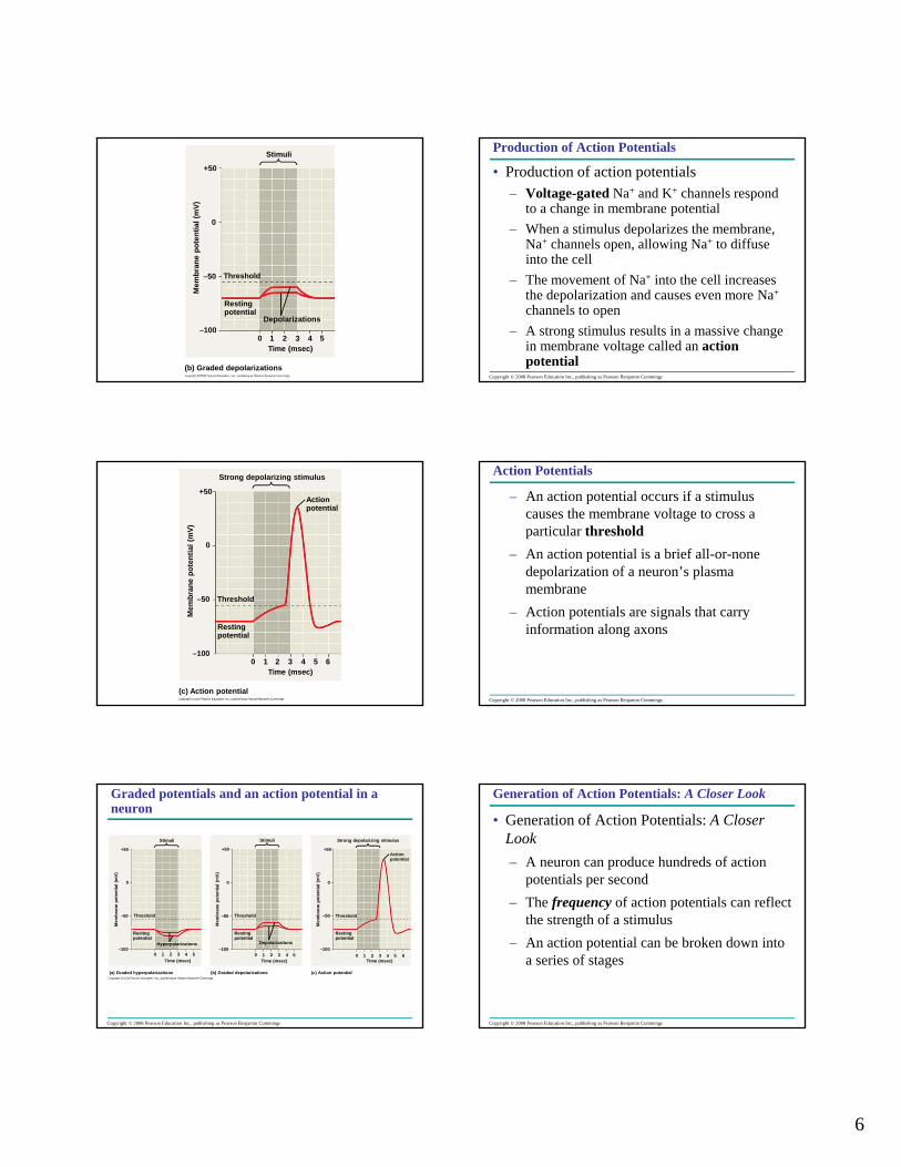
Production of Action Potentials
• Production of action potentials– Voltage-gated Na+ and K+ channels respond
to a change in membrane potential– When a stimulus depolarizes the membrane,
Na+ channels open, allowing Na+ to diffuse into the cell
– The movement of Na+ into the cell increases the depolarization and causes even more Na+
channels to open– A strong stimulus results in a massive change
in membrane voltage called an action potential
Strong depolarizing stimulus
+50
Mem
bran
e po
tent
ial (
mV)
–50 Threshold
Restingpotential
–1000 2 3 4
Time (msec)
(c) Action potential
1 5
0
Actionpotential
6
Copyright © 2008 Pearson Education Inc., publishing as Pearson Benjamin Cummings
Action Potentials
– An action potential occurs if a stimulus causes the membrane voltage to cross a particular threshold
– An action potential is a brief all-or-none depolarization of a neuron’s plasma membrane
– Action potentials are signals that carry information along axons
Copyright © 2008 Pearson Education Inc., publishing as Pearson Benjamin Cummings
Stimuli
+50 +50
Stimuli
00
Mem
bran
e po
tent
ial (
mV)
Mem
bran
e po
tent
ial (
mV)
–50 –50Threshold Threshold
Restingpotential
Restingpotential
Hyperpolarizations–100 –100
0 1 2 3 4 5Time (msec)
(a) Graded hyperpolarizations
Time (msec)
(b) Graded depolarizations
Depolarizations
0 1 2 3 4 5
Strong depolarizing stimulus
+50
0
Mem
bran
e po
tent
ial (
mV)
–50 Threshold
Restingpotential
–100
Time (msec)0 1 2 3 4 5 6
(c) Action potential
Actionpotential
Graded potentials and an action potential in a neuron
Copyright © 2008 Pearson Education Inc., publishing as Pearson Benjamin Cummings
Generation of Action Potentials: A Closer Look
• Generation of Action Potentials: A Closer Look– A neuron can produce hundreds of action
potentials per second– The frequency of action potentials can reflect
the strength of a stimulus– An action potential can be broken down into
a series of stages
7
Copyright © 2008 Pearson Education Inc., publishing as Pearson Benjamin Cummings
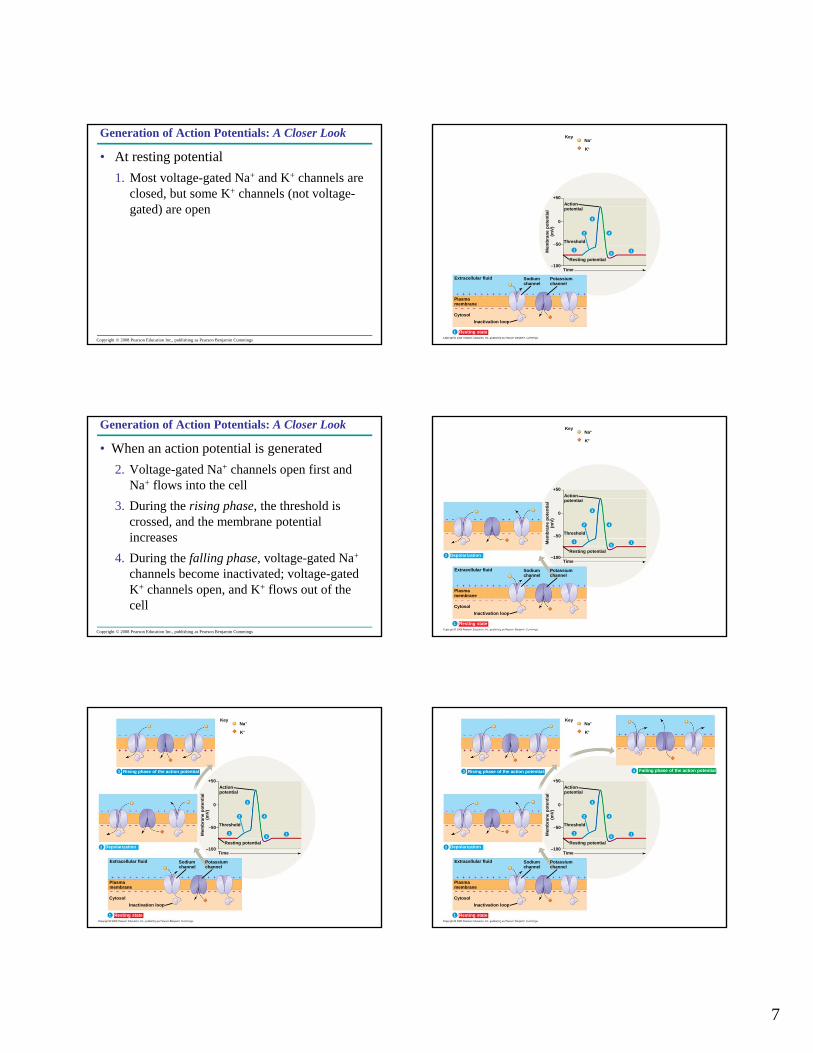
Generation of Action Potentials: A Closer Look
• At resting potential1. Most voltage-gated Na+ and K+ channels are
closed, but some K+ channels (not voltage-gated) are open
KeyNa+
K+
+50Actionpotential
Threshold
0
1
4
51
–50
Resting potential
Mem
bran
e po
tent
ial
(mV)
–100Time
Extracellular fluid
Plasmamembrane
CytosolInactivation loop
Resting state
Sodiumchannel
Potassiumchannel
Depolarization
2
3
1
Copyright © 2008 Pearson Education Inc., publishing as Pearson Benjamin Cummings
Generation of Action Potentials: A Closer Look
• When an action potential is generated2. Voltage-gated Na+ channels open first and
Na+ flows into the cell3. During the rising phase, the threshold is
crossed, and the membrane potential increases
4. During the falling phase, voltage-gated Na+
channels become inactivated; voltage-gated K+ channels open, and K+ flows out of the cell
KeyNa+
K+
+50Actionpotential
Threshold
0
1
4
51
–50
Resting potentialM
embr
ane
pote
ntia
l(m
V)–100
Time
Extracellular fluid
Plasmamembrane
CytosolInactivation loop
Resting state
Sodiumchannel
Potassiumchannel
Depolarization
2
3
2
1
KeyNa+
K+
+50Actionpotential
Threshold
0
1
4
51
–50
Resting potential
Mem
bran
e po
tent
ial
(mV)
–100Time
Extracellular fluid
Plasmamembrane
CytosolInactivation loop
Resting state
Sodiumchannel
Potassiumchannel
Depolarization
Rising phase of the action potential
2
3
2
1
3
KeyNa+
K+
+50Actionpotential
Threshold
0
1
4
51
–50
Resting potential
Mem
bran
e po
tent
ial
(mV)
–100Time
Extracellular fluid
Plasmamembrane
CytosolInactivation loop
Resting state
Sodiumchannel
Potassiumchannel
Depolarization
Rising phase of the action potential Falling phase of the action potential
2
3
2
1
3 4
8
Copyright © 2008 Pearson Education Inc., publishing as Pearson Benjamin Cummings
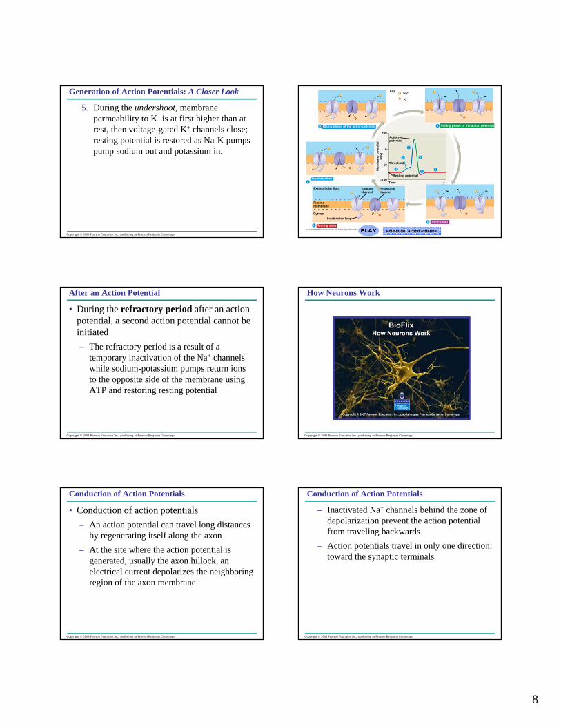
Generation of Action Potentials: A Closer Look
5. During the undershoot, membrane permeability to K+ is at first higher than at rest, then voltage-gated K+ channels close; resting potential is restored as Na-K pumps pump sodium out and potassium in.
KeyNa+
K+
+50Actionpotential
Threshold
0
1
4
51
–50
Resting potential
Mem
bran
e po
tent
ial
(mV)
–100Time
Extracellular fluid
Plasmamembrane
CytosolInactivation loop
Resting state
Sodiumchannel
Potassiumchannel
Depolarization
Rising phase of the action potential Falling phase of the action potential
5 Undershoot
2
3
2
1
3 4
Animation: Action Potential
Copyright © 2008 Pearson Education Inc., publishing as Pearson Benjamin Cummings
After an Action Potential
• During the refractory period after an action potential, a second action potential cannot be initiated– The refractory period is a result of a
temporary inactivation of the Na+ channels while sodium-potassium pumps return ions to the opposite side of the membrane using ATP and restoring resting potential
Copyright © 2008 Pearson Education Inc., publishing as Pearson Benjamin Cummings
How Neurons Work
Copyright © 2008 Pearson Education Inc., publishing as Pearson Benjamin Cummings
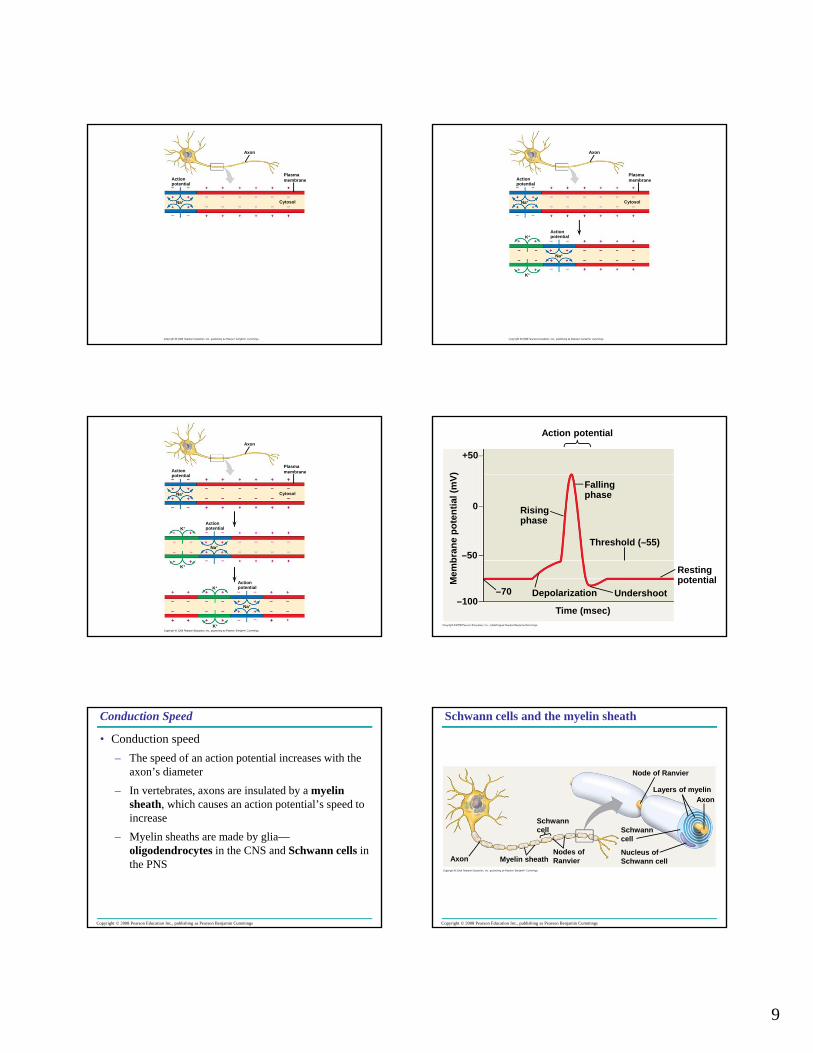
Conduction of Action Potentials
• Conduction of action potentials– An action potential can travel long distances
by regenerating itself along the axon– At the site where the action potential is
generated, usually the axon hillock, an electrical current depolarizes the neighboring region of the axon membrane
Copyright © 2008 Pearson Education Inc., publishing as Pearson Benjamin Cummings
Conduction of Action Potentials
– Inactivated Na+ channels behind the zone of depolarization prevent the action potential from traveling backwards
– Action potentials travel in only one direction: toward the synaptic terminals
9
Axon
Plasmamembrane
Cytosol
Actionpotential
Na+
Axon
Plasmamembrane
Cytosol
Actionpotential
Na+
Actionpotential
Na+
K+
K+
Axon
Plasmamembrane
Cytosol
Actionpotential
Na+
Actionpotential
Na+
K+
K+
ActionpotentialK+
K+
Na+
Action potential
Fallingphase
Risingphase
Threshold (–55)
Restingpotential
Undershoot
Time (msec)
Depolarization–70–100
–50
0
+50M
embr
ane
pote
ntia
l (m
V)
Copyright © 2008 Pearson Education Inc., publishing as Pearson Benjamin Cummings
Conduction Speed
• Conduction speed– The speed of an action potential increases with the
axon’s diameter– In vertebrates, axons are insulated by a myelin
sheath, which causes an action potential’s speed to increase
– Myelin sheaths are made by glia—oligodendrocytes in the CNS and Schwann cells in the PNS
Copyright © 2008 Pearson Education Inc., publishing as Pearson Benjamin Cummings
Axon Myelin sheath
Schwanncell
Nodes ofRanvier
Schwanncell
Nucleus ofSchwann cell
Node of Ranvier
Layers of myelinAxon
Schwann cells and the myelin sheath
10
Copyright © 2008 Pearson Education Inc., publishing as Pearson Benjamin Cummings
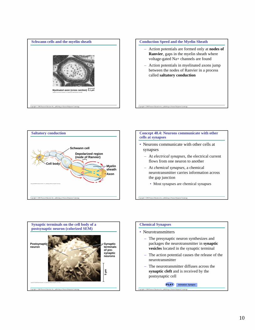
Myelinated axon (cross section) 0.1 µm
Schwann cells and the myelin sheath
Copyright © 2008 Pearson Education Inc., publishing as Pearson Benjamin Cummings
Conduction Speed and the Myelin Sheath
– Action potentials are formed only at nodes of Ranvier, gaps in the myelin sheath where voltage-gated Na+ channels are found
– Action potentials in myelinated axons jump between the nodes of Ranvier in a process called saltatory conduction
Copyright © 2008 Pearson Education Inc., publishing as Pearson Benjamin Cummings
Cell body
Schwann cell
Depolarized region(node of Ranvier)
MyelinsheathAxon
Saltatory conduction
Copyright © 2008 Pearson Education Inc., publishing as Pearson Benjamin Cummings
Concept 48.4: Neurons communicate with other cells at synapses
• Neurons communicate with other cells at synapses – At electrical synapses, the electrical current
flows from one neuron to another– At chemical synapses, a chemical
neurotransmitter carries information across the gap junction• Most synapses are chemical synapses
Copyright © 2008 Pearson Education Inc., publishing as Pearson Benjamin Cummings
Postsynapticneuron
Synapticterminalsof pre-synapticneurons
5 µm
Synaptic terminals on the cell body of a postsynaptic neuron (colorized SEM)
Copyright © 2008 Pearson Education Inc., publishing as Pearson Benjamin Cummings
Chemical Synapses
• Neurotransmitters– The presynaptic neuron synthesizes and
packages the neurotransmitter in synaptic vesicles located in the synaptic terminal
– The action potential causes the release of the neurotransmitter
– The neurotransmitter diffuses across the synaptic cleft and is received by the postsynaptic cell
Animation: Synapse
11
Copyright © 2008 Pearson Education Inc., publishing as Pearson Benjamin Cummings
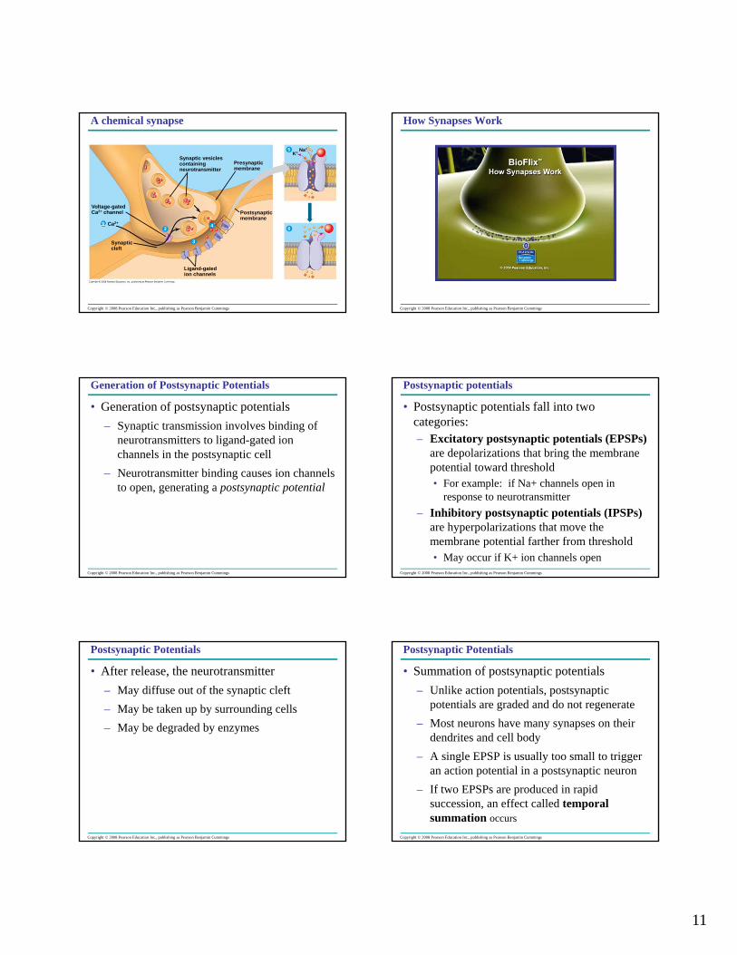
Voltage-gatedCa2+ channel
Ca2+12
3
4
Synapticcleft
Ligand-gatedion channels
Postsynapticmembrane
Presynapticmembrane
Synaptic vesiclescontainingneurotransmitter
5
6
K+ Na+
A chemical synapse
Copyright © 2008 Pearson Education Inc., publishing as Pearson Benjamin Cummings
How Synapses Work
Copyright © 2008 Pearson Education Inc., publishing as Pearson Benjamin Cummings
Generation of Postsynaptic Potentials
• Generation of postsynaptic potentials– Synaptic transmission involves binding of
neurotransmitters to ligand-gated ion channels in the postsynaptic cell
– Neurotransmitter binding causes ion channels to open, generating a postsynaptic potential
Copyright © 2008 Pearson Education Inc., publishing as Pearson Benjamin Cummings
Postsynaptic potentials
• Postsynaptic potentials fall into two categories:– Excitatory postsynaptic potentials (EPSPs)
are depolarizations that bring the membrane potential toward threshold• For example: if Na+ channels open in
response to neurotransmitter– Inhibitory postsynaptic potentials (IPSPs)
are hyperpolarizations that move the membrane potential farther from threshold• May occur if K+ ion channels open
Copyright © 2008 Pearson Education Inc., publishing as Pearson Benjamin Cummings
Postsynaptic Potentials
• After release, the neurotransmitter– May diffuse out of the synaptic cleft– May be taken up by surrounding cells – May be degraded by enzymes
Copyright © 2008 Pearson Education Inc., publishing as Pearson Benjamin Cummings
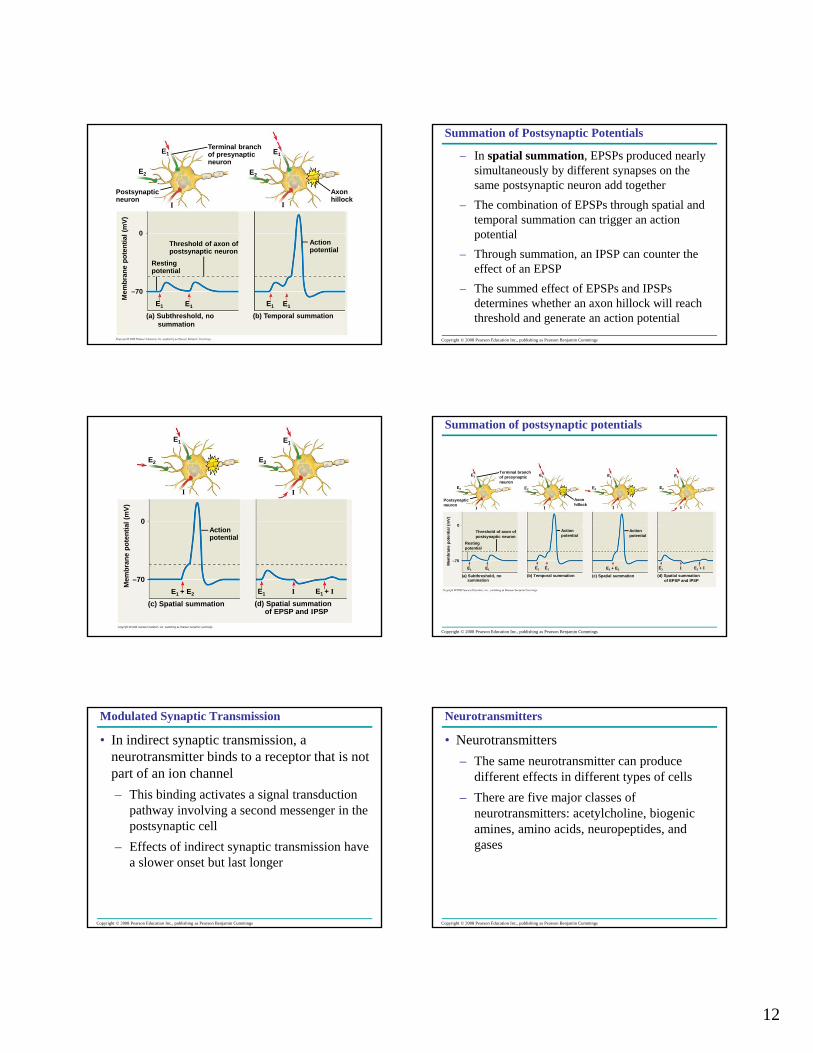
Postsynaptic Potentials
• Summation of postsynaptic potentials– Unlike action potentials, postsynaptic
potentials are graded and do not regenerate– Most neurons have many synapses on their
dendrites and cell body– A single EPSP is usually too small to trigger
an action potential in a postsynaptic neuron– If two EPSPs are produced in rapid
succession, an effect called temporal summation occurs
12
E1
E2
Postsynapticneuron
I
Terminal branchof presynapticneuron
E2
0
–70
Threshold of axon ofpostsynaptic neuron
Restingpotential
Mem
bran
e po
tent
ial (
mV)
E1E1
(a) Subthreshold, nosummation
(b) Temporal summation
E1 E1
Actionpotential
I
Axonhillock
E1
Copyright © 2008 Pearson Education Inc., publishing as Pearson Benjamin Cummings
Summation of Postsynaptic Potentials
– In spatial summation, EPSPs produced nearly simultaneously by different synapses on the same postsynaptic neuron add together
– The combination of EPSPs through spatial and temporal summation can trigger an action potential
– Through summation, an IPSP can counter the effect of an EPSP
– The summed effect of EPSPs and IPSPs determines whether an axon hillock will reach threshold and generate an action potential
E1
E2
Mem
bran
e po
tent
ial (
mV)
Actionpotential
I
E1 + E2
(c) Spatial summation (d) Spatial summationof EPSP and IPSP
E1
E1 E1 + II
I
E2
0
–70
Copyright © 2008 Pearson Education Inc., publishing as Pearson Benjamin Cummings
Terminal branchof presynapticneuron
E1
E2
I
Postsynapticneuron
Threshold of axon ofpostsynaptic neuron
Restingpotential
E1 E1
0
–70
Mem
bran
e po
tent
ial (
mV)
(a) Subthreshold, nosummation
(b) Temporal summation
E1 E1
Actionpotential
I
Axonhillock
E1
E2 E2
E1
I
Actionpotential
E1 + E2
(c) Spatial summation
I
E1 E1 + I
(d) Spatial summationof EPSP and IPSP
E2
E1
I
Summation of postsynaptic potentials
Copyright © 2008 Pearson Education Inc., publishing as Pearson Benjamin Cummings
Modulated Synaptic Transmission
• In indirect synaptic transmission, a neurotransmitter binds to a receptor that is not part of an ion channel– This binding activates a signal transduction
pathway involving a second messenger in the postsynaptic cell
– Effects of indirect synaptic transmission have a slower onset but last longer
Copyright © 2008 Pearson Education Inc., publishing as Pearson Benjamin Cummings
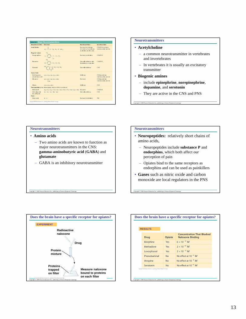
Neurotransmitters
• Neurotransmitters– The same neurotransmitter can produce
different effects in different types of cells– There are five major classes of
neurotransmitters: acetylcholine, biogenic amines, amino acids, neuropeptides, and gases
13
Copyright © 2008 Pearson Education Inc., publishing as Pearson Benjamin Cummings
Neurotransmitters
• Acetylcholine – a common neurotransmitter in vertebrates
and invertebrates– In vertebrates it is usually an excitatory
transmitter
• Biogenic amines – include epinephrine, norepinephrine,
dopamine, and serotonin– They are active in the CNS and PNS
Copyright © 2008 Pearson Education Inc., publishing as Pearson Benjamin Cummings
Neurotransmitters
• Amino acids– Two amino acids are known to function as
major neurotransmitters in the CNS: gamma-aminobutyric acid (GABA) and glutamate
– GABA is an inhibitory neurotransmitter
Copyright © 2008 Pearson Education Inc., publishing as Pearson Benjamin Cummings
Neurotransmitters
• Neuropeptides: relatively short chains of amino acids, – Neuropeptides include substance P and
endorphins, which both affect our perception of pain
– Opiates bind to the same receptors as endorphins and can be used as painkillers
• Gases such as nitric oxide and carbon monoxide are local regulators in the PNS
Copyright © 2008 Pearson Education Inc., publishing as Pearson Benjamin Cummings
EXPERIMENT
Radioactivenaloxone
Drug
Proteinmixture
Proteinstrapped on filter
Measure naloxonebound to proteinson each filter
Does the brain have a specific receptor for opiates?
Copyright © 2008 Pearson Education Inc., publishing as Pearson Benjamin Cummings
RESULTS
Does the brain have a specific receptor for opiates?
14
Copyright © 2008 Pearson Education Inc., publishing as Pearson Benjamin Cummings
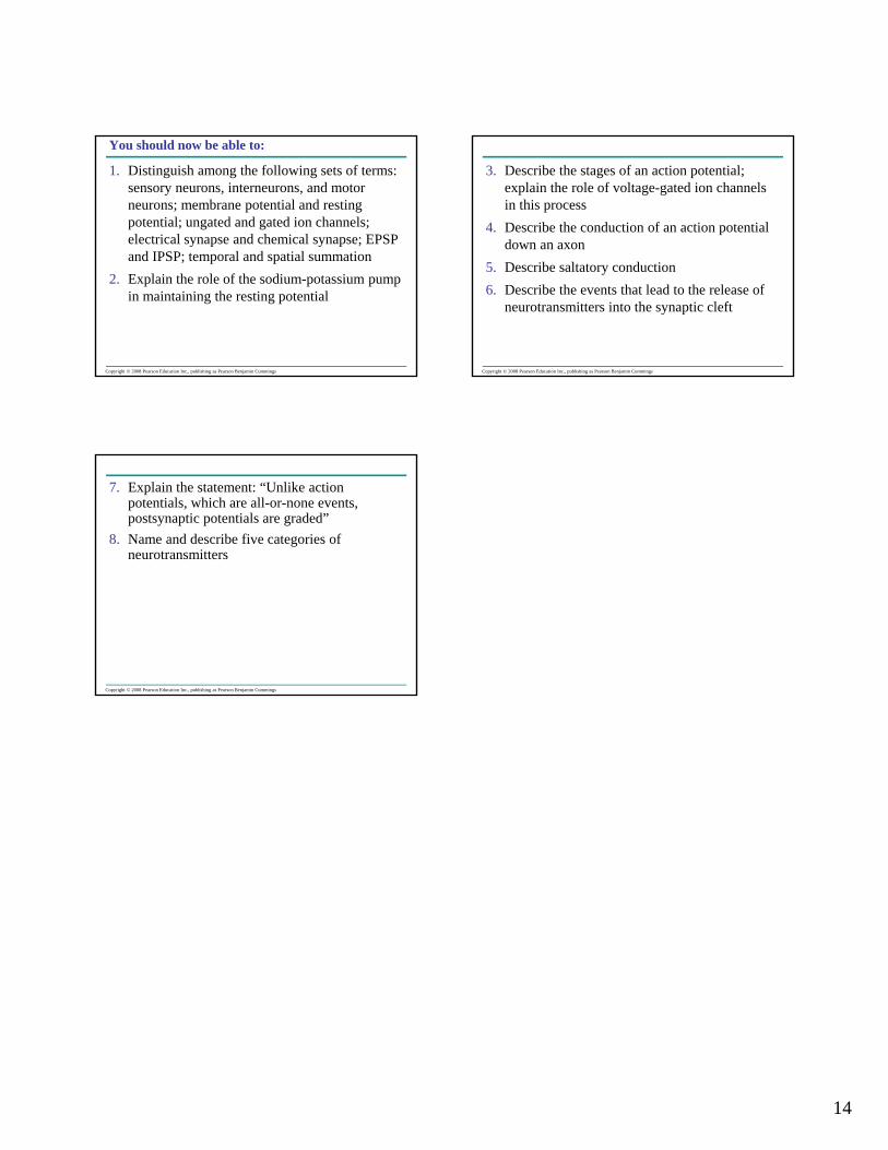
You should now be able to:
1. Distinguish among the following sets of terms: sensory neurons, interneurons, and motor neurons; membrane potential and resting potential; ungated and gated ion channels; electrical synapse and chemical synapse; EPSP and IPSP; temporal and spatial summation
2. Explain the role of the sodium-potassium pump in maintaining the resting potential
Copyright © 2008 Pearson Education Inc., publishing as Pearson Benjamin Cummings
3. Describe the stages of an action potential; explain the role of voltage-gated ion channels in this process
4. Describe the conduction of an action potential down an axon
5. Describe saltatory conduction6. Describe the events that lead to the release of
neurotransmitters into the synaptic cleft
Copyright © 2008 Pearson Education Inc., publishing as Pearson Benjamin Cummings
7. Explain the statement: “Unlike action potentials, which are all-or-none events, postsynaptic potentials are graded”
8. Name and describe five categories of neurotransmitters