Direct induction of neural progenitor cells transiently...
15
Direct induction of neural progenitor cells transiently passes through a partially reprogrammed state Rui Gao b, c, 1 , Wenchao Xiu a, 1 , Linfeng Zhang a , Ruge Zang a , Lei Yang d , Chenfei Wang a , Min Wang e , Mingzhu Wang a , Li Yi a , Yuanyuan Tang d , Yawei Gao a , Hong Wang a , Jiajie Xi a , Wenqiang Liu a , Yixuan Wang a , Xuejun Wen f , Yongchun Yu e , Yong Zhang a , Liang Chen b, c, ** , Jiayu Chen a, * , Shaorong Gao a, * a Clinical and Translational Research Center of Shanghai First Maternity & Infant Hospital, School of Life Sciences and Technology, Tongji University, Shanghai, 200092, China b School of Life Sciences, Tsinghua University, Beijing, 100084, China c National Institute of Biological Sciences, NIBS, Beijing, 102206, China d Shanghai First Maternity and Infant Hospital, Tongji University School of Medicine, Shanghai, 200092, China e Institute of Neurobiology, Institutes of Brain Science and State Key Laboratory of Medical Neurobiology, Fudan University, Shanghai, 200032, China f The Institute for Translational Nanomedicine, Shanghai East Hospital, Institute of Biomedical Engineering and Nanoscience, Tongji University School of Medicine, Shanghai, 200092, China article info Article history: Received 1 November 2016 Received in revised form 7 December 2016 Accepted 7 December 2016 Available online 10 December 2016 Keywords: Direct induction giNPCs Partially reprogrammed state abstract The generation of functional neural progenitor cells (NPCs) holds great promise for both research and clinical applications in neurodegenerative diseases. Traditionally, NPCs are derived from embryonic stem cells (ESCs) and induced pluripotent stem cells (iPSCs), or NPCs can be directly converted from somatic cells by sets of transcription factors or by a combination of chemical cocktails and/or hypoxia. However, the ethical issues of ESCs, the risk of tumorigenesis from iPSCs and transgenic integration from exoge- nous genes as well as complicated manipulation and time-consuming of chemical induced NPCs (ciNPCs) limit the applications of these strategies. Here, we describe a novel method for generating growth factor- induced neural progenitor cells (giNPCs) from mouse embryonic and adult fibroblasts by using inductive and/or permissive signaling culture conditions. These giNPCs closely resemble brain-derived NPCs in terms of transcription networks and neural lineage differentiation potentials. Moreover, this somatic cell to NPC induction is a gradual process that includes initiation, intermediate, maturation and stabilization stages. Importantly, gene expression and histone modification analyses further indicate a partially reprogrammed state during the generation process of induced NPCs, in which lineage specific genes and pluripotency associated genes are transiently activated. Our study therefore describes the potential safety problems that also exist in the transgene-free direct induction strategy and highlights the importance of excluding the possibility of residual partially reprogrammed and/or teratoma-like cells from the generated NPCs for future clinical trials. © 2016 Published by Elsevier Ltd. 1. Introduction The generation of induced pluripotent stem cells (iPSCs) [1e3] and induced neuronal cells [4e7] from somatic cells provides not only new cell sources for biomedical research but also potential cell therapy for neurological dysfunction, such as Alzheimer's disease and Parkinson's disease [8e12]. However, the risk of tumorigenicity observed in most iPSC lines may lead to the subsequently differ- entiated neuronal cells producing certain safety threats [13,14]. On the other hand, the long induction time course, the low efficiency * Corresponding authors. Clinical and Translational Research Center of Shanghai First Maternity & Infant Hospital, School of Life Sciences and Technology, Tongji University, Shanghai, 200092, China. ** Corresponding author. National Institute of Biological Sciences, NIBS, Beijing, 102206, China. E-mail addresses: [email protected] (L. Chen), [email protected] (J. Chen), [email protected] (S. Gao). 1 These authors contributed equally to this work. Contents lists available at ScienceDirect Biomaterials journal homepage: www.elsevier.com/locate/biomaterials http://dx.doi.org/10.1016/j.biomaterials.2016.12.007 0142-9612/© 2016 Published by Elsevier Ltd. Biomaterials 119 (2017) 53e67
Transcript of Direct induction of neural progenitor cells transiently...
lable at ScienceDirect
Biomaterials 119 (2017) 53e67
Contents lists avai
Biomaterials
journal homepage: www.elsevier .com/locate/biomater ia ls
Direct induction of neural progenitor cells transiently passes through apartially reprogrammed state
Rui Gao b, c, 1, Wenchao Xiu a, 1, Linfeng Zhang a, Ruge Zang a, Lei Yang d, Chenfei Wang a,Min Wang e, Mingzhu Wang a, Li Yi a, Yuanyuan Tang d, Yawei Gao a, Hong Wang a,Jiajie Xi a, Wenqiang Liu a, Yixuan Wang a, Xuejun Wen f, Yongchun Yu e, Yong Zhang a,Liang Chen b, c, **, Jiayu Chen a, *, Shaorong Gao a, *
a Clinical and Translational Research Center of Shanghai First Maternity & Infant Hospital, School of Life Sciences and Technology, Tongji University,Shanghai, 200092, Chinab School of Life Sciences, Tsinghua University, Beijing, 100084, Chinac National Institute of Biological Sciences, NIBS, Beijing, 102206, Chinad Shanghai First Maternity and Infant Hospital, Tongji University School of Medicine, Shanghai, 200092, Chinae Institute of Neurobiology, Institutes of Brain Science and State Key Laboratory of Medical Neurobiology, Fudan University, Shanghai, 200032, Chinaf The Institute for Translational Nanomedicine, Shanghai East Hospital, Institute of Biomedical Engineering and Nanoscience, Tongji University School ofMedicine, Shanghai, 200092, China
a r t i c l e i n f o
Article history:Received 1 November 2016Received in revised form7 December 2016Accepted 7 December 2016Available online 10 December 2016
Keywords:Direct inductiongiNPCsPartially reprogrammed state
* Corresponding authors. Clinical and TranslationalFirst Maternity & Infant Hospital, School of Life ScieUniversity, Shanghai, 200092, China.** Corresponding author. National Institute of Biolo102206, China.
E-mail addresses: [email protected] (L. Che(J. Chen), [email protected] (S. Gao).
1 These authors contributed equally to this work.
http://dx.doi.org/10.1016/j.biomaterials.2016.12.0070142-9612/© 2016 Published by Elsevier Ltd.
a b s t r a c t
The generation of functional neural progenitor cells (NPCs) holds great promise for both research andclinical applications in neurodegenerative diseases. Traditionally, NPCs are derived from embryonic stemcells (ESCs) and induced pluripotent stem cells (iPSCs), or NPCs can be directly converted from somaticcells by sets of transcription factors or by a combination of chemical cocktails and/or hypoxia. However,the ethical issues of ESCs, the risk of tumorigenesis from iPSCs and transgenic integration from exoge-nous genes as well as complicated manipulation and time-consuming of chemical induced NPCs (ciNPCs)limit the applications of these strategies. Here, we describe a novel method for generating growth factor-induced neural progenitor cells (giNPCs) from mouse embryonic and adult fibroblasts by using inductiveand/or permissive signaling culture conditions. These giNPCs closely resemble brain-derived NPCs interms of transcription networks and neural lineage differentiation potentials. Moreover, this somatic cellto NPC induction is a gradual process that includes initiation, intermediate, maturation and stabilizationstages. Importantly, gene expression and histone modification analyses further indicate a partiallyreprogrammed state during the generation process of induced NPCs, in which lineage specific genes andpluripotency associated genes are transiently activated. Our study therefore describes the potential safetyproblems that also exist in the transgene-free direct induction strategy and highlights the importance ofexcluding the possibility of residual partially reprogrammed and/or teratoma-like cells from thegenerated NPCs for future clinical trials.
© 2016 Published by Elsevier Ltd.
Research Center of Shanghainces and Technology, Tongji
gical Sciences, NIBS, Beijing,
1. Introduction
The generation of induced pluripotent stem cells (iPSCs) [1e3]and induced neuronal cells [4e7] from somatic cells provides notonly new cell sources for biomedical research but also potential celltherapy for neurological dysfunction, such as Alzheimer's diseaseand Parkinson's disease [8e12]. However, the risk of tumorigenicityobserved in most iPSC lines may lead to the subsequently differ-entiated neuronal cells producing certain safety threats [13,14]. Onthe other hand, the long induction time course, the low efficiency
R. Gao et al. / Biomaterials 119 (2017) 53e6754
and the restricted proliferative potential of induced differentiatedneuronal cells limit the clinical applications of this strategy. Bycontrast, induced neural progenitor cells (iNPCs), which are capableof self-renewing and differentiating into a wide range of neural celllineages, hold great promise for therapeutic approaches [15,16].Currently, diverse mouse [17e22] and human [23] cell types havebeen successfully converted to iNPCs via different direct inductionstrategies with the overexpression of transcription factors invarious combinations. These direct conversion approaches provedto be highly efficient and time-saving, but the risk of genomicintegration of the viral vectors sustains debates regarding safety.Alternatively, a footprint-free strategy for reprograming humanurine epithelial-like cells into NPCs using an episomal system hasbeen established [24]. More recently, an important advance hasindicated that small molecules can be used to generate chemical-induced neural progenitor cells (ciNPCs) from somatic cellswithout introducing exogenous genes [25,26]. This new means ofinduction has considerable advantages because the small mole-cules are nonimmunogenic and cost-effective. Moreover, their ef-fects on activating and inhibiting the function of specific factors arereversible and can be finely modulated by adjusting their concen-trations. However, this direct induction strategy involves compli-cated and replicated manipulation of chemical cocktails and a strictphysiological hypoxic condition [25].
Previously, direct conversion at themolecular level was typicallyachieved by ectopic expression of sets of cell type-specific tran-scription factors [27,28]. However, a more general lineage conver-sion approach is to transiently overexpress the conventionalYamanaka factors [17,19,29,30]. This strategy suggested that thetransient expression of pluripotency-associated genes couldgenerate a plastic intermediate state, which further serves as acellular platform for transdifferentiation into various lineages inthe presence of lineage-specifying extracellular signaling inputs.These processes did not involve the establishment of a pluripotentstate because mouse somatic cells subjected to Yamanaka-factor-based conversion approaches failed to activate transgenic plurip-otent reporters such as Oct4-GFP [17,19]. However, two recent in-dependent studies using different lineage tracing systemsconflicted with these issues [31,32]. The studies both concludedthat transdifferentiation systems using pulsed expression ofpluripotency-associated factors in combination withdifferentiation-inducing signals indeed involved a short-livedOct4-positive iPSC-like state. Then, the cells rapidly differentiatedinto the specified lineages [31,32]. Notably, passage through atransient pluripotent state might be the result of usingpluripotency-associated transcription factors. Thus, our interestsarose from two unsolved issues regarding: 1) Whether externalinductive and/or permissive signaling culture conditions couldinduced neural progenitor cells from somatic cells? 2) If the directcell type switch from somatic cells to functional NPCs withoutintroducing exogenous genes also involves a similar transientstage? Answering these questions will increase our understandingof the molecular mechanism underlying the establishment ofinduced neural progenitor cells. Further, answering these questionswill provide safety verification information for generating func-tionally desirable cell types with potential regenerative applica-tions by cell fate reprogramming using specific growth factors orchemical compounds instead of genetic manipulation.
Here, we present a novel method for generating growth factor-induced neural progenitor cells (giNPCs) by using inductive andpermissive signaling culture conditions with the combination ofonly a few growth factors. We show that these giNPCs are verysimilar to neonatal mouse brain-derived NPCs in terms of tran-scription networks and neural lineage differentiation potentialin vivo and in vitro. We demonstrate that somatic cell to NPC
induction is a gradual process that includes initiation, intermediate,maturation and stabilization stages with the erasure of a somatic-specific platform and establishment of an NPC-specific network.Importantly, our data further suggest that both our giNPC andpreviously reported ciNPC induction go through a partiallyreprogrammed state inwhich lineage genes from three germ layersand pluripotency-associated genes are transiently activated withthe corresponding reconstitution of histone modifications.
2. Materials and methods
2.1. Mouse and cell culture
The specific pathogen-free mice were housed in the animal fa-cility of Tongji University. All our study procedures were consistentwith the Tongji University Guide for the care and use of laboratoryanimals. Mouse embryonic fibroblasts (MEFs) were isolated from13.5-dpc embryos. Tail-tip fibroblasts (TTFs) were isolated from theadult Nestin-GFP transgenic mice. MEFs and mouse TTFs weremaintained in somatic cell culture medium [DMEM (Life Technol-ogies) supplemented with 10% (vol/vol) FBS (Gibco) and 1 mM L-glutamine (Merck Millipore)] at 37 �C with 5% CO2. Mouse brain-neural progenitor cells (Brain-NPCs) were derived from neonatalmice, digested by Accutase (Sigma) and cultured in NPC expansionmedium [DMEM/F12 supplemented with 1% N2 (Invitrogen) and2% B27 (Invitrogen), 20 ng/mL basic fibroblast growth factor (bFGF;R&D Systems) and 10 ng/mL epidermal growth factor (EGF;Peprotech)], as previously described [33].
2.2. Generation of giNPCs
For induction of neural progenitor cells, 1.5e2 � 105 MEFs orTTFs were initially plated in a well of a 24-well plate in initiationmedium [DMEM/F12 with the following supplements: 1% Gluta-MAX (Gibco), 2% B27 minus vitamin A (Gibco), 1 mg/mL heparin(Stem Cell Technologies), 1000 units/mL LIF (Merck Millipore),20 ng/mL bFGF and 20 ng/mL EGF]. Cells were gently pipetted eachday for the first week to prevent them from attaching to the dishbottom and to generate good sphere formation. After two weeks,the cells were digested by trypsin/EDTA (Invitrogen) and expandedto a 12-well or a 35-mm tissue culture dish in the presence of NPCexpansion medium. For MEF-giNPC induction, 7 days later, theneural rosettes were pipetted and passaged in suspension ontoultralow attachment plates (Costar) to form the growth factor-induced neural progenitor cells. For TTF-giNPC induction, theneural rosettes were pipetted and passaged in suspension after 2weeks. These formed MEF-giNPCs or TTF-giNPCs were cultured insuspension or under single-cell monolayer conditions for 3e4rounds of passaging to select for the fully induced giNPCs. Duringthe giNPC induction process, the culture medium was partiallychanged every other day.
2.3. Generation of ciNPCs
The chemical induced neural progenitor cells (ciNPCs) weregenerated as previously reported [25]. Briefly, initial 2 � 105 MEFswere seeded in each well of 6-well plates in KSR medium withchemical compounds (0.5 mM VPA, 3 mM CHIR99021, 1 mM Repsoxand 2 mM Parnate). As indicated previously [25], VPA is an HDACinhibitor and Parnate is an H3K4 demethylation inhibitor. The ef-fects of these two small molecules suggest that H3K4 demethyla-tion and histone deacetylation could be two critical epigeneticbarriers to reprogramming. CHIR99021 is a GSK3-b inhibitor andRepsox is a TGF-b signaling inhibitor. Either of them could effi-ciently replace Sox2 for reprogramming. Cells were cultured at
R. Gao et al. / Biomaterials 119 (2017) 53e67 55
37 �C under 5% O2 (hypoxia) and 5% CO2. Medium containingchemical compounds was changed every other day. 10 days later,cell mixtures were digested by trypsin/EDTA and expanded in NPCexpansion medium under 20% O2 (normoxic) conditions. ciNPCswere formed around day 20 and were enriched by rounds of neu-rosphere suspension culture.
2.4. In vitro differentiation of giNPCs
For spontaneous differentiation into all neural types, 5 � 104
giNPCs were plated onto PDL (Invitrogen)/Laminin (Sigma)-coatedglass coverslips in 12 wells containing NPC expansion medium.After 24 h, the medium was switched to NPC expansion mediumwithout growth factors for 1e2 weeks [23]. Robust astrogenesiswas induced by adding 1% FBS to NPC expansion medium withoutgrowth factors for one week [23]. For the analysis of the neuronaldifferentiation potential of the giNPCs, the cells were plated ontoPDL/Laminin-coated glass coverslips in 12 wells in medium con-sisting of a 1:3 mix of DMEM/F12 and Neurobasal (Invitrogen), 1%N2 and 2% B27 supplements, and 6.7 ng/mL bFGF and 20 ng/mLbrain-derived neurotrophic factor (BDNF; PeproTech). After 3 days,the media was replaced with freshmediumwith a reduced amountof 5 ng/mL bFGF and an increased amount of 30 ng/mL BDNF. Thecells were maintained under these conditions for an additional10e15 days, and the mediumwas partially changed every 2e3 days[34,35]. For oligodendrocyte differentiation, the cells were platedonto PDL/Laminin-coated coverslips and cultured in N2B27 me-diumwith 10 ng/mL bFGF and 10 ng/mL PDGF-AA (Peprotech) for 3days and then with 100 ng/mL T3 (Sigma-Aldrich) for 4e6 days[36].
2.5. RNA preparation and quantitative real-time PCR
Total RNA was extracted using Trizol reagent (Invitrogen) andreverse transcribed using 5X All-in-One RT MasterMix (AppliedBiological Materials) according to the manufacturer's recommen-dations. Quantitative RT-PCR was performed using SYBR Premix ExTaq II (Takara), and the signals were detectedwith an ABI7500 Real-Time PCR System (Applied BioSystems). All samples were run intriplicate. Glyceraldehyde 3-phosphate dehydrogenase (Gapdh)was used as an endogenous control. The values were normalizedbased on Gapdh expression. All the primers used were synthesizedat SangonBiotech (Shanghai) Co., Ltd, and are listed in Table S1.
2.6. Confocal microscopy, immunofluorescent staining andquantitative analyses
For immunofluorescent staining, MEFs, mouse TTFs, giNPCs,Brain-NPCs or neural cells were plated onto PDL-coated glass cov-erslips (Thermo). The cells were fixed in 4% paraformaldehyde(Sigma) overnight at 4 �C. After washing in phosphate bufferedsaline (PBS), the cells were permeabilized for 15 min in PBS con-taining 0.5% Triton X-100 and, subsequently, were blocked with2.5% bovine serum albumin (BSA) in PBS for 1 h at room temper-ature. Then, the cells were incubated overnight at 4 �C in PBScontaining 2.5% BSA with the primary antibodies. The primaryantibodies used in this study were as follows: mouse anti-Nestin(1:1000; Chemicon), goat anti-Sox1 (1:1000, R&D Systems),mouse anti-Sox2 (1:500; Santa Cruz Biotechnology), rabbit anti-b-III-Tubulin (Tuj1, 1:1000; Covance), mouse anti-Map2 (1:500; Mil-lipore), and rabbit anti-Gfap (1:1000; Millipore). Then, the cellswere washed three times with PBS and incubated for 1 h at roomtemperature with secondary antibodies conjugated with Alexa-Fluor 488, AlexaFluor 594, or AlexaFluor 633 (1:500; Invitrogen).The DNA was labeled with 40, 6-diamidino-2-phenylindole (DAPI).
The glass coverslips were observed with a NIKON ECLIPSE 80i mi-croscope or ZEISS LSM 880 microscope using the Plan Fluor 20X or40X DIC or 63X Oil objective.
For quantitative analyses of giNPCs' differentiation efficiency,giNPCs were plated onto PDL/Laminin-coated glass coverslips in12-well plates containing certain differentiation medium. MEFsand Brain-NPCs were set as negative and positive controls,respectively. In order to exclude mutual interference between pri-mary antibodies, each coverslip was stained with only one primaryantibody. Three independent experimental replicates were con-ducted. For each immunostaining experiment, three vision fields ofone kind of differentiation were randomly selected. The “Percent-age (%)” was calculated as the total marker positive cells (immu-noreactive cells) over total DAPI positive cells. The actual number ofcells showing immunoreactivity for each marker as well as thewhole population under certain differentiation conditions areprovided in Table S2.
2.7. Electrophysiological analysis
Electrophysiological experiments were performed using giNPC1#- or Brain-NPC-derived neurons. Whole-cell patch clamp re-cordings in either voltage or current clamp mode were conductedto measure voltage-activated sodium/potassium currents and ac-tion potentials. The output signals from the Multiclamp 700Bamplifier were digitized using a DigiData 1440 A/D D/A board, low-pass filtered at 5 KHz. Glass micropipettes (2.0e4.0 MU) containinga solution of 130 mM Kþ-gluconate, 20 mM KCl, 10 mM HEPES,0.2 mM EGTA, 4 mM Mg2ATP, 0.3 mM Na2GTP, and 10 mM Na2þ-phosphocreatine (at pH 7.3, 290e310 mOsm) were used for patch-clamp recordings. The bath solution contained 140 mMNaCl, 5 mMKCl, 2 mM CaCl2, 2 mMMgCl2, 10 mMHEPES and 10mM glucose (atpH 7.4). Membrane potentials were hold around�100mV, and stepcurrents with an increment of 20 pA were injected to elicit actionpotentials. A ramp protocol (from�80 toþ60mV)was used to elicitinward sodium current and outward potassium currents. Datawereanalyzed using pClamp10 (Clampft).
2.8. In vivo transplantation
In vivo transplantation was performed as previously described[37]. Briefly, uterine horns of E13.5 gestation stage pregnant C57BL/6 mice were maintained in pathogen-free condition. 2 ml PBS con-taining around 20 GFP-giNPC line 1# neurospheres with neuro-sphere diameter less than 80 mm was injected into the embryoniccerebral ventricle through a bevelled, calibrated glass micropipette.After that, the uterine horns were replaced, the peritoneal cavitywas lavaged with 10 mL warm PBS containing antibiotics and thewound was closed. 1 month after birth, mice were anesthetized onice or with pentobarbital sodium and brains section were preparedas described above for further analysis. The primary antibodiesused in the following study were as follows: mouse anti-GFP(1:100; Proteintech) and rabbit anti-b-III-Tubulin (Tuj1, 1:1000;Covance). The brains section were observed with a Zeiss LSM 880with airyscan microscope using the Plan Fluor 25X or 63X Oilobjective.
2.9. Teratoma assay and H&E staining
A total of 5 � 105 cells, including the induction cells from Day 0,Day 1, Day 4, Day 7, Day 10, and Day 14 as well as giNPC (lines 1#and 2#), brain-NPCs and iPSCs, were individually suspended in250 ml of PBS and subcutaneously injected into the SCID mouse totest their teratoma formation ability. Four weeks post-injection,subcutaneous tissue were dissected and processed for

R. Gao et al. / Biomaterials 119 (2017) 53e6756
hematoxylin-eosin (H&E) staining, which was performed at theShanghai Yichang Biological Technology Co., Ltd.
2.10. RNA-seq library generation and illumina HiSeq 2500sequencing
During the giNPC induction process, cell samples were har-vested at various induction days including Day 1, Day 4, Day 7, Day10, Day 14, Day 17 and Day 21. Simultaneously, samples from pri-mary neurosphere-like cluster (pre-NPCs), an established giNPCcell line at passage 6 (giNPCs P6), Brain-NPCs as well as teratomacells were also collected. Total RNA was isolated from cell pelletsusing Trizol reagent, and RNA-seq libraries were generated usingKAPA Stranded mRNA-Seq Kits according to the manufacturer'srecommendations. Briefly, the mRNAwas enriched using oligo (dT)magnetic beads and sheared to create short fragments of approxi-mately 300 bp. cDNA was synthesized using random hexamerprimers and purified using 1.8X Agencourt AMPure XP beads(Beckman). Finally, the sequencing primers linked to the cDNAfragments (approximately 300 bp) were isolated by gel electro-phoresis and enriched by PCR amplification to construct the library.The sequencing was performed at the Berry Genomics Co Ltd. andthe Sequencing Center in National Institute of Biological Sciences,Beijing, using the HiSeq 2500 system developed by Illumina.Paired-end sequencing was applied.
2.11. Sequencing analysis
Public RNA-seq datasets including MEFs (GSE43986), mouseESCs (GSE59463) and iPSCs (GSE54619) were downloaded from theGEO data repository and integrated in the analysis. All RNA-seqreads were mapped to the mouse genome using the Tophat soft-ware (version: v2.0.12) [38]. The expression level for each genewasquantified to the reads count using htseq-count software (version:0.6.0) and to FPKM using Cufflinks software (version: v2.2.1) [39].Then, the clustering analysis was performed by heatmap and PCApackages within custom R script software. Differential expressionanalysis was conducted using edgeR software (version: 3.12.0) onthe basis of a comprehensive consideration of the P value (cutoff to0.05) and fold-change (cutoff to 4.5) via the ‘Cuffdiff’ command[40].
2.12. Chromatin immunoprecipitation (ChIP)
ChIP experiments were performed using the MAGnify™ Chro-matin Immunoprecipitation System (Invitrogen) according to themanufacturer's recommendations. Briefly, ~105 cells were resus-pended in lysis buffer, and chromatinwas sonicated to 200e500 bpwith the Covaris M220 system. Then, the sonicated chromatin wasimmunoprecipitated with the following antibodies: anti-H3K4me3(Abcam) and anti-H3K27me3 (Millipore). A fraction of ‘whole-cellextract’ obtained without antibody was retained as an input con-trol, whereas DNA fragments obtainedwith normal rabbit or mouseIgG were applied as negative controls. The DNA isolated from theChIP was quantified using a Qubit 2.0 Fluorometer (Life Technolo-gies), and qRT-PCR was performed to validate the enrichmentamounts. The primers are listed in Table S1.
2.13. Statistics
The Holm-Sidak test (for ANOVA) or Student's test was per-formed using GraphPad Prism 5 software for statistics comparison.
2.14. Accession numbers
The RNA-seq datasets have been deposited in Gene ExpressionOmnibus (GEO) and are accessible under the GEO accession num-ber GSE76857.
3. Results
3.1. Growth factor induction of mouse neural progenitor cells(NPCs)
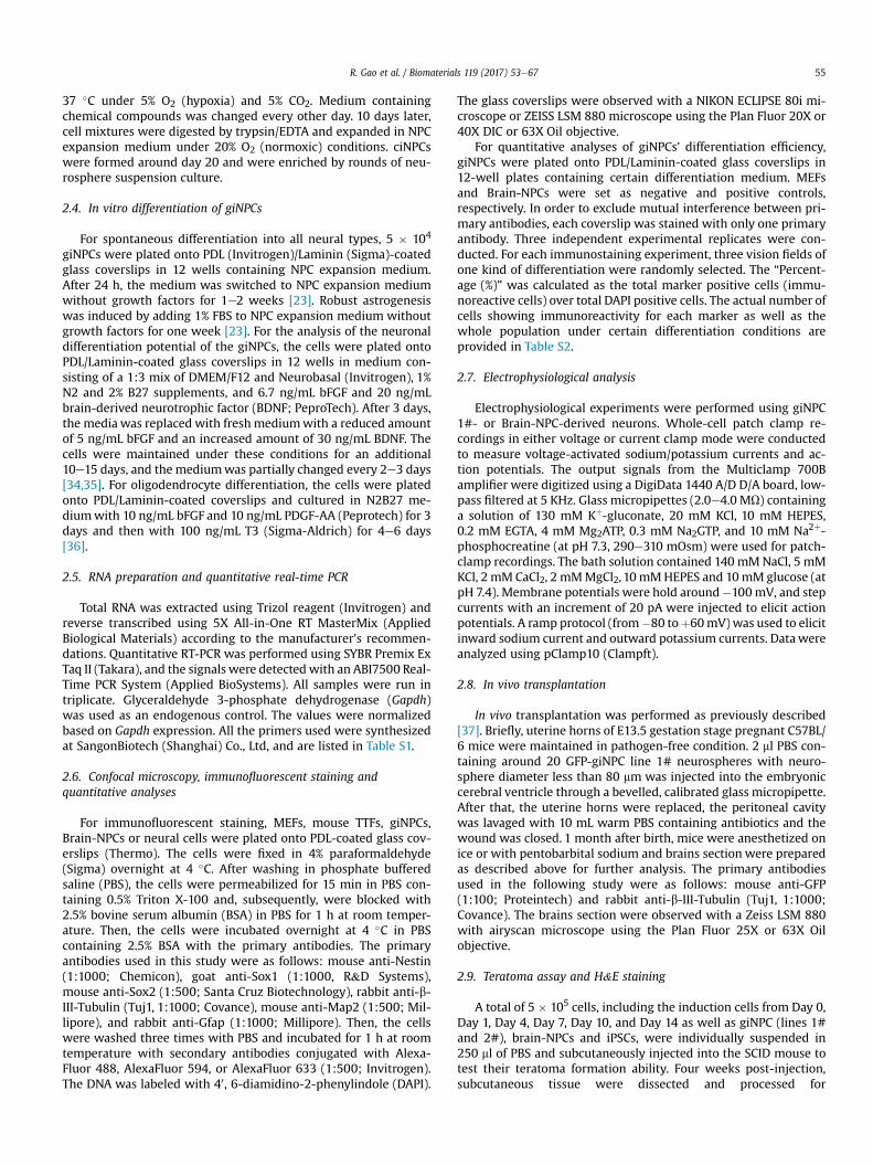
Previous studies provided evidence that direct lineage conver-sion of different somatic cells to self-renewal, multipotent andneural lineage-restricted NPCs could be achieved by sets of definedtranscription factors [17e23] or by using the combination ofchemical cocktails and/or hypoxia [25,26]. However, due to theintroduction of exogenous genes as well as the complicatedmanipulation of chemicals, there are still a lot of concerns comingalong these techniques. Here, we presented a novel method usinginductive and permissive signaling culture conditions with thecombination of only a few growth factors including B27 minusvitamin A, heparin, LIF, bFGF and EGF, which could successfullyinduce NPCs from differentiated somatic cells. Firstly, mouse em-bryonic fibroblasts (MEFs) were plated and cultured in the initia-tion medium with the following supplements: B27 minus vitaminA, heparin, LIF, bFGF and EGF. Cells were gently pipetted each dayfor the first week to prevent them from attaching to the dish bot-tom, and sphere morphology was visualized (Fig. 1A and B, Day 1and Day 7). Secondly, these sphere-like colonies attached to thebottom of the culture dishes, followed by cell mixtures migratingand gradually forming monolayer structures in the following week(Fig. 1A and B, Day 14). Thirdly, cell mixtures were digested andexpanded in the presence of NPC expansion medium with thefollowing supplements: N2, B27, bFGF and EGF (Fig. 1A and B, Day15) to establish primary neurosphere-like networks (Fig. 1A and B,Day 21). Lastly, primary NPC-like cells (Fig. 1A and B, Pre-NPCs)were isolated and subjected to 3e4 rounds of passaging to selectfor the fully induced neural progenitor cells (giNPCs). Free-floatingclusters were observed when these giNPCs were cultured in sus-pension (Fig. 1A and B, giNPCs in suspension). Alternatively, undersingle-cell monolayer culture conditions, NPC-like bipolarmorphology was observed (Fig. 1A and B, adherent giNPCs). Inaddition, the flow cytometric analysis and imaging of this growthfactor mediated induction process by using Nestin-GFP MEF cellsfurther indicated a gradual acquisition of the neural phenotype(Fig. 1CeD and S1A-S1B). Simultaneously, we verified the extent ofcell death and cell viability during this process. Early apoptosisunder the long induction period displayed many minor fluctua-tions, whereas the extent of late apoptosis and cell death graduallydecreased (Fig. S1C). Overall, the extent of cell death under thegrowth factor mediated induction process is not significant.
3.2. giNPCs show transcriptional profiles similar to those of brain-NPCs
To characterize the identity of growth factor-induced NPCs(giNPCs), we performed comprehensive molecular analysis at thetranscription and protein levels. We randomly chose four giNPClines, which were derived from several independent experimentalreplicates and were cultured in suspension (giNPC lines 1# and 2#)or adherent (giNPC lines 5# and 6#) culture conditions. All giNPCsdisplayed typical NPC morphologies (Fig. 1B, giNPCs in suspensionand adherent giNPCs), and themean doubling times were similar tothat of brain-derived NPCs (Brain-NPCs) (Fig. S2A). Quantitativeanalysis demonstrated that giNPCs expressed typical NPC markers,
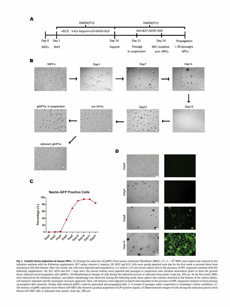
Fig. 1. Growth Factor induction of mouse NPCs. (A) Strategy for induction of giNPCs from mouse embryonic fibroblasts (MEFs). 1.5e2 � 105 MEFs were plated and cultured in theinitiation medium with the following supplements: B27 minus vitamin A, heparin, LIF, bFGF and EGF. Cells were gently pipetted each day for the first week to prevent them fromattaching to the dish bottom. After two weeks, the cells were digested and expanded to a 12-well or a 35-mm tissue culture dish in the presence of NPC expansion mediumwith thefollowing supplements: N2, B27, bFGF and EGF. 7 days later, the neural rosettes were pipetted and passaged in suspension onto ultralow attachment plates to form the growthfactor-induced neural progenitor cells (giNPCs). (B) Morphological changes of cells during the induction process at indicated time points. Scale bar, 200 mm. At the first week, MEFswere induced by the initiation medium, and sphere morphology was observed. During the following week, these sphere-like colonies attached to the bottom of the culture dishes,cell mixtures migrated and the monolayer structure appeared. Then, cell mixtures were digested at Day14 and expanded in the presence of NPC expansion medium to form primaryneurosphere-like networks. Finally, fully induced giNPCs could be generated and propagated after 3e4 rounds of passages under suspension or monolayer culture conditions. (C)The kinetics of giNPC induction from Nestin-GFP MEF cells showed a gradual acquisition of GFP positive signals. (D) Representative images of cells during the induction process fromNestin-GFP MEF cells at indicated time points. Scale bar, 200 mm.

R. Gao et al. / Biomaterials 119 (2017) 53e6758
including Nestin, Pax6 and Sox2, instead of fibroblast-associatedgenes Thy1 and Col3a1, which was similar to the expression ofBrain-NPCs (Fig. 2A and S2B). Immunofluorescent staining further
Fig. 2. Characterization of giNPCs. (A) qRT-PCR analysis of fibroblast- and NPC- specific gecultured in NPC expansion medium. Brain-NPCs (passage 6) and MEFs are set as controls. Thspecific markers in giNPC line 1# at passage 6. These giNPCs are subjected to monolayer cu50 mm. (C) Heatmap and hierarchical clustering of gene expression profiles in giNPCs, Brainlines 1# and 2# at passage 22. Brain-NPCs (passage 26) and MEFs are set as controls. Thespecific markers in giNPC line 1# at passage 22. These giNPCs are subjected to monolayer cu***p < 0.001 by ANOVA or Student's t-test for comparison.
confirmed that giNPCs, like Brian-NPCs, were consistently positivefor NPC markers, including Nestin, Sox2 and Sox1 (Fig. 2B and S2C).However, no morphological changes or Nestin-, Sox1-, or Sox2-
nes in giNPC lines 1# and 2# at passage 6. giNPC lines 1# and 2# are suspended ande expression levels were normalized to Gapdh. (B) Immunofluorescent staining of NPClture. Brain-NPCs and MEFs are positive and negative controls, respectively. Scale bar,-NPCs and MEFs. (D) qRT-PCR analysis of fibroblast- and NPC- specific genes in giNPCexpression levels were normalized to Gapdh. (E) Immunofluorescent staining of NPClture. Scale bar, 50 mm. Data in (A) and (D) are represented as the mean ± SEM (n ¼ 3).

Fig. 3. Generation of giNPCs from mouse adult fibroblasts. (A) Morphological changes of cells during the induction process at indicated time points. Tail-tip fibroblasts (TTFs)were derived from Nestin-GFP transgenic mice. No Nestin-GFP positive cells were observed in the starting TTFs, while Nestin-GFP positive and neurosphere-like cells graduallyappeared. Nestin-GFP positive TTF-giNPCs were then generated. Scale bar, 200 mm. (B) qRT-PCR analysis of fibroblast- and NPC-specific gene expression in TTF-giNPCs. Brain-NPCsand TTFs are set as controls. The expression levels were normalized toTTF. (C) Immunofluorescent staining of NPC specific markers in TTF-giNPCs. Brain-NPCs and TTFs are positiveand negative controls, respectively. Scale bar, 100 mm. Data in (B) are represented as the mean ± SEM (n ¼ 3). ***p < 0.001 by Student's t-test for comparison.
R. Gao et al. / Biomaterials 119 (2017) 53e67 59
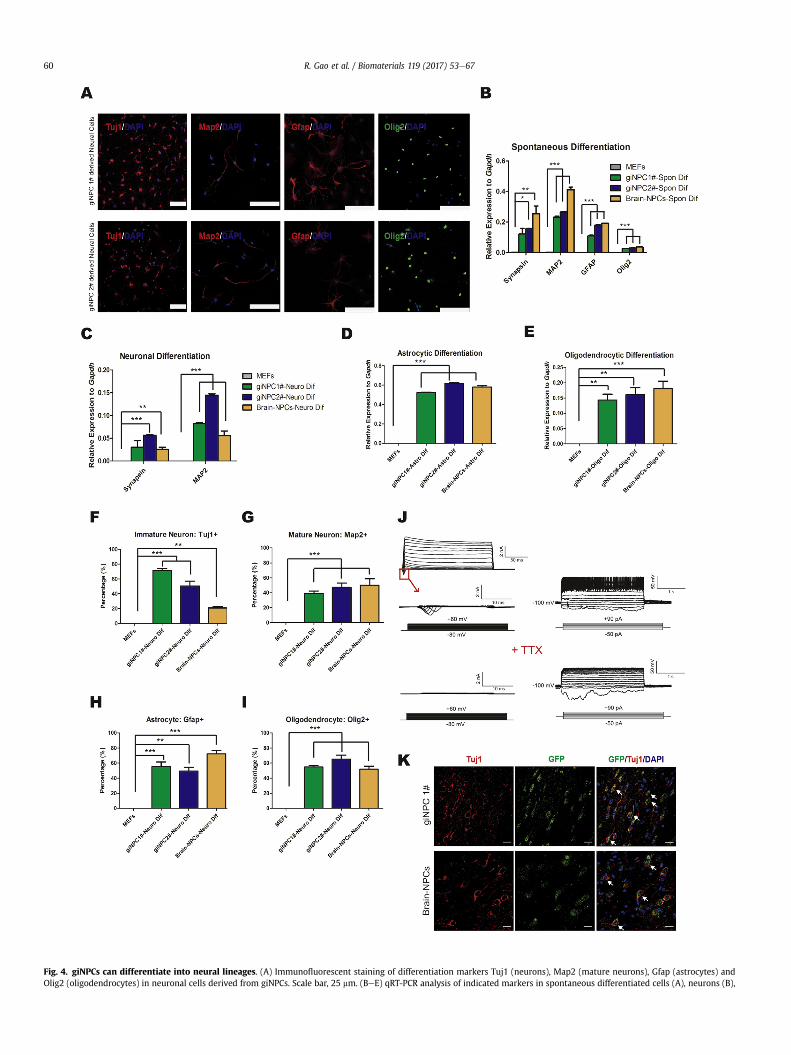
Fig. 4. giNPCs can differentiate into neural lineages. (A) Immunofluorescent staining of differentiation markers Tuj1 (neurons), Map2 (mature neurons), Gfap (astrocytes) andOlig2 (oligodendrocytes) in neuronal cells derived from giNPCs. Scale bar, 25 mm. (BeE) qRT-PCR analysis of indicated markers in spontaneous differentiated cells (A), neurons (B),
R. Gao et al. / Biomaterials 119 (2017) 53e6760
R. Gao et al. / Biomaterials 119 (2017) 53e67 61
positive cells were observed when MEFs were cultured in somaticcell culture medium (Fig. 2B). Importantly, the global geneexpression profiles of giNPCs resembled Brain-NPCs but were quitedistinct from those of MEFs (Fig. 2C and S2D). Moreover, thesegiNPCs maintained the NPC characteristics after prolonged culturein their morphologies, proliferation potency or gene expression(Fig. 2DeE and S2E-S2F). Taken together, these data suggest thathomogenous expandable giNPCs resembling Brain-NPC propertiescan be successfully generated from MEFs in our system.
3.3. Generation of giNPCs from adult mouse fibroblasts
To rule out the possibility that giNPCs were induced from certainneural cell types in the startingMEF populations, we treated tail-tipfibroblasts (TTFs) from adult Nestin-GFP transgenic mice in thesame induction system. No Nestin-GFP positive cells were observedin the starting TTFs, whereas during the induction process, Nestin-GFP positive cells appeared at an early stage, and the GFP signalinggradually increased (Fig. 3A). In all, the morphological changes ingiNPCs induced from TTFs were similar to those from MEFs, andmature TTF-giNPCs were successfully obtained despite a prolongedinduction period. These Nestin-GFP positive TTF-giNPCs had typicalNPC morphology and neurosphere-forming ability. The qRT-PCRand immunostaining analyses further confirmed that TTF-giNPCsexpressed NPC markers, including Nestin, Pax6 and Sox2, as Brain-NPCs did (Fig. 3B and C). However, Thy1 and Col3a1, the fibroblastspecific genes, were down-regulated (Fig. 3B). Moreover, no NPC-specific markers were expressed in the starting TTFs (Fig. 3B andC). In conclusion, giNPCs can be induced not only from mouseembryonic fibroblasts but also from mouse adult fibroblasts.
3.4. giNPCs can differentiate into neural lineages
We then analyzed the developmental potential of giNPCs byassessing their capacity for differentiation into the major subtypesof the neural lineage. By removing growth factors from the NPCexpansion medium for 1e2 weeks, giNPCs were able to differen-tiate into neurons (Tuj1-positive or Map2-positive) or astrocytes(Gfap-positive), which were similar to Brain-NPCs (Fig. 4AeB andS3A). For mature neuronal differentiation, the differentiated cellsfrom giNPCs showed typical neuronal morphology and expressedneuronal cell markersMap2 and Synapsin (Fig. 4A and C). Moreover,these cells displayed expression of glutamatergic and GABAergicneuron markers, which indicated mature synapse formation (Fig.S3B and S3D). Quantitative analysis of the immunostaining re-sults further showed that efficiency of glutamatergic differentiationbetween giNPCs and Brain-NPCs were comparable (Fig. S3C). Be-sides, giNPCs could also differentiate into astrocytes (Gfape-positive) and oligodendrocytes (Olig2-positive) as Brain-NPCs did(Fig. 4A and D-E). In addition, quantitative analysis demonstratedthat the in vitro differentiation efficiencies of giNPCs into majorneural lineages was quite similar to those of Brain-NPCs (Fig. 4FeI).In contrast, the starting MEFs did not express neuronal cellmarkers, which was quite different from cells differentiated fromgiNPCs (Fig. 4AeI and S3A-S3C).
The maturity and function of giNPCs were further assessed byin vitro and in vivo analyses. By whole-cell patch-clamp analysis, the
astrocytes (C) and oligodendrocytes (D) derived from giNPCs. MEFs and neuronal cells derlevels were normalized to Gapdh. (FeI) Quantitative analysis of indicated immature neurongiNPC differentiation. MEFs and neuronal cells derived from Brain-NPCs are set as negativneurons differentiated from giNPC line 1#. Whole cell recordings show voltage-gated curreinward currents can be blocked by adding tetrodotoxin (TTX). (K) Immunostaining of in vivcontrol. Arrows indicate GFP þ cells expressing Tuj1. Nuclei were counterstained with DAPI(F)e(I) are represented as the mean ± SEM (n ¼ 9). *p < 0.05; **p < 0.01; ***p < 0.001 by
differentiated neurons from giNPCs, similar to those differentiatedfrom Brain-NPCs, expressed voltage-gated ion channels and hadthe ability to generate single or multiple action potentials evokedby current injection, indicating functional membrane properties(Fig. 4J and S3E). Thus, giNPCs are capable of differentiating intomajor neural lineages in vitro, which are quite similar to theircounterparts derived from newborn brain tissue. To determine thegiNPC's developmental potential in vivo, giNPCs weremicroinjectedinto the embryonic cerebral ventricle of E13.5 pups, and their sur-vival and differentiation were evaluated about 1 month after thesepups were born [37]. Remarkably, neither obvious developmentalabnormality nor tumors or deformity was observed in thesetransplanted pups. Further immunofluorescent analysis demon-strated that cells differentiated from transplanted giNPCs were ableto survive and migrate into diverse brain sections, which closelyresembled Brain-NPCs (Fig. 4K).
3.5. Induction of giNPCs is a gradual process
To gain insight into the molecular mechanisms involved in thegiNPC induction process, we harvested samples at different timepoints and compared the global gene expression profiles of thesecells by RNA-seq analysis. The global genome heatmap indicated agenome-wide transcriptional conversion from a somatic state to anNPC state (Fig. S4A). Accordingly, both principal component anal-ysis (PCA) and unsupervised hierarchical clustering analysis indi-cated that this somatic to NPC induction was a gradual process,which included the initiation, intermediate, maturation and stabi-lization stages (Fig. 5A and B). At the initiation stage, MEFs wereinduced by the initiation medium, and sphere morphology wasobserved within the first week (Fig. 1B and 5A-B, Day 1, Day 4 andDay 7). At the intermediate stage, the monolayer structureappeared, and cells started to express NPC specific genes (Fig. 1Band 5A-B, Day 10 and Day 14). Surprisingly, cells in this stageshowed a transcriptional profile similar to that of teratomas, whichare known to express genes from three germ layers. At the matu-ration stage, primary neurosphere-like networks formed, and NPCspecific genes were prominently up-regulated (Fig. 1B and 5A-B,Day 17 and Day 21). At the stabilization stage, free floating giNPCscould be generated and propagated after 3e4 rounds of passages,showing a transcriptional pattern similar to that of Brain-NPCs (Fig.1B and 5A-B). These various transcriptional patterns were furtherconfirmed by hierarchical clustering and Pearson analysis, whichdemonstrated that the global gene expression patterns of giNPCswere similar to those of brain-NPCs but were different from thoseof ESCs, iPSCs and MEFs (Fig. 5C).
3.6. Transcriptome dynamics in the direct induction system
Based on the gene-expression dynamics, we clustered thesignificantly changed genes into eight groups (3106 genes in total)during the giNPC induction process (Fig. 6A). A large set of genes(clusters I, II, III and IV) including fibroblast markers were repressedacross this process. Meanwhile, previously reported Mesenchymal-to-Epithelial Transition (MET) associated genes, such as Thy1,Col1a1 as well as Col3a1, were included (Fig. 6A, clusters II and IIIand Figs. S4BeS4C) [41]. By contrast, neural specific markers, such
ived from Brain-NPCs are negative and positive controls, respectively. The expressions (F), mature neurons (G), astrocytes (H) and oligodendrocytes (I) obtained followinge and positive controls, respectively. (J) Electrophysiological properties of functionalnts and evoked action potentials in response to the injected current. Simultaneously,o transplanted GFP labeled giNPC line 1#. The transplanted brain-NPCs are set as the. Scale bar, 20 mm. Data in (B)e(E) are represented as the mean ± SEM (n ¼ 3). Data inANOVA or Student's t-test for comparison.

Fig. 5. Induction from MEFs into giNPCs is a gradual process. (A) Three-dimensional principal component analysis (PCA) of gene expression in cell samples during the giNPCinduction process at indicated time points. Sequencing reads of MEFs (GSE43986), mouse ESCs (GSE59463) and iPSCs (GSE54619) are cited from public datasets. The arrow indicatesa gradual induction process from the MEF state to the NPC state. (B) Unsupervised hierarchical clustering of gene expression profiles clearly shows that the giNPC induction processgoes through four stages. (C) Hierarchical clustering and Pearson correlation analysis of different time points during the giNPC induction process. The color represents the Pearsoncorrelation coefficient (red for higher correlation and blue for lower correlation), and the values are indicated. (For interpretation of the references to colour in this figure legend, thereader is referred to the web version of this article.)
R. Gao et al. / Biomaterials 119 (2017) 53e6762
as Pax6, Sox2 as well as Nestin, were gradually reactivated from theintermediate stage and highly expressed in the maturation andstabilization stages (Fig. 6A, clusters V, VI and VII and Fig. 6BeC).
Additionally, expression of brain region-specific genes, includingventral brain-specific markers (such as Olig2), dorsal brain markers(such as Sox3), forebrainmarkers (such as Foxg1), midbrainmarkers
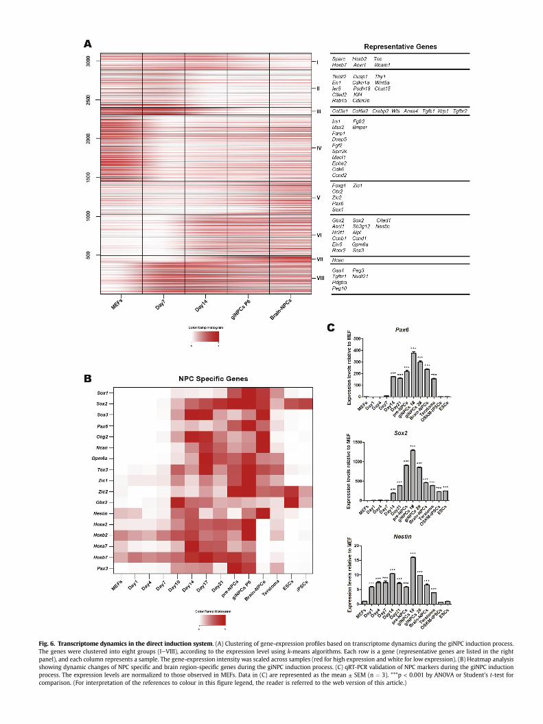
Fig. 6. Transcriptome dynamics in the direct induction system. (A) Clustering of gene-expression profiles based on transcriptome dynamics during the giNPC induction process.The genes were clustered into eight groups (IeVIII), according to the expression level using k-means algorithms. Each row is a gene (representative genes are listed in the rightpanel), and each column represents a sample. The gene-expression intensity was scaled across samples (red for high expression and white for low expression). (B) Heatmap analysisshowing dynamic changes of NPC specific and brain region-specific genes during the giNPC induction process. (C) qRT-PCR validation of NPC markers during the giNPC inductionprocess. The expression levels are normalized to those observed in MEFs. Data in (C) are represented as the mean ± SEM (n ¼ 3). ***p < 0.001 by ANOVA or Student's t-test forcomparison. (For interpretation of the references to colour in this figure legend, the reader is referred to the web version of this article.)
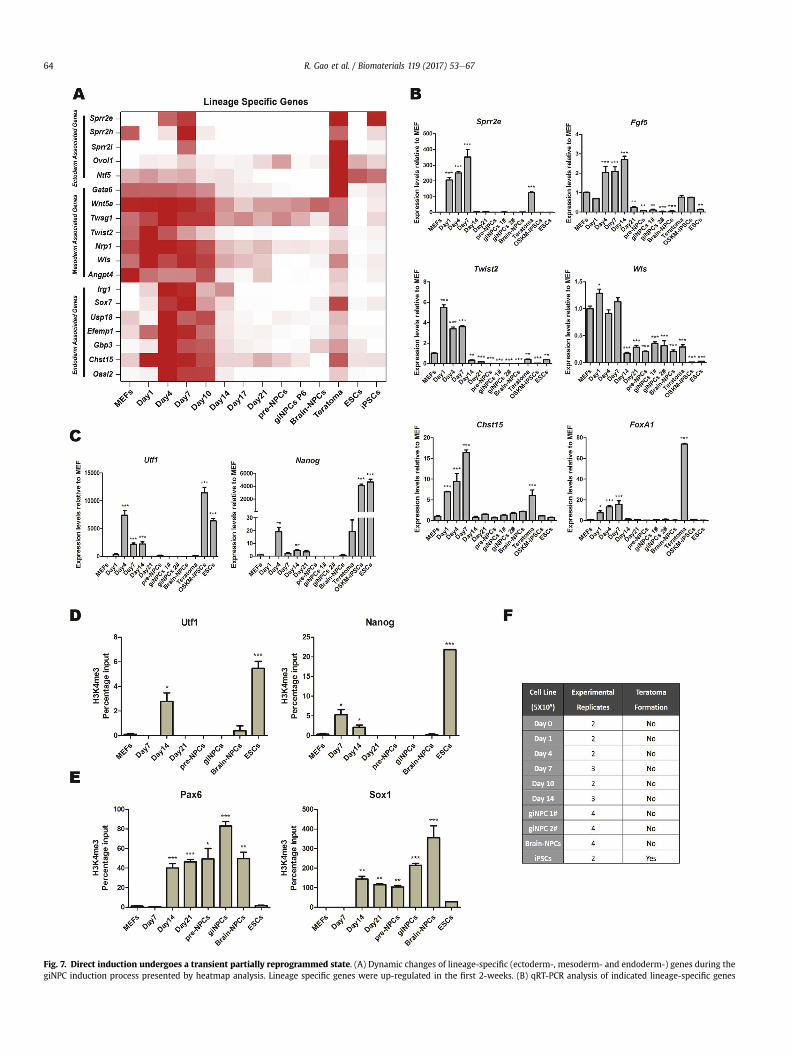
Fig. 7. Direct induction undergoes a transient partially reprogrammed state. (A) Dynamic changes of lineage-specific (ectoderm-, mesoderm- and endoderm-) genes during thegiNPC induction process presented by heatmap analysis. Lineage specific genes were up-regulated in the first 2-weeks. (B) qRT-PCR analysis of indicated lineage-specific genes
R. Gao et al. / Biomaterials 119 (2017) 53e6764
R. Gao et al. / Biomaterials 119 (2017) 53e67 65
(such as Gbx2 and En1) and posterior brain markers (such asHoxb7), were also up-regulated during this process and maintainedin giNPCs (Fig. 6B). Together, these data suggest that induction ofMEFs into giNPCs is a gradual process that occurs by removal ofsomatic structure (Fig. 5AeB, 6A and S4B) and establishment of anNPC specific network (Fig. 5AeB and 6A-B). Moreover, similarprocesses were also observed both in the fibroblast-giNPC induc-tion (Fig. S5A and S5B) and in the recently reported ciNPC inductionsystem (Fig. S5Ce5F).
3.7. Direct induction undergoes a transient partially reprogrammedstate
To better understand how the transgene-free iNPC network wasset up, we sought insight into the molecular mechanism of theentire induction process. Using PCA and unsupervised hierarchicalclustering analyses, we noticed a particular period in which cellsshowed a transient partially reprogrammed transcriptional plat-form and presented a transcriptome similar to that of teratomas,which are known to express genes from three germ layers(Fig. 5AeB and 7A). Specifically, several lineage specific (ectoderm-,mesoderm- and endoderm-) genes were up-regulated at the initi-ation stage from Day 1 and maintained relatively high expressionlevels for about two weeks until the maturation stage (Fig. 7A).Moreover, qRT-PCR analysis demonstrated that ectoderm associ-ated genes (Sprr2e, Fgf5 and Sprr2i), mesoderm associated genes(Twist2 andWls) and endoderm associated genes (Chst15, FoxA1, Afpand Sox17) were up-regulated from Day 1 to Day 7 and were down-regulated thereafter (Fig. 7B and S6A). Interestingly, the fibroblast-giNPC induction process as well as the ciNPC induction system alsoshowed a similar transcriptional tendency of these lineage specificgenes (Fig. S7A, S8A and S9A). Surprisingly, we noticed that certaincore pluripotent transcription factors, such as Utf1, Nanog, Oct4,Rex1, Dppa4 and Fgf4 (Fig. 7C and S6B), were transiently up-regulated in giNPC induction. Moreover, the up-regulation of corepluripotent genes was also noticed in fibroblast-giNPC and ciNPCsystem (Fig. S7B, S8B and S9B). In addition, chromatin immuno-precipitation analysis indicated that promoters of Utf1 and Nanogunderwent a transient burst of activating H3K4 trimethylation anda concomitant down-regulation of repressive H3K27me3 marksduring the giNPC induction both from MEFs (Fig. 7D and S6C) andfibroblasts (Fig. S7C and S7E). Importantly, despite a relatively lowlevel, these histone modification changes at the pluripotent pro-moters had a pattern similar to that of mouse ESCs, which furtherindicated that giNPC induction undergoes a transient partiallyreprogrammed state. In contrast, the promoter regions of Pax6 andSox1 gradually gained H3K4 trimethylation and reduced H3K27trimethylation from Day 14 onward, respectively (Fig. 7E, S6D, S7Dand S7F).
As the cells during the giNPC induction process showed atransient partially reprogrammed state, the tumorigenicity of thesecells deserves our concern. Thus, teratoma formation assay wasfurther performed to test the potential tumorigenic ability of thesecells as well as the established giNPCs. When subcutaneouslyinjected into immune-deficient mice, neither these partiallyreprogrammed cells nor giNPCs could generate any teratomas,which was quite similar to Brain-NPCs (Fig. 7F and S10A-S10C). As
(ectoderm associated genes-Sprr2e and Fgf5, mesoderm associated genes-Twist2 and Wls aprocess. The expression levels were normalized to those observed in MEFs. (C) qRT-PCR analynormalized to those observed in MEFs. (DeE) Time course analysis of histone modification dupromoter loci (E) were analyzed by chromatin immunoprecipitation with antibodies againsinput chromatin amount, analyzed by qPCR. (F) Table summarizing the teratoma formationDay 10, and Day 14 as well as giNPC lines 1# and 2# could not generate teratomas in immuData in (B)e(E) are represented as the mean ± SEM (n ¼ 3). *p < 0.05; **p < 0.01; ***p < 0
positive controls, iPS cells were capable of forming the teratomawith derivatives from all three germ layers (Fig. 7F and S10B).
Collectively, these results strongly support the notion that directinduced mouse NPCs undergo a partially reprogrammed state,during which certain lineage specific and pluripotency associatedgenes will be transiently activated, whereas NPC specific markersare gradually up-regulated. Then, the fully induced NPCs wereestablished.
4. Discussion
Generation of desirable functional cells holds great potential forboth research and clinical applications. Recently, several effectivestrategies have been developed to obtain neural cells and neuralprogenitor cells, which, in the future, may help treat the selectivedysfunctional neurons in most neurodegenerative disorders[2,3,5,8e12]. Thus, it is important to understand technical andfunctional advantages and disadvantages as well as the underlyingmolecular mechanism of such methods. Although functional NPCscould be efficiently derived from embryonic stem cells (ESCs) andinduced pluripotent stem cells (iPSCs) [2,9e12], the ethical issues ofESCs and the risk of tumorigenesis observed in iPSCs limit their usein clinical treatment. Similarly, the low yield of neurons generatedby the direct reprogramming approach also restricts their furtheruse [5,8]. Thus, the direct conversion of somatic cells into multi-potent and lineage-restricted NPCs is highly desirable. Although theoverexpression of master transcription factors is considered themajor determinant of cellular fate conversion, transdifferentiationmediated by small molecules has great advantages, including non-immunogenicity and non-transgene integration, despite that theunderlying mechanism remains elusive [25,26,42e45].
Ectopic expression of the NPC-related factors or transientexpression of pluripotent-related factors with neural-lineage sig-nals during NPC conversion from fibroblasts would direct theexogenous factors to recognize specific loci and recruit and/orcooperate with other endogenous regulators to establish a NPCidentity [17e21,23,24]. Meanwhile, small molecules that have theability to activate neuro-lineage signaling or modulate epigeneticmodifications also help for the NPC conversion [25,26]. However,the same situation is that both methods break the original steadysomatic state by exogenous substances (exogenous transcriptionfactors or small molecules) and then activating endogenous neuralrelated transcription factors and/or signaling pathways[17e20,23e26]. Accordingly, we hypothesized that externalinductive and/or permissive signaling conditions might also havethe same potential. Based on this speculation, we established anovel growth factor based culture system and successfully used it toachieve a direct lineage conversion by inducing somatic cells intofunctional neural progenitor cells (giNPCs) in the present study (Fig.1 and 3). These giNPCs exhibited similar morphological and mo-lecular features as well as transcriptional network to those ofneonatal mouse brainederived NPCs, but were quite different fromthe starting cells (Fig. 1B, 2, 3 and S2). Notably, these giNPCsmaintained their characteristics over prolonged expansion (>20passages) (Fig. 2DeE and S2E-S2F) and could give rise to majorneural lineages in vivo and in vitro (Fig. 4 and S3). Thus, inconclusion, the giNPCs showed typical features of Brain-NPCs,
s well as endoderm associated genes-Chst15 and FoxA1) during the giNPC inductionsis of pluripotent genes during the giNPC induction process. The expression levels werering the giNPC induction process. Utf1 and Nanog promoter loci (D) and Pax6 and Sox1t the epigenetic marks H3K4me3 during this process. The data are percentages of theability of indicated cells. Briefly, the induction of cells from Day 0, Day 1, Day 4, Day 7,ne-deficient mice within 1 months' observation. iPSCs were set as the positive control..001 by ANOVA or Student's t-test for comparison.
R. Gao et al. / Biomaterials 119 (2017) 53e6766
which are self-renewal and neural multipotent.We have also tested whether human fibroblasts can be induced
into NPCs by this growth factor mediated method. When humanfibroblast cells were induced by the same procedures, compact cellcolonies resembling those obtained in growth factor treated mousecells could emerge after 30 days (data not shown). However,despite that they show similar morphologies to that of mouse NPCs,these human fibroblasts derived colonies cannot efficiently up-regulate NPC-specific genes. Thus, these results indicated that thegrowth factors in current protocol may not be enough to activateendogenous genes in human system. Therefore, this strategyshould be further optimized to obtain human giNPCs.
Because induced pluripotency is established in a step-wisemanner, previous studies hypothesized that brief reactivation ofreprogramming factors in somatic cells would generate a highlyplastic intermediate state (Oct4-GFP negative) instead of a matureiPSC state (Oct4-GFP positive), and that, thereafter, the inducedcells could further differentiate into different lineages under certaininductive signals [17,29]. However, recent findings from two groupsrefuted this argument and demonstrated that pluripotency-associated transcription factor mediated lineage conversion in-volves a transient passage through an iPSC stage [31,32]. As theanalyses conducted in previous studies were based on ectopictransgene mediated transdifferentiation, the understanding of themolecular mechanisms underlying transgene-free iNPC generationremains elusive. In the present study, we demonstrated that bothour growth factor-induced and recently reported chemical-inducedNPC undergo a gradual induction process that included the removalof somatic structure and the establishment of an NPC specificnetwork, which typically includes initiation, intermediate, matu-ration and stabilization stages (Fig. 5AeB, 6B-C, S4B-S4C and S5).Most importantly, we noticed a special period in this growth factormediated lineage conversion process, during which cells showed atransient partially reprogrammed transcriptional platform andactivated not only genes from the three germ layers (Fig. 5AeB, 7A-B, S6A and S7A) but also pluripotent genes (Fig. 7C, S6B and S7B) toa certain extent. The induction of a developmental open-chromatinstate was further marked by a transient burst of activating H3K4trimethylation (Fig. 7D and S7C) and a contaminant down-regulation of repressive H3K27me3 marks (Fig. S6C and S7E)within the promoters of Utf1 and Nanog. Moreover, a similartransient up-regulation of lineage specific genes as well as plurip-otent genes was also noticed in the previously reported chemicalbased means (Fig. S8 and S9) [25]. Although those partiallyreprogrammed cells did not contribute to teratomas in immune-deficient mice, we still could not ignore potential safety concerns(Fig. 7F and S10). Thus, a more in-depth understanding of fully- andpartially-reprogrammed stages is required to provide safety veri-fication information for generating functionally desirable cell typesfor potential regenerative applications in the future.
5. Conclusion
In summary, we describe a novel method for generating growthfactor-induced neural progenitor cells (giNPCs) by using inductiveand permissive signaling culture conditions with the combinationof only a few growth factors. Thus, ours and other recent studies[25,26,42e44] provide evidence that direct lineage-specific con-version can be achieved without introducing ectopic transcrip-tional factors. Importantly, we demonstrate that the direct cell typeswitch from somatic cells (both MEFs and adult fibroblasts) tofunctional NPCs by growth factors or small molecules both gothrough a transient partially reprogrammed state. Our studytherefore highlights the importance of excluding the possibility ofresidual partially reprogrammed or teratoma-like cells in the
transgene-free direct induction strategy for future clinical trials.
Author contributions
R.G. and W.X.: conception and design, collection and/or as-sembly of data, data analysis and interpretation and manuscriptwriting. L.Z., R.Z., L.Y., C.W., M.W., M. W., L.Y., Y.T., Y. G., H.W., J.X.,W.L., Y.W., X.W., Y.Y. and Y.Z.: provision of study materials and dataanalysis and interpretation. J.C., L.C. and S.G.: conception anddesign, financial support, data analysis and interpretation, manu-script writing and final approval of the manuscript.
Acknowledgements
We are grateful to our colleagues in the laboratory for theirassistance with the experiments and in the preparation of thismanuscript. We thank Prof. Yuqiang Ding for sharing the Nestin-GFP transgenic mice. This project was supported by the NationalNatural Science Foundation of China (31401247, 31401266,31325019, 91319306, 31471392, 31430056 and 31501196), theMinistry of Science and Technology of China (grants 2014CB964601,2015CB964503, 2016YFA0100400 and 2015CB964800), the Scienceand Technology Commission of Shanghai Municipality(YF1403900), the Shanghai Municipal Education Commission(14CG16) and the Program for Young Excellent Talents in TongjiUniversity (grants 2000219115, 2000219117 and 1515219023).
Appendix A. Supplementary data
Supplementary data related to this article can be found at http://dx.doi.org/10.1016/j.biomaterials.2016.12.007.
References
[1] K. Takahashi, K. Tanabe, M. Ohnuki, M. Narita, T. Ichisaka, K. Tomoda, et al.,Induction of pluripotent stem cells from adult human fibroblasts by definedfactors, Cell 131 (2007) 861e872.
[2] I.H. Park, N. Arora, H. Huo, N. Maherali, T. Ahfeldt, A. Shimamura, et al., Dis-ease-specific induced pluripotent stem cells, Cell 134 (2008) 877e886.
[3] F. Soldner, D. Hockemeyer, C. Beard, Q. Gao, G.W. Bell, E.G. Cook, et al., Par-kinson's disease patient-derived induced pluripotent stem cells free of viralreprogramming factors, Cell 136 (2009) 964e977.
[4] Z.P.P. Pang, N. Yang, T. Vierbuchen, A. Ostermeier, D.R. Fuentes, T.Q. Yang, etal., Induction of human neuronal cells by defined transcription factors, Nature476 (2011). 220-U122.
[5] L. Qiang, R. Fujita, T. Yamashita, S. Angulo, H. Rhinn, D. Rhee, et al., Directedconversion of Alzheimer's disease patient skin fibroblasts into functionalneurons, Cell 146 (2011) 359e371.
[6] M. Caiazzo, M.T. Dell'Anno, E. Dvoretskova, D. Lazarevic, S. Taverna, D. Leo, etal., Direct generation of functional dopaminergic neurons from mouse andhuman fibroblasts, Nature 476 (2011). 224-U151.
[7] E.Y. Son, J.K. Ichida, B.J. Wainger, J.S. Toma, V.F. Rafuse, C.J. Woolf, et al.,Conversion of mouse and human fibroblasts into functional spinal motorneurons, Cell Stem Cell 9 (2011) 205e218.
[8] J. Jiao, Y.Y. Yang, Y.W. Shi, J.Y. Chen, R. Gao, Y. Fan, et al., Modeling Dravetsyndrome using induced pluripotent stem cells (iPSCs) and directly convertedneurons, Hum. Mol. Genet. 22 (2013) 4241e4252.
[9] T. Yagi, D. Ito, Y. Okada, W. Akamatsu, Y. Nihei, T. Yoshizaki, et al., Modelingfamilial Alzheimer's disease with induced pluripotent stem cells, Hum. Mol.Genet. 20 (2011) 4530e4539.
[10] K.J. Brennand, A. Simone, J. Jou, C. Gelboin-Burkhart, N. Tran, S. Sangar, et al.,Modelling schizophrenia using human induced pluripotent stem cells, Nature473 (2011) 221.
[11] M.A. Israel, S.H. Yuan, C. Bardy, S.M. Reyna, Y.L. Mu, C. Herrera, et al., Probingsporadic and familial Alzheimer's disease using induced pluripotent stemcells, Nature 482 (2012). 216-U107.
[12] M.C.N. Marchetto, C. Carromeu, A. Acab, D. Yu, G.W. Yeo, Y.L. Mu, et al.,A model for neural development and treatment of Rett syndrome using hu-man induced pluripotent stem cells, Cell 143 (2010) 527e539.
[13] M. Wernig, A. Meissner, J.P. Cassady, R. Jaenisch, c-Myc is dispensable fordirect reprogramming of mouse fibroblasts, Cell Stem Cell 2 (2008) 10e12.
[14] J.Y. Chen, Y.W. Gao, H. Huang, K. Xu, X.H. Chen, Y.H. Jiang, et al., The combi-nation of Tet1 with Oct4 generates high-quality mouse-induced pluripotentstem cells, Stem Cells 33 (2015) 686e698.
R. Gao et al. / Biomaterials 119 (2017) 53e67 67
[15] G. Martino, S. Pluchino, The therapeutic potential of neural stem cells, Nat.Rev. Neurosci. 7 (2006) 395e406.
[16] P. Taupin, The therapeutic potential of adult neural stem cells, Curr. Opin. Mol.Ther. 8 (2006) 225e231.
[17] J. Kim, J.A. Efe, S.Y. Zhu, M. Talantova, X. Yuan, S.F. Wang, et al., Directreprogramming of mouse fibroblasts to neural progenitors, Proc. Natl. Acad.Sci. U. S. A. 108 (2011) 7838e7843.
[18] E. Lujan, S. Chanda, H. Ahlenius, T.C. Sudhof, M. Wernig, Direct conversion ofmouse fibroblasts to self-renewing, tripotent neural precursor cells, Proc. Natl.Acad. Sci. U. S. A. 109 (2012) 2527e2532.
[19] M. Thier, P. Worsdorfer, Y.B. Lakes, R. Gorris, S. Herms, T. Opitz, et al., Directconversion of fibroblasts into stably expandable neural stem cells, Cell StemCell 10 (2012) 473e479.
[20] D.W. Han, N. Tapia, A. Hermann, K. Hemmer, S. Hoing, M.J. Arauzo-Bravo, etal., Direct reprogramming of fibroblasts into neural stem cells by definedfactors, Cell Stem Cell 10 (2012) 465e472.
[21] C. Sheng, Q.Y. Zheng, J.Y. Wu, Z. Xu, L.B. Wang, W. Li, et al., Direct reprog-ramming of Sertoli cells into multipotent neural stem cells by defined factors,Cell Res. 22 (2012) 208e218.
[22] J.P. Cassady, A.C. D'Alessio, S. Sarkar, V.S. Dani, Z.P. Fan, K. Ganz, et al., Directlineage conversion of adult mouse liver cells and B lymphocytes to neuralstem cells, Stem Cell Rep. 3 (2014) 948e956.
[23] K.L. Ring, L.M. Tong, M.E. Balestra, R. Javier, Y. Andrews-Zwilling, G. Li, et al.,Direct reprogramming of mouse and human fibroblasts into multipotentneural stem cells with a single factor, Cell Stem Cell 11 (2012) 100e109.
[24] L.H. Wang, L.L. Wang, W.H. Huang, H.X. Su, Y.T. Xue, Z.H. Su, et al., Generationof integration-free neural progenitor cells from cells in human urine, Nat.Methods 10 (2013) 84eU124.
[25] L. Cheng, W.X. Hu, B.L. Qiu, J. Zhao, Y.C. Yu, W.Q. Guan, et al., Generation ofneural progenitor cells by chemical cocktails and hypoxia, Cell Res. 24 (2014)665e679.
[26] M. Zhang, Y.H. Lin, Y.J. Sun, S. Zhu, J. Zheng, K. Liu, et al., Pharmacologicalreprogramming of fibroblasts into neural stem cells by signaling-directedtranscriptional activation, Cell Stem Cell 18 (2016) 653e667.
[27] T. Graf, T. Enver, Forcing cells to change lineages, Nature 462 (2009) 587e594.[28] T. Vierbuchen, M. Wernig, Direct lineage conversions: unnatural but useful?
Nat. Biotechnol. 29 (2011) 892e907.[29] J.A. Efe, S. Hilcove, J. Kim, H. Zhou, K. Ouyang, G. Wang, et al., Conversion of
mouse fibroblasts into cardiomyocytes using a direct reprogramming strat-egy, Nat. Cell Biol. 13 (2011), 215-U61.
[30] S.Y. Zhu, M. Rezvani, J. Harbell, A.N. Mattis, A.R. Wolfe, L.Z. Benet, et al., Mouseliver repopulation with hepatocytes generated from human fibroblasts, Na-ture 508 (2014) 93e97.
[31] O. Bar-Nur, C. Verheul, A.G. Sommer, J. Brumbaugh, B.A. Schwarz, I. Lipchina,et al., Lineage conversion induced by pluripotency factors involves transient
passage through an iPSC stage, Nat. Biotechnol. 33 (2015) 761e768.[32] I. Maza, I. Caspi, A. Zviran, E. Chomsky, Y. Rais, S. Viukov, et al., Transient
acquisition of pluripotency during somatic cell transdifferentiation with iPSCreprogramming factors, Nat. Biotechnol. 33 (2015) 769e774.
[33] A.D. Ebert, E.L. McMillan, C.N. Svendsen, Isolating, expanding, and infectinghuman and rodent fetal neural progenitor cells, Curr. Protoc. Stem Cell Biol.(2008 Sep), http://dx.doi.org/10.1002/9780470151808.sc02d02s6 (Chapter 2):Unit 2D.
[34] C.H. Tian, Q. Liu, K.M. Ma, Y.X. Wang, Q. Chen, R. Ambroz, et al., Character-ization of induced neural progenitors from skin fibroblasts by a novel com-bination of defined factors, Sci. Rep. U. K. (2013) 3.
[35] Y.Y. Yang, J. Jiao, R. Gao, R.R. Le, X.C. Kou, Y.H. Zhao, et al., Enhanced rejuve-nation in induced pluripotent stem cell-derived neurons compared withdirectly converted neurons from an aged mouse, Stem Cells Dev. 24 (2015)2767e2777.
[36] L. Cheng, W.X. Hu, B.L. Qiu, J. Zhao, Y.C. Yu, W.Q. Guan, et al., Generation ofneural progenitor cells by chemical cocktails and hypoxia (vol 24, pg 665,2014), Cell Res. 25 (2015) 645e646.
[37] Y.C. Yu, S. He, S. Chen, Y. Fu, K.N. Brown, X.H. Yao, et al., Preferential electricalcoupling regulates neocortical lineage-dependent microcircuit assembly,Nature 486 (2012) 113e117.
[38] C. Trapnell, L. Pachter, S.L. Salzberg, TopHat: discovering splice junctions withRNA-Seq, Bioinformatics 25 (2009) 1105e1111.
[39] C. Trapnell, B.A. Williams, G. Pertea, A. Mortazavi, G. Kwan, M.J. van Baren, etal., Transcript assembly and quantification by RNA-Seq reveals unannotatedtranscripts and isoform switching during cell differentiation, Nat. Biotechnol.28 (2010) 511e515.
[40] M.D. Robinson, D.J. McCarthy, G.K. Smyth, edgeR: a Bioconductor package fordifferential expression analysis of digital gene expression data, Bioinformatics26 (2010) 139e140.
[41] P. Samavarchi-Tehrani, A. Golipour, L. David, H.K. Sung, T.A. Beyer, A. Datti, etal., Functional genomics reveals a BMP-driven mesenchymal-to-epithelialtransition in the initiation of somatic cell reprogramming, Cell Stem Cell 7(2010) 64e77.
[42] X. Li, X.H. Zuo, J.Z. Jing, Y.T. Ma, J.M. Wang, D.F. Liu, et al., Small-molecule-driven direct reprogramming of mouse fibroblasts into functional neurons,Cell Stem Cell 17 (2015) 195e203.
[43] W.X. Hu, B.L. Qiu, W.Q. Guan, Q.Y. Wang, M. Wang, W. Li, et al., Direct con-version of normal and Alzheimer's disease human fibroblasts into neuronalcells by small molecules, Cell Stem Cell 17 (2015) 204e212.
[44] Y.B. Fu, C.W. Huang, X.X. Xu, H.F. Gu, Y.Q. Ye, C.Z. Jiang, et al., Direct reprog-ramming of mouse fibroblasts into cardiomyocytes with chemical cocktails,Cell Res. 25 (2015) 1013e1024.
[45] J. Xu, Y.Y. Du, H.K. Deng, Direct lineage reprogramming: strategies, mecha-nisms, and applications, Cell Stem Cell 16 (2015) 119e134.