Direct cusp replacement in the molar region using a ... · CASE REPORT Direct cusp replacement in...
15
CASE REPORT Direct cusp replacement in the molar region using a thermoviscous bulk-fill composite restorative material – a clinical case report Prof. Dr. Jürgen Manhart, Munich Summary: Today, direct composites restorations in posterior teeth are a crucial part of the standard therapy spectrum in modern restorative dentistry. The performance of this treatment method in the masticatory load-bearing posterior region has been conclusively proven in many clinical studies, even for extensive composite restorations with cuspal coverage. These restorations are usually carried out in an elaborate incremental layering technique. Aside from the possibilities that highly esthetic composites offer in the application of polychromatic multiple-layer techniques, there is also a great market demand for the most simple and quick and therefore economical to place bulk-fill composite materials for posterior teeth. A new development in this class of materials is a bulk-fill composite with thermally controlled viscosity behavior. 1. Introduction In recent years, the indications for direct resin-based composite restorations were continuously expanded due to improvements in the technology of composite materials and related adhesive systems, as well as an optimization of clinical treatment protocols in adhesive dentistry [1-14]. Today, direct resin bonded composites are becoming first choice for many dental practitioners for the restoration of posterior defects, even extensive cavities in load-bearing areas are considered suitable for the direct adhesive technique [9, 12, 15-17]. The maximum preservation of hard tooth tissues using direct composites as an alternative to indirect onlays and partial crowns is one of the major advantages and key elements when restoring severely damaged teeth with cuspal involvement [2, 9, 18-29]. The replacement of single cusps with direct composite
Transcript of Direct cusp replacement in the molar region using a ... · CASE REPORT Direct cusp replacement in...

CASE REPORT
Direct cusp replacement in the molar region using a thermoviscous bulk-fill composite restorative material – a clinical case report
Prof. Dr. Jürgen Manhart, Munich
Summary:
Today, direct composites restorations in posterior teeth are a crucial part of the standard
therapy spectrum in modern restorative dentistry. The performance of this treatment
method in the masticatory load-bearing posterior region has been conclusively proven in
many clinical studies, even for extensive composite restorations with cuspal coverage.
These restorations are usually carried out in an elaborate incremental layering technique.
Aside from the possibilities that highly esthetic composites offer in the application of
polychromatic multiple-layer techniques, there is also a great market demand for the
most simple and quick and therefore economical to place bulk-fill composite materials
for posterior teeth. A new development in this class of materials is a bulk-fill composite
with thermally controlled viscosity behavior.
1. Introduction
In recent years, the indications for direct resin-based composite restorations were
continuously expanded due to improvements in the technology of composite materials
and related adhesive systems, as well as an optimization of clinical treatment protocols
in adhesive dentistry [1-14]. Today, direct resin bonded composites are becoming first
choice for many dental practitioners for the restoration of posterior defects, even
extensive cavities in load-bearing areas are considered suitable for the direct adhesive
technique [9, 12, 15-17]. The maximum preservation of hard tooth tissues using direct
composites as an alternative to indirect onlays and partial crowns is one of the major
advantages and key elements when restoring severely damaged teeth with cuspal
involvement [2, 9, 18-29]. The replacement of single cusps with direct composite

CASE REPORT
restorations is meanwhile an accepted treatment method and scientifically proven [30].
However, when the replacement of several cusps is needed in very large defects, indirect
restorations - requiring additional substance removal in many cases - are still the
preferred option for most dentists [9, 17]. Longevity studies on posterior composite
restorations including cusp replacement show an acceptable performance and qualify
this treatment option as an alternative to conventional indirect restorations in selected
clinical cases [16, 31-34].
To date, incremental layering is considered to be the gold standard for placing light-
curing composite materials [35]. Generally, conventional composites are placed in
individual layers of maximum 2 mm thickness due to their particular polymerization
properties and limited depth of cure. Each increment is polymerized separately for 10 to
40 s, depending on the light intensity of the curing device used, the shade and
translucency level of the respective composite paste and the light initiator system of the
composite material [36]. Thicker layers of these conventional composites, however, do
not polymerize properly and therefore produce poor mechanical and biological properties
[37-39].
Especially in the case of large-volume posterior cavities, the conventional incremental
technique can be a very time-consuming and complicated, technology-sensitive
procedure [30]. That is why many dentists are looking for an alternative to this complex
multi-layer placement technique, so that direct composites can be processed in less time
and thus more economically and at the same time with greater product safety [40-43].
The bulk-fill composites have been developed in recent years in response to this growing
demand for more efficiency. Using a simplified application protocol these materials can
be placed into cavities in increments of 4 to 5 mm thickness with short polymerization
times of 10 to 20 s per increment when a high-intensity curing-light is engaged [36, 40,
44-47].
Bulk-fill composites are usually offered in two versions that require completely different
application technique:
1. Low-viscosity, flowable bulk-fill composites, which flow well onto the cavity floor
and the cavity walls and optimally wet the interior line and point angles of the
preparations. These flowable bulk-fill composites must be protected on the
occlusal surface by an additional capping layer (2 mm thickness) made of a

CASE REPORT
regular hybrid composite which is qualified for load-bearing posterior restorations
[30, 48, 49], since the flowable bulk-fill composites have a reduced filler content
and contain comparatively large fillers in order to lower polymerization stress. As a
result, however, they have poorer mechanical and aesthetic properties compared
with conventional hybrid composites: for example a lower modulus of elasticity, a
reduced wear resistance, an increased surface roughness and an inferior
polishability [36, 50-54]. In addition, the capping layer allows to create the
functional contouring of occlusal anatomical structures, as this would be very
difficult or even impossible to manage with a flowable composite material.
2. Regular to high-viscosity, sculptable bulk-fill composites that can reach up to the
occlusal surface and do not require an additional protective capping layer. Thus,
no additional composite material is required.
Bulk-fill composite materials in both viscosity versions allow a single layer thickness of
4-5 mm due to optimized depth of cure. This means that the high-viscosity bulk-fill
composites can be used in a single-layer technique in a cavity which depth corresponds
at most to the depth of cure of the material. If deeper defects are to be restored or if the
flowable bulk-fill composite variants are used, this always requires a two-phase
procedure with an additional composite layer. Technically, the present bulk-fill
composites that are available for the simplified restoration of posterior teeth are not
really bulk-fill materials, because in particular many proximal cavities extend into areas
that are deeper than the maximum curing depth of these materials (4 – 5 mm) [55, 56].
A new approach is taken by the thermoviscous bulk-fill composite VisCalor bulk (VOCO,
Cuxhaven). This is a high-viscosity composite material at room and body temperature,
which is converted to a flowable consistency by heating to a temperature of 68 °C in a
composite oven or a special dispenser with heating function (Thermo-Viscous-
Technology). In the heated phase, the material flows perfectly onto the cavity walls. Even
in narrow and undercut areas of the defect as well as in internal line and point angles,
an excellent wetting is observed, and thus facilitates the application of the restorative
material into the cavity. VisCalor bulk again reaches body temperature within a short
time when it comes to tooth contact and thus returns to the high-viscosity, sculptable
state. VisCalor bulk thus combines the flowability of a low-viscosity composite during

CASE REPORT
application with the sculpting ability of a high-viscosity composite within one single
restorative composite material. Since the entire cavity can be filled with the same
composite material, there is also a time saving compared to combined systems of
flowable and sculptable composite materials. VisCalor bulk can be manipulated in layers
up to 4 mm thickness. It is available in 4 shades (universal shade, A1, A2, A3). It
exhibits a polymerization shrinkage of 1.44 vol.-% with simultaneously low shrinkage
stress (4.6 MPa). With a flexural strength of 164 MPa, the material shows a high
mechanical stability. VisCalor bulk ensures good color stability and stable mechanical
properties thanks to low water absorption. The application compule is headed by a
narrow, flexible cannula, which perfectly enables direct application of the thermoviscous
composite to hard-to-reach and narrow cavity areas.
2. Clinical Case Presentation
A 50-year old female patient requested in our dental office the replacement of her
composite restoration in tooth 46 (first lower right molar) (Fig. 1).
The tooth showed an insufficiently shaped direct composite restoration especially in the
areas of the replaced distolingual cusp and distal marginal ridge with lack of a sufficient
distal proximal contact which resulted in frequent food impaction with respective
negative consequences. During the clinical inspection, the tooth reacted sensitively in
the cold test and showed no negative reaction to the percussion test. In consultation with
the patient and after an explanation of the possible restorative alternatives and treatment
fees, the patient decided on a direct bulk-fill restoration using VisCalor bulk (VOCO
GmbH, Cuxhaven).
Fig. 1: Preoperative situation: insufficient old
composite restoration with cuspal
involvement in a first lower molar.
Fig. 1: Preoperative situation: insufficient old
composite restoration with cuspal
involvement in a first lower molar.
Fig. 1: Preoperative situation: insufficient old composite restoration with cuspal involvement in a first lower molar.

CASE REPORT
Treatment started with thoroughly cleaning the affected tooth of external deposits using
a fluoride-free prophylaxis paste and a rubber cup. Shade determination was done on the
moist tooth prior to the application of rubber dam. After administration of local
anesthetics, the old insufficient composite restoration was carefully removed while
conserving the remaining hard tissues. After excavation, the cavity was completely
prepared and finished with a fine-grit diamond bur. In the area of the distal proximal
box, the defect extended clearly subgingival. The distolingual cusp was missing
completely and subsequently had to be reconstructed with composite (Fig. 2). The old
composite restoration in tooth 47 was refurbished on the mesial surface as it had a
nonphysiological contour (Fig. 2).
The tooth was subsequently isolated with rubber dam (Fig. 3).
A metal matrix was used to delimit the cavity. The matrix band was sealed at the mesial
gingival margin using a wooden wedge (Fig. 4).
Fig. 2: Situation after careful removal of the old restoration and
cavity preparation. In the area of the distal proximal box, the
defect extended clearly subgingival and the distolingual cusp
was missing completely.
Fig. 3: Application of rubber dam.
Fig. 4: Placement of a metal matrix band.

CASE REPORT
At the distal proximal box, the matrix band was stabilized using a light-curing provisional
composite material (Clip, VOCO GmbH, Cuxhaven). A distal wedge was omitted because
of the risk of dislocating the cervical part of the metal band onto the floor of the proximal
box (Fig. 5).
The universal adhesive Futurabond M+ (VOCO) was chosen for the adhesive pretreatment
of the dental hard tissue. Futurabond M+ is a state-of-the-art universal one-bottle
adhesive that is compatible with all common conditioning techniques and adhesive
strategies currently in use (multimode adhesive): the self-etch technique without the use
of phosphoric acid and both phosphoric acid-based "etch-and-rinse"-conditioning
techniques (selective enamel etching with phosphoric acid or complete total-etch
pretreatment of enamel and dentin with phosphoric acid). Also in these universal
adhesives, the preliminary conditioning of enamel using phosphoric acid (selective
enamel etching) results in better adhesion promotion [57-59]. Unlike former traditional
self-etch adhesives, the new universal adhesives are insensitive to phosphoric acid
etching of dentin [60-64]. The possibility of being able to vary the application protocol
at short notice when using these universal adhesives without changing the adhesion
promoter reduces the technique sensitivity and gives the necessary freedom to the
dentist to react flexibly to different clinical situations (e.g. dentin close to the pulp, risk
of bleeding of the adjacent gingiva, etc.).
In this clinical case, the total-etch adhesive pretreatment using phosphoric acid was
used. 35% phosphoric acid (Vococid, VOCO GmbH, Cuxhaven) was applied along the
enamel margins first for a reaction time of 15 s, followed by an additional conditioning
of the dentin for further 15 s (Fig. 5). Subsequently the cavity was washed thoroughly for
20 s with the air-water-spray to remove the acid and precipitation residues. The cavity
was then gently air-dried from excessive moisture avoiding desiccation of the dentin (Fig.
6).
Fig. 5: Conditioning of enamel and dentin with 35% phosphoric acid.

CASE REPORT
Ample amounts of the adhesive Futurabond M+ were applied and distributed generously
in the area of the cavity using a microbrush (Fig. 7).
It must be ensured that all cavity areas are sufficiently covered by the adhesive. After at
least 20 seconds of carefully scrubbing the adhesive into the tooth surface, the solvent
was carefully evaporated with dry, oil-free compressed air from the bonding agent until a
glossy, immobile adhesive film resulted. Then, the bonding agent was subsequently
light-cured for 10 seconds (Fig. 8). The result was a shiny cavity surface, evenly covered
with adhesive (Fig. 9).
This should be carefully checked before placing the restorative material, as any areas of
the cavity that appear matte are an indication that insufficient amount of adhesive has
been applied to those sites. In the worst case, this could result in reduced bond strength
of the restoration to these areas and, at the same time, in inadequate dentin sealing,
Fig. 6: Situation after thoroughly rinsing the conditioning agent and gentle air-drying the cavity avoiding desiccation of the dentin.
Fig. 7: Adhesive pretreatment of the dental tissues with the universal adhesive Futurabond M+.
Fig. 8: After careful evaporation of the solvent of the adhesive, the bonding was light cured for 10 seconds.
Fig. 9: A shiny cavity surface means evenly sealing dentin and enamel with adhesive.

CASE REPORT
which may lead to persistent postoperative sensitivity in vital teeth. This complication,
which often requires the replacement of a newly-made bonded dental restoration, can
usually be avoided by a careful adhesive protocol. If such dull-looking areas, not or
inferior covered by adhesive, are detected in the visual inspection, additional bonding
agent is selectively applied to them to optimize the adhesive layer.
The thermoviscous composite VisCalor bulk (VOCO, Cuxhaven) was heated in a
composite oven (Caps Warmer, VOCO, Cuxhaven) at 68 °C (Fig. 10 and 11).
The heated composite material was first applied only in a small amount on the floor of
the distal proximal box (Fig. 12).
The narrow, flexible cannula of the VisCalor bulk compule facilitates direct application of
the composite even in hard-to-reach areas and narrow cavity areas (Fig. 11).
A special hand instrument (Easy Contact Point, Helmut Zepf Medizintechnik GmbH,
Seitingen-Oberflacht) was inserted into the unpolymerized, still plastic composite
material to create a physiologically correct formed proximal area with tight contact to the
adjacent tooth (Fig. 13).
Fig. 10: The thermoviscous composite VisCalor bulk (VOCO, Cuxhaven) was heated in a composite oven (Caps Warmer, VOCO, Cuxhaven) at 68 °C.
Fig. 11: The narrow, flexible cannula of the VisCalor bulk compule facilitates direct application of the composite even in hard-to-reach areas and narrow cavity areas.
Fig. 12: The heated composite material was first applied only in a small amount on the floor of the distal proximal box.
CASE REPORT
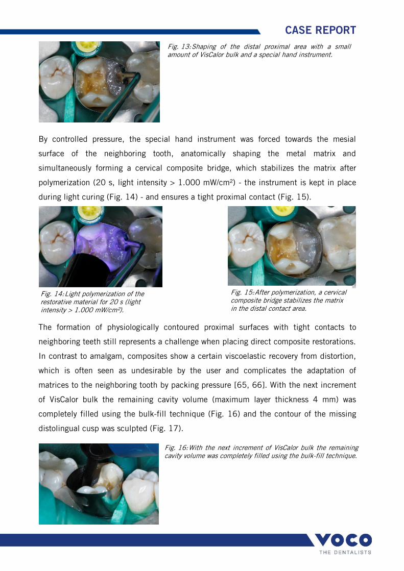
By controlled pressure, the special hand instrument was forced towards the mesial
surface of the neighboring tooth, anatomically shaping the metal matrix and
simultaneously forming a cervical composite bridge, which stabilizes the matrix after
polymerization (20 s, light intensity > 1.000 mW/cm²) - the instrument is kept in place
during light curing (Fig. 14) - and ensures a tight proximal contact (Fig. 15).
The formation of physiologically contoured proximal surfaces with tight contacts to
neighboring teeth still represents a challenge when placing direct composite restorations.
In contrast to amalgam, composites show a certain viscoelastic recovery from distortion,
which is often seen as undesirable by the user and complicates the adaptation of
matrices to the neighboring tooth by packing pressure [65, 66]. With the next increment
of VisCalor bulk the remaining cavity volume (maximum layer thickness 4 mm) was
completely filled using the bulk-fill technique (Fig. 16) and the contour of the missing
distolingual cusp was sculpted (Fig. 17).
Fig. 13: Shaping of the distal proximal area with a small amount of VisCalor bulk and a special hand instrument.
Fig. 14: Light polymerization of the restorative material for 20 s (light intensity > 1.000 mW/cm²).
Fig. 15: After polymerization, a cervical composite bridge stabilizes the matrix in the distal contact area.
Fig. 16: With the next increment of VisCalor bulk the remaining cavity volume was completely filled using the bulk-fill technique.

CASE REPORT
The composite material was again polymerized with a high-performance curing light for
20 s (light intensity > 1.000 mW/cm²). After removal of the metal matrix band, the
restoration was checked for imperfections. Additional 10 s light curing cycles from
mesio-lingual, mesio-buccal, disto-lingual and disto-buccal in the region of both
proximal boxes, especially at the gingival seat, were executed to ensure that all areas
covered before by the metal matrix band experienced sufficient polymerization.
After removal of rubber dam, the fissure relief and the fossae of the occlusal anatomy
were finished with a pear-shaped fine-grit diamond bur. In the next step of the standard
finishing sequence, a point-shaped fine-grit diamond was then used to finish the
convexity of the cusps and triangular ridges. After the elimination of occlusal
interferences and adjustment of the static and dynamic occlusion, the accessible
proximal areas were contoured and prepolished with abrasive disks. The use of diamond-
impregnated composite polishers (Dimanto, VOCO, Cuxhaven) achieved a satin matte,
lustrous finish on the surface of the restoration. Subsequent high-gloss polishing was
completed using the same Dimanto polishers with reduced pressure to optimize the
luster of the restorative material. Figure 18 shows the completed direct bulk-fill
composite restoration with cusp replacement, reconstructing the original tooth shape
with an anatomical and functional occlusal surface, physiological formed proximal
contact areas, and an excellent esthetic appearance. To complete the treatment, a
fluoride varnish (Bifluorid 12, VOCO, Cuxhaven) was applied to the affected tooth using
a foam pellet.
Fig. 17: The contour of the missing distolingual cusp was sculpted. The last increment of the composite was again polymerized for 20 s.
Abb. 16: Final result: the direct bulk-fill composite restoration with cusp replacement blends in well to the surrounding hard dental tissue.

CASE REPORT
3. Conclusion
Composite-based direct restorative materials will gain in importance in the years to
come. These restorations present a scientifically proved, high-quality permanent
treatment option for the masticatory load-bearing posterior region and their reliability has
been documented in literature [11, 67-73]. The results of a comprehensive review have
shown that the annual failure rates of direct posterior composite restorations (2.2%) are
not statistically different to amalgam restorations (3.0%) [69]. Even cuspal coverage
direct composite restorations are meanwhile used frequently and prove to be a viable
alternative to conventional indirect restorations in selected clinical cases [16, 31-34].
The growing economic pressure on the health care system and, in many cases, a lack of
financial means on the part of patients with regard to additional payments adequate to
services are creating a need for reliable, easy-to-use and faster-to-complete and therefore
more economical basic posterior restorative treatment options as an alternative to the
time-consuming high-end solutions [42]. In addition to the universal hybrid composites,
which are available in various shades and levels of opacity, new bulk-fill composites with
optimized depth of cure have lately emerged on the market. They are specially designed
for use in posterior dentition, where they produce esthetically pleasing restorations. The
placement procedure is economically more efficient than that of conventional hybrid
composites [74, 75]. Supplementary to low-viscosity and high-viscosity bulk-fill
composite materials, the material options in the sector of light-activated direct
placement restoratives with increased curing depth were recently expanded by a bulk-fill
composite with thermally controlled viscosity behavior.

CASE REPORT
BIOGRAPHY
Dr. Manhart is Professor, Department of Restorative Dentistry, Dental School of LMU-
University in Munich, Germany. He offers seminars and practical hands-on workshops in
esthetic restorative dentistry, such as direct composite restorations, all-ceramic
restorations, veneers, post-endodontic treatment, management of severely worn
dentition, and treatment planning in esthetic dentistry. He can be reached via e-mail at
Correspondence Address:
Prof. Dr. Juergen Manhart, DDS Department of Restorative Dentistry
Dental School of the Ludwig-Maximilians-University
Goethe Street 70
80336 Munich
Germany
E-Mail: [email protected]
www.manhart.com
www.dental.education

CASE REPORT
References
1. Wolff, D., H.J. Staehle, and C. Frese, Komplexe Zahnaufbauten als Alternative zur Überkronung. ZWR, 2015. 124(1): p. 30-34.
2. Hickel, R., et al., Direct composite restorations: extended use in anterior and posterior situations. Clinical Oral Investigations, 2004. 8(2): p. 43-44.
3. Frese, C., D. Wolff, and H. Staehle, Proximal box elevation with resin composite and the dogma of biological width: clinical r2-technique and critical review. Oper Dent, 2014. 39(1): p. 22-31.
4. Frese, C., D. Wolff, and H.J. Staehle, Die R2-Technik: zweiphasige direkte Kompositrestauration. Restaurative Versorgung extrem tiefer Kavitäten. Zahnärztliche Mitteilungen, 2014. 104(5): p. 50-59.
5. Frese, C., D. Wolff, and H.J. Staehle, Komplexe Seitenzahnrestaurationen in der R1- und R2-Technik. Schwierige Ausgangssituationen und deren Lösung bei direkter Versorgung mit Kompositmaterialien. DFZ Der Freie Zahnarzt, 2014. 58(12): p. 72-81.
6. Frese, C., et al., Recontouring teeth and closing diastemas with direct composite buildups: a 5-year follow-up. J Dent, 2013. 41(11): p. 979-85.
7. Roggendorf, M.J., et al., Effect of proximal box elevation with resin composite on marginal quality of resin composite inlays in vitro. J Dent, 2012. 40(12): p. 1068-73.
8. Manhart, J. and R. Hickel, "Bulk Fill"-Komposite. Neuartige Einsatztechnik von Kompositen im Seitenzahnbereich. Swiss Dental Journal, 2014. 124(1): p. 19-28.
9. Lynch, C.D., et al., Guidance on posterior resin composites: Academy of Operative Dentistry - European Section. J Dent, 2014. 42(4): p. 377-83.
10. Staehle, H.J., Minimally invasive restorative treatment. J Adhes Dent, 1999. 1(3): p. 267-84. 11. Heintze, S.D. and V. Rousson, Clinical effectiveness of direct class II restorations - a meta-
analysis. J Adhes Dent, 2012. 14(5): p. 407-31. 12. Deliperi, S. and D.N. Bardwell, Direct cuspal-coverage posterior resin composite restorations: A
case report. Oper Dent, 2006. 31(1): p. 143-50. 13. Frese, C. and H.J. Staehle, Wie invasiv ist minimalinvasiv? Management von Einzelzahnlücken aus
konservierender Sicht. DFZ Der Freie Zahnarzt, 2018. 62(3): p. 70-77. 14. Staehle, H.J., Erweiterte Anwendungsgebiete für Komposite. wissen kompakt, 2007. 1: p. 29-38. 15. Demarco, F.F., et al., Longevity of posterior composite restorations: not only a matter of materials.
Dent Mater, 2012. 28(1): p. 87-101. 16. Scholtanus, J.D. and M. Ozcan, Clinical longevity of extensive direct composite restorations in
amalgam replacement: up to 3.5 years follow-up. J Dent, 2014. 42(11): p. 1404-10. 17. Laegreid, T., et al., Clinical decision making on extensive molar restorations. Oper Dent, 2014.
39(6): p. E231-40. 18. Plotino, G., et al., Fracture resistance of endodontically treated molars restored with extensive
composite resin restorations. J Prosthet Dent, 2008. 99(3): p. 225-32. 19. Denehy, G. and D. Cobb, Impression matrix technique for cusp replacement using direct
composite resin. J Esthet Restor Dent, 2004. 16(4): p. 227-233. 20. Brackett, W.W., et al., Effect of restoration size on the clinical performance of posterior "packable"
resin composites over 18 months. Oper Dent, 2007. 32(3): p. 212-6. 21. Fennis, W.M., et al., Fatigue resistance of teeth restored with cuspal-coverage composite
restorations. Int J Prosthodont, 2004. 17(3): p. 313-7. 22. Segura, A. and R. Riggins, Fracture resistance of four different restorations for cuspal
replacement. J Oral Rehabil, 1999. 26(12): p. 928-31. 23. Macpherson, L.C. and B.G. Smith, Replacement of missing cusps: an in vitro study. J Dent, 1994.
22(2): p. 118-20. 24. Mondelli, R.F., et al., Conservative approach to restore the first molar with extensive destruction: A
30-month follow-up. Quintessence Int, 2013. 44(6): p. 385-91. 25. Kois, D.E., et al., Evaluation of fracture resistance and failure risks of posterior partial coverage
restorations. J Esthet Restor Dent, 2013. 25(2): p. 110-22. 26. Kantardzic, I., et al., Influence of cavity design preparation on stress values in maxillary premolar:
a finite element analysis. Croat Med J, 2012. 53(6): p. 568-76. 27. Xie, K.X., et al., Fracture resistance of root filled premolar teeth restored with direct composite
resin with or without cusp coverage. Int Endod J, 2012. 45(6): p. 524-9.

CASE REPORT
28. ElAyouti, A., et al., Influence of cusp coverage on the fracture resistance of premolars with endodontic access cavities. Int Endod J, 2011. 44(6): p. 543-9.
29. Kuijs, R.H., et al., A randomized clinical trial of cusp-replacing resin composite restorations: efficiency and short-term effectiveness. Int J Prosthodont, 2006. 19(4): p. 349-54.
30. Federlin, M., et al., Kompositrestaurationen im Seitenzahnbereich. S1-Handlungsempfehlung (Langversion). AWMF-Registernummer: 083–028; Stand: Oktober 2016; gültig bis: Oktober 2021. Deutsche Zahnärztliche Zeitschrift, 2017. 72(1): p. 75-82.
31. Laegreid, T., N.R. Gjerdet, and A.K. Johansson, Extensive composite molar restorations: 3 years clinical evaluation. Acta Odontol Scand, 2012. 70(4): p. 344-52.
32. Deliperi, S. and D.N. Bardwell, Clinical evaluation of direct cuspal coverage with posterior composite resin restorations. J Esthet Restor Dent, 2006. 18(5): p. 256-265.
33. Opdam, N.J., et al., Seven-year clinical evaluation of painful cracked teeth restored with a direct composite restoration. J Endod, 2008. 34(7): p. 808-11.
34. Fennis, W.M., et al., Randomized control trial of composite cuspal restorations: five-year results. J Dent Res, 2014. 93(1): p. 36-41.
35. Park, J., et al., How should composite be layered to reduce shrinkage stress: incremental or bulk filling? Dent Mater, 2008. 24(11): p. 1501-5.
36. Ilie, N. and B. Stawarczyk, Bulk-Fill-Komposite: neue Entwicklungen oder doch herkömmliche Komposite? ZMK, 2014. 30(3): p. 90-97.
37. Tauböck, T.T., Bulk-Fill-Komposite. Wird die Füllungstherapie einfacher, schneller und erfolgreicher? teamwork J Cont Dent Educ, 2013. 16(4): p. 318-323.
38. Ferracane, J.L. and E.H. Greener, The effect of resin formulation on the degree of conversion and mechanical properties of dental restorative resins. J Biomed Mater Res, 1986. 20(1): p. 121-31.
39. Caughman, W.F., et al., Correlation of cytotoxicity, filler loading and curing time of dental composites. Biomaterials, 1991. 12(8): p. 737-40.
40. Manhart, J., Muss es immer Kaviar sein? – Die Frage nach dem Aufwand für Komposite im Seitenzahnbereich. ZMK, 2011. 27(Sonderausgabe März 2011): p. 10-15.
41. Burtscher, P., Von geschichteten Inkrementen zur Vier-Millimeter-Bulk-Fill-Technik – Anforderungen an Komposit und Lichthärtung. DZW Die Zahnarzt Woche, 2011. Ausgabe 39/2011(39): p. 6-8.
42. Margeas, R., New Bulk-Fill Material Simplifies Restorations to One Step. Inside Dentistry, 2014. 10(10): p. 86-90.
43. Margeas, R.C., Bulk-Fill Materials: Simplify Restorations, Reduce Chairtime. Compend Contin Educ Dent, 2015. 36(1): p. e1-e4.
44. Czasch, P. and N. Ilie, In vitro comparison of mechanical properties and degree of cure of bulk fill composites. Clin Oral Investig, 2013. 17(1): p. 227-235.
45. Finan, L., et al., The influence of irradiation potential on the degree of conversion and mechanical properties of two bulk-fill flowable RBC base materials. Dent Mater, 2013. 29(8): p. 906-12.
46. Manhart, J., Neues Konzept zum Ersatz von Dentin in der kompositbasierten Seitenzahnversorgung. ZWR Das Deutsche Zahnärzteblatt, 2010. 119(3): p. 118-125.
47. Fleming, G.J., et al., The potential of a resin-composite to be cured to a 4mm depth. Dental Materials, 2008. 24(4): p. 522-529.
48. Ilie, N., A. Kessler, and J. Durner, Influence of various irradiation processes on the mechanical properties and polymerisation kinetics of bulk-fill resin based composites. J Dent, 2013. 41(8): p. 695-702.
49. Ferracane, J., G. Alex, and R. Margeas, Question: Are Bulk-Fill Composites a Good Idea? Inside Dentistry, 2014. 10(10): p. 42-44.
50. Hickel, R., Neueste Komposite - viele Behauptungen. BZB Bayerisches Zahnärzteblatt, 2012. 49(9): p. 50-53.
51. Ilie, N., S. Bucuta, and M. Draenert, Bulk-fill resin-based composites: an in vitro assessment of their mechanical performance. Oper Dent, 2013. 38(6): p. 618-25.
52. Condon, J.R. and J.L. Ferracane, Evaluation of composite wear with a new multi-mode oral wear simulator. Dent Mater, 1996. 12(4): p. 218-26.
53. Condon, J.R. and J.L. Ferracane, In vitro wear of composite with varied cure, filler level, and filler treatment. Journal of Dental Research, 1997. 76(7): p. 1405-1411.
54. Poggio, C., et al., Surface roughness of flowable resin composites eroded by acidic and alcoholic drinks. J Conserv Dent, 2012. 15(2): p. 137-40.

CASE REPORT
55. Frankenberger, R., et al., Bulk-Fill-Komposite: Mit dicken Schichten einfacher zum Erfolg? Quintessenz, 2012. 65(5): p. 579-584.
56. Frankenberger, R., et al., Die richtige Basisversorgung - Expertenzirkel. Dental Magazin, 2012. 30(1): p. 12-24.
57. de Goes, M.F., M.S. Shinohara, and M.S. Freitas, Performance of a new one-step multi-mode adhesive on etched vs non-etched enamel on bond strength and interfacial morphology. J Adhes Dent, 2014. 16(3): p. 243-50.
58. Hanabusa, M., et al., Bonding effectiveness of a new 'multi-mode' adhesive to enamel and dentine. J Dent, 2012. 40(6): p. 475-84.
59. McLean, D.E., et al., Enamel Bond Strength of New Universal Adhesive Bonding Agents. Oper Dent, 2015. 40(4): p. 410-7.
60. Takamizawa, T., et al., Influence of different etching modes on bond strength and fatigue strength to dentin using universal adhesive systems. Dent Mater, 2016. 32(2): p. e9-21.
61. Wagner, A., et al., Bonding performance of universal adhesives in different etching modes. J Dent, 2014. 42(7): p. 800-7.
62. Lenzi, T.L., et al., Bonding Performance of a Multimode Adhesive to Artificially-induced Caries-affected Primary Dentin. J Adhes Dent, 2015. 17(2): p. 125-31.
63. Loguercio, A.D., et al., A new universal simplified adhesive: 36-Month randomized double-blind clinical trial. J Dent, 2015. 43(9): p. 1083-92.
64. Munoz, M.A., et al., In vitro longevity of bonding properties of universal adhesives to dentin. Oper Dent, 2015. 40(3): p. 282-92.
65. Manhart, J., Eine Alternative zu Amalgam? Hochvisköse stopfbare Komposite: Überblick, Eigenschaften und Verarbeitungshinweise. KONS-Journal, 2001. 3: p. 21-26.
66. Kunzelmann, K.H. and R. Hickel, Klinische Aspekte der Adhäsivtechnik mit plastischen Werkstoffen, in Die Adhäsivtechnologie. Ein Leitfaden für Theorie und Praxis., M. ESPE, Editor. 2001, 3M ESPE: Seefeld, Germany. p. 46-67.
67. Da Rosa Rodolpho, P.A., et al., 22-Year clinical evaluation of the performance of two posterior composites with different filler characteristics. Dent Mater, 2011. 27(10): p. 955-63.
68. van de Sande, F.H., et al., 18-year survival of posterior composite resin restorations with and without glass ionomer cement as base. Dent Mater, 2015. 31(6): p. 669-75.
69. Manhart, J., et al., Review of the clinical survival of direct and indirect restorations in posterior teeth of the permanent dentition. Oper Dent, 2004. 29(5): p. 481-508.
70. Opdam, N.J., et al., Longevity of posterior composite restorations: a systematic review and meta-analysis. J Dent Res, 2014. 93(10): p. 943-9.
71. Opdam, N.J., et al., 12-year survival of composite vs. amalgam restorations. J Dent Res, 2010. 89(10): p. 1063-7.
72. Pallesen, U. and J.W. van Dijken, A randomized controlled 30 years follow up of three conventional resin composites in Class II restorations. Dent Mater, 2015. 31(10): p. 1232-44.
73. Pallesen, U. and J.W. van Dijken, A randomized controlled 27 years follow up of three resin composites in Class II restorations. J Dent, 2015. 43(12): p. 1547-58.
74. Manhart, J., H.Y. Chen, and R. Hickel, Three-year results of a randomized controlled clinical trial of the posterior composite QuiXfil in class I and II cavities. Clin Oral Investig, 2009. 13(3): p. 301-7.
75. Burke, F.J., et al., The current status of materials for posterior composite restorations: the advent of low shrink. Dent Update, 2009. 36(7): p. 401-402.