Dipeptidyl peptidase-4 inhibitor, linagliptin, ameliorates ...
8
Dipeptidyl peptidase-4 inhibitor, linagliptin, ameliorates endothelial dysfunction and atherogenesis in normoglycemic apolipoprotein-E deficient mice Hotimah Masdan Salim a , Daiju Fukuda a, ⁎, Yasutomi Higashikuni b , Kimie Tanaka c , Yoichiro Hirata d , Shusuke Yagi a , Takeshi Soeki a , Michio Shimabukuro e , Masataka Sata a a Department of Cardiovascular Medicine, Institute of Biomedical Sciences, Tokushima University Graduate School, Japan b Department of Cardiovascular Medicine, The University of Tokyo, Japan c Division for Health Service Promotion, The University of Tokyo, Japan d Department of Pediatrics, The University of Tokyo Hospital, Japan e Department of Cardio-Diabetes Medicine, Institute of Biomedical Sciences, Tokushima University Graduate School, Japan abstract article info Article history: Received 20 June 2015 Received in revised form 31 July 2015 Accepted 11 August 2015 Available online 13 August 2015 Keywords: DPP-4 inhibitor Linagliptin GLP-1 Endothelial function Inflammation Oxidative stress Background: Dipeptidyl peptidase-4 (DPP-4) inhibitors have vasoprotective effects. This study investigated whether a recently approved DPP-4 inhibitor, linagliptin (Lina), suppresses atherogenesis in non-diabetic apolipoprotein-E deficient (ApoE -/- ) mice, and examined its effects on endothelial function. Methods and results: Lina (10 mg/kg/day) was administered orally to ApoE -/- mice for 20 weeks. Lina reduced atherogenesis without alteration of metabolic parameters including blood glucose level compared with control (P b 0.05). Results of immunohistochemical analyses and quantitative RT-PCR demonstrated that Lina signifi- cantly decreased inflammatory molecule expression and macrophage infiltration in the atherosclerotic aorta. Lina administration to ApoE -/- mice for 9 weeks ameliorated endothelium-dependent vasodilation compared with that in untreated mice. Plasma active glucagon-like peptide-1 (GLP-1) level was significantly higher in the treated group (P b 0.05). Exendin-4 (Ex-4), a GLP-1 analog, ameliorated endothelium-dependent vasodila- tion impaired by palmitic acid (PA) in wild-type mouse aortic segments. Ex-4 promoted phosphorylation of eNOS Ser1177 and Akt, both of which were abrogated by PA, in human umbilical vein endothelial cells. In addition, Lina administration to ApoE -/- mice decreased oxidative stress, as determined by urinary 8-OHdG secretion and NADPH oxidase subunit expression in the abdominal aorta. Conclusion: Lina inhibited atherogenesis in non-diabetic ApoE -/- mice. Amelioration of endothelial dysfunction associated with a reduction of oxidative stress by GLP-1 contributes to the atheroprotective effects of Lina. © 2015 Elsevier Inc. All rights reserved. 1. Introduction Chronic inflammation plays a central role in the pathogenesis of atherosclerosis [1]. Endothelial dysfunction caused by cardiovascular risk factors is a key initiator of vascular inflammation [2]. Dysfunction of endothelial cells alters vascular responses and induces the expression of adhesion molecules and chemokines which stimulate monocyte- endothelial cell interaction, leading to the development of atherosclero- sis [3]. Therefore, abrogation of endothelial dysfunction is an attractive strategy for preventing vascular inflammation and atherosclerosis. Dipeptidyl peptidase-4 (DPP-4) inhibitors are a new class of antidi- abetic drugs that improve glucose metabolism by raising the active con- centration and duration of action of glucose-like peptide (GLP)-1, a gut hormone secreted in response to nutrient ingestion that stimulates glucose-dependent insulin secretion [4]. In addition to their anti- diabetic property, recent studies have suggested that DPP-4 inhibitors have anti-inflammatory effects independent of their glucose-lowering effect [5–9]. The GLP-1 receptor mediates major function of GLP-1. The GLP-1 receptor is known to be expressed in pancreatic β cells, although recent studies demonstrated its expression in endothelial cells, suggest- ing vasoprotective effects independent of blood glucose level [10,11]. Vascular Pharmacology 79 (2016) 16–23 Abbreviations: DPP-4, dipeptidyl peptidase-4; Lina, linagliptin; ApoE -/- , apolipoprotein- E deficient; GLP-1, glucagon-like peptide-1; Ex-4, exendin-4; PA, palmitic acid; WTD, western-type diet; CMC, carboxymethyl cellulose; 8-OHdG, 8-hydroxy-2′-deoxyguanosine; MCP-1, monocyte chemoattractant protein-1; VCAM-1, vascular cellular adhesion molecule-1; Mac-3, macrophage antigen-3; Ach, acetylcholine; SNP, sodium nitroprusside; HUVEC, human umbilical vein endothelial cell; VEGF, vascular endothelial growth factor; qPCR, quantitative real-time PCR; ROS, reactive oxygen species. ⁎ Corresponding author at: Department of Cardiovascular Medicine, Institute of Biomedical Sciences, Tokushima University Graduate School, 3-18-15 Kuramoto-cho, Tokushima 770-8503, Japan. E-mail address: [email protected] (D. Fukuda). http://dx.doi.org/10.1016/j.vph.2015.08.011 1537-1891/© 2015 Elsevier Inc. All rights reserved. Contents lists available at ScienceDirect Vascular Pharmacology journal homepage: www.elsevier.com/locate/vph brought to you by CORE View metadata, citation and similar papers at core.ac.uk provided by Tokushima University Institutional Repository
Transcript of Dipeptidyl peptidase-4 inhibitor, linagliptin, ameliorates ...
Vascular Pharmacology 79 (2016) 16–23
Contents lists available at ScienceDirect
Vascular Pharmacology
j ourna l homepage: www.e lsev ie r .com/ locate /vph
brought to you by COREView metadata, citation and similar papers at core.ac.uk
provided by Tokushima University Institutional Repository
Dipeptidyl peptidase-4 inhibitor, linagliptin, ameliorates endothelialdysfunction and atherogenesis in normoglycemic apolipoprotein-Edeficient mice
Hotimah Masdan Salim a, Daiju Fukuda a,⁎, Yasutomi Higashikuni b, Kimie Tanaka c, Yoichiro Hirata d,Shusuke Yagi a, Takeshi Soeki a, Michio Shimabukuro e, Masataka Sata a
a Department of Cardiovascular Medicine, Institute of Biomedical Sciences, Tokushima University Graduate School, Japanb Department of Cardiovascular Medicine, The University of Tokyo, Japanc Division for Health Service Promotion, The University of Tokyo, Japand Department of Pediatrics, The University of Tokyo Hospital, Japane Department of Cardio-Diabetes Medicine, Institute of Biomedical Sciences, Tokushima University Graduate School, Japan
Abbreviations: DPP-4, dipeptidyl peptidase-4; Lina, linagE deficient; GLP-1, glucagon-like peptide-1; Ex-4, exenwestern-type diet; CMC, carboxymethyl cellulose; 8-OHdGMCP-1, monocyte chemoattractant protein-1; VCAM-molecule-1; Mac-3, macrophage antigen-3; Ach, acetylchoHUVEC, human umbilical vein endothelial cell; VEGF, vasqPCR, quantitative real-time PCR; ROS, reactive oxygen spe⁎ Corresponding author at: Department of Cardiov
Biomedical Sciences, Tokushima University Graduate STokushima 770-8503, Japan.
E-mail address: [email protected] (D. Fu
http://dx.doi.org/10.1016/j.vph.2015.08.0111537-1891/© 2015 Elsevier Inc. All rights reserved.
a b s t r a c t
a r t i c l e i n f oArticle history:
Received 20 June 2015Received in revised form 31 July 2015Accepted 11 August 2015Available online 13 August 2015Keywords:DPP-4 inhibitorLinagliptinGLP-1Endothelial functionInflammationOxidative stress
Background: Dipeptidyl peptidase-4 (DPP-4) inhibitors have vasoprotective effects. This study investigatedwhether a recently approved DPP-4 inhibitor, linagliptin (Lina), suppresses atherogenesis in non-diabeticapolipoprotein-E deficient (ApoE−/−) mice, and examined its effects on endothelial function.Methods and results: Lina (10 mg/kg/day) was administered orally to ApoE−/− mice for 20 weeks. Lina reducedatherogenesis without alteration of metabolic parameters including blood glucose level compared with control(P b 0.05). Results of immunohistochemical analyses and quantitative RT-PCR demonstrated that Lina signifi-cantly decreased inflammatory molecule expression and macrophage infiltration in the atherosclerotic aorta.Lina administration to ApoE−/− mice for 9 weeks ameliorated endothelium-dependent vasodilation comparedwith that in untreated mice. Plasma active glucagon-like peptide-1 (GLP-1) level was significantly higher inthe treated group (P b 0.05). Exendin-4 (Ex-4), a GLP-1 analog, ameliorated endothelium-dependent vasodila-tion impaired by palmitic acid (PA) in wild-type mouse aortic segments. Ex-4 promoted phosphorylation ofeNOSSer1177 and Akt, both of whichwere abrogated by PA, in human umbilical vein endothelial cells. In addition,
Lina administration to ApoE−/−mice decreased oxidative stress, as determined by urinary 8-OHdG secretion andNADPH oxidase subunit expression in the abdominal aorta.Conclusion: Lina inhibited atherogenesis in non-diabetic ApoE−/− mice. Amelioration of endothelial dysfunctionassociated with a reduction of oxidative stress by GLP-1 contributes to the atheroprotective effects of Lina.© 2015 Elsevier Inc. All rights reserved.
1. Introduction
Chronic inflammation plays a central role in the pathogenesis ofatherosclerosis [1]. Endothelial dysfunction caused by cardiovascularrisk factors is a key initiator of vascular inflammation [2]. Dysfunction
liptin; ApoE−/−, apolipoprotein-din-4; PA, palmitic acid; WTD,, 8-hydroxy-2′-deoxyguanosine;1, vascular cellular adhesionline; SNP, sodium nitroprusside;cular endothelial growth factor;cies.ascular Medicine, Institute ofchool, 3-18-15 Kuramoto-cho,
kuda).
of endothelial cells alters vascular responses and induces the expressionof adhesion molecules and chemokines which stimulate monocyte-endothelial cell interaction, leading to the development of atherosclero-sis [3]. Therefore, abrogation of endothelial dysfunction is an attractivestrategy for preventing vascular inflammation and atherosclerosis.
Dipeptidyl peptidase-4 (DPP-4) inhibitors are a new class of antidi-abetic drugs that improve glucosemetabolism by raising the active con-centration and duration of action of glucose-like peptide (GLP)-1, a guthormone secreted in response to nutrient ingestion that stimulatesglucose-dependent insulin secretion [4]. In addition to their anti-diabetic property, recent studies have suggested that DPP-4 inhibitorshave anti-inflammatory effects independent of their glucose-loweringeffect [5–9]. The GLP-1 receptor mediates major function of GLP-1. TheGLP-1 receptor is known to be expressed in pancreatic β cells, althoughrecent studies demonstrated its expression in endothelial cells, suggest-ing vasoprotective effects independent of blood glucose level [10,11].
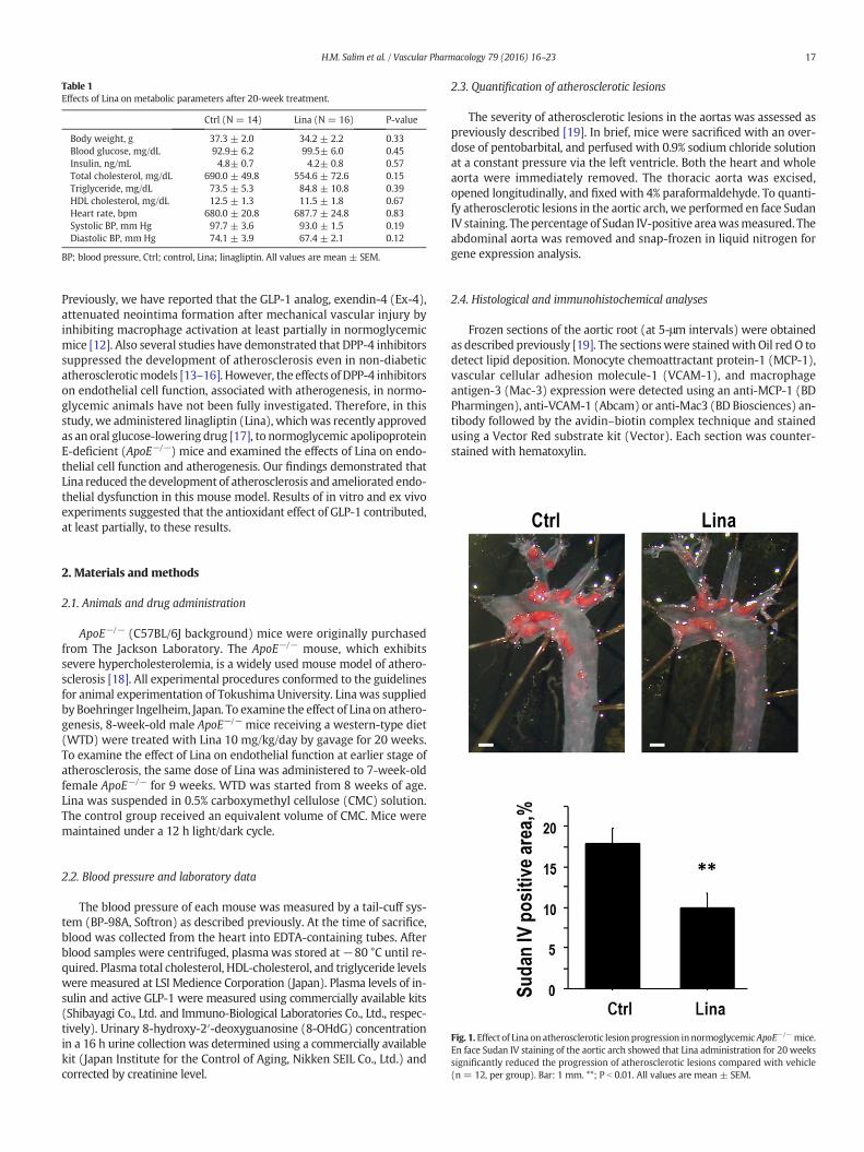
Table 1Effects of Lina on metabolic parameters after 20-week treatment.
Ctrl (N = 14) Lina (N = 16) P-value
Body weight, g 37.3 ± 2.0 34.2 ± 2.2 0.33Blood glucose, mg/dL 92.9± 6.2 99.5± 6.0 0.45Insulin, ng/mL 4.8± 0.7 4.2± 0.8 0.57Total cholesterol, mg/dL 690.0 ± 49.8 554.6 ± 72.6 0.15Triglyceride, mg/dL 73.5 ± 5.3 84.8 ± 10.8 0.39HDL cholesterol, mg/dL 12.5 ± 1.3 11.5 ± 1.8 0.67Heart rate, bpm 680.0 ± 20.8 687.7 ± 24.8 0.83Systolic BP, mm Hg 97.7 ± 3.6 93.0 ± 1.5 0.19Diastolic BP, mm Hg 74.1 ± 3.9 67.4 ± 2.1 0.12
BP; blood pressure, Ctrl; control, Lina; linagliptin. All values are mean ± SEM.
17H.M. Salim et al. / Vascular Pharmacology 79 (2016) 16–23
Previously, we have reported that the GLP-1 analog, exendin-4 (Ex-4),attenuated neointima formation after mechanical vascular injury byinhibiting macrophage activation at least partially in normoglycemicmice [12]. Also several studies have demonstrated that DPP-4 inhibitorssuppressed the development of atherosclerosis even in non-diabeticatheroscleroticmodels [13–16]. However, the effects of DPP-4 inhibitorson endothelial cell function, associated with atherogenesis, in normo-glycemic animals have not been fully investigated. Therefore, in thisstudy, we administered linagliptin (Lina), whichwas recently approvedas an oral glucose-lowering drug [17], to normoglycemic apolipoproteinE-deficient (ApoE−/−) mice and examined the effects of Lina on endo-thelial cell function and atherogenesis. Our findings demonstrated thatLina reduced the development of atherosclerosis and ameliorated endo-thelial dysfunction in this mouse model. Results of in vitro and ex vivoexperiments suggested that the antioxidant effect of GLP-1 contributed,at least partially, to these results.
2. Materials and methods
2.1. Animals and drug administration
ApoE−/− (C57BL/6J background) mice were originally purchasedfrom The Jackson Laboratory. The ApoE−/− mouse, which exhibitssevere hypercholesterolemia, is a widely used mouse model of athero-sclerosis [18]. All experimental procedures conformed to the guidelinesfor animal experimentation of Tokushima University. Linawas suppliedby Boehringer Ingelheim, Japan. To examine the effect of Lina on athero-genesis, 8-week-old male ApoE−/− mice receiving a western-type diet(WTD) were treated with Lina 10 mg/kg/day by gavage for 20 weeks.To examine the effect of Lina on endothelial function at earlier stage ofatherosclerosis, the same dose of Lina was administered to 7-week-oldfemale ApoE−/− for 9 weeks. WTD was started from 8 weeks of age.Lina was suspended in 0.5% carboxymethyl cellulose (CMC) solution.The control group received an equivalent volume of CMC. Mice weremaintained under a 12 h light/dark cycle.
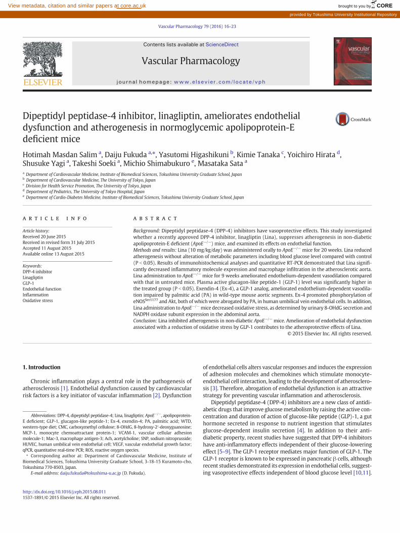
Fig. 1. Effect of Lina on atherosclerotic lesion progression innormoglycemic ApoE−/−mice.En face Sudan IV staining of the aortic arch showed that Lina administration for 20 weekssignificantly reduced the progression of atherosclerotic lesions compared with vehicle(n = 12, per group). Bar: 1 mm. **; P b 0.01. All values are mean ± SEM.
2.2. Blood pressure and laboratory data
The blood pressure of each mouse was measured by a tail-cuff sys-tem (BP-98A, Softron) as described previously. At the time of sacrifice,blood was collected from the heart into EDTA-containing tubes. Afterblood samples were centrifuged, plasmawas stored at−80 °C until re-quired. Plasma total cholesterol, HDL-cholesterol, and triglyceride levelswere measured at LSI Medience Corporation (Japan). Plasma levels of in-sulin and active GLP-1 were measured using commercially available kits(Shibayagi Co., Ltd. and Immuno-Biological Laboratories Co., Ltd., respec-tively). Urinary 8-hydroxy-2′-deoxyguanosine (8-OHdG) concentrationin a 16 h urine collection was determined using a commercially availablekit (Japan Institute for the Control of Aging, Nikken SEIL Co., Ltd.) andcorrected by creatinine level.
2.3. Quantification of atherosclerotic lesions
The severity of atherosclerotic lesions in the aortas was assessed aspreviously described [19]. In brief, mice were sacrificed with an over-dose of pentobarbital, and perfused with 0.9% sodium chloride solutionat a constant pressure via the left ventricle. Both the heart and wholeaorta were immediately removed. The thoracic aorta was excised,opened longitudinally, and fixed with 4% paraformaldehyde. To quanti-fy atherosclerotic lesions in the aortic arch, we performed en face SudanIV staining. The percentage of Sudan IV-positive areawasmeasured. Theabdominal aorta was removed and snap-frozen in liquid nitrogen forgene expression analysis.
2.4. Histological and immunohistochemical analyses
Frozen sections of the aortic root (at 5-μm intervals) were obtainedas described previously [19]. The sectionswere stainedwith Oil red O todetect lipid deposition. Monocyte chemoattractant protein-1 (MCP-1),vascular cellular adhesion molecule-1 (VCAM-1), and macrophageantigen-3 (Mac-3) expression were detected using an anti-MCP-1 (BDPharmingen), anti-VCAM-1 (Abcam) or anti-Mac3 (BD Biosciences) an-tibody followed by the avidin–biotin complex technique and stainedusing a Vector Red substrate kit (Vector). Each section was counter-stained with hematoxylin.
18 H.M. Salim et al. / Vascular Pharmacology 79 (2016) 16–23
2.5. Vascular reactivity assay
Analysis of vascular reactivity was performed as described previous-ly [20]. In brief, the descending thoracic aortas frommice were cut into2-mm rings with special care to preserve the endothelium, andmounted in organ baths filled with modified Krebs–Henseleit buffer(KHB; 118.4 mM NaCl, 4.7 mM KCl, 2.5 mM CaCl2, 1.2 mM KH2PO4,1.2 mM MgSO4, 25 mM NaHCO3, 11.1 mM glucose) aerated with 95%O2 and 5% CO2 at 37 °C. The preparationswere attached to a force trans-ducer, and isometric tension was recorded on a polygraph. Vessel ringswere primed with 31.4 mM KCl, and then pre-contracted with phenyl-ephrine, producing submaximal (60% of maximum) contraction.After the plateau was attained, the rings were exposed to increasing
Fig. 2. Effect of Lina on characteristics of atherosclerotic plaques. (A) Representative Oil redC) Representative MCP-1 and VCAM-1 staining of atherosclerotic plaques. Lina significan(D) Representative Mac3 staining of atherosclerotic plaques. Lina tended to decrease macrophare mean ± SEM.
concentrations of acetylcholine (Ach; 10−9 to 10−4 M) and sodium ni-troprusside (SNP; 10−9 to 10−4 M) to obtain cumulative concentra-tion–response curves. In some experiments, aortic segments wereincubated with 200 μM palmitic acid (PA, Chem Service) in the pres-ence/absence of Ex-4 for 4 h before analysis of vascular reactivity.
2.6. Cell culture
Human umbilical vein endothelial cells (HUVEC) were purchasedfrom Life Technologies and cultured in EGM-2 (Lonza). HUVEC (pas-sages 4–6) were treated with 1 or 10 nM Ex-4 (Sigma-Aldrich) inEBM-2 (Lonza). For inflammatory activation, cells were stimulated
O staining of aortic root. Lina significantly decreased lipid deposition in plaques. (B andtly reduced the expression of MCP-1 (B) and VCAM-1 (C) in atherosclerotic lesions.age accumulation in plaques. (n = 11–12, per group). Bar: 100 μm. *; P b 0.05. All values
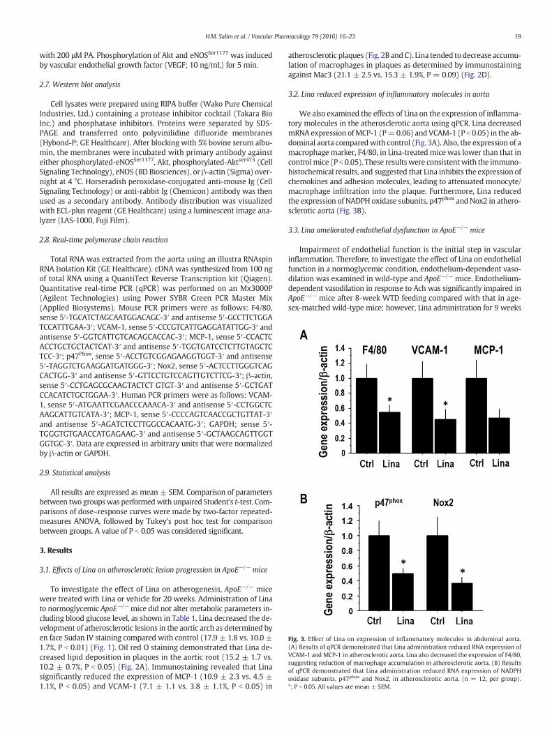
Fig. 3. Effect of Lina on expression of inflammatory molecules in abdominal aorta.(A) Results of qPCR demonstrated that Lina administration reduced RNA expression ofVCAM-1 and MCP-1 in atherosclerotic aorta. Lina also decreased the expression of F4/80,suggesting reduction of macrophage accumulation in atherosclerotic aorta. (B) Resultsof qPCR demonstrated that Lina administration reduced RNA expression of NADPHoxidase subunits, p47phox and Nox2, in atherosclerotic aorta. (n = 12, per group).*; P b 0.05. All values are mean ± SEM.
19H.M. Salim et al. / Vascular Pharmacology 79 (2016) 16–23
with 200 μM PA. Phosphorylation of Akt and eNOSSer1177 was inducedby vascular endothelial growth factor (VEGF; 10 ng/mL) for 5 min.
2.7. Western blot analysis
Cell lysates were prepared using RIPA buffer (Wako Pure ChemicalIndustries, Ltd.) containing a protease inhibitor cocktail (Takara BioInc.) and phosphatase inhibitors. Proteins were separated by SDS-PAGE and transferred onto polyvinilidine difluoride membranes(Hybond-P; GE Healthcare). After blocking with 5% bovine serum albu-min, the membranes were incubated with primary antibody againsteither phosphorylated-eNOSSer1177, Akt, phosphorylated-Aktser473 (CellSignaling Technology), eNOS (BD Biosciences), or β-actin (Sigma) over-night at 4 °C. Horseradish peroxidase-conjugated anti-mouse Ig (CellSignaling Technology) or anti-rabbit Ig (Chemicon) antibody was thenused as a secondary antibody. Antibody distribution was visualizedwith ECL-plus reagent (GE Healthcare) using a luminescent image ana-lyzer (LAS-1000, Fuji Film).
2.8. Real-time polymerase chain reaction
Total RNA was extracted from the aorta using an illustra RNAspinRNA Isolation Kit (GE Healthcare). cDNA was synthesized from 100 ngof total RNA using a QuantiTect Reverse Transcription kit (Qiagen).Quantitative real-time PCR (qPCR) was performed on an Mx3000P(Agilent Technologies) using Power SYBR Green PCR Master Mix(Applied Biosystems). Mouse PCR primers were as follows: F4/80,sense 5′-TGCATCTAGCAATGGACAGC-3′ and antisense 5′-GCCTTCTGGATCCATTTGAA-3′; VCAM-1, sense 5′-CCCGTCATTGAGGATATTGG-3′ andantisense 5′-GGTCATTGTCACAGCACCAC-3′; MCP-1, sense 5′-CCACTCACCTGCTGCTACTCAT-3′ and antisense 5′-TGGTGATCCTCTTGTAGCTCTCC-3′; p47Phox, sense 5′-ACCTGTCGGAGAAGGTGGT-3′ and antisense5′-TAGGTCTGAAGGATGATGGG-3′; Nox2, sense 5′-ACTCCTTGGGTCAGCACTGG-3′ and antisense 5′-GTTCCTGTCCAGTTGTCTTCG-3′; β-actin,sense 5′-CCTGAGCGCAAGTACTCT GTGT-3′ and antisense 5′-GCTGATCCACATCTGCTGGAA-3′. Human PCR primers were as follows: VCAM-1, sense 5′-ATGAATTCGAACCCAAACA-3′ and antisense 5′-CCTGGCTCAAGCATTGTCATA-3′; MCP-1, sense 5′-CCCCAGTCAACCGCTGTTAT-3′and antisense 5′-AGATCTCCTTGGCCACAATG-3′; GAPDH; sense 5′-TGGGTGTGAACCATGAGAAG-3′ and antisense 5′-GCTAAGCAGTTGGTGGTGC-3′. Data are expressed in arbitrary units that were normalizedby β-actin or GAPDH.
2.9. Statistical analysis
All results are expressed as mean ± SEM. Comparison of parametersbetween two groupswas performedwith unpaired Student's t-test. Com-parisons of dose–response curves were made by two-factor repeated-measures ANOVA, followed by Tukey's post hoc test for comparisonbetween groups. A value of P b 0.05 was considered significant.
3. Results
3.1. Effects of Lina on atherosclerotic lesion progression in ApoE−/− mice
To investigate the effect of Lina on atherogenesis, ApoE−/− micewere treated with Lina or vehicle for 20 weeks. Administration of Linato normoglycemic ApoE−/− mice did not alter metabolic parameters in-cluding blood glucose level, as shown in Table 1. Lina decreased the de-velopment of atherosclerotic lesions in the aortic arch as determined byen face Sudan IV staining compared with control (17.9 ± 1.8 vs. 10.0 ±1.7%, P b 0.01) (Fig. 1). Oil red O staining demonstrated that Lina de-creased lipid deposition in plaques in the aortic root (15.2 ± 1.7 vs.10.2 ± 0.7%, P b 0.05) (Fig. 2A). Immunostaining revealed that Linasignificantly reduced the expression of MCP-1 (10.9 ± 2.3 vs. 4.5 ±1.1%, P b 0.05) and VCAM-1 (7.1 ± 1.1 vs. 3.8 ± 1.1%, P b 0.05) in
atherosclerotic plaques (Fig. 2B andC). Lina tended to decrease accumu-lation of macrophages in plaques as determined by immunostainingagainst Mac3 (21.1 ± 2.5 vs. 15.3 ± 1.9%, P = 0.09) (Fig. 2D).
3.2. Lina reduced expression of inflammatory molecules in aorta
We also examined the effects of Lina on the expression of inflamma-tory molecules in the atherosclerotic aorta using qPCR. Lina decreasedmRNA expression ofMCP-1 (P=0.06) andVCAM-1 (P b 0.05) in the ab-dominal aorta comparedwith control (Fig. 3A). Also, the expression of amacrophage marker, F4/80, in Lina-treated mice was lower than that incontrolmice (Pb 0.05). These resultswere consistentwith the immuno-histochemical results, and suggested that Lina inhibits the expression ofchemokines and adhesion molecules, leading to attenuated monocyte/macrophage infiltration into the plaque. Furthermore, Lina reducedthe expression of NADPH oxidase subunits, p47phox and Nox2 in athero-sclerotic aorta (Fig. 3B).
3.3. Lina ameliorated endothelial dysfunction in ApoE−/− mice
Impairment of endothelial function is the initial step in vascularinflammation. Therefore, to investigate the effect of Lina on endothelialfunction in a normoglycemic condition, endothelium-dependent vaso-dilation was examined in wild-type and ApoE−/− mice. Endothelium-dependent vasodilation in response to Ach was significantly impaired inApoE−/− mice after 8-week WTD feeding compared with that in age-sex-matched wild-type mice; however, Lina administration for 9 weeks
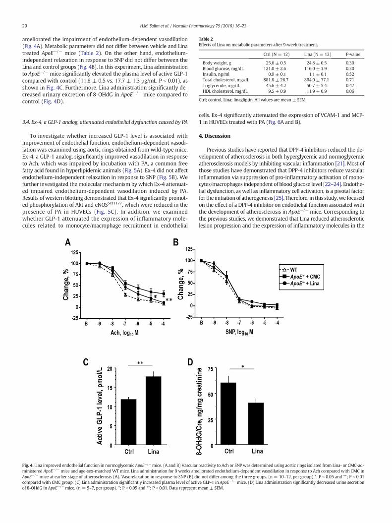
Table 2Effects of Lina on metabolic parameters after 9-week treatment.
Ctrl (N = 12) Lina (N = 12) P-value
Body weight, g 25.6 ± 0.5 24.8 ± 0.5 0.30Blood glucose, mg/dL 121.0 ± 2.6 116.0 ± 3.9 0.30Insulin, ng/ml 0.9 ± 0.1 1.1 ± 0.1 0.52Total cholesterol, mg/dL 881.8 ± 26.7 864.0 ± 37.1 0.71Triglyceride, mg/dL 45.6 ± 4.2 50.7 ± 5.4 0.47HDL cholesterol, mg/dL 9.5 ± 0.9 11.9 ± 0.9 0.06
Ctrl; control, Lina; linagliptin. All values are mean ± SEM.
20 H.M. Salim et al. / Vascular Pharmacology 79 (2016) 16–23
ameliorated the impairment of endothelium-dependent vasodilation(Fig. 4A). Metabolic parameters did not differ between vehicle and Linatreated ApoE−/− mice (Table 2). On the other hand, endothelium-independent relaxation in response to SNP did not differ between theLina and control groups (Fig. 4B). In this experiment, Lina administrationto ApoE−/− mice significantly elevated the plasma level of active GLP-1compared with control (11.8 ± 0.5 vs. 17.7 ± 1.3 pg/mL, P b 0.01), asshown in Fig. 4C. Furthermore, Lina administration significantly de-creased urinary excretion of 8-OHdG in ApoE−/− mice compared tocontrol (Fig. 4D).
3.4. Ex-4, a GLP-1 analog, attenuated endothelial dysfunction caused by PA
To investigate whether increased GLP-1 level is associated withimprovement of endothelial function, endothelium-dependent vasodi-lation was examined using aortic rings obtained from wild-type mice.Ex-4, a GLP-1 analog, significantly improved vasodilation in responseto Ach, which was impaired by incubation with PA, a common freefatty acid found in hyperlipidemic animals (Fig. 5A). Ex-4 did not affectendothelium-independent relaxation in response to SNP (Fig. 5B). Wefurther investigated the molecular mechanism by which Ex-4 attenuat-ed impaired endothelium-dependent vasodilation induced by PA.Results of western blotting demonstrated that Ex-4 significantly promot-ed phosphorylation of Akt and eNOSSer1177, which were reduced in thepresence of PA in HUVECs (Fig. 5C). In addition, we examinedwhether GLP-1 attenuated the expression of inflammatory mole-cules related to monocyte/macrophage recruitment in endothelial
Fig. 4. Lina improved endothelial function in normoglycemic ApoE−/−mice. (A and B) Vascularministered ApoE−/− mice and age-sex-matched WT mice. Lina administration for 9 weeks amApoE−/− mice at earlier stage of atherosclerosis (A). Vasorelaxation in response to SNP (B) dicompared with CMC group. (C) Lina administration significantly increased plasma level of actiof 8-OHdG in ApoE−/− mice. (n = 5–7, per group). *; P b 0.05 and **; P b 0.01. Data represent
cells. Ex-4 significantly attenuated the expression of VCAM-1 and MCP-1 in HUVECs treated with PA (Fig. 6A and B).
4. Discussion
Previous studies have reported that DPP-4 inhibitors reduced the de-velopment of atherosclerosis in both hyperglycemic and normoglycemicatherosclerosis models by inhibiting vascular inflammation [21]. Most ofthose studies have demonstrated that DPP-4 inhibitors reduce vascularinflammation via suppression of pro-inflammatory activation of mono-cytes/macrophages independent of blood glucose level [22–24]. Endothe-lial dysfunction, as well as inflammatory cell activation, is a pivotal factorfor the initiation of atherogenesis [25]. Therefore, in this study,we focusedon the effect of a DPP-4 inhibitor on endothelial function associated withthe development of atherosclerosis in ApoE−/− mice. Corresponding tothe previous studies, we demonstrated that Lina reduced atheroscleroticlesion progression and the expression of inflammatory molecules in the
reactivity to Ach or SNP was determined using aortic rings isolated from Lina- or CMC-ad-eliorated endothelium-dependent vasodilation in response to Ach compared with CMC ind not differ among the three groups. (n = 10–12, per group) *; P b 0.05 and **; P b 0.01ve GLP-1 in ApoE−/− mice. (D) Lina administration significantly decreased urine secretionmean ± SEM.
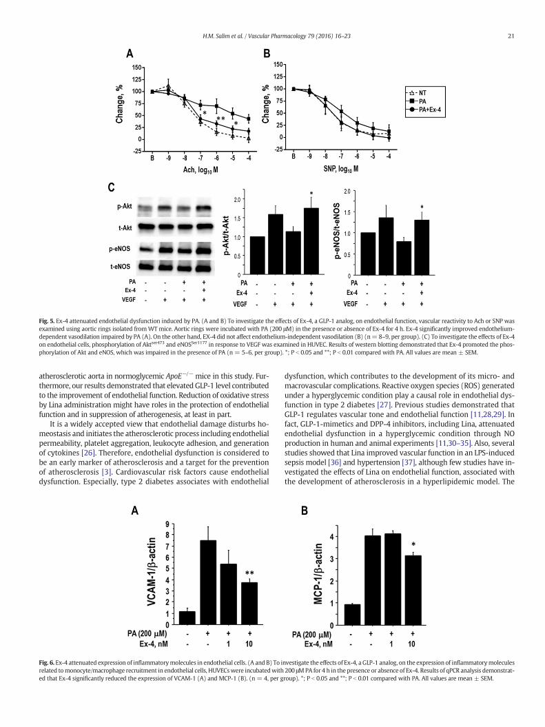
Fig. 5. Ex-4 attenuated endothelial dysfunction induced by PA. (A and B) To investigate the effects of Ex-4, a GLP-1 analog, on endothelial function, vascular reactivity to Ach or SNP wasexamined using aortic rings isolated fromWT mice. Aortic rings were incubated with PA (200 μM) in the presence or absence of Ex-4 for 4 h. Ex-4 significantly improved endothelium-dependent vasodilation impaired by PA (A). On the other hand, EX-4 did not affect endothelium-independent vasodilation (B) (n= 8–9, per group). (C) To investigate the effects of Ex-4on endothelial cells, phosphorylation of Aktser473 and eNOSSer1177 in response to VEGF was examined in HUVEC. Results of western blotting demonstrated that Ex-4 promoted the phos-phorylation of Akt and eNOS, which was impaired in the presence of PA (n = 5–6, per group). *; P b 0.05 and **; P b 0.01 compared with PA. All values are mean ± SEM.
21H.M. Salim et al. / Vascular Pharmacology 79 (2016) 16–23
atherosclerotic aorta in normoglycemic ApoE−/− mice in this study. Fur-thermore, our results demonstrated that elevatedGLP-1 level contributedto the improvement of endothelial function. Reduction of oxidative stressby Lina administration might have roles in the protection of endothelialfunction and in suppression of atherogenesis, at least in part.
It is a widely accepted view that endothelial damage disturbs ho-meostasis and initiates the atherosclerotic process including endothelialpermeability, platelet aggregation, leukocyte adhesion, and generationof cytokines [26]. Therefore, endothelial dysfunction is considered tobe an early marker of atherosclerosis and a target for the preventionof atherosclerosis [3]. Cardiovascular risk factors cause endothelialdysfunction. Especially, type 2 diabetes associates with endothelial
Fig. 6. Ex-4 attenuated expression of inflammatorymolecules in endothelial cells. (A and B) To irelated tomonocyte/macrophage recruitment in endothelial cells, HUVECswere incubatedwithed that Ex-4 significantly reduced the expression of VCAM-1 (A) and MCP-1 (B). (n = 4, per g
dysfunction, which contributes to the development of its micro- andmacrovascular complications. Reactive oxygen species (ROS) generatedunder a hyperglycemic condition play a causal role in endothelial dys-function in type 2 diabetes [27]. Previous studies demonstrated thatGLP-1 regulates vascular tone and endothelial function [11,28,29]. Infact, GLP-1-mimetics and DPP-4 inhibitors, including Lina, attenuatedendothelial dysfunction in a hyperglycemic condition through NOproduction in human and animal experiments [11,30–35]. Also, severalstudies showed that Lina improved vascular function in an LPS-inducedsepsis model [36] and hypertension [37], although few studies have in-vestigated the effects of Lina on endothelial function, associated withthe development of atherosclerosis in a hyperlipidemic model. The
nvestigate the effects of Ex-4, a GLP-1 analog, on the expression of inflammatorymolecules200 μMPA for 4 h in the presence or absence of Ex-4. Results of qPCR analysis demonstrat-roup). *; P b 0.05 and **; P b 0.01 compared with PA. All values are mean ± SEM.
22 H.M. Salim et al. / Vascular Pharmacology 79 (2016) 16–23
results of our present study demonstrated that endothelium-dependentvasodilation was improved in Lina-treated ApoE−/− mice, indicating itsprotective effects on endothelial cell function in a hyperlipidemic condi-tion, one of the major cardiovascular risk factors.
Lina elevated plasma active GLP-1 concentration in ApoE−/− mice.Therefore, we investigated the effect of GLP-1 on endothelial function.Our ex vivo experiment using aortic rings obtained fromWTmice dem-onstrated that Ex-4, a GLP-1 analog, improved endothelium-dependentvasodilationwhichwas impaired by the presence of PA, one of themostcommon free fatty acids found in hyperlipidemic animals [38,39]. Previ-ous studies have shown that PA-induced oxidative stress contributes toendothelial dysfunction and atherogenesis [40,41]. Our in vitro experi-ments using HUVEC demonstrated that Ex-4 increased phosphorylationof eNOSSer1177 and its upstream kinase, Aktser473, in response to VEGF[42], which was affected by the presence of PA. This result correspondsto previous studies that reported Akt/eNOS activation by Ex-4 [43,44].Phosphorylation eNOS at Ser1177 increases NO production and medi-ates endothelial function [20]. These results suggested that the antioxi-dant property of GLP-1 attenuated endothelial dysfunction caused byhyperlipidemia. In addition, Lina administration decreased ROS genera-tion as determined by urinary excretion of 8-OHdG and the expressionof NADPH oxidase subunits Nox2 and p47phox in the aorta. The expres-sion of Nox2 and p47phox is associated with vascular oxidative stress,leading to the development of atherosclerosis. Especially, several stud-ies have shown that Nox2-derived oxidative stress has a pathogenicrole in the functional changes of the arterial wall, including endothelialcell function, in hyperlipidemia [45–47]. In fact, Lina-treated miceshowed lower expression of inflammatory molecules in the abdominalaorta in our study. The result of in vitro experiments using HUVECwhich demonstrated inhibitory effect of EX-4 on inflammatory mole-cule expression supported this in vivo data. These results suggest thatthe antioxidative property of Lina, which is derived from elevatedGLP-1 level, contributes at least partially to the reduction of endothelialdamage and atherogenesis caused by hyperlipidemia. Recent evidencesuggests the cardioprotective effects of DPP-4 inhibitors independentof incretins [48]. DPP-4 inhibitors attenuate degradation of stromalcell-derived factor-1 and substance P as well as incretins, which maycontribute to vasoprotection at least in part [49]. Also, recent studiessuggest direct effects of DPP-4 inhibitors on vascular cells [48,50,51].Further studies are needed to elucidate these effects of Lina, howeverthose studies will help clarify its vasoprotective effects and underlyingmechanisms.
In conclusion, our results demonstrated that Lina inhibited thedevelopment atherosclerotic lesions in ApoE−/− mice, independent-ly of blood glucose level. The beneficial effect of Lina was associatedwith attenuation of endothelial dysfunction, at least in part by areduction of oxidative stress. Taken together with the results ofprevious studies, our results show that DPP-4 inhibitors have anti-inflammatory effects on various cell types in the vasculature. Ourpresent study supports the results of recent studies that suggestedatheroprotective effects of DPP-4 inhibitors. These non-glycemicproperties of DPP-4 inhibitors may provide attractive therapeuticoptions for atherosclerosis.
Sources of funding
This work was partially supported by JSPS Kakenhi Grants (Number25460369 to D.F., Number 25860586 to Y. Higashikuni, Number25860843 to Y. Hirata, Number 23591314 to M. Shimabukuro, andNumbers 24659392, 22390159, 25670390, 25293184 to M. Sata),MEXT KAKENHI Grant Number 21117007 (M. Sata) and a Grant fromThe Vehicle Racing Commemorative Foundation (K.T. and M. Sata). Dr.Sata received research funding from Boehringer Ingelheim, Japan(J171500100). The funders had no role in study design, data collectionand analysis, or preparation of the manuscript.
Disclosures
Dr. Sata received research funding from Boehringer Ingelheim,Japan. Other authors declare that they have no conflict of interest.
Acknowledgments
The authors thank Hiromi Kato, Yumi Sugawara, Yumiko Saga, andEtsuko Uematsu for their technical assistance.
References
[1] R. Ross, Atherosclerosis — an inflammatory disease, N. Engl. J. Med. 340 (2) (1999)115–126.
[2] U. Hink, H. Li, H. Mollnau, M. Oelze, E. Matheis, M. Hartmann, M. Skatchkov, F.Thaiss, R.A. Stahl, A. Warnholtz, T. Meinertz, K. Griendling, D.G. Harrison, U.Forstermann, T. Munzel, Mechanisms underlying endothelial dysfunction in diabetesmellitus, Circ. Res. 88 (2) (2001) E14–E22.
[3] J. Davignon, P. Ganz, Role of endothelial dysfunction in atherosclerosis, Circulation109 (23 Suppl. 1) (2004) III27–III32.
[4] D.J. Drucker, Incretin action in the pancreas: potential promise, possible perils, andpathological pitfalls, Diabetes 62 (10) (2013) 3316–3323.
[5] I. Jialal, M. Bajaj, DPP-4 inhibitors and atherosclerosis: the promise, Atherosclerosis227 (2) (2013) 224–225.
[6] J. Matsubara, S. Sugiyama, E. Akiyama, S. Iwashita, H. Kurokawa, K. Ohba, H. Maeda,K. Fujisue, E. Yamamoto, K. Kaikita, S. Hokimoto, H. Jinnouchi, H. Ogawa, Dipeptidylpeptidase-4 inhibitor, sitagliptin, improves endothelial dysfunction in associationwith its anti-inflammatory effects in patients with coronary artery disease anduncontrolled diabetes, Circ. J. 77 (5) (2013) 1337–1344.
[7] T. Murohara, Dipeptidyl peptidase-4 inhibitor: another player for cardiovascularprotection, J. Am. Coll. Cardiol. 59 (3) (2012) 277–279.
[8] A.J. Scheen, Cardiovascular effects of gliptins, Nat. Rev. Cardiol. 10 (2) (2013) 73–84.[9] J.R. Ussher, D.J. Drucker, Cardiovascular actions of incretin-based therapies, Circ. Res.
114 (11) (2014) 1788–1803.[10] K. Ban, M.H. Noyan-Ashraf, J. Hoefer, S.S. Bolz, D.J. Drucker, M. Husain, Cardioprotective
and vasodilatory actions of glucagon-like peptide 1 receptor aremediated throughbothglucagon-like peptide 1 receptor-dependent and -independent pathways, Circulation117 (18) (2008) 2340–2350.
[11] T. Nystrom, M.K. Gutniak, Q. Zhang, F. Zhang, J.J. Holst, B. Ahren, A. Sjoholm, Effectsof glucagon-like peptide-1 on endothelial function in type 2 diabetes patients withstable coronary artery disease, Am. J. Physiol. Endocrinol. Metab. 287 (6) (2004)E1209–E1215.
[12] Y. Hirata, H. Kurobe, C. Nishio, K. Tanaka, D. Fukuda, E. Uematsu, S. Nishimoto, T.Soeki, N. Harada, H. Sakaue, T. Kitagawa, M. Shimabukuro, Y. Nakaya, M. Sata,Exendin-4, a glucagon-like peptide-1 receptor agonist, attenuates neointimal hyper-plasia after vascular injury, Eur. J. Pharmacol. 699 (1–3) (2013) 106–111.
[13] N. Ervinna, T. Mita, E. Yasunari, K. Azuma, R. Tanaka, S. Fujimura, D. Sukmawati, T.Nomiyama, A. Kanazawa, R. Kawamori, Y. Fujitani, H. Watada, Anagliptin, a DPP-4inhibitor, suppresses proliferation of vascular smooth muscles and monocyte in-flammatory reaction and attenuates atherosclerosis in male apo E-deficient mice,Endocrinology 154 (3) (2013) 1260–1270.
[14] Z. Shah, T. Kampfrath, J.A. Deiuliis, J. Zhong, C. Pineda, Z. Ying, X. Xu, B. Lu, S. Moffatt-Bruce, R. Durairaj, Q. Sun, G. Mihai, A. Maiseyeu, S. Rajagopalan, Long-termdipeptidyl-peptidase 4 inhibition reduces atherosclerosis and inflammation via ef-fects on monocyte recruitment and chemotaxis, Circulation 124 (21) (2011)2338–2349.
[15] M. Terasaki, M. Nagashima, K. Nohtomi, K. Kohashi, M. Tomoyasu, K. Sinmura, Y.Nogi, Y. Katayama, K. Sato, F. Itoh, T. Watanabe, T. Hirano, Preventive effect ofdipeptidyl peptidase-4 inhibitor on atherosclerosis is mainly attributable toincretin's actions in nondiabetic and diabetic apolipoprotein E-null mice, PLoS One8 (8) (2013) e70933.
[16] Y. Zeng, C. Li, M. Guan, Z. Zheng, J. Li, W. Xu, L. Wang, F. He, Y. Xue, The DPP-4 inhib-itor sitagliptin attenuates the progress of atherosclerosis in apolipoprotein-E-knockout mice via AMPK- and MAPK-dependent mechanisms, Cardiovasc. Diabetol.13 (2014) 32.
[17] J. Doupis, Linagliptin: from bench to bedside, Drug Des. Devel. Ther. 8 (2014)431–446.
[18] M.H. Moghadasian, L.B. Nguyen, S. Shefer, B.M. McManus, J.J. Frohlich, Histologic, he-matologic, and biochemical characteristics of apo E-deficient mice: effects of dietarycholesterol and phytosterols, Lab. Invest. 79 (3) (1999) 355–364.
[19] D. Fukuda, M. Sata, N. Ishizaka, R. Nagai, Critical role of bone marrow angiotensin IItype 1 receptor in the pathogenesis of atherosclerosis in apolipoprotein E deficientmice, Arterioscler. Thromb. Vasc. Biol. 28 (1) (2008) 90–96.
[20] S. Matsumoto, M. Shimabukuro, D. Fukuda, T. Soeki, K. Yamakawa, H. Masuzaki, M.Sata, Azilsartan, an angiotensin II type 1 receptor blocker, restores endothelial func-tion by reducing vascular inflammation and by increasing the phosphorylation ratioSer(1177)/Thr(497) of endothelial nitric oxide synthase in diabetic mice,Cardiovasc. Diabetol. 13 (2014) 30.
[21] S.G. Chrysant, G.S. Chrysant, Clinical implications of cardiovascular preventing pleio-tropic effects of dipeptidyl peptidase-4 inhibitors, Am. J. Cardiol. 109 (11) (2012)1681–1685.
[22] M. Arakawa, T. Mita, K. Azuma, C. Ebato, H. Goto, T. Nomiyama, Y. Fujitani, T. Hirose,R. Kawamori, H. Watada, Inhibition of monocyte adhesion to endothelial cells and
23H.M. Salim et al. / Vascular Pharmacology 79 (2016) 16–23
attenuation of atherosclerotic lesion by a glucagon-like peptide-1 receptor agonist,exendin-4, Diabetes 59 (4) (2010) 1030–1037.
[23] Y. Dai, X. Wang, Z. Ding, D. Dai, J.L. Mehta, DPP-4 inhibitors repress foam cell forma-tion by inhibiting scavenger receptors through protein kinase C pathway, ActaDiabetol. 51 (3) (2014) 471–478.
[24] V. Matheeussen, Y. Waumans, W. Martinet, S. Van Goethem, P. Van der Veken, S.Scharpe, K. Augustyns, G.R. De Meyer, I. De Meester, Dipeptidyl peptidases inatherosclerosis: expression and role in macrophage differentiation, activation andapoptosis, Basic Res. Cardiol. 108 (3) (2013) 350.
[25] P.M.Vanhoutte, Endothelial dysfunction: thefirst step toward coronary arteriosclerosis,Circ. J. 73 (4) (2009) 595–601.
[26] P.O. Bonetti, L.O. Lerman, A. Lerman, Endothelial dysfunction: a marker of athero-sclerotic risk, Arterioscler. Thromb. Vasc. Biol. 23 (2) (2003) 168–175.
[27] H. Oeseburg, R.A. de Boer, H. Buikema, P. van der Harst, W.H. van Gilst, H.H. Sillje,Glucagon-like peptide 1 prevents reactive oxygen species-induced endothelial cellsenescence through the activation of protein kinase A, Arterioscler. Thromb. Vasc.Biol. 30 (7) (2010) 1407–1414.
[28] J. Oyama, Y. Higashi, K. Node, Do incretins improve endothelial function?Cardiovasc. Diabetol. 13 (2014) 21.
[29] Z. Shah, C. Pineda, T. Kampfrath, A. Maiseyeu, Z. Ying, I. Racoma, J. Deiuliis, X. Xu, Q.Sun, S. Moffatt-Bruce, F. Villamena, S. Rajagopalan, Acute DPP-4 inhibition modulatesvascular tone through GLP-1 independent pathways, Vasc. Pharmacol. 55 (1–3)(2011) 2–9.
[30] K. Nakamura, H. Oe, H. Kihara, K. Shimada, S. Fukuda, K. Watanabe, T. Takagi, K.Yunoki, T. Miyoshi, K. Hirata, J. Yoshikawa, H. Ito, DPP-4 inhibitor and alpha-glucosidase inhibitor equally improve endothelial function in patients with type 2diabetes: EDGE study, Cardiovasc. Diabetol. 13 (2014) 110.
[31] F.K. Saraiva, A.C. Sposito, Cardiovascular effects of Glucagon-like peptide 1 (GLP-1)receptor agonists, Cardiovasc. Diabetol. 13 (1) (2014) 142.
[32] S.M. Salheen, U. Panchapakesan, C.A. Pollock, O.L. Woodman, The DPP-4 inhibitorlinagliptin and the GLP-1 receptor agonist exendin-4 improve endothelium-dependent relaxation of rat mesenteric arteries in the presence of high glucose,Pharmacol. Res. 94 (2015) 26–33.
[33] L. Ding, J. Zhang, Glucagon-like peptide-1 activates endothelial nitric oxide synthasein human umbilical vein endothelial cells, Acta Pharmacol. Sin. 33 (1) (2012) 75–81.
[34] T. Gaspari, H. Liu, I. Welungoda, Y. Hu, R.E. Widdop, L.B. Knudsen, R.W. Simpson, A.E.Dear, A GLP-1 receptor agonist liraglutide inhibits endothelial cell dysfunction andvascular adhesion molecule expression in an ApoE−/− mouse model, Diab. Vasc.Dis. Res. 8 (2) (2011) 117–124.
[35] H. Nakagami, Z. Pang, T. Shimosato, T. Moritani, H. Kurinami, H. Koriyama, A. Tenma,M. Shimamura, R. Morishita, The dipeptidyl peptidase-4 inhibitor teneligliptinimproved endothelial dysfunction and insulin resistance in the SHR/NDmcr-cp ratmodel of metabolic syndrome, Hypertens. Res. 37 (7) (2014) 629–635.
[36] S. Kroller-Schon, M. Knorr, M. Hausding, M. Oelze, A. Schuff, R. Schell, S. Sudowe, A.Scholz, S. Daub, S. Karbach, S. Kossmann, T. Gori, P. Wenzel, E. Schulz, S. Grabbe, T.Klein, T. Munzel, A. Daiber, Glucose-independent improvement of vascular dysfunc-tion in experimental sepsis by dipeptidyl-peptidase 4 inhibition, Cardiovasc. Res. 96(1) (2012) 140–149.
[37] N. Koibuchi, Y. Hasegawa, T. Katayama, K. Toyama, K. Uekawa, D. Sueta, H. Kusaka,M. Ma, T. Nakagawa, B. Lin, S. Kim-Mitsuyama, DPP-4 inhibitor linagliptin amelio-rates cardiovascular injury in salt-sensitive hypertensive rats independently ofblood glucose and blood pressure, Cardiovasc. Diabetol. 13 (2014) 157.
[38] R.H. Knopp, B. Retzlaff, C. Walden, B. Fish, B. Buck, B. McCann, One-year effects ofincreasingly fat-restricted, carbohydrate-enriched diets on lipoprotein levels infree-living subjects, Proc. Soc. Exp. Biol. Med. 225 (3) (2000) 191–199.
[39] C. Sastre, A. Rubio-Navarro, I. Buendia, C. Gomez-Guerrero, J. Blanco, S. Mas, J. Egido,L.M. Blanco-Colio, A. Ortiz, J.A. Moreno, Hyperlipidemia-associated renal damagedecreases Klotho expression in kidneys from ApoE knockout mice, PLoS One 8(12) (2013) e83713.
[40] F. Kim, K.A. Tysseling, J. Rice, M. Pham, L. Haji, B.M. Gallis, A.S. Baas, P. Paramsothy,C.M. Giachelli, M.A. Corson, E.W. Raines, Free fatty acid impairment of nitric oxideproduction in endothelial cells is mediated by IKKbeta, Arterioscler. Thromb. Vasc.Biol. 25 (5) (2005) 989–994.
[41] C.H. Lee, S.D. Lee, H.C. Ou, S.C. Lai, Y.J. Cheng, Eicosapentaenoic acid protects againstpalmitic acid-induced endothelial dysfunction via activation of the AMPK/eNOSpathway, Int. J. Mol. Sci. 15 (6) (2014) 10334–10349.
[42] A. Uruno, A. Sugawara, H. Kanatsuka, H. Kagechika, A. Saito, K. Sato, M. Kudo, K.Takeuchi, S. Ito, Upregulation of nitric oxide production in vascular endothelialcells by all-trans retinoic acid through the phosphoinositide 3-kinase/Akt pathway,Circulation 112 (5) (2005) 727–736.
[43] W. Chai, Z. Dong, N. Wang, W. Wang, L. Tao, W. Cao, Z. Liu, Glucagon-like peptide 1recruits microvasculature and increases glucose use in muscle via a nitric oxide-dependent mechanism, Diabetes 61 (4) (2012) 888–896.
[44] O. Erdogdu, D. Nathanson, A. Sjoholm, T. Nystrom, Q. Zhang, Exendin-4 stimulatesproliferation of human coronary artery endothelial cells through eNOS-, PKA- andPI3K/Akt-dependent pathways and requires GLP-1 receptor, Mol. Cell. Endocrinol.325 (1–2) (2010) 26–35.
[45] T.J. Guzik, J. Sadowski, B. Guzik, A. Jopek, B. Kapelak, P. Przybylowski, K. Wierzbicki,R. Korbut, D.G. Harrison, K.M. Channon, Coronary artery superoxide production andnox isoform expression in human coronary artery disease, Arterioscler. Thromb.Vasc. Biol. 26 (2) (2006) 333–339.
[46] L. Loffredo, F. Martino, R. Carnevale, P. Pignatelli, E. Catasca, L. Perri, C.M. Calabrese,M.M. Palumbo, F. Baratta, M. Del Ben, F. Angelico, F. Violi, Obesity and hypercholester-olemia are associated with NOx2 generated oxidative stress and arterial dysfunction,J. Pediatr. 161 (6) (2012) 1004–1009.
[47] P. Haddad, S. Dussault, J. Groleau, J. Turgeon, F. Maingrette, A. Rivard, Nox2-derivedreactive oxygen species contribute to hypercholesterolemia-induced inhibition ofneovascularization: effects on endothelial progenitor cells and mature endothelialcells, Atherosclerosis 217 (2) (2011) 340–349.
[48] A.R. Aroor, J.R. Sowers, G. Jia, V.G. DeMarco, Pleiotropic effects of thedipeptidylpeptidase-4 inhibitors on the cardiovascular system, Am. J. Physiol.Heart Circ. Physiol. 307 (4) (2014) H477–H492.
[49] J.K. Damas, T. Waehre, A. Yndestad, T. Ueland, F. Muller, H.G. Eiken, A.M. Holm, B.Halvorsen, S.S. Froland, L. Gullestad, P. Aukrust, Stromal cell-derived factor-1alphain unstable angina: potential antiinflammatory and matrix-stabilizing effects, Circula-tion 106 (1) (2002) 36–42.
[50] W.S. da Silva Junior, A.F. de Godoy-Matos, L.G. Kraemer-Aguiar, Dipeptidyl peptidase4: a new link between diabetes mellitus and atherosclerosis? Biomed. Res. Int. 2015(2015) 816164.
[51] R. Morishita, H. Nakagami, Teneligliptin: expectations for its pleiotropic action,Expert. Opin. Pharmacother. 16 (3) (2015) 417–426.