The Potassium Content Gonyaulax Phase Changes Circadian Rhythm of

Dinophyta II. – Diversity, Evolution • dinoflagellates belong to both zoological and botanical systematics
• Alveolata (Cilliates, Dinophyta, Apicomplexa)
• traditional systematics (Dodge 1982) - 12 orders

Traditional systematics • groups (orders) defined by different numbers and shapes of
cellulose plates
• plate reduction model vs. plate increase model

Traditional systematics plate reduction model – heterotrophic ancestor
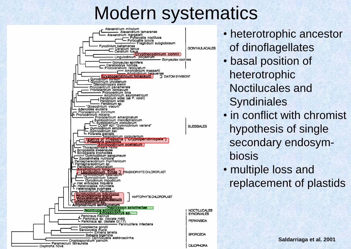
Modern systematics • heterotrophic ancestor
of dinoflagellates
• basal position of
heterotrophic
Noctilucales and
Syndiniales
• in conflict with chromist
hypothesis of single
secondary endosym-
biosis
• multiple loss and
replacement of plastids
Saldarriaga et al. 2001

• repeated loss of cellulose
plates during the evolution
• morphologically defined orders
Peridiniales and Gymnodiniales
are not monophyletic
• most of the genera
monophyletic!
Saldarriaga et al. 2004

Modern concept of dinoflagellate evolution history
Zhang et al. 2007
• Mitochondrial phylogeny

Modern concept of dinoflagellate evolution history
Oxyrrhis marina
– brackish species, toxic
red tides
– primitive features: open
mitosis, histons presented
– unclear position (SSU
nested in dinoflagellates,
protein coding: near the
base – probably one of the
oldes lineages of
dinoflagellates
gradual
transformation of
nucleus
Saldarriaga et al. 2004

Modern concept of dinoflagellate evolution history
Most dinoflagellates evolved from peridinin-containing ancestor. Those lineages containing
green algal, diatom, and haptophyte plastids evolved by a terciary endosymbiosis.

Modern systematics
Dinophysiales I
Dinophysiales II
• minimum of 7 independent
terciary endosymbioses
Kryptoperidinium
Karenia
Lepidodinium
Gymnodinium aeruginosum
Podolampas

Dinophysiales I Kryptoperidinium Karenia
Lepidodinium Gymnodinium aeruginosum Podolampas
Dinophysiales II
D. norvegica D. mitra
CRYPTO HAPTO
Durinskia, P. quinquecorne
BACILLARIO HAPTO
CHLORO
P. bipes
Karenia sp.
L. viride
CRYPTO DICTYOCHO

traditional features to recognize particular genera
• naked vs. thecate
• cingulum position (displacement)
• differences in plate ornamentation
features correlated with molecular data
• type of apical groove
• ultrastructural features
(stigma types)

• ecto- and endoparasites of marine invertebrates and fish
Syndinium – release of zoospores
Syndiniales (M)

endoparasite in Copepods
Syndiniales (M)
Blastodinium

Noctilucales (M)
• marine Noctiluca scintillans
• free-living flagellates without plates, large vacuolised cells
(up to 1.2 mm in diameter) – Na+, K+ replaced by H+
• heterotrophic
• epitheca and hypotheca are not differentiated in adult
cells, cingululm transformef in groove with tentacle

bioluminescence
Noctilucales (M)

zoosporogenesis
Noctilucales (M)

Gymnodiniales • unarmored dinoflagellates
• polyphyletic

Gymnodinium • free-living flagellates, sulcus + cingulum
• dorsiventrally flattened cells without visible plates
• autotrophic and heterotrophic species
• freshwater and marine

Gymnodinium (M)
saxitoxin - PSP !

Gymnodinium (F)
G. aeruginosum G. carinatum G. paradoxum
G. veris G. aeruginosum

Gymnodinium (F)
G. fuscum

Polykrikos (M)
coenocytic organisation
Polykrikos
schwartzii

Amphidinium (F, M)
conspicuously small epitheca
ciguatoxin - CFP !
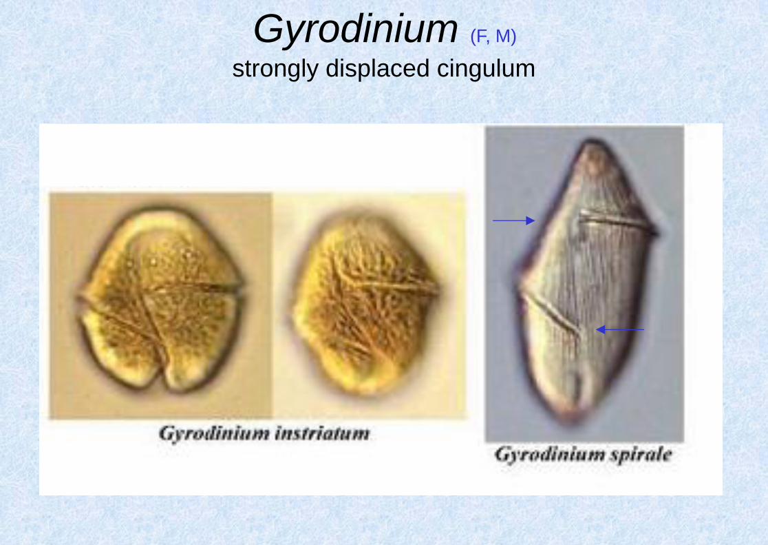
Gyrodinium (F, M)
strongly displaced cingulum

parasite on crustacean eggs
Dissodinium pseudolunula (M)

parasite on crustacean eggs
1 – dinospore is attaching the cell
5 – trophont separation (after 90 min)
Dissodinium pseudolunula (M)

parasite on crustacean eggs
1 – dinospore is attaching the cell
5 – trophont separation (after 90 min)
6 – several trophonts on one cell
10 – primary cyst
12 – secondaty cyst
13 – secondary cysts with mature
dinospores
Dissodinium pseudolunula (M)

parasite on crustacean eggs
1 – dinospore is attaching the cell
5 – trophont separation (after 90 min)
6 – several trophonts on one cell
10 – primary cyst
12 – secondaty cyst
13 – secondary cysts with mature
dinospores
Dissodinium pseudolunula (M)

Karlodinium (M)
• massive blooms in coastal waters, toxin production
• paralysis and mortality of finfish, copepods, nematods, and even
polychaete-larvae

Phytodiniales • coccoid dinoflagellates
• epifytes on filamentous algae

Dinococcus (F)

Stylodinium (F)

Peridiniales • flagellates, mostly dorsiventrally flattened cells
• polygonal plates, sulcus + cingulum
• marine and freshwater species

Peridinium (F)
formula 4´, 2a-3a, 7´´, 5´´´, 2´´´´
• autotrophic, freshwater, 5-6 cingular plates

Peridinium (F)
shapes of relevant plates
ortho meta para quadra penta hexa

Peridinium (F)
Peridinium willei – no pore, symmetrical plates

Peridinium (F)
Peridinium cinctum – typical shape of 3´ plate

Peridinium (F)
Peridinium umbonatum – pore, small cell dimensions

Peridinium (F)
Peridinium bipes – pore, symmetrical plates
2a
3a

Protoperidinium (M)
formula 4´, 3a, 7´´, 5´´´, 2´´´´
• heterotrophic, marine, only 3 cingular plates

Protoperidinium (M)
Protoperidinium depressum – 1´ ortho, 2a quadra
1´
2a

Protoperidinium (M)
Protoperidinium achromaticum – 1´ ortho, 2a hexa
1´
2a

Protoperidinium (M)
Protoperidinium pellucidum – 1´ para, 2a hexa
1´
2a

Peridiniopsis (F)
vzorec 3-5´, 0a-1a, 6-8´´, 5´´´, 2´´´´
• autotrophic, freshwater and brackish species
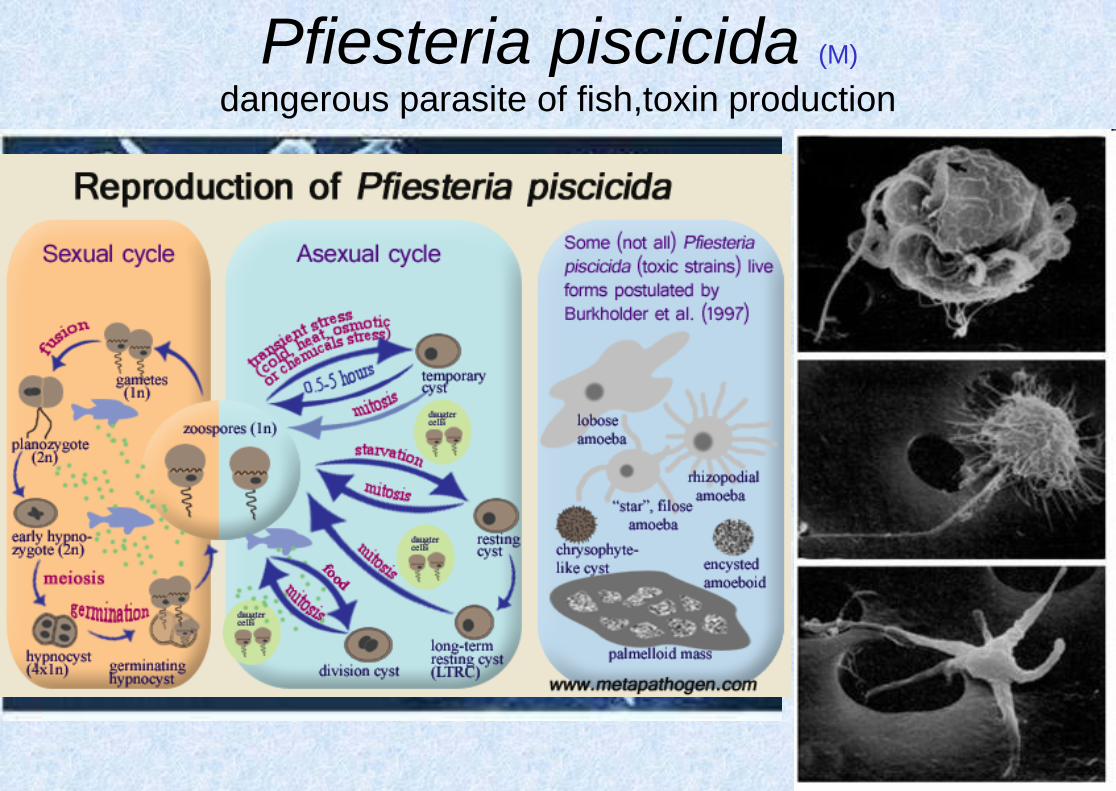
dangerous parasite of fish,toxin production
Pfiesteria piscicida (M)

Pfisteria piscicida (M)

• blood lesions
Pfisteria piscicida (M)

Gonyaulacales • armored flagellates
• distinguished from the Peridiniales
mostly by the arrangement and
number of the thecal plates
• the first apical plate assymetrical

Ceratium (F, M)
formula 4´, 5´´, 5´´´, 2´´´´

Ceratium (F)
Ceratium hirundinella Ceratium cornutum

Ceratium (M)
Ceratium furca Ceratium fusus Ceratium lineatum
Ceratium longipes Ceratium macroceros Ceratium tripos

Gonyaulax (F, M)
thick, ornamented plates, species determined according to the
hypotheca morphology, hard to determine particular species

bioluminescence
Pyrocystis (M)
Pyrocystis fusiformis Pyrocystis noctiluca

Alexandrium (M)
thin plates, pore at 1´ plate, often filamentous

Alexandrium (M)
thin plates, pore at 1´ plate, often filamentous
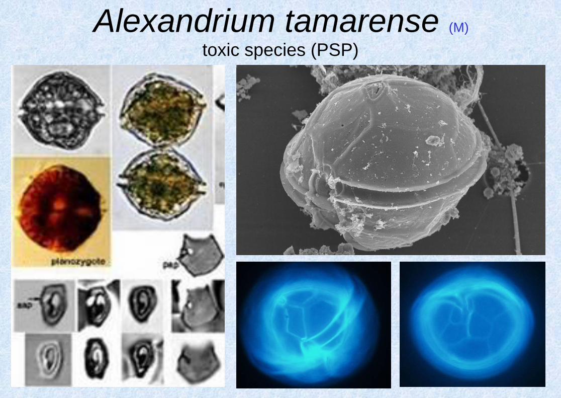
Alexandrium tamarense (M)
toxic species (PSP)

Thoracosphaerales • coccoid dinoflagellates, in Thoracosphaera cells
surrounded by calcareous cell wall
• molecular data support a relationship with thecate
flagellates of Peridiniales s.l.

Gloeodinium montanum • capsal thallus
• peat bogs (N Europe, N America, New Zaeland)
• separate species (genus) or only a life stage?

Hemidinium nasutum (F)
„Gloeodinium“
stage

Hemidinium nasutum (F)

assymetric cells, incomplete cingulum
Hemidinium (F)

Suessiales - Symbiodinium (M)
http://www.youtube.com/watch?feature=player_detailpage&v=9Lm9hUj2h_0
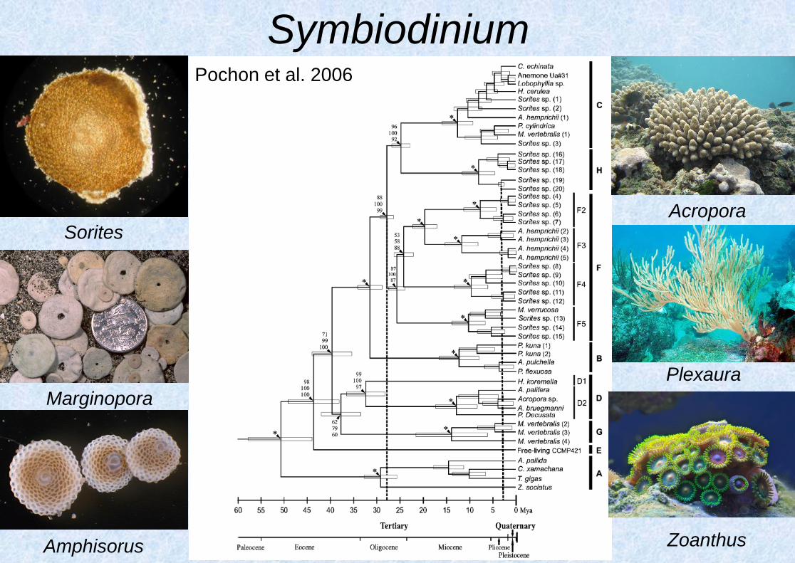
Symbiodinium
Sorites
Marginopora
Amphisorus
Acropora
Plexaura
Pochon et al. 2006
Zoanthus

Symbiodinium • Symbiodinium lineages exhibit different physiologies in
response to variations in light and temperature

Symbiodinium • Coral bleeching - the loss of intracellular endosymbionts
• increase of temperature, solar radiation, acidification, ...

Symbiodinium • To prevent bleeching, corals host specifically adapted
Symbiodinium
• clade C – sensitive to higher temperatures, PSII damage
• clade D – tolerant to higher temp., photoprotection
Rowan 2004

Symbiodinium • Habitat adapted symbiosis
Byler et al. 2013
cla
de
A
cla
de
C

Symbiodinium
• Enormous cryptic diversity
• Strong phylogeograhic
structure
• Host-specific lineages
LaJeunesse et al. 2011
LaJeunesse et al. 2011

Symbiodinium • Host-symbiont specificity, ecological differences
• C3-z: offshore; C15: thermally tolerant
LaJeunesse et al. 2010

Dinophysiales • flagellates, lateraly flattened cells
• thecate, epitheca + hypotheca, left and right half of the cell
separated by longitudinal suture
• only marine species

Dinophysis (M)
toxic species (DSP)

Dinophysis (M)
non-toxic species
Dinophysis norvegica Dinophysis rotundata
(= Phalacroma)
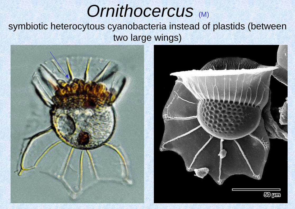
Ornithocercus (M)
symbiotic heterocytous cyanobacteria instead of plastids (between
two large wings)

Histioneis (M)

Amphisolenia (M)
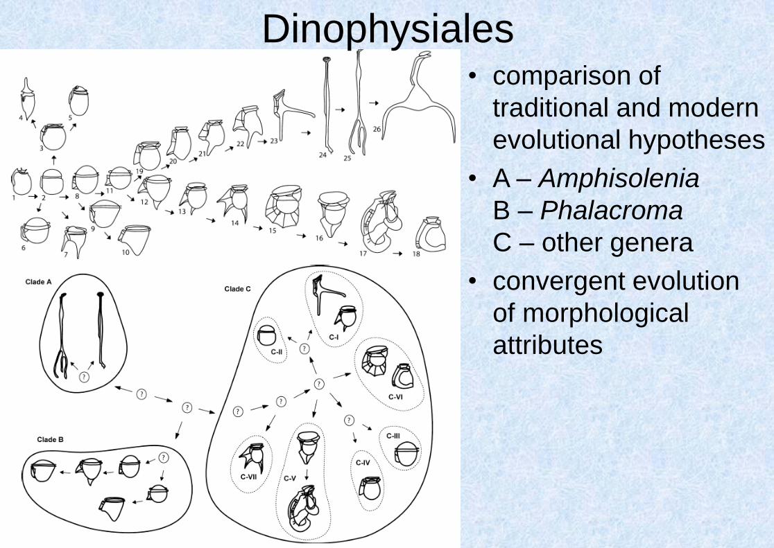
Dinophysiales • comparison of
traditional and modern
evolutional hypotheses
• A – Amphisolenia
B – Phalacroma
C – other genera
• convergent evolution
of morphological
attributes

Prorocentrales • flagellates, lateraly flattened cells
• thecate, 2 big plates separated by longitudinal suture +
several minute plates near apical pores
• only marine species, autotrophic

Prorocentrum (M)

Prorocentrum micans (M)
planktonic species, red tides
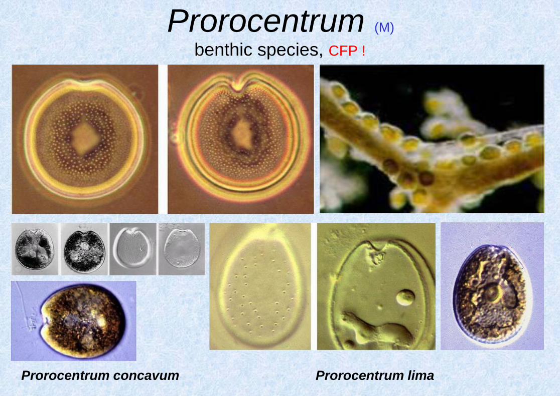
Prorocentrum (M)
benthic species, CFP !
Prorocentrum lima Prorocentrum concavum