Dinitrosyl Iron Complexes with Thiol-Containing Ligands as ... · involve iron, thiol sulfur and...
7
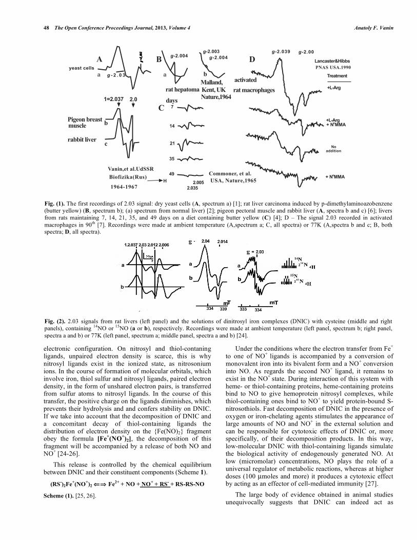
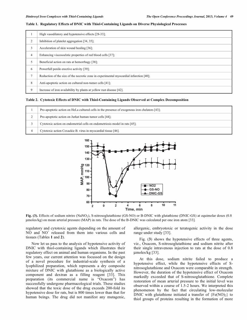
Send Orders for Reprints to [email protected] The Open Conference Proceedings Journal, 2013, 4, 47-53 47 2210-2892/13 2013 Bentham Open Open Access Dinitrosyl Iron Complexes with Thiol-Containing Ligands as a Base for New-Generation Drugs (Review) Anatoly F. Vanin * Semyonov Institute of Chemical Physics, Russian Academy of Sciences, Kosygin Str. 4, 119991 Moscow, Russia Abstract: Some present-day concepts of biological activities of dinitrosyl iron complexes (DNIC) with thiol-containing ligands are reviewed. These activities are determined by the ability of DNIC to act as donors of NO and nitrosonium ions (NO + ) in various body cells and tissues. DNIC are endowed with strong vasodilator activity. Due to the latter DNIC single dose injection into animal and human organisms induces long-lasting hypotension. Moreover, DNIC suppress platelet aggregation, increase red blood cell elasticity, accelerate skin wound healing, induce penis erection in animals and exert pronounced anti-apoptotic effect on cultured non-tumor cells. These regulatory effects are due to the slow release of NO and NO + from intact DNIC at both organism and in cell cultures. Fast decomposition of DNIC, e.g., in the presence of exogenous or endogenous iron-chelating agents, results in a toxic effect of DNIC on various cells and tissues and causes apoptosis of, e.g., HeLa and Jurkat human tumor cells. More recent model studies established the ability of DNIC to suppress endometriosis in rats. Keywords: Dinitrosyl iron complexes, endometriosis, hypotension, penile erection, platelet aggregation, vasodilation, wound healing. INTRODUCTION As established now, dinitrosyl iron complexes (briefly DNIC) with thiol-containing ligands are synthesized by cells and tissues able to generate nitric oxide. For the first time, these complexes were identified in 1960’s by our group by a characteristic electron paramagnetic resonance (EPR) signal in yeasts, pigeon pectoral muscle and rabbit liver with g av. =2.03 (2.03 signal) (Fig. 1, A). A similar, but less intense signal was detected in rat liver by our colleagues from Great Britain and the U.S.A. at early stages of chemically induced carcinogenesis (Fig. 1, B and C) [1-6]. Thirty years after acknowledgement of the physiological role of endogenously synthesized nitric oxide as a universal regulator of a vast array of metabolic reactions, the 2.03 signal was recorded in many cells and tissues [7-21]. Among them, are activated macrophages able to synthesize nitric oxide by an enzymatic route (Fig. 1, D) [7]. Identification of paramagnetic centers responsible for the 2.03 signal as DNIC with thiol-containing ligands was first performed in our laboratory [22] and, some time later, in the U.S.A [23]. These studies established that the major characteristics of the 2.03 signal fully coincided with those of the EPR signal recorded in frozen solutions of DNIC with low-molecular thiol-containing ligands (Fig. 2). The identity of the characteristics of the 2.03 signal recorded in biological objects to those of the EPR signal observed in frozen solutions of DNIC with low-molecular thiol-containing *Address correspondence to this author at the Semyonov Institute of Chemical Physics, Russian Academy of Sciences, Kosygin Str. 4, 119991 Moscow, Russia; Tel: +7-495-939-7535; Fax: +7-495-651-2191; E-mail: [email protected] ligands and the nature of changes in these signals initiated by treatment of biological objects and low-molecular DNIC with various chemical agents [6] led us to conclude that the centres responsible for these signals have the same origin. The only difference between them is that in biological objects it is protein-bound DNIC containing protein residues of cysteine that are responsible for the 2.03 signal. Evidence for the protein origin of these DNIC can be derived from the preservation of the anisotropic shape of their EPR signal with the increase in the registration temperature from 77К to ambient (Fig. 2, left panel, signals a and b). The low mobility of the protein globule at ambient temperature is insufficient to initiate the averaging of anisotropy of the g- factor responsible for the anisotropic shape of the 2.03 signal. This averaging is characteristic of highly mobile low- molecular DNIC, as a result of which their EPR signal diminishes in size after the increase in the registration temperature from 77К to ambient and acquires the shape of a narrow symmetric singlet with a well-resolved hyperfine structure (HFS) (Fig. 2, middle and left panels). An analysis of the hyperfine structure of their EPR signals at ambient temperature (Fig. 2, right panel) showed that these DNIC contained one iron atom, two nitrosyl ligands and two thiol-containing ligands. More recent studies revealed that DNIC with thiol-containing ligands are represented by two forms, viz., a paramagnetic mononuclear form (М-DNIC) responsible for 2.03 signal and a more stable diamagnetic binuclear form (B-DNIC) characterized by the formulas [(RS) 2 Fe (NO) 2 ] and [(RS) 2 Fe 2 (NO) 4 ], respectively. In addition, these studies made it possible to establish the distribution of spin density in M-DNIC, which is characterized by the formula [(RS - ) 2 Fe + (NO + ) 2 ] [24-26]. As can be seen, the unpaired electron density is predominantly localized on the iron atom, which has a d 7
Transcript of Dinitrosyl Iron Complexes with Thiol-Containing Ligands as ... · involve iron, thiol sulfur and...
Send Orders for Reprints to [email protected]
The Open Conference Proceedings Journal, 2013, 4, 47-53 47
2210-2892/13 2013 Bentham Open
Open Access
Dinitrosyl Iron Complexes with Thiol-Containing Ligands as a Base for New-Generation Drugs (Review)
Anatoly F. Vanin*
Semyonov Institute of Chemical Physics, Russian Academy of Sciences, Kosygin Str. 4, 119991 Moscow, Russia
Abstract: Some present-day concepts of biological activities of dinitrosyl iron complexes (DNIC) with thiol-containing ligands are reviewed. These activities are determined by the ability of DNIC to act as donors of NO and nitrosonium ions (NO+) in various body cells and tissues. DNIC are endowed with strong vasodilator activity. Due to the latter DNIC single dose injection into animal and human organisms induces long-lasting hypotension. Moreover, DNIC suppress platelet aggregation, increase red blood cell elasticity, accelerate skin wound healing, induce penis erection in animals and exert pronounced anti-apoptotic effect on cultured non-tumor cells. These regulatory effects are due to the slow release of NO and NO+ from intact DNIC at both organism and in cell cultures. Fast decomposition of DNIC, e.g., in the presence of exogenous or endogenous iron-chelating agents, results in a toxic effect of DNIC on various cells and tissues and causes apoptosis of, e.g., HeLa and Jurkat human tumor cells. More recent model studies established the ability of DNIC to suppress endometriosis in rats.
Keywords: Dinitrosyl iron complexes, endometriosis, hypotension, penile erection, platelet aggregation, vasodilation, wound healing.
INTRODUCTION
As established now, dinitrosyl iron complexes (briefly DNIC) with thiol-containing ligands are synthesized by cells and tissues able to generate nitric oxide. For the first time, these complexes were identified in 1960’s by our group by a characteristic electron paramagnetic resonance (EPR) signal in yeasts, pigeon pectoral muscle and rabbit liver with gav.=2.03 (2.03 signal) (Fig. 1, A). A similar, but less intense signal was detected in rat liver by our colleagues from Great Britain and the U.S.A. at early stages of chemically induced carcinogenesis (Fig. 1, B and C) [1-6]. Thirty years after acknowledgement of the physiological role of endogenously synthesized nitric oxide as a universal regulator of a vast array of metabolic reactions, the 2.03 signal was recorded in many cells and tissues [7-21]. Among them, are activated macrophages able to synthesize nitric oxide by an enzymatic route (Fig. 1, D) [7].
Identification of paramagnetic centers responsible for the 2.03 signal as DNIC with thiol-containing ligands was first performed in our laboratory [22] and, some time later, in the U.S.A [23]. These studies established that the major characteristics of the 2.03 signal fully coincided with those of the EPR signal recorded in frozen solutions of DNIC with low-molecular thiol-containing ligands (Fig. 2). The identity of the characteristics of the 2.03 signal recorded in biological objects to those of the EPR signal observed in frozen solutions of DNIC with low-molecular thiol-containing
*Address correspondence to this author at the Semyonov Institute of Chemical Physics, Russian Academy of Sciences, Kosygin Str. 4, 119991 Moscow, Russia; Tel: +7-495-939-7535; Fax: +7-495-651-2191; E-mail: [email protected]
ligands and the nature of changes in these signals initiated by treatment of biological objects and low-molecular DNIC with various chemical agents [6] led us to conclude that the centres responsible for these signals have the same origin. The only difference between them is that in biological objects it is protein-bound DNIC containing protein residues of cysteine that are responsible for the 2.03 signal. Evidence for the protein origin of these DNIC can be derived from the preservation of the anisotropic shape of their EPR signal with the increase in the registration temperature from 77К to ambient (Fig. 2, left panel, signals a and b). The low mobility of the protein globule at ambient temperature is insufficient to initiate the averaging of anisotropy of the g-factor responsible for the anisotropic shape of the 2.03 signal. This averaging is characteristic of highly mobile low-molecular DNIC, as a result of which their EPR signal diminishes in size after the increase in the registration temperature from 77К to ambient and acquires the shape of a narrow symmetric singlet with a well-resolved hyperfine structure (HFS) (Fig. 2, middle and left panels). An analysis of the hyperfine structure of their EPR signals at ambient temperature (Fig. 2, right panel) showed that these DNIC contained one iron atom, two nitrosyl ligands and two thiol-containing ligands. More recent studies revealed that DNIC with thiol-containing ligands are represented by two forms, viz., a paramagnetic mononuclear form (М-DNIC) responsible for 2.03 signal and a more stable diamagnetic binuclear form (B-DNIC) characterized by the formulas [(RS)2 Fe (NO)2] and [(RS)2 Fe2 (NO)4], respectively. In addition, these studies made it possible to establish the distribution of spin density in M-DNIC, which is characterized by the formula [(RS-)2 Fe+ (NO+)2] [24-26]. As can be seen, the unpaired electron density is predominantly localized on the iron atom, which has a d7
48 The Open Conference Proceedings Journal, 2013, Volume 4 Anatoly F. Vanin
electronic configuration. On nitrosyl and thiol-contaning ligands, unpaired electron density is scarce, this is why nitrosyl ligands exist in the ionized state, as nitrosonium ions. In the course of formation of molecular orbitals, which involve iron, thiol sulfur and nitrosyl ligands, paired electron density, in the form of unshared electron pairs, is transferred from sulfur atoms to nitrosyl ligands. In the course of this transfer, the positive charge on the ligands diminishes, which prevents their hydrolysis and and confers stability on DNIC. If we take into account that the decomposition of DNIC and a concomitant decay of thiol-containing ligands the distribution of electron density on the {Fe(NO)2} fragment obey the formula [Fe+(NO+)2], the decomposition of this fragment will be accompanied by a release of both NO and NO+ [24-26]. This release is controlled by the chemical equilibrium between DNIC and their constituent components (Scheme 1).
(RS-)2Fe+(NO+)2 ⇐⇒ Fe2+ + NO + NO+ + RS- + RS-RS-NO
Scheme (1). [25, 26].
Under the conditions where the electron transfer from Fe+ to one of NO+ ligands is accompanied by a conversion of monovalent iron into its bivalent form and a NO+ conversion into NO. As regards the second NO+ ligand, it remains to exist in the NO+ state. During interaction of this system with heme- or thiol-containing proteins, heme-containing proteins bind to NO to give hemoprotein nitrosyl complexes, while thiol-containing ones bind to NO+ to yield protein-bound S-nitrosothiols. Fast decomposition of DNIC in the presence of oxygen or iron-chelating agents stimulates the appearance of large amounts of NO and NO+ in the external solution and can be responsible for cytotoxic effects of DNIC or, more specifically, of their decomposition products. In this way, low-molecular DNIC with thiol-containing ligands simulate the biological activity of endogenously generated NO. At low (micromolar) concentrations, NO plays the role of a universal regulator of metabolic reactions, whereas at higher doses (100 µmoles and more) it produces a cytotoxic effect by acting as an effector of cell-mediated immunity [27]. The large body of evidence obtained in animal studies unequivocally suggests that DNIC can indeed act as
Fig. (1). The first recordings of 2.03 signal: dry yeast cells (A, spectrum a) [1]; rat liver carcinoma induced by p-dimethylaminoazobenzene (butter yellow) (B, spectrum b); (a) spectrum from normal liver) [2]; pigeon pectoral muscle and rabbit liver (A, spectra b and c) [6]; livers from rats maintaining 7, 14, 21, 35, and 49 days on a diet containing butter yellow (C) [4]; D – The signal 2.03 recorded in activated macrophages in 90th [7]. Recordings were made at ambient temperature (A,spectrum a; C, all spectra) or 77K (A,spectra b and c; B, both spectra; D, all spectra).
Fig. (2). 2.03 signals from rat livers (left panel) and the solutions of dinitrosyl iron complexes (DNIC) with cysteine (middle and right panels), containing 14NO or 15NO (a or b), respectively. Recordings were made at ambient temperature (left panel, spectrum b; right panel, spectra a and b) or 77K (left panel, spectrum a; middle panel, spectra a and b) [24].
���������
� �
�
� � � � �
���� �� ��
������ ����������
����������
�
�
����������������������� ���!
�"#$%&"#$'
� �
�
��
��
��
��
���(�)����
��*�
�� ��� ��
��� ���� �
��� �
+������
,��� ��"#$%-�����-
���� �������������,��� ��"#$.
/
���������
������ )(����
��� �� ���
�,�������"##0���������������
���������
����
���� ��!�""�#
!#�$$���#�
��!�""�#
Dinitrosyl Iron Complexes with Thiol-Containing Ligands The Open Conference Proceedings Journal, 2013, Volume 4 49
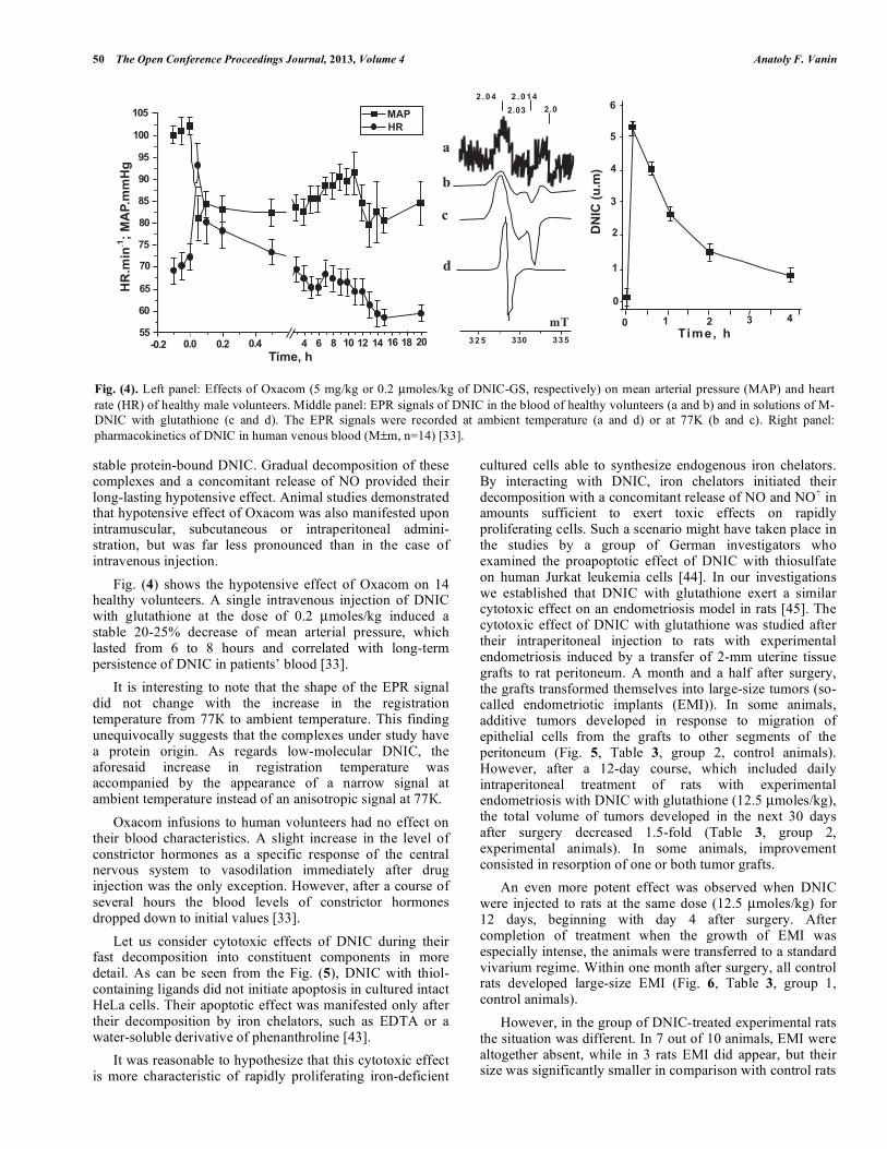
regulatory and cytotoxic agents depending on the amount of NO and NO+ released from them into various cells and tissues (Tables 1 and 2). Now let us pass to the analysis of hypotensive activity of DNIC with thiol-containing ligands which illustrates their regulatory effect on animal and human organisms. In the past few years, our current attention was focussed on the design of a novel procedure for industrial-scale synthesis of a lyophilized preparation, which represents a dry composite mixture of DNIC with glutathione as a biologically active component and dextran as a filling reagent [33]. This preparation (its commercial name is “Oxacom”) has successfully undergone pharmacological trials. These studies showed that the toxic dose of the drug exceeds 200-fold its hypotensive dose for rats, but is 800 times lower than that for human beings. The drug did not manifest any mutagenic,
allergenic, embryotoxic or teratogenic activity in the dose range under study [33]. Fig. (3) shows the hypotensive effects of three agents, viz., Oxacom, S-nitrosoglutathione and sodium nitrite after their single intravenous injection to rats at the dose of 0.8 µmoles/kg [33]. At this dose, sodium nitrite failed to produce a hypotensive effect, while the hypotensive effects of S-nitrosoglutathione and Oxacom were comparable in strength. However, the duration of the hypotensive effect of Oxacom markedly exceeded that of S-nitrosoglutathione. Complete restoration of mean arterial pressure to the initial level was observed within a course of 1.5-2 hours. We interpreted this phenomenon by the fact that circulating low-molecular DNIC with glutathione initiated a transfer of {Fe(NO)2} to thiol groups of proteins resulting in the formation of more
Table 1. Regulatory Effects of DNIC with Thiol-Containing Ligands on Diverse Physiological Processes
1 High vasodilatory and hypotensive effects [28-33];
2 Inhibition of platelet aggregation [34, 35];
3 Acceleration of skin wound healing [36];
4 Enhancing viscosoelstic properties of red blood cells [37];
5 Beneficial action on rats at hemorrhagy [38];
6 Powerfull penile erective activity [39];
7 Reduction of the size of the necrotic zone in experimental myocardial infarction [40];
8 Anti-apoptotic action on cultured non-tumor cells [41];
9 Increase of iron availability by plants at yellow rust disease [42].
Table 2. Cytotoxic Effects of DNIC with Thiol-Containing Ligands Observed at Complex Decomposition
1 Pro-apoptotic action on HeLa cultured cells in the presence of exogenous iron chelators [43];
2 Pro-apoptotic action on Jurkat human tumor cells [44];
3 Cytotoxic action on endometrial cells on endometriosis model in rats [45];
4 Cytotoxic action Coxackie B. virus in myocardial tissue [46].
Fig. (3). Effects of sodium nitrire (NaNO2), S-nitrosoglutathione (GS-NO) or B-DNIC with glutathione (DNIC-GS) at equimolar doses (0.8 µmoles/kg) on mean arterial pressure (MAP) in rats. The dose of the B-DNIC was calculated per one iron atom [33].
"�&'����
�%
��
��
��
��
��
�
�
(
�
% � � � ( � ��
����'����
)!*+,-,-!.!.�
%
50 The Open Conference Proceedings Journal, 2013, Volume 4 Anatoly F. Vanin
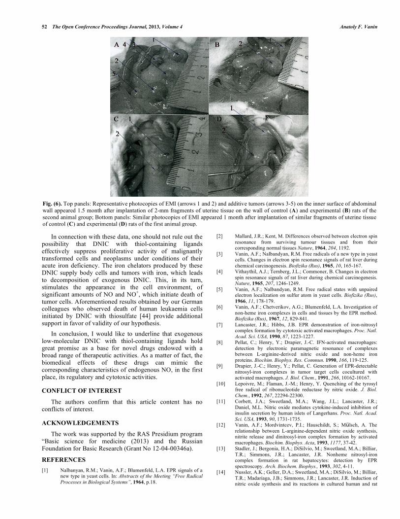
stable protein-bound DNIC. Gradual decomposition of these complexes and a concomitant release of NO provided their long-lasting hypotensive effect. Animal studies demonstrated that hypotensive effect of Oxacom was also manifested upon intramuscular, subcutaneous or intraperitoneal admini-stration, but was far less pronounced than in the case of intravenous injection. Fig. (4) shows the hypotensive effect of Oxacom on 14 healthy volunteers. A single intravenous injection of DNIC with glutathione at the dose of 0.2 µmoles/kg induced a stable 20-25% decrease of mean arterial pressure, which lasted from 6 to 8 hours and correlated with long-term persistence of DNIC in patients’ blood [33]. It is interesting to note that the shape of the EPR signal did not change with the increase in the registration temperature from 77К to ambient temperature. This finding unequivocally suggests that the complexes under study have a protein origin. As regards low-molecular DNIC, the aforesaid increase in registration temperature was accompanied by the appearance of a narrow signal at ambient temperature instead of an anisotropic signal at 77К. Oxacom infusions to human volunteers had no effect on their blood characteristics. A slight increase in the level of constrictor hormones as a specific response of the central nervous system to vasodilation immediately after drug injection was the only exception. However, after a course of several hours the blood levels of constrictor hormones dropped down to initial values [33]. Let us consider cytotoxic effects of DNIC during their fast decomposition into constituent components in more detail. As can be seen from the Fig. (5), DNIC with thiol-containing ligands did not initiate apoptosis in cultured intact HeLa cells. Their apoptotic effect was manifested only after their decomposition by iron chelators, such as EDTA or a water-soluble derivative of phenanthroline [43]. It was reasonable to hypothesize that this cytotoxic effect is more characteristic of rapidly proliferating iron-deficient
cultured cells able to synthesize endogenous iron chelators. By interacting with DNIC, iron chelators initiated their decomposition with a concomitant release of NО and NO+ in amounts sufficient to exert toxic effects on rapidly proliferating cells. Such a scenario might have taken place in the studies by a group of German investigators who examined the proapoptotic effect of DNIC with thiosulfate on human Jurkat leukemia cells [44]. In our investigations we established that DNIC with glutathione exert a similar cytotoxic effect on an endometriosis model in rats [45]. The cytotoxic effect of DNIC with glutathione was studied after their intraperitoneal injection to rats with experimental endometriosis induced by a transfer of 2-mm uterine tissue grafts to rat peritoneum. A month and a half after surgery, the grafts transformed themselves into large-size tumors (so-called endometriotic implants (EMI)). In some animals, additive tumors developed in response to migration of epithelial cells from the grafts to other segments of the peritoneum (Fig. 5, Table 3, group 2, control animals). However, after a 12-day course, which included daily intraperitoneal treatment of rats with experimental endometriosis with DNIC with glutathione (12.5 µmoles/kg), the total volume of tumors developed in the next 30 days after surgery decreased 1.5-fold (Table 3, group 2, experimental animals). In some animals, improvement consisted in resorption of one or both tumor grafts. An even more potent effect was observed when DNIC were injected to rats at the same dose (12.5 µmoles/kg) for 12 days, beginning with day 4 after surgery. After completion of treatment when the growth of EMI was especially intense, the animals were transferred to a standard vivarium regime. Within one month after surgery, all control rats developed large-size EMI (Fig. 6, Table 3, group 1, control animals). However, in the group of DNIC-treated experimental rats the situation was different. In 7 out of 10 animals, EMI were altogether absent, while in 3 rats EMI did appear, but their size was significantly smaller in comparison with control rats
Fig. (4). Left panel: Effects of Oxacom (5 mg/kg or 0.2 µmoles/kg of DNIC-GS, respectively) on mean arterial pressure (MAP) and heart rate (HR) of healthy male volunteers. Middle panel: EPR signals of DNIC in the blood of healthy volunteers (a and b) and in solutions of M-DNIC with glutathione (c and d). The EPR signals were recorded at ambient temperature (a and d) or at 77K (b and c). Right panel: pharmacokinetics of DNIC in human venous blood (M±m, n=14) [33].
� �
�
��
�
(�
(
��
�
%�
%
�� �� � �� �� � % ( � �� �� �% �( �
����'�/
�0����
��1�"�&����
"�&�0
��� �� ���
�6
�
�
�
�
� � � � � � � � � ��
�� � ��
�
%
�
�
�
�
� � � � ����' �/
)!*+�23��4

Dinitrosyl Iron Complexes with Thiol-Containing Ligands The Open Conference Proceedings Journal, 2013, Volume 4 51
Fig. (5). Upper panel: The histograms illustrating the lack effect of DNIC with glutathione 1:20 on the state of DNA in HeLa cells, the lack of the HeLa subpopulation with the DNA content less the diploid level (<2c, late apoptosis) on the fluorescence channels with the channel number <75. DNIC concentration 100 mM (a), 200 mM (b), and 500 mM (c). The cells were incubated in Eagle`s medium supplemented with fetal calf serum. Solid line – control; dotted line – incubation with DNIC. Bottom panel: The histograms illustrating the pro-apoptotic effect of DNIC with thiosulphate (left side) on HeLa cells preincubated in Versene`s solution containing 0.5 mM EDTA and DNIC with glutathione after incubation of HeLa cells in Eagle`s medium supplemented with fetal calf serum in the presence of bathophenantroline disulfide (BPDS) (right side). Line 1- control; lines 2-4 (left side) – DNIC with thiosulphate (50, 100, and 200 mM, respectively). Lines 2 and 3 – 200 mM DNIC with glutathione or DNIC with glutathione + 50 mM BPDS. <2c, –2c and –4c – subpopulations of cells with the DNA content less than the diploid level, with the diploid and tetraploid content of DNA, respectively. Ordinate – number of cells (in relative units) [43]. Table 3. Results of Statistical Treatments of the Data Obtained in Experiments on Rat Endometriosis Model. Volumes (mm3) of
Endometriotic Implants (EMI) are Shown
Median (min-maz) Mean + SD
Control (10 rats) 36 (2-599) 113 + 179 Group 1
Experiment (10 rats) 0 (0-73) 7 + 17
P < 0.001
Control (10 rats) 30 (2-866) 150 + 230 Group 2
Experiment (10 rats) 7 (0-759) 106 + 232
P < 0.008
(Fig. 6, Table 3, group 1, experimental animals). These findings explicitly suggest that treatment of rats with DNIC had a substantially stronger effect on EMI when DNIC injections were given in the course of their proliferative growth. It might be expected that the less pronounced effect of DNIC on the growth of EMI tumors in second group rats was due to lower rates of tumor growth one month after transplantation. However, histopathological analysis of EMI tumors established a lack of endometrial cysts in
experimental rats, while EMI tumors from control rats displayed the presence of multiple cysts. These data prompt a conclusion that endometrial cells of second group rats, whose proliferative activity provided the growth of EMI tumors, were also susceptible to cytotoxic effect of DNIC. Evidently, the cytotoxic activity of DNIC could not be directed against fibrous and other EMI cells, which developed on the 30th post-transplantation day and retained their intactness by the moment of sacrifice on day 45 (Table 3).
,��
�� �
���
���� �
�
� � ,���� ����(���
/,3�&4�"00��� +
&2�&
&%�&
�
52�&�
�
� �
/,3�&4�200��� +�
,���� ����(���
%
,��
�� �
���
����
,��
�� �
���
���� %
�
�
,���� ����(���
/,3�&4�"��+�
� ��
,���� ����(���� �
�
�
,��
�� �
���
����
/,3�&6�7�8/6�
2 9"
%
� � 0
200
%00
,��
�� �
���
����
/,3�&4�7)(��� ���
9
2"
,���� ����(���
52 The Open Conference Proceedings Journal, 2013, Volume 4 Anatoly F. Vanin
In connection with these data, one should not rule out the possibility that DNIC with thiol-containing ligands effectively suppress proliferative activity of malignantly transformed cells and neoplasms under conditions of their acute iron deficiency. The iron chelators produced by these DNIC supply body cells and tumors with iron, which leads to decomposition of exogenous DNIC. This, in its turn, stimulates the appearance in the cell environment, of significant amounts of NO and NO+, which initiate death of tumor cells. Aforementioned results obtained by our German colleagues who observed death of human leukaemia cells initiated by DNIC with thiosulfate [44] provide additional support in favor of validity of our hypothesis. In conclusion, I would like to underline that exogenous low-molecular DNIC with thiol-containing ligands hold great promise as a base for novel drugs endowed with a broad range of therapeutic activities. As a matter of fact, the biomedical effects of these drugs can mimic the corresponding characteristics of endogenous NO, in the first place, its regulatory and cytotoxic activities.
CONFLICT OF INTEREST
The authors confirm that this article content has no conflicts of interest.
ACKNOWLEDGEMENTS
The work was supported by the RAS Presidium program “Basic science for medicine (2013) and the Russian Foundation for Basic Research (Grant No 12-04-00346a). REFERENCES [1] Nalbanyan, R.M.; Vanin, A.F.; Blumenfeld, L.A. EPR signals of a
new type in yeast cells. In: Abstracts of the Meeting “Free Radical Processes in Biological Systems”, 1964, p.18.
[2] Mallard, J.R.; Kent, M. Differences observed between electron spin resonance from surviving tumour tissues and from their corresponding normal tissues Nature, 1964, 204, 1192.
[3] Vanin, A.F.; Nalbandyan, R.M. Free radicals of a new type in yeast cells. Changes in electron spin resonance signals of rat liver during chemical carcinogenesis. Biofizika (Rus), 1965, 10, 165-167.
[4] Vithaythil, A.J.; Ternberg, J.L.; Commoner, B. Changes in electron spin resonance signals of rat liver during chemical carcinogenesis. Nature, 1965, 207, 1246-1249.
[5] Vanin, A.F.; Nalbandyan, R.M. Free radical states with unpaired electron localization on sulfur atom in yeast cells. Biofizika (Rus), 1966, 11, 178-179.
[6] Vanin, A.F.; Chetverikov, A.G.; Blumenfeld, L.A. Investigation of non-heme iron complexes in cells and tissues by the EPR method. Biofizika (Rus), 1967, 12, 829-841.
[7] Lancaster, J.R.; Hibbs, J.B. EPR demonstration of iron-nitrosyl complex formation by cytotoxic activated macrophages. Proc. Natl. Acad. Sci. USA, 1990, 87, 1223-1227.
[8] Pellat, C.; Henry, Y.; Drapier, J.-C. IFN-activated macrophages: detection by electronic paramagnetic resonance of complexes between L-arginine-derived nitric oxide and non-heme iron proteins. Biochim. Biophys. Res. Commun. 1990, 166, 119-125.
[9] Drapier, J.-C.; Henry, Y.; Pellat, C. Generation of EPR-detectable nitrosyl-iron complexes in tumor target cells cocultured with activated macrophages. J. Biol. Chem., 1991, 266, 10162-10167.
[10] Lepoivre, M.; Flaman, J.-M.; Henry, Y. Quenching of the tyrosyl free radical of ribonucleotide reductase by nitric oxide. J. Biol. Chem., 1992, 267, 22294-22300.
[11] Corbett, J.A.; Sweetland, M.A.; Wang, J.L.; Lancaster, J.R.; Daniel, M.L. Nitric oxide mediates cytokine-induced inhibition of insulin secretion by human islets of Langerhans. Proc. Natl. Acad. Sci. USA, 1993, 90, 1731-1735.
[12] Vanin, A.F.; Mordvintcev, P.I.; Hauschildt, S.; Mülsch, A. The relationship between L-arginine-dependent nitric oxide synthesis, nitrite release and dinitrosyl-iron complex formation by activated macrophages. Biochim. Biophys. Acta, 1993, 1177, 37-42.
[13] Stadler, J.; Bergonia, H.A.; DiSilvio, M.; Sweetland, M.A.; Billiar, T.R.; Simmons, J.R.; Lancaster, J.R. Nonheme nitrosyl-iron complex formation in rat hepatocytes: detection by EPR spectroscopy. Arch. Biochem. Biophys., 1993, 302, 4-11.
[14] Nussler, A.K.; Geller, D.A.; Sweetland, M.A.; DiSilvio, M.; Billiar, T.R.; Madariaga, J.B.; Simmons, J.R.; Lancaster, J.R. Induction of nitric oxide synthesis and its reactions in cultured human and rat
Fig. (6). Top panels: Representative photocopies of EMI (arrows 1 and 2) and additive tumors (arrows 3-5) on the inner surface of abdominal wall appeared 1.5 month after implantation of 2-mm fragments of uterine tissue on the wall of control (A) and experimental (B) rats of the second animal group; Bottom panels: Similar photocopies of EMI appeared 1 month after implantation of similar fragments of uterine tissue of control (C) and experimental (D) rats of the first animal group.
Dinitrosyl Iron Complexes with Thiol-Containing Ligands The Open Conference Proceedings Journal, 2013, Volume 4 53
hepatocytes stimulated with cytokines plus LPS. Biochim. Biophys. Res. Commun. 1993, 194, 826-835.
[15] Mülsch, A.; Mordvintcev, P.I.; Vanin, A.F.; Busse, R., Formation and release of dinitrosyl iron complexes by endothelial cells. Biochim. Biophys. Res. Commun. 1993, 196, 1303-1308.
[16] Geng, Y.-L.; Pettersson, A.-S.; Wennmalm, A.; Hannson, G. Cytokine-induced expression of nitric oxide synthase results in nitrosylation of heme and nonheme iron proteins in vascular smooth muscle cells. Exp. Cell Res., 1994, 214, 418-424.
[17] Bastian, N.R.; Yim, C.-Y.; Hibbs, J.B.; Samlowsky, W.E. Induction of iron-derived EPR signals in murine cancers by nitric oxide. J. Biol. Chem., 1994, 269, 5127-5131.
[18] Muller, B.; Kleschyov, A.L.; Stoclet, J.-C. Evidence for N-acetylcysteine-sensitive nitric oxide storage as dinitrosyl-iron complexes in lipopolysaccharide-treated rat aorta. Br. J. Pharmacol., 1996, 119, 1281-1285.
[19] Odoi, K.; Akaike, T.; Horie, H.; Noguchi, Y.; Fujii, S.; Beppu, N.; Ogawa, M.; Maeda, H. Excessive production of nitric oxide in rat solid tumor and its application to rapid tumor growth. Cancer, 1996, 77, 1598-1604.
[20] Mikoyan, V.D.; Serezhenkov, V.A.; Brazhnikova, N.V.; Kubrina, L.N.; Khatchryan, G.V.; Vanin, A.F. Formation of paramagnetic nitrosyl complexes of nonheme iron in the animal organism from exogenous and endogenous sources. Biofizika (Rus), 2004, 41, 110-116.
[21] Watts, R.N.; Hawkins, C.; Ponka, P.; Richardson, D. Nitrogen monoxide (NO)-mediated iron release from cells is linked to NO-induced glutathione efflux via multidrug resistance-associated protein 1. Proc. Natl Acad. Sci. USA, 2006, 103, 7670-7675.
[22] Vanin, A.F. Identification of divalent iron complexes with cysteine in biological systems by the EPR method. Biokhimiya (Rus), 1967, 32, 1240-1249.
[23] Woolum, J.C.; Commoner, B. Isolation and identification of a paramagnetic complex from the livers of carcinogen-treated rats. Biochim. Biophys. Acta, 1970, 201, 131-140.
[24] Vanin, A.F. Dinitrosyl iron complexes with thiolate ligands: Physico-chemistry, biochemistry and physiology. Nitric Oxide Biol. Chem., 2009, 21, 1-13.
[25] Vanin, A.F.; Burbaev, D.Sh. Electronic and spatial structures of water-soluble dinitrosyl iron complexes with thiol-containing ligands underlying their ability to act as nitric oxide and nitrosonium ion donors. J. Biophys., 2011, 2011, 14 (Article ID 878236).
[26] Borodulin, R.R.; Kubrina, L.N.; Mikoyan, V.D.; Poltorakov, A.P.; Shvydkiy, V.O.; Burbaev, D.Sh.; Serezhenkov, V.A.; Yakhontova, E.R.; Vanin, A.F. Dinitrosyl iron complexes with glutathione as NO and NO+ donors. Nitric Oxide Biol Chem., 2013, 29, 4-16.
[27] Ignarro, L.J. Nitric Oxide: Biology and Pharmacology, Academic Press, San Diego, 2000.
[28] Vedernikov, Y.P.; Mordvintcev, P.I.; Malenkova, I.V.; Vanin, A.F. Similarity between the vasorelaxing activity of dinitrosyl iron complexes and endothelium-derived relaxing factor. Eur. J. Pharmacol. 1992, 211, 313-317.
[29] Vanin. A.F.; Mokh, V.P.; Serezhenkov, V.A.; Chazov E.I. Vasorelaxing activity of stable powder preparations of dinitrosyl iron complexes with cysteine or glutathione. Nitric Oxide Biol Chem., 2007, 16, 323-326.
[30] Flitney, F.W.; Megson, I.L.; Flitney, D.E.; Butler, A.R. Iron-sulfur cluster nitrosyl, a novel class of nitric oxide generator: mechanism of vasodilator action on rat isolated tail artery. Br. J. Pharmacol., 1992, 107, 842-848.
[31] Kleschyov, A.L.; Mordvintcev, P.I.; Vanin, A.F. Role of nitric oxide and iron in hypotensive action of nitrosyl iron complexes with various anion ligands. Stud Biophys., 1985, 105, 93-102.
[32] Lakomkin, V.L.; Vanin, A.F.; Timoshin, A.A.; Kapelko, V.I.; Chazov, E.I. Long-lasting hypotensive action of stable preparations 0f dinitrosyl iron complexes with thiol-containing ligands in conscious normotensive and hypertensive rats. Nitric Oxide Biol. Chem., 2007, 16, 413-418.
[33] Chazov, E.I.; Rodnenkov, O.V.; Zorin, A.V.; Lakomkin, V.L.; Gramovich, V.V.; Vyborov, O.N.; Dragnev, A.G.; Timoshin, A.A.; Buryachkovskayay, L.I.; Abramov, A.A.; Massenko, V.P.; Arzamastsev, E.V.; Kapelko, V.I.; Vanin, A.F. Hypotensive effect of Oxacom containing a dinitrosyl iron complex with glutathione: Animal studies and clinical trials on healthy volunteers. Nitric Oxide Biol. Chem., 2012, 26, 148-156.
[34] Mordvintcev, P.I.; Rudneva, V.G.; Vanin, A.F.; Shimkevich, L.L.; Khodorov, B.I. Inhibiting effect of dinitrosyl iron complexes with low molecular weight ligands on platelet aggregation. Biokhimiya (Rus), 1986, 51, 1851-1856.
[35] Arkhipova, M.M.; Mikoyan, V.D.; Vanin, A.F. Effect of exogenous donors of nitric oxide and inhibitors of its enzymatic synthesis on experimental ischemic thrombosis on conjunctive veins in rabbit eyes. Biofizika (Rus), 2008, 53, 315-325.
[36] Shekhter, A.B.; Rudenko, T.G.; Serezhenkov, V.A.; Vanin, A.F. Dinitrosyl-iron complexes with cysteine or glutathione accelerate skin wound healing. Biofizika (Rus), 2007, 52, 539-547.
[37] Shamova, E.V.; Bichan, O.D.; Drozd, E.S.; Gorudko, I.V.; Chizhik, S.A.; Shumaev, K.B.; Cherenkevich, S.N.; Vanin, A.F. Regulation of the functional and mechanical properties of platelet and red blood cells by nitric oxide donors. Biofizika (Rus), 2011, 56, 265-271.
[38] Remizova, M.I.; Kochetygov, N.I.; Kerbout, K.A.; Lakomkin, V.L.; Timoshin, A.A.; Burgova, E.N.; Vanin, A.F. Effect of dinitrosyl iron complex with glutathione on hemorrhagic shock followed by saline treatment. Eur. J. Pharmacol., 2011, 662, 40-46.
[39] Andreev-Andriyevsky, A.A.; Mikoyan, V.D.; Serezhenkov, V.A.; Vanin, A.F. Penile erectile activity of dinitrosyl iron complexes with thiol-containing ligands. Nitric Oxide Biol. Chem., 2011, 24, 217-223.
[40] Pisarenko, O.I.; Serebryakova, L.I.; Tskitishvili, O.V.; Studneva, I.M.; Vanin, A.F.; Chazov, E.I. Cardioprotective effect of dinitrosyl iron complex with l-cysteine in rats in vivo. Izvestia Akademii Nauk, Seriya Biologicheskaya(Rus), 2008, No.1, pp.110-114.
[41] Kim, Y.M.; Ghung, H.T.; Simmons, R.L.; Billiar, T.R. Cellular non-heme iron content is a determinant of nitric oxide-mediated apoptosis, necrosis, and caspase inhibition. J. Biol. Chem. 2000, 275, 10954-10961.
[42] Graziano, M.; Lamattina, L. Nitric oxide and iron in plants: an emerging and converging story. Trends Plant Sci., 2005, 10, 4-8.
[43] Giliano, N.Y.; Konevega, L.V.; Noskin, L.A.; Serezhenkov, V.A.; Poltorakov, A.P.; Vanin, A.F. Dinitrosyl iron complexes with thiol-containing ligands and apopotosis: Studies with HeLa cell culture. Nitric Oxide Biol. Chem., 2011, 24, 151-159.
[44] Kleschyov, A.L.; Strand, S.; Schmitt, S.; Gottfried, D.; Skatchkov, M.; Sjakste, N.; Daiber, A.; Umansky, V.; Munzel, T. Dinitrosyl-iron triggers apoptosis in Jurkat cells despite overexpression of Bcl-2. Free Radic. Biol. Med. 2006, 40, 1340-1348.
[45] Adamyan, L.V.; Burgova, E.N.; Tkachev, N.A.; Mikoyan, V.D.; Stapanyan, A.A.; Sonova M.M.; Galkin, A.V.; Vanin, A.F. Dinitrosyl iron complexes with glutathione largely relieve rats of experimental endometriosis. Biophysics, 2013, 58, 222-227.
[46] Badroff, C.; Fichtscherer, B.; Muelsch, A.; Zeiher, A.M.; Dimeller, S. Selective delivery of nitric oxide to a cellular target: a pseudosubstrate-coupled dinitrosyl iron complex inhibits the enteroviral protease 2 A. Nitric Oxide Biol. Chem., 2001, 6, 305-312.
Received: April 18, 2013 Revised: June 07, 2013 Accepted: July 03, 2013
© Anatoly F. Vanin; Licensee Bentham Open.
This is an open access article licensed under the terms of the Creative Commons Attribution Non-Commercial License (http://creativecommons.org/licenses/by-nc/3.0/), which permits unrestricted, non-commercial use, distribution and reproduction in any medium, provided the work is properly cited.