
Dimer N-terminal the APC
5
Proc. Natl. Acad. Sci. USA Vol. 90, pp. 11109-11113, December 1993 Biochemistry Dimer formation by an N-terminal coiled coil in the APC protein (adenomatous polyposis coli/heptad repeat region/subdomain) GEOFF JOSLYN*t, DIANE S. RICHARDSON*, RAY WHITE*, AND TOM ALBERt§¶ *Department of Human Genetics and Howard Hughes Medical Institute, University of Utah School of Medicine, Salt Lake City, UT 84132; and 'Department of Biochemistry, University of Utah School of Medicine, Salt Lake City, UT 84132 Contributed by Ray White, August 6, 1993 ABSTRACT Mutations in the human APC gene are asso- ciated with an inherited predisposition to colon cancer. APC codes for polypeptides of =2800 amino acids, with sequence homologies to coiled-coil proteins in the first 900 residues. To determine the oligomerization properties of the APC protein, we used genetic and biochemical approaches to examine the ability of APC fragments to self-associate. A subdomain com- prising the first 55 amino acids of APC was found to form a stable, parallel, helical dimer, as expected for a coiled coil. The location of a key dimerization element at the N terminus of the protein supports models in which mutations in APC exert effects through dimerization of the mutant gene products. Familial adenomatous polyposis (FAP) is a dominant genetic disease defined by early onset of large numbers of colonic polyps that are precursors of colon cancer (1). FAP is caused by mutations in APC, a gene of unknown function located on human chromosome 5 (2-7). Alternatively spliced APC mRNAs encode predicted polypeptides of 2742 and 2843 amino acids, with no extensive sequence homology to other proteins. The first =900 residues of the APC protein, how- ever, contain proline-free blocks with heptad repeats of hydrophobic residues characteristic of coiled coils (5, 6, 8). This pattern suggests that APC may function in association with itself or with other cellular factors. Coiled coils consist of associated a-helices that form superhelical ropes (8). The interface between the helices generally contains hydrophobic amino acids that occur at the first and fourth positions of a recurring seven-residue motif, the heptad repeat. Polar amino acids are common at the other positions in coiled-coil sequences, with charged groups often bracketing succeeding buried hydrophobic groups. Excep- tions to these patterns are commonly tolerated in long coiled coils, but short coiled coils, such as leucine zippers, can be destabilized by replacements of even single interface residues (9). Because the blocks of heptad repeat sequences in APC are relatively short and contain violations of the character- istic sequence patterns, experimental tests are needed to determine oligomerization potential. In addition, the number and the orientation of helices in coiled coils is variable (8). Examples of parallel and antipar- allel dimeric, trimeric, and tetrameric coiled coils have been reported (8, 10-12). Furthermore, some coiled coils, such as laminins (13) and the leucine zippers of Fos and Jun (14), preferentially associate with heterologous sequences. We describe here the use of genetic and biochemical methods to investigate the oligomerization potential of sequences in the first 686 residues of the APC protein. We show that the first 55 amino acids of APC are sufficient to form a stable, parallel, helical dimer, as expected for a two-stranded parallel coiled coil. The publication costs of this article were defrayed in part by page charge payment. This article must therefore be hereby marked "advertisement" in accordance with 18 U.S.C. §1734 solely to indicate this fact. MATERIALS AND METHODS Dimerization of A Repressor by APC Sequences. APC gene fragments flanked by in-frame Sal I and BamHI restriction sites were produced by PCR using appropriate cDNA clones as templates (4). Gene fragments encoding amino acids 1-112 and 1-456 were obtained by digesting an APC cDNA clone in the pFLAG vector (15) with BamHI and BstYI or BamHI and Bgl II, respectively. To make A repressor fusions, the APC restriction fragments were inserted between the Sal I and BamHI sites of the plasmid pJH391 (9). Automated DNA sequencing (4) confirmed the structures of the plasmids. Plasmids were introduced into Escherichia coli AG1688 (9), and the resulting strains were plated with 100-1000 A phages at 37°C. The tester phage, AKH54 (9), has undergone a deletion of the cI repressor. The positive control for phage sensitivity, A'434 (16), contains the phage 434 immunity re- gion, which is not recognized by A repressor. Expression and Purification of APC Fragments. The DNA sequence coding for amino acids 1-55 of APC (APC-55) was amplified by PCR (4), using primers that introduced flanking restriction sites (Nde I at the initiator methionine and BamHI after a termination codon or, alternatively, flanking Xho I sites). To replace Ile-55 with Cys, the change in codon 55 was included in an alternative PCR primer. The PCR products were digested with restriction enzymes and inserted into Nde I/BamHI-digested pAED4 (17) or Xho I-digested pFLAG (15) expression vectors. Sequencing the inserts (5) revealed the expected order of nucleotides except for a T -* A change that converted Asp-7 to Glu in the APC sequences in the otherwise wild-type FLAG fusion protein. Nonetheless, this peptide was characterized biochemically, because residue 7, which would occur on the outside of a coiled coil, was expected to have no influence on oligomerization. The peptide APC-55 was expressed from the phage T7 promoter in the pAED4-based plasmid in E. coli BL21(DE3) (18). Cultures were grown at 37°C in LB medium (33) supplemented with ampicillin at 50 ,ug/ml. When the optical density at 600 nm reached 0.76, cultures were induced with 0.4 mM isopropyl ,B D-thiogalactoside (IPTG). After 2 hr, induced cultures were harvested, resuspended in buffer con- taining 50 mM Tris'HCl at pH 8.0, 20%o (vol/vol) glycerol, 200 mM KCl, and 0.2 mM EDTA (buffer A) and were lysed by sonication. The lysate was brought to pH 2.0 by addition of HCI and centrifuged 10 min at 10,000 rpm in a Beckman JA-20 rotor to remove cell debris and precipitated protein. The pellet was washed with buffer A, and the combined super- natants were brought to pH 8.0 with NaOH. After 2-fold dilution with water, the supernatant was loaded on a DEAE- Sephadex A-25 column and eluted with a gradient of 0.1-1.0 Abbreviations: FAP, familial adenomatous polyposis; APC, product of the APC (adenomatous polyposis coli) gene. tPresent address: Fred Hutchinson Cancer Research Center, 1124 Columbia Street, Seattle, WA 98104. §Present address: Department of Molecular and Cell Biology, Stan- ley/Donner ASU, University of California, Berkeley, CA 94720. ¶To whom reprint requests should be addressed. 11109 Downloaded by guest on February 23, 2022
Transcript of Dimer N-terminal the APC
Proc. Natl. Acad. Sci. USAVol. 90, pp. 11109-11113, December 1993Biochemistry
Dimer formation by an N-terminal coiled coil in the APC protein(adenomatous polyposis coli/heptad repeat region/subdomain)
GEOFF JOSLYN*t, DIANE S. RICHARDSON*, RAY WHITE*, AND TOM ALBERt§¶
*Department of Human Genetics and Howard Hughes Medical Institute, University of Utah School of Medicine, Salt Lake City, UT 84132;and 'Department of Biochemistry, University of Utah School of Medicine, Salt Lake City, UT 84132
Contributed by Ray White, August 6, 1993
ABSTRACT Mutations in the human APC gene are asso-ciated with an inherited predisposition to colon cancer. APCcodes for polypeptides of =2800 amino acids, with sequencehomologies to coiled-coil proteins in the first 900 residues. Todetermine the oligomerization properties of the APC protein,we used genetic and biochemical approaches to examine theability of APC fragments to self-associate. A subdomain com-prising the first 55 amino acids of APC was found to form astable, parallel, helical dimer, as expected for a coiled coil. Thelocation of a key dimerization element at the N terminus of theprotein supports models in which mutations in APC exerteffects through dimerization of the mutant gene products.
Familial adenomatous polyposis (FAP) is a dominant geneticdisease defined by early onset of large numbers of colonicpolyps that are precursors of colon cancer (1). FAP is causedby mutations in APC, a gene ofunknown function located onhuman chromosome 5 (2-7). Alternatively spliced APCmRNAs encode predicted polypeptides of 2742 and 2843amino acids, with no extensive sequence homology to otherproteins. The first =900 residues of the APC protein, how-ever, contain proline-free blocks with heptad repeats ofhydrophobic residues characteristic of coiled coils (5, 6, 8).This pattern suggests that APC may function in associationwith itself or with other cellular factors.
Coiled coils consist of associated a-helices that formsuperhelical ropes (8). The interface between the helicesgenerally contains hydrophobic amino acids that occur at thefirst and fourth positions of a recurring seven-residue motif,the heptad repeat. Polar amino acids are common at the otherpositions in coiled-coil sequences, with charged groups oftenbracketing succeeding buried hydrophobic groups. Excep-tions to these patterns are commonly tolerated in long coiledcoils, but short coiled coils, such as leucine zippers, can bedestabilized by replacements ofeven single interface residues(9). Because the blocks of heptad repeat sequences in APCare relatively short and contain violations of the character-istic sequence patterns, experimental tests are needed todetermine oligomerization potential.
In addition, the number and the orientation of helices incoiled coils is variable (8). Examples of parallel and antipar-allel dimeric, trimeric, and tetrameric coiled coils have beenreported (8, 10-12). Furthermore, some coiled coils, such aslaminins (13) and the leucine zippers of Fos and Jun (14),preferentially associate with heterologous sequences. Wedescribe here the use of genetic and biochemical methods toinvestigate the oligomerization potential of sequences in thefirst 686 residues of the APC protein. We show that the first55 amino acids ofAPC are sufficient to form a stable, parallel,helical dimer, as expected for a two-stranded parallel coiledcoil.
The publication costs of this article were defrayed in part by page chargepayment. This article must therefore be hereby marked "advertisement"in accordance with 18 U.S.C. §1734 solely to indicate this fact.
MATERIALS AND METHODSDimerization of A Repressor by APC Sequences. APC gene
fragments flanked by in-frame Sal I and BamHI restrictionsites were produced by PCR using appropriate cDNA clonesas templates (4). Gene fragments encoding amino acids 1-112and 1-456 were obtained by digesting an APC cDNA clonein the pFLAG vector (15) with BamHI and BstYI or BamHIand Bgl II, respectively. To make A repressor fusions, theAPC restriction fragments were inserted between the Sal Iand BamHI sites of the plasmid pJH391 (9). Automated DNAsequencing (4) confirmed the structures of the plasmids.Plasmids were introduced into Escherichia coli AG1688 (9),and the resulting strains were plated with 100-1000 A phagesat 37°C. The tester phage, AKH54 (9), has undergone adeletion of the cI repressor. The positive control for phagesensitivity, A'434 (16), contains the phage 434 immunity re-gion, which is not recognized by A repressor.
Expression and Purification of APC Fragments. The DNAsequence coding for amino acids 1-55 of APC (APC-55) wasamplified by PCR (4), using primers that introduced flankingrestriction sites (Nde I at the initiator methionine and BamHIafter a termination codon or, alternatively, flanking Xho Isites). To replace Ile-55 with Cys, the change in codon 55 wasincluded in an alternative PCR primer. The PCR productswere digested with restriction enzymes and inserted into NdeI/BamHI-digested pAED4 (17) or Xho I-digested pFLAG(15) expression vectors. Sequencing the inserts (5) revealedthe expected order of nucleotides except for a T -* A changethat converted Asp-7 to Glu in the APC sequences in theotherwise wild-type FLAG fusion protein. Nonetheless, thispeptide was characterized biochemically, because residue 7,which would occur on the outside of a coiled coil, wasexpected to have no influence on oligomerization.The peptide APC-55 was expressed from the phage T7
promoter in the pAED4-based plasmid in E. coli BL21(DE3)(18). Cultures were grown at 37°C in LB medium (33)supplemented with ampicillin at 50 ,ug/ml. When the opticaldensity at 600 nm reached 0.76, cultures were induced with0.4 mM isopropyl ,B D-thiogalactoside (IPTG). After 2 hr,induced cultures were harvested, resuspended in buffer con-taining 50mM Tris'HCl at pH 8.0, 20%o (vol/vol) glycerol, 200mM KCl, and 0.2 mM EDTA (buffer A) and were lysed bysonication. The lysate was brought to pH 2.0 by addition ofHCI and centrifuged 10 min at 10,000 rpm in a Beckman JA-20rotor to remove cell debris and precipitated protein. Thepellet was washed with buffer A, and the combined super-natants were brought to pH 8.0 with NaOH. After 2-folddilution with water, the supernatant was loaded on a DEAE-Sephadex A-25 column and eluted with a gradient of 0.1-1.0
Abbreviations: FAP, familial adenomatous polyposis; APC, productof the APC (adenomatous polyposis coli) gene.tPresent address: Fred Hutchinson Cancer Research Center, 1124Columbia Street, Seattle, WA 98104.§Present address: Department of Molecular and Cell Biology, Stan-ley/Donner ASU, University of California, Berkeley, CA 94720.¶To whom reprint requests should be addressed.
11109
Dow
nloa
ded
by g
uest
on
Feb
ruar
y 23
, 202
2
Proc. Natl. Acad. Sci. USA 90 (1993)
M KCl in 0.5x buffer A. The peptide was concentrated byprecipitation with 70% saturated ammonium sulfate, resus-pended, and dialyzed against deionized water. After dialysis,the sample was clarified by centrifugation for 10 min at 10,000rpm in a Beckman JA-20 rotor.The Asp-7 -- Glu and Ile-55 -- Cys mutant APC-55
fragments fused to the FLAG epitope (15) were namedFLAG-APC-55 and FLAG-APC-CYS55, respectively. Thesepeptides were obtained by osmotic shock of induced culturesof E. coli strains harboring a pFLAG derivative containingthe desired APC sequences (15). The shock supematantswere dialyzed against 20 mM Tris HCl, pH 7.6/150 mM NaCl(buffer B) and chromatographed on DEAE-Sephadex A-25with a gradient of 0.15-1.0 M NaCl in buffer B.Peptides APC-55, FLAG-APC-55, and FLAG-APC-
CYS55 were further purified by HPLC (Beckman SystemGold) on a C18 column (Vydac, 2 x 250 mm) with a lineargradient (1%/min) of 80% (vol/vol) acetonitrile in 0.1%trifluoroacetic acid. The purity .>i the peptides wasjudged byreverse-phase chromatography on an analytical C18 column(Vydac, Hesperia, CA) and by electrophoresis on N-[tris(hy-droxymethyl)methyl]glycine (Tricine)/NaDodSO4 gels (19).Absorbance and CD Spectroscopy. Peptide concentrations
were determined (20) by using an Aviv 118 spectrophotom-eter. Extinction coefficients of peptides APC-55, FLAG-APC-55, and FLAG-APC-CYS55 were 1338, 2665, and 2725M-1 cm-1, respectively. CD spectra were recorded in bufferB, using 0.1-cm or 1.0-cm path-length cells in Aviv 62DSinstruments (21). Temperature was regulated with a thermo-electric cell holder under computer control. For thermaldenaturations, the temperature was changed in 2°C steps with2-min equilibration at each temperature.
Protein Sequencing and Mass Spectrometry. The N-termi-nal sequences of the purified peptides were determined byusing an Applied Biosystems 477A pulsed liquid proteinsequencer. The results showed that the initiator methioninewas absent from each peptide.The molecular weights of the APC-55 and FLAG-APC-55
peptides were determined by electrospray ionization in a VGFisons Trio 2000 mass spectrometer. The respective exper-imental m/z values of6103.4 and 9130.25 compared favorablywith the molecular weights (6103.4 for APC-55 and 9129.6 forFLAG-APC-55) calculated from the deduced amino acidsequences of the peptides.
Analytical Ultracentrifugation. APC-55 and FLAG-APC-55peptides (in buffer B) were sedimented to equilibrium in aBeckman XL-A analytical ultracentrifuge. Radial distribu-tions ofpeptides were monitored by optical absorbance at 235or 280 nm. Partial specific volumes were calculated from the
1 200 400 6001
rLTnFnF
amino acid composition of each peptide, and a value of 1.008g/cm3 was used for the solvent density.
Exclusion Chromatography. The purified peptides werechromatographed on a Superdex 75 FPLC column (Pharma-cia) in buffer B containing 1 mM EDTA. Samples were loadedat 0.5 ml/min and the column was developed at 1.7 ml/min.To ascertain the effect of disulfide bond formation on mo-lecular weight, the FLAG-APC-CYS55 peptide was chro-matographed in the presence and absence of 10 mM reduceddithiothreitol. Globular protein standards (thyroglobulin,gamma globulin, ovalbumin, and myoglobin) were obtainedfrom Bio-Rad.
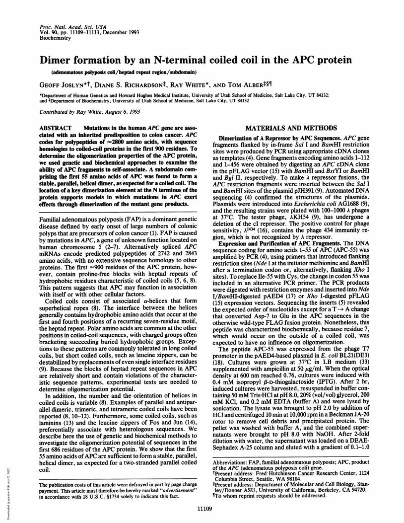
RESULTSThe dimerization potential of the APC heptad repeats wasinitially assessed by testing their ability to provide a dimer-ization domain for the cI repressor ofphage A (9). A repressorfunctions as a dimer, with DNA binding and dimerizationmediated by the N- and C-terminal domains, respectively.The C-terminal dimerization domain can be functionallyreplaced by heterologous motifs such as the GCN4 leucinezipper, a short sequence that forms a two-stranded parallelcoiled coil (9, 22).Fragments of the APC gene (Fig. 1) were cloned in a A
repressor test system (9), and E. coli strains harboring theresulting plasmids were challenged with A KH54 (Ad) andA'434 phages. Resistance to A KH54 and sensitivity to A'434,which contains operator DNA sequences not recognized byA repressor, suggests that a functional A-repressor-APC di-mer is expressed from the test plasmid. When joined to theA repressor DNA-binding domain, APC residues 1-55 con-ferred resistance to infection by A KH54 (Fig. 1), suggestingthat this first block of heptad repeats is sufficient for dimerformation.The structure of the first block of heptad repeats was ana-
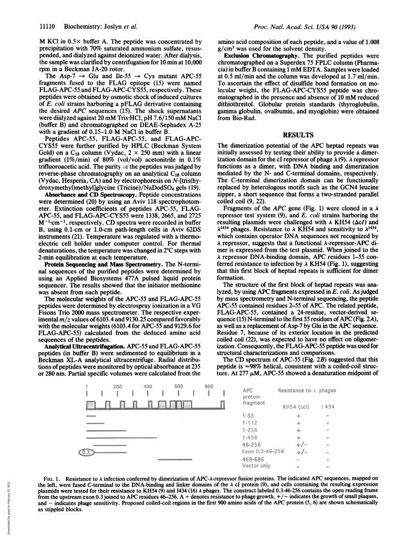
lyzed, by using APC fragments expressed in E. coli. Asjudgedby mass spectrometry and N-terminal sequencing, the peptideAPC-55 contained residues 2-55 of APC. The related peptide,FLAG-APC-55, contained a 24-residue, vector-derived se-quence (15) N-terminal to the first 55 residues ofAPC (Fig. 2A),as well as a replacement ofAsp-7 by Glu in the APC sequence.Residue 7, because of its exterior location in the predictedcoiled coil (22), was expected to have no effect on oligomer-ization. Consequently, the FLAG-APC-55 peptide was used forstructural characterizations and comparisons.The CD spectrum of APC-55 (Fig. 2B) suggested that this
peptide is =98% helical, consistent with a coiled-coil struc-ture. At 277 ,M, APC-55 showed a denaturation midpoint of
800
= I
In
APCproteinfragment
Resistance to i. phages
KH54 (.\cl) 434
1-551-1 121-2561-45646-256Exon 0.3-46-256469-686Vector only
+/+/
FIG. 1. Resistance to A infection conferred by dimerization of APC-A-repressor fusion proteins. The indicated APC sequences, mapped onthe left, were fused C-terminal to the DNA-binding and linker domains of the A cI protein (9), and cells containing the resulting expressionplasmids were tested for their resistance to KH54 (9) and I434 (16) A phages. The construct labeled 0.3-46-256 contains the open reading framefrom the upstream exon 0.3 joined to APC residues 46-256. A + denotes resistance to phage growth, +/- indicates the growth of small plaques,and - indicates phage sensitivity. Proposed coiled-coil regions in the first 900 amino acids of the APC protein (5, 6) are shown schematicallyas stippled blocks.
11110 Biochemistry: Joslyn et al.
Dow
nloa
ded
by g
uest
on
Feb
ruar
y 23
, 202
2
Proc. Natl. Acad. Sci. USA 90 (1993) 11111
A6AASYDQLLKQVEALKMENSNLRQELEDNSNHLTKLETEASNMKEVLKQLQGSI
DYKDDDDKLEFSRDIVDRSLESTHM-(APC-55)
Bm 5_o :X0
x-5
'aE
-15
-25
N
T -35 -
cC
o0
x
0)
Ela-
E
N
N
cmN
.
I
I I
* U
IU I~1`U,~
5.- - - - w w w w w w .
190 200 210 220 230Wavelength, nm
04000
0
0
0
0
0
0
30 50Temperature, °C
FIG. 2. (A) Amino acid sequences ofAPC-55APC-55 (lower) listed in the one-letter code. Fcontained the substitution Asp-7 -- Glu in ti
Residues at the characteristic hydrophobic posirepeats are underlined. (B) CD spectra of A]APC-55 (A), and FLAG-APC-CYS55 (o). Peptwere 55, 19, and 73 ,uM, respectively, in buffer I
dependence of the CD signal at 222 nm for Alconcentration was 277 ,uM in buffer B.
46°C at neutral pH (Fig. 2C). CD spectra of tFLAG-APC-55 (Fig. 2B) also are consisteclusion that the APC sequence adopts a helicTo discern the oligomerization states
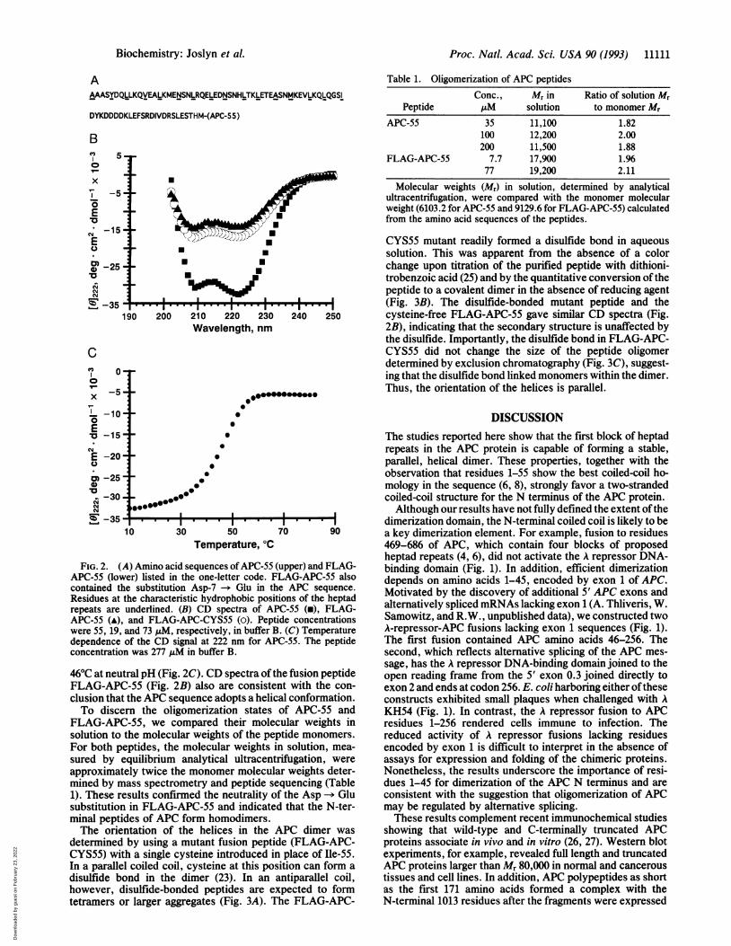
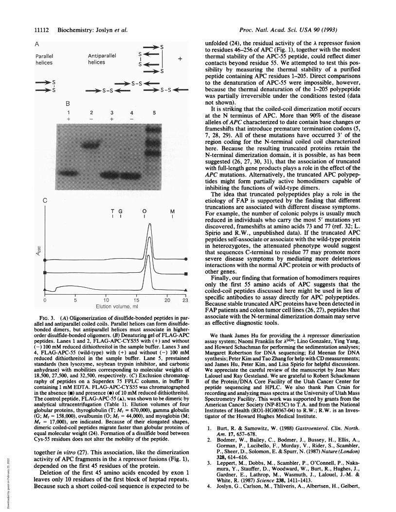
FLAG-APC-55, we compared their molesolution to the molecular weights of the peFor both peptides, the molecular weights isured by equilibrium analytical ultracentapproximately twice the monomer moleculmined by mass spectrometry and peptide si1). These results confirmed the neutralitysubstitution in FLAG-APC-55 and indicateminal peptides of APC form homodimers.The orientation of the helices in the
determined by using a mutant fusion peptiCYS55) with a single cysteine introduced iiIn a parallel coiled coil, cysteine at this po:disulfide bond in the dimer (23). In anhowever, disulfide-bonded peptides are etetramers or larger aggregates (Fig. 3A). 1
Table 1. Oligomerization of APC peptidesConc., Mr in Ratio of solution Mr
Peptide ,uM solution to monomer MrAPC-55 35 11,100 1.82
100 12,200 2.00200 11,500 1.88
FLAG-APC-55 7.7 17,900 1.9677 19,200 2.11
Molecular weights (Mr) in solution, determined by analyticalultracentrifugation, were compared with the monomer molecularweight (6103.2 for APC-55 and 9129.6 for FLAG-APC-55) calculatedfrom the amino acid sequences of the peptides.
CYS55 mutant readily formed a disulfide bond in aqueoussolution. This was apparent from the absence of a colorchange upon titration of the purified peptide with dithioni-trobenzoic acid (25) and by the quantitative conversion ofthepeptide to a covalent dimer in the absence of reducing agent(Fig. 3B). The disulfide-bonded mutant peptide and the
240 250 cysteine-free FLAG-APC-55 gave similar CD spectra (Fig.2B), indicating that the secondary structure is unaffected bythe disulfide. Importantly, the disulfide bond in FLAG-APC-CYS55 did not change the size of the peptide oligomerdetermined by exclusion chromatography (Fig. 3C), suggest-ing that the disulfide bond linked monomers within the dimer.
09960~0Thus, the orientation of the helices is parallel.
DISCUSSIONThe studies reported here show that the first block of heptadrepeats in the APC protein is capable of forming a stable,parallel, helical dimer. These properties, together with theobservation that residues 1-55 show the best coiled-coil ho-mology in the sequence (6, 8), strongly favor a two-strandedcoiled-coil structure for the N terminus of the APC protein.Although our results have not fully defined the extent ofthe
dimerization domain, the N-terminal coiled coil is likely to be70 90 a key dimerization element. For example, fusion to residues
469-686 of APC, which contain four blocks of proposedheptad repeats (4, 6), did not activate the A repressor DNA-
(upper) and FLAG- binding domain (Fig. 1). In addition, efficient dimerization
he APC also depends on amino acids 1-45, encoded by exon 1 of APC.te Af sequence. Motivated by the discovery of additional 5' APC exons and
PC-55 (m), FLAG- alternatively spliced mRNAs lacking exon 1 (A. Thliveris, W.tide concentrations Samowitz, and R.W., unpublished data), we constructed twoB. (C) Temperature A-repressor-APC fusions lacking exon 1 sequences (Fig. 1).PC-55. The peptide The first fusion contained APC amino acids 46-256. The
second, which reflects alternative splicing of the APC mes-sage, has the A repressor DNA-binding domain joined to the
the fusion peptide open reading frame from the 5' exon 0.3 joined directly tont with the con- exon 2 and ends at codon 256. E. coli harboring either of thesecal conformation. constructs exhibited small plaques when challenged with Aof APC-55 and KH54 (Fig. 1). In contrast, the A repressor fusion to APCcular weights in residues 1-256 rendered cells immune to infection. Theptide monomers. reduced activity of A repressor fusions lacking residuesin solution, mea- encoded by exon 1 is difficult to interpret in the absence oftrifugation, were assays for expression and folding of the chimeric proteins.ar weights deter- Nonetheless, the results underscore the importance of resi-equencing (Table dues 1-45 for dimerization of the APC N terminus and areif the Asp -- Glu consistent with the suggestion that oligomerization of APCd that the N-ter- may be regulated by alternative splicing.
These results complement recent immunochemical studiesAPC dimer was showing that wild-type and C-terminally truncated APC[de (FLAG-APC- proteins associate in vivo and in vitro (26, 27). Western blotn place of Ile-55. experiments, for example, revealed full length and truncatedsition can form a APC proteins larger than Mr 80,000 in normal and cancerousantiparallel coil, tissues and cell lines. In addition, APC polypeptides as shortxpected to form as the first 171 amino acids formed a complex with the'he FLAG-APC- N-terminal 1013 residues after the fragments were expressed
Biochemistry: Joslyn et al.
Dow
nloa
ded
by g
uest
on
Feb
ruar
y 23
, 202
2
Proc. Natl. Acad. Sci. USA 90 (1993)
A
Parallelhelices
SS.S
B
Antiparallelhelices
2 3-4-
TG
s-*
-*--I
5
0 M
1, J
E1.j't.:-, eJI !.p.. |IFIG. 3. (A) Oligomerization of disulfide-bonded peptides in par-
allel and antiparallel coiled coils. Parallel helices can form disulfide-bonded dimers, but antiparallel helices must associate in higher-order disulfide-bonded oligomers. (B) Denaturing gel of FLAG-APCpeptides. Lanes 1 and 2, FLAG-APC-CYS55 with (+) and without(-) 100 mM reduced dithiothreitol in the sample buffer. Lanes 3 and4, FLAG-APC-55 (wild-type) with (+) and without (-) 100 mMreduced dithiothreitol in the sample buffer. Lane 5, prestainedstandards (hen lysozyme, soybean trypsin inhibitor, and carbonicanhydrase) with mobilities corresponding to molecular weights of18,500, 27,500, and 32,500, respectively. (C) Exclusion chromatog-raphy of peptides on a Superdex 75 FPLC column, in buffer Bcontaining 1 mM EDTA. FLAG-APC-CYS55 was chromatographedin the absence (u) and presence (e) of 10 mM reduced dithiothreitol.The control peptide, FLAG-APC-55 (A), was shown to be dimeric byanalytical ultracentrifugation (Table 1). Elution volumes of fourglobular proteins, thyroglobulin (T; Mr = 670,000), gamma globulin(G; Mr = 158,000), ovalbumin (0; Mr = 44,000), and myoglobin (M;M, = 17,000), are indicated. Because of their elongated shapes,dimeric coiled-coil peptides migrate faster than globular proteins ofequal molecular weight (24). Formation of a disulfide bond betweenCys-55 residues does not alter the mobility of the peptide.
together in vitro (27). This association, like the dimerizationactivity of APC fragments in the A repressor fusions (Fig. 1),depended on the first 45 residues of the protein.
Deletion of the first 45 amino acids encoded by exon 1leaves only 10 residues of the first block of heptad repeats.Because such a short coiled-coil sequence is expected to be
unfolded (24), the residual activity of the A repressor fusionto residues 46-256 of APC (Fig. 1), together with the modestthermal stability of the APC-55 peptide, could reflect dimercontacts beyond residue 55. We attempted to test this pos-sibility by measuring the thermal stability of a purifiedpeptide containing APC residues 1-205. Direct comparisonsto the denaturation of APC-55 were impossible, however,because the thermal denaturation of the 1-205 polypeptidewas partially irreversible under the conditions tested (datanot shown).
It is striking that the coiled-coil dimerization motif occursat the N terminus of APC. More than 90% of the diseasealleles ofAPC characterized to date contain base changes orframeshifts that introduce premature termination codons (5,7, 28, 29). All of these mutations have occurred 3' of theregion coding for the N-terminal coiled coil characterizedhere. Because the resulting truncated proteins retain theN-terminal dimerization domain, it is possible, as has beensuggested (26, 27, 30, 31), that the association of truncatedwith full-length gene products plays a role in the effect of theAPC mutations. Alternatively, the truncated APC polypep-tides might form partially active homodimers capable ofinhibiting the functions of wild-type dimers.The idea that truncated polypeptides play a role in the
etiology of FAP is supported by the finding that differenttruncations are associated with different disease symptoms.For example, the number of colonic polyps is usually muchreduced in individuals who carry the most 5' mutations yetdiscovered, frameshifts at amino acids 73 and 77 (ref. 32; L.Spirio and R.W., unpublished data). If the truncated APCpeptides self-associate or associate with the wild-type proteinin heterozygotes, the attenuated phenotype would suggestthat sequences C-terminal to residue 77 may promote moresevere disease symptoms by mediating more deleteriousinteractions with the normal APC protein or with products ofother genes.
Finally, our finding that formation ofhomodimers requiresonly the first 55 amino acids of APC suggests that thecoiled-coil peptides discussed here might be used in lieu ofspecific antibodies to assay directly for APC polypeptides.Because stable truncated APC proteins have been detected inFAP patients and colon tumor cell lines (26, 27), peptides thatassociate with the N-terminal dimerization domain may serveas effective diagnostic tools.
We thank James Hu for providing the A repressor dimerizationassay system; Naomi Franklin for A434; Lino Gonzalez, Ying Yang,and Howard Schachman for performing the sedimentation analyses;Margaret Robertson for DNA sequencing; Ed Meenan for DNAsynthesis; Peter Kim and Tao Zhang for help with CD measurements;and James Hu, Peter Kim, and Lisa Spirio for helpful discussions.We appreciate the careful review of the manuscript by Jean MarcLalouel and Ray Gesteland. We are grateful to Robert Schackmannof the Protein/DNA Core Facility of the Utah Cancer Center forpeptide sequencing and HPLC. We also thank Pam Crain forrecording and analyzing mass spectra at the University of Utah MassSpectrometry Facility. This work was supported by grants from theAmerican Cancer Society (NP-815C) to T.A. and from the NationalInstitutes of Health (ROl-HG00367-04) to R.W.; R.W. is an Inves-tigator of the Howard Hughes Medical Institute.
1. Burt, R. & Samowitz, W. (1988) Gastroenterol. Clin. North.Am. 17, 657-678.
2. Bodmer, W., Bailey, C., Bodmer, J., Bussey, H., Ellis, A.,Gorman, P., Lucibello, F., Murday, V., Rider, S., Scambler,P., Sheer, D., Solomon, E. & Spurr, N. (1987) Nature (London)328, 614-616.
3. Leppert, M., Dobbs, M., Scambler, P., O'Connell, P., Naka-mura, Y., Stauffer, D., Woodward, W., Burt, R., Hughes, J.,Gardner, E., Lathrop, M., Wasmuth, J., Lalouel, J.-M. &White, R. (1987) Science 238, 1411-1413.
4. Joslyn, G., Carlson, M., Thliveris, A., Albertsen, H., Gelbert,
11112 Biochemistry: Joslyn et al.
s -S -*----*- s s -*-- 0- s s -0-
Dow
nloa
ded
by g
uest
on
Feb
ruar
y 23
, 202
2
Proc. Natl. Acad. Sci. USA 90 (1993) 11113
L., Samowitz, W., Groden, J., Stevens, J., Spirio, L., Rob-ertson, M., Sargeant, L., Krapcho, K., Wolff, E., Burt, R.,Hughes, J., Warrington, J., McPherson, J., Wasmuth, J., LePasilier, D., Abderrahim, H., Cohen, D., Leppert, M. & White,R. (1991) Cell 66, 601-613.
5. Groden, J., Thliveris, A., Samowitz, W., Carlson, M., Gelbert,L., Albertsen, H., Joslyn, G., Stevens, J., Spirio, L., Robert-son, M., Sargeant, L., Krapcho, K., Wolff, E., Burt, R.,Hughes, J., Warrington, J., McPherson, J., Wasmuth, J., LePaslier, D., Abderrahim, J., Cohen, D., Leppert, M. & White,R. (1991) Cell 66, 589-600.
6. Kinzler, K., Nilbert, M., Su, L.-K., Vogelstein, B., Bryan, T.,Levy, D., Smith, K., Preisinger, A., Hedge, P., McKechnie,D., Finnear, R., Markham, A., Groffen, J., Boguski, M.,Altschul, S., Horii, A., Ando, H., Miyoshi, Y., Miki, Y.,Nishisho, I. & Nakamura, Y. (1991) Science 253, 661-665.
7. Nishisho, I., Nakamura, Y., Miyoshi, Y., Miki, Y., Ando, H.,Horii, A., Koyama, K., Utsunomiya, J., Baba, S., Hedge, P.,Markham, A., Krush, A., Petersen, G., Hamilton, S., Nilbert,M., Levy, D., Bryan, T., Preisinger, A., Smith, K., Su, L.-K.,Kinzler, K. & Vogelstein, B. (1991) Science 253, 665-669.
8. Cohen, C. & Parry, D. (1986) Trends Biochem. Sci. 11, 245-248.
9. Hu, J. C., O'Shea, E. K., Kim, P. S. & Sauer, R. T. (1990)Science 250, 1400-1403.
10. Banner, D. W., Kokkinidis, M. & Tsernoglou, D. (1987) J. Mol.Biol. 196, 657-675.
11. Cusack, S., Berthet-Colominas, C., Hartlein, M., Nassar, N. &Leberman, R. (1990) Nature (London) 347, 249-255.
12. Lovejoy, B., Choe, S., Cascio, D., McRorie, D. K., DeGrado,W. F. & Eisenberg, D. (1993) Science 259, 1288-1293.
13. Hunter, I., Schulthess, T. & Engel, J. (1992),J. Biol. Chem. 267,6006-6011.
14. O'Shea, E. K., Rutkowski, R., Stafford, W. F., III, & Kim,P. S. (1989) Science 245, 646-648.
15. Hopp, T. P., Prickett, K. S., Price, V. L., Libby, R. T.,March, C. J., Cerretti, D. P., Urdal, D. L. & Conlon, P. J.(1988) BiolTechnology 6, 1204-1210.
16. Kaiser, A. D. & Jacob, F. (1957) Virology 4, 509-521.17. Doering, D. S. (1992) Dissertation (Massachusetts Institute of
Technology, Cambridge).
18. Studier, F. W., Rosenberg, A. H., Dunn, J. J. & Dubendorf,J. W. (1990) Methods Enzymol. 185, 60-89.
19. Schaegger, H. & von Jagow, G. (1987) Anal. Biochem. 166,368-379.
20. Gill, S. C. & von Hippel, P. H. (1989) Anal. Biochem. 182,319-326.
21. O'Shea, E. K., Rutkowski, R. & Kim, P. S. (1989) Science 243,538-542.
22. O'Shea, E. K., Klemm, J. D., Kim, P. S. & Alber, T. (1991)Science 254, 539-544.
23. Zhou, N. E., Kay, C. M. & Hodges, R. S. (1992) Biochemistry31, 5739-5746.
24. Lau, S. Y. M., Taneja, A. K. & Hodges, R. S. (1984) J. Biol.Chem. 259, 13253-13261.
25. Creighton, T. E. (1990) in Protein Structure: A Practical Ap-proach, ed. Creighton, T. E. (Oxford Univ. Press, Oxford), pp.155-167.
26. Smith, K. J., Johnson, K. A., Bryan, T. M., Hill, D. E., Mar-kowitz, S., Willson, J. K. V., Paraskeva, C., Petersen, G. M.,Hamilton, S. R., Vogelstein, B. & Kinzler, K. W. (1993) Proc.Natl. Acad. Sci. USA 90, 2846-2850.
27. Su, L.-K., Johnson, K. A., Smith, K. J., Hill, D. E., Vo-gelstein, B. & Kinzler, K. W. (1993) Cancer Res. 53, 2728-2731.
28. Groden, J., Gelbert, L., Thliveris, A., Nelson, L., Robertson,M., Joslyn, G., Samowitz, W., Spirio, L., Carlson, M., Burt,R., Leppert, M. & White, R. (1993) Am. J. Hum. Genet. 52,263-272.
29. Miyoshi, Y., Ando, H., Nagase, H., Nishisho, I., Horii, A.,Miki, Y., Mori, T., Utsunomiya, J., Baba, S., Petersen, G.,Hamilton, S., Kinzler, K., Vogelstein, B. & Nakamura, Y.(1992) Proc. Natl. Acad. Sci. USA 89, 4452-4456.
30. Bourne, H. (1991) Nature (London) 351, 188-190.31. Bourne, H. (1991) Nature (London) 353, 696-697.32. Spirio, L., Otterud, B., Stauffer, D., Lynch, H., Lynch, P.,
Watson, P., Lanspa, S., Smyrk, T., Cavalieri, J., Howard, L.,Burt, R., White, R. & Leppert, M. (1992) Am. J. Hum. Genet.51, 92-100.
33. Maniatis, T., Fritsch, E. F. & Sambrook, J. (1982) MolecularCloning: A Laboratory Manual (Cold Spring Harbor Lab.Press, Plainview, NY), p. 440.
Biochemistry: Joslyn et al.
Dow
nloa
ded
by g
uest
on
Feb
ruar
y 23
, 202
2


![INDEX [meanwell.com]meanwell.com/Upload/PDF/meanwell_LED.pdf · APC-8, APC-12, APC-16, APC-25, APC-35 3 APV-8E, APV-12E, APV-16E 4 APC-8E, APC-12E, APC-16E LP ... Over voltage protection](https://static.fdocuments.in/doc/165x107/5b619e107f8b9a40488c919f/index-apc-8-apc-12-apc-16-apc-25-apc-35-3-apv-8e-apv-12e-apv-16e-4.jpg)
