Digital Imaging and Communications in Medicine (DICOM ...dicom.nema.org/Dicom/supps/sup45_08.pdf ·...
25
ASE • ACR • NEMA Digital Imaging and Communications in Medicine (DICOM) Supplement 45: Ultrasound Protocol Supplement Prepared by: DICOM Standards Committee, Working Group 12 Ultrasound 1300 N. 17 th Street Rosslyn, Virginia 22209 USA Version: 0.7
-
Upload
phunghuong -
Category
Documents
-
view
227 -
download
0
Transcript of Digital Imaging and Communications in Medicine (DICOM ...dicom.nema.org/Dicom/supps/sup45_08.pdf ·...

ASE • ACR • NEMA
Digital Imaging and Communications in Medicine (DICOM)
Supplement 45: Ultrasound Protocol Supplement
Prepared by:
DICOM Standards Committee, Working Group 12 Ultrasound
1300 N. 17th Street
Rosslyn, Virginia 22209 USA
Version: 0.7

ii
ii
8 June, 1999

Supplement 45: Ultrasound ProtocolPage i
Foreword..........................................................................................................................................................iii
Scope and Field of Application....................................................................................................................... ivA.1.4 Overview of the Composite IOD Module Content.....................................................................2A.6.4 US Image IOD Module Table ...................................................................................................4A.7.4 US Multi-Frame Image IOD Module Table...............................................................................6
C.8.5.x US Series Module ........................................................................................................7C.8.5.y US Protocol Module .....................................................................................................9
6. REGISTRY OF DICOM DATA ELEMENTS .......................................................................................15
ii
ii
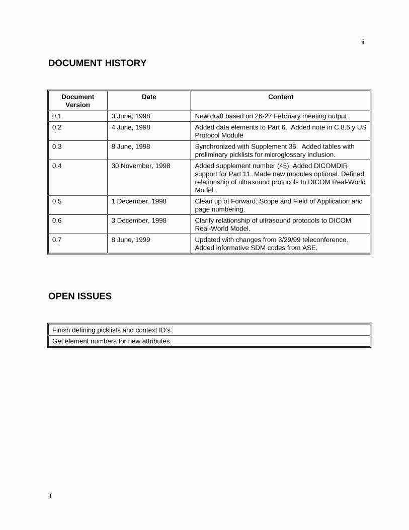
DOCUMENT HISTORY
DocumentVersion
Date Content
0.1 3 June, 1998 New draft based on 26-27 February meeting output
0.2 4 June, 1998 Added data elements to Part 6. Added note in C.8.5.y USProtocol Module
0.3 8 June, 1998 Synchronized with Supplement 36. Added tables withpreliminary picklists for microglossary inclusion.
0.4 30 November, 1998 Added supplement number (45). Added DICOMDIRsupport for Part 11. Made new modules optional. Definedrelationship of ultrasound protocols to DICOM Real-WorldModel.
0.5 1 December, 1998 Clean up of Forward, Scope and Field of Application andpage numbering.
0.6 3 December, 1998 Clarify relationship of ultrasound protocols to DICOMReal-World Model.
0.7 8 June, 1999 Updated with changes from 3/29/99 teleconference.Added informative SDM codes from ASE.
OPEN ISSUES
Finish defining picklists and context ID’s.
Get element numbers for new attributes.
Supplement 45: Ultrasound ProtocolPage iii
Foreword
This draft Supplement to the DICOM Standard was developed according to NEMA Procedures. ThisSupplement to the Standard is developed in liaison with other standardization organizations including CENTC251 in Europe, MEDIS-DC and JIRA in Japan, with review also by other organizations including IEEE,HL7 and ANSI in the USA.
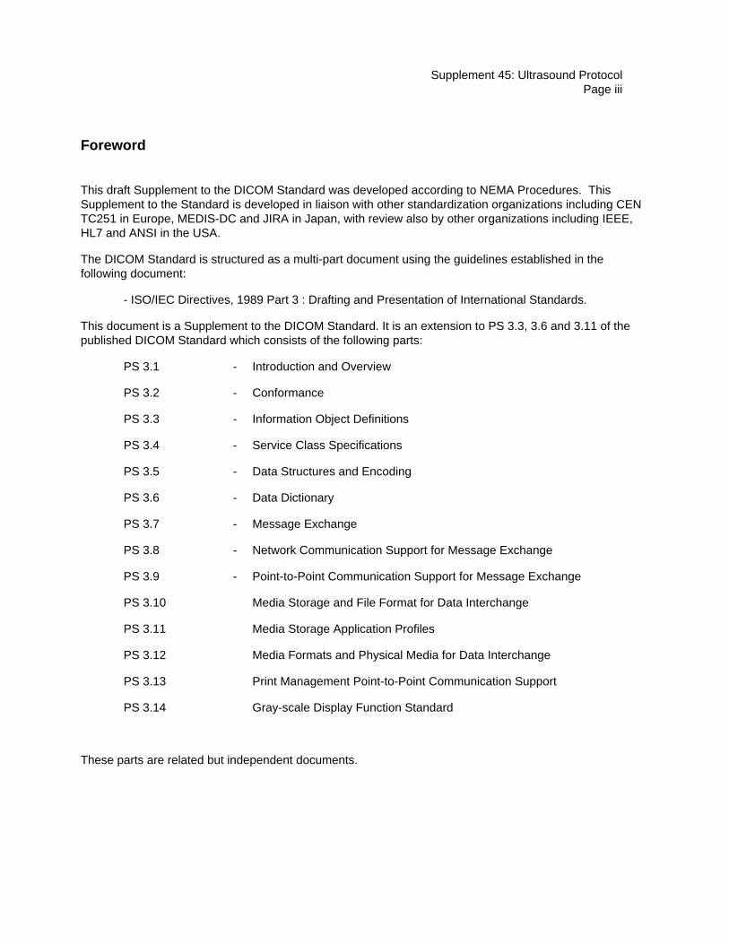
The DICOM Standard is structured as a multi-part document using the guidelines established in thefollowing document:
- ISO/IEC Directives, 1989 Part 3 : Drafting and Presentation of International Standards.
This document is a Supplement to the DICOM Standard. It is an extension to PS 3.3, 3.6 and 3.11 of thepublished DICOM Standard which consists of the following parts:
PS 3.1 - Introduction and Overview
PS 3.2 - Conformance
PS 3.3 - Information Object Definitions
PS 3.4 - Service Class Specifications
PS 3.5 - Data Structures and Encoding
PS 3.6 - Data Dictionary
PS 3.7 - Message Exchange
PS 3.8 - Network Communication Support for Message Exchange
PS 3.9 - Point-to-Point Communication Support for Message Exchange
PS 3.10 Media Storage and File Format for Data Interchange
PS 3.11 Media Storage Application Profiles
PS 3.12 Media Formats and Physical Media for Data Interchange
PS 3.13 Print Management Point-to-Point Communication Support
PS 3.14 Gray-scale Display Function Standard
These parts are related but independent documents.

iv
iv
Scope and Field of Application
This Supplement to the DICOM Standard specifies changes to the DICOM Image Information ObjectDefinition (IOD) for Ultrasound (US) and Ultrasound Multi-frame (USMF) Images. It specifies the newmodules necessary to identify images acquired as part of an ultrasound acquisition protocol.
Since this document proposes changes to existing Parts of DICOM the reader should have a workingunderstanding of the Standard.
This proposed Supplement includes a number of Addenda to existing Parts of DICOM:
1. PS 3.3 Addenda (Extension to Annex A and Annex C)
2. PS 3.6 Addenda (Extension to Section 6)
3. PS 3.11 Addenda (Extension to Annex C)

Supplement 45: Ultrasound ProtocolPage 1
Changes to:
NEMA Standards Publication PS 3.3-1998
Digital Imaging and Communications in Medicine (DICOM)
Part 3: Information Object Definitions

2
3.X Ultrasound
The following definitions are commonly used in the specification of the Ultrasound IOD’s.
3.X.1 Acquisition Protocol
An Acquisition Protocol is a Procedure Type that defines the particular ultrasound images that will beacquired.
Note: These can be well known or locale specific.
3.X.2 Staged Protocol
A Staged Protocol is an Acquisition Protocol that is repeated at more than one specific instance of time.
Note: These instances commonly occur due to simple events. The introduction of contrast media, drugs or stressto the patient are typical events.
Item: Add to PS 3.3 Table A.1-1
A.1.4 Overview of the Composite IOD Module Content
Table A.1-1 provides an overview of the Modules used throughout the Composite IODs. This table is forinformative purposes only. It is based on the IOD definitions found in the remaining sections of Annex Awhich are normative.
Table A.1-1COMPOSITE INFORMATION OBJECT MODULES OVERVIEW
IODs
ModulesUS US-
mf
Patient M M
PatientSummary
GeneralStudy
M M
Patient study U U
Study Content
GeneralSeries
M M
CR Series
NM Series
US Series U U
Frame OfReference
U U
U S Frame ofRef.
C C
GeneralEquipment
M M
SC Equipment
Supplement 45: Ultrasound ProtocolPage 3
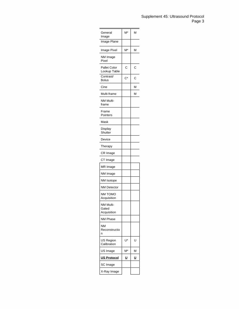
GeneralImage
M* M
Image Plane
Image Pixel M* M
NM ImagePixel
Pallet ColorL o oku p Ta b le
C C
Contrast/Bolus
C* C
Cine M
Multi-frame M
NM Multi-frame
FramePointers
Mask
DisplayShutter
Device
Therapy
CR Image
CT Image
MR Image
NM Image
NM Isotope
NM Detector
NM TOMOAcquisition
NM Multi-GatedAcquisition
NM Phase
NMRe co n str u ct io n
US RegionCalibration
U* U
US Image M* M
US Protocol U U
SC Image
X-Ray Image

4
X-RayAcquisition
X-RayCollimator
X-Ray Table
XRFPositioner
XRF TomoAcquisition
XA Positioner
Bi-PlaneSequence
Bi-PlaneImage
OverlayIdentification
Overlay Plane U*
Multi-frameOverlay
Bi-PlaneOverlay
CurveIdentification
M* M*
Curve M* M*
Audio U U
Modality LUT
VOI LUT U* U
LUTIdentification
SOPCommon
M* M*
Item: Add to PS 3.3 Table A.6.4-1 - for clarity, those modules that are changed from the existing standardor new are shown in Bold and Underlined
A.6.4 US Image IOD Module Table
Table A.6.4-1US IMAGE IOD MODULES
IE Module Reference Usage

Supplement 45: Ultrasound ProtocolPage 5
Patient Patient C.7.1.1 M
Study General Study C.7.2.1 M
Patient Study C.7.2.2 U
Series General Series C.7.3.1 M
US Series C.8.5.x U – Suggested if image ispart of an acquisition
protocol
Frame ofReference
Frame of Reference C.7.4.1 U
US Frame of Reference C.8.5.4 C - Required if images arespatially related
Equipment General Equipment C.7.5.1 M
Image General Image C.7.6.1 M
(See A.6.4.1) Image Pixel C.7.6.3 M
Contrast/bolus C.7.6.4 C - Required if contrastmedia was used in this
image
Palette Color Lookup table C.7.9 C - Required if PhotometicInterpretation (0028,0004)has a value of PALETTE
COLOR
US Region Calibration C.8.5.5 U
US Image C.8.5.6 M
US Staged Protocol C.8.5.y U – Suggested if image ispart of a staged protocol
Overplay Plane C.9.2 U
VOI LUT C.11.2 U
SOP Common C.12.1 M
Curve Curve Identification C.10.1 M
(See A.6.4.1) Curve C.10.2 M
Audio C.10.3 U
SOP Common C.12.1 M
A.6.4.1 Mutually Exclusive IEs
The Image and Curve IEs are mutually exclusive. Each SOP Instance using this IOD shall contain exactlyone of these IEs.
A.6.5 US Image IOD Relationship to the DICOM Real-World Model
Ultrasound images that make use of the US Series & Protocol Modules have a more specificrelationship to the DICOM Real-World Model than non-protocol images. An ultrasound acquisitionprotocol is considered one Requested Procedure and has one Procedure Plan. The ProcedurePlan will have one or more Action Items. An ultrasound acquisition protocol will typically consist

6
of one Scheduled Procedure Step containing one or more Action Items. Assuming the acquisitionprotocol was successfully completed, this will result in one Modality Performed Procedure Step.Therefore, all images that are part of this particular instance of an ultrasound acquisition protocolwill have the same Procedure Code Sequence (0032,1064). It is recommended that all the imagesthat are part of this particular instance of an ultrasound acquisition protocol have the samePerformed Procedure Step ID. In an environment with information system connectivity, theProcedure Code Sequence can be used to identify all the images that were acquired as part of thatprotocol. If no information system connectivity is available, then Performed Procedure Step IDmay be used to identify all the images that were acquired as part of that protocol.
It is recommended that each stage should map to a separate series.
Item: Add to PS 3.3 Table A.7.4-1 - for clarity, those modules that are changed from the existing standardor new are shown in Bold and Underlined
A.7.4 US Multi-Frame Image IOD Module Table
Table A.7.4-1US MULTI-FRAME IMAGE IOD MODULES
IE Module Reference Usage
Patient Patient C.7.1.1 M
Study General Study C.7.2.1 M
Patient Study C.7.2.2 U
Series General Series C.7.3.1 M
US Series C.8.5.x U – Suggested if image ispart of an acquisition
protocol
Frame ofReference
Frame of Reference C.7.4.1 U
US Frame of Reference C.8.5.4 C - Required if images arespatially related
Equipment General Equipment C.7.5.1 M
Image General Image C.7.6.1 M
(See A.7.4.1) Image Pixel C.7.6.3 M
Contrast/bolus C.7.6.4 C - Required if contrastmedia was used in this
image.
Cine C.7.6.5 M
Multi-frame C.7.6.6 M
Palette Color Lookup Table C.7.9 C - Required if PhotometricInterpretation (0028,0004)has a value of PALETTE
COLOR

Supplement 45: Ultrasound ProtocolPage 7
US Region Calibration C.8.5.5 U
US Image C.8.5.6 M
US Staged Protocol C.8.5.y U – Suggested if image ispart of a staged protocol
VOI LUT C.11.2 U
SOP Common C.12.1 M
Curve Curve Identification C.10.1 M
(see A.7.4.1) Curve C.10.2 M
Audio C.10.3 U
SOP Common C.12.1 M
A.7.4.1 Mutually Exclusive IEs
The Image and Curve IEs are mutually exclusive. Each SOP Instance using this IOD shall contain exactlyone of these IEs.
A.7.5 US Multi-Frame Image IOD Relationship to the DICOM Real-World Model
Ultrasound images that make use of the US Series & Protocol Modules have a more specificrelationship to the DICOM Real-World Model than non-protocol images. An ultrasound acquisitionprotocol is considered one Requested Procedure and has one Procedure Plan. The ProcedurePlan will have one or more Action Items. An ultrasound acquisition protocol will typically consistof one Scheduled Procedure Step containing one or more Action Items. Assuming the acquisitionprotocol was successfully completed, this will result in one Modality Performed Procedure Step.Therefore, all images that are part of this particular instance of an ultrasound acquisition protocolwill have the same Procedure Code Sequence (0032,1064). It is recommended that all the imagesthat are part of this particular instance of an ultrasound acquisition protocol have the samePerformed Procedure Step ID. In an environment with information system connectivity, theProcedure Code Sequence can be used to identify all the images that were acquired as part of thatprotocol. If no information system connectivity is available, then Performed Procedure Step IDmay be used to identify all the images that were acquired as part of that protocol.
It is recommended that each stage should map to a separate series.
Item: Add to PS 3.3 New Sections C.8.5.x and C.8.5.y
C.8.5.x US Series Module
Table C.8-x contains IOD attributes that describe an ultrasound series.

8
Table C.8-xUS SERIES MODULE ATTRIBUTES
Attribute Name Tag Type Attribute Description
Protocol Name (0018,1030) 1C User-defined description of the conditionsunder which the Series was performed.Required if Protocol Type Sequence(0018,xxxx) has no value.
Protocol Type Sequence (0018,xxxx) 2 Sequence of one or more Items thatidentifies the protocol used to acquire thisimage.
See section C.8.5.x.1.y
>Include ‘Code Sequence Macro‘ Table 8.8-1 Baseline Context ID is ???.
>Protocol Type Modifier Sequence (0018,xxxx) 3 Sequence of one or more Items thatmodifies the primary protocol type used toacquire this image.
See section C.8.5.x.1.y
>>Include ‘Code Sequence Macro‘ Table 8.8-1 Baseline Context ID is ???.
Performed Procedure Step ID (0040,0253) 1C Identification of that Procedure that hasbeen carried out within this step. Required ifimage was acquired in an acquisitionprotocol.
C.8.5.x.1 US Series Attributes Descriptions
C.8.5.x.1.y Protocol Type Sequence
These Data Elements are intended to replace Protocol Name (0018,1030) Data Element.
Protocol Type Sequence (0018,xxxx) describes the acquisition protocol used to acquire this image.
The Coding Scheme Designator (0008,0102) shall be SNM3.
The Code Value (0008,0100) shall be drawn from the SNOMED DICOM Microglossary Context ID XXX.
Note: The existence of this attribute means that this image is part of an acquisition protocol.
Note: The following will not be included in the standard.
Code Meaning(0008,0104)
Code Value(0008,0100)
Transesophageal echocardiography P5-B3002
Transthoracic echocardiography P5-B3003
Epicardial echocardiography P5-B3004
Intravascular echocardiography P5-B3005
Intracardiac echocardiography P5-B3006
Limited M-mode only echocardiography P5-B301F
Limited Doppler only echocardiography P5-B303F
Exercise stress echocardiography P5-B3050
Maximal stress echocardiography P5-B3051

Supplement 45: Ultrasound ProtocolPage 9
Submaximal stress echocardiography P5-B3052
Treadmill exercise stress echocardiography P5-B3053
Bruce treadmill stress echocardiography P5-B3054
Modified Bruce treadmill stress echocardiography P5-B3055
Naughton treadmill stress echocardiography P5-B3056
Bicycle exercise stress echocardiography P5-B3058
Echocardiography with administered drug stress P5-B3060
Dobutamine stress echocardiography P5-B3061
High dose dobutamine stress echocardiography P5-B3062
Low dose dobutamine stress echocardiography P5-B3063
Arbutamine stress echocardiography P5-B3065
Dipyridamole stress echocardiography P5-B3066
Cardiac pacing echocardiography P5-B3070
Adult echocardiography P5-B3081
Pediatric echocardiography P5-B3082
Intraoperative echocardiography P5-B3083
Upright echocardiography P5-B3084
Supine echocardiography P5-B3085
Contrast echocardiography P5-B3090
Contrast left ventricular opacification echocardiography P5-B3091
Contrast perfusion echocardiography P5-B3092
Contrast Doppler enhancement echocardiography P5-B3093
2D complete echocardiography P5-B3191
Limited 2D only echocardiography P5-B3192
Fetal echocardiography P5-B8215
Protocol Type Modifier Sequence (0018,xxxx) modifies the primary acquisition protocol stage when theimage was acquired.
The Coding Scheme Designator (0008,0102) shall be SNM3.
The Code Value (0008,0100) shall be drawn from the SNOMED DICOM Microglossary Context ID XXX.
C.8.5.y US Staged Protocol Module
Table C.8-y contains IOD attributes that describe an ultrasound staged protocol.
Table C.8-yUS STAGED PROTOCOL MODULE ATTRIBUTES
Attribute Name Tag Type Attribute Description
Number of Stages (0008,2124) 2 Number of Stages in this protocol.
Number of Views in Stage (0008,212A) 2 Number of views in this Stage.

10
Stage Name (0008,2120) 1C A Stage is a particular time slice of aprotocol in which a set of images arecollected. The names can be free form text.Recommended text for Stress Echo stagenames are:
PRE-EXERCISE, POST-EXERCISE, PEAK-EXERCISE, RECOVERY, BASELINE, LOW DOSE, PEAK DOSE
Required if Stage Type Sequence(0008,xxxx) has no value.
Stage Type Sequence (0008,xxxx) 2 Sequence of one or more items thatidentifies the acquisition protocol stagewhen the image was acquired.
See Section 8.5.y.1.1
>Include ‘Code Sequence Macro‘ Table 8.8-1 Baseline Context ID is ???.
>Stage Type Modifier Sequence (0008,xxxx) 3 Sequence of one or more Items thatmodifies the primary stage type in thisimage.
See Section 8.5.y.1.1
>>Include ‘Code Sequence Macro‘ Table 8.8-1 Baseline Context ID is ???.
Stage Number (0008,2122) 1 A number that identifies the Stage. StageNumber starts at one.
See Section C.8.5.y.1.4
View Number (0008,2128) 2 A number that identifies the View. Can beempty if View Number is unknown or ifknown, View Number starts at one.
See Section C.8.5.y.1.5
Transducer Position Sequence (0008,2240) 2 Sequence of one or more Items thatidentifies the transducer position used in thisimage.
See section C.8.5.y.1.2.
>Include ‘Code Sequence Macro‘ Table 8.8-1 Baseline Context ID is 4.
> Transducer Position ModifierSequence
(0008,2242) 3 Sequence of one or more Items thatmodifies the primary transducer position ofinterest in this image.
See Section C.8.5.y.1.2.
>>Include ‘Code Sequence Macro‘ Table 8.8-1 Baseline Context ID is 5.
Transducer Orientation Sequence (0008,2244) 2 Sequence of one or more Items thatidentifies the Transducer Orientation used inthis image.
See section C.8.5.y.1.3.
Supplement 45: Ultrasound ProtocolPage 11
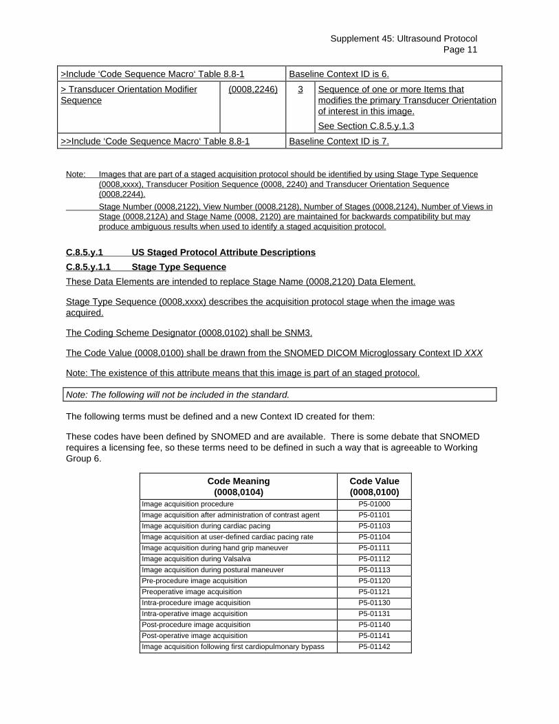
>Include ‘Code Sequence Macro‘ Table 8.8-1 Baseline Context ID is 6.
> Transducer Orientation ModifierSequence
(0008,2246) 3 Sequence of one or more Items thatmodifies the primary Transducer Orientationof interest in this image.
See Section C.8.5.y.1.3
>>Include ‘Code Sequence Macro‘ Table 8.8-1 Baseline Context ID is 7.
Note: Images that are part of a staged acquisition protocol should be identified by using Stage Type Sequence(0008,xxxx), Transducer Position Sequence (0008, 2240) and Transducer Orientation Sequence(0008,2244).
Stage Number (0008,2122), View Number (0008,2128), Number of Stages (0008,2124), Number of Views inStage (0008,212A) and Stage Name (0008, 2120) are maintained for backwards compatibility but mayproduce ambiguous results when used to identify a staged acquisition protocol.
C.8.5.y.1 US Staged Protocol Attribute Descriptions
C.8.5.y.1.1 Stage Type Sequence
These Data Elements are intended to replace Stage Name (0008,2120) Data Element.
Stage Type Sequence (0008,xxxx) describes the acquisition protocol stage when the image wasacquired.
The Coding Scheme Designator (0008,0102) shall be SNM3.
The Code Value (0008,0100) shall be drawn from the SNOMED DICOM Microglossary Context ID XXX
Note: The existence of this attribute means that this image is part of an staged protocol.
Note: The following will not be included in the standard.
The following terms must be defined and a new Context ID created for them:
These codes have been defined by SNOMED and are available. There is some debate that SNOMEDrequires a licensing fee, so these terms need to be defined in such a way that is agreeable to WorkingGroup 6.
Code Meaning(0008,0104)
Code Value(0008,0100)
Image acquisition procedure P5-01000
Image acquisition after administration of contrast agent P5-01101
Image acquisition during cardiac pacing P5-01103
Image acquisition at user-defined cardiac pacing rate P5-01104
Image acquisition during hand grip maneuver P5-01111
Image acquisition during Valsalva P5-01112
Image acquisition during postural maneuver P5-01113
Pre-procedure image acquisition P5-01120
Preoperative image acquisition P5-01121
Intra-procedure image acquisition P5-01130
Intra-operative image acquisition P5-01131
Post-procedure image acquisition P5-01140
Post-operative image acquisition P5-01141
Image acquisition following first cardiopulmonary bypass P5-01142

12
Image acquisition following second cardiopulmonarybypass
P5-01143
Image acquisition following third cardiopulmonary bypass P5-01144
Image acquisition during stress procedure P5-01200
Image acquisition at baseline P5-01201
Pre-stress image acquisition P5-01202
Mid-stress image acquisition P5-01203
Peak-stress image acquisition P5-01204
Image acquisition during recovery P5-01205
Image acquisition after drug administration P5-01300
Image acquisition at user-defined dobutamine dose P5-01310
Image acquisition at low-dose dobutamine P5-01311
Image acquisition at mid-dose dobutamine P5-01312
Image acquisition at peak dose dobutamine P5-01313
Image acquisition at dobutamine 5 mcg/kg/min P5-01314
Image acquisition at dobutamine 10 mcg/kg/min P5-01315
Image acquisition at dobutamine 20 mcg/kg/min P5-01316
Image acquisition at dobutamine 30 mcg/kg/min P5-01317
Image acquisition at dobutamine 40 mcg/kg/min P5-01318
Image acquisition at dobutamine 50 mcg/kg/min P5-01319
Image at dobutamine 40 mcg/kg/min plus atropine P5-0131A
Image acquisition at dobutamine 50 mcg/kg/min plusatropine
P5-0131B
Image acquisition at peak Arbutamine dose P5-01323
Image acquisition at peak dipyridamole P5-01333
Image acquisition after nitroglycerin P5-01341
Image acquisition after amyl nitrite P5-01342
Image acquisition after adenosine P5-01343
Stage Type Modifier Sequence (0008,xxxx) modifies the primary acquisition protocol stage when theimage was acquired.
The Coding Scheme Designator (0008,0102) shall be SNM3.
The Code Value (0008,0100) shall be drawn from the SNOMED DICOM Microglossary Context ID XXX.
C.8.5.y.1.2 Transducer Position Sequence
Transducer Position Sequence (0008,2240) identifies the transducer position used in this image.
The Coding Scheme Designator (0008,0102) shall be SNM3.
The Code Value (0008,0100) shall be drawn from the SNOMED DICOM Microglossary Context ID 4.
Transducer Position Modifier Sequence (0008,2242) modifies the transducer position used in this image.
The Coding Scheme Designator (0008,0102) shall be SNM3.
The Code Value (0008,0100) shall be drawn from the SNOMED DICOM Microglossary Context ID 5.

Supplement 45: Ultrasound ProtocolPage 13
C.8.5.y.1.3 Transducer Orientation Sequence
Transducer Orientation Sequence (0008,2244) identifies the transducer orientation used in this image.
The Coding Scheme Designator (0008,0102) shall be SNM3.
The Code Value (0008,0100) shall be drawn from the SNOMED DICOM Microglossary Context ID 6.
Transducer Orientation Modifier Sequence (0008,2246) modifies the transducer orientation used in thisimage.
The Coding Scheme Designator (0008,0102) shall be SNM3.
The Code Value (0008,0100) shall be drawn from the SNOMED DICOM Microglossary Context ID 7.
C.8.5.y.1.4 Stages
The Number of Stages (0008,2124) shall be the total number of stages performed in this instance of theprotocol. Stage Number (0008,2122) shall be the number that indicates the default order in which thestages are to be presented. Stage Number shall start from 1.
C.8.5.y.1.5 Views
The View Number (0008,2128) shall be used identify the same view across stages and defines a defaultorder for presentation within and across stages. If a particular view appears in one stage but not in asubsequent stage, then that View Number will not be used during that subsequent stage. View Numbershall start from 1. The Number of Views in Stage (0008,212A) shall be used to indicate how many uniqueView Numbers exist for this stage.
The addition of modification of the value for this attribute shall not by itself force a SOP Instance UIDchange.

14
Changes to:
NEMA Standards Publication PS 3.6-1998
Digital Imaging and Communications in Medicine (DICOM)
Part 6: Data Dictionary

Supplement 45: Ultrasound ProtocolPage 15
6. REGISTRY OF DICOM DATA ELEMENTS
Add the following Data Elements to PS 3.6, Section 6.
Tag Name VR VM(0008,xxxx) Stage Type Sequence SQ 1(0008,xxxx) Stage Type Modifier Sequence SQ 1(0018,xxxx) Protocol Type Sequence SQ 1(0018,xxxx) Protocol Type Modifier Sequence SQ 1
16
Changes to:
NEMA Standards Publication PS 3.11-1998
Digital Imaging and Communications in Medicine (DICOM)
Part 11: Media Storage Application Profiles
Supplement 45: Ultrasound ProtocolPage 17
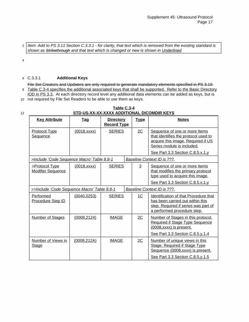
Item: Add to PS 3.11 Section C.3.3.1 - for clarity, that text which is removed from the existing standard is2
shown as Strikethrough and that text which is changed or new is shown in Underlined
4
C.3.3.1 Additional Keys6
File Set Creators and Updaters are only required to generate mandatory elements specified in PS 3.10.Table C.3-4 specifies the additional associated keys that shall be supported. Refer to the Basic Directory8
IOD in PS 3.3. At each directory record level any additional data elements can be added as keys, but isnot required by File Set Readers to be able to use them as keys.10
Table C.3-4STD-US-XX-XX-XXXX ADDITIONAL DICOMDIR KEYS12
Key Attribute Tag DirectoryRecord Type
Type Notes
Protocol TypeSequence
(0018,xxxx) SERIES 2C Sequence of one or more Itemsthat identifies the protocol used toacquire this image. Required if USSeries module is included.
See Part 3.3 Section C.8.5.x.1.y
>Include ‘Code Sequence Macro‘ Table 8.8-1 Baseline Context ID is ???.
>Protocol TypeModifier Sequence
(0018,xxxx) SERIES 3 Sequence of one or more Itemsthat modifies the primary protocoltype used to acquire this image.
See Part 3.3 Section C.8.5.x.1.y
>>Include ‘Code Sequence Macro‘ Table 8.8-1 Baseline Context ID is ???.
PerformedProcedure Step ID
(0040,0253) SERIES 1C Identification of that Procedure thathas been carried out within thisstep. Required if series was part ofa performed procedure step.
Number of Stages (0008,2124) IMAGE 2C Number of Stages in this protocol.Required if Stage Type Sequence(0008,xxxx) is present.
See Part 3.3 Section C.8.5.y.1.4
Number of Views inStage
(0008,212A) IMAGE 2C Number of unique views in thisStage. Required if Stage TypeSequence (0008,xxxx) is present.
See Part 3.3 Section C.8.5.y.1.5

18
Stage TypeSequence
(0008,xxxx) IMAGE 2C Sequence of one or more items thatidentifies the acquisition protocolstage when the image wasacquired. Required US Protocolmodule is present.
See Part 3.3 Section 8.5.y.1.1
>Include ‘Code Sequence Macro‘ Table 8.8-1 Baseline Context ID is ???.
>Stage TypeModifier Sequence
(0008,xxxx) IMAGE 3 Sequence of one or more Itemsthat modifies the primary stage typein this image.
See Part 3.3 Section 8.5.y.1.1
>>Include ‘Code Sequence Macro‘ Table 8.8-1 Baseline Context ID is ???.
Stage Number (0008,2122) IMAGE 1C A number that identifies the Stage.Stage Number starts at one.Required if Stage Type Sequence(0008,xxxx) is present.
See Part 3.3 Section C.8.5.y.1.4
View Number (0008,2128) IMAGE 1C A number that identifies the View.View Number starts at one.Required if Stage Type Sequence(0008,xxxx) is present.
See Part 3.3 Section C.8.5.y.1.5
TransducerPosition Sequence
(0008,2240) IMAGE 2C Sequence of one or more Itemsthat identifies the transducerposition used in this image.Required if Stage Type Sequence(0008,xxxx) is present.
See Part 3.3 Section C.8.5.y.1.2.
>Include ‘Code Sequence Macro‘ Table 8.8-1 Baseline Context ID is 4.
> TransducerPosition ModifierSequence
(0008,2242) IMAGE 3 Sequence of one or more Itemsthat modifies the primarytransducer position of interest inthis image.
See Part 3.3 Section C.8.5.y.1.2.
>>Include ‘Code Sequence Macro‘ Table 8.8-1 Baseline Context ID is 5.
TransducerOrientationSequence
(0008,2244) IMAGE 2C Sequence of one or more Itemsthat identifies the TransducerOrientation used in this image.Required if Stage Type Sequence(0008,xxxx) is present.
See Part 3.3 Section C.8.5.y.1.3.
>Include ‘Code Sequence Macro‘ Table 8.8-1 Baseline Context ID is 6.

Supplement 45: Ultrasound ProtocolPage 19
> TransducerOrientation ModifierSequence
(0008,2246) IMAGE 3 Sequence of one or more Itemsthat modifies the primaryTransducer Orientation of interestin this image.
See Part 3.3 Section C.8.5.y.1.3
>>Include ‘Code Sequence Macro‘ Table 8.8-1 Baseline Context ID is 7.
2