
Digital Commons - Washington University in St. Louis
8
Washington University School of Medicine Digital Commons@Becker Open Access Publications 2018 TREM2-dependent effects on microglia in Alzheimer's Disease Yingyue Zhou Washington University School of Medicine in St. Louis Tyler K. Ulland Washington University School of Medicine in St. Louis Marco Colonna Washington University School of Medicine in St. Louis Follow this and additional works at: hps://digitalcommons.wustl.edu/open_access_pubs is Open Access Publication is brought to you for free and open access by Digital Commons@Becker. It has been accepted for inclusion in Open Access Publications by an authorized administrator of Digital Commons@Becker. For more information, please contact [email protected]. Recommended Citation Zhou, Yingyue; Ulland, Tyler K.; and Colonna, Marco, ,"TREM2-dependent effects on microglia in Alzheimer's Disease." Frontiers in Aging Neuroscience.10,. 202. (2018). hps://digitalcommons.wustl.edu/open_access_pubs/7019
Transcript of Digital Commons - Washington University in St. Louis
Washington University School of MedicineDigital Commons@Becker
Open Access Publications
2018
TREM2-dependent effects on microglia inAlzheimer's DiseaseYingyue ZhouWashington University School of Medicine in St. Louis
Tyler K. UllandWashington University School of Medicine in St. Louis
Marco ColonnaWashington University School of Medicine in St. Louis
Follow this and additional works at: https://digitalcommons.wustl.edu/open_access_pubs
This Open Access Publication is brought to you for free and open access by Digital Commons@Becker. It has been accepted for inclusion in OpenAccess Publications by an authorized administrator of Digital Commons@Becker. For more information, please contact [email protected].
Recommended CitationZhou, Yingyue; Ulland, Tyler K.; and Colonna, Marco, ,"TREM2-dependent effects on microglia in Alzheimer's Disease." Frontiers inAging Neuroscience.10,. 202. (2018).https://digitalcommons.wustl.edu/open_access_pubs/7019
MINI REVIEWpublished: 09 July 2018
doi: 10.3389/fnagi.2018.00202
TREM2-Dependent Effects onMicroglia in Alzheimer’s DiseaseYingyue Zhou, Tyler K. Ulland and Marco Colonna*
Department of Pathology and Immunology, Washington University in St. Louis, St. Louis, MO, United States
Edited by:Alberto Serrano-Pozo,
Massachusetts General Hospital,Harvard Medical School,
United States
Reviewed by:Eloise Hudry,
Harvard NeuroDiscovery Center,Harvard Medical School,
United StatesMarc Suárez-Calvet,
BarcelonaBeta Brain ResearchCenter, Spain
*Correspondence:Marco Colonna
Received: 05 April 2018Accepted: 13 June 2018Published: 09 July 2018
Citation:Zhou Y, Ulland TK and Colonna M
(2018) TREM2-Dependent Effects onMicroglia in Alzheimer’s Disease.Front. Aging Neurosci. 10:202.doi: 10.3389/fnagi.2018.00202
Alzheimer’s disease (AD) is a late-onset dementia characterized by the deposition ofamyloid plaques and formation of neurofibrillary tangles (NFTs) which lead to neuronalloss and cognitive deficits. Abnormal protein aggregates in the AD brain are alsoassociated with reactive microglia and astrocytes. Whether this glial response isbeneficial or detrimental in AD pathology is under debate. Microglia are the residentinnate immune cells in the central nervous system (CNS) that survey the surroundingenvironment. Genome-wide association studies (GWAS) have identified the R47Hvariant of triggering receptor expressed on myeloid cell 2 (TREM2) as a risk factorfor late-onset AD (LOAD) with an odds ratio of 4.5. TREM2 is an immunoreceptorprimarily present on microglia in the CNS that binds to polyanionic molecules. Thetransmembrane domain of TREM2 signals through DAP12, an adaptor protein thatcontains an immunoreceptor tyrosine-based activation motif (ITAM), which mediatesTREM2 signaling and promotes microglial activation and survival. In mouse models ofAD, Trem2 haplodeficiency and deficiency lead to reduced microglial clustering aroundamyloid β (Aβ) plaques, suggesting TREM2 is required for plaque-associated microglialresponses. Recently, TREM2 has been shown to enhance microglial metabolismthrough the mammalian target of rapamycin (mTOR) pathway. Although aberrantmetabolism has long been associated with AD, not much was known regarding howmetabolism in microglia might affect disease progression. In this review, we discussthe role of TREM2 and metabolism in AD pathology, highlighting how TREM2-mediatedmicroglial metabolism modulates AD pathogenesis.
Keywords: Alzheimer’s disease, microglia, TREM2, metabolism, autophagy
ALZHEIMER’S DISEASE AND GENETIC RISK FACTORS
Alzheimer’s disease (AD), the most common form of dementia, is a progressive neurodegenerativedisorder clinically distinguished by loss of memory and deficits in cognitive functions.Histologically, the hallmarks of AD are aggregation and accumulation of extracellular β amyloid(Aβ) plaques and intracellular tau protein neurofibrillary tangles (NFTs), which results in extensiveneuronal death (Holtzman et al., 2011). Plaque forming Aβ peptides are derived from amyloidprecursor proteins (APP) that are sequentially cleaved by β-secretase and γ-secretase (Holtzmanet al., 2011). On the other hand, hyperphosphorylated, aggregated microtubule-binding protein taudissociates frommicrotubules and forms NFT. Accumulation of Aβ plaques precedes tau-mediatedneuronal dysfunction and cognitive decline in both autosomal dominant and late-onset AD(LOAD) patients (Jack et al., 2010; Bateman et al., 2012). Whether tau pathology is independent or
Frontiers in Aging Neuroscience | www.frontiersin.org 1 July 2018 | Volume 10 | Article 202
Zhou et al. TREM2, Microglial Metabolism and AD
downstream of Aβ remains elusive. Abnormal protein aggregatesin the AD brain are also associated with a glial response, whichincludes activation and recruitment of microglia and astrocytesto amyloid plaques (Sastre et al., 2006; Ransohoff, 2016). Theimpact of AD-associated gliosis remains a topic of extensiveresearch.
Aging is the greatest risk factor for sporadic AD. Besidesthat, genetic, epigenetic, and environmental factors all contributeto the complexity of AD. Mutations in APP, presenilin 1(PSEN1), and presenilin 2 (PSEN2) cause a rare form ofAD that occurs in an autosomal dominant fashion (Bertramet al., 2010). Patients with familial AD develop symptoms asearly as their 20 s or 30 s. Mutations in APP, PSEN1 andPSEN2 cause increased total Aβ level or an increased ratio ofAβ42 to Aβ40, leading to familial AD (Takasugi et al., 2003).The genetics of the more common LOAD is more complex.Many genetic risk factors have been implicated in increasingsusceptibility for LOAD, among which apolipoprotein E (APOE)confers the highest odds ratio. One copy of the ε4 allele ofAPOE increases the risk of AD by ∼4 fold (Strittmatter et al.,1993), and individuals carrying two alleles of APOE4 havea risk of developing AD 12-fold higher than individualswith two copies of APOE3. In contrast, the APOE2 allele isneuroprotective and reduces risk of AD by 50% compared withAPOE3 (Bertram et al., 2007). Genome-wide association studies(GWAS) have identified additional genes associated with AD.Multiple variants associated with an increased risk of developingLOAD are in genes related to immune functions, includingtriggering receptor expressed on myeloid cells 2 (TREM2),CD33, CR1, EPHA1 and ABCA7 (Hollingworth et al., 2011; Najet al., 2011; Lambert et al., 2013). Notably, multiple humanheterozygous rare variants in TREM2 were found with highrisks of LOAD (Guerreiro et al., 2013; Jonsson et al., 2013).Themost common variant within TREM2, rs75932628, encodingan arginine to histidine at position 47 (R47H) that imparts apartial loss of function, increases the risk for developing LOADby 4-fold (Guerreiro et al., 2013; Jonsson et al., 2013). OtherTREM2 variants, including R62H, D87N, T96K, E151K, H157Yand L211P, have been associated with LOAD, although theirfunctional effects vary and the impacts on TREM2 signalingrequire further investigations (Guerreiro et al., 2013; Jin et al.,2014; Song et al., 2017). Altogether, these genetic studieshighlighted the important role of microglia in regulating ADprogression.
MICROGLIA AND AD
Microglia are the resident innate immune cells in the centralnervous system (CNS) that account for ∼10%–15% of cells.Microglia are yolk sac-derived and represent a self-renewingpopulation that requires colony-stimulating factor 1 receptor(CSF1R) signaling for development and survival (Ginhouxet al., 2010; Wang et al., 2012; Elmore et al., 2014). Besidestheir function in brain immunosurveillance, microglia play animportant role in brain development and synaptic plasticityby constantly surveying the surroundings. In steady state,microglia engulf synapses through the complement pathway,
which is essential for synaptic connectivity and normal braindevelopment (Stevens et al., 2007; Schafer et al., 2012; Honget al., 2016). Cognitively, mice depleted of microglia showdefective learning and memory formation abilities (Parkhurstet al., 2013).
The exact role microglia play in AD is not completelyclear. In vitro, Aβ oligomers can induce the production ofproinflammatory cytokines such as interleukin 1 beta (IL-1β) andtumor necrosis factor alpha (TNFα) in microglia primed by LPSor IFNγ, as well as trigger reactive oxygen species (ROS) andnitric oxide (NO) production (Meda et al., 1995; Parajuli et al.,2013), leading to the hypothesis that microglia are neurotoxicand contribute to a chronic neuroinflammatory environmentin neurodegeneration. Consistent with the idea, knockout ofthe NLRP3 inflammasome pathway in APP/PS1 mice skewsmicroglia to anti-inflammatory states and protects the mice frommemory loss (Heneka et al., 2013). Additionally, Aβ depositioninduces inflammasome-dependent ASC specks formation inmicroglia, which in turn seed Aβ oligomers and aggregatesand increase Aβ pathology in a feed forward loop. Thisseeding is absent in ASC-deficient mice (Venegas et al., 2017),suggesting that microglia facilitate plaque formation and play adetrimental role in AD pathology. In contrast to these findings,several studies support a neuroprotective role of microglia.One striking feature of microglia in both AD mouse modelsand AD patients is that they cluster around plaques (Ulrichet al., 2014; Condello et al., 2015; Jay et al., 2015; Wang et al.,2015; Yuan et al., 2016), providing a protective barrier betweenneurons and Aβ and thus preventing neuronal dystrophy.Furthermore, primary microglia cultures are able to phagocytoseAβ complexed with apolipoproteins (Yeh et al., 2016). However,in transiently microglia depleted AD mice, the overall Aβ
level is not affected, compared to wild-type (WT) AD controls(Spangenberg et al., 2016), suggesting microglia’s dispensablerole in Aβ phagocytosis, which might be compensated byastrocytes.
Single-cell RNA sequencing analysis has allowed finercharacterization of disease-associated microglia (DAM; alsodefined as microglial neurodegenerative phenotype (MGnD)),which localize to plaques in ADmousemodels and are also foundin other neurodegenerative models, namely amyotrophic lateralsclerosis (ALS) and experimental autoimmune encephalomyelitis(EAE; Keren-Shaul et al., 2017; Krasemann et al., 2017).DAM downregulate homeostatic microglial genes such asP2ry12, Tmem119 and Cx3cr1, while inducing the expressionof several AD associated activation markers, such as Apoe,Tyrobp and Trem2 (Keren-Shaul et al., 2017; Krasemann et al.,2017). Keren-Shaul et al. (2017) proposed that TREM2 isrequired for induction of fully activated DAM, which ispreceded by an intermediate state of microglial activationinitiated in a TREM2-independent manner. On the otherhand, Krasemann et al. (2017) showed that induction ofMGnD can be initiated by phagocytosis of apoptotic neuronsand is mediated by TREM2-induced expression of ApoE andmiR-155. TREM2 is a critical regulator of DAM activationyet the exact role of TREM2 in this process needs furtherinvestigations.
Frontiers in Aging Neuroscience | www.frontiersin.org 2 July 2018 | Volume 10 | Article 202
Zhou et al. TREM2, Microglial Metabolism and AD
FUNCTIONS OF TREM2
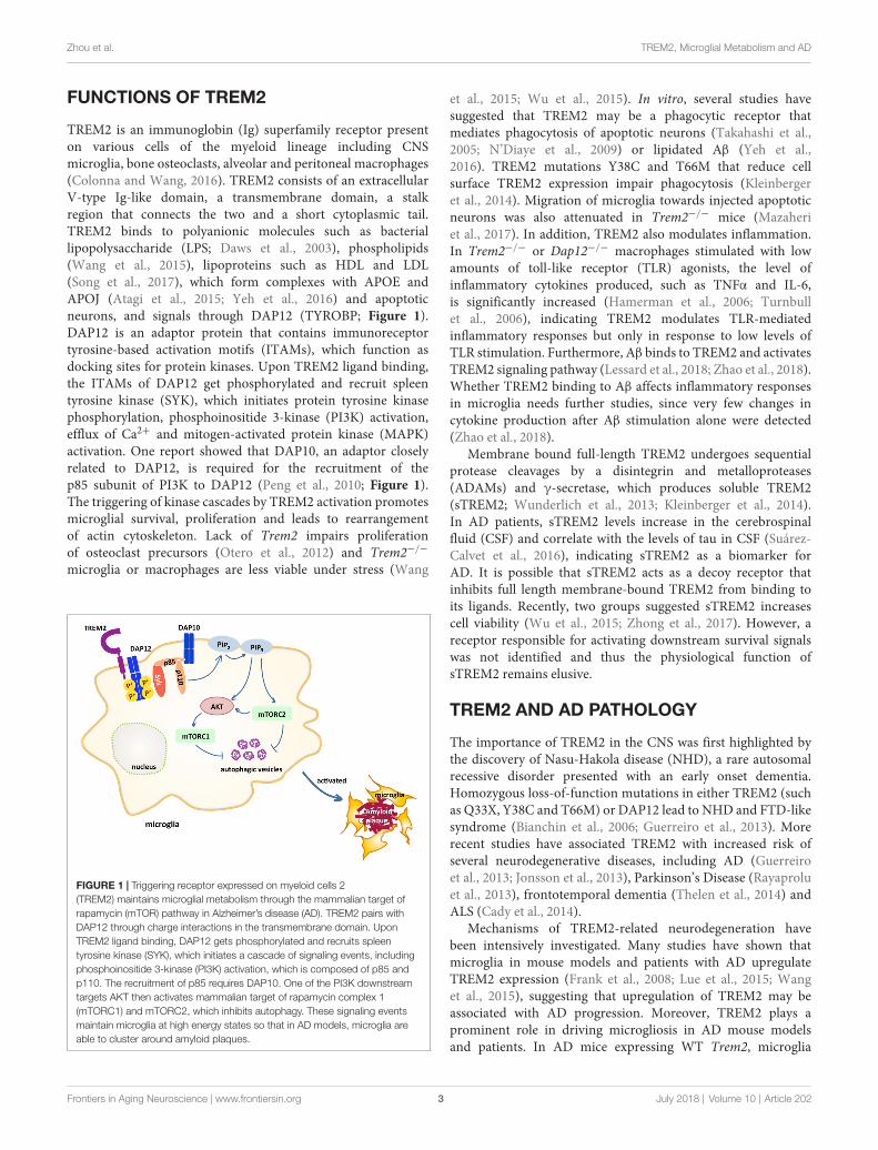
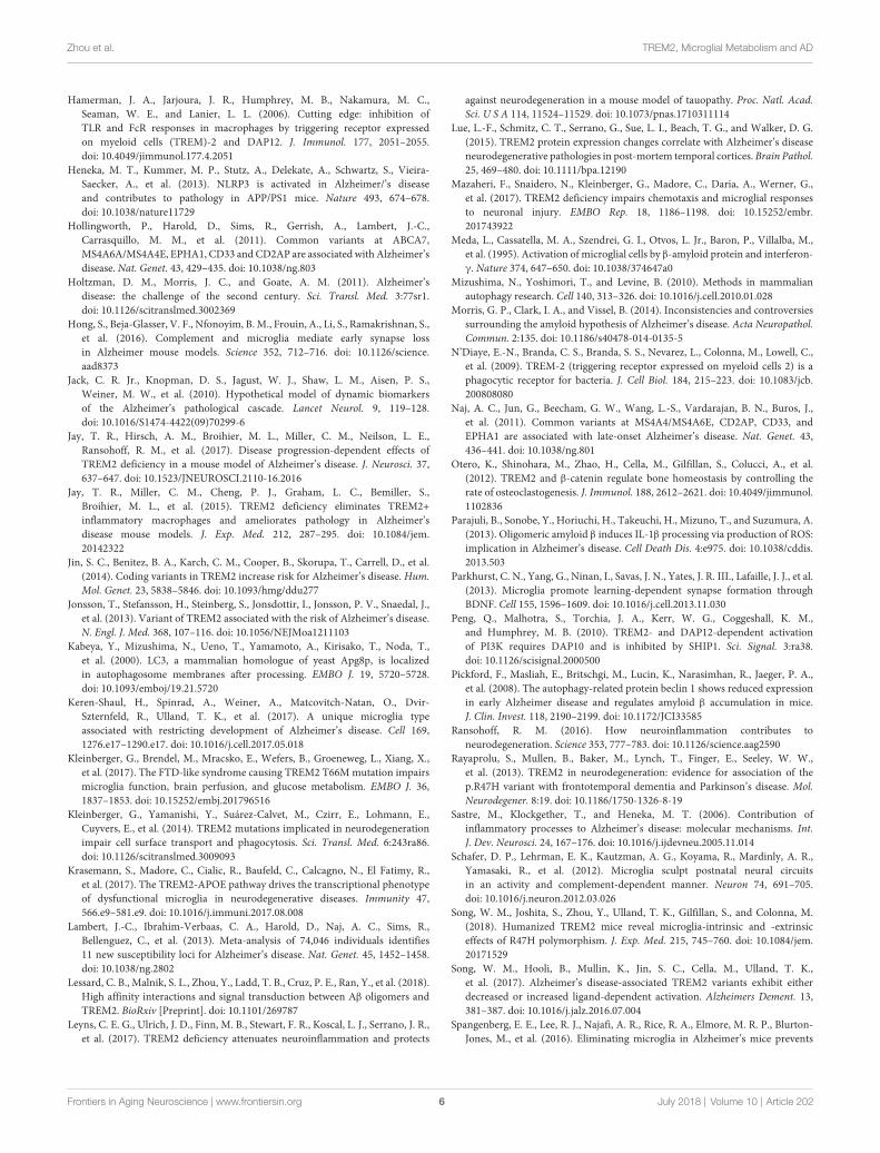
TREM2 is an immunoglobin (Ig) superfamily receptor presenton various cells of the myeloid lineage including CNSmicroglia, bone osteoclasts, alveolar and peritoneal macrophages(Colonna and Wang, 2016). TREM2 consists of an extracellularV-type Ig-like domain, a transmembrane domain, a stalkregion that connects the two and a short cytoplasmic tail.TREM2 binds to polyanionic molecules such as bacteriallipopolysaccharide (LPS; Daws et al., 2003), phospholipids(Wang et al., 2015), lipoproteins such as HDL and LDL(Song et al., 2017), which form complexes with APOE andAPOJ (Atagi et al., 2015; Yeh et al., 2016) and apoptoticneurons, and signals through DAP12 (TYROBP; Figure 1).DAP12 is an adaptor protein that contains immunoreceptortyrosine-based activation motifs (ITAMs), which function asdocking sites for protein kinases. Upon TREM2 ligand binding,the ITAMs of DAP12 get phosphorylated and recruit spleentyrosine kinase (SYK), which initiates protein tyrosine kinasephosphorylation, phosphoinositide 3-kinase (PI3K) activation,efflux of Ca2+ and mitogen-activated protein kinase (MAPK)activation. One report showed that DAP10, an adaptor closelyrelated to DAP12, is required for the recruitment of thep85 subunit of PI3K to DAP12 (Peng et al., 2010; Figure 1).The triggering of kinase cascades by TREM2 activation promotesmicroglial survival, proliferation and leads to rearrangementof actin cytoskeleton. Lack of Trem2 impairs proliferationof osteoclast precursors (Otero et al., 2012) and Trem2−/−
microglia or macrophages are less viable under stress (Wang
FIGURE 1 | Triggering receptor expressed on myeloid cells 2(TREM2) maintains microglial metabolism through the mammalian target ofrapamycin (mTOR) pathway in Alzheimer’s disease (AD). TREM2 pairs withDAP12 through charge interactions in the transmembrane domain. UponTREM2 ligand binding, DAP12 gets phosphorylated and recruits spleentyrosine kinase (SYK), which initiates a cascade of signaling events, includingphosphoinositide 3-kinase (PI3K) activation, which is composed of p85 andp110. The recruitment of p85 requires DAP10. One of the PI3K downstreamtargets AKT then activates mammalian target of rapamycin complex 1(mTORC1) and mTORC2, which inhibits autophagy. These signaling eventsmaintain microglia at high energy states so that in AD models, microglia areable to cluster around amyloid plaques.
et al., 2015; Wu et al., 2015). In vitro, several studies havesuggested that TREM2 may be a phagocytic receptor thatmediates phagocytosis of apoptotic neurons (Takahashi et al.,2005; N’Diaye et al., 2009) or lipidated Aβ (Yeh et al.,2016). TREM2 mutations Y38C and T66M that reduce cellsurface TREM2 expression impair phagocytosis (Kleinbergeret al., 2014). Migration of microglia towards injected apoptoticneurons was also attenuated in Trem2−/− mice (Mazaheriet al., 2017). In addition, TREM2 also modulates inflammation.In Trem2−/− or Dap12−/− macrophages stimulated with lowamounts of toll-like receptor (TLR) agonists, the level ofinflammatory cytokines produced, such as TNFα and IL-6,is significantly increased (Hamerman et al., 2006; Turnbullet al., 2006), indicating TREM2 modulates TLR-mediatedinflammatory responses but only in response to low levels ofTLR stimulation. Furthermore, Aβ binds to TREM2 and activatesTREM2 signaling pathway (Lessard et al., 2018; Zhao et al., 2018).Whether TREM2 binding to Aβ affects inflammatory responsesin microglia needs further studies, since very few changes incytokine production after Aβ stimulation alone were detected(Zhao et al., 2018).
Membrane bound full-length TREM2 undergoes sequentialprotease cleavages by a disintegrin and metalloproteases(ADAMs) and γ-secretase, which produces soluble TREM2(sTREM2; Wunderlich et al., 2013; Kleinberger et al., 2014).In AD patients, sTREM2 levels increase in the cerebrospinalfluid (CSF) and correlate with the levels of tau in CSF (Suárez-Calvet et al., 2016), indicating sTREM2 as a biomarker forAD. It is possible that sTREM2 acts as a decoy receptor thatinhibits full length membrane-bound TREM2 from binding toits ligands. Recently, two groups suggested sTREM2 increasescell viability (Wu et al., 2015; Zhong et al., 2017). However, areceptor responsible for activating downstream survival signalswas not identified and thus the physiological function ofsTREM2 remains elusive.
TREM2 AND AD PATHOLOGY
The importance of TREM2 in the CNS was first highlighted bythe discovery of Nasu-Hakola disease (NHD), a rare autosomalrecessive disorder presented with an early onset dementia.Homozygous loss-of-function mutations in either TREM2 (suchas Q33X, Y38C and T66M) or DAP12 lead to NHD and FTD-likesyndrome (Bianchin et al., 2006; Guerreiro et al., 2013). Morerecent studies have associated TREM2 with increased risk ofseveral neurodegenerative diseases, including AD (Guerreiroet al., 2013; Jonsson et al., 2013), Parkinson’s Disease (Rayaproluet al., 2013), frontotemporal dementia (Thelen et al., 2014) andALS (Cady et al., 2014).
Mechanisms of TREM2-related neurodegeneration havebeen intensively investigated. Many studies have shown thatmicroglia in mouse models and patients with AD upregulateTREM2 expression (Frank et al., 2008; Lue et al., 2015; Wanget al., 2015), suggesting that upregulation of TREM2 may beassociated with AD progression. Moreover, TREM2 plays aprominent role in driving microgliosis in AD mouse modelsand patients. In AD mice expressing WT Trem2, microglia
Frontiers in Aging Neuroscience | www.frontiersin.org 3 July 2018 | Volume 10 | Article 202

Zhou et al. TREM2, Microglial Metabolism and AD
cluster around plaques, providing a barrier to the surroundingneurons (Ulrich et al., 2014; Jay et al., 2015; Wang et al.,2015, 2016; Yuan et al., 2016). On the contrary, the numberof plaque-associated microglia is significantly reduced in Trem2deficient AD mice, and the effect is Trem2 gene dose-dependent(Jay et al., 2015; Wang et al., 2015). In both transgenic miceexpressing the human TREM2 R47H or patients with the R47Hvariant, a similar reduction in microgliosis is observed (Yuanet al., 2016; Song et al., 2018), supporting that R47H is a loss-of-function mutation. Consistently, a larger number of dystrophicneurites accumulate around plaques in Trem2−/− 5xFAD mice,compared to WT Trem2 5xFAD. The observations of plaque-associated microglia are consistent with the identification ofDAM (Keren-Shaul et al., 2017; Krasemann et al., 2017). AsDAM are phagocytic, one would hypothesize that in Trem2−/−
AD mice, lack of DAM would lead to increased Aβ burden.However, observations on plaque load in Trem2-deficiency ADmice are inconsistent (Ulrich et al., 2014; Jay et al., 2015; Wanget al., 2015), which is likely due to different mouse modelsused or timing at which the analyses were done (Jay et al.,2017). Despite discrepancies on plaque load, changes in plaquemorphology have been reported by several groups. In Trem2−/−
5xFAD mice, amyloid plaques appear with loosely packed coresand more diffuse structures extending outward (Wang et al.,2016; Yuan et al., 2016), which is also observed in R47Hcarriers (Yuan et al., 2016), suggesting that TREM2-mediatedmicroglia-plaque interaction may be critical for compactingamyloid fibrils. Whether the changes in plaque structure arerelevant to the role of TREM2 in AD is uncertain, sincediffuse non-fibrillar plaques also occur in cognitively normalindividuals (Morris et al., 2014). More studies are needed toinvestigate if compositions of plaques are different. Besides,whether the increased neuritic dystrophy in Trem2 deficientmice is due to impaired clearance of damaged neurites byTrem2−/− microglia or the result of elevated damage by looselycompacted plaques also remains unclear. Collectively, thesestudies indicate that TREM2 signaling promotes microglialresponses to Aβ in AD.
The effects of TREM2 on tau-driven AD models have notbeen intensely investigated. Recently, two groups that studied taumodels showed inconsistent results. In the PS19 tau transgenicmice that express a human tau with the P301S mutation, lackof Trem2 leads to less brain atrophy but no change in tauphosphorylation or aggregation. These tau mice lacking Trem2show reduced microgliosis and decreased microglial activation at9-month old (Leyns et al., 2017). In contrast, in another studyusing a mouse model expressing the full-length human tau gene,Trem2 deficiency resulted in elevated hyperphosphorylation andaggregation of tau (Bemiller et al., 2017). As with Aβ models,controversy on effects of TREM2 in tau pathology remains. Therelationship of Aβ and tau and how it is affected by TREM2 areinteresting topics for future studies.
TREM2 AND METABOLISM
Metabolic dysfunctions have long been associated with AD,while most studies focused on neuronal metabolisms. Our group
and others have recently linked defective microglial function tometabolism in dementia. Kleinberger et al. (2017) demonstratedthat cerebral metabolic rates of glucose slowed down as shownby reduced FDG-uPET signal in a Trem2 T66M knock-in mousemodel of NHD. The reduced glucose usage could be due todefective microglial function that impairs the metabolic statesof the brain, hinting that dysfunctional TREM2 might alter thebrain metabolism and thus promote pathogenesis (Kleinbergeret al., 2017).
In another study, it was shown that Trem2-deficient bonemarrow-derived macrophages exhibit a defective energeticand anabolic state, which is further exacerbated by stress,such as CSF1 reduction (Ulland et al., 2017). This metabolicdefect is a result of reduced mammalian target of rapamycin(mTOR) signaling, which is ameliorated by activation ofDectin-1, a receptor that activates downstream PI3K andmTOR independent of TREM2, or cyclocreatine, a creatineanalog that restores ATP level. Impaired mTOR signalingwas also observed in microglia sorted from Trem2−/−
5xFAD mice. These results suggest that TREM2 maintainsmicroglia at high metabolic states through enhancedactivation of the mTOR pathway (Figure 1). Furthermore,increased autophagy is detected in Trem2-deficient microgliaand in AD patients carrying one allele of the R47H orR62H variant of TREM2, suggesting microglia attempt tocompensate the mTOR defects with autophagy as a survivalmechanism.
AUTOPHAGY AND AD
Autophagy is a self-eating process where cells clear misfoldedproteins and damaged organelles as a housekeeping mechanismand as a survival mechanism during stress and starvation.The formation of an autophagophore, an isolation membranethat initiates autophagy and becomes the autophagosome later,requires the ULK complex and the class III PI-3 kinase (PI3K)complex composed of Vps34, Beclin-1, p150 and ATG14.The ULK complex is inactivated by mammalian target ofrapamycin complex 1 (mTORC1) and positively regulated byAMP-activated protein kinase (AMPK). Activation of ULKand PI3K complexes recruit additional autophagy related(ATG) proteins to drive autophagosome nucleation. Thenthe ATG12-ATG5-ATG16 complex is recruited to facilitatelipidation of microtubule-associated protein 1 light chain 3(LC3), resulting in the conversion of LC3-I to LC3-II, asthe isolation membrane expands to form the autophagosome(Kabeya et al., 2000). In mammalian cells, only LC3-II remainson autophagosome membranes until after their fusion withlysosomes, allowing it to be a marker for autophagy (Mizushimaet al., 2010).
Autophagy has been associated with AD. The levels ofautophagy protein Beclin 1 are decreased in AD patients butnot in AD mouse models (Pickford et al., 2008), pointingto the possibility that Beclin 1 reduction occurs upstream ofamyloid pathology. Genetic ablation of one copy of Beclin1reduces autophagy in cultured primary neurons and increases Aβ
deposition and neuronal loss in an ADmodel. On the other hand,
Frontiers in Aging Neuroscience | www.frontiersin.org 4 July 2018 | Volume 10 | Article 202
Zhou et al. TREM2, Microglial Metabolism and AD
an increase of CD68 immunoreactivity, an activation marker,with the absence of change in Iba1 marks microglial activationin Beclin 1+/− AD mice without affecting microglial number(Pickford et al., 2008). These findings indicate that neuronalautophagy plays a protective role in AD progression. In linewith this, overexpression of another autophagy gene, p62, byadeno-associated virus (AAV) infection in neurons improvescognitive functions and reduces Aβ pathology in an autophagy-mediated manner (Caccamo et al., 2017). Pharmaceutically,feeding mice with rapamycin, an inhibitor of the mTORpathway, induces autophagy in two mouse models of AD butnot in non-AD littermate controls, suggesting high Aβ levelis a trigger of autophagy. This rapamycin-mediated mTORinhibition leads to a reduction of Aβ42 level in the hippocampusand an amelioration in memory deficits by increasing autophagy(Caccamo et al., 2010; Spilman et al., 2010). Although all thesestudies suggest a protective function of autophagy, whether thiseffect is only mediated by neurons or if glia also play a role isunclear.
Rather, our study demonstrated a role of TREM2 inattenuatingmicroglial autophagy (Ulland et al., 2017). Comparedto control, microglia lacking TREM2 contain more autophagicvesicles as shown by electron microscopy. Increased autophagyin Trem2-deficient microglia is a result of reduced mTORsignaling in response to a low metabolic state of microgliaand this increase in autophagy is not sufficient to rescuemicroglia from dying under stress, such as neuroinflammation.Interestingly, dietary supplementation with cyclocreatine rescuesTrem2-deficient microglia from autophagy and partially correctsthe defect in microglial clustering in Trem2−/− 5xFAD. Theseresults suggest that TREM2 sustains microglial metabolism thusallowing them to function properly. This study links TREM2 to
metabolic states of microglia and allows us to revisit the role ofTREM2 and microglia in AD pathology.
FUTURE PERSPECTIVES
The role of microglia in neurodegeneration has been hotlydebated while no concluding agreement has been made. Suchan agreement is difficult to achieve as different groups usingsimilar models show different results, perhaps due to microbiota.Notably, studies on germ-free mice in AD models are currentlybeing conducted. As TREM2 is required to turn homeostaticmicroglia into activated microglia, whether these activatedmicroglia acquire a unique metabolic state is an intriguing topic.It remains unclear what exact role TREM2 plays. Does it sensechanges in environment or just provide a tonic signal to keepmicroglia ready to go? Further studies are needed to distinguishthe two possibilities. Defining the ligands of TREM2 in vivo andhow it is activated will help answer the question. Microglia can bea double-edged sword. Deciphering the role of DAMwill advanceour therapeutic approaches in neurodegenerative diseases.
AUTHOR CONTRIBUTIONS
YZ drafted the manuscript. TU and MC reviewed and edited themanuscript.
FUNDING
This work was funded by the National Institutes of Health(NIH) grant RF1 AG05148501 and a Cure Alzheimer’s Fundgrant to MC.
REFERENCES
Atagi, Y., Liu, C.-C., Painter, M. M., Chen, X.-F., Verbeeck, C., Zheng, H., et al.(2015). Apolipoprotein E is a ligand for triggering receptor expressed onmyeloid cells 2 (TREM2). J. Biol. Chem. 290, 26043–26050. doi: 10.1074/jbc.M115.679043
Bateman, R. J., Xiong, C., Benzinger, T. L. S., Fagan, A. M., Goate, A.,Fox, N. C., et al. (2012). Clinical and biomarker changes in dominantlyinherited Alzheimer’s disease. N. Engl. J. Med. 367, 795–804.doi: 10.1056/NEJMoa1202753
Bemiller, S. M., McCray, T. J., Allan, K., Formica, S. V., Xu, G., Wilson, G., et al.(2017). TREM2 deficiency exacerbates tau pathology through dysregulatedkinase signaling in a mouse model of tauopathy. Mol. Neurodegener. 12:74.doi: 10.1186/s13024-017-0216-6
Bertram, L., Lill, C. M., and Tanzi, R. E. (2010). The genetics of Alzheimer disease:back to the future. Neuron 68, 270–281. doi: 10.1016/j.neuron.2010.10.013
Bertram, L., McQueen, M. B., Mullin, K., Blacker, D., and Tanzi, R. E. (2007).Systematic meta-analyses of Alzheimer disease genetic association studies: theAlzGene database. Nat. Genet. 39, 17–23. doi: 10.1038/ng1934
Bianchin, M. M., Lima, J. E., Natel, J., and Sakamoto, A. C. (2006). Thegenetic causes of basal ganglia calcification, dementia, and bone cysts:DAP12 and TREM2. Neurology 66, 615–616. doi: 10.1212/01.wnl.0000216105.11788.0f
Caccamo, A., Ferreira, E., Branca, C., and Oddo, S. (2017). p62 improvesAD-like pathology by increasing autophagy. Mol. Psychiatry 22, 865–873.doi: 10.1038/mp.2016.139
Caccamo, A., Majumder, S., Richardson, A., Strong, R., and Oddo, S. (2010).Molecular interplay between mammalian target of rapamycin (mTOR),
amyloid-β, and Tau effects on cognitive impairments. J. Biol. Chem. 285,13107–13120. doi: 10.1074/jbc.M110.100420
Cady, J., Koval, E. D., Benitez, B. A., Zaidman, C., Jockel-Balsarotti, J., Allred, P.,et al. (2014). TREM2 variant p.R47H as a risk factor for sporadic amyotrophiclateral sclerosis. JAMANeurol. 71, 449–453. doi: 10.1001/jamaneurol.2013.6237
Colonna, M., and Wang, Y. (2016). TREM2 variants: new keys to decipherAlzheimer disease pathogenesis. Nat. Rev. Neurosci. 17, 201–207.doi: 10.1038/nrn.2016.7
Condello, C., Yuan, P., Schain, A., and Grutzendler, J. (2015). Microglia constitutea barrier that prevents neurotoxic protofibrillar Aβ42 hotspots around plaques.Nat. Commun. 6:6176. doi: 10.1038/ncomms7176
Daws, M. R., Sullam, P. M., Niemi, E. C., Chen, T. T., Tchao, N. K., andSeaman, W. E. (2003). Pattern recognition by TREM-2: binding of anionicligands. J. Immunol. 171, 594–599. doi: 10.4049/jimmunol.171.2.594
Elmore, M. R. P., Najafi, A. R., Koike, M. A., Dagher, N. N., Spangenberg, E. E.,Rice, R. A., et al. (2014). Colony-stimulating factor 1 receptor signaling isnecessary for microglia viability, unmasking a microglia progenitor cell in theadult brain. Neuron 82, 380–397. doi: 10.1016/j.neuron.2014.02.040
Frank, S., Burbach, G. J., Bonin, M., Walter, M., Streit, W., Bechmann, I., et al.(2008). TREM2 is upregulated in amyloid plaque-associated microglia in agedAPP23 transgenic mice. Glia 56, 1438–1447. doi: 10.1002/glia.20710
Ginhoux, F., Greter, M., Leboeuf, M., Nandi, S., See, P., Gokhan, S.,et al. (2010). Fate mapping analysis reveals that adult microglia derivefrom primitive macrophages. Science 330, 841–845. doi: 10.1126/science.1194637
Guerreiro, R., Wojtas, A., Bras, J., Carrasquillo, M., Rogaeva, E., Majounie, E., et al.(2013). TREM2 variants in Alzheimer’s disease. N. Engl. J. Med. 368, 117–127.doi: 10.1056/NEJMoa1211851
Frontiers in Aging Neuroscience | www.frontiersin.org 5 July 2018 | Volume 10 | Article 202
Zhou et al. TREM2, Microglial Metabolism and AD
Hamerman, J. A., Jarjoura, J. R., Humphrey, M. B., Nakamura, M. C.,Seaman, W. E., and Lanier, L. L. (2006). Cutting edge: inhibition ofTLR and FcR responses in macrophages by triggering receptor expressedon myeloid cells (TREM)-2 and DAP12. J. Immunol. 177, 2051–2055.doi: 10.4049/jimmunol.177.4.2051
Heneka, M. T., Kummer, M. P., Stutz, A., Delekate, A., Schwartz, S., Vieira-Saecker, A., et al. (2013). NLRP3 is activated in Alzheimer/’s diseaseand contributes to pathology in APP/PS1 mice. Nature 493, 674–678.doi: 10.1038/nature11729
Hollingworth, P., Harold, D., Sims, R., Gerrish, A., Lambert, J.-C.,Carrasquillo, M. M., et al. (2011). Common variants at ABCA7,MS4A6A/MS4A4E, EPHA1, CD33 andCD2AP are associated with Alzheimer’sdisease. Nat. Genet. 43, 429–435. doi: 10.1038/ng.803
Holtzman, D. M., Morris, J. C., and Goate, A. M. (2011). Alzheimer’sdisease: the challenge of the second century. Sci. Transl. Med. 3:77sr1.doi: 10.1126/scitranslmed.3002369
Hong, S., Beja-Glasser, V. F., Nfonoyim, B. M., Frouin, A., Li, S., Ramakrishnan, S.,et al. (2016). Complement and microglia mediate early synapse lossin Alzheimer mouse models. Science 352, 712–716. doi: 10.1126/science.aad8373
Jack, C. R. Jr., Knopman, D. S., Jagust, W. J., Shaw, L. M., Aisen, P. S.,Weiner, M. W., et al. (2010). Hypothetical model of dynamic biomarkersof the Alzheimer’s pathological cascade. Lancet Neurol. 9, 119–128.doi: 10.1016/S1474-4422(09)70299-6
Jay, T. R., Hirsch, A. M., Broihier, M. L., Miller, C. M., Neilson, L. E.,Ransohoff, R. M., et al. (2017). Disease progression-dependent effects ofTREM2 deficiency in a mouse model of Alzheimer’s disease. J. Neurosci. 37,637–647. doi: 10.1523/JNEUROSCI.2110-16.2016
Jay, T. R., Miller, C. M., Cheng, P. J., Graham, L. C., Bemiller, S.,Broihier, M. L., et al. (2015). TREM2 deficiency eliminates TREM2+inflammatory macrophages and ameliorates pathology in Alzheimer’sdisease mouse models. J. Exp. Med. 212, 287–295. doi: 10.1084/jem.20142322
Jin, S. C., Benitez, B. A., Karch, C. M., Cooper, B., Skorupa, T., Carrell, D., et al.(2014). Coding variants in TREM2 increase risk for Alzheimer’s disease. Hum.Mol. Genet. 23, 5838–5846. doi: 10.1093/hmg/ddu277
Jonsson, T., Stefansson, H., Steinberg, S., Jonsdottir, I., Jonsson, P. V., Snaedal, J.,et al. (2013). Variant of TREM2 associated with the risk of Alzheimer’s disease.N. Engl. J. Med. 368, 107–116. doi: 10.1056/NEJMoa1211103
Kabeya, Y., Mizushima, N., Ueno, T., Yamamoto, A., Kirisako, T., Noda, T.,et al. (2000). LC3, a mammalian homologue of yeast Apg8p, is localizedin autophagosome membranes after processing. EMBO J. 19, 5720–5728.doi: 10.1093/emboj/19.21.5720
Keren-Shaul, H., Spinrad, A., Weiner, A., Matcovitch-Natan, O., Dvir-Szternfeld, R., Ulland, T. K., et al. (2017). A unique microglia typeassociated with restricting development of Alzheimer’s disease. Cell 169,1276.e17–1290.e17. doi: 10.1016/j.cell.2017.05.018
Kleinberger, G., Brendel, M., Mracsko, E., Wefers, B., Groeneweg, L., Xiang, X.,et al. (2017). The FTD-like syndrome causing TREM2 T66Mmutation impairsmicroglia function, brain perfusion, and glucose metabolism. EMBO J. 36,1837–1853. doi: 10.15252/embj.201796516
Kleinberger, G., Yamanishi, Y., Suárez-Calvet, M., Czirr, E., Lohmann, E.,Cuyvers, E., et al. (2014). TREM2 mutations implicated in neurodegenerationimpair cell surface transport and phagocytosis. Sci. Transl. Med. 6:243ra86.doi: 10.1126/scitranslmed.3009093
Krasemann, S., Madore, C., Cialic, R., Baufeld, C., Calcagno, N., El Fatimy, R.,et al. (2017). The TREM2-APOE pathway drives the transcriptional phenotypeof dysfunctional microglia in neurodegenerative diseases. Immunity 47,566.e9–581.e9. doi: 10.1016/j.immuni.2017.08.008
Lambert, J.-C., Ibrahim-Verbaas, C. A., Harold, D., Naj, A. C., Sims, R.,Bellenguez, C., et al. (2013). Meta-analysis of 74,046 individuals identifies11 new susceptibility loci for Alzheimer’s disease. Nat. Genet. 45, 1452–1458.doi: 10.1038/ng.2802
Lessard, C. B., Malnik, S. L., Zhou, Y., Ladd, T. B., Cruz, P. E., Ran, Y., et al. (2018).High affinity interactions and signal transduction between Aβ oligomers andTREM2. BioRxiv [Preprint]. doi: 10.1101/269787
Leyns, C. E. G., Ulrich, J. D., Finn, M. B., Stewart, F. R., Koscal, L. J., Serrano, J. R.,et al. (2017). TREM2 deficiency attenuates neuroinflammation and protects
against neurodegeneration in a mouse model of tauopathy. Proc. Natl. Acad.Sci. U S A 114, 11524–11529. doi: 10.1073/pnas.1710311114
Lue, L.-F., Schmitz, C. T., Serrano, G., Sue, L. I., Beach, T. G., and Walker, D. G.(2015). TREM2 protein expression changes correlate with Alzheimer’s diseaseneurodegenerative pathologies in post-mortem temporal cortices. Brain Pathol.25, 469–480. doi: 10.1111/bpa.12190
Mazaheri, F., Snaidero, N., Kleinberger, G., Madore, C., Daria, A., Werner, G.,et al. (2017). TREM2 deficiency impairs chemotaxis and microglial responsesto neuronal injury. EMBO Rep. 18, 1186–1198. doi: 10.15252/embr.201743922
Meda, L., Cassatella, M. A., Szendrei, G. I., Otvos, L. Jr., Baron, P., Villalba, M.,et al. (1995). Activation of microglial cells by β-amyloid protein and interferon-γ. Nature 374, 647–650. doi: 10.1038/374647a0
Mizushima, N., Yoshimori, T., and Levine, B. (2010). Methods in mammalianautophagy research. Cell 140, 313–326. doi: 10.1016/j.cell.2010.01.028
Morris, G. P., Clark, I. A., and Vissel, B. (2014). Inconsistencies and controversiessurrounding the amyloid hypothesis of Alzheimer’s disease. Acta Neuropathol.Commun. 2:135. doi: 10.1186/s40478-014-0135-5
N’Diaye, E.-N., Branda, C. S., Branda, S. S., Nevarez, L., Colonna, M., Lowell, C.,et al. (2009). TREM-2 (triggering receptor expressed on myeloid cells 2) is aphagocytic receptor for bacteria. J. Cell Biol. 184, 215–223. doi: 10.1083/jcb.200808080
Naj, A. C., Jun, G., Beecham, G. W., Wang, L.-S., Vardarajan, B. N., Buros, J.,et al. (2011). Common variants at MS4A4/MS4A6E, CD2AP, CD33, andEPHA1 are associated with late-onset Alzheimer’s disease. Nat. Genet. 43,436–441. doi: 10.1038/ng.801
Otero, K., Shinohara, M., Zhao, H., Cella, M., Gilfillan, S., Colucci, A., et al.(2012). TREM2 and β-catenin regulate bone homeostasis by controlling therate of osteoclastogenesis. J. Immunol. 188, 2612–2621. doi: 10.4049/jimmunol.1102836
Parajuli, B., Sonobe, Y., Horiuchi, H., Takeuchi, H., Mizuno, T., and Suzumura, A.(2013). Oligomeric amyloid β induces IL-1β processing via production of ROS:implication in Alzheimer’s disease. Cell Death Dis. 4:e975. doi: 10.1038/cddis.2013.503
Parkhurst, C. N., Yang, G., Ninan, I., Savas, J. N., Yates, J. R. III., Lafaille, J. J., et al.(2013). Microglia promote learning-dependent synapse formation throughBDNF. Cell 155, 1596–1609. doi: 10.1016/j.cell.2013.11.030
Peng, Q., Malhotra, S., Torchia, J. A., Kerr, W. G., Coggeshall, K. M.,and Humphrey, M. B. (2010). TREM2- and DAP12-dependent activationof PI3K requires DAP10 and is inhibited by SHIP1. Sci. Signal. 3:ra38.doi: 10.1126/scisignal.2000500
Pickford, F., Masliah, E., Britschgi, M., Lucin, K., Narasimhan, R., Jaeger, P. A.,et al. (2008). The autophagy-related protein beclin 1 shows reduced expressionin early Alzheimer disease and regulates amyloid β accumulation in mice.J. Clin. Invest. 118, 2190–2199. doi: 10.1172/JCI33585
Ransohoff, R. M. (2016). How neuroinflammation contributes toneurodegeneration. Science 353, 777–783. doi: 10.1126/science.aag2590
Rayaprolu, S., Mullen, B., Baker, M., Lynch, T., Finger, E., Seeley, W. W.,et al. (2013). TREM2 in neurodegeneration: evidence for association of thep.R47H variant with frontotemporal dementia and Parkinson’s disease. Mol.Neurodegener. 8:19. doi: 10.1186/1750-1326-8-19
Sastre, M., Klockgether, T., and Heneka, M. T. (2006). Contribution ofinflammatory processes to Alzheimer’s disease: molecular mechanisms. Int.J. Dev. Neurosci. 24, 167–176. doi: 10.1016/j.ijdevneu.2005.11.014
Schafer, D. P., Lehrman, E. K., Kautzman, A. G., Koyama, R., Mardinly, A. R.,Yamasaki, R., et al. (2012). Microglia sculpt postnatal neural circuitsin an activity and complement-dependent manner. Neuron 74, 691–705.doi: 10.1016/j.neuron.2012.03.026
Song, W. M., Joshita, S., Zhou, Y., Ulland, T. K., Gilfillan, S., and Colonna, M.(2018). Humanized TREM2 mice reveal microglia-intrinsic and -extrinsiceffects of R47H polymorphism. J. Exp. Med. 215, 745–760. doi: 10.1084/jem.20171529
Song, W. M., Hooli, B., Mullin, K., Jin, S. C., Cella, M., Ulland, T. K.,et al. (2017). Alzheimer’s disease-associated TREM2 variants exhibit eitherdecreased or increased ligand-dependent activation. Alzheimers Dement. 13,381–387. doi: 10.1016/j.jalz.2016.07.004
Spangenberg, E. E., Lee, R. J., Najafi, A. R., Rice, R. A., Elmore, M. R. P., Blurton-Jones, M., et al. (2016). Eliminating microglia in Alzheimer’s mice prevents
Frontiers in Aging Neuroscience | www.frontiersin.org 6 July 2018 | Volume 10 | Article 202
Zhou et al. TREM2, Microglial Metabolism and AD
neuronal loss without modulating amyloid-β pathology. Brain 139, 1265–1281.doi: 10.1093/brain/aww016
Spilman, P., Podlutskaya, N., Hart, M. J., Debnath, J., Gorostiza, O., Bredesen, D.,et al. (2010). Inhibition of mTOR by rapamycin abolishes cognitive deficits andreduces amyloid-β levels in a mouse model of Alzheimer’s disease. PLOS One5:e9979. doi: 10.1371/journal.pone.0009979
Stevens, B., Allen, N. J., Vazquez, L. E., Howell, G. R., Christopherson, K. S.,Nouri, N., et al. (2007). The classical complement cascade mediatesCNS synapse elimination. Cell 131, 1164–1178. doi: 10.1016/j.cell.2007.10.036
Strittmatter, W. J., Saunders, A. M., Schmechel, D., Pericak-Vance, M.,Enghild, J., Salvesen, G. S., et al. (1993). Apolipoprotein E: high-aviditybinding to β-amyloid and increased frequency of type 4 allele in late-onsetfamilial Alzheimer disease. Proc. Natl. Acad. Sci. U S A 90, 1977–1981.doi: 10.1073/pnas.90.5.1977
Suárez-Calvet, M., Araque Caballero, M. Á., Kleinberger, G., Bateman, R. J.,Fagan, A. M., Morris, J. C., et al. (2016). Early changes in CSF sTREM2 indominantly inherited Alzheimer’s disease occur after amyloid depositionand neuronal injury. Sci. Transl. Med. 8:369ra178. doi: 10.1126/scitranslmed.aag1767
Takahashi, K., Rochford, C. D. P., andNeumann, H. (2005). Clearance of apoptoticneurons without inflammation by microglial triggering receptor expressed onmyeloid cells-2. J. Exp. Med. 201, 647–657. doi: 10.1084/jem.20041611
Takasugi, N., Tomita, T., Hayashi, I., Tsuruoka, M., Niimura, M., Takahashi, Y.,et al. (2003). The role of presenilin cofactors in the γ-secretase complex.Nature422, 438–441. doi: 10.1038/nature01506
Thelen, M., Razquin, C., Hernández, I., Gorostidi, A., Sánchez-Valle, R., Ortega-Cubero, S., et al. (2014). Investigation of the role of rare TREM2 variants infrontotemporal dementia subtypes. Neurobiol. Aging 35, 2657.e13–2657.e19.doi: 10.1016/j.neurobiolaging.2014.06.018
Turnbull, I. R., Gilfillan, S., Cella, M., Aoshi, T., Miller, M., Piccio, L., et al. (2006).Cutting edge: TREM-2 attenuates macrophage activation. J. Immunol. 177,3520–3524. doi: 10.4049/jimmunol.177.6.3520
Ulland, T. K., Song, W. M., Huang, S. C.-C., Ulrich, J. D., Sergushichev, A.,Beatty, W. L., et al. (2017). TREM2 Maintains microglial metabolic fitness inAlzheimer’s disease. Cell 170, 649.e13–663.e13. doi: 10.1016/j.cell.2017.07.023
Ulrich, J. D., Finn, M. B., Wang, Y., Shen, A., Mahan, T. E., Jiang, H., et al. (2014).Altered microglial response to Aβ plaques in APPPS1–21 mice heterozygousfor TREM2.Mol. Neurodegener. 9:20. doi: 10.1186/1750-1326-9-20
Venegas, C., Kumar, S., Franklin, B. S., Dierkes, T., Brinkschulte, R., Tejera, D.,et al. (2017). Microglia-derived ASC specks cross-seed amyloid-β inAlzheimer’s disease. Nature 552, 355–361. doi: 10.1038/nature25158
Wang, Y., Cella, M., Mallinson, K., Ulrich, J. D., Young, K. L., Robinette, M. L.,et al. (2015). TREM2 lipid sensing sustains the microglial response in anAlzheimer’s disease model. Cell 160, 1061–1071. doi: 10.1016/j.cell.2015.01.049
Wang, Y., Szretter, K. J., Vermi,W., Gilfillan, S., Rossini, C., Cella, M., et al. (2012).IL-34 is a tissue-restricted ligand of CSF1R required for the development ofLangerhans cells and microglia. Nat. Immunol. 13, 753–760. doi: 10.1038/ni.2360
Wang, Y., Ulland, T. K., Ulrich, J. D., Song, W., Tzaferis, J. A., Hole, J. T.,et al. (2016). TREM2-mediated early microglial response limits diffusion andtoxicity of amyloid plaques. J. Exp. Med. 213, 667–675. doi: 10.1084/jem.20151948
Wu, K., Byers, D. E., Jin, X., Agapov, E., Alexander-Brett, J., Patel, A. C.,et al. (2015). TREM-2 promotes macrophage survival and lung disease afterrespiratory viral infection. J. Exp. Med. 212, 681–697. doi: 10.1084/jem.20141732
Wunderlich, P., Glebov, K., Kemmerling, N., Tien, N. T., Neumann, H., andWalter, J. (2013). Sequential proteolytic processing of the triggering receptorexpressed on myeloid cells-2 (TREM2) protein by ectodomain sheddingand γ-secretase-dependent intramembranous cleavage. J. Biol. Chem. 288,33027–33036. doi: 10.1074/jbc.m113.517540
Yeh, F. L., Wang, Y., Tom, I., Gonzalez, L. C., and Sheng, M. (2016). TREM2 bindsto apolipoproteins, including APOE and CLU/APOJ and thereby facilitatesuptake of amyloid-β by microglia. Neuron 91, 328–340. doi: 10.1016/j.neuron.2016.06.015
Yuan, P., Condello, C., Keene, C. D., Wang, Y., Bird, T. D., Paul, S. M.,et al. (2016). TREM2 haplodeficiency in mice and humans impairs themicroglia barrier function leading to decreased amyloid compaction andsevere axonal dystrophy. Neuron 90, 724–739. doi: 10.1016/j.neuron.2016.05.003
Zhao, Y., Wu, X., Li, X., Jiang, L.-L., Gui, X., Liu, Y., et al. (2018). TREM2 isa receptor for β-amyloid that mediates microglial function. Neuron 97,1023.e7–1031.e7. doi: 10.1016/j.neuron.2018.01.031
Zhong, L., Chen, X.-F., Wang, T., Wang, Z., Liao, C., Wang, Z., et al.(2017). Soluble TREM2 induces inflammatory responses and enhancesmicroglial survival. J. Exp. Med. 214, 597–607. doi: 10.1084/jem.20160844
Conflict of Interest Statement: The authors declare that the research wasconducted in the absence of any commercial or financial relationships that couldbe construed as a potential conflict of interest.
Copyright © 2018 Zhou, Ulland and Colonna. This is an open-access articledistributed under the terms of the Creative Commons Attribution License (CC BY).The use, distribution or reproduction in other forums is permitted, provided theoriginal author(s) and the copyright owner(s) are credited and that the originalpublication in this journal is cited, in accordance with accepted academic practice.No use, distribution or reproduction is permitted which does not comply with theseterms.
Frontiers in Aging Neuroscience | www.frontiersin.org 7 July 2018 | Volume 10 | Article 202