Digestive system · Web viewThe alimentary canal or gastrointestinal (GI) tract _____ Alimentary...
23
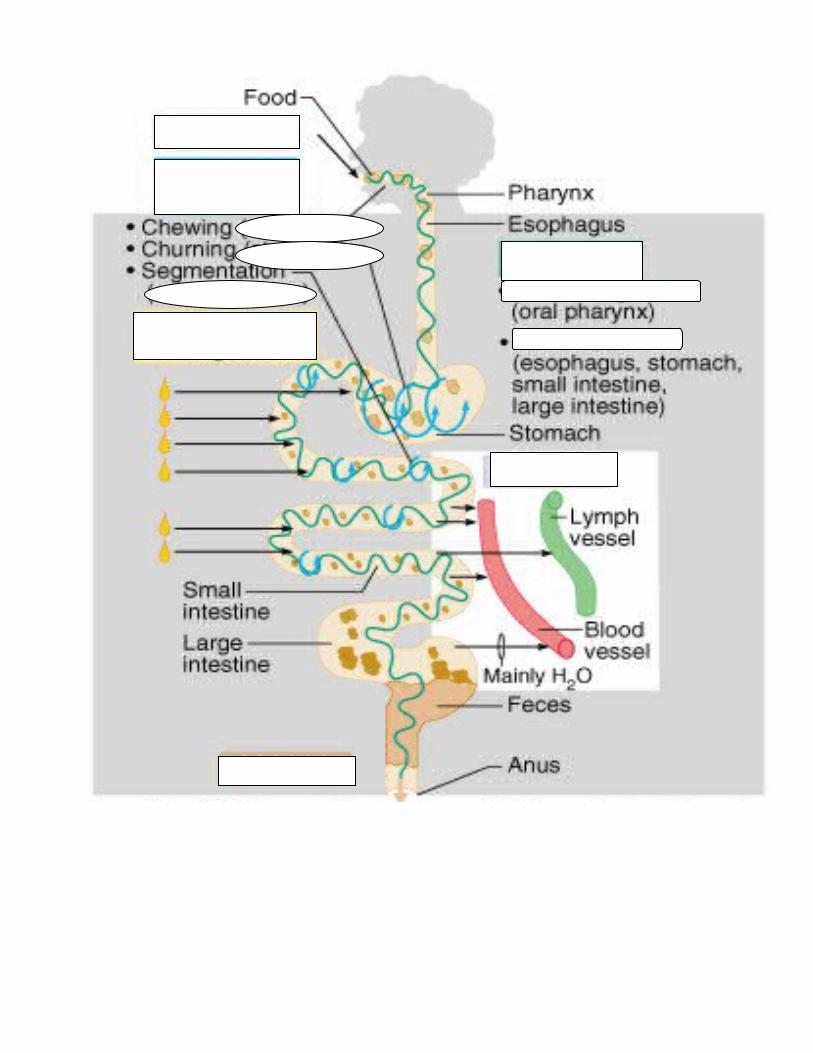
Digestive system _____________ in food. ______________ food down into nutrient molecules _____________ these molecules into blood stream ________body of indigestible remains • The alimentary canal or gastrointestinal (GI) tract _____________________________ • Alimentary canal – • Accessory digestive organs – Digestion • Mechanism for nourishing the body • Most nutrients in food require either degradation or release prior to absorption • Digestion occurs in GI Tract • Digestion • ______________________ • chewing and peristalsis • ____________________ • enzymes, HCl • The GI tract is a _____________________________________________________ • Nutrients become more available to the body in each step • There are six essential activities: • - • - • - • - • - • - Essential Activities of Digestion Ingestion – Propulsion – Peristalsis – Mechanical digestion – Movement of digestive materials • Visceral smooth muscle shows rhythmic cycles of activity • Pacemaker cells • Peristalsis • Waves that move a _________________: • Segmentation • Churn and __________________ a bolus
Transcript of Digestive system · Web viewThe alimentary canal or gastrointestinal (GI) tract _____ Alimentary...

Digestive system_____________ in food.______________ food down into nutrient molecules_____________ these molecules into blood stream________body of indigestible remains
• The alimentary canal or gastrointestinal (GI) tract _____________________________• Alimentary canal –
• Accessory digestive organs –
Digestion• Mechanism for nourishing the body• Most nutrients in food require either degradation or release prior to absorption• Digestion occurs in GI Tract• Digestion
• ______________________• chewing and peristalsis
• ____________________• enzymes, HCl
• The GI tract is a _____________________________________________________• Nutrients become more available to the body in each step
• There are six essential activities: • -• -• -• -• -• -
Essential Activities of Digestion Ingestion – Propulsion –
Peristalsis – Mechanical digestion –
Movement of digestive materials• Visceral smooth muscle shows rhythmic cycles of activity
• Pacemaker cells• Peristalsis
• Waves that move a _________________:• Segmentation
• Churn and __________________ a bolus
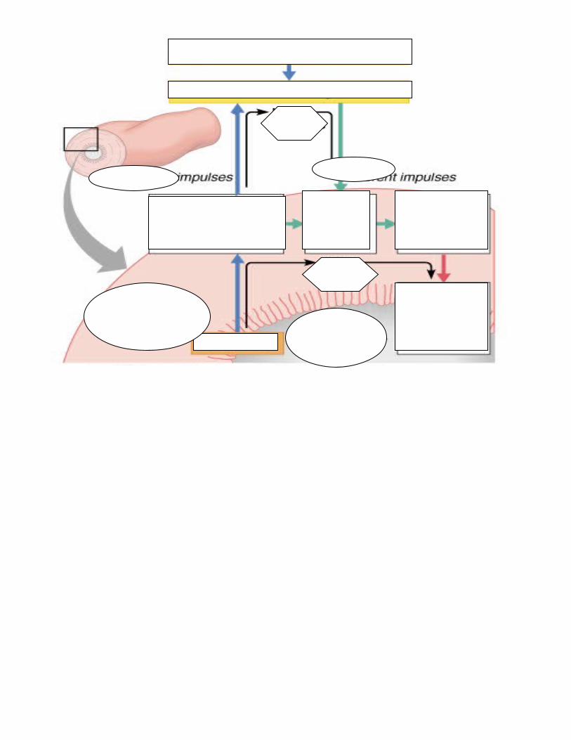
Control of the digestive system• Movement of materials along the digestive tract is controlled by:
• -• Parasympathetic and local reflexes
• -• Enhance or inhibit smooth muscle contraction
• -• Coordinate response to changes in pH or chemical stimuli
Chemical digestion – Absorption – Defecation –


GI Tract• External environment for the digestive process• Regulation of digestion involves:
• -• -• -
• Mechano- and chemoreceptors respond to:• ________________________________• Osmolarity(solute concentration) and pH• Presence of substrate or • End products of digestion
• They initiate reflexes that:• ________________________________digestive galnds• __________lumen contents and ____________them along
Control of the digestive system• Movement of materials along the digestive tract is controlled by:
• _________________mechanisms • Parasympathetic and local reflexes
• ___________________ mechanisms • Enhance or inhibit smooth muscle contraction
• _________________ mechanisms • Coordinate response to changes in pH or chemical stimuli
Nervous Control of the GI Tract• Intrinsic controls-gut brain• Extrinsic controls-involves CNS

Digestive System Organs and Peritoneum • Peritoneum –
• Visceral – • Parietal –
• Peritoneal cavity• -• Allows them to slide across one another
• Mesentery – • -supplies __________________ ___________________• -• Stores fat
• Retroperitoneal organs – organs __________________________ the peritoneum• Peritoneal organs (intraperitoneal) – organs surrounded by peritoneum• Mesenteries
• -Sheets of serous membranes that support portions of the digestive tract• ________________________________________ lies anterior to abdominal viscera
• Provides ______________________________________________________ • __________________________________________
Blood Supply: Splanchnic Circulation• Arteries that branch off abdominal aorata and the organs they serve include
• The hepatic--_______________________, splenic,_____________________ and left gastric: ______________________________
• Inferior and superior mesenteric: ______________________________________ • Hepatic portal circulation:
• Collects nutrient-rich venous blood from the digestive viscera(organs)• Delivers it to the _____________________________ for metabolic processing and storage
Histology of the Alimentary Canal • From esophagus to anal canal -- walls of the GI tract have same _____________________
• From the lumen outward they are the:• ______________________• _______________________ • _________________________• _______________________(serosa is technically not present on the esophagus)
• Each tunic has a predominant tissue type and specific digestive functionMucosa
• _____________________________________________lines lumen of alimentary canal• Its three major functions are:
• -_____________________of mucus• -_____________________of end products of digestion• -_____________________aginst infectious diseases
• Consists of three layers: • a lining epithelium, • lamina propria, • muscularis mucosae
Epithelial Lining• Consists of _______________________________________________________________• The mucus secretions:
• _________________ digestive organs from digesting themselves• ____________ food along tract
• Stomach and small intestine mucosa contain:• -• -
Mucosa: Lamina Propria and Muscularis Mucosae• Lamina Propria
• -• Nourishes epithelium and absorbs nutrients• Contains lymph nodes
• (part of MALT-Mucosa Associated Lymphatic Tissue) •
• Muscularis mucosae– smooth muscle cells that produce local movements of mucosaMucosa: Other Sublayers
• Submucosa – dense ___________________________ containing• _______________________,• _________________________________________________________ vessels, • _____________________________________________• ____________________
• Muscularis externa – • responsible for _________________________________________________
• Serosa – the protective visceral peritoneum• Replaced by the ______________________________________ in the esophagus • Retroperitoneal organs have both
Enteric Nervous System• Composed of two major intrinsic nerve plexuses
• -• – regulates glands and smooth muscle in the mucosa
• -• Major nerve supply that controls GI tract mobility• Segmentation and peristalsis
• Linked to the CNS via long autonomic reflex arc : • parasympathetic enhances • sympathetic inhibits
Functional Anatomy of Digestive SystemMouth: oral or buccal cavity
Bounded by lips, cheeks, palate, and tongue; oral orifice as its anterior opening; oropharynx posteriorly
• To withstand abrasions: • mouth lined with stratified squamous epithelium • gums, hard palate, and dorsum of the tongue are slightly keratinized
Lips and Cheeks core of skeletal muscles; Vestibule; Oral cavity proper; Labial frenulum
PalateHard palate Soft palate: closes off the nasopharynx during swallowing; Uvula projects downward Palatoglossal and palatopharyngeal arches form fauces
TongueFunctions: 1)Gripping and repositioning food during chewing 2) Mixing food with saliva and forming the bolus 3)Initiation of swallowing, and speechMuscles change shape of tongue and positionLingual frenulum secures the tongue to the floor of the mouth; Ankyloglossia=tongue tied
• Superior surface bears three types of papillae• _________________________– give the tongue roughness and provide friction • ___________________– scattered widely over the tongue and give it a reddish hue• _____________________ – V-shaped row in back of tongue
• Sulcus terminalis – groove that separates the tongue into two areas:• Anterior 2/3 residing in the oral cavity• Posterior third residing in the oropharynx
Salivary GlandsProduce and secrete saliva
• -• -• -• -
Three pairs: -_____________________anterior to ear, between masseter and skin; opens next to 2nd
upper molar -_____________________lies along medial aspect of mandible; opens under tongue -____________________anterior to submandibular; opens 10-12 ducts into floor of mouth
buccal glands – scattered throughout the oral mucosaMUMPS-
Saliva: Source and Composition• Secreted from serous and mucous cells of salivary glands• A 97-99.5% water, hypo-osmotic, slightly acidic solution containing
• __________________________ – Na+, K+, Cl–, PO42–, HCO3–• Digestive enzyme – ___________________________________• ___________________________ – mucin, lysozyme, defensins, and IgA• ___________________________________ – urea and uric acid
• Intrinsic glands keep the mouth moist• Extrinsic salivary glands secrete serous, enzyme-rich saliva in response to:
• _______________________________ which stimulates chemoreceptors + pressoreceptors • The ______________________________• Strong sympathetic stimulation inhibits salivation and results in dry mouth

Teeth • Primary – 20 deciduous teeth that erupt at intervals between 6 and 24 months• Permanent – enlarge / develop causing deciduous teeth to fall out between of 6 -12 years
• All but the third molars have erupted by the end of adolescence• There are usually _______________ permanent teeth
• classified according to shape and function• ____________________ – chisel-shaped teeth adapted for cutting or nipping• _________________________ – conical or fanglike teeth that tear or pierce• _____________________________________________________ – have broad crowns
with rounded tips and are best suited for grinding or crushingForce of teeth is 170 N
Tooth Structure• _______________ – exposed part of the tooth above the gingiva (gum) encapsulated by enamel
• Enamel – acelluar, brittle material composed of calcium salts and hydroxyapatite crystals is the hardest substance in the body
• ___________ – portion of the tooth embedded in the jawbone; root canal therapy
Tooth and Gum DiseaseDental caries Periodontitis
Pharynx: allows passage of ____________ and fluids to the esophagus and _______ to the tracheaLined with stratified squamous epithelium and mucus glands
Esophagus• Muscular tube from the laryngopharynx to the stomach• Travels through the mediastinum and pierces the diaphragm • Joins the stomach at the _________________________________________________
GERD gastroesophageal reflux disease or Heartburn- burning, radiating substernal pain caused by acidic gastric juice regurgitated into the esophagus
Caused by Hiatus hernia – structural abnormality in which the superior part of the stomach protrudes slightly above
the diaphragmLeads to ulcers and cancer
Esophageal Characteristicsmucosa – nonkeratinized stratified squamous epithelium empty esophagus is________________________ and _______________________ Glands secrete mucus as a bolus moves through the esophagusSkeletal muscle at superior portion (voluntary); smooth muscle inferior portion (involuntary)
Digestive Processes in the MouthIngestionMasticationSalivary amylase-breaks down ______________Deglutition: ____________________________________________________
Stomach: variations-high and horizontal in short persons; elongated in tall persons15-25cm (6-10 inches); empty=50ml; fully distended=1 gallon and may extend into pelvisWhen empty collapses in on itself into large folds called rugae
• Holds ingested food• Degrades it both physically and chemically• Chemical breakdown of proteins begins and food is converted to chyme• Delivers chyme to the small intestine• Enzymatically digests proteins with pepsin• Secretes intrinsic factor required for absorption of vitamin B12

Microscopic Anatomy of the Stomach• Muscularis: additional _______________________ layer
• Allows stomach to churn, mix and pummel food physically • Epithelial lining composed of:
• ______________________________ that produce a coat of _____________________• Gastric pits containing gastric glands that secrete:
• -• -• -
Glands of the Stomach Fundus and BodyMucous neck cells – _______________________________________________________Parietal (oxyntic) cells – ____________________________________________________ Chief (zymogenic) cells – __________________________________________________
Pepsinogen activated to pepsin by: ______________ in stomachPepsin by ______________________________ mechanism
__________________________________ cells – secrete: gastrin, histamine, endorphins, serotonin, cholecystokinin (CCK), and somatostatin into the lamina propria
Stomach Lining• exposed to the harshest conditions in the digestive tract• mucosal barrier with:
• thick coat of ____________________________________-rich mucus on the stomach wall• Epithelial cells that are joined by ___________________________________________• Gastric glands that have cells impermeable to HCl
• Damaged epithelial cells are quickly replaced
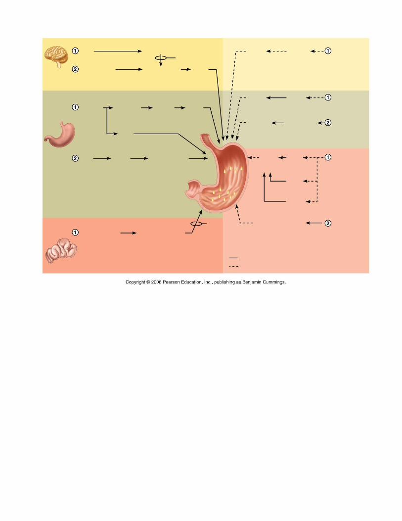
Regulation of Gastric SecretionNeural and hormonal mechanisms
• _____________________________________phase: prior to food entry• Excitatory events include:
• Sight or thought of food• Stimulation of taste or smell receptors
• Inhibitory events include:• Loss of appetite or depression• Decrease in stimulation of the parasympathetic division
• ______________________________ phase: once food enters the stomach• Excitatory events include:
• Stomach distension • Activation of stretch receptors (neural activation)• Activation of chemoreceptors by peptides, caffeine, and rising pH • Release of gastrin to the blood
• Inhibitory events include:• A pH lower than 2 • Emotional upset which overrides the parasympathetic division
• ___________________________ phase: as partially digested food enters the duodenum • Excitatory phase – low pH and partially digested food enters the duodenum• Inhibitory phase – distension of duodenum, presence of fatty, acidic, or hypertonic chyme,
and/or irritants in the duodenum• Closes the pyloric sphincter• Releases enterogastrones that inhibit gastric secretion


Regulation and Mechanism of HCl Secretion
• stimulated by ACh, histamine, and gastrin• Release of hydrochloric acid:
Gastric motility and emptyingResponse of the Stomach to Filling: Stomach pressure remains constant until about 1L of food is ingested
• ______________________ relaxation – as food travels in the esophagus, stomach muscles relax• ______________________relaxation – the stomach dilates in response to gastric filling• Plasticity – intrinsic ability of smooth muscle to exhibit the stress-relaxation response
Gastric Contractile Activity• Peristaltic waves move toward pylorus at the rate of 3 per minute• basic electrical rhythm (BER) is initiated by pacemaker cells (cells of Cajal)• Most vigorous peristalsis and mixing occurs near the pylorus• Chyme is either:
• Delivered in small amounts to duodenum • Forced backward for further mixing
Regulation of Gastric Emptying ;Rate of gastric emptying depends on contents of duodenum
Stomach usually empties within 4 hrsliquids faster; larger meals faster!!
• Carbohydrate-rich chyme moves through the duodenum ___________________________• Fat-laden chyme is digested ____________________________ causing food to remain in the stomach longer
Vomiting (emesis) – the stomach empties via a different route (oral)• Causes include extreme stretching, irritants such as bacterial toxins, excessive alcohol, spicy
foods, and certain drugs

Small Intestine: Gross Anatomy• from pyloric sphincter to the ileocecal valve • three subdivisions:
• -• -• -
• The bile duct and main pancreatic duct:• Join the duodenum at the hepatopancreatic ampulla • Are controlled by the sphincter of Oddi
• ileum joins large intestine at ______________________ valve
Microscopic Anatomy of the Small Intestine• Structural modifications of the small intestine wall increase surface area
• _______________________________: deep circular folds of the mucosa and submucosa• ______________: fingerlike extensions of the mucosa• __________________________: tiny projections of absorptive mucosal cells’ plasma
membranes; in microvilli are enzymes that complete digestion
Small Intestine: Histology of the Wall• The epithelium of the mucosa is made up of:
• -• - • -
• Intestinal crypts cells secrete intestinal juice• Secreted by intestine glands in response to distension or irritation of the mucosa• slightly alkaline and isotonic with blood plasma• largely water, enzyme-poor, but contains mucus
• Peyer’s patches are found in the submucosa •
• Brunner’s glands in the duodenum secrete _____________________________
Liver• largest gland in the body; weighs about 3 lbs in an adult• Superficially has four lobes – right, left, caudate, and quadrate• falciform ligament:• ligamentum teres:• The hepatic blood vessels enter the liver at the porta hepatis• The gallbladder rests in a recess on the inferior surface of the right lobe• Bile leaves the liver via
• Bile ducts which fuse into the common hepatic duct • The common hepatic duct fuses with the cystic duct• These two ducts form the bile duct
Microscopic Anatomy of the Liver• Hexagonal-shaped liver lobules are the structural and functional units of the liver
• Composed of hepatocyte (liver cell) plates radiating outward from a central vein• Portal triads are found at each of the six corners of each liver lobule
• Portal triads:• Bile duct • Hepatic artery(oxygen-rich blood)• Hepatic portal vein – carries venous blood with nutrients from digestive viscera
• Liver sinusoids – enlarged, leaky capillaries located between hepatic plates• _____________________________________ – hepatic macrophages found in liver sinusoids• Hepatocytes’ functions include:
• Production of___________________• Processing blood borne nutrients• Storage of __________________________________• D___________________________
• Secreted bile flows between hepatocytes toward the bile ducts in the portal triads Hepatitis – inflammation of the liver often due to viral infectionCirrhosis – diffuse and progressive chronic inflammation of the liverComposition of Bile
• yellow-green,____________________ solution: bile salts, bile pigments, cholesterol, neutral fats, phospholipids, electrolytes
• Bile salts are cholesterol derivatives that:• Emulsify _____________________• Facilitate fat and cholesterol ___________________________• Help make cholesterol ____________________________
• The chief bile pigment is ______________________, a waste product of heme from red blood cells

The Gallbladder• Thin-walled, green muscular sac on the ventral surface of the liver• Stores and concentrates bile by absorbing its water and ions• Releases bile via the cystic duct which flows into the bile duct
Regulation of Bile Release Acidic, fatty chyme causes duodenum to release: Cholecystokinin (CCK) and secretin Bile salts, secretin stimulate liver to produce bile Cholecystokinin causes: gallbladder contractions, hepatopancreatic sphincter relaxes
bile enters the duodenum Gallstones – crystallization of cholesterol which can obstruct the flow of bileCholecystectomy-surgery to remove gallbladderPancreas• Lies deep to the greater curvature of the stomach; head by the duodenum; tail near the spleen
• Exocrine function• Secretes pancreatic juice which breaks down all categories of foodstuff
• Water solution of enzymes and electrolytes (primarily HCO3)• Neutralizes acid chyme• Provides optimal environment for pancreatic enzymes• Enzymes are released in inactive form and activated in the duodenum
• Acini (clusters of secretory cells) contain zymogen granules with digestive enzymes • Endocrine function – release of insulin and glucagonRegulation of Pancreatic Secretion
• CCK induces the secretion of enzyme-rich pancreatic juice• Secretin causes secretion of bicarbonate-rich pancreatic juice • Vagus nerve
Digestion in the Small Intestine• As chyme enters the duodenum
• Carbohydrates and proteins are only partially digested• No fat digestion has taken place
• Digestion continues in the small intestine• Chyme is released slowly into the duodenum • Because it is hypertonic and has low pH, mixing is required for proper digestion• Required substances needed are supplied by the liver • Virtually all nutrient absorption takes place in the small intestine
Motility of the Small Intestine• most common motion of the small intestine is segmentation
• After nutrients are absorbed: Meal remnants, bacteria, mucosal cells, and debris move to:Large Intestine
• three unique features:• Teniae coli – three bands of longitudinal smooth muscle in its muscularis• Haustra – pocketlike sacs caused by the tone of the teniae coli• Epiploic appendages – fat-filled pouches of visceral peritoneum
• subdivided into the cecum, appendix, colon, rectum, and anal canal• The saclike cecum: Contains a wormlike vermiform appendix
• Appendicitis – inflammation of the appendix resulting from blockage that traps infectious bacteria in its lumen